tuberin and pras40 are anti-apoptotic gatekeepers during early human amniotic fluid stem-cell...
TRANSCRIPT
Tuberin and PRAS40 are anti-apoptotic gatekeepersduring early human amniotic fluid stem-celldifferentiation
Christiane Fuchs, Margit Rosner, Helmut Dolznig, Mario Mikula, Nina Kramer
and Markus Hengstschlager∗
Institute of Medical Genetics, Medical University of Vienna, Wahringer Strasse 10, Vienna 1090, Austria
Received September 12, 2011; Revised and Accepted November 13, 2011
Embryoid bodies (EBs) are three-dimensional multicellular aggregates allowing the in vitro investigation ofstem-cell differentiation processes mimicking early embryogenesis. Human amniotic fluid stem (AFS) cellsharbor high proliferation potential, do not raise the ethical issues of embryonic stem cells, have a lowerrisk for tumor development, do not need exogenic induction of pluripotency and are chromosomal stable.Starting from a single human AFS cell, EBs can be formed accompanied by the differentiation into cells ofall three embryonic germ layers. Here, we report that siRNA-mediated knockdown of the endogenous tuber-ous sclerosis complex-2 (TSC2) gene product tuberin or of proline-rich Akt substrate of 40 kDa (PRAS40), thetwo major negative regulators of mammalian target of rapamycin (mTOR), leads to massive apoptotic celldeath during EB development of human AFS cells without affecting the endodermal, mesodermal and ecto-dermal cell differentiation spectrum. Co-knockdown of endogenous mTOR demonstrated these effects to bemTOR-dependent. Our findings prove this enzyme cascade to be an essential anti-apoptotic gatekeeper ofstem-cell differentiation during EB formation. These data allow new insights into the regulation of earlystem-cell maintenance and differentiation and identify a new role of the tumor suppressor tuberin and theoncogenic protein PRAS40 with the relevance for a more detailed understanding of the pathogenesis ofdiseases associated with altered activities of these gene products.
INTRODUCTION
The description of Oct4-positive stem cells within human am-niotic fluid (1) initiated a new and promising research field(2,3). Descending from a single-immunoselected CD117(c-Kit)-positive human amniotic fluid stem (AFS) cell, linescan be established, which can be expanded as immature pluri-potent stem cells able to differentiate along all three embryon-ic germ layers (4). These monoclonal AFS cell lines can beexpanded in culture with high proliferative capacity, arechromosomal stable and do not need exogenic treatment tomaintain pluripotency. Furthermore, AFS cells have a lowerrisk for tumor development and do not raise the ethicalissues of embryonic stem (ES) cells. Accordingly, besidestheir putative functions in specific cell-based therapies, AFScells became increasingly accepted as an ideal tool to studycell differentiation processes (2–5).
Pluripotent stem cells are also defined by their potential tospontaneously form three-dimensional multicellular aggre-gates called embryoid bodies (EBs). EBs allow the in vitrorecapitulation and investigation of the three-dimensionaland tissue level contexts of the cell differentiation phenom-ena during early mammalian embryogenesis (6,7). Recently,it was demonstrated that starting from one single humanAFS cell, expressing the stem-cell markers CD117 andOct4, EBs can be formed. When cultured in suspension,under conditions in which they are unable to attach to thesurface of culture dishes, monoclonal AFS cells form EBsconsisting of many different cells expressing ectodermal,endodermal and mesodermal markers (8). This in vitro recap-itulation of the pluripotent AFS cell differentiation potentialtogether with the recently established protocol for prolongedefficient siRNA-mediated gene silencing in human AFS cells(9) provides an optimal tool to study the relevance of specific
∗To whom correspondence should be addressed. Tel: +43 14016056500; Fax: +43 140160956501; Email: [email protected]
# The Author 2011. Published by Oxford University Press. All rights reserved.For Permissions, please email: [email protected]
Human Molecular Genetics, 2012, Vol. 21, No. 5 1049–1061doi:10.1093/hmg/ddr535Advance Access published on November 16, 2011
by guest on June 12, 2013http://hm
g.oxfordjournals.org/D
ownloaded from
endogenous gene functions for early mammalian differenti-ation processes.
The serine/threonine protein kinase mammalian target ofrapamycin (mTOR) is part of two distinct protein complexesin mammalian cells and is the central player within theinsulin signaling pathway regulating cell size, tumor develop-ment and differentiation. The protein complex mTORC1, con-taining raptor and mLST8, phosphorylates the eukaryoticinitiation factor 4EBP1 and the ribosomal p70S6 kinase (atT389 to activate the ribosomal protein S6), both being regula-tors of mRNA translation. mTORC2 contains rictor, mLST8,sin1 and protor, and phosphorylates the oncogenic kinaseAkt at S473. Within the insulin signaling pathway, thephosphatidylinositol-3-kinase regulates the phosphoinositide-dependent kinase-1 to phosphorylate Akt at T308, what inconjunction with the above-mentioned mTORC2-mediatedphosphorylation drives full activation of Akt. So, activatedAkt stimulates mTORC1 activity via phosphorylating andthereby inhibiting the function of the two major negative reg-ulators of mTORC1, the tuberous sclerosis complex-2 (TSC2)gene product tuberin and proline-rich Akt substrate of 40 kDa(PRAS40) (10–12).
Using siRNA-mediated endogenous gene silencing inhuman AFS cells during EB formation, we found tuberinand PRAS40 to be potent anti-apoptotic gatekeepers in earlymammalian stem-cell differentiation. The discovery of thismTOR-dependent potential provides new insights into the de-velopment of tumors and human genetic diseases associatedwith functional loss of either tuberin or PRAS40.
RESULTS
Tuberin and PRAS40 regulate apoptosis during EBformation of AFS cells
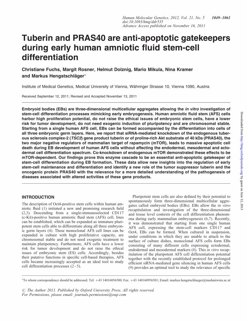
siRNA-mediated knockdown of endogenous PRAS40 ortuberin triggered a pronounced degradation of EBs withinseveral days of EB formation of human AFS cells (Fig. 1Aand B). It is important to note that the used siRNA approachtriggers gene knockdown for about 2 weeks in human AFScells (9). Detailed microscopical investigation demonstratedsignificant morphological changes in the structures of theEBs. The inner area consists of cells with morphologiesearlier described to be characteristic for AFS cell-derivedEBs (8). Upon tuberin or PRAS40 knockdown, however, anouter area appeared consisting of less compacted, brightershaped cells being typically described as the consequence ofapoptotic processes (Fig. 1C) (13). Whereas the whole EBarea remained unchanged (Fig. 1D), the inner diameter andthe area of the EBs dramatically decreased upon PRAS40 ortuberin knockdown, accompanied by an increase in the sur-rounding outer ring and by decreased total cell number(Fig. 1E–G) in a time-dependent manner (Fig. 1H and I).These data demonstrate that tuberin and PRAS40 are essentialgatekeepers inhibiting EB degradation during early humanAFS cell differentiation.
Besides the detection of the already mentioned typicalapoptotic cell morphology changes, we used additionalapproaches to prove that the outer ring of the EBs representstuberin- and PRAS40-regulated apoptotic cell death. Hoechst
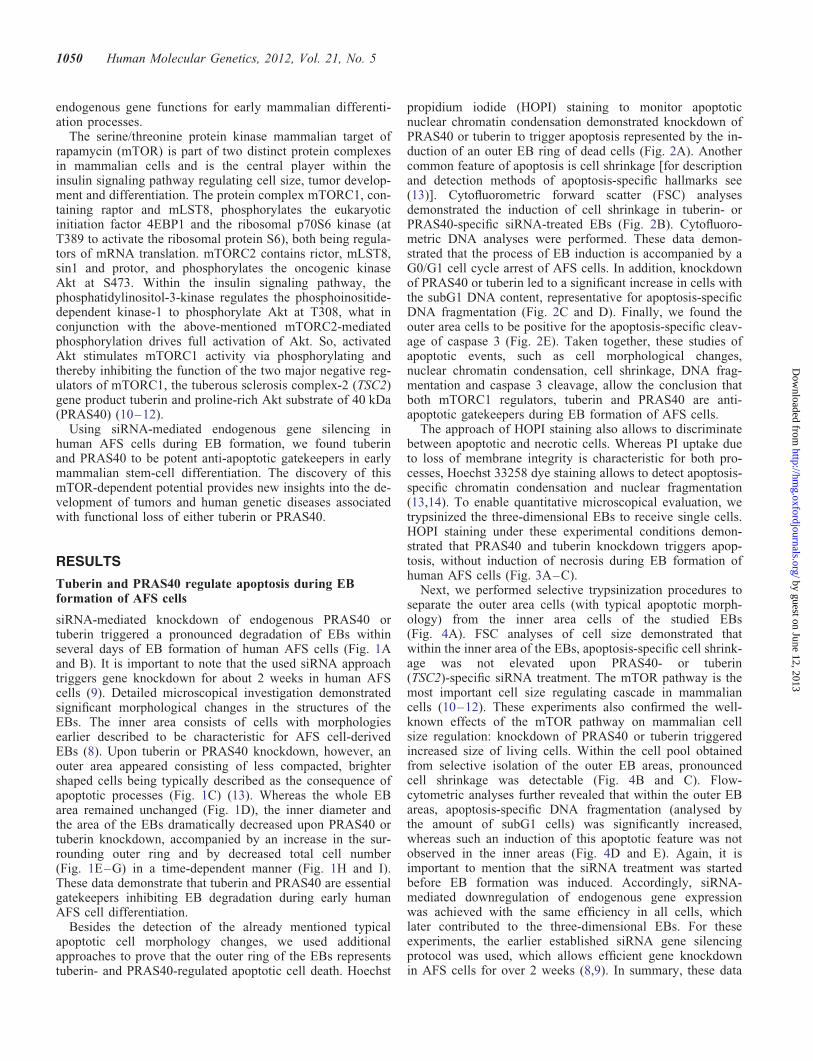
propidium iodide (HOPI) staining to monitor apoptoticnuclear chromatin condensation demonstrated knockdown ofPRAS40 or tuberin to trigger apoptosis represented by the in-duction of an outer EB ring of dead cells (Fig. 2A). Anothercommon feature of apoptosis is cell shrinkage [for descriptionand detection methods of apoptosis-specific hallmarks see(13)]. Cytofluorometric forward scatter (FSC) analysesdemonstrated the induction of cell shrinkage in tuberin- orPRAS40-specific siRNA-treated EBs (Fig. 2B). Cytofluoro-metric DNA analyses were performed. These data demon-strated that the process of EB induction is accompanied by aG0/G1 cell cycle arrest of AFS cells. In addition, knockdownof PRAS40 or tuberin led to a significant increase in cells withthe subG1 DNA content, representative for apoptosis-specificDNA fragmentation (Fig. 2C and D). Finally, we found theouter area cells to be positive for the apoptosis-specific cleav-age of caspase 3 (Fig. 2E). Taken together, these studies ofapoptotic events, such as cell morphological changes,nuclear chromatin condensation, cell shrinkage, DNA frag-mentation and caspase 3 cleavage, allow the conclusion thatboth mTORC1 regulators, tuberin and PRAS40 are anti-apoptotic gatekeepers during EB formation of AFS cells.
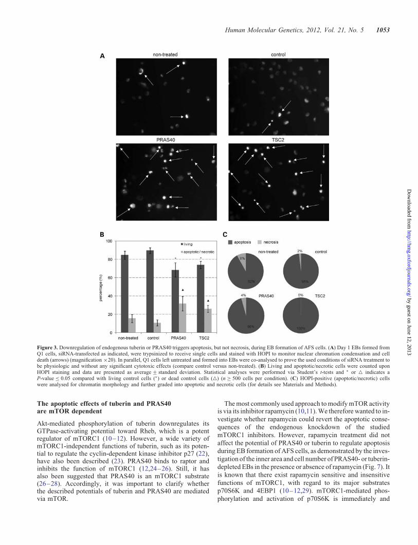
The approach of HOPI staining also allows to discriminatebetween apoptotic and necrotic cells. Whereas PI uptake dueto loss of membrane integrity is characteristic for both pro-cesses, Hoechst 33258 dye staining allows to detect apoptosis-specific chromatin condensation and nuclear fragmentation(13,14). To enable quantitative microscopical evaluation, wetrypsinized the three-dimensional EBs to receive single cells.HOPI staining under these experimental conditions demon-strated that PRAS40 and tuberin knockdown triggers apop-tosis, without induction of necrosis during EB formation ofhuman AFS cells (Fig. 3A–C).
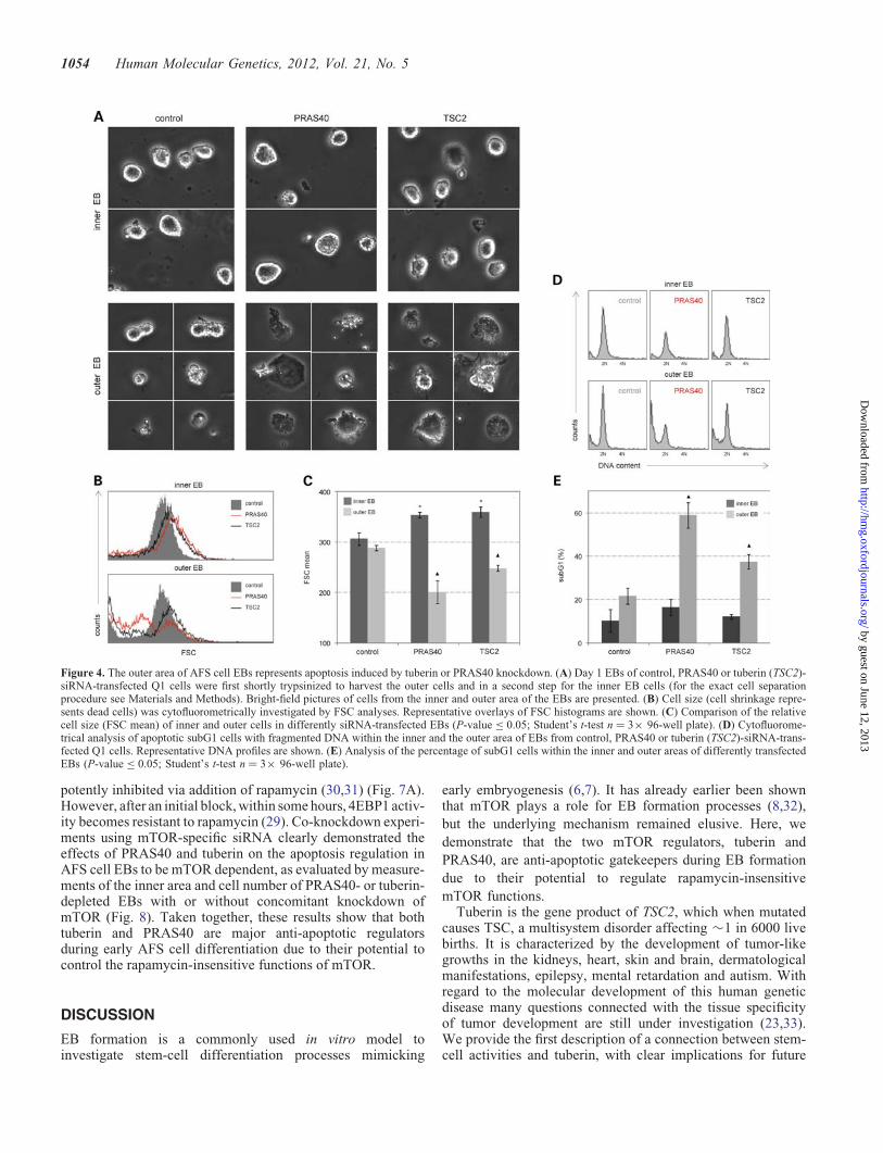
Next, we performed selective trypsinization procedures toseparate the outer area cells (with typical apoptotic morph-ology) from the inner area cells of the studied EBs(Fig. 4A). FSC analyses of cell size demonstrated thatwithin the inner area of the EBs, apoptosis-specific cell shrink-age was not elevated upon PRAS40- or tuberin(TSC2)-specific siRNA treatment. The mTOR pathway is themost important cell size regulating cascade in mammaliancells (10–12). These experiments also confirmed the well-known effects of the mTOR pathway on mammalian cellsize regulation: knockdown of PRAS40 or tuberin triggeredincreased size of living cells. Within the cell pool obtainedfrom selective isolation of the outer EB areas, pronouncedcell shrinkage was detectable (Fig. 4B and C). Flow-cytometric analyses further revealed that within the outer EBareas, apoptosis-specific DNA fragmentation (analysed bythe amount of subG1 cells) was significantly increased,whereas such an induction of this apoptotic feature was notobserved in the inner areas (Fig. 4D and E). Again, it isimportant to mention that the siRNA treatment was startedbefore EB formation was induced. Accordingly, siRNA-mediated downregulation of endogenous gene expressionwas achieved with the same efficiency in all cells, whichlater contributed to the three-dimensional EBs. For theseexperiments, the earlier established siRNA gene silencingprotocol was used, which allows efficient gene knockdownin AFS cells for over 2 weeks (8,9). In summary, these data
1050 Human Molecular Genetics, 2012, Vol. 21, No. 5
by guest on June 12, 2013http://hm
g.oxfordjournals.org/D
ownloaded from
show the induction of the outer area during AFS cell EB for-mation to represent apoptosis mediated by endogenous knock-down of the mTOR inhibitors PRAS40 or tuberin.
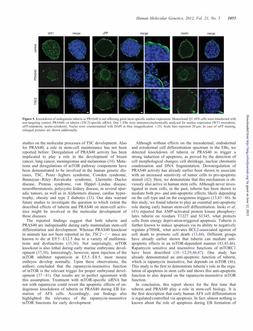
Downregulation of endogenous tuberin or PRAS40 iswithout effects on germ layer-specific marker expression
Starting from one single AFS cell, the formation of EBs, com-posed of cells of all three germlayers, is induced. This AFScell-derived EB formation is accompanied by a decrease inthe expression of the stem-cell markers nodal and Oct-4 andby induction of the differentiation markers Pax 6 (ectodermal),Flk1 (endothelial), E-cadherin (epithelial), GATA4 (endoder-mal), T (brachyury; mesodermal) and HBE1 (mesodermal).AFS cell-derived EBs are also positive for laminin andnestin. In addition, as for ES cell-derived EBs, AFS cell-derived EBs also form the characteristic distinct peripheral
layer on the outer surface, including cells expressinga-fetoprotein (aFP) (8).
Accordingly, it was interesting to investigate whether theabove-described apoptotic effects of PRAS40 or tuberinknockdown were accompanied by effects on the cell differen-tiation spectrum within the EBs. We immunocytochemicallyanalysed the expression of the mesodermal marker Wilmstumor 1 (WT1) (5,15), of the endodermal marker aFP (8,16)and of the ectodermal marker nestin (4,16). Specificity ofthe used antibodies was confirmed by analyzing aFP-negativeIMR-90 fibroblasts (17), aFP-positive Hela cells (18) (Supple-mentary Material, Fig. S1A), nestin-negative Jurkat cells andnestin-positive SK-N-SH cells (19) (Supplementary Material,Fig. S1B), WT1-negative MCF-7 cells (20) and WT1-positiveembryonic kidney cells (5,21) (Supplementary Material,Fig. S1C). We found that although the size of the inner areaEBs decreased, neither PRAS40 knockdown nor tuberin
Figure 1. The role of tuberin and PRAS40 in EB development of AFS cells. (A) Monoclonal Q1 human AFS cells were transfected with non-targeting controlsiRNA, tuberin (TSC2)-specific siRNA or PRAS40-specific siRNA, and knockdown of endogenous tuberin and PRAS40 was confirmed by western blot analysis.a-Tubulin served as a control for equal loading. Lysates of non-transfected (non-treated) Q1 cells were analysed in parallel and were included to prove the usedconditions of siRNA treatment to be physiologic and technically sound (compare control versus non-treated). (B) The number of EBs formed from theso-transfected cells was counted on day 1 (24 h post-induction of EB formation) and day 6 and the incidence of EB formation is given as a percentage ofthe number of cell aggregates initially seeded and induced to form EBs. (C) Left panel: representative bright-field pictures of day 1 EBs generated from dif-ferently transfected Q1 cells (magnification ×10). Middle panel: enlargement of the EB area marked in the left panel (arrows mark representative apoptoticcells). Right panel: schematic presentation of the inner area of the EB (dark red) and the outer area (light red). For detailed explanation see the text. (D)The whole area (outline of the outer area) of formed EBs was analysed at day 1. For each calculation, 10 EBs were photographed, the area was analysedvia Cell D image software (Olympus) and the data are presented as average+ standard deviation. (E) The inner area (outline of the inner area) of formedEBs was analysed at day 1. For each calculation, 10 EBs were photographed, the area was analysed via Cell D image software (Olympus) and the data arepresented as average+ standard deviation. (F) The mean diameter was deduced from measurements of the inner area of day 1 EBs. (G) The cell number ofday 1 EBs (32 EBs for each calculation) was determined by a CASY cell counter and presented relative to the control. (H) Representative bright-field picturesof 12 h and 24 h EBs formed from Q1 cells transfected as indicated (magnification ×10). (I) The inner area of the EBs formed from transfected Q1 cells wasanalysed at the indicated time points. For all statistical analyses in this figure, Student’s t-tests (unpaired, two-tailed) were performed and ∗ indicates statisticalsignificance with a P-value ≤ 0.05.
Human Molecular Genetics, 2012, Vol. 21, No. 5 1051
by guest on June 12, 2013http://hm
g.oxfordjournals.org/D
ownloaded from
knockdown affected the expression pattern of the studiedmesodermal, endodermal or ectodermal markers within theEBs (Fig. 5). These findings provide evidence that these twomTOR inhibitors are anti-apoptotic gatekeepers during earlyAFS cell differentiation, but are not involved in lineage-specific regulations of differentiation.
So far, in the study presented here, we have proved that theouter cells (appearing upon siRNA-knockdown of tuberin orPRAS40) are apoptotic rather than, for example, differentiat-ing (analyses of cell shrinkage, DNA fragmentation, chroma-tin condensation, caspase 3 cleavage). In addition, thesemarker stainings confirm that the outer cells are not differen-tiating since they are, for example, aFP negative (see theenlarged pictures of aFP staining in Fig. 5).
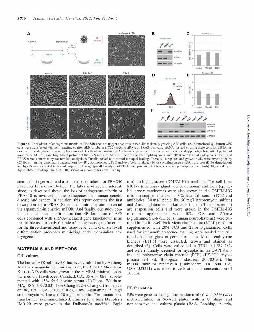
Downregulation of endogenous tuberin or PRAS40does not induce apoptosis in two-dimensionally growingAFS cells
To investigate whether the described apoptotic effects ofsiRNA-mediated knockdown of tuberin or PRAS40 specific-ally occur in the course of cell differentiation processesduring EB formation, we performed experiments in two-dimensionally (2D) growing AFS cells. Downregulation ofendogenous tuberin or PRAS40 in AFS cells growing in2D and high serum (Fig. 6A and B) had neither effects on
the cell morphology (Fig. 6A), on chromatin condensationanalysed by HOPI staining (Fig. 6C), on cell shrinkage flow-cytometrically investigated by FSC analyses (Fig. 6D), onDNA degradation studied by subG1 analyses (Fig. 6E) noron caspase cleavage analysed by western blot (Fig. 6F).These results demonstrate that the apoptotic effects oftuberin- or PRAS40-knockdown specifically occur in thecourse of EB formation.
In the western blot analyses of caspase 3 cleavage, weco-analysed protein extracts obtained upon EB formation asapoptotic controls (Fig. 6F). Once again, these experimentsconfirmed the EB-specific apoptotic effects of knockdownof both tuberin and PRAS40. These western blot analyses ofcaspase 3 cleavage also provided evidence that the effects ofPRAS40 are more severe than the effects of tuberin knock-down (Fig. 6F). Although all the data obtained in this studydemonstrate that tuberin is also an anti-apoptotic gatekeeperduring stem-cell differentiation, the effects of PRAS40 modu-lation appeared to be more pronounced in all performedexperiments (see especially Figs 1, 2 and 4). Although theefficiency of siRNA-mediated downregulation of PRAS40and tuberin in AFS cells seemed always to be very comparable(Figs 1A and 6B), it is still hard to say whether these differ-ences are a consequence of different starting levels ofPRAS40 and tuberin protein amounts or are due to differentbiochemical functions of these two regulators.
Figure 2. Knockdown of endogenous tuberin or PRAS40 triggers apoptosis during EB development of AFS cells. (A) Day 1 EBs, derived from siRNA-treatedQ1 cells as described in Figure 1, were stained with HOPI for 1 h at 378C and then analysed on a confocal microscope (magnification ×25). Scale bars represent50 mm. In addition, enlarged pictures (the white dotted line marks the boundary between inner and outer area) and bright-field pictures are presented. (B) Day 1EBs were trypsinized and relative cell size was studied via cytofluorometric FSC analyses. A representative overlay of FSC histograms is presented. (C) In thesame samples, apoptotic cells were cytofluorometrically detected by their subG1 DNA content (DNA content ,2N). Representative DNA profiles are presented.The log Q1 picture represents non-transfected Q1 cells logarithmically growing in petri dishes without induction of EB formation. (D) Quantitative investigationof the amount of subG1 cells in EBs analysed in (C). Student’s t-test was performed and ∗ indicates statistical significance (P-value ≤ 0.05; n ¼ 3× 96-wellplate). (E) Immoncytochemical detection of cleaved caspase 3 in the so-formed EBs (the white dotted line marks the boundary between inner and outer area).
1052 Human Molecular Genetics, 2012, Vol. 21, No. 5
by guest on June 12, 2013http://hm
g.oxfordjournals.org/D
ownloaded from
The apoptotic effects of tuberin and PRAS40are mTOR dependent
Akt-mediated phosphorylation of tuberin downregulates itsGTPase-activating potential toward Rheb, which is a potentregulator of mTORC1 (10–12). However, a wide variety ofmTORC1-independent functions of tuberin, such as its poten-tial to regulate the cyclin-dependent kinase inhibitor p27 (22),have also been described (23). PRAS40 binds to raptor andinhibits the function of mTORC1 (12,24–26). Still, it hasalso been suggested that PRAS40 is an mTORC1 substrate(26–28). Accordingly, it was important to clarify whetherthe described potentials of tuberin and PRAS40 are mediatedvia mTOR.
The most commonly used approach to modify mTOR activityis via its inhibitor rapamycin (10,11). We therefore wanted to in-
vestigate whether rapamycin could revert the apoptotic conse-
quences of the endogenous knockdown of the studied
mTORC1 inhibitors. However, rapamycin treatment did not
affect the potential of PRAS40 or tuberin to regulate apoptosis
during EB formation of AFS cells, as demonstrated by the inves-
tigation of the inner area and cell number of PRAS40- or tuberin-
depleted EBs in the presence or absence of rapamycin (Fig. 7). It
is known that there exist rapamycin sensitive and insensitive
functions of mTORC1, with regard to its major substrates
p70S6K and 4EBP1 (10–12,29). mTORC1-mediated phos-
phorylation and activation of p70S6K is immediately and
Figure 3. Downregulation of endogenous tuberin or PRAS40 triggers apoptosis, but not necrosis, during EB formation of AFS cells. (A) Day 1 EBs formed fromQ1 cells, siRNA-transfected as indicated, were trypsinized to receive single cells and stained with HOPI to monitor nuclear chromation condensation and celldeath (arrows) (magnification ×20). In parallel, Q1 cells left untreated and formed into EBs were co-analysed to prove the used conditions of siRNA treatment tobe physiologic and without any significant cytotoxic effects (compare control versus non-treated). (B) Living and apoptotic/necrotic cells were counted uponHOPI staining and data are presented as average+ standard deviation. Statistical analyses were performed via Student’s t-tests and ∗ or △ indicates aP-value ≤ 0.05 compared with living control cells (∗) or dead control cells (△) (n ≥ 500 cells per condition). (C) HOPI-positive (apoptotic/necrotic) cellswere analysed for chromatin morphology and further graded into apoptotic and necrotic cells (for details see Materials and Methods).
Human Molecular Genetics, 2012, Vol. 21, No. 5 1053
by guest on June 12, 2013http://hm
g.oxfordjournals.org/D
ownloaded from
potently inhibited via addition of rapamycin (30,31) (Fig. 7A).However, after an initial block, within some hours, 4EBP1 activ-ity becomes resistant to rapamycin (29). Co-knockdown experi-ments using mTOR-specific siRNA clearly demonstrated theeffects of PRAS40 and tuberin on the apoptosis regulation inAFS cell EBs to be mTOR dependent, as evaluated by measure-ments of the inner area and cell number of PRAS40- or tuberin-depleted EBs with or without concomitant knockdown ofmTOR (Fig. 8). Taken together, these results show that bothtuberin and PRAS40 are major anti-apoptotic regulatorsduring early AFS cell differentiation due to their potential tocontrol the rapamycin-insensitive functions of mTOR.
DISCUSSION
EB formation is a commonly used in vitro model toinvestigate stem-cell differentiation processes mimicking
early embryogenesis (6,7). It has already earlier been shownthat mTOR plays a role for EB formation processes (8,32),
but the underlying mechanism remained elusive. Here, we
demonstrate that the two mTOR regulators, tuberin and
PRAS40, are anti-apoptotic gatekeepers during EB formation
due to their potential to regulate rapamycin-insensitive
mTOR functions.Tuberin is the gene product of TSC2, which when mutated
causes TSC, a multisystem disorder affecting �1 in 6000 livebirths. It is characterized by the development of tumor-likegrowths in the kidneys, heart, skin and brain, dermatologicalmanifestations, epilepsy, mental retardation and autism. Withregard to the molecular development of this human geneticdisease many questions connected with the tissue specificityof tumor development are still under investigation (23,33).We provide the first description of a connection between stem-cell activities and tuberin, with clear implications for future
Figure 4. The outer area of AFS cell EBs represents apoptosis induced by tuberin or PRAS40 knockdown. (A) Day 1 EBs of control, PRAS40 or tuberin (TSC2)-siRNA-transfected Q1 cells were first shortly trypsinized to harvest the outer cells and in a second step for the inner EB cells (for the exact cell separationprocedure see Materials and Methods). Bright-field pictures of cells from the inner and outer area of the EBs are presented. (B) Cell size (cell shrinkage repre-sents dead cells) was cytofluorometrically investigated by FSC analyses. Representative overlays of FSC histograms are shown. (C) Comparison of the relativecell size (FSC mean) of inner and outer cells in differently siRNA-transfected EBs (P-value ≤ 0.05; Student’s t-test n ¼ 3× 96-well plate). (D) Cytofluorome-trical analysis of apoptotic subG1 cells with fragmented DNA within the inner and the outer area of EBs from control, PRAS40 or tuberin (TSC2)-siRNA-trans-fected Q1 cells. Representative DNA profiles are shown. (E) Analysis of the percentage of subG1 cells within the inner and outer areas of differently transfectedEBs (P-value ≤ 0.05; Student’s t-test n ¼ 3× 96-well plate).
1054 Human Molecular Genetics, 2012, Vol. 21, No. 5
by guest on June 12, 2013http://hm
g.oxfordjournals.org/D
ownloaded from
studies on the molecular processes of TSC development. Alsofor PRAS40, a role in stem-cell maintenance has not beenreported before. Deregulation of PRAS40 activity has beenimplicated to play a role in the development of breastcancer, lung cancer, meningiomas and melanomas (34). Muta-tions and deregulations of mTOR pathway components havebeen demonstrated to be involved in the human genetic dis-eases, TSC, Peutz–Jeghers syndrome, Cowden syndrome,Bannayan–Riley–Ruvalcaba syndrome, Lhermitte–Duclosdisease, Proteus syndrome, von Hippel–Lindau disease,neurofibromatosis, polycystic kidney disease, in several spor-adic tumors, as well as in Alzheimers disease, cardiac hyper-trophy, obesity and type 2 diabetes (33). Our data warrantfuture studies to investigate the question to which extent thedescribed effects of tuberin and PRAS40 on stem-cell activ-ities might be involved in the molecular development ofthese diseases.
The reported findings suggest that both tuberin andPRAS40 are indispensable during early mammalian stem-celldifferentiation and development. Whereas PRAS40 knockoutin animals has not been reported so far, TSC22/2 mice areknown to die at E9.5–E12.5 due to a variety of malforma-tions and dysfunctions (35,36). Not surprisingly, mTORknockout is also lethal during early murine embryonic devel-opment (37,38). Interestingly, however, upon injection of themTOR inhibitor rapamycin at E5.5–E8.5, most mouseembryos develop normally. Upon these observations, theauthors concluded that the rapamycin-insensitive functionof mTOR is the relevant trigger for proper embryonal devel-opment (37–41). Our results are in perfect agreement withthis assumption. Treatment with mTOR-specific siRNA butnot with rapamycin could revert the apoptotic effects of en-dogenous knockdown of tuberin or PRAS40 during EB for-mation of AFS cells. Accordingly, our findings alsohighlighted the relevance of the rapamycin-insensitivemTOR functions for early development.
Although without effects on the mesodermal, endodermaland ectodermal cell differentiation spectrum in the EBs, wedetected knockdown of tuberin or PRAS40 to trigger astrong induction of apoptosis, as proved by the detection ofcell morphological changes, cell shrinkage, nuclear chromatincondensation and DNA fragmentation. Downregulation ofPRAS40 activity has already earlier been shown to associatewith an increased sensitivity of tumor cells to pro-apoptoticstimuli (42). Here, we demonstrate that this mechanism is ob-viously also active in human stem cells. Although never inves-tigated in stem cells, in the past, tuberin has been shown tomediate both pro- and anti-apoptotic effects, likely dependingon the cell type and on the exogenous triggers (13,43–46). Inthis study, we found tuberin to play an essential anti-apoptoticrole during early human stem-cell differentiation. Inoki et al.(43) reported that AMP-activated protein kinase phosphory-lates tuberin on residues T1227 and S1345, what protectscells from energy deprivation-triggered apoptosis. Tuberin isfurther known to induce apoptosis via its ability to negativelyregulate p70S6K, what activates BCL2-associated agonist ofcell death to promote cell death (13,44). Different groupshave already earlier shown that tuberin can mediate anti-apoptotic effects in an mTOR-dependent manner (43,45,46).Rapamycin sensitive and insensitive functions of mTORC1have been described (10–12,29,46,47). One study hasalready demonstrated an anti-apoptotic function of tuberin,which is rapamycin insensitive, but depends on mTOR (46).This study is the first to demonstrate tuberin’s role in the regu-lation of apoptosis in stem cells and shows this anti-apoptoticfunction to also depend on the rapamycin-insensitive mTORfunction.
In conclusion, this report shows for the first time thattuberin and PRAS40 play a role in stem-cell biology. It isthe first description that early human AFS cell differentiationis regulated/controlled via apoptosis. In fact, almost nothing isknown about the role of apoptosis during EB formation of
Figure 5. Knockdown of endogenous tuberin or PRAS40 is not affecting germ layer-specific marker expression. Monoclonal Q1 AFS cells were transfected withnon-targeting control, PRAS40- or tuberin (TSC2)-specific siRNA. Day 1 EBs were immunocytochemically analysed for marker expression (WT1-mesoderm;aFP-endoderm; nestin-ectoderm). Nuclei were counterstained with DAPI in blue (magnification ×25). Scale bars represent 20 mm. In case of aFP staining,enlarged pictures are shown additionally.
Human Molecular Genetics, 2012, Vol. 21, No. 5 1055
by guest on June 12, 2013http://hm
g.oxfordjournals.org/D
ownloaded from
stem cells in general, and a connection to tuberin or PRAS40has never been drawn before. The latter is of special interest,since, as described above, the loss of endogenous tuberin orPRAS40 is involved in the pathogenesis of human geneticdisease and cancer. In addition, this report contains the firstdescription of a PRAS40-mediated anti-apoptotic potentialvia rapamycin-insensitive mTOR. And finally, our study con-tains the technical confirmation that EB formation of AFScells combined with siRNA-mediated gene knockdown is aninvaluable tool to study the role of endogenous gene functionsfor the three-dimensional and tissue level context of stem-celldifferentiation processes mimicking early mammalian em-bryogenesis.
MATERIALS AND METHODS
Cell culture
The human AFS cell line Q1 has been established by AnthonyAtala via magnetic cell sorting using the CD117 MicroBeadKit (4). AFS cells were grown in the a-MEM minimal essen-tial medium (Invitrogen, Carlsbad, CA, USA, 41061), supple-mented with 15% fetal bovine serum (HyClone, Waltham,MA, USA, 30070.03), 18% Chang B, 2% Chang C (Irvine Sci-entific, CA, USA, C100, C106), 2 mM L-glutamine, 50 mg/lstreptomycin sulfate and 30 mg/l penicillin. The human non-transformed, non-immortalized, primary fetal lung fibroblastsIMR-90 were grown in the Dulbecco’s modified Eagle
medium-high glucose (DMEM-HG) medium. The cell linesMCF-7 (mammary gland adenocarcinoma) and Hela (epithe-lial cervix carcinoma) were also grown in the DMEM-HGmedium supplemented with 10% fetal calf serum (FCS) andantibiotics (30 mg/l penicillin, 50 mg/l streptomycin sulfate)and 2 mM L-glutamine. Jurkat cells (human T cell leukemia)are suspension cells and were grown in the DMEM-HGmedium supplemented with 10% FCS and 2.5 mM
L-glutamine. SK-N-SH cells (human neuroblastoma) were cul-tured in the Roswell Park Memorial Institute (RPMI) mediumsupplemented with 20% FCS and 2 mM L-glutamine. Cellsused for immunofluorescence staining were seeded and cul-tured on either glass or permanox slides. Mouse embryonickidneys (E11.5) were dissected, grown and stained asdescribed (5). Cells were cultivated at 378C and 5% CO2
and were routinely screened for mycoplasma via DAPI stain-ing and polymerase chain reaction (PCR) (EZ-PCR myco-plasma test kit, Biological Industries, 20-700-20). ThemTOR inhibitor rapamycin (Calbiochem, La Jolla, CA,USA, 553211) was added to cells at a final concentration of100 nM.
EB formation
EBs were generated using a suspension method with 0.3% (w/v)methylcellulose in 96-well plates with a U shape andnon-adhesive cell culture plastic (PAA, Pasching, Austria,
Figure 6. Knockdown of endogenous tuberin or PRAS40 does not trigger apoptosis in two-dimensionally growing AFS cells. (A) Monoclonal Q1 human AFScells were transfected with non-targeting control siRNA, tuberin (TSC2)-specific siRNA or PRAS40-specific siRNA. Instead of using these cells for EB forma-tion, in this study, the cells were replated under 2D cell culture conditions. A schematic presentation of the used experimental approach, a bright-field picture ofnon-treated AFS cells and bright-field pictures of the siRNA-treated AFS cells before and after replating are shown. (B) Knockdown of endogenous tuberin andPRAS40 was confirmed by western blot analysis. a-Tubulin served as a control for equal loading. These cells, replated and grown in 2D, were investigated by(C) HOPI staining (chromatin condensation), by (D) cytofluorometric FSC analyses (cell shrinkage), by (E) cytofluorometric subG1 analyses (DNA degradation)and by (F) western blot detection of caspase 3 cleavage (parallel analyses of EB-derived protein extracts served as apoptotic-positive controls). Glyceraldehyde3-phosphate dehydrogenase (GAPDH) served as a control for equal loading.
1056 Human Molecular Genetics, 2012, Vol. 21, No. 5
by guest on June 12, 2013http://hm
g.oxfordjournals.org/D
ownloaded from
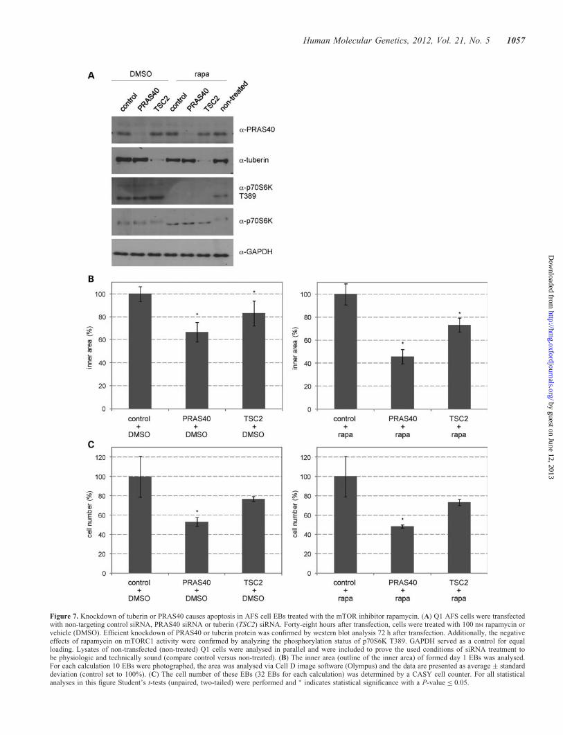
Figure 7. Knockdown of tuberin or PRAS40 causes apoptosis in AFS cell EBs treated with the mTOR inhibitor rapamycin. (A) Q1 AFS cells were transfectedwith non-targeting control siRNA, PRAS40 siRNA or tuberin (TSC2) siRNA. Forty-eight hours after transfection, cells were treated with 100 nM rapamycin orvehicle (DMSO). Efficient knockdown of PRAS40 or tuberin protein was confirmed by western blot analysis 72 h after transfection. Additionally, the negativeeffects of rapamycin on mTORC1 activity were confirmed by analyzing the phosphorylation status of p70S6K T389. GAPDH served as a control for equalloading. Lysates of non-transfected (non-treated) Q1 cells were analysed in parallel and were included to prove the used conditions of siRNA treatment tobe physiologic and technically sound (compare control versus non-treated). (B) The inner area (outline of the inner area) of formed day 1 EBs was analysed.For each calculation 10 EBs were photographed, the area was analysed via Cell D image software (Olympus) and the data are presented as average+ standarddeviation (control set to 100%). (C) The cell number of these EBs (32 EBs for each calculation) was determined by a CASY cell counter. For all statisticalanalyses in this figure Student’s t-tests (unpaired, two-tailed) were performed and ∗ indicates statistical significance with a P-value ≤ 0.05.
Human Molecular Genetics, 2012, Vol. 21, No. 5 1057
by guest on June 12, 2013http://hm
g.oxfordjournals.org/D
ownloaded from
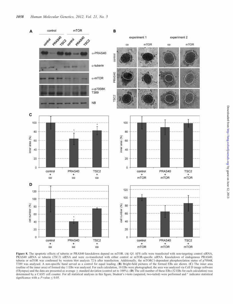
Figure 8. The apoptotic effects of tuberin or PRAS40 knockdown depend on mTOR. (A) Q1 AFS cells were transfected with non-targeting control siRNA,PRAS40 siRNA or tuberin (TSC2) siRNA and were co-transfected with either control or mTOR-specific siRNA. Knockdown of endogenous PRAS40,tuberin or mTOR was confirmed by western blot analysis 72 h after transfection. Additionally, the mTORC1-dependent phosphorylation status of p70S6KT389 was analysed. A non-specific band served as a control for equal loading. (B) Bright-field pictures of the formed EBs are shown. (C) The inner area(outline of the inner area) of formed day 1 EBs was analysed. For each calculation, 10 EBs were photographed, the area was analysed via Cell D image software(Olympus) and the data are presented as average+ standard deviation (control set to 100%). (D) The cell number of these EBs (32 EBs for each calculation) wasdetermined by a CASY cell counter. For all statistical analyses in this figure, Student’s t-tests (unpaired, two-tailed) were performed and ∗ indicates statisticalsignificance with a P-value ≤ 0.05.
1058 Human Molecular Genetics, 2012, Vol. 21, No. 5
by guest on June 12, 2013http://hm
g.oxfordjournals.org/D
ownloaded from
34296X) as described (8). A single EB is composed of 1000–1500 cells in 100 ml medium. For analysis of cell number, cellsize, DNA content and protein expression of whole EBs, EBswere directly harvested, washed three times in ice-cold 1×phosphate buffered saline (PBS) and then trypsinized for15–20 min to receive a single cell suspension. Separatingthe inner and outer cells of EBs was achieved by harvestingEBs, washing them carefully with ice-cold 1× PBS, followedby 2–3 min incubation with trypsin-EDTA (TE). Reactionwas stopped by adding serum-containing medium to EBs.EBs and single cells (outer cells) were separated by slow cen-trifugation (300 rpm, 5 min, 48C). Supernatant was collected,transferred into new tubes and centrifuged for 10 min at1000 rpm and at 48C. The cell pellet was then used forfurther analysis. Remaining EBs were washed with 1× PBSand then trypsinized for 15–20 min to receive single cells(inner cells). So obtained cell pellets were further processedfor analysis.
siRNA treatment
RNA silencing was achieved using siRNA specific for humanPRAS40, tuberin and mTOR (ON-TARGETplus SMART poolreagents, Dharmacon, Lafayette, CO, USA) at a final concen-tration of 50 nM (9). siRNA was delivered to cells usingLipofectamine RNAiMAX reagent (Invitrogen). A pool offour non-targeting siRNAs was used as control fornon-sequence-specific effects for each transfection. In experi-ments, where single knockdowns were compared with thesimultaneous knockdown of two genes, the amount of gene-specific siRNA was reduced to 25 nM while keeping theoverall amount of siRNA for each reaction (50 nM) constantby adding non-targeting siRNA. EB formation of siRNA-treated cells was performed 72 h after transfection.
Flow cytometry and CASY cell counter
Single cells from 2D cultures (cells grown on plates) and 3Dcultures (whole EB, inner EB and outer EB) were fixed byrapid submersion in ice-cold 85% ethanol. After fixation over-night at 2208C, DNA was stained with 0.25 mg/ml propidiumiodide (PI), 0.05 mg/ml RNAse A, 0.1% Triton X-100 incitrate buffer, pH 7.8 and relative cell size (FSC), and DNAcontent and subG1 fraction were analyzed on a Beckton Dick-inson FACSCalibur (Beckton Dickinson, San Jose, CA, USA)(48). For flow-cytometric analysis, a 96-well plate with EBswas used in triplicate. For cell number determination, EBswere trypsinized to obtain single cells and the number ofvital cells was analysed on a CASY Cell Counter (Innovatis,Roche, Basel, Switzerland). For each measurement, 32 EBswere trypsinized to calculate the cell number of a single EB.Measurements were performed in triplicate.
Microscopy and area measurement
For phase contrast images, EBs were photographed with anOlympus IX51 equipped with a XC50 camera (Olympus,Tokio, Japan). For the measurements of the ‘inner area’ and‘whole area’ of EBs, pictures were randomly taken fromthree 96-well plates and the area was determined using the
microscopic imaging software Cell^D (Olympus). The innerarea of the EB is defined as the round-shaped inner structure.The cells situated out of the inner EB are integrated in themeasurement of the ‘whole EB’ (for a scheme see Fig. 1C).The mean diameter of the EB was deduced from measure-ments of the inner area of day 1 EBs (24 h after inductionof EB formation). All immunostained samples were analysedon either an Olympus IX51 microscope with a XC50 camera(Olympus) or a Zeiss LSM Exciter confocal microscope(Carl Zeiss, Oberkochen, Germany). The microscopes werenot changed within one experiment.
Immunocytochemistry
Cells on slides were fixed in 4% paraformaldehyde for 10 minat room temperature. Fixed cells were washed with PBS andthen permeabilized in 0.1% Triton X-100 in PBS (PBS/T)for 10 min, followed by blocking with 0.5% bovine serumalbumin in PBS/T for 30 min at room temperature. Cellswere subsequently incubated with primary antibody overnightat 48C. Antibodies specific for the following proteins wereused: anti-aFP antibody (R&D Systems, Minneapolis, MN,USA, MAB4305), anti-laminin antibody (Sigma, St Gallen,Switzerland, L9393), anti-nestin antibody (Neuromics,Edina, MN, USA, MO15012) and anti-WT1 antibody (Dako,Glostrup, Denmark, M3561). Thereafter, cells were washedwith PBS and incubated with labeled secondary antibody(1:100 in PBS) for 30 min at room temperature: goat tetra-methyl rhodamine iso-thiocyanate a-rabbit (Sigma) or goatfluorescein iso-thiocyanate a-mouse (Sigma). Cells werewashed again and DAPI (4,6-diamino-2-phenylindole; 1 mg/ml) was added for nuclei staining.
EBs were incubated with 4% paraformaldehyde for 30 minat room temperature and then carefully washed with PBS, fol-lowed by washing 15 min in 0.1% Triton X-100 in TBS (TBS/T) and subsequently 15 min in PBS/T. EBs were blocked for1 h in 1% BSA in PBS/T. EBs were washed and incubatedwith primary antibody overnight at 48C. Antibodies specificfor the following proteins were used: cleaved caspase 3,Asp175 (clone 5A1E) (Cell Signaling, Danvers, MA, USA,9664), aFP (R&D Systems), nestin (Neuromics) and WT1(Dako). After primary antibody incubation, EBs werewashed and afterwards stained with labeled secondary anti-bodies: Alexa Fluor 488 goat a-mouse (Molecular Probes,Invitrogen, A11029) or Alexa Fluor 488 goat a-rabbit (Mo-lecular Probes, Invitrogen, A11034) and DAPI overnight at48C. The next day, stained EBs were washed three timeswith 1× PBS and then transferred onto glass slides. Imageswere obtained with a Zeiss LSM Exciter confocal microscope(Carl Zeiss).
HOPI staining
The cell culture medium with a final concentration of 5 mg/mlHoechst 3358 dye (HO) and 2 mg/ml PI was prepared. Foranalysis of cells from 2D cultures (cells grown on plates)and 3D cultures (whole EB), cells/EBs were washed withPBS and treated with TE (for dissociation of whole EBs,EBs were processed as described above). The trypsinized,single cells were harvested in an Eppendorf tube and incubated
Human Molecular Genetics, 2012, Vol. 21, No. 5 1059
by guest on June 12, 2013http://hm
g.oxfordjournals.org/D
ownloaded from
with the HOPI stain for 1 h at 378C. After incubation, the cellsneeded to be carefully resuspended to receive a single cell so-lution, were put on a glass slide and pictures were taken withthe Olympus microscope (up to 10 pictures per sample). EBswithout prior dissociation were directly incubated with theHOPI dye in a 96-well plate and were analyzed on a confocalmicroscope after 1 h incubation. The analysis of stained,single cells was done by counting viable, apoptotic and necrot-ic cells. The Hoechst 33258 dye stains the nuclei of all cells.Nuclear changes, such as chromatin condensation and nuclearfragmentation, are associated with apoptosis. PI uptake indi-cates the loss of membrane integrity being characteristic fornecrosis and apoptosis. Necrosis is characterized by nuclearPI uptake into cells without chromatin condensation ornuclear fragmentation (14).
Protein extraction
Total protein of cells from 2D cultures (cells grown on plates)and 3D cultures (whole EB) was extracted by physical disrup-tion of cell membranes by repeated freeze and thaw cycles. Inbrief, cells/EBs were harvested by trypsinization to receivesingle cells (for dissociation of whole EBs, EBs were pro-cessed as described above). Cell pellets were washed withPBS and lysed in buffer A containing 20 mM Hepes, pH 7.9,0.4 M NaCl, 25% glycerol, 1 mM EDTA, 0.5 mM dithiothreitol,1 mM phenylmethylsulfonyl fluoride, 0.5 mM NaF, 0.5 mM
Na3VO4 supplemented with 2 mg/ml aprotinin, 2 mg/ml leu-peptin, 0.3 mg/ml benzamidinchlorid, 10 mg/ml trypsininhibi-tor by repeated freeze and thaw cycles. After incubation onice and centrifugation at 15000 rpm for 20 min at 48C, super-natants were collected and protein lysates stored at 2808C.Protein concentrations were determined using the Bio-Radprotein assay (22).
Immunoblotting
Proteins (7–15 mg/lane) were run on a sodium dodecylsulfate-polyacrylamide gel and transferred onto a nitrocellu-lose membrane. Blots were stained with Ponceau-S to visual-ize the amount of loaded protein. For immunodetection,antibodies specific for the following proteins were used:tuberin C-20 (Santa Cruz Biotechnology, Santa Cruz, CA,USA, sc-892), phospho-PRAS40 T246 (Cell Signaling,2997), PRAS40 (clone D23C7) (Cell Signaling, 2691),mTOR (Cell Signaling, 2972), phospho-p70S6K T389 (clone108D2) (Cell Signaling, 9234), p70S6K (Cell Signaling,9202), cleaved caspase 3, Asp175 (clone 5A1E) (Cell Signal-ing, 9664), a-tubulin (clone DM1A) (Calbiochem, CP06) andglyceraldehyde 3-phosphate dehydrogenase (GAPDH) (Trevi-gen, Gaithersburg, MD, USA, 2275-PC-100). Rabbit poly-clonal and monoclonal antibodies were detected using antirabbit IgG, an horseradish peroxidase (HRP)-linked heavyand light chain antibody from goat (A120-101P, Bethyl La-boratories, Montgomery, TX, USA); mouse monoclonal anti-bodies were detected using anti-mouse IgG, an HRP-linkedheavy and light chain antibody from goat (A90-116P, BethylLaboratories). Signals were detected using the enhancedchemiluminescence method (Pierce).
Statistical analyses
Analyses of ‘inner’ and ‘outer’ (whole) area of EBs, flow cyto-metry data, incidence of EB formation and cell number dataare all presented as average+ standard deviation (SD). Allcomparisons between groups were calculated using Student’st-test (unpaired, two-tailed) with P-values ≤ 0.05 indicatingstatistical significance.
SUPPLEMENTARY MATERIAL
Supplementary Material is available at HMG online.
Conflict of Interest statement. None declared.
FUNDING
Research in our laboratory is supported by the OsterreichischeNationalbank.
REFERENCES
1. Prusa, A.R., Marton, E., Rosner, M., Bernaschek, G. and Hengstschlager,M. (2003) Oct4 expressing cells in human amniotic fluid: a new source forstem cell research? Hum. Reprod., 18, 1489–1493.
2. Aboushwareb, T. and Atala, A. (2008) Stem cells in urology. Nat. Clin.Pract. Urol., 5, 621–631.
3. Rosner, M., Mikula, M., Preitschopf, A., Feichtinger, M., Schipany, K.and Hengstschlager, M. (2011) Neurogenic differentiation of amnioticfluid stem cells. Amino Acids, doi 10.1007/s00726-011-0929-8.
4. De Coppi, P., Bartsch, G., Siddiqui, M.M., Xu, T., Santos, T.X., Perin, L.,Mostoslavsky, G., Serre, A.C., Snyder, E.Y., Yoo, J.J. et al. (2007)Isolation of amniotic stem cell lines with potential for therapy. Nat.Biotechnol., 25, 100–106.
5. Siegel, N., Rosner, M., Unbekandt, M., Fuchs, C., Slabina, N., Dolznig,H., Davies, J.A., Lubec, G. and Hengstschlager, M. (2010) Contribution ofhuman amniotic fluid stem cells to renal tissue formation depends onmTOR. Hum. Mol. Genet., 19, 3320–3331.
6. Koike, M., Sakaki, S., Amano, Y. and Kurosawa, H. (2007)Characterization of embryoid bodies of mouse embryonic stem cellsformed under various culture conditions and estimation of differentiationstatus of such bodies. J. Biosci. Bioeng., 104, 294–299.
7. Ungrin, M.D., Joshi, C., Nica, A., Bauwens, C. and Zandstra, P.W. (2008)Reproducible, ultra high-troughput formation of multicellularorganization from single cell suspension-derived human embryonic stemcell aggregates. PLoS ONE, 3, e1565.
8. Valli, A., Rosner, M., Fuchs, C., Siegel, N., Bishop, C.E., Dolznig, H.,Madel, U., Feichtinger, W., Atala, A. and Hengstschlager, M. (2010)Embryoid body formation of human amniotic fluid stem cells depends onmTOR. Oncogene, 29, 966–977.
9. Rosner, M., Siegel, N., Fuchs, C., Slabina, N., Dolznig, H. andHengstschlager, M. (2010) Efficient siRNA-mediated prolonged genesilencing in human amniotic fluid stem cells. Nat. Protoc., 5, 1081–1095.
10. Yang, Q. and Guan, K.-L. (2007) Expanding mTOR signaling. Cell Res.,17, 666–681.
11. Wang, X. and Proud, C.G. (2009) Nutrient control of TORC1, a cell-cycleregulator. Trends Cell Biol., 19, 260–267.
12. Sengupta, S., Peterson, T.R. and Sabatini, D.M. (2010) Regulation of themTOR complex 1 pathway by nutrients, growth factors, and stress. Mol.Cell, 40, 310–322.
13. Freilinger, A., Rosner, M., Krupitza, G., Nishino, M., Lubec, G.,Korsmeyer, S.J. and Hengstschlager, M. (2006) Tuberin activates theproapoptotic molecule BAD. Oncogene, 25, 6467–6479.
14. Grusch, M., Polgar, D., Gfatter, S., Leuhuber, K., Huettenbrenner, S.,Leisser, C., Fuhrmann, G., Kassie, F., Steinkellner, H., Smid, K. et al.(2002) Maintenance of ATP favours apoptosis over necrosis triggered bybenzamide riboside. Cell Death Differ., 9, 169–178.
1060 Human Molecular Genetics, 2012, Vol. 21, No. 5
by guest on June 12, 2013http://hm
g.oxfordjournals.org/D
ownloaded from
15. Armstrong, J.F., Pritchard-Jones, K., Bickmore, W.A., Hastie, N.D. andBard, J.B. (1993) The expression of the Wilms’ tumour gene, WT1, in thedeveloping mammalian embryo. Mech. Dev., 40, 85–97.
16. Cai, J., Chen, J., Liu, Y., Miura, T., Luo, Y., Loring, J.F., Freed, W.J.,Rao, M.S. and Zeng, X. (2006) Assessing self-renewal and differentiationin human embryonic stem cell lines. Stem. Cells, 24, 516–530.
17. Ren, X.W., Liang, M., Meng, X., Ye, X., Ma, H., Zhao, Y., Guo, J., Cai,N., Chen, H.Z., Ye, S.L. and Hu, F. (2006) A tumor-specific conditionallyreplicative adenovirus vector expressing TRAIL for gene therapy ofhepatocellular carcinoma. Cancer Gene Ther., 13, 159–168.
18. Ogden, S.K., Lee, K.C., Wernke-Dollries, K., Stratton, S.A., Aronow, B.and Barton, MC. (2001) p53 targets chromatin structure alteration torepress alpha-fetoprotein gene expression. J. Biol. Chem., 276, 42057–42062.
19. Mahller, Y.Y., Williams, J.P., Baird, W.H., Mitton, B., Grossheim, J.,Saeki, Y., Cancelas, J.A., Ratner, N. and Cripe, T.P. (2009)Neuroblastoma cell lines contain pluripotent tumor initiating cells that aresusceptible to a targeted oncolytic virus. PLoS ONE, 4, e4235.
20. Loeb, D.M., Evron, E., Patel, C.B., Sharma, P.M., Niranjan, B., Buluwela,L., Weitzman, S.A., Korz, D. and Sukumar, S. (2001) Wilms’ tumorsuppressor gene (WT1) is expressed in primary breast tumors despitetumor-specific promoter methylation. Cancer Res., 61, 921–925.
21. Unbekandt, M. and Davies, J.A. (2010) Dissociation of embryonickidneys followed by reaggregation allows the formation of renal tissues.Kidney Int., 77, 407–416.
22. Rosner, M., Freilinger, A., Hanneder, M., Fujita, N., Lubec, G., Tsuruo, T.and Hengstschlager, M. (2007) p27Kip1 localization depends on thetumor suppressor protein tuberin. Hum. Mol. Genet., 16, 1541–1556.
23. Neuman, N.A. and Henske, E.P. (2011) Non-canonical functions of thetuberous sclerosis complex-Rheb signalling axis. EMBO Mol. Med., 3,1–12.
24. Sancak, Y., Thoreen, C.C., Peterson, T.R., Lindquist, R.A., Kang, S.A.,Spooner, E., Carr, S.A. and Sabatini, D.M. (2007) PRAS40 is aninsulin-regulated inhibitor of the mTORC1 protein kinase. Mol. Cell, 25,903–915.
25. Wang, L., Harris, T.E., Roth, R.A. and Lawrence, J.C. Jr. (2007) PRAS40regulates mTORC1 kinase activity by functioning as a direct inhibitor ofsubstrate binding. J. Biol. Chem., 282, 20036–20044.
26. Thedieck, K., Polak, P., Kim, M.L., Molle, K.D., Cohen, A., Jeno, P.,Arrieumerlou, C. and Hall, M.N. (2007) PRAS40 and PRR5-like proteinare new mTOR interactors that regulate apoptosis. PLoS ONE, 2, e1217.
27. Fonseca, B.D., Smith, E.M., Lee, V.H., MacKintosh, C. and Proud, C.G.(2007) PRAS40 is a target for mammalian target of rapamycin complex 1and is required for signaling downstream of this complex. J. Biol. Chem.,282, 24514–24524.
28. Oshiro, N., Takahashi, R., Yoshino, K., Tanimura, K., Nakashima, A.,Eguchi, S., Miyamoto, T., Hara, K., Takehana, K., Avruch, J., Kikkawa,U. and Yonezawa, K. (2007) The proline-rich Akt substrate of 40 kDa(PRAS40) is a physiological substrate of mammalian target of rapamycincomplex 1. J. Biol. Chem., 282, 20329–20339.
29. Choo, A.Y., Yoon, S.O., Kim, S.G., Roux, P.P. and Blenis, J. (2008)Rapamycin differently inhibits S6Ks and 4E-BP1 to mediate celltype-specific repression of mRNA translation. Proc. Natl Acad. Sci. USA,105, 17414–17419.
30. Sarbassov, D.D., Ali, S.M., Sengupta, S., Sheen, J.H., Hsu, P.P., Bagley,A.F., Markhard, A.L. and Sabatini, D.M. (2006) Prolonged rapamycintreatment inhibits mTORC2 assembly and Akt/PKB. Mol. Cell, 22,159–168.
31. Rosner, M. and Hengstschlager, M. (2008) Cytoplasmic and nucleardistribution of the protein complex mTORC1 and mTORC2: rapamycintriggers dephosphorylation and delocalization of the mTORC2components rictor and sin1. Hum. Mol. Genet., 17, 2934–2948.
32. Zhou, J., Su, P., Wang, L., Chen, J., Zimmermann, M., Genbacev, O.,Afonja, O., Horne, M.C., Tanaka, T., Duan, E. et al. (2009) mTOR
supports long-term self-renewal and suppresses mesoderm and endodermactivites of human ambryonic stem cells. Proc. Natl Acad. Sci. USA, 106,7840–7845.
33. Rosner, M., Hanneder, M., Siegel, N., Valli, A., Fuchs, C. andHengstschlager, M. (2008) The mTOR pathway and its role in humangenetic diseases. Mutat. Res. Rev. Mutat. Res., 659, 284–292.
34. Nascimento, E.B.M. and Ouwens, D.M. (2009) PRAS40: Target ormodulator of mTORC1 signalling and insulin action? Arch. Physiol.
Biochem., 115, 163–175.
35. Onda, H., Lueck, A., Marks, P.W., Warren, H.B. and Kwiatkowski, D.J.(1999) Tsc2(+/-) mice develop tumors in multiple sites that expressgelsolin and are influenced by genetic background. J. Clin. Invest., 104,687–695.
36. Kobayashi, T., Minowa, O., Kuno, J., Mitani, H., Hino, O. and Noda, T.(1999) Renal carcinogenesis, hepatic hemangiomatosis, and embryoniclethality caused by a germ-line Tsc2 mutation in mice. Cancer Res., 59,1206–1211.
37. Gangloff, Y.G., Mueller, M., Dann, S.G., Svoboda, P., Sticker, M., Spetz,J.F., Um, S.H., Brown, E.J., Cereghini, S., Thomas, G. and Kozma, S.C.(2004) Disruption of the mouse mTOR gene leads to earlypostimplantation lethality and prohibits embryonic stem cell development.Mol. Cell Biol., 24, 9508–9516.
38. Murakami, M., Ichisaka, T., Maeda, M., Oshiro, N., Hara, K., Edenhofer,F., Kiyama, H., Yonezawa, K. and Yamanaka, S. (2004) mTOR isessential for growth and proliferation in early mouse embryos andembryonic stem cells. Mol. Cell Biol., 24, 6710–6718.
39. Hentges, K.E., Sirry, B., Gingeras, A.C., Sarbassov, D., Sonenberg, N.,Sabatini, D. and Peterson, A.S. (2001) FRAP/mTOR is required forproliferation and patterning during embryonic development in the mouse.Proc. Natl Acad. Sci. USA, 98, 13796–13801.
40. Jirmanova, L., Afanassieff, M., Gobert-Gosse, S., Markossian, S. andSavatier, P. (2002) Differential contributions of ERK and PI3-kinaseto the regulation of cyclin D1 expression and to the control of theG1/S transition in moue embryonic stem cells. Oncogene, 21,5515–5528.
41. Kawasome, H., Papst, P., Webb, S., Keller, G.M., Johnson, G.L., Gelfand,E.W. and Terada, N. (1998) Targeted disruption of p70(S6K) defines itsrole in protein synthesis and rapamycin sensitivity. Proc. Natl Acad. Sci.
USA, 95, 5033–5038.
42. Madhunapantula, S.V., Sharma, A. and Robertson, G.P. (2007) PRAS40deregulates apoptosis in malignant melanoma. Cancer Res., 67,3626–3636.
43. Inoki, K., Zhu, T. and Guan, K-L. (2003) TSC2 mediates cellular energyresponse to control cell growth and survival. Cell, 115, 577–590.
44. Freilinger, A., Rosner, M., Hanneder, M. and Hengstschlager, M. (2008)Ras mediates cell survival by regulating tuberin. Oncogene, 27,2072–2083.
45. Ghosh, S., Tergaonkar, V., Rothlin, C.V., Correa, R.G., Bottero, V., Bist,P., Verma, I.M. and Hunter, T. (2006) Essential role of tuberous sclerosisgenes TSC1 and TSC2 in NF-kappaB activation and cell survival. Cancer
Cell, 10, 215–226.
46. Kang, Y.J., Lu, M.K. and Guan, K-L. (2011) The TSC1 and TSC2 tumorsuppressors are required for proper ER stress response and protect cellsfrom ER stress-induced apoptosis. Cell Death Differ., 18, 133–144.
47. Thoreen, C.C., Kang, S.A., Chang, J.W., Liu, Q., Zhang, J., Gao, Y.,Reichling, L.J., Sim, T., Sabatini, D.M. and Gray, N.S. (2009) AnATP-competitive mammalian target of rapamycin inhibitor revealsrapamycin-resistant functions of mTORC1. J. Biol. Chem., 284,8023–8032.
48. Rosner, M., Hofer, K., Kubista, M. and Hengstschlager, M. (2003) Cellsize regulation by the human TSC tumor suppressor proteins depends onPI3K and FKBP38. Oncogene, 22, 4786–4798.
Human Molecular Genetics, 2012, Vol. 21, No. 5 1061
by guest on June 12, 2013http://hm
g.oxfordjournals.org/D
ownloaded from