trypanosoma brucei cathepsin-l increases arrhythmogenic sarcoplasmic reticulum-mediated calcium...
TRANSCRIPT
. . . . . . . . . . . . . . . . . . . . . . . . . . . . . . . . . . . . . . . . . . . . . . . . . . . . . . . . . . . . . . . . . . . . . . . . . . . . . . . . . . . . . . . . . . . . . . . . . . . . . . . . . . . . . . . . . . . . . . . . . . . . . . . . . . . . . . . . . . . . . . . . . . . . . . . . . . . . . . . . . . . . . . . . . . . . . . . . . . . . .
. . . . . . . . . . . . . . . . . . . . . . . . . . . . . . . . . . . . . . . . . . . . . . . . . . . . . . . . . . . . . . . . . . . . . . . . . . . . . . . . . . . . . . . . . . . . . . . . . . . . . . . . . . . . . . . . . . . . . . . . . . . . . . . . . . . . . . . . . . . . . . . . . . . . . . . . . . . . . . . . . . . . . . . . . . . . . . . . . . . . .
Trypanosoma brucei cathepsin-L increasesarrhythmogenic sarcoplasmic reticulum-mediatedcalcium release in rat cardiomyocytesElspeth B. Elliott1†, Douglas McCarroll1†, Hisashi Hasumi2, Claire E. Welsh1,Amanda A. Panissidi1, Nathaniel G. Jones3, Charlotte L. Rossor1, Andy Tait4,GodfreyL.Smith1, JeremyC.Mottram3,LiamJ.Morrison5†,andChristopherM.Loughrey1†*
1College of Medical, Veterinary and Life Sciences, Institute of Cardiovascular and Medical Sciences, University of Glasgow, Glasgow Cardiovascular Research Centre, University Place, GlasgowG12 8TA, UK; 2Department of Cardiovascular Medicine, Dokkyo Medical University School of Medicine, Tochigi, Japan; 3Wellcome Trust Centre for Molecular Parasitology, Institute of Infection,Immunity and Inflammation,College of Medical, Veterinary and Life Sciences, Universityof Glasgow, 120 University Place, Glasgow G12 8TA, UK; 4College of Medical, Veterinary and Life Sciences,School of Veterinary Medicine, Garscube Campus, Bearsden Road, Glasgow G61 1QH, UK; and 5Roslin Institute, University of Edinburgh, Easter Bush, Midlothian EH25 9RG, UK
Received 27 March 2013; revised 1 July 2013; accepted 20 July 2013; online publish-ahead-of-print 26 July 2013
Time for primary review: 25 days
Aims African trypanosomiasis, causedbyTrypanosoma brucei species, leads tobothneurological andcardiac dysfunction andcanbe fatal if untreated. While the neurological-related pathogenesis is well studied, the cardiac pathogenesis remainsunknown. The current study exposed isolated ventricular cardiomyocytes and adult rat hearts to T. brucei to testwhether trypanosomes can alter cardiac function independent of a systemic inflammatory/immune response.
Methodsand results
Using confocal imaging, T. brucei and T. brucei culture media (supernatant) caused an increased frequency of arrhythmo-genic spontaneous diastolic sarcoplasmic reticulum (SR)-mediated Ca2+ release (Ca2+ waves) in isolated adult rat ven-tricular cardiomyocytes. Studies utilising inhibitors, recombinant protein and RNAi all demonstrated that this altered SRfunction was due to T. brucei cathepsin-L (TbCatL). Separate experiments revealed that TbCatL induced a 10–15% in-crease of SERCA activity but reduced SR Ca2+ content, suggesting a concomitant increased SR-mediated Ca2+ leak.This conclusion was supported by data demonstrating that TbCatL increased Ca2+ wave frequency. These effectswere abolished by autocamtide-2-related inhibitory peptide, highlighting a role for CaMKII in the TbCatL action on SRfunction. Isolated Langendorff perfused whole heart experiments confirmed that supernatant caused an increasednumber of arrhythmic events.
Conclusion These data demonstrate for the first time that African trypanosomes alter cardiac function independent of a systemicimmune response, via a mechanism involving extracellular cathepsin-L-mediated changes in SR function.
- - - - - - - - - - - - - - - - - - - - - - - - - - - - - - - - - - - - - - - - - - - - - - - - - - - - - - - - - - - - - - - - - - - - - - - - - - - - - - - - - - - - - - - - - - - - - - - - - - - - - - - - - - - - - - - - - - - - - - - - - - - - - - - - - - - - - - - - - - - - - - - - - - - - - - - - - - -Keywords Sarcoplasmic reticulum † Cardiomyocyte † Calcium † Trypanosomiasis † Trypanosome
1. IntroductionHuman African trypanosomiasis (HAT) is a neglected disease caused
by subspecies of Trypanosoma brucei, which are blood-borne extracellu-
lar protozoa transmitted by tsetse fly (Glossina ssp). HAT is fatal if un-
treated. In early HAT, parasites are intravascular (haemolymphatic/
Stage I disease) but as infection progresses, parasites cross endothelia
and invade extravascular tissue within different organs. When parasites
traverse the blood brain barrier (BBB), neuropsychiatric disturbances
develop (‘sleeping sickness’/Stage II disease). Whilst central nervoussystem (CNS) involvement is the clinical focus of patient screens,cardiac disturbances are now recognized as significant symptoms inHAT. A recent field study observed high prevalence of cardiac electricalabnormalities in HAT (�55% of Stage I, �70% of Stage II patients).1,2
† E.B.E. and D.M. are joint first authors and L.J.M. and C.M.L. contributed to the work equally.
* Corresponding author. Tel: +44 1413302753; Email: [email protected]
&The Author 2013. Published by Oxford University Press on behalf of the European Society of Cardiology. This is an Open Access article distributed under the terms of the Creative CommonsAttribution Non-Commercial License (http://creativecommons.org/licenses/by-nc/3.0/), which permits non-commercial re-use, distribution, and reproduction in any medium, provided theoriginal work is properly cited. For commercial re-use, please contact [email protected].
Cardiovascular Research (2013) 100, 325–335doi:10.1093/cvr/cvt187
by guest on March 18, 2016
Dow
nloaded from

Of note was the increased proportion of HAT patients experiencing pal-pitations.1,2 Other reported cardiac-related abnormalities include con-duction block, low voltage abnormalities, ventricular dilatation and heartfailure;1– 7 23% ofHATpatientshaveNT-proBNP levels (N-terminal prob-type natriuretic peptide; a biomarker of excessive cardiomyocytestretching) consistent with left ventricular dysfunction.2 Both experi-mentally infected animals and between 70–100% of human autopsiesshow clear heart pathology including pancarditis.8– 10 Experimentalanimal models demonstrate significant trypanosome numbers withinmyocardial interstitium, with or without a mononuclear cellular infil-trate.9 Despite the large number of studies demonstrating heart involve-ment in African trypanosomiasis, the appreciation of cardiac dysfunctionas a clinical feature, and consequently our understanding of the basiccardiac pathogenesis, is very limited. In contrast, a significant body ofrecent work has focused on the neuro-pathogenesis of the diseaseshowing that both an inflammatory response and trypanosome inter-action with BBB cells are important.11,12 Therefore, one inference isthat the cardiac-related clinical signs in African trypanosomiasis resultfrom the inflammatory response. Whilst this may indeed play a role,an alternative hypothesis of parasites interacting with cardiomyocytesand altering heart function is untested.
Sarcoplasmic reticulum(SR)-mediatedCa2+ releaseduringexcitation–contraction coupling causes cardiomyocyte and whole heart contraction(systole). Cardiomyocytes relax (diastole) by lowering intracellular Ca2+
concentration ([Ca2+]i,) predominantly by SR-mediated Ca2+ uptake viaSERCA and sarcolemmal extrusion via the Na+/Ca2+ exchanger(NCX). Under certain circumstances (e.g. heart failure), SR-mediatedCa2+ release can also occur spontaneously without electrical excitation,as propagating Ca2+ waves. These events are linked to impaired contrac-tion, abnormal electrical activity, ventricular premature complexes (VPC)(which can cause palpitations), and the triggering of fatal arrhythmias.13
Previous in vitro studies on trypanosome interaction with brain micro-vascular endothelial cells (BMECs) demonstrated that trypanosomesinduce changes in [Ca2+]i dynamics which correlated with the parasite’sability to cross the BMEC monolayer.12 Given that trypanosomes affecthost cell [Ca2+]i dynamics, and the pivotal role of Ca2+ in cardiomyocyteand heart function, the aim of this study was to utilize isolated cardio-myocytes and whole hearts to investigate the hypothesis that Africantrypanosomes alter intra-cardiomyocyte Ca2+ handling and wholeheart function.
2. Methods
2.1 Adult cardiomyocyte isolationAdult male Wistar rats (200–300 g) were euthanized by schedule one pro-cedure (concussion followed by cervical dislocation) in accordance with theUK Animals (Scientific Procedures) Act 1986, Directive 2010/63/EU of theEuropean Parliament and University of Glasgow ethical review panel. Cardi-omyocytes were isolated as previously described14 (see Supplementary ma-terial online). Cardiomyocytes were re-suspended in a Modified IsolationKrebs–Henseleit (MIKH), 1.8 mM [Ca2+]o.
2.2 Preparation of trypanosomes, media,and supernatantParasites were cultured in HMI-9, 20% v/v Serum Plusw (SAFC Biosciences)(378C, 5% CO2; see Supplementary material online); referred to as live try-panosomes. Parasite number was equivalent to that found during human andlivestock infections (�5.0×10⁵ parasites mL21). Supernatant was preparedby centrifugation of live trypanosome suspension (857 g, 10 min) and
subsequent removal of supernatant. To ensure no trypanosome contamin-ation, supernatant was filtered (0.2 mm filter, Sartorius Stedim). HMI-9 with20% v/v Serum Plusw without trypanosomes is referred to as media and wastreated identically to supernatant (i.e. centrifuged and filtered).
2.3 Spontaneous contractile eventmeasurements in isolated rat cardiomyocytesIsolated adult cardiomyocytes were incubated for 30 min [room tempera-ture (RT)] with live trypanosomes, supernatant, or media and then placedin a cell bath containing the corresponding solution. Cardiomyocytes wereviewed under a light microscope and contractile events measured (see Sup-plementary material online).
2.4 Confocal imaging of spontaneousSR-mediated Ca21 releaseIntact cardiomyocytes in MIKH, 1.0 mM [Ca2+]o were loaded withCa2+-sensitive fluorophore (5.0 mM Fluo-3AM, Biotium;10 min incubation).MIKH was removed and cardiomyocytes re-suspended in media or super-natant for a further 30 min to ensure complete de-esterification. Cardio-myocytes were then perfused with media or supernatant and fieldstimulated (1.0 Hz, 45 s, RT) to allow for the steady-state SR Ca2+
content. Field stimulation was terminated and a 45 s rest period enabledspontaneous Ca2+ release event visualization and measurement (see Sup-plementary material online).
2.5 Epifluorescence measurements of fieldstimulated Ca21 transientsIntact cardiomyocytes (1.0 mM [Ca2+]o) were loaded with Ca2+-sensitivefluorophore (5.0 mM Fura-4FAM, Invitrogen). Cardiomyocytes were incu-bated (30 min) in media or supernatant and allowed to settle in a cell bath(Cell Microcontrols) followed by superfusion with media/supernatant(378C) and field stimulation (1.0 Hz, 2.0 ms, voltage set to 1.5× threshold).In separate experiments, cardiomyocytes were field stimulated and perfusedfirstly with media (60 s) and then supernatant (60 s). Caffeine (10 mM, 20 s;without field-stimulation) was applied before the protocol, after perfusionwith media and after supernatant. The three caffeine and two solutionchanges were all performed on the same isolated cardiomyocyte, enablingaccurate paired-assessment of the SR Ca2+ content between media andsupernatant (see Supplementary material online).
2.6 Epifluorescence measurements of diastolic[Ca21]i during SR inhibitionFura-2AM loaded cardiomyocytes were incubated with thapsigargin (1 mM;Merckmillipore) and ryanodine (1 mM; Merck Millipore) for 30 min to inhibitSR function and then perfused with media and/or supernatant for 5 min inthe continued presence of inhibitors. Changes in diastolic [Ca2+]i werethen measured (see Supplementary material online).
2.7 RNA interference of T. brucei cathepsin-L(TbCatL)TbCatL RNAi (see Supplementary material online) was induced with1 mg mL21 tetracycline and triplicate growth curves performed for induced/uninduced cultures, initiated with 1.0 × 10⁵ cells mL21 and counted at 24,48, and 72 h post-induction. Quantitative PCR was performed to measurerelative TbCatL gene expression (see Supplementary material online).
2.8 Langendorff perfused rat heartsIsolated hearts were cannulated via the aorta, perfused with media or super-natant and pseudo-ECG measurements performed (iworx; see Supplemen-tary material online).
E.B. Elliott et al.326by guest on M
arch 18, 2016D
ownloaded from
2.9 Statistical analysisData expressed as means+ SEM. For Ca2+ transient amplitude andCa2+ wave parameters paired Student’s t-test was used, with ANOVA formultiple comparisons. Multiple linear regression analysis was used tocompare nominal categorical data with continuous variables. Normal distri-bution was assessed by plotting of residuals. Differences were consideredsignificant when P , 0.05.
3. Results
3.1 Trypanosomes increase spontaneouscontractile events in isolatedcardiomyocytesTo investigate whether Trypanosoma brucei altered cardiomyocyte func-tion, spontaneous contractile events were quantified. When cardiomyo-cytes were incubated with live trypanosomes (Figure 1A) thecardiomyocyte percentage producing spontaneous contractile eventsincreased to145%ofmedia levels (52.1+1.6vs. 75.4+5.3%cardiomyo-cytes exhibiting at least one spontaneous contractile event min21; mediavs. live trypanosomes; P , 0.001; Figure 1B). Interestingly, this effect wasmaintained when cardiomyocytes were incubated with supernatant,which increased spontaneous contractile events to 130% (52.1+1.6vs. 67.5+1.9% of cardiomyocytes exhibiting at least one spontaneouscontractileevent min21;mediavs. supernatant;P , 0.001; Figure1B). Sep-arate experiments assessed this effect with reduction of external [Ca2+]oby equivalent amounts in both media and supernatant. Under these con-ditions, the supernatant effect was maintained with an increased percent-age of cardiomyocytes demonstrating spontaneous contractile eventsabove media levels (17.4+3.2 vs. 34.5+2.5% of cardiomyocytes exhi-biting at least one spontaneous contractile event min21; media vs. super-natant; P , 0.05; data not shown). Extracellular [Ca2+]o and pH were notsignificantly different between media or supernatant. If supernatant washeated to 808C prior to incubation, the spontaneous contractile eventincrease was abolished (data not shown).
3.2 Spontaneous contractile eventsare due to Ca21 wavesConfocal imaging was performed to characterize the cardiomyocyte[Ca2+]i dynamics underpinning the spontaneous contractile events(Figure 1C). The line-scan mode produced images of time (x) vs. distance(y) across the cardiomyocyte (Figure 1Di and ii). Spontaneous contractileevents (denoted by an inward deflection in the y-axis of the image,Figure 1E) were preceded by a fluorescence and therefore [Ca2+]i rise,which propagated from one region of the cell to another (Figure 1Diand ii). These events were characteristic of spontaneous SR-mediatedCa2+ release (Ca2+ waves). Ca2+ wave frequency in cardiomyocytesincubated with supernatant increased to 243% of media levels(0.07+0.02 vs. 0.17+0.03waves s21; media vs. supernatant; P ,
0.05; Figure 1Diii). Ca2+ wave velocity, calculated as the Ca2+ wavepropagation gradient (Figure 1Ei and ii), increased in supernatant to107% of media (110.2+ 2.2 vs. 118.4+1.7 mm s21; media vs. super-natant; P , 0.05; Figure 1Eiii).
3.3 Supernatant increases the decay rateconstant of the stimulated Ca21 transientCardiomyocytes loaded with Fura-4F were used to determine the elec-trically stimulated systolic Ca2+ transient characteristics without
movement artefact (Figure 2A). The mean Ca2+ transient maximumand minimum ratios were unaltered by supernatant perfusion (datanot shown), therefore, resulting in no significant change in Ca2+ transi-ent amplitude (106.42% of media; P . 0.05; Figure 2Bi and ii). Similarly,the Ca2+ transient mean maximum rates of rise and fall were unalteredby supernatant (data not shown), although the rate of fall tended to befaster (127.8% of media), albeit not significantly (P ¼ 0.09). In contrast,supernatant significantly increased Ca2+ transient decay rate constantto 138.6% of media levels (11.3+ 1.06 vs. 15.68+ 1.41 s21; media vs.supernatant; P , 0.05; Figure 2Biii).
3.4 Supernatant reduces SR Ca21 contentThe SR Ca2+ content significantly influences Ca2+ transient characteris-tics. TheSRCa2+ contentwasdeterminedby theassessmentof theCa2+-induced Ca2+ release amplitude, induced by 10 mM caffeine application(20 s) once after perfusion of cardiomyocytes with media and then againafter supernatant perfusion in the same cell (Figure 2C). This paired proto-col permitted accurate determination of small SR Ca2+ content changesbetween media and supernatant. As in Figure 2B, the Ca2+ transient mini-mum,maximum, andamplitudebetweenmediaandsupernatantwerenotsignificantly different (Figure 2Di and ii right panel), yet the Ca2+ transientdecay rate constant was significantly increased (12.2+1.2 vs. 13.4+1.4 s21; media vs. supernatant; P , 0.05; Figure 2Diii right panel). Thesame results were obtained when Ca2+ transient amplitude wasreducedby loweringextracellular [Ca2+]o, i.e. nochange inCa2+ transientamplitude but an increased decay rate constant induced by supernatant(8.5+0.4 vs. 9.4+0.4 s21; media vs. supernatant; P , 0.05; Figure 2Diiileft panel). The amplitude of the caffeine-induced Ca2+ release wasreduced by supernatant at both normal [Ca2+]o (760.9+42.9 vs.698.3+39.4 nM; media vs. supernatant; P , 0.05; Figure 2Ei rightpanel) and lower [Ca2+]o (958.7+52.2 vs. 769.4+41.9 nM; media vs.supernatant; P , 0.05; Figure 2Ei left panel) suggesting that supernatantsignificantly reduced the SR Ca2+ content. In the continued presence ofcaffeine, the SR cannot re-accumulate Ca2+ and removal from the cell ispredominantly by sarcolemmal extrusion. The caffeine-induced Ca2+
transient time constant of decline was not significantly altered by super-natant at either normal or lower extracellular [Ca2+]o (Figure 2Eii).
3.5 Supernatant increases diastolic [Ca21]ivia a non-SR dependent routeTo assess whether supernatant could alter diastolic [Ca2+]i in theabsence of SR-mediated Ca2+ release or uptake, the higher affinitydye Fura-2AM was utilized in cardiomyocytes where SR function wasinhibited by thapsigargin and ryanodine (Figure 2F). In the absence ofSR-mediated Ca2+ release, supernatant caused a persistent, significantelevation of diastolic [Ca2+]i over the 4 min period compared withmedia (Figure 2F). One possible route through which diastolic [Ca2+]i
may have increased was through the L-type Ca2+ channel. To assessthis possibility, the amplitude of the first Ca2+ transient after 10 mM caf-feine application (an index of Ca2+ influx via the L-type Ca2+ channel)was measured during media or supernatant perfusion (Figure 2Di). Nosignificant difference was detected in the first Ca2+ transient amplitudebetween media and supernatant (Figure 2G).
3.6 Alteration of SR function by supernatantis CaMKII dependentTo examine whether CaMKII or PKA is involved in the supernatantaction on SR function, inhibitors were utilized in separate experimental
Cathepsin-L alters sarcoplasmic reticulum function 327by guest on M
arch 18, 2016D
ownloaded from

cohorts (as in Figure 1B). The increase in spontaneous contractile eventswas abolished by the CaMKII inhibitor autocamtide-2 related inhibitorypeptide (AIP) (to 104% of media + AIP levels; Figure 3Ai) but not bythe PKA inhibitor H89 (N-[2-(p-bromocinnamylamino)ethyl]-5-iso-quinolinesulphonamide) (Sigma-Aldrich) (to 129% of media + H89
levels; Figure 3Aii). An additional cohort of cells pre-incubated in AIPunderwent field stimulation during the same protocol shown inFigure 2C. No change in Ca2+ transient amplitude was apparent.However, AIP abolished the supernatant-induced Ca2+ transient ratedecay constant increase and SR Ca2+ content decrease (Figure 3B).
Figure 1 (A) Isolated rat cardiomyocyte incubated with Trypanosoma brucei (grey arrowheads). (B) % cardiomyocytes with spontaneous contractileevents (n ¼ cardiomyocytes with number of hearts in parentheses). (C) Protocol used in separate confocal experiments. (D) Upper (i and ii) confocal line-scan images of cardiomyocytes from bracketed region in (C). Lower (i and ii) respective line profile trace taken from a 20 pixel region (denoted by yellowlines in upper image). (iii) Mean Ca2+ wave frequency; media (n ¼ 18) and supernatant (n ¼ 21). (Ei and ii) Individual Ca2+ waves, (iii) mean Ca2+ wavevelocity in media (n ¼ 53 waves from 18 cells) and supernatant (n ¼ 158 waves from 21 cells).
E.B. Elliott et al.328by guest on M
arch 18, 2016D
ownloaded from

Figure 2 (A) Protocol used in epifluorescence Ca2+measurements. (Bi) Average trace from last 12 stimulated Ca2+ transients in media (n ¼ 10; grey) orsupernatant (n ¼ 14; black); (inset) normalized traces overlayed. (Bii) mean Ca2+ transient amplitude and (iii) decay rate constant. (C ) Protocol used inseparate epifluorescence experiments. (D) Typical trace of [Ca2+]i during protocol in (C). (Dii) Ca2+ transient parameters and (iii) decay rate constantat low [Ca2+]o (left panel; n ¼ 8) and normal [Ca2+]o (right panel; n ¼ 13). (Ei) Caffeine-induced Ca2+ transient parameters and (ii) decay rate constantat low[Ca2+]o (left panel) and normal [Ca2+]o (right panel). (F) Fura-2AM ratioduring SR inhibition with media (n ¼ 10) and supernatant (n ¼ 11). (G) FirstCa2+ transient amplitude post-caffeine during protocol C at low [Ca2+]o (left panel) and normal [Ca2+]o (right panel).
Cathepsin-L alters sarcoplasmic reticulum function 329by guest on M
arch 18, 2016D
ownloaded from

Toassesswhether the non-SRdependentdiastolic [Ca2+]i elevation wasCaMKII-dependent as shown in Figure 2F, the same protocol was usedbut in cardiomyocytes pre-incubated with AIP. The CaMKII inhibitordid not abolish the supernatant-induced diastolic [Ca2+]i elevation(Figure 3C).
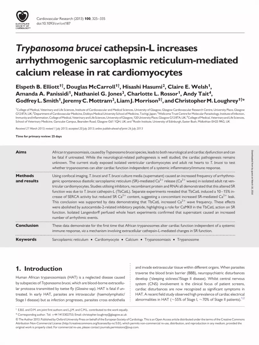
3.7 Supernatant effects on the stimulatedCa21 transient during b-adrenergicstimulationThe sympathetic nervous system and b-adrenergic signalling pathwayactivate in response to physiological and pathophysiological stresses.To examine supernatant effects under b-adrenergic stimulation,isoproterenol was added (ISO,100 nM; Figure 4A). Under these con-ditions, supernatant did not significantly alter the mean maximum [Ca2+]ior Ca2+ transient amplitude, but did increase the minimum [Ca2+]i
between field stimulations to 115% of control levels (0.20+ 0.010 vs.0.22+ 0.006 mM; media vs. supernatant; P , 0.05; Figure 4Bi– iii, andCii). The maximum rate of rise of the Ca2+ transient was not altered bysupernatant but supernatant increased the maximum rate of fall to 133%of control levels (16.0+1.7 vs. 21.4+2.0 mM ms21; media vs.
supernatant; P , 0.05; Figure 4Cv). This latter result is confirmed by the sig-nificant increase in Ca2+ transient decay constant by supernatant to 126%of control values; 16.54+1.34 vs. 20.86+1.31 s21; media vs. super-natant; P , 0.05; Figure 4Cvi).
As shown in Figure 4Bi vs. ii, supernatant increased the appearance ofCa2+ waves between stimulated Ca2+ transients. Cardiomyocytessuperfused with supernatant and ISO demonstrated a Ca2+ wave fre-quency increase over time, plateauing at 90 s; (Figure 4Di). The meanCa2+ wave frequency (measured over 90–120 s) increased to 866%of control levels during perfusion with supernatant compared withmedia (0.03+0.01 vs. 0.26+0.09 Ca2+ wave s21; media vs. super-natant; P , 0.05; (Figure 4Dii). The Ca2+ wave amplitude also increasedto 632% of control levels (9.4+6.4 vs. 59.7+21.0 nM [Ca2+]i; mediavs. supernatant; P , 0.05; Figure 4Diii).
3.8 Ca21 wave increase is abolished byinhibition of parasite cathepsin-LThe trypanosome factor associated with BMEC cytosolic Ca2+ riseswas T. brucei cathepsin-L (TbCatL).12 Trypanosoma brucei expressestwo Clan CA, Family C1 cysteine proteases; TbCatL and
Figure 3 (A) % Change in cardiomyocytes with spontaneous contractile events supernatant + AIP (n ¼ 307) vs. media + AIP (n ¼ 121) or (ii) H89 +supernatant (n ¼ 377) vs. media + H89 (n ¼ 305). (B) % change in Ca2+ transient and caffeine-induced parameters with AIP. (C) Fura-2AM ratio during SRinhibition + AIP with media (n ¼ 25) and supernatant (n ¼ 21).
E.B. Elliott et al.330by guest on M
arch 18, 2016D
ownloaded from
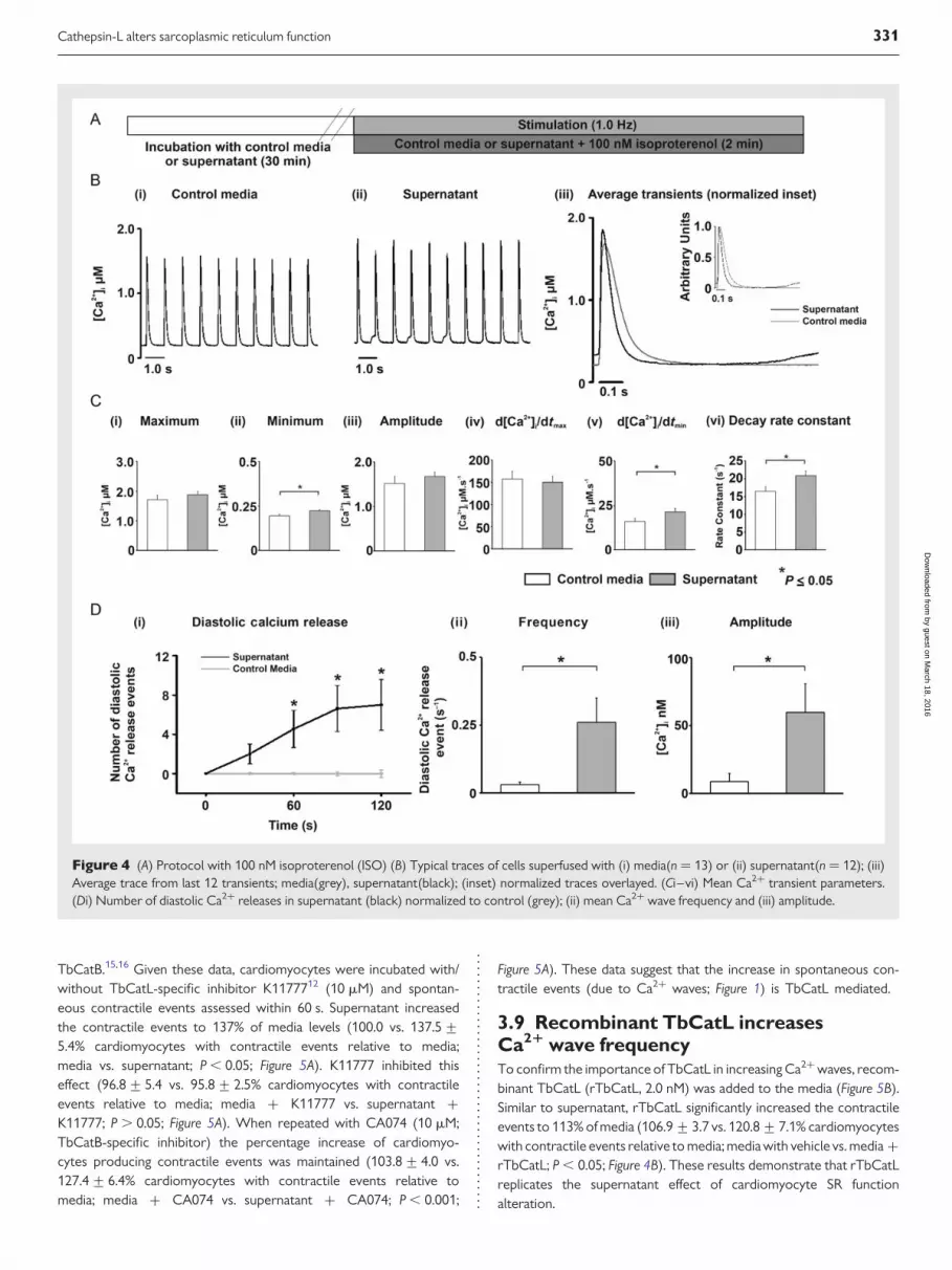
TbCatB.15,16 Given these data, cardiomyocytes were incubated with/without TbCatL-specific inhibitor K1177712 (10 mM) and spontan-eous contractile events assessed within 60 s. Supernatant increasedthe contractile events to 137% of media levels (100.0 vs. 137.5+5.4% cardiomyocytes with contractile events relative to media;media vs. supernatant; P , 0.05; Figure 5A). K11777 inhibited thiseffect (96.8+5.4 vs. 95.8+ 2.5% cardiomyocytes with contractileevents relative to media; media + K11777 vs. supernatant +K11777; P . 0.05; Figure 5A). When repeated with CA074 (10 mM;TbCatB-specific inhibitor) the percentage increase of cardiomyo-cytes producing contractile events was maintained (103.8+ 4.0 vs.127.4+ 6.4% cardiomyocytes with contractile events relative tomedia; media + CA074 vs. supernatant + CA074; P , 0.001;
Figure 5A). These data suggest that the increase in spontaneous con-tractile events (due to Ca2+ waves; Figure 1) is TbCatL mediated.
3.9 Recombinant TbCatL increasesCa21 wave frequencyTo confirm the importance of TbCatL in increasing Ca2+ waves, recom-binant TbCatL (rTbCatL, 2.0 nM) was added to the media (Figure 5B).Similar to supernatant, rTbCatL significantly increased the contractileevents to 113% of media (106.9+3.7 vs. 120.8+ 7.1% cardiomyocyteswith contractile events relative to media; media with vehicle vs. media +rTbCatL; P , 0.05; Figure 4B). These results demonstrate that rTbCatLreplicates the supernatant effect of cardiomyocyte SR functionalteration.
Figure 4 (A) Protocol with 100 nM isoproterenol (ISO) (B) Typical traces of cells superfused with (i) media(n ¼ 13) or (ii) supernatant(n ¼ 12); (iii)Average trace from last 12 transients; media(grey), supernatant(black); (inset) normalized traces overlayed. (Ci–vi) Mean Ca2+ transient parameters.(Di) Number of diastolic Ca2+ releases in supernatant (black) normalized to control (grey); (ii) mean Ca2+ wave frequency and (iii) amplitude.
Cathepsin-L alters sarcoplasmic reticulum function 331by guest on M
arch 18, 2016D
ownloaded from
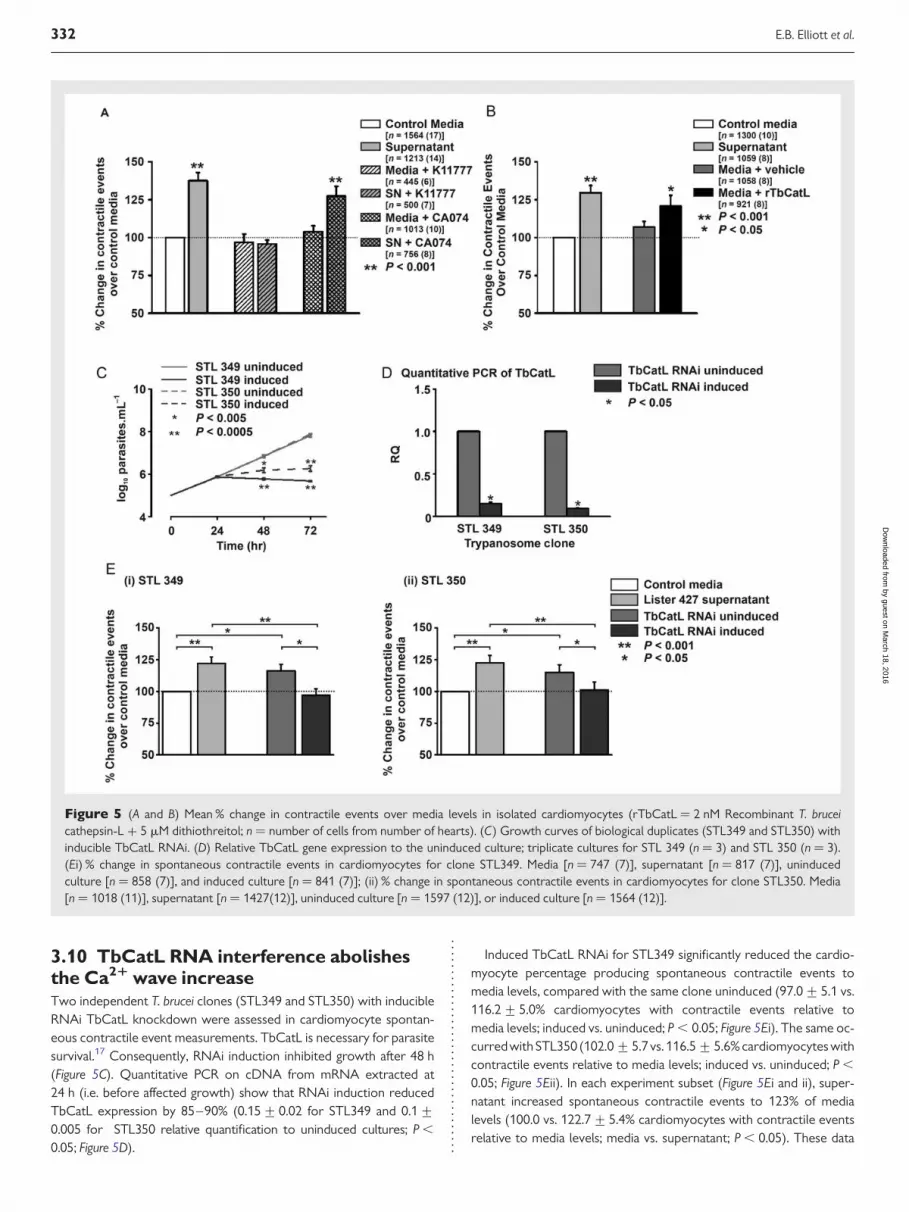
3.10 TbCatL RNA interference abolishesthe Ca21 wave increaseTwo independent T. brucei clones (STL349 and STL350) with inducibleRNAi TbCatL knockdown were assessed in cardiomyocyte spontan-eous contractile event measurements. TbCatL is necessary for parasitesurvival.17 Consequently, RNAi induction inhibited growth after 48 h(Figure 5C). Quantitative PCR on cDNA from mRNA extracted at24 h (i.e. before affected growth) show that RNAi induction reducedTbCatL expression by 85–90% (0.15+0.02 for STL349 and 0.1+0.005 for STL350 relative quantification to uninduced cultures; P ,
0.05; Figure 5D).
Induced TbCatL RNAi for STL349 significantly reduced the cardio-myocyte percentage producing spontaneous contractile events tomedia levels, compared with the same clone uninduced (97.0+ 5.1 vs.116.2+ 5.0% cardiomyocytes with contractile events relative tomedia levels; induced vs. uninduced; P , 0.05; Figure 5Ei). The same oc-curredwith STL350 (102.0+ 5.7 vs. 116.5+ 5.6%cardiomyocytes withcontractile events relative to media levels; induced vs. uninduced; P ,
0.05; Figure 5Eii). In each experiment subset (Figure 5Ei and ii), super-natant increased spontaneous contractile events to 123% of medialevels (100.0 vs. 122.7+5.4% cardiomyocytes with contractile eventsrelative to media levels; media vs. supernatant; P , 0.05). These data
Figure 5 (A and B) Mean % change in contractile events over media levels in isolated cardiomyocytes (rTbCatL ¼ 2 nM Recombinant T. bruceicathepsin-L + 5 mM dithiothreitol; n ¼ number of cells from number of hearts). (C) Growth curves of biological duplicates (STL349 and STL350) withinducible TbCatL RNAi. (D) Relative TbCatL gene expression to the uninduced culture; triplicate cultures for STL 349 (n ¼ 3) and STL 350 (n ¼ 3).(Ei) % change in spontaneous contractile events in cardiomyocytes for clone STL349. Media [n ¼ 747 (7)], supernatant [n ¼ 817 (7)], uninducedculture [n ¼ 858 (7)], and induced culture [n ¼ 841 (7)]; (ii) % change in spontaneous contractile events in cardiomyocytes for clone STL350. Media[n ¼ 1018 (11)], supernatant [n ¼ 1427(12)], uninduced culture [n ¼ 1597 (12)], or induced culture [n ¼ 1564 (12)].
E.B. Elliott et al.332by guest on M
arch 18, 2016D
ownloaded from

show that the supernatant-induced increased Ca2+ waves are abolishedif TbCatL gene expression is reduced to �10% of control levels.
3.11 Supernatant leads to arrhythmicevents in whole heartsGiven the established link between spontaneous SR-mediatedCa2+ release and arrhythmias, the arrhythmogenic potential of super-natant was investigated in ex vivo whole hearts. The pseudo-ECGwas examined for VPCs (Figure 6A and B), known to be associatedwith abnormal diastolic Ca2+ release. Each VPC was counted as onearrhythmic event. There was no significant difference in mean arrhyth-mic frequency between the different time periods of the protocol inthe time-control group; (0.40+ 0.28 vs. 0.33+ 0.19 vs. 0.53+ 0.27VPCs min21; first, second, and third 10 min period, respectively;P . 0.05; Figure 6C). However, in hearts perfused with supernatantthere was a significant increase in mean arrhythmic event frequencywhich then reduced during the washout period (0.60+ 0.26 vs.5.15+ 2.49 vs. 1.36+ 0.73 PCs min21; first, second, and third10 min period, respectively; P , 0.05 between first and secondperiod; Figure 6C). There was no significant difference in heartrate between hearts perfused with media (282+ 17 b.p.m.) orsupernatant (278+ 12 b.p.m.).
4. DiscussionUsing isolated cardiomyocytes and whole hearts,wehavedemonstratedfor the first time that Trypanosoma brucei can alter cardiac functionwithout a systemic immune response or autonomic nervous systemeffect. Prior to this study, the only suggested mechanism forcardiac-related clinical symptoms has been the immune/inflammatoryresponse to the parasite. Our data demonstrate that this is not thesole mechanism and present a new paradigm for trypanosome-inducedcardiac dysfunction.
4.1 Alteration of spontaneous SR-mediatedCa21 release in isolated cardiomyocytesIsolated cardiomyocytes show increased spontaneous contractile activ-ity when exposed to both parasites and supernatant (Figure 1). Suchspontaneous contractile activity can be associated with spontaneousSR-mediatedCa2+ release in the formofCa2+waves,whichwasdemon-strated in this study (Figure 1). A similar effect has been observed inBMECs, where [Ca2+]i spontaneous releases occurred despite theremoval of the parasite from the media.12 It is clear therefore that inboth cardiomyocytes and BMECs, the [Ca2+]i alteration is mediatedby a trypanosome-derived (secreted/excreted) factor.
Figure 6 (A) Langendorff perfused heart protocol. (B) typical pseudo-ECG recordings. (C ) Mean number of ventricular premature complexes (VPCs)per min for time control (n ¼ 6) and supernatant-perfused hearts (n ¼ 12) in media (first 10 min; white bar), switch to media or supernatant (second10 min; black bar), and media washout (third 10 min; grey bar).
Cathepsin-L alters sarcoplasmic reticulum function 333by guest on M
arch 18, 2016D
ownloaded from

4.2 Ca21 wave velocity and frequencyare increased by supernatantAltered Ca2+ wave characteristics indicate altered SR function.18 In thisstudy, two notable effects on Ca2+ wave characteristics occurred post-field stimulation. In agreement with spontaneous contractile event mea-surements (Figure 1A), an increased Ca2+ wave frequency was observedin cardiomyocytes incubated with supernatant. Ca2+waves are linked toboth arrhythmias and contractile dysfunction and increased Ca2+ wavefrequency is likely to increase whole heart arrhythmias.13 Ca2+wave fre-quency is determined by the time taken to reach a SR Ca2+ threshold.13
SERCA stimulation by a direct or indirect effect of a supernatant factorwould increase the SR Ca2+accumulation rate and hence decrease thetime between spontaneous events.13,19 O’Neill et al.19 also showedthat Ca2+ wave velocity was directly related to SERCA activity, andthus the increased Ca2+ wave velocity observed in the current studymay be an expected additional effect of enhanced SERCA activity.
4.3 Effects of supernatant on SR functionThe proposal that exposure to supernatant increases cardiac SERCAactivity is consistent with the supernatant effects on the stimulatedCa2+ transient (Figures 2 and 4), which showed an increased decayrate constant. SERCA activity (KSERCA) was estimated by measuringthe rate constant of decay of the electrically stimulated Ca2+ transient.However, sarcolemmal efflux from the cell also contributes to this rateconstant of decay. When the rate constant of decay of thecaffeine-induced Ca2+ transient (which includes sarcolemmal effluxbut no SR uptake contribution) is subtracted from that of the electricallystimulated Ca2+ transient (which includes the combined SR uptake andsarcolemmal efflux)20 the % change in KSERCA induced by the super-natant was 114.3+ 2.2% of media control. This compares well withthe value (110.4+1.2; Figure 2Diii) when using the rate decay constantof the electrically induced transient alone. Therefore, both values arevery similar and we conclude that SERCA activity is significantlyincreased by the supernatant by 10–15%. An alternative explanationfor the increased decay time constant is alteration of myofilamentCa2+ responsiveness. However, the supernatant had no effect on myo-filament sensitivity to [Ca2+]i (Supplementary material online). Ifenhanced SERCA activity was the exclusive action of the supernatanton SR function then the SR Ca2+ content may be expected to haveincreased. However, Figure 2 clearly shows that the supernatantdecreases the SR Ca2+ content. This decrease cannot be explained byincreased sarcolemmal efflux, as the caffeine-induced Ca2+ transientdecay rate constant and diastolic [Ca2+]i were unaltered. Therefore,we hypothesize that in addition to mild (10–15%) stimulation ofSERCA activity, TbCatL also enhances SR-mediated Ca2+ leak; the neteffect being reduction in the SR Ca2+ content. An increasedSR-mediated Ca2+ leak could potentially occur via the ryanodine recep-tor (RyR). A large body of work has confirmed that increased RyR openprobability may lead to: (i) unaltered stimulated Ca2+ transient ampli-tude, (ii) reduction in the SR Ca2+ content, (iii) increased Ca2+wave fre-quency in resting cells, and (iv) sustained and increased Ca2+ wavesduring electrical stimulation in the presence ofb-adrenergic stimulation(reviewed in13). The data presenteddemonstrate the above features andtherefore support the hypothesis that TbCatL induces SR-mediatedCa2+ leak with parallel SERCA activity increase. CaMKII (but not PKA)inhibition abolished the supernatant effect on SR Ca2+ uptake andcontent. Importantly, this in turn led to the abolishment of the expectedincreased in Ca2+ waves (Figure 3). Enhanced SR Ca2+ leak and a
subsequent reduction of SR Ca2+ content can be prevented by AIP incardiomyocytes overexpressing CaMKII without significantly alteringthe Ca2+ transient amplitude.21 Furthermore, SR-localized AIP canalso inhibit CaMKII-dependent regulation of SERCA-mediated Ca2+
uptake.22 Therefore, these data support the proposed mechanism of aparallel effect of the supernatant on abnormal SR Ca2+ release anduptake that leads to the increased Ca2+ wave frequency observed.
4.4 Effects of supernatant on diastolic[Ca21]iIsolated cardiomyocyte experiments where SR-mediated Ca2+ releaseand uptake was inhibited (Figure 2F) revealed that supernatant elevated[Ca2+]i compared with control media. Our experiments do not discrim-inate between reduced Ca2+ efflux or enhanced Ca2+ influx, butincreased diastolic [Ca2+]i may favour the production of abnormal SRCa2+ release. One potential mechanism for increased diastolic [Ca2+]i
is reduced NCX activity, the main sarcolemmal Ca2+ extrusionprocess. However, Ca2+ extrusion rate in the presence of caffeinewas unaltered by supernatant (Figure 2), suggesting NCX activity was un-affected. Alternatively, supernatant may have enhanced Ca2+ entry intocardiomyocytes, but in quiescent cells this is unlikely to be via the L-typeCa2+ channel (see Figure 2G). Interestingly, this effect was not AIP-sensitive (Figure 3). The changes in basal and store-based Ca2+ signallingobserved in cardiomyocytes parallel those seen in BMECs,12 an effectmediated by TbCatL action on a GPCR mediated pathway.23 Activationof this or a similar pathway in cardiomyocytes may underlie the super-natant effect on the heart. Overall, it is possible that supernatant elevatesdiastolic [Ca2+]i (which could be substantial within subcellular compart-ments) causing CaMKII activation, which then increases SR-mediatedCa2+ leak and uptake with a net effect being an increase in Ca2+
waves and reduced SR Ca2+.
4.5 Ca21 waves are caused by TbCatLThe cathepsin-L inhibitor K11777 prevented the supernatant-mediatedincrease in Ca2+waves. This demonstrated for the first time thatTbCatLincreases spontaneous Ca2+ release from the cardiomyocyte SR. Theseresults are consistent with rTbCatL increasing Ca2+ waves (gain offunction) and RNAi reducing TbCatL gene expression and decreasingCa2+ waves (loss of function). The combined pharmacological, recom-binant protein, and RNAi approaches confirmed that trypanosome-produced extracellular TbCatL causes the increased arrhythmogenicSR-mediated Ca2+ release in adult rat ventricular cardiomyocytes, inde-pendent of an immune/inflammatory response. TbCatL is essential toparasite survival (hence RNAi use rather than gene knockout, which islethal).17 Current HAT drugs are difficult to administer in the field andcause severe toxic side effects in 5–10% of cases (average case-fatalityrate of 50%).24 Cysteine protease peptide inhibitors are currentlybeing tested as novel trypanocides.17 Our study suggests that in additionto being trypanocidal, drugs that target TbCatL may limit both BBB path-ology and cardiac dysfunction.
4.6 Supernatant causes arrhythmic events inwhole heartsField studies report a range of ECG abnormalities and palpitations inHAT patients that can result from VPCs.2 The link is establishedbetween Ca2+ waves, delayed after-depolarization and subsequentVPC production in the whole heart which may induce fatal arrhythmias(reviewed in13). Supernatant is able to increase VPCs independent of
E.B. Elliott et al.334by guest on M
arch 18, 2016D
ownloaded from

an immune response under b-adrenergic stimulation conditions. This isthe first time that trypanosomes have been shown to contribute directlyto whole heart arrhythmias. It is unclear to what degree HAT patientsunder stress (e.g. exercise/pathological states) would have an increasedarrhythmia risk, but from our findings we hypothesize that HAT patientswould be more prone to arrhythmic events under stress conditions.Indeed, there are reports of sudden death in HAT patients beforefull progression of the disease, and in these cases suddenarrhythmic-induced cardiac death cannot be ruled out.5,6
4.7 Study implications and conclusionsExtracellular cathepsin-L not only disrupts Ca2+ release from intracell-ular stores in the brain but as we have shown for the first time, also in theheart. This common pathogenic mechanism provides a platform tosupport future investigations that centre on normalization of theeffects of cathepsin-L via drugs that act on SR/ER-mediated Ca2+ signal-ling in order to address the neurological and cardiovascular pathologyassociated with HAT. Previous studies have shown that arrhythmiasassociated with disrupted SR function can be treated with flecainideand K201(JTV-519)14 and this suggests a potential therapeutic approachfor treatment of cathepsin-L induced cardiac dysfunction.
The findings of this study also have implications for other pathogens,such as Trypanosoma cruzi, which causes Chagas disease. Notable cardiacpathology is present in Chagas disease, and T. cruzi induces abnormalCa2+handling in cardiomyocytes.25 Another important implication con-cerns coronary heart disease (CHD). It is well established that patientswith CHD have abnormal SR-mediated Ca2+ release, contractile dys-function, increased arrhythmia risk, and elevated levels of endogenouscathepsin-L in serum, yet the effect of mammalian-derived extracellularcathepsins on SR function remains unknown.
Supplementary materialSupplementary material is available at Cardiovascular Research online.
AcknowledgementsWe thank Jim McKerrow and Ana Paula Lima for the kind gift of recom-binant TbCatL.This work was supported by the Wellcome Trust. TheWellcome Trust Centre for Molecular Parasitology is supported bycore funding from the Wellcome Trust [085349].
Conflict of interest: none declared.
FundingThis work was supported by the Royal College of Veterinary Surgeons Trust(GR000 652 to C.L. and L.M.); University of Glasgow Medical Fund (to C.L.and L.M.); Biotechnology and Biological Sciences Research Council (BBSRC)doctoral training grant (to D.M. and C.W.), the Wellcome Trust and theRoyal Society (University Research Fellowship UF090083 to L.M.). TheWellcome Trust Centre for Molecular Parasitology is supported by corefunding from the Wellcome Trust (085349) (to J.M.).
References1. Blum JA, Schmid C,Burri C,HatzC,Olson C, FungulaB et al. Cardiacalterations in human
African trypanosomiasis (T.b. gambiense) with respect to the disease stage and antipar-asitic treatment. PLoS Negl Trop Dis 2009;3:e383.
2. Blum JA, Burri C, Hatz C, Kazumba L, Mangoni P, Zellweger MJ. Sleeping hearts: the roleof the heart in sleeping sickness (human African trypanosomiasis). Trop Med Int Health2007;12:1422–1432.
3. Hidron A, Vogenthaler N, Santos-Preciado JI, Rodriguez-Morales AJ, Franco-Paredes C,Rassi A Jr. Cardiac involvement with parasitic infections. Clin Microbiol Rev 2010;23:324–349.
4. Jones IG, Lowenthal MN, Buyst H. Electrocardiographic changes in African trypanosom-iasis caused by Trypanosoma brucei rhodesiense. Trans R Soc Trop Med Hyg 1975;69:388–395.
5. Bertrand E. Cardiac involvement in human African trypanosomiasis. Med Trop (Mars)1987;47:91–93.
6. Chappuis F, Loutan L, Simarro P, Lejon V, Buscher P. Options for field diagnosis ofhuman African trypanosomiasis. Clin Microbiol Rev 2005;18:133–146.
7. Franco-Paredes C, Rouphael N, Mendez J, Folch E, Rodriguez-Morales AJ, Santos JI et al.Cardiac manifestations of parasitic infections. Part 2: parasitic myocardial disease. ClinCardiol 2007;30:218–222.
8. Poltera AA, Owor R, Cox JN. Pathological aspects of human African trypanosomiasis(HAT) in Uganda. A post-mortem survey of fourteen cases. Virchows Arch A Pathol AnatHistol 1977;373:249–265.
9. Morrison WI, Murray M, Sayer PD, Preston JM. The pathogenesis of experimentallyinduced Trypanosoma brucei infection in the dog. I. Tissue and organ damage. Am JPathol 1981;102:168–181.
10. Poltera AA, Cox JN, Owor R. Pancarditis affecting the conducting system and all valves inhuman African trypanosomiasis. Br Heart J 1976;38:827–837.
11. Grab DJ, Kennedy PG. Traversal of human and animal trypanosomes across the blood-brain barrier. J Neurovirol 2008;14:344–351.
12. Nikolskaia OV, de A Lima AP, Kim YV, Lonsdale-Eccles JD, Fukuma T, Scharfstein J et al.Blood-brain barrier traversal by African trypanosomes requires calcium signalinginduced by parasite cysteine protease. J Clin Invest 2006;116:2739–2747.
13. Venetucci LA, Trafford AW, O’Neill SC, Eisner DA. The sarcoplasmic reticulum andarrhythmogenic calcium release. Cardiovasc Res 2008;77:285–292.
14. Elliott EB, Hasumi H, Otani N, Matsuda T, Matsuda R, Kaneko N et al. K201 (JTV-519)alters the spatiotemporal properties of diastolic Ca2+ release and the associated diastol-ic contractionduring beta-adrenergic stimulation in rat ventricularcardiomyocytes.BasicRes Cardiol 2011;106:1009–1022.
15. Abdulla MH, O’Brien T, Mackey ZB, Sajid M, Grab DJ, McKerrow JH. RNA interferenceof Trypanosoma brucei cathepsin B and L affects disease progression in a mouse model.PLoS Negl Trop Dis 2008;2:e298.
16. Berriman M, Ghedin E, Hertz-Fowler C, Blandin G, Renauld H, Bartholomeu DC et al.The genome of the African trypanosome Trypanosoma brucei. Science 2005;309:416–422.
17. Steverding D, Sexton DW, Wang X, Gehrke SS, Wagner GK, Caffrey CR. Trypanosomabrucei: chemical evidence that cathepsin L is essential for survival and a relevant drugtarget. Int J Parasitol 2012;42:481–488.
18. Loughrey CM, Smith GL, MacEachern KE. Comparison of Ca2+ release and uptake char-acteristics of the sarcoplasmic reticulum in isolatedhorse and rabbit cardiomyocytes. AmJ Physiol Heart Circ Physiol 2004;287:H1149–H1159.
19. O’Neill SC, Miller L, Hinch R, Eisner DA. Interplay between SERCA and sarcolemmalCa2+ efflux pathways controls spontaneous release of Ca2+ from the sarcoplasmicreticulum in rat ventricular myocytes. J Physiol 2004;559:121–128.
20. Bode EF, Briston SJ, Overend CL, O’Neill SC, Trafford AW, Eisner DA. Changes ofSERCA activity have only modest effects on sarcoplasmic reticulum Ca2+ content inrat ventricular myocytes. J Physiol 2011;589:4723–4729.
21. Kohlhaas M, Zhang T, Seidler T, Zibrova D, Dybkova N, Steen A et al.Increased sarcoplasmic reticulum calcium leak but unaltered contractility by acuteCaMKII overexpression in isolated rabbit cardiac myocytes. Circ Res 2006;98:235–244.
22. Picht E, DeSantiago J, Huke S, Kaetzel MA, Dedman JR, Bers DM. CaMKII inhibition tar-geted to the sarcoplasmic reticulum inhibits frequency-dependent acceleration of relax-ation and Ca2+ current facilitation. J Mol Cell Cardiol 2007;42:196–205.
23. Grab DJ, Garcia-Garcia JC, Nikolskaia OV, Kim YV, Brown A, Pardo CA et al. Proteaseactivated receptor signaling is required for African trypanosome traversal of humanbrain microvascular endothelial cells. PLoS Negl Trop Dis 2009;3:e479.
24. Legros D, Ollivier G, Gastellu-Etchegorry M, Paquet C, Burri C, Jannin J et al. Treatmentof human African trypanosomiasis-present situation and needs for research and dev-elopment. Lancet Infect Dis 2002;2:437–440.
25. Barr SC, Han W, Andrews NW, Lopez JW, Ball BA, Pannabecker TL et al. A factor fromTrypanosoma cruzi induces repetitive cytosolic free Ca2+ transients in isolated primarycanine cardiac myocytes. Infect Immunity 1996;64:1770–1777.
Cathepsin-L alters sarcoplasmic reticulum function 335by guest on M
arch 18, 2016D
ownloaded from