treatment of hepatic epithelioid hemangioendothelioma: finding new uses for thalidomide
TRANSCRIPT
Treatment of Hepatic Epithelioid Hemangioendothelioma WithLiver Transplantation
IGNAZIO R. MARINO, MD, SATORU TODO, MD, ANDREAS G. TZAKIS, MD, GORANKLINTMALM, MD, PhD*, MICHAEL KELLEHER, MD, SHUNZABURO IWATSUKI, MD, THOMASE. STARZL, MD, PhD, and CARLOS O. ESQUIVEL, MD, PhDDepartment of Surgery, Pathology, and Medicine, University Health Center of Pittsburgh, and theVeterans Administration Medical Center, Pittsburgh, Pennsylvania*Department of Surgery, Baylor University Medical Center, Dallas, Texas
AbstractTen patients received liver transplants for unresectable epithelioid hemangioendothelioma (EHE).At the time of transplantation, four patients had microscopic metastases to the hilar lymph nodes,and one of the four also had metastases to a rib. The fifth patient had metastases to the lung, pleura,and diaphragm. The remaining five patients were believed to be free of metastatic disease. Two ofthese five patients died of metastatic disease at 3 and 16 months, respectively, after transplantation.Interestingly, all five patients with metastatic involvement are currently alive 40.6 ± 22 months (mean± standard error of mean [SEM]) after transplantation, although one of these patients currently hasmetastatic disease to the lungs and mediastinum. Thus, the projected 5-year actuarial survival rate is76%, with two patients at risk after the third year. In conclusion, liver transplantation is a reasonableprocedure Cor bulky, otherwise unresectable, EHE even in the presence of metastatic disease.
Epithelioid hemangioendothelioma (EHE) is a soft tissue malignant tumor that is characterizedby its epithelioid appearance and vascular endothelial histogenesis. It was specificallyidentified histologically by Weiss and Enzinger1 in 1982. In 1975, a similar if not identicaltumor occurring in the lung was described by Dail and Liebow who proposed the term“intravascular bronchioalveolar tumor.”2 In 1984, Ishak et al. reported for the first time 32patients with primary EHE of the liver.3
The current definition of EHE as a unique form of vascular lesion consisting of endothelialcells is based on the presence of immunohistochemical staining for Factor VIII-related antigenin the tumor.1–5 Using this specific definition, only a few cases of EHE have been describedin the literature, particularly as primary hepatic malignancies, although EHE may occur moreoften than it is reported.1,6–8
The therapeutic approaches used in the clinical management of this tumor have been variablebecause of the limited clinical experience with this malignancy. There have been two separatesingle case reports of patients with EHE who have been treated with orthotopic livertransplantation (OLT).9,10
Herein we report the results with hepatic transplantation in ten patients with EHE. In each ofthese patients, the tumor was unresectable using any of several different conventional subtotalhepatectomy procedures.
Address for reprints: Carlos O. Esquivel, MD, PhD, Pacific Presbyterian Medical Center, Division of Transplantation, Box 7999, SanFrancisco, CA 94120..
NIH Public AccessAuthor ManuscriptCancer. Author manuscript; available in PMC 2010 November 23.
Published in final edited form as:Cancer. 1988 November 15; 62(10): 2079–2084.
NIH
-PA Author Manuscript
NIH
-PA Author Manuscript
NIH
-PA Author Manuscript
Patients and MethodsPatient Profile
Between March 1963 and October 1987, 1281 patients had transplants performed at either theUniversity of Colorado or University of Pittsburgh Health Centers or the Baylor UniversityMedical Center. In 91 of these patients (7.1 %), the indication for OLT was a primary hepaticmalignancy that could not otherwise be resected. The histologic diagnosis in ten of these 91patients was EHE. Only one of these ten patients has been reported currently.3 Their agesranged from 24 to 52.5 years (median age, 29.5 years). Six often patients were female.
After OLT, the immunosuppression consisted of azathioprine and prednisone for the first twopatients and cyclosporine and prednisone for the subsequent eight patients.
All ten patients were evaluated initially in different institutions. They were referred to one ofour hospitals for liver transplantation because the malignant lesions were deemed unresectableexcept as a total hepatectomy. Before transplantation, the patients were subjected to a thoroughevaluation to rule out the presence of any metastatic disease. This included an ultrasonographicexamination of the abdomen (with particular attention to the presence of any vascular lesions),computerized axial tomography of the chest, abdomen, and head, a bone scan for the detectionof metastases, complete upper and lower gastrointestinal endoscopy, and chestroentgenograms. Occasionally, an arteriogram or laparoscopy also was performed.
The correct diagnosis of EHE was known preoperatively in six patients. In two other patients,the diagnosis was not made until the excised liver was examined histologically and Factor VIII-related antigen was detected immunocytochemically as being present within the tumor cells.The initial histopathologic diagnosis was erroneous in two patients (the first two). A recentreview of their histologic material showed the correct diagnosis.
Clinical Features, Operation, and FindingsComplete clinical data were available in nine of ten patients. In one patient, only the operativerecords and histopathology were available.
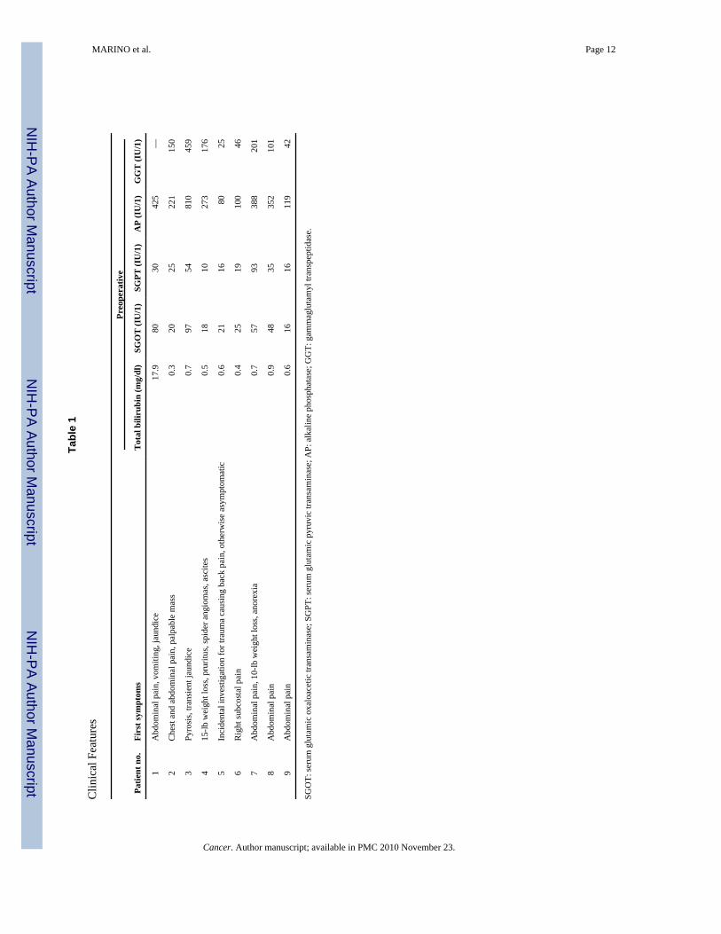
As shown in Table 1, pain was the most common initial symptom of the disease, with anonspecific intestinal disturbance being the second most common complaint. Two patients hadjaundice, one with ascites and one with a palpable hepatic mass. Another patient experiencedobstruction of large intrahepatic biliary ducts with cholestasis and symptoms of veno-occlusivedisease. Liver injury variables often showed nonspecific abnormalities, and all but one patienthad an increased alkaline phosphatase and gamma-glutamyl transpeptidase (GGT) level.
Although a standard protocol for chemotherapy has not been instituted yet, four patients didreceive postoperative adjuvant chemotherapy after transplantation consisting of Adriamycin(Adria Laboratories, Columbus, OH) (Table 2). The one patient who originally was thought tohave a fibrolamellar hepatocellular carcinoma received intraarterial hepatic chemotherapy and2400 cGy of external hepatic radiation before OLT. The patient with metastasis to the leftsecond rib underwent rib resection after radiation therapy to the area.
The technique for liver replacement was similar to that reported elsewhere, except that therecipient hepatectomy included the gastrohepatic ligament, the hepatoduodenal ligament, andskeletonization of all vascular structures in the hepatic hilum.11 The common bile duct wastransected distally as it passed behind the duodenum and the biliary reconstruction wasperformed using an end-to-side donor choledochus to a recipient Roux-en-Y jejunal limb. Inone patient, a portion of the diaphragm was excised because of local tumor invasion. Thediaphragmatic defect in this case was repaired with Marlex mesh.
MARINO et al. Page 2
Cancer. Author manuscript; available in PMC 2010 November 23.
NIH
-PA Author Manuscript
NIH
-PA Author Manuscript
NIH
-PA Author Manuscript
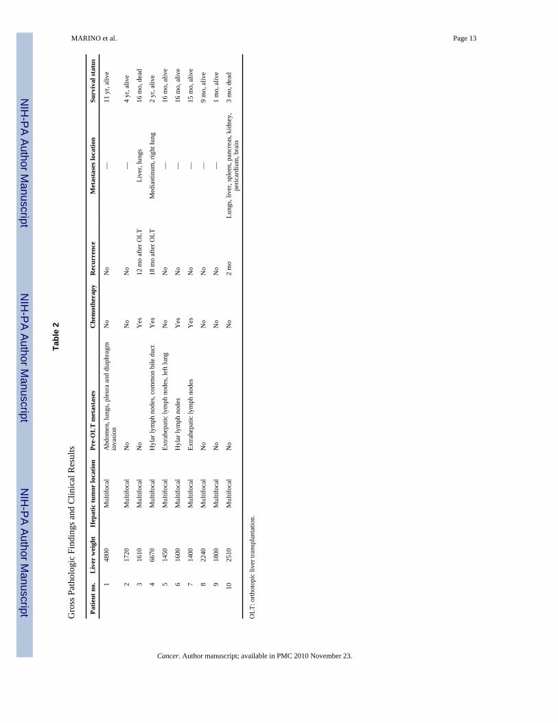
One patient was known to have metastatic disease involving the left second rib before OLT.In addition, four other patients were found to have metastases at the time of the transplantsurgery. One of these had involvement of the lung, pleura, and diaphragm. The other three hadmicroscopic involvement of their hilar lymph nodes (Table 2).
Pathologic StudiesSix of 10 patients had a preoperative biopsy diagnosis of EHE. In each of those patients, theoriginal surgical pathology was reviewed before OLT. All hepatectomy specimens underwenta complete pathologic examination according to a standard liver transplant protocol used atour institutions. In each case, the tumor was weighed and fixed in 10% formalin. The extentof tumor within the liver, the surgical margins, and the number of lymph nodes included weredocumented and appropriate sections were removed and studied. Besides routine hematoxylinand eosin (H & E) stains, eight of ten cases were studied immunocytochemically for FactorVIII-related antigen to confirm the diagnosis. In each of these cases, cytokeratin and alpha-fetoprotein immunocytochemistry was negative. Those patients in whom recurrent diseasedeveloped had all of their pathologic material reviewed to ensure that the recurrences wereidentical to the original tumor.
Statistical AnalysisThe collected data were incorporated into the existing liver transplant database of theDepartment of Surgery at the University of Pittsburgh. Data analysis was performed using theLife Table and Survival Function Programs of the BMDP statistical software package(Statistical Software, Inc., Los Angeles, CA).12
ResultsSurvival and Tumor Recurrence
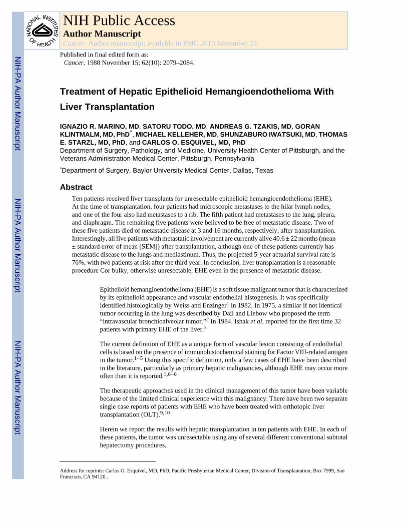
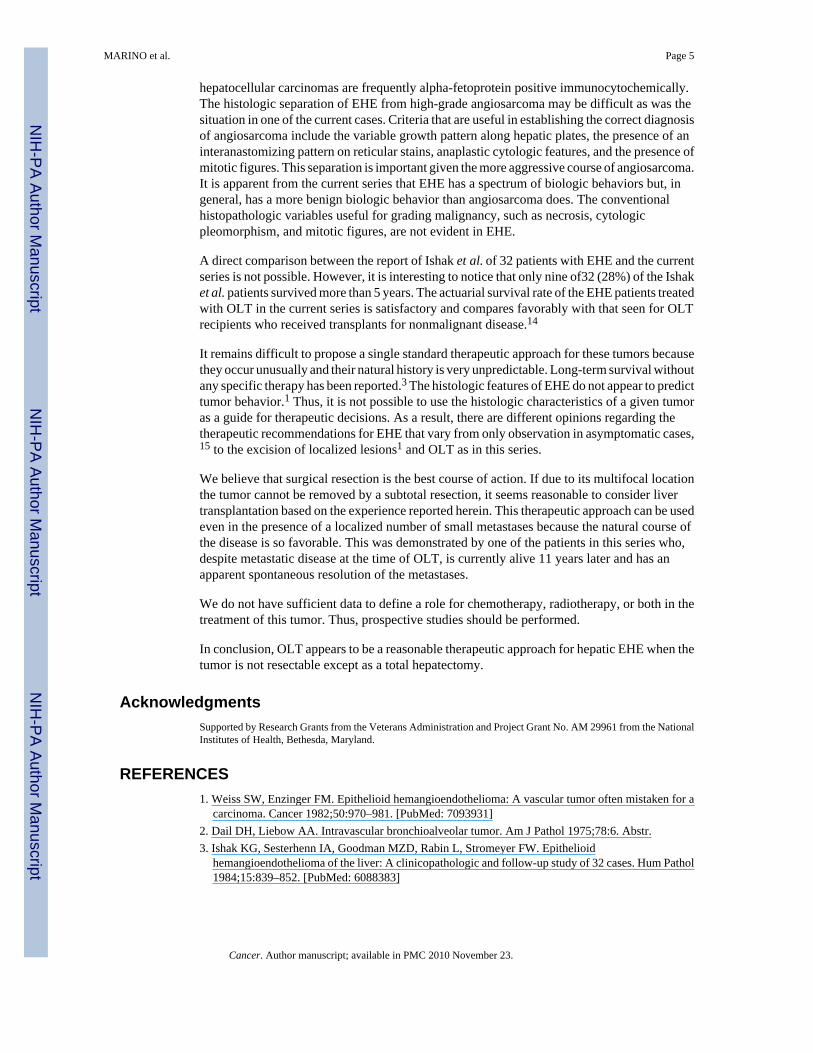
The projected 5-year actuarial survival rate of the ten patients is 76%, with eight patients atrisk after the first year, three after the second year, and two after the third year (Fig. 1).
Table 2 reports the clinical outcome. None of the ten patients died of a complication related tothe transplant procedure. Two died of recurrence of EHE at 3 and 16 months, respectively,after transplantation. Recurrence in the first of these two patients was noticed 2 months afterOLT, with tumor being found in the liver allograft and lungs. The postmortem examination inthis case also showed tumor involving the brain, spleen, pancreas, kidneys, and pericardium.The second patient had a recurrence in the hepatic allograft 12 months after surgery. An autopsyalso documented recurrent tumor in the lungs.
Six patients are alive at 1 month (Patient 9), 9 months (Patient 8), 16 months (Patients 5 and6), 4 years (Patient 2), and 11 years (Patient 1), respectively, after transplantation without anyevidence of recurrence. Our first case might represent a case of tumor regression after surgerybecause this patient had intra-abdominal, pleural, and pulmonary metastases at the time of OLT(Table 2). This patient, who did not receive adjuvant therapy, is alive and free of malignantdisease 11 years after OLT.
Two patients are alive despite the presence of recurrent or residual tumor. The first patient(Patient 4) underwent transplantation 2 years ago and recurrence was first detected in themediastinum 18 months after transplantation. In June 1987, he underwent surgery for resectionof this metastatic mass, during which several additional metastases in the right lung weredetected. The second patient (Patient 7), who was transplanted 15 months ago, had a ribmetastasis that was recognized before OLT (Table 2).
MARINO et al. Page 3
Cancer. Author manuscript; available in PMC 2010 November 23.
NIH
-PA Author Manuscript
NIH
-PA Author Manuscript
NIH
-PA Author Manuscript
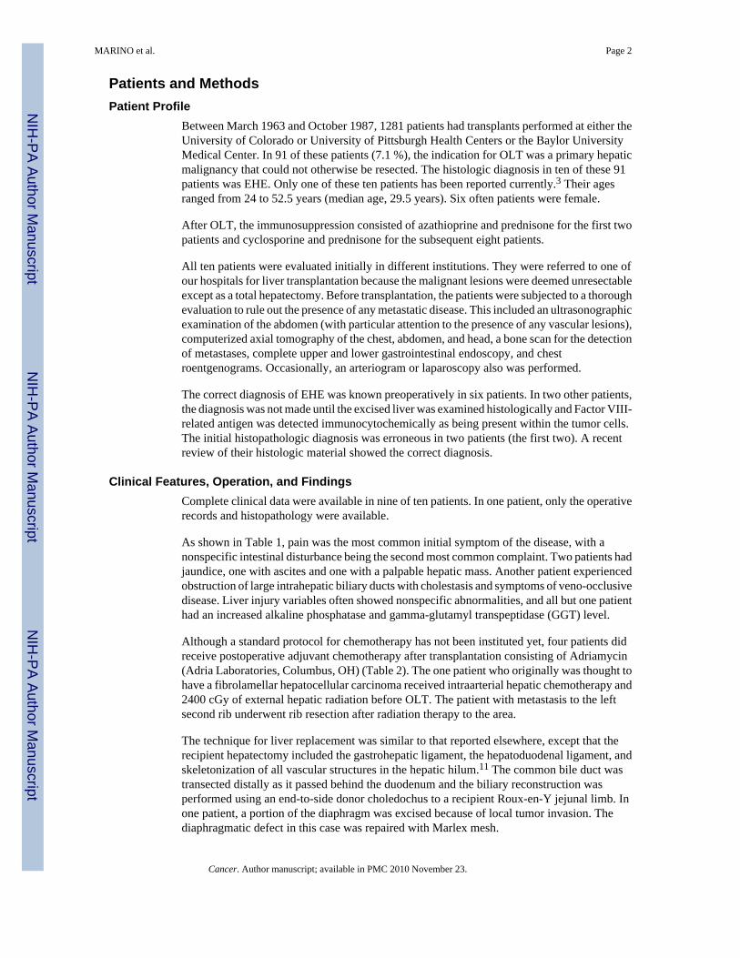
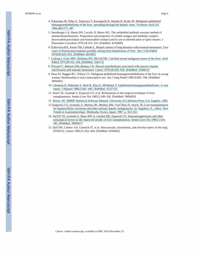
Pathologic FindingsThe liver resection specimens weighed between 1400 and 6670 g. The capsular surface in allhepatectomy specimens was smooth except for the presence of some white tumor depressionsranging from 0.5 to 2.5 cm in diameter. Cross-sections of the resected liver showed multiplenon-encapsulated white nodules throughout both lobes ranging from 1 to 5 cm in greatestdiameter. Some of these nodules were seen in continuity with the hepatic capsule. Nopreferential localization within the hepatic parenchyma was evident (Fig. 2). The interveningnonmalignant hepatic parenchyma was normal except for cholestasis.
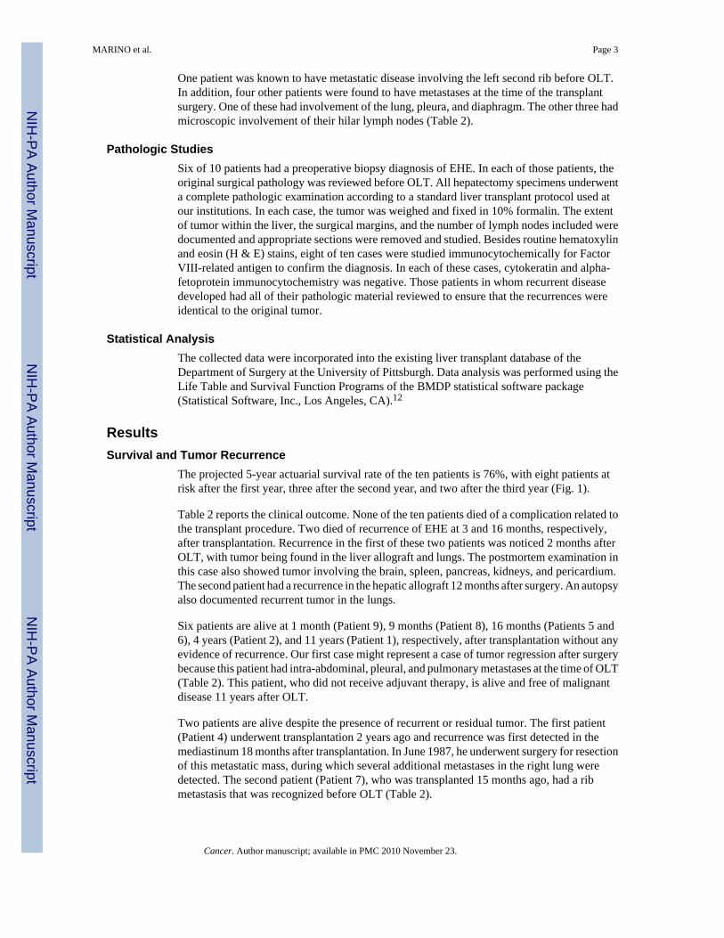
On low-power examination, the tumor nodules appeared to have a variable cellularity with ill-defined margins. At the gross anatomic periphery of the tumor, infiltration of adjacent hepaticsinusoids (Fig. 3) was evident, whereas the central areas showed both necrosis and sclerosis.Tumor invasion into the central and portal veins was observed with varying degrees ofocclusion of their lumens being a common finding (Fig. 3 Inset).
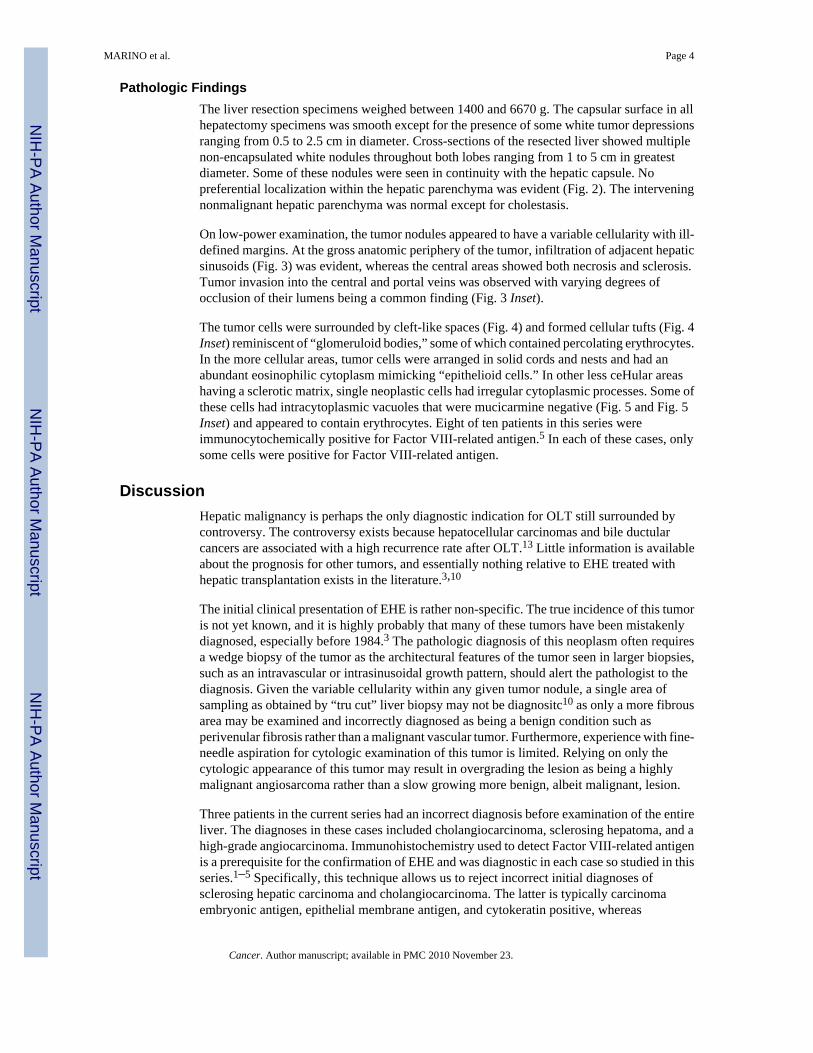
The tumor cells were surrounded by cleft-like spaces (Fig. 4) and formed cellular tufts (Fig. 4Inset) reminiscent of “glomeruloid bodies,” some of which contained percolating erythrocytes.In the more cellular areas, tumor cells were arranged in solid cords and nests and had anabundant eosinophilic cytoplasm mimicking “epithelioid cells.” In other less ceHular areashaving a sclerotic matrix, single neoplastic cells had irregular cytoplasmic processes. Some ofthese cells had intracytoplasmic vacuoles that were mucicarmine negative (Fig. 5 and Fig. 5Inset) and appeared to contain erythrocytes. Eight of ten patients in this series wereimmunocytochemically positive for Factor VIII-related antigen.5 In each of these cases, onlysome cells were positive for Factor VIII-related antigen.
DiscussionHepatic malignancy is perhaps the only diagnostic indication for OLT still surrounded bycontroversy. The controversy exists because hepatocellular carcinomas and bile ductularcancers are associated with a high recurrence rate after OLT.13 Little information is availableabout the prognosis for other tumors, and essentially nothing relative to EHE treated withhepatic transplantation exists in the literature.3,10
The initial clinical presentation of EHE is rather non-specific. The true incidence of this tumoris not yet known, and it is highly probably that many of these tumors have been mistakenlydiagnosed, especially before 1984.3 The pathologic diagnosis of this neoplasm often requiresa wedge biopsy of the tumor as the architectural features of the tumor seen in larger biopsies,such as an intravascular or intrasinusoidal growth pattern, should alert the pathologist to thediagnosis. Given the variable cellularity within any given tumor nodule, a single area ofsampling as obtained by “tru cut” liver biopsy may not be diagnositc10 as only a more fibrousarea may be examined and incorrectly diagnosed as being a benign condition such asperivenular fibrosis rather than a malignant vascular tumor. Furthermore, experience with fine-needle aspiration for cytologic examination of this tumor is limited. Relying on only thecytologic appearance of this tumor may result in overgrading the lesion as being a highlymalignant angiosarcoma rather than a slow growing more benign, albeit malignant, lesion.
Three patients in the current series had an incorrect diagnosis before examination of the entireliver. The diagnoses in these cases included cholangiocarcinoma, sclerosing hepatoma, and ahigh-grade angiocarcinoma. Immunohistochemistry used to detect Factor VIII-related antigenis a prerequisite for the confirmation of EHE and was diagnostic in each case so studied in thisseries.1–5 Specifically, this technique allows us to reject incorrect initial diagnoses ofsclerosing hepatic carcinoma and cholangiocarcinoma. The latter is typically carcinomaembryonic antigen, epithelial membrane antigen, and cytokeratin positive, whereas
MARINO et al. Page 4
Cancer. Author manuscript; available in PMC 2010 November 23.
NIH
-PA Author Manuscript
NIH
-PA Author Manuscript
NIH
-PA Author Manuscript
hepatocellular carcinomas are frequently alpha-fetoprotein positive immunocytochemically.The histologic separation of EHE from high-grade angiosarcoma may be difficult as was thesituation in one of the current cases. Criteria that are useful in establishing the correct diagnosisof angiosarcoma include the variable growth pattern along hepatic plates, the presence of aninteranastomizing pattern on reticular stains, anaplastic cytologic features, and the presence ofmitotic figures. This separation is important given the more aggressive course of angiosarcoma.It is apparent from the current series that EHE has a spectrum of biologic behaviors but, ingeneral, has a more benign biologic behavior than angiosarcoma does. The conventionalhistopathologic variables useful for grading malignancy, such as necrosis, cytologicpleomorphism, and mitotic figures, are not evident in EHE.
A direct comparison between the report of Ishak et al. of 32 patients with EHE and the currentseries is not possible. However, it is interesting to notice that only nine of32 (28%) of the Ishaket al. patients survived more than 5 years. The actuarial survival rate of the EHE patients treatedwith OLT in the current series is satisfactory and compares favorably with that seen for OLTrecipients who received transplants for nonmalignant disease.14
It remains difficult to propose a single standard therapeutic approach for these tumors becausethey occur unusually and their natural history is very unpredictable. Long-term survival withoutany specific therapy has been reported.3 The histologic features of EHE do not appear to predicttumor behavior.1 Thus, it is not possible to use the histologic characteristics of a given tumoras a guide for therapeutic decisions. As a result, there are different opinions regarding thetherapeutic recommendations for EHE that vary from only observation in asymptomatic cases,15 to the excision of localized lesions1 and OLT as in this series.
We believe that surgical resection is the best course of action. If due to its multifocal locationthe tumor cannot be removed by a subtotal resection, it seems reasonable to consider livertransplantation based on the experience reported herein. This therapeutic approach can be usedeven in the presence of a localized number of small metastases because the natural course ofthe disease is so favorable. This was demonstrated by one of the patients in this series who,despite metastatic disease at the time of OLT, is currently alive 11 years later and has anapparent spontaneous resolution of the metastases.
We do not have sufficient data to define a role for chemotherapy, radiotherapy, or both in thetreatment of this tumor. Thus, prospective studies should be performed.
In conclusion, OLT appears to be a reasonable therapeutic approach for hepatic EHE when thetumor is not resectable except as a total hepatectomy.
AcknowledgmentsSupported by Research Grants from the Veterans Administration and Project Grant No. AM 29961 from the NationalInstitutes of Health, Bethesda, Maryland.
REFERENCES1. Weiss SW, Enzinger FM. Epithelioid hemangioendothelioma: A vascular tumor often mistaken for a
carcinoma. Cancer 1982;50:970–981. [PubMed: 7093931]2. Dail DH, Liebow AA. Intravascular bronchioalveolar tumor. Am J Pathol 1975;78:6. Abstr.3. Ishak KG, Sesterhenn IA, Goodman MZD, Rabin L, Stromeyer FW. Epithelioid
hemangioendothelioma of the liver: A clinicopathologic and follow-up study of 32 cases. Hum Pathol1984;15:839–852. [PubMed: 6088383]
MARINO et al. Page 5
Cancer. Author manuscript; available in PMC 2010 November 23.
NIH
-PA Author Manuscript
NIH
-PA Author Manuscript
NIH
-PA Author Manuscript
4. Fukayama M, Nihei Z, Takizawa T, Kawaguchi K, Harada H, Koike M. Malignant epithelioidhemangioendothelioma of the liver, spreading through the hepatic veins. Virchows Arch [A]1984;404:275–287.
5. Sternberger LA, Hardy PH, Cuculis JJ, Meyer HG. The unlabelled antibody enzyme method ofimmunohistochemistry: Preparation and properties of soluble antigen and antibody complex(horseradish peroxidase-anti-horseradish oxidase) and its use in identification of spiro-chaetes. JHistochem Cytochem 1970;18:315–333. [PubMed: 4192899]
6. Echeverria RA, Arean VM, Galindo L. Hepatic tumors of long duration with eventual metastases: Twocases of leiomyosarcomatosis possibly arising from hamartomas of liver. Am J Clin Pathol1978;69:624–631. [PubMed: 665583]
7. Ludwig J, Grier MW, Hoffman HN, McGill DB. Calcified mixed malignant tumor of the liver. ArchPathol 1975;99:162–166. [PubMed: 164173]
8. Persaud V, Bateson EM, Bankay CD. Pleural mesothelioma associated with massive hepaticcalcification and unusual metastases. Cancer 1970;26:920–928. [PubMed: 5506613]
9. Dean PJ, Haggitt RC, O'Hara CG. Malignant epithelioid hemangioendothelioma of the liver in youngwomen: Relationship to oral contraceptive use. Am J Surg Pathol 1985;9:695–704. [PubMed:3904492]
10. Clements D, Hubscher S, West R, Elias E, McMaster P. Epithelioid hemangioendothelioma: A casereport. J Hepatol 1986;2:441–449. [PubMed: 3522725]
11. Starzl TE, Iwatsuki S, Esquivel CO, et al. Refinements in the surgical technique of livertransplantation. Semin Liver Dis 1985;5:349–356. [PubMed: 3909429]
12. Dixon, WJ. BMDP Statistical Software Manual. University of California Press; Los Angeles: 1985.13. Esquivel, CO.; Iwatsuki, S.; Marino, IR.; Markus, BH.; Van Thiel, D.; Starzl, TE. Liver transplantation
for hepatocellular carcinoma and other primary hepatic malignancies. In: Sugahara, K., editor. NewTrends in Gastroenterology. Shinkoshu; Kyoto, Japan: 1987. p. 323-332.
14. StaTZI TE, Iwatsuki S, Shaw BW Jr, Gordon RD, Esquivel CO. Immunosuppression and othernonsurgical factors in the improved results of liver transplantation. Semin Liver Dis 1985;5:334–343. [PubMed: 3909427]
15. Dail DH, Liebow AA, Gmelich JT, et al. Intravascular, bronchiolar, and alveolar tumor of the lung(IVBAT). Cancer 1983;51:452–464. [PubMed: 6295602]
MARINO et al. Page 6
Cancer. Author manuscript; available in PMC 2010 November 23.
NIH
-PA Author Manuscript
NIH
-PA Author Manuscript
NIH
-PA Author Manuscript
FIG. 1.Five-year actuarial survival rate of ten patients with hepatic EHE who had OLT.
MARINO et al. Page 7
Cancer. Author manuscript; available in PMC 2010 November 23.
NIH
-PA Author Manuscript
NIH
-PA Author Manuscript
NIH
-PA Author Manuscript
FIG. 2.Cross-section of liver showing multiple white nodules, some of which are seen in continuitywith the capsule.
MARINO et al. Page 8
Cancer. Author manuscript; available in PMC 2010 November 23.
NIH
-PA Author Manuscript
NIH
-PA Author Manuscript
NIH
-PA Author Manuscript
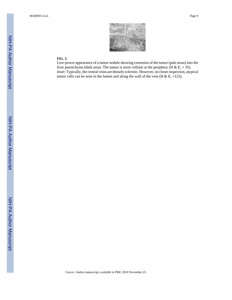
FIG. 3.Low-power appearance of a tumor nodule showing extension of the tumor (pale areas) into theliver parenchyma (dark area). The tumor is more cellular at the periphery (H & E, × 35).Inset: Typically, the central veins are densely sclerotic. However, on closer inspection, atypicaltumor cells can be seen in the lumen and along the wall of the vein (H & E, ×125).
MARINO et al. Page 9
Cancer. Author manuscript; available in PMC 2010 November 23.
NIH
-PA Author Manuscript
NIH
-PA Author Manuscript
NIH
-PA Author Manuscript
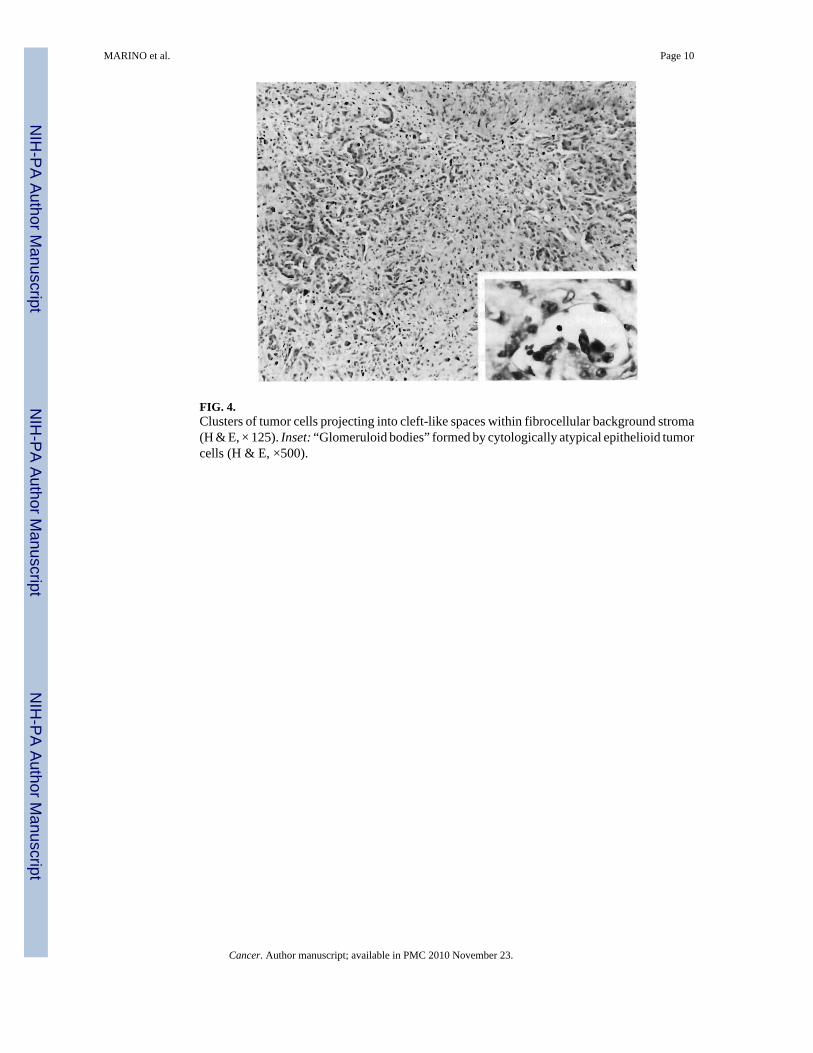
FIG. 4.Clusters of tumor cells projecting into cleft-like spaces within fibrocellular background stroma(H & E, × 125). Inset: “Glomeruloid bodies” formed by cytologically atypical epithelioid tumorcells (H & E, ×500).
MARINO et al. Page 10
Cancer. Author manuscript; available in PMC 2010 November 23.
NIH
-PA Author Manuscript
NIH
-PA Author Manuscript
NIH
-PA Author Manuscript
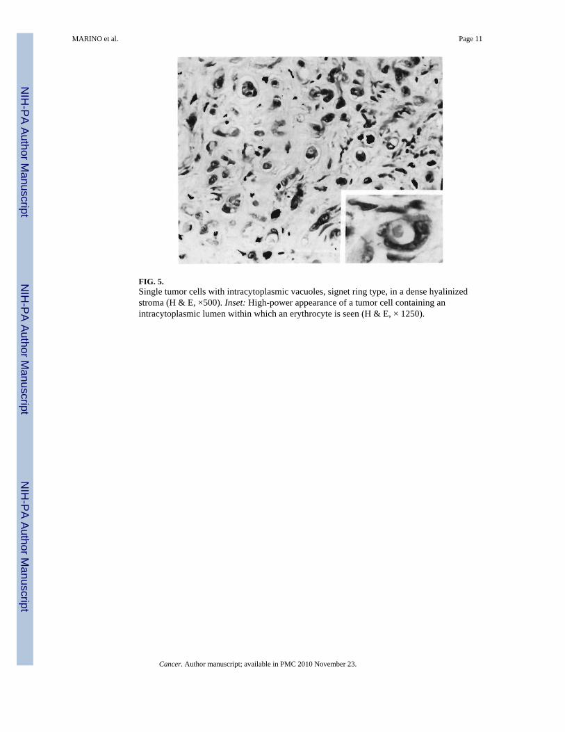
FIG. 5.Single tumor cells with intracytoplasmic vacuoles, signet ring type, in a dense hyalinizedstroma (H & E, ×500). Inset: High-power appearance of a tumor cell containing anintracytoplasmic lumen within which an erythrocyte is seen (H & E, × 1250).
MARINO et al. Page 11
Cancer. Author manuscript; available in PMC 2010 November 23.
NIH
-PA Author Manuscript
NIH
-PA Author Manuscript
NIH
-PA Author Manuscript
NIH
-PA Author Manuscript
NIH
-PA Author Manuscript
NIH
-PA Author Manuscript
MARINO et al. Page 12
Tabl
e 1
Clin
ical
Fea
ture
s
Preo
pera
tive
Patie
nt n
o.Fi
rst s
ympt
oms
Tot
al b
iliru
bin
(mg/
dl)
SGO
T (I
U/1
)SG
PT (I
U/1
)A
P (I
U/1
)G
GT
(IU
/1)
1A
bdom
inal
pai
n, v
omiti
ng, j
aund
ice
17.9
8030
425
—
2C
hest
and
abd
omin
al p
ain,
pal
pabl
e m
ass
0.3
2025
221
150
3Py
rosi
s, tra
nsie
nt ja
undi
ce0.
797
5481
045
9
415
-lb w
eigh
t los
s, pr
uritu
s, sp
ider
ang
iom
as, a
scite
s0.
518
1027
317
6
5In
cide
ntal
inve
stig
atio
n fo
r tra
uma
caus
ing
back
pai
n, o
ther
wis
e as
ympt
omat
ic0.
621
1680
25
6R
ight
subc
osta
l pai
n0.
425
1910
046
7A
bdom
inal
pai
n, 1
0-lb
wei
ght l
oss,
anor
exia
0.7
5793
388
201
8A
bdom
inal
pai
n0.
948
3535
210
1
9A
bdom
inal
pai
n0.
616
1611
942
SGO
T: se
rum
glu
tam
ic o
xalo
acet
ic tr
ansa
min
ase;
SG
PT: s
erum
glu
tam
ic p
yruv
ic tr
ansa
min
ase;
AP:
alk
alin
e ph
osph
atas
e; G
GT:
gam
mag
luta
myl
tran
spep
tidas
e.
Cancer. Author manuscript; available in PMC 2010 November 23.
NIH
-PA Author Manuscript
NIH
-PA Author Manuscript
NIH
-PA Author Manuscript
MARINO et al. Page 13
Tabl
e 2
Gro
ss P
atho
logi
c Fi
ndin
gs a
nd C
linic
al R
esul
ts
Patie
nt n
o.L
iver
wei
ght
Hep
atic
tum
or lo
catio
nPr
e-O
LT
met
asta
ses
Che
mot
hera
pyR
ecur
renc
eM
etas
tase
s loc
atio
nSu
rviv
al st
atus
148
00M
ultif
ocal
Abd
omen
, lun
gs, p
leur
a an
d di
aphr
agm
inva
sion
No
No
—11
yr,
aliv
e
217
20M
ultif
ocal
No
No
No
—4
yr, a
live
316
10M
ultif
ocal
No
Yes
12 m
o af
ter O
LTLi
ver,
lung
s16
mo,
dea
d
466
70M
ultif
ocal
Hyl
ar ly
mph
nod
es, c
omm
on b
ile d
uct
Yes
18 m
o af
ter O
LTM
edia
stin
um, r
ight
lung
2 yr
, aliv
e
514
50M
ultif
ocal
Extra
hepa
tic ly
mph
nod
es, l
eft l
ung
No
No
—16
mo,
aliv
e
616
00M
ultif
ocal
Hyl
ar ly
mph
nod
esY
esN
o—
16 m
o, a
live
714
00M
ultif
ocal
Extra
hepa
tic ly
mph
nod
esY
esN
o—
15 m
o, a
live
822
40M
ultif
ocal
No
No
No
—9
mo,
aliv
e
918
00M
ultif
ocal
No
No
No
—1
mo,
aliv
e
1025
10M
ultif
ocal
No
No
2 m
oLu
ngs,
liver
, spl
een,
pan
crea
s, ki
dney
,pe
ricar
dium
, bra
in3
mo,
dea
d
OLT
: orth
otop
ic li
ver t
rans
plan
tatio
n.
Cancer. Author manuscript; available in PMC 2010 November 23.