transition from glycogen to starch metabolism in archaeplastida
TRANSCRIPT

Seediscussions,stats,andauthorprofilesforthispublicationat:https://www.researchgate.net/publication/256609862
TransitionfromglycogentostarchmetabolisminArchaeplastida
ARTICLEinTRENDSINPLANTSCIENCE·SEPTEMBER2013
ImpactFactor:12.93·DOI:10.1016/j.tplants.2013.08.004·Source:PubMed
CITATIONS
12
READS
143
6AUTHORS,INCLUDING:
UgoCenci
DalhousieUniversity
6PUBLICATIONS126CITATIONS
SEEPROFILE
MartinSteup
UniversitätPotsdam
127PUBLICATIONS3,894CITATIONS
SEEPROFILE
ChristopheColleoni
UniversitédesSciencesetTechnologiesdeLi…
36PUBLICATIONS1,180CITATIONS
SEEPROFILE
StevenGBall
UniversityofLilleNorddeFrance
112PUBLICATIONS6,922CITATIONS
SEEPROFILE
Availablefrom:UgoCenci
Retrievedon:04February2016

TRPLSC-1085; No. of Pages 11
Transition from glycogen to starchmetabolism in ArchaeplastidaUgo Cenci1, Felix Nitschke2, Martin Steup2, Berge A. Minassian3,4,Christophe Colleoni1, and Steven G. Ball1
1 Laboratoire de Chimie Biologique, Universite des Sciences et Technologies de Lille, CNRS, UMR8576, Cite Scientifique,
59655 Villeneuve d’Ascq, France2 Institute of Biochemistry and Biology and Plant Physiology, University of Potsdam, Karl-Liebknecht-Str. 24–25,
14476 Potsdam-Golm, Germany3 Division of Neurology, Department of Pediatrics, The Hospital for Sick Children, 555 University Avenue, Toronto,
ON M5G 1X8, Canada4 Institute of Medical Sciences, University of Toronto, 1 King’s College Circle, Toronto, ON M5S 1A8, Canada
Opinion
In this opinion article we propose a scenario detailing howtwo crucial components have evolved simultaneously toensure the transition of glycogen to starch in the cytosolof the Archaeplastida last common ancestor: (i) the re-cruitment of an enzyme from intracellular Chlamydiaepathogens to facilitate crystallization of a-glucan chains;and (ii) the evolution of novel types of polysaccharide(de)phosphorylating enzymes from preexisting glycogen(de)phosphorylation host pathways to allow the turnoverof such crystals. We speculate that the transition to starchbenefitted Archaeplastida in three ways: more carboncould be packed into osmotically inert material; the hostcould resume control of carbon assimilation from thechlamydial pathogen that triggered plastid endosymbio-sis; and cyanobacterial photosynthate export could beintegrated in the emerging Archaeplastida.
Starch and glycogen metabolismStarch and glycogen define storage polysaccharides com-posed solely of glucosyl moieties that are predominantlylinked by a-1,4 and a-1,6 bonds [1]. Starch, unlike hydro-soluble glycogen, is deposited as granules that have a semi-crystalline structure and unlimited size and that aretemporarily unavailable to hydrosoluble enzymes (Box 1).Typically, two types of polyglucan are present in starch:amylopectin, the major branched component, is largelyresponsible for building the internal structure of the gran-ule, whereas the minor, moderately branched amylose isdispensable for granule formation and is synthesized bygranule-bound starch synthase (GBSS), the sole enzymeworking in a starch-bound environment [1,2]. However,GBSS activity requires a preformed solid starch granule [3].
1360-1385/$ – see front matter
� 2013 Elsevier Ltd. All rights reserved. http://dx.doi.org/10.1016/
j.tplants.2013.08.004
Corresponding author: Ball, S.G. ([email protected]).Keywords: evolution of plastids; starch and glycogen metabolism; polyglucandebranching reactions; starch and glycogen (de)phosphorylation; Chlamydia-likebacteria; Lafora disease.
Studies of Chlamydomonas reinhardtii, cereal, and Ara-bidopsis thaliana mutants have established that duringsynthesis [4–8] a debranching enzyme is required fornormal starch synthesis. It is generally assumed that thisenzyme splices out those a-1,6 branches that preventpolysaccharide assembly to yield a crystalline structure.In the absence of this debranching enzyme, mutant plantsand algae accumulate glycogen. Because semicrystallinepolysaccharides escape efficient degradation by the hydro-soluble enzymes of eukaryotic glycogen catabolism, wepropose in this opinion article that enzymes have coevolvedto degrade such structures. In all photosynthetic eukar-yotes, the so-called glucan, water dikinases (GWDs) andphosphoglucan, water dikinases (PWDs) phosphorylatethe hydrophobic crystalline structure of amylopectin [9–12]. These enzymes transfer the b-phosphate of ATP to theC3 or the C6 of glucosyl residues within the crystallinearrays [13]. This action appears to loosen hydrophobiccrystals by generating more hydrophilic sections of thegranule, which become accessible to hydrosoluble enzymes[14]. Mutants with a defective GWD or PWD are impairedin starch degradation and with time exhibit a starch-excess(SEX) phenotype. Furthermore, both mutants are compro-mised in growth. In Arabidopsis, all these phenotypicalfeatures are more pronounced when GWD is lacking [9,15–17]. Leaves of a rice mutant lacking functional GWDpossess approximately tenfold higher levels of transitorystarch having a low glucosyl 6-phosphate content andreduced grain yield, but vegetative growth is similar tothat of the wild type [17].
Starch metabolism specifically developed in photosyn-thetic eukaryotes and is found in the three major Archae-plastida lineages that emerged after plastid endosymbiosis:Glaucophyta, Rhodophyceae (red algae), and Chloroplastida(green algae and land plants) [18,19]. In all these lines,including those evolved from Archaeplastida through sec-ondary endosymbiosis, starch can be considered as a cyto-solic carbohydrate store, with no larger a-glucans beingsynthesized in plastids. However, an important exceptionis the Chloroplastida, which typically form starch in plastidsonly [18,19]. Only a few wild type plants are capable ofsynthesizing both starch and glycogen-like polysaccharides
Trends in Plant Science xx (2013) 1–11 1
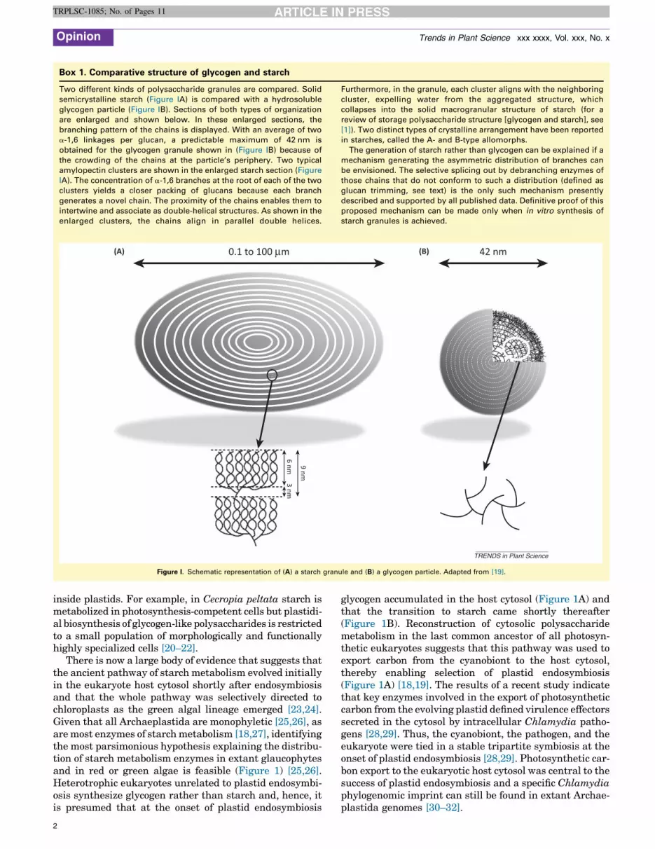
Box 1. Comparative structure of glycogen and starch
Two different kinds of polysaccharide granules are compared. Solid
semicrystalline starch (Figure IA) is compared with a hydrosoluble
glycogen particle (Figure IB). Sections of both types of organization
are enlarged and shown below. In these enlarged sections, the
branching pattern of the chains is displayed. With an average of two
a-1,6 linkages per glucan, a predictable maximum of 42 nm is
obtained for the glycogen granule shown in (Figure IB) because of
the crowding of the chains at the particle’s periphery. Two typical
amylopectin clusters are shown in the enlarged starch section (Figure
IA). The concentration of a-1,6 branches at the root of each of the two
clusters yields a closer packing of glucans because each branch
generates a novel chain. The proximity of the chains enables them to
intertwine and associate as double-helical structures. As shown in the
enlarged clusters, the chains align in parallel double helices.
Furthermore, in the granule, each cluster aligns with the neighboring
cluster, expelling water from the aggregated structure, which
collapses into the solid macrogranular structure of starch (for a
review of storage polysaccharide structure [glycogen and starch], see
[1]). Two distinct types of crystalline arrangement have been reported
in starches, called the A- and B-type allomorphs.
The generation of starch rather than glycogen can be explained if a
mechanism generating the asymmetric distribution of branches can
be envisioned. The selective splicing out by debranching enzymes of
those chains that do not conform to such a distribution (defined as
glucan trimming, see text) is the only such mechanism presently
described and supported by all published data. Definitive proof of this
proposed mechanism can be made only when in vitro synthesis of
starch granules is achieved.
42 nm0.1 to 100 μm(A) (B)
9 nm
6 nm3 nm
TRENDS in Plant Science
Figure I. Schematic representation of (A) a starch granule and (B) a glycogen particle. Adapted from [19].
Opinion Trends in Plant Science xxx xxxx, Vol. xxx, No. x
TRPLSC-1085; No. of Pages 11
inside plastids. For example, in Cecropia peltata starch ismetabolized in photosynthesis-competent cells but plastidi-al biosynthesis of glycogen-like polysaccharides is restrictedto a small population of morphologically and functionallyhighly specialized cells [20–22].
There is now a large body of evidence that suggests thatthe ancient pathway of starch metabolism evolved initiallyin the eukaryote host cytosol shortly after endosymbiosisand that the whole pathway was selectively directed tochloroplasts as the green algal lineage emerged [23,24].Given that all Archaeplastida are monophyletic [25,26], asare most enzymes of starch metabolism [18,27], identifyingthe most parsimonious hypothesis explaining the distribu-tion of starch metabolism enzymes in extant glaucophytesand in red or green algae is feasible (Figure 1) [25,26].Heterotrophic eukaryotes unrelated to plastid endosymbi-osis synthesize glycogen rather than starch and, hence, itis presumed that at the onset of plastid endosymbiosis
2
glycogen accumulated in the host cytosol (Figure 1A) andthat the transition to starch came shortly thereafter(Figure 1B). Reconstruction of cytosolic polysaccharidemetabolism in the last common ancestor of all photosyn-thetic eukaryotes suggests that this pathway was used toexport carbon from the cyanobiont to the host cytosol,thereby enabling selection of plastid endosymbiosis(Figure 1A) [18,19]. The results of a recent study indicatethat key enzymes involved in the export of photosyntheticcarbon from the evolving plastid defined virulence effectorssecreted in the cytosol by intracellular Chlamydia patho-gens [28,29]. Thus, the cyanobiont, the pathogen, and theeukaryote were tied in a stable tripartite symbiosis at theonset of plastid endosymbiosis [28,29]. Photosynthetic car-bon export to the eukaryotic host cytosol was central to thesuccess of plastid endosymbiosis and a specific Chlamydiaphylogenomic imprint can still be found in extant Archae-plastida genomes [30–32].

SS-ADP
SS-ADP
DPE2
Pho
AGPase
BE
Glg SS-UDP
SS-ADP
ISO
BE
GlgSS-UDP
Pho
BAM
GlgX
iDBE
Calvincycle
(A)
(B)
CO2
Pi
3-PGA
ADP-G
ADP-G
UDP-G
UDP-G
GlcG-1-P
α-glucan
β-maltose
DPE2
DPE2
BAM
Pho
iDBE Pho
G-1-PGlc
α-glucan
β-maltose
G-1-P
AMP
AMP
–
+
AGPase
Calvincycle CO2
Pi
3-PGA
PDP-G
P
ADP-G
ADP-G
AMP
AMP
–
+
GBSSGBSS
GWD
PWDP
TRENDS in Plant Science
Figure 1. Reconstruction of storage polysaccharide metabolism in the common ancestor of Archaeplastida. (A) The storage polysaccharide network at the onset of
endosymbiosis. In this reconstruction, the transition to starch in the common ancestor cytosol has not yet occurred and glycogen has accumulated. The direct debranching
enzyme (labeled GlgX) of bacterial phylogeny has not yet been recruited for amylopectin crystallization and is secreted as a virulence effector by Chlamydia pathogens
(depicted as a brown inclusion vesicle containing white pathogens attached to this membrane by their type-three secretion system, shown in orange, which is responsible
for secretion of the SS-ADP and GlgX functions). The cyanobiont is shown in blue and green. The bacterial direct debranching enzyme still displays its ancestral bacterial
function, detailed in Box 2. The eukaryotic indirect debranching enzyme has a similar function. However, the chlamydial enzyme releases the maltotetraose outer chains
(labeled a-glucan) in the cytosol, which may have been subjected to degradation by a combination of one of the two DPE2 amylomaltase isoforms still found in extant
Rhodophyceae and Glaucophyta and glycogen phosphorylase. The cytosolic dual-substrate pathway of glycogen accumulation relies on both UDP-glucose (UDP-G)
generated through host biochemical networks in its cytosol according to host needs and ADP-glucose (ADP-G) generated by cyanobacterial ADP-glucose
pyrophosphorylase (AGPase), which is activated by 3-phosphoglyceric acid (3-PGA) and inhibited by orthophosphate according to the networks and physiology of the
cyanobiont. To be incorporated into cytosolic glycogen, this substrate must be exported by a nucleotide–sugar translocator of host origin (shown in beige on the inner
membrane of the cyanobiont) that exchanges ADP-G with AMP (for a review, see [18]). The ADP-G substrate in the cytosol must be incorporated through an ADP-G-specific
glucan synthase (labeled SS-ADP). By contrast, the host UDP-G pools will be directed to glycogen according to the highly regulated eukaryotic UDP-G-specific glucan
synthase (labeled SS-UDP). This enzyme, unlike the bacterial glucan synthase, requires a primer to elongate a glucan. This primer is defined by glycogenin, an
(Figure legend continued on the bottom of the next page.)
Opinion Trends in Plant Science xxx xxxx, Vol. xxx, No. x
TRPLSC-1085; No. of Pages 11
3

Opinion Trends in Plant Science xxx xxxx, Vol. xxx, No. x
TRPLSC-1085; No. of Pages 11
As intracellular pathogens, Chlamydia presumably se-creted enzymes that metabolize storage polysaccharides inthe host cytosol to induce glycogen synthesis at the begin-ning of the infectious cycle. At the end of this cycle, storeswere mobilized by a secreted chlamydial glycogen phos-phorylase, thereby bypassing the highly regulated eukary-otic glycogen phosphorylase (Box 1) [28]. Hence stores werereadily available to the pathogens, because glycogen deg-radation was no longer controlled solely through the host.
In this opinion article, we focus on those events that wepropose may have been involved in the evolution of starchmetabolism from the preexisting glycogen metabolismnetwork active in the host cytosol of plastid endosymbiosis.From detailed phylogenetic and biochemical consider-ations, we propose that the switch of glycogen to starchmetabolism in the Archaeplastida ancestors was uniqueand required the concomitant appearance of a debranchingenzyme generating insoluble crystalline polysaccharidestructures and of a dikinase allowing their mobilization.
Recruitment of a direct debranching enzyme fromChlamydia pathogens may explain the switch fromglycogen to starchTo ensure crystallization of vicinal a-glucan chains ofamylopectin, clustering of branches is required (Box 1).It has been proposed that isoamylase isozyme1 (isa1),which encodes the catalytic subunit of the isoamylasedebranching enzyme, is chiefly responsible for trimmingout the misplaced branches in a pre-amylopectin hydro-philic precursor, thereby leading to cluster formation andpolysaccharide crystallization. This hypothesis, known asthe ‘glucan-trimming model’ is consistent with a very largebody of data [33–35]. However, definitive proof of thismodel would still require the establishment of an in vitrostarch-synthesizing system demonstrating the require-ment for glucan trimming by isoamylase to generatestarch-like granules. Nevertheless isoamylase has beenproven to be required for aggregation of starch granulesin vivo in all plant systems tested. Detailed phylogeneticanalysis (Figure 2) supports the notion that all Archae-plastida direct debranching enzymes, including isoamy-lase but excluding pullulanases, originate from a lateralgene-transfer event involving a chlamydial gene [30–32].This gene is related to the bacterial GlgX debranchingenzymes, which appear to be involved in glycogen catabo-lism [36]. In Escherichia coli, GlgX-type enzymes areknown to restrict their debranching activity to those outer
autoglucosylating protein (GLG). The glucans elongated through both glucan synthases
outer chains will be degraded through either b-amylase (BAM) or glycogen phospho
metabolized by the disproportionating isozyme 2 (DPE2) amylomaltase. Enzymes of hos
those of chlamydial origin in red. At this stage the chlamydial genes encoded by the
nucleus. (B) Cytosolic storage polysaccharide metabolism has been reconstructed by hyp
of the starch metabolism network within the three distinct Archaeplastida lineages (for
transition from glycogen to starch has occurred. The glycogen metabolism network i
required the duplication and evolution of the bacterial direct debranching enzyme into a
transfer (LGT) of the chlamydial gene to the host genome. This enzyme processes
generated by BEs. The debranched glucans (labeled a-glucan) are metabolized through
gene fusion between a carbohydrate-binding module and a dikinase domain enables
crystals (depicted as a circled P attached to the white starch granules). This fusion ge
dikinase (PWD) novel activities (shown in gray), which were required to initiate starch ca
polysaccharides aggregated into semicrystalline starch granules enabled the binding a
bound to starch) responsible for amylose synthesis within the polysaccharide matrix. T
maintenance of at least part of the cyanobacterial storage polysaccharide metabolism
inclusion vesicle are depicted as in (A). (B) is adapted from [19].
4
chains that have already been shortened by glycogenphosphorylase [36,37]. Glycogen phosphorylase stopsphosphorolysis four glucose residues upstream from thebranch (see Box 2 for details on debranching-enzyme func-tion in bacteria and eukaryotes). GlgX has high substratespecificity and will not debranch longer outer chains. Inbacteria this high specificity prevents futile cycles owing tothe presence of branching enzymes that transfer oligosac-charides at the a-1,6 position of at least six glucose resi-dues in length. The restrictive substrate specificity of GlgXwill not allow degradation of these novel branches unlessthe ensuing outer chains have first been shortened byglycogen phosphorylase (Box 2). In Chloroplastida thereare three genes that encode distinct GlgX-derived deb-ranching enzymes [33]. Isa3 may be defined as a catabolicenzyme because it has retained the restricted substratespecificity of GlgX [33]. Indeed, isa3 is involved in thedegradation of oligosaccharides released from starch bythe action of GWDs in the presence of both b-amylase andphosphorylase [8,38]. Despite all the problems of phylo-genetic signal erosion witnessed in the trees, the phylog-eny supports that Isa2 may have evolved from duplicationof the isa3 gene to generate a non-catalytic ‘regulatory’ or‘scaffolding’ subunit of the large-size heteromultimericisoamylase enzyme containing the isa1 catalytic subunit[28] (Figure 2). However, the exact position of the isa1group within the chlamydial isoamylase-GlgX clade islikely to remain obscure because of the high level ofphylogenetic signal erosion and the ensuing poor boot-strap support of the relevant nodes. As mentioned above,the starch metabolism network was rewired from the hostcytosol to the evolving chloroplast in the emerging greenalgae [23,24]. This redirection of the whole network intothe stroma was a problematic process given that isolatedduplications followed by transit peptide acquisition ofgenes duplicated from the cytosolic starch metabolismnetwork would yield no selective advantage unless otherenzymes of this pathway simultaneously underwent sim-ilar alterations [24]. See [24] for further discussion of ahypothesis to explain how the rewiring was neverthelessmade possible by sequential steps leading to first thesynthesis of oligosaccharides, followed by that of glyco-gen. Finally, plastidial starch emerged and cytosolicstarch metabolism was lost. Hence this stepwise processsuggests that the chloroplastic polysaccharide turnoverevolved later and independently from the acquisition ofstarch metabolism in the cytosol [24]. It nevertheless
will then be branched into glycogen by the branching enzyme (BE). The glycogen
rylase (PHO) to generate maltose and glucose-1-P, respectively. The maltose will
t phylogenetic origin are colored in beige; those of cyanobacterial origin in blue and
pathogens located in the inclusion vesicle have not been transferred to the host
othesizing the simplest enzyme set that would explain the distribution of the genes
a review, see [18]). This early stage corresponds to the common ancestor after the
llustrated in (A) is a simplification of this reconstruction. The transition to starch
functional isoamylase (iso), which is now encoded by the nucleus after lateral gene
the branches generated randomly on the hydrophilic branched polysaccharides
a combination of DPE2-like amylomaltases and phosphorylases. Simultaneously a
the phosphorylation and loosening of the otherwise undegradable amylopectin
nerates the archaeplastidal glucan, water dikinase (GWD)–phosphoglucan, water
tabolism through the b-amylase and phosphorylases (see above). The presence of
nd function of the cyanobacterial granule-bound starch synthase (GBSS) (shown
he late transfer of the cyanobacterial GBSS gene to the host nucleus suggests the
network until the transition to starch occurred. The cyanobiont and chlamydial
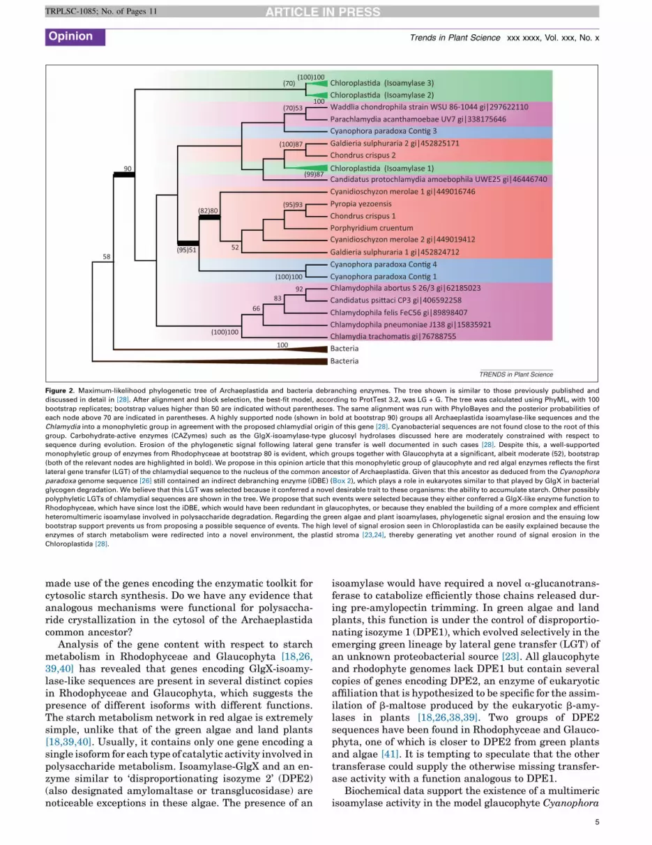
Chloroplas�da (Isoamylase 3)Chloroplas�da (Isoamylase 2)Waddlia chondrophila strain WSU 86-1044 gi|297622110Parachlamydia acanthamoebae UV7 gi|338175646Cyanophora paradoxa Con�g 3Galdieria sulphuraria 2 gi|452825171Chondrus crispus 2Chloroplas�da (Isoamylase 1)Candidatus protochlamydia amoebophila UWE25 gi|46446740Cyanidioschyzon merolae 1 gi|449016746Pyropia yezoensisChondrus crispus 1Porphyridium cruentumCyanidioschyzon merolae 2 gi|449019412Galdieria sulphuraria 1 gi|452824712Cyanophora paradoxa Con�g 4Cyanophora paradoxa Con�g 1Chlamydophila abortus S 26/3 gi|62185023Candidatus psi�aci CP3 gi|406592258Chlamydophila felis FeC56 gi|89898407Chlamydophila pneumoniae J138 gi|15835921Chlamydia trachoma�s gi|76788755BacteriaBacteria
100
(100)100
(100)100
(95)51
(82)80
90
(95)93
(99)87
(100)87
(70)53100
(100)100(70)
5258
6683
92
TRENDS in Plant Science
Figure 2. Maximum-likelihood phylogenetic tree of Archaeplastida and bacteria debranching enzymes. The tree shown is similar to those previously published and
discussed in detail in [28]. After alignment and block selection, the best-fit model, according to ProtTest 3.2, was LG + G. The tree was calculated using PhyML, with 100
bootstrap replicates; bootstrap values higher than 50 are indicated without parentheses. The same alignment was run with PhyloBayes and the posterior probabilities of
each node above 70 are indicated in parentheses. A highly supported node (shown in bold at bootstrap 90) groups all Archaeplastida isoamylase-like sequences and the
Chlamydia into a monophyletic group in agreement with the proposed chlamydial origin of this gene [28]. Cyanobacterial sequences are not found close to the root of this
group. Carbohydrate-active enzymes (CAZymes) such as the GlgX-isoamylase-type glucosyl hydrolases discussed here are moderately constrained with respect to
sequence during evolution. Erosion of the phylogenetic signal following lateral gene transfer is well documented in such cases [28]. Despite this, a well-supported
monophyletic group of enzymes from Rhodophyceae at bootstrap 80 is evident, which groups together with Glaucophyta at a significant, albeit moderate (52), bootstrap
(both of the relevant nodes are highlighted in bold). We propose in this opinion article that this monophyletic group of glaucophyte and red algal enzymes reflects the first
lateral gene transfer (LGT) of the chlamydial sequence to the nucleus of the common ancestor of Archaeplastida. Given that this ancestor as deduced from the Cyanophora
paradoxa genome sequence [26] still contained an indirect debranching enzyme (iDBE) (Box 2), which plays a role in eukaryotes similar to that played by GlgX in bacterial
glycogen degradation. We believe that this LGT was selected because it conferred a novel desirable trait to these organisms: the ability to accumulate starch. Other possibly
polyphyletic LGTs of chlamydial sequences are shown in the tree. We propose that such events were selected because they either conferred a GlgX-like enzyme function to
Rhodophyceae, which have since lost the iDBE, which would have been redundant in glaucophytes, or because they enabled the building of a more complex and efficient
heteromultimeric isoamylase involved in polysaccharide degradation. Regarding the green algae and plant isoamylases, phylogenetic signal erosion and the ensuing low
bootstrap support prevents us from proposing a possible sequence of events. The high level of signal erosion seen in Chloroplastida can be easily explained because the
enzymes of starch metabolism were redirected into a novel environment, the plastid stroma [23,24], thereby generating yet another round of signal erosion in the
Chloroplastida [28].
Opinion Trends in Plant Science xxx xxxx, Vol. xxx, No. x
TRPLSC-1085; No. of Pages 11
made use of the genes encoding the enzymatic toolkit forcytosolic starch synthesis. Do we have any evidence thatanalogous mechanisms were functional for polysaccha-ride crystallization in the cytosol of the Archaeplastidacommon ancestor?
Analysis of the gene content with respect to starchmetabolism in Rhodophyceae and Glaucophyta [18,26,39,40] has revealed that genes encoding GlgX-isoamy-lase-like sequences are present in several distinct copiesin Rhodophyceae and Glaucophyta, which suggests thepresence of different isoforms with different functions.The starch metabolism network in red algae is extremelysimple, unlike that of the green algae and land plants[18,39,40]. Usually, it contains only one gene encoding asingle isoform for each type of catalytic activity involved inpolysaccharide metabolism. Isoamylase-GlgX and an en-zyme similar to ‘disproportionating isozyme 2’ (DPE2)(also designated amylomaltase or transglucosidase) arenoticeable exceptions in these algae. The presence of an
isoamylase would have required a novel a-glucanotrans-ferase to catabolize efficiently those chains released dur-ing pre-amylopectin trimming. In green algae and landplants, this function is under the control of disproportio-nating isozyme 1 (DPE1), which evolved selectively in theemerging green lineage by lateral gene transfer (LGT) ofan unknown proteobacterial source [23]. All glaucophyteand rhodophyte genomes lack DPE1 but contain severalcopies of genes encoding DPE2, an enzyme of eukaryoticaffiliation that is hypothesized to be specific for the assim-ilation of b-maltose produced by the eukaryotic b-amy-lases in plants [18,26,38,39]. Two groups of DPE2sequences have been found in Rhodophyceae and Glauco-phyta, one of which is closer to DPE2 from green plantsand algae [41]. It is tempting to speculate that the othertransferase could supply the otherwise missing transfer-ase activity with a function analogous to DPE1.
Biochemical data support the existence of a multimericisoamylase activity in the model glaucophyte Cyanophora
5
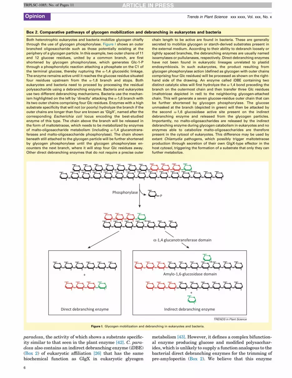
Box 2. Comparative pathways of glycogen mobilization and debranching in eukaryotes and bacteria
Both heterotrophic eukaryotes and bacteria mobilize glycogen chiefly
through the use of glycogen phosphorylase. Figure I shows an outer
branched oligosaccharide such as those potentially existing at the
periphery of a glycogen particle. In this example, two outer chains of 11
and 12 glucose residues, united by a common branch, are first
shortened by glycogen phosphorylase, which generates Glc-1-P
through a phosphorolytic reaction attaching a phosphate on the C1 of
the terminal glucose, thereby rupturing the a-1,4 glucosidic linkage.
The enzyme remains active until it reaches the glucose residue situated
four residues upstream from the a-1,6 branch and stops. Both
eukaryotes and bacteria need to proceed by processing the residual
polysaccharide using a debranching enzyme. Bacteria and eukaryotes
use two different debranching mechanisms. Bacteria use the mechan-
ism highlighted on the left by ‘directly’ attacking the a-1,6 branch with
its two outer chains comprising four Glc residues. Enzymes with a high
substrate specificity that will not (or poorly) hydrolyze the branch if the
outer chains are longer than four are known as ‘GlgX’, named after the
corresponding Escherichia coli locus encoding the best-studied
enzyme of this type. The chain above the branch will be released in
the form of maltotetraose, which needs to be metabolized by enzymes
of malto-oligosaccharide metabolism (including a-1,4 glucanotrans-
ferases and malto-oligosaccharide phosphorylase). The chain shown
beneath still attached to the glycogen particle will be further shortened
by glycogen phosphorylase until the glycogen phosphorylase en-
counters the next branch, where it will stop four Glc residues away.
Other direct debranching enzymes that do not require a precise outer
chain length to be active are found in bacteria. These are generally
secreted to mobilize glycogen or starch-derived substrates present in
the external medium. According to their ability to debranch loosely or
tightly spaced branches, the debranching enzymes are usually named
isoamylases or pullulanases, respectively. Direct debranching enzymes
have not been found in eukaryotic lineages unrelated to plastid
endosymbiosis. In such eukaryotes, the product resulting from
glycogen phosphorylase action (defined as glycogen with outer chains
comprising four Glc residues) will be processed as shown on the right-
hand side of the drawing. An enzyme called iDBE containing two
distinct catalytic sites will first hydrolyze the a-1,4 bond preceding the
branch on the outermost chain and then transfer three Glc residues
(maltotriose depicted in red) to the neighboring glycogen-attached
chain. This will generate a seven glucose-residue outer chain that can
be further shortened by glycogen phosphorylase. The glucose
unmasked at the branch (depicted in green) will then be attacked by
the second a-1,6 glucosidase active site present on the indirect
debranching enzyme and released from the glycogen particles.
Importantly, no malto-oligosaccharides are released by the indirect
debranching enzyme during glycogen catabolism in eukaryotes and no
enzymes able to catabolize malto-oligosaccharides are therefore
present in the cytosol of eukaryotes. This difference may be used by
extant Chlamydia pathogens, which possibly trigger maltotetraose
production through secretion of their own GlgX-type effector in the
host cytosol, triggering the formation of a substrate that only they can
further metabolize.
PPhosphorylase
α-1,4 glucanotransferase domain
+
+
Amylo-1,6-glucosidase domain
Direct debranching enzyme Indirect debranching enzyme
TRENDS in Plant Science
Figure I. Glycogen mobilization and debranching in eukaryotes and bacteria.
Opinion Trends in Plant Science xxx xxxx, Vol. xxx, No. x
TRPLSC-1085; No. of Pages 11
paradoxa, the activity of which shows a substrate specific-ity similar to that seen in the plant enzyme [42]. C. para-doxa also contains an indirect debranching enzyme (iDBE)(Box 2) of eukaryotic affiliation [26] that has the samebiochemical function as GlgX in eukaryotic glycogen
6
metabolism [43]. However, it defines a complex bifunction-al enzyme producing glucose and modified polysacchar-ides, which is unlikely to supply a function analogous to thebacterial direct debranching enzymes for the trimming ofpre-amylopectin (Box 2). We believe that this enzyme
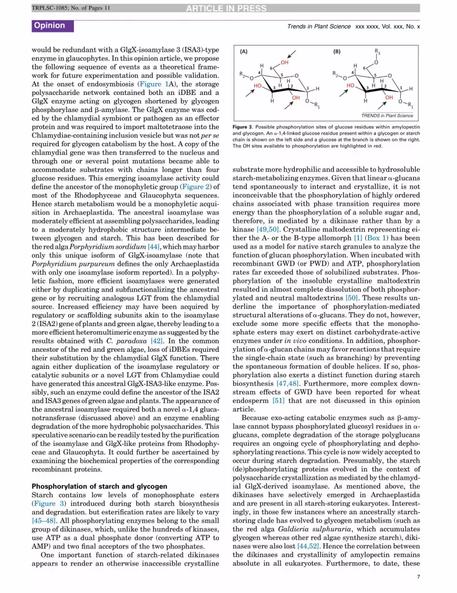
HR2
R1 R1
R2
R3
O
H H
H HH
H
H H
O
O
O
O
O
O
1 12 23 3
4 46 6
(A) (B)
5 5H
OH OH
OH
HO HO
TRENDS in Plant Science
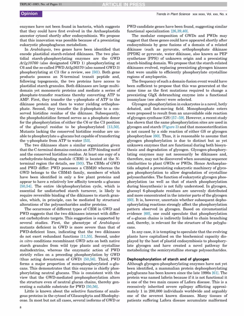
Figure 3. Possible phosphorylation sites of glucose residues within amylopectin
and glycogen. An a-1,4-linked glucose residue present within a glycogen or starch
chain is shown on the left side and a glucose at the branch is shown on the right.
The OH sites available to phosphorylation are highlighted in red.
Opinion Trends in Plant Science xxx xxxx, Vol. xxx, No. x
TRPLSC-1085; No. of Pages 11
would be redundant with a GlgX-isoamylase 3 (ISA3)-typeenzyme in glaucophytes. In this opinion article, we proposethe following sequence of events as a theoretical frame-work for future experimentation and possible validation.At the onset of endosymbiosis (Figure 1A), the storagepolysaccharide network contained both an iDBE and aGlgX enzyme acting on glycogen shortened by glycogenphosphorylase and b-amylase. The GlgX enzyme was cod-ed by the chlamydial symbiont or pathogen as an effectorprotein and was required to import maltotetraose into theChlamydiae-containing inclusion vesicle but was not per serequired for glycogen catabolism by the host. A copy of thechlamydial gene was then transferred to the nucleus andthrough one or several point mutations became able toaccommodate substrates with chains longer than fourglucose residues. This emerging isoamylase activity coulddefine the ancestor of the monophyletic group (Figure 2) ofmost of the Rhodophyceae and Glaucophyta sequences.Hence starch metabolism would be a monophyletic acqui-sition in Archaeplastida. The ancestral isoamylase wasmoderately efficient at assembling polysaccharides, leadingto a moderately hydrophobic structure intermediate be-tween glycogen and starch. This has been described forthe red alga Porphyridium sordidum [44], which may harboronly this unique isoform of GlgX-isoamylase (note thatPorphyridium purpureum defines the only Archaeplastidawith only one isoamylase isoform reported). In a polyphy-letic fashion, more efficient isoamylases were generatedeither by duplicating and subfunctionalizing the ancestralgene or by recruiting analogous LGT from the chlamydialsource. Increased efficiency may have been acquired byregulatory or scaffolding subunits akin to the isoamylase2 (ISA2) gene of plants and green algae, thereby leading to amore efficient heteromultimeric enzyme as suggested by theresults obtained with C. paradoxa [42]. In the commonancestor of the red and green algae, loss of iDBEs requiredtheir substitution by the chlamydial GlgX function. Thereagain either duplication of the isoamylase regulatory orcatalytic subunits or a novel LGT from Chlamydiae couldhave generated this ancestral GlgX-ISA3-like enzyme. Pos-sibly, such an enzyme could define the ancestor of the ISA2and ISA3 genes of green algae and plants. The appearance ofthe ancestral isoamylase required both a novel a-1,4 gluca-notransferase (discussed above) and an enzyme enablingdegradation of the more hydrophobic polysaccharides. Thisspeculative scenario can be readily tested by the purificationof the isoamylase and GlgX-like proteins from Rhodophy-ceae and Glaucophyta. It could further be ascertained byexamining the biochemical properties of the correspondingrecombinant proteins.
Phosphorylation of starch and glycogenStarch contains low levels of monophosphate esters(Figure 3) introduced during both starch biosynthesisand degradation. but esterification rates are likely to vary[45–48]. All phosphorylating enzymes belong to the smallgroup of dikinases, which, unlike the hundreds of kinases,use ATP as a dual phosphate donor (converting ATP toAMP) and two final acceptors of the two phosphates.
One important function of starch-related dikinasesappears to render an otherwise inaccessible crystalline
substrate more hydrophilic and accessible to hydrosolublestarch-metabolizing enzymes. Given that linear a-glucanstend spontaneously to interact and crystallize, it is notinconceivable that the phosphorylation of highly orderedchains associated with phase transition requires moreenergy than the phosphorylation of a soluble sugar and,therefore, is mediated by a dikinase rather than by akinase [49,50]. Crystalline maltodextrin representing ei-ther the A- or the B-type allomorph [1] (Box 1) has beenused as a model for native starch granules to analyze thefunction of glucan phosphorylation. When incubated withrecombinant GWD (or PWD) and ATP, phosphorylationrates far exceeded those of solubilized substrates. Phos-phorylation of the insoluble crystalline maltodextrinresulted in almost complete dissolution of both phosphor-ylated and neutral maltodextrins [50]. These results un-derline the importance of phosphorylation-mediatedstructural alterations of a-glucans. They do not, however,exclude some more specific effects that the monopho-sphate esters may exert on distinct carbohydrate-activeenzymes under in vivo conditions. In addition, phosphor-ylation of a-glucan chains may favor reactions that requirethe single-chain state (such as branching) by preventingthe spontaneous formation of double helices. If so, phos-phorylation also exerts a distinct function during starchbiosynthesis [47,48]. Furthermore, more complex down-stream effects of GWD have been reported for wheatendosperm [51] that are not discussed in this opinionarticle.
Because exo-acting catabolic enzymes such as b-amy-lase cannot bypass phosphorylated glucosyl residues in a-glucans, complete degradation of the storage polyglucansrequires an ongoing cycle of phosphorylating and depho-sphorylating reactions. This cycle is now widely accepted tooccur during starch degradation. Presumably, the starch(de)phosphorylating proteins evolved in the context ofpolysaccharide crystallization as mediated by the chlamyd-ial GlgX-derived isoamylase. As mentioned above, thedikinases have selectively emerged in Archaeplastidaand are present in all starch-storing eukaryotes. Interest-ingly, in those few instances where an ancestrally starch-storing clade has evolved to glycogen metabolism (such asthe red alga Galdieria sulphuraria, which accumulatesglycogen whereas other red algae synthesize starch), diki-nases were also lost [44,52]. Hence the correlation betweenthe dikinases and crystallinity of amylopectin remainsabsolute in all eukaryotes. Furthermore, to date, these
7
Opinion Trends in Plant Science xxx xxxx, Vol. xxx, No. x
TRPLSC-1085; No. of Pages 11
enzymes have not been found in bacteria, which suggeststhat they could have first evolved in the Archaeplastidaancestor cytosol shortly after endosymbiosis. We proposethat this innovation was possible because of a preexistingeukaryotic phosphoglucan metabolism.
In Arabidopsis, two genes have been identified thatencode plastidial starch-related dikinases. The two plas-tidial starch-phosphorylating enzymes are the GWDAt1g10760 (also designated GWD 1) phosphorylating atC6 and the so-called PWD At5g26570 (also named GWD 3)phosphorylating at C3 (for a review, see [50]). Both geneproducts possess an N-terminal transit peptide and,following topogenesis, the two proteins have access toplastidial starch granules. Both dikinases are large multi-domain yet monomeric proteins and mediate a series ofphosphate-transfer reactions thereby converting ATP toAMP. First, they transfer the g-phosphate of ATP to thedikinase protein and then to water yielding orthophos-phate. Second, they transfer the b-phosphate to a con-served histidine residue of the dikinase. Subsequently,the phosphohistidine formed serves as a phosphate donorfor the phosphorylation of either the C6 or the C3 positionof the glucosyl residue to be phosphorylated [13,50].Mutants lacking the conserved histidine residue are un-able to phosphorylate a-glucans but capable of transferringthe g-phosphate from ATP to water [53].
The two dikinases share a similar organization giventhat the C-terminal domains contain an ATP-binding motifand the conserved histidine residue. At least one copy of acarbohydrate-binding module (CBM) is located at the N-terminal region (for details, see [50]). The CBMs of GWDand PWD differ: PWD possesses a CBM20 whereas theGWD belongs to the CBM45 family, members of whichhave been identified in only a few plant proteins andappear to have a relatively low affinity towards a-glucans[50,54]. The entire (de)phosphorylation cycle, which isessential for undisturbed starch turnover, is likely torequire reversible binding of the dikinases to starch gran-ules, which, in principle, can be mediated by structuralalterations of the polysaccharides and/or proteins.
The occurrence of different types of CBM in GWD andPWD suggests that the two dikinases interact with differ-ent carbohydrate targets. This suggestion is supported byseveral studies. First, the phenotype of Arabidopsismutants deficient in GWD is more severe than that ofPWD-deficient lines, indicating that the two dikinasesdo not exert redundant functions [11,55]. Second, underin vitro conditions recombinant GWD acts on both nativestarch granules from wild type plants and crystallinemaltodextrins, whereas the enzymatic action of PWDstrictly relies on a preceding phosphorylation by GWD(thus acting downstream of GWD) [50,56]. Third, PWDforms a major proportion of monophosphorylated a-glu-cans. This demonstrates that this enzyme is chiefly phos-phorylating neutral glucans. This is consistent with theview that the GWD-mediated prephosphorylation altersthe structure even of neutral glucan chains, thereby gen-erating a suitable substrate for PWD [50,56].
Little is known about the selective functions of analo-gous proteins in the cytosol of Glaucophyta and Rhodophy-ceae. In most but not all cases, several isoforms of GWD or
8
PWD candidate genes have been found, suggesting similarfunctional specialization [26,39,40].
The modular composition of GWDs and PWDs maysuggest that these genes could have appeared shortly afterendosymbiosis by gene fusions of a domain of a relateddikinase (such as pyruvate, orthophosphate dikinase[PPDK] or pyruvate, water dikinase, also known as PEPsynthetase [PPS]) of unknown origin and a preexistingstarch-binding domain. We propose that the starch-relateddikinases evolved, replacing preexisting glycogen kinasesthat were unable to efficiently phosphorylate crystallineregions of amylopectin.
The frequency of such a domain-fusion event would havebeen sufficient to propose that this was generated at thesame time as the first mutations required to change apreexisting GlgX debranching enzyme into a functionalisoamylase (see above) were selected.
Glycogen phosphorylation in eukaryotes is a novel, hotlydebated, and fast-moving field. Monophosphate esterswere proposed to result from an unavoidable side reactionof glycogen synthase (GS) [57–59]. However, a recent studyhas shown that the same phosphorylation sites are used inglycogen and starch (Figure 3) and that monoesterificationis not caused by a side reaction of either GS or glycogenphosphorylase [60]. Thus, it is reasonable to assume thatglycogen phosphorylation is due to distinct but as yetunknown enzymes that are functional during both biosyn-thesis and degradation of glycogen. Glycogen-phosphory-lating enzymes may not necessarily be dikinases and,therefore, may not be discovered when assuming sequencesimilarities to plant GWDs or PWDs. Hence Archaeplas-tida adapted a preexisting eukaryotic machinery of glyco-gen phosphorylation to allow degradation of crystallinepolysaccharides. The function of eukaryotic glycogen phos-phorylation (as well as that of starch phosphorylationduring biosynthesis) is not fully understood. In glycogen,glucosyl 6-phosphate residues are unevenly distributedand more concentrated in the interior parts of the glycogen[60]. It is, however, uncertain whether subsequent depho-sphorylating reactions strongly affect the phosphorylationpattern observed in glycogen. Based on circumstantialevidence [60], one could speculate that phosphorylationof a-glucan chains is indirectly linked to chain branchingand, thereby, is relevant for the structure of the polyglu-cans.
In any case, it is tempting to speculate that the evolvingplants have capitalized on the biochemical capacity dis-played by the host of plastid endosymbiosis to phosphory-late glycogen and have created a novel pathway formetabolizing the semicrystalline storage polysaccharides.
Dephosphorylation of starch and of glycogenAlthough glycogen-phosphorylating enzymes have not yetbeen identified, a mammalian protein dephosphorylatingpolyglucans has been known since the late 1990s [61]. Theprotein was named laforin because if it is not functional itis one of the two main causes of Lafora disease. This is arecessively inherited severe epilepsy afflicting approxi-mately 1 in 200,000 individuals worldwide and arguablyone of the severest known diseases. Many tissues ofpatients suffering Lafora disease accumulate malformed

Opinion Trends in Plant Science xxx xxxx, Vol. xxx, No. x
TRPLSC-1085; No. of Pages 11
polyglucans that are poorly branched, hyperphosphory-lated, and insoluble [62,63]. Lafora disease is caused byloss-of-function mutations of the gene that encodes thelaforin glycogen phosphatase [61] or of the gene encodingthe malin ubiquitin E3 ligase [64], which appears to func-tion in the regulation of laforin [65].
Laforin belongs to the large group of dual-specificityphosphatases (DSPs), which dephosphorylate phosphotyr-osine, phosphoserine, and phosphothreonine residues and,in some cases, also act on non-proteinaceous substrates[66]. Laforin is the only known human DSP possessing aCBM (CBM20 family). In plants, a plastidial DSP has beenidentified that carries both a CBM and a DSP motif.Initially, this protein was named PTPKIS1, assuming thatphosphoproteins were the target of the phosphatase[67,68]. In Arabidopsis, the same locus was identified byscreening chemically mutagenized lines for a SEX pheno-type and the gene product was designated SEX4 [49,68,69].Despite some phenotypical differences, the results of thescreen strongly suggest that in higher plants starch isnormally turned over, provided both phosphorylatingand dephosphorylating enzyme activities are functional[50].
Acting on phosphorylated glucans, laforin and SEX4 areless selective than GWD or PWD. Both phosphataseshydrolyze monophosphate esters at C3 and at C6 and alsoact on hydrosoluble a-glucans [49,69–72]. Likewise, glyco-gen from laforin-deficient mice contains elevated levels ofglucosyl 6-phosphate residues [59]. Possibly, laforin and/orSEX4 also exert various actions on (phospho)proteins inaddition to that on phosphorylated a-glucans [58]. Twoother plastidial DSPs from Arabidopsis that have beenrecently characterized are not discussed here. For a review,see [51].
In vivo, laforin and SEX4 appear to be functionallysimilar and functional human laforin complements theSEX4-deficient Arabidopsis mutant [71,72]. However,the two proteins are not orthologs. In SEX4 the CBM20and DSP motifs are located at the C and N termini,respectively, but the intramolecular order of the twodomains is reversed in laforin [58,73]. Thus, laforin andSEX4 appear to be generated by independently performedfusions of domains.
True laforin orthologs have been reported for red algaesuch as Chondrus crispus, Porphyridium cruentum, andCyanidioschyzon merolae (and possibly also C. paradoxa)[18,26,39,40]. Apparently, during the targeting of thestarch pathway to the plastid, a novel laforin-like proteinwas generated, but the reasons for this novelty remainunclear.
Recently, two additional plastidial starch-related DSPshave been identified in A. thaliana that are designatedLike-Sex-Four1 (LSF1) and Like-Sex-Four2 (LSF2). Ara-bidopsis mutants lacking functional LSF1 or LSF2 possessa SEX phenotype. For a review, see [50]. Based on in vitrostudies, LSF1 does not exhibit a noticeable phosphataseactivity when assayed with phosphorylated a-glucans.Likewise, transitory starch of the LSF1-deficient mutantspossesses similar glucose-based contents of glucose 6-phos-phate and glucose 3-phosphate levels [74]. Currently, thebiochemical function of LSF1 is unknown. By contrast,
LSF2 possesses a phosphatase activity, selectively hydro-lyzing monophosphates at C3 of starch-related glucosylmoieties [75]. In contrast to SEX4 and LSF1, the phospha-tase LSF2 does not contain a classical CBM that is sepa-rated from the catalytic domain. LSF2 does, however,undergo multiple interactions with amylopectin. Site-spe-cific mutations and structural analyses of LSF2 revealedthat all glucan-binding sites are located within the cata-lytically active phosphatase domain [76]. These resultshave important implications for the search of furtherphosphatases acting on phosphorylated a-glucans.
As discussed above for the phosphorylating enzymes, wepropose that chloroplast-containing eukaryotic cells estab-lished novel starch-dephosphorylating enzymes andadapted these enzymes to specific features of starch(and, possibly, to the novel intracellular compartmentali-zation). However, the novel enzymes were generated usinga preexisting mode of removing monophosphates.
Concluding remarks: why do Archaeplastidaaccumulate starch?When considering the physicochemical differences ofstarch and glycogen, the question arises of the possiblereasons underlying the evolution of glycogen metabolisminto starch. At first one might propose that the vastamounts of carbon made available by photosynthesis re-quired a novel packing of glucose into insoluble stores. If so,one would expect that all cyanobacteria would accumulatestarch-like polysaccharides. However, there are only a fewcyanobacteria synthesizing starch-like granules and thevast majority of cyanobacteria remain bona fide glycogenaccumulators [77]. Looking closer at the physiology ofstarch-accumulating cyanobacteria, it appears that thoselineages metabolizing semicrystalline polysaccharides areall unicellular and diazotrophic. Because nitrogenase isinhibited or inactivated by molecular oxygen, unicellulardiazotrophy can be sustained only if phototrophy anddiazotrophy are temporally separated by tight circadian-clock control of cellular metabolism. Polysaccharides syn-thesized in the light are metabolized in darkness to reachanoxia by respiration of large amounts of glucose. Simul-taneously, vast amounts of ATP and reducing power areprovided to nitrogenase. Hence cyanobacteria with such aphysiology require a larger carbon store compared withother cyanobacteria [18]. However, if more carbon can bestored in starch than in glycogen, why is starch not a morewidespread form of storage in eukaryotes and bacteria?The answer may rest in the ease with which peripheralglucosyl residues may become available to cellular metab-olism. One can speculate that the flexibility offered byglycogen breakdown may not be matched by starch andthat turnover of carbon in the light may define a desirablefine-tuning mechanism for photosynthesis optimization.Hence there may be a balance between the selective advan-tages offered by flexible glycogen mobilization and theadditional efforts associated with storage of starch.
Although it is easy to understand why unicellular dia-zotrophic cyanobacteria have resorted to starch metabo-lism, it is less obvious why the Archaeplastida ancestorevolved semicrystalline starch. First, the cytosolic locali-zation of the storage polysaccharide may have prevented
9

Opinion Trends in Plant Science xxx xxxx, Vol. xxx, No. x
TRPLSC-1085; No. of Pages 11
its use to optimize photosynthesis through carbon turnoverin the light. Hence the advantages offered by the flexibilityof glycogen degradation may not have applied as in cya-nobacteria. Second, because Chlamydia pathogens mayhave been involved by establishing the metabolic linkbetween the protoplastid and its host, it is reasonable toassume that the pathogens may have had the ability to tapdirectly into the host glycogen stores [26]. Hence theeukaryotic host had lost its monopoly on the mobilizationof its own glucose stores and the switch from glycogen tostarch may have been prompted by the advantage affordedby an increase in carbon-sink strength in the cytosol andbecause solid starch escaped direct mobilization of storageby Chlamydia pathogens. Indeed, the initial step of starchmobilization was then defined by the host GWDs with nodirect access to these stores by the unregulated chlamydialeffector glycogen phosphorylases.
Finally, the escape of the glucose stores from directdegradation by the glycogen catabolism enzymes enabledthe evolution of the first mechanisms integrating storagepolysaccharide mobilization in the cytosol with photosyn-thate supply by the cyanobiont. The highly integratedpathway of host glycogen catabolism control was not tai-lored to take into account the timing and amounts ofcarbohydrates afforded by cyanobacterial synthesis. Bythe evolution of a novel first step defined by the requiredphosphorylation of the semicrystalline glucans, naturalselection allowed the evolution of novel circadian clock-mediated controls specifically exerted on this novel bio-chemical step without interfering with the highly integrat-ed phosphorylation cascades responsible for host glycogenphosphorylase regulation. This innovation also facilitatedthe evolution of novel diurnal assessment mechanismsinforming the host about the extent of the stores suppliedby photosynthesis. These would have facilitated the emer-gence of a novel regulation of storage polysaccharide me-tabolism, taking into account the amount and activity ofthe biochemical networks of the cyanobiont.
The results of phylogenetic analysis of the starch deb-ranching enzyme are consistent with a monophyletic ac-quisition of starch in the cytosol of the Archaeplastidaancestor. [28] However, the detailed biochemical proper-ties of all of the distinct GlgX-isoamylase-type debranchingenzymes from the Rhodophyceae and Glaucophyta shownin Figure 2 need to be ascertained to confirm this idea.
AcknowledgmentsThis research was funded by the French Ministry of Education, theCentre National de la Recherche Scientifique (CNRS), the AgenceNationale pour la Recherche (Menage a Trois), the European Union,and the Region Nord Pas de Calais.
References1 Buleon, A. et al. (1998) Starch granules: structure and biosynthesis. Int.
J. Biol. Macromol. 23, 85–1122 Ball, S. et al. (1998) Progress in understanding the biosynthesis of
amylose. Trends Plant Sci. 3, 462–4673 Dauvillee, D. et al. (1999) Novel starch-like polysaccharides are
synthesized by a soluble form of granule-bound starch synthase inglycogen accumulating mutants of Chlamydomonas reinhardtii. PlantPhysiol. 119, 321–330
4 James, M.G. et al. (1995) Characterization of the maize gene sugary1, adeterminant of starch composition in kernels. Plant Cell 7, 417–429
10
5 Mouille, G. et al. (1996) Preamylopectin processing: a mandatory stepfor starch biosynthesis in plants. Plant Cell 8, 1353–1366
6 Nakamura, Y. et al. (1997) Correlation between activities of starchdebranching enzymes and a-polyglucan structure in endosperms ofsugary-1 mutants of rice. Plant J. 12, 143–153
7 Zeeman, S.C. et al. (1998) A mutant of Arabidopsis lacking achloroplastic isoamylase accumulates both starch andphytoglycogen. Plant Cell 10, 1699–1712
8 Wattebled, F. et al. (2005) Mutants of Arabidopsis lacking achloroplastic isoamylase accumulate phytoglycogen and an abnormalform of amylopectin. Plant Physiol. 138, 184–195
9 Lorberth, R. et al. (1998) Inhibition of a starch-granule-bound proteinleads to modified starch and repression of cold sweetening. Nat.Biotechnol. 16, 473–477
10 Ritte, G. et al. (2002) The starch-related R1 protein is an a-glucan,water dikinase. Proc. Natl. Acad. Sci. U.S.A. 99, 7166–7171
11 Kotting, O. et al. (2005) Identification of a novel enzyme required forstarch metabolism in Arabidopsis leaves. The phosphoglucan, waterdikinase. Plant Physiol. 137, 242–252
12 Baunsgaard, L. et al. (2005) A novel isoform of glucan, water dikinasephosphorylates pre-phosphorylated alpha-glucans and is involved instarch degradation in Arabidopsis. Plant J. 41, 595–605
13 Ritte, G. et al. (2006) Phosphorylation of C6 and C3 positions of glucosylresidues in starch is catalyzed by two distinct dikinases. FEBS Lett.580, 4872–4876
14 Hejazi, M. et al. (2008) Glucan, water dikinase phosphorylatescrystalline maltodextrins and thereby initiates solubilization. PlantJ. 55, 323–334
15 Yu, T.S. et al. (2001) The Arabidopsis sex1 mutant is defective in the R1protein, a general regulator of starch degradation in plants, and not inthe chloroplast hexose transporter. Plant Cell 13, 1907–1918
16 Vriet, C. et al. (2010) A suite of Lotus japonicus starch mutants revealsboth conserved and novel features of starch metabolism. Plant Physiol.154, 643–655
17 Hirose, T. et al. (2013) Disruption of a rice gene for a-glucan waterdikinase, OsGWD1, leads to hyperaccumulation of starch in leaves butexhibits limited effects on growth. Front. Plant Sci. 4, 147
18 Deschamps, P. et al. (2007) Metabolic symbiosis and the birth of theplant kingdom. Mol. Biol. Evol. 25, 536–548
19 Ball, S. et al. (2011) The evolution of glycogen and starch metabolism ineukaryotes gives molecular clues to understand the establishment ofplastid endosymbiosis. J. Exp. Bot. 62, 1775–1801
20 Rickson, F.R. (1971) Glycogen plastids in Mu llerian body cells ofCecropia peltata – a higher green plant. Science 173, 344–347
21 Marshall, J.J. and Rickson, F.R. (1973) Characterization of the a-D-glucan from the plastids of Cecropia peltata as a glycogen-typepolysaccharide. Carbohydr. Res. 28, 31–37
22 Bischof, S. et al. (2013) Cecropia peltata accumulates starch or solubleglycogen by differentially regulating starch biosynthetic genes. PlantCell 25, 1400–1415
23 Deschamps, P. et al. (2008) Early gene duplication withinChloroplastida and its correspondence with relocation of starchmetabolism to chloroplasts. Genetics 178, 2373–2387
24 Deschamps, P. et al. (2008) The relocation of starch metabolism tochloroplasts: when, why and how. Trends Plant Sci. 13, 574–582
25 Rodrıguez-Ezpeleta, N. et al. (2005) Monophyly of primaryphotosynthetic eukaryotes: green plants, red algae, andglaucophytes. Curr. Biol. 15, 1325–1330
26 Price, D.C. et al. (2012) Cyanophora paradoxa genome elucidates originof photosynthesis in algae and plants. Science 335, 843–847
27 Patron, N.J. and Keeling, P.K. (2005) Common evolutionary origin ofstarch biosynthetic enzymes in green and red algae. J. Phycol. 41,1131–1141
28 Ball, S.G. et al. (2013) Metabolic effectors secreted by bacterialpathogens; essential facilitators of plastid endosymbiosis? Plant Cell25, 7–21
29 Baum, D. (2013) The origin of primary plastids: a pas de deux or amenage a trois? Plant Cell 25, 4–6
30 Huang, J. and Gogarten, J.P. (2007) Did an ancient chlamydialendosymbiosis facilitate the establishment of primary plastids?Genome Biol. 8, R99
31 Becker, B. et al. (2008) Chlamydial genes shed light on the evolution ofphotoautotrophic eukaryotes. BMC Evol. Biol. 8, 20

Opinion Trends in Plant Science xxx xxxx, Vol. xxx, No. x
TRPLSC-1085; No. of Pages 11
32 Moustafa, A. et al. (2008) Chlamydiae has contributed at least 55 genesto Plantae with predominantly plastid functions. PLoS ONE 3, e2205
33 Hussain, H. et al. (2003) Three isoforms of isoamylase contributedifferent catalytic properties for the debranching of potato glucans.Plant Cell 15, 133–149
34 Ball, S.G. et al. (1996) From glycogen to amylopectin: a modelexplaining the biogenesis of the plant starch granule. Cell 86, 349–352
35 Myers, A.M. et al. (2000) Recent progress toward understandingbiosynthesis of the amylopectin crystal. Plant Physiol. 122, 989–998
36 Dauvillee, D. et al. (2005) Role of the E. coli glgX gene in glycogenmetabolism. J. Bacteriol. 187, 1465–1473
37 Jeanningros, R.N. et al. (1976) Purification and properties ofdebranching enzyme from E. coli. Biochim. Biophys. Acta 438, 186–199
38 Delatte, T. et al. (2006) Evidence for distinct mechanisms of starchgranule breakdown in plants. J. Biol. Chem. 285, 12050–12055
39 Collen, J. et al. (2013) Genome structure and metabolic features in thered seaweed Chondrus crispus shed light on evolution of theArchaeplastida. Proc. Natl. Acad. Sci. U.S.A. 110, 5247–5252
40 Bhattacharya, D. et al. (2013) Genome of the red alga Porphyridiumcruentum. Nat. Commun. 4, 1941
41 Arias, M.C. et al. (2012) Eukaryote to gut bacteria transfer of aglycoside hydrolase gene essential for starch breakdown in plants.Mob. Genet. Elements 2, 81–87
42 Plancke, C. et al. (2008) Pathway of cytosolic starch synthesis in themodel glaucophyte Cyanophora paradoxa. Eukaryot. Cell 7, 247–257
43 Teste, M.A. et al. (2000) The Saccharomyces cerevisiae YPR184w geneencodes the glycogen debranching enzyme. FEMS Microbiol. Lett. 193,105–110
44 Shimonaga, T. et al. (2008) Variations in storage a-glucans of thePorphyridiales (Rhodophyta). Plant Cell Physiol. 49, 103–116
45 Hizukuri, S. et al. (1970) Studies on starch phosphate. 1. Estimation ofglucose 6-phosphate residues on starch and the presence of other boundphosphate(s). Starch/Sta rke 45, 417–420
46 Tabata, S. and Hizukuri, S. (1971) Studies on starch phosphate. Part 2.Isolation of glucose 3-phosphate and maltose phosphate by acidhydrolysis of potato starch. Starch/Sta rke 23, 267–272
47 Nielsen, T.H. et al. (1994) Starch phosphorylation in potato tubersproceeds concurrently with de novo biosynthesis of starch. PlantPhysiol. 105, 111–117
48 Ritte, G. et al. (2004) Phosphorylation of transitory starch is increasedduring degradation. Plant Physiol. 135, 2068–2077
49 Hejazi, M. et al. (2010) The laforin-like dual-specificity phosphataseSEX4 from Arabidopsis hydrolyzes both C6- and C3-phosphate estersintroduced by starch-related dikinases and thereby affects phasetransition of a-glucans. Plant Physiol. 152, 711–722
50 Hejazi, M. et al. (2012) Starch phosphorylation and dephosphorylation.In Starch: Origins, Structure and Metabolism (Essential Reviews inExperimental Biology) (Vol. 5) (Tetlow, I., ed.), pp. 279–310, SEB
51 Ral, J-P. et al. (2012) Down-regulation of glucan, water-dikinaseactivity in wheat endosperm increases vegetative biomass and yield.Plant Biotechnol. J. 10, 871–882
52 Schonknecht, G. et al. (2013) Gene transfer from bacteria and archeafacilitated evolution of an extremophilic eukaryote. Science 239, 1207–1210
53 Hejazi, M. et al. (2012) The plastidial glucan, water dikinase (GWD)catalyses multiple phosphotransfer reactions. FEBS J. 279, 1953–1966
54 Glaring, M.A. et al. (2011) Starch-binding domains in the CBM45family – low affinity domains from glucan, water dikinase and a-amylase involved in plastidial starch metabolism. FEBS J. 278,1175–1185
55 Baunsgaard, L. et al. (2005) A novel isoform of glucan, water dikinasephosphorylates prephosphorylated a-glucans and is involved in starchdegradation in Arabidopsis. Plant J. 41, 595–605
56 Hejazi et al. (2009) The two plastidial starch-related dikinasessequentially phosphorylate glucosyl residues at the surface of both
the A- and the B-allomorph of crystallized maltodextrins but the modeof action differs. Plant Physiol. 150, 962–976
57 Tagliabracci, V.S. et al. (2011) Phosphate incorporation duringglycogen synthesis and Lafora disease. Cell Metab. 13, 274–282
58 Roach, P.J. et al. (2012) Glycogen and its metabolism: some newdevelopments and old themes. Biochem. J. 441, 763–787
59 Gentry, M.S. et al. (2013) Laforin, a protein with many faces: glucanphosphatase, adapter protein, et alii. FEBS J. 280, 526–537
60 Nitschke, F. et al. (2013) Hyperphosphorylation of glucosyl C6 carbonsand altered structure of glycogen in the neurodegenerative epilepsyLafora disease. Cell Metab. 17, 756–767
61 Minassian, B.A. et al. (1998) Mutations in a gene encoding a novelprotein tyrosine phosphatase cause progressive myoclonus epilepsy.Nat. Genet. 20, 171–174
62 Sakai, M. et al. (1970) Studies in myoclonus epilepsy (lafora body form).II. Polyglucosans in the systemic deposits of myoclonus epilepsy and incorpora amylacea. Neurology 20, 160–176
63 Tagliabracci, V.S. et al. (2008) Abnormal metabolism of glycogenphosphate as a cause for Lafora disease. J. Biol. Chem. 283, 33816–33825
64 Chan, E.M. et al. (2003) Mutations in NHLRC1 cause progressivemyoclonus epilepsy. Nat. Genet. 35, 125–127
65 Tiberia, E. et al. (2012) Increased laforin and laforin binding toglycogen underlie lafora body formation in malin-deficient Laforadisease. J. Biol. Chem. 287, 25650–25659
66 Pulido, R. and Hooft van Huijsduijnen, R. (2008) Protein tyrosinephosphatases: dual-specificity phosphatases in health and disease.FEBS J. 275, 848–866
67 Fordham-Skelton, A.P. et al. (2002) A novel higher plant proteintyrosine phosphatase interacts with SNF1-related protein kinasesvia a KIS (kinase interaction sequence) domain. Plant J. 29, 705–715
68 Kerk, D. et al. (2006) A chloroplast-localized dual-specificity proteinphosphatase in Arabidopsis contains a phylogenetically dispersed andancient carbohydrate-binding domain, which binds the polysaccharidestarch. Plant J. 46, 400–413
69 Kotting, O. et al. (2009) STARCH-EXCESS4 is a laforin-likephosphoglucan phosphatase required for starch degradation inArabidopsis thaliana. Plant Cell 21, 334–346
70 Worby, C.A. et al. (2006) Laforin, a dual specificity phosphatase thatdephosphorylates complex carbohydrates. J. Biol. Chem. 281, 30412–30418
71 Niittyla, T. et al. (2006) Similar protein phosphatases control starchmetabolism in plants and glycogen metabolism in mammals. J. Biol.Chem. 281, 11815–11818
72 Gentry, M.S. et al. (2007) The phosphatase laforin crosses evolutionaryboundaries and links carbohydrate metabolism to neuronal disease. J.Cell Biol. 178, 477–488
73 Gentry, M.S. et al. (2009) Lafora disease: insights intoneurodegeneration from plant metabolism. Trends Biochem. Sci. 34,628–639
74 Comparat-Moss, S. et al. (2010) A putative phosphatase, LSF1, isrequired for normal starch turnover in Arabidopsis leaves. PlantPhysiol. 152, 685–697
75 Santelia et al. (2011) The phosphoglucan phosphatase like SEXFour2dephosphorylates starch at the C3-position in Arabidopsis. Plant Cell23, 4096–4111
76 Meekins, D.A. et al. (2013) Structure of the Arabidopsis glucanphosphatase LIKE SEX FOUR2 reveals a unique mechanism forstarch dephosphorylation. Plant Cell 25, 2302–2314
77 Colleoni, C. and Suzuki, E. (2012) Storage polysaccharide metabolismin cyanobacteria. In Starch: Origins, Structure and Metabolism(Essential Reviews in Experimental Biology) (Vol. 5) (Tetlow, I., ed.),pp. 217–253, SEB
11