transcriptomic, biochemical, and histopathological responses of the clam ruditapes decussatus from a...
TRANSCRIPT
Transcriptomic, Biochemical, and Histopathological Responsesof the Clam Ruditapes decussatus from a Metal-ContaminatedTunis Lagoon
Houssem Chalghmi1,2 • Jean-Paul Bourdineaud2 • Zohra Haouas3 •
Pierre-Yves Gourves2 • Ines Zrafi4 • Dalila Saidane-Mosbahi1
Received: 9 March 2015 / Accepted: 10 June 2015
� Springer Science+Business Media New York 2015
Abstract This study was designed to investigate the
molecular (transcriptional expression), biochemical (ox-
idative stress and neurotoxicity), and histopathological
effects of metal contamination in the gill of clams (Rudi-
tapes decussatus) sampled from the Tunis lagoon. The
concentrations of five heavy metals (Cd, Pb, Hg, Cu, and
Zn) in surface sediments and their accumulation in soft
tissues of R. decussatus were evaluated in three sites (Z1,
Z2, and Z3). A metal contamination state of Tunis lagoon
sediments was noted with spatial variations with relatively
high levels at Z2. Biomarker analyses showed an increase
in glutathione S-transferase and catalase activities and lipid
peroxidation levels and a decrease in acetylcholinesterase
activity in the studied sites. Molecular investigation
showed a significant overexpression of: cytochrome c oxi-
dase subunit I, ribosomal RNA 16S, Cu/Zn superoxide
dismutase, heat shock protein 70, and metallothioneins in
the three sampling sites. Moreover, our data were
correlated to severe and diverse histopathological alter-
ations in the clam gills. The principal component analysis
showed that the Z2 region is more affected by metal con-
tamination than Z1 and Z3 regions. Current field results
suggest the use of several combined biomarkers at different
cell levels instead of individual ones in monitoring
programs.
Coastal lagoons are complex and dynamic ecosystems,
characterized by constant changes in environmental con-
ditions (Porter et al. 2001; Kamel et al. 2014). Some
Mediterranean lagoons are among the most extensively
modified and threatened ecosystems mainly due to urban-
ization, industrialization, and tourism (Ben Khedher et al.
2013; Matozzo et al. 2010). Therefore, several contami-
nants are continuously released into these systems deteri-
orating water quality, imposing severe restrictions to
organisms and possibly causing a decrease in natural
resources (Cravo et al. 2012; Ben Khedher et al. 2014).
Trace metals are among the most relevant contaminants
that may affect both biotope and biota quality in marine
Electronic supplementary material The online version of thisarticle (doi:10.1007/s00244-015-0185-0) contains supplementarymaterial, which is available to authorized users.
& Houssem Chalghmi
Jean-Paul Bourdineaud
Zohra Haouas
Pierre-Yves Gourves
Ines Zrafi
Dalila Saidane-Mosbahi
1 Laboratory of Analysis Treatment and Valorization of
Environmental Pollutants and Products, Faculty of Pharmacy,
5000 Monastir, Tunisia
2 UMR CNRS 5805 EPOC, University of Bordeaux, Arcachon
Marine Station, Place du Dr Peyneau, 33120 Arcachon,
France
3 Laboratory of Histology Cytology and Genetics, Faculty of
Medicine, 5019 Monastir, Tunisia
4 Centre of Water Researches and Technologies, Technopark
Borj Cedria, BP. 273, 8020 Soliman, Tunisia
123
Arch Environ Contam Toxicol
DOI 10.1007/s00244-015-0185-0
ecosystems in general and lagoon ecosystems in particular
(Sfriso et al. 2008; Ennouri et al. 2010; Ben Khedher et al.
2013).
Trace metals naturally occur in marine environments.
However, human activities have introduced high amounts
of these elements into the environment, making it difficult
to distinguish natural contributions from anthropogenic
ones. Heavy metals, which readily accumulate in marine
sediments, can reach significant accumulation levels in the
tissues of aquatic organisms (Fu et al. 2014). Various trace
metals, such as copper (Cu) and zinc (Zn), are essential for
aquatic organisms, but they are toxic above a given
threshold (Kucuksezgin et al. 2006). Nonessential metals,
such as cadmium (Cd), lead (Pb), and mercury (Hg), are
toxic even at trace levels. An in vivo study showed that the
phagocytic capacity in mussel P. viridis was inhibited by
exposure to high Cu levels (50–200 lg L-1) (Nicholson
2003). Zebrafish exposed to 1.9 lg L-1 of Cd showed a
pro-apoptotic response and mitochondrial damage (Gon-
zalez et al. 2006). Acute lethality of Pb to aquatic inver-
tebrates and fish occurs at concentrations of 1 to 500
mg g-1, but mortality and adverse effects on growth and
biochemical responses have been observed in chronic
studies at concentrations as low as 0.007–0.020 mg g-1
(Demayo et al. 1982).
These heavy metals are often concentrated and ampli-
fied in organisms at higher levels of the food chain,
particularly in benthic animals. Heavy metal concentra-
tions found in the tissues of marine organisms do not
always reflect the quantity of metals present in a toxic
form within the cell, as demonstrated for Cu, which can
be complexed in nonactive inorganic complexes (Viar-
engo and Nott 1993). Thus, there is a need to develop
strategies to assess whether a given environment is under
stress or not. Therefore, techniques based on measuring
the biological effects are critical for any pollution-moni-
toring program (Nasci et al. 2000; Viarengo et al. 2007).
The application of biomarkers in field conditions has been
proposed by many authors in order to assess chronic
responses in exposed aquatic populations under environ-
mental realistic conditions (De la Torre et al. 2005;
Morales-Caselles et al. 2008).
Over the years, some marine bivalves, particularly
mussels and clams, have been used widely and successfully
as bioindicators for monitoring pollution in many coastal
environments (O’Connor 2002; Geret et al. 2003). Many
studies have demonstrated that several bivalve species have
a great ability to accumulate organic pollutants and heavy
metals and, thus, can be used as sensitive in situ indicators
for pollution assessment (Wetzel and Van Vleet 2004; Oros
and Ross 2005; Zhu et al. 2005). Molecular approaches
have been investigated by many authors to monitor the
contamination impact on aquatic organisms and to better
understand the early cellular events associated with the
sensing and protective responses that occur upon exposure
to contaminants (Negri et al. 2013; Arini et al. 2014; Banni
et al. 2014; Dedeh et al. 2015).
Located in northern Tunisia, the Tunis lagoon is of great
economic importance to the country. It is a Mediterranean
eutrophic coastal lagoon covering 40 km2. Tunis, Tunisia’s
capital city where the lagoon in located, is a densely
populated area characterized by the presence of a variety of
economic activities, such as harbors, tourism, agriculture,
and several industries (e.g., chemical manufacturing plants,
metal processing factories and electronic and mechanic
industries). The Tunis lagoon drains a watershed of 40 km2,
including 15 km2 occupied by industrial areas. It thus
receives approximately 5500 m3 day-1 of industrial
wastewater, rich in heavy metals, and hydrocarbons. As a
consequence, high levels of chemicals were found in the
lagoon sediments. These chemicals include polycyclic
aromatic hydrocarbons (levels varied from 452 to 1411 ng
g-1) (Mzoughi and Chouba 2011) and trace metals, such as
Pb (13.1–18.8 lg g-1), Cr (54.5–62 lg g-1), Ni (21–25.8
lg g-1), Cu (11.3–15.8 lg g-1), and Zn (213.7–231 lgg-1) (Hellal et al. 2011). To our knowledge, no studies
have assessed the levels of metal compounds in this region
and have investigated their effects on clams.
The purpose of the present study was to investigate the
potential use of the biochemical, transcriptomic, and
histopathological responses of wild clams (Ruditapes
decussatus) as biomarkers of environmental pollution and
water quality assessment in productive ecosystems, such as
the Tunis lagoon.
Materials and Methods
Study Area
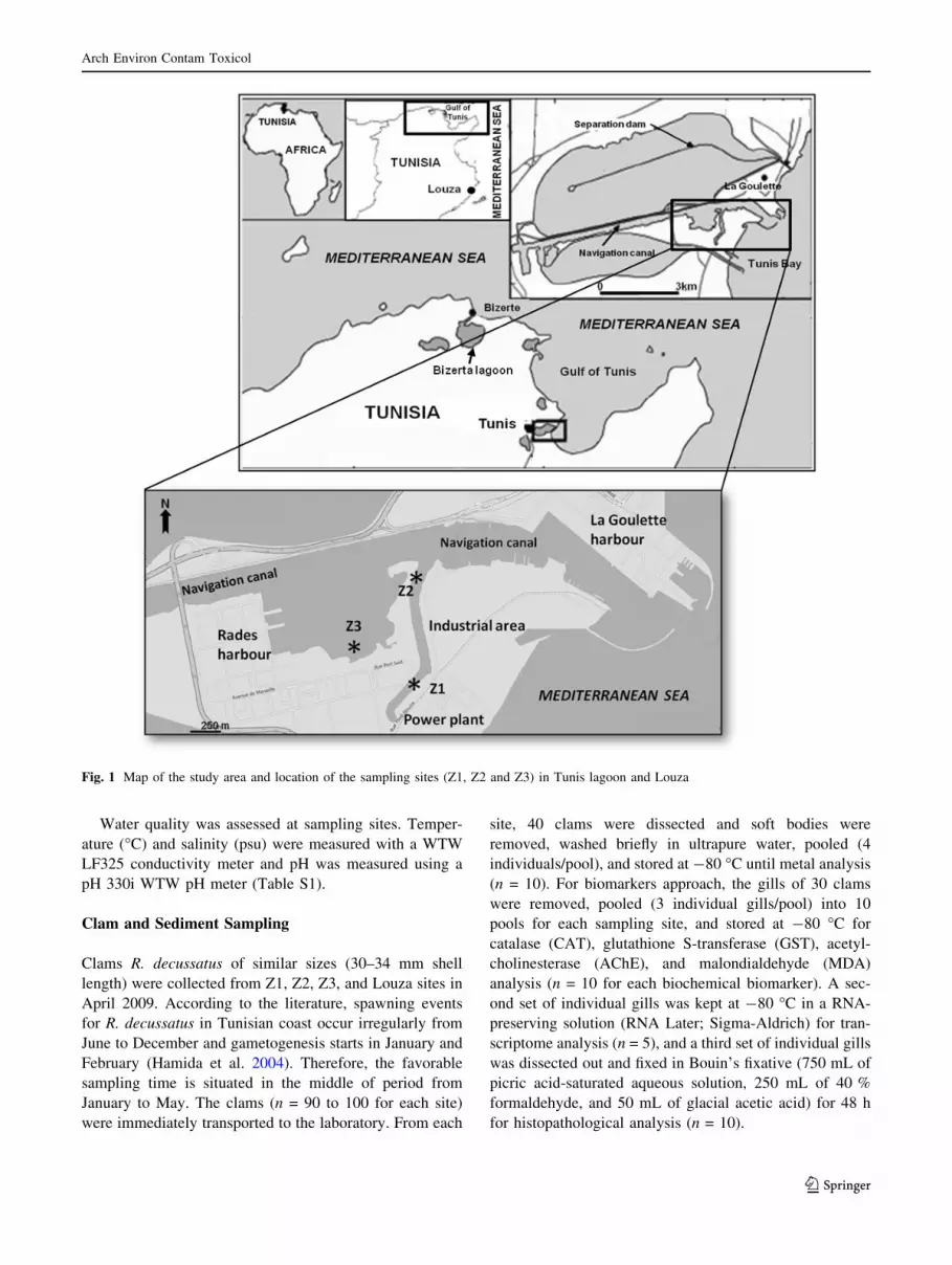
Three sampling sites were chosen in the Tunis lagoon
because they are geographically located near contamina-
tion sources (Fig. 1). The sampling site Z1 (36�48002.200N10�16055.500E) is located near a chemical industrial area,
the sampling site Z2 (36�48032.000N 10�16058.000E) is
located in the navigation canal and near the industrial area,
and Z3 (36�48013.000N 10�16036.100E) is very close to Radesharbour—one of the largest harbours in Tunisia, which has
the most important industrial complexes and intense com-
mercial transport activities.
Control samples of surface sediments and clams were
collected from Louza site (35�01011.100N 11�00024.600E).This site was chosen in another region 258 km far from the
Tunis lagoon. Louza site has been considered as a refer-
ence site in monitoring programs along the Tunisian coasts
(Banni et al. 2009).
Arch Environ Contam Toxicol
123
Water quality was assessed at sampling sites. Temper-
ature (�C) and salinity (psu) were measured with a WTW
LF325 conductivity meter and pH was measured using a
pH 330i WTW pH meter (Table S1).
Clam and Sediment Sampling
Clams R. decussatus of similar sizes (30–34 mm shell
length) were collected from Z1, Z2, Z3, and Louza sites in
April 2009. According to the literature, spawning events
for R. decussatus in Tunisian coast occur irregularly from
June to December and gametogenesis starts in January and
February (Hamida et al. 2004). Therefore, the favorable
sampling time is situated in the middle of period from
January to May. The clams (n = 90 to 100 for each site)
were immediately transported to the laboratory. From each
site, 40 clams were dissected and soft bodies were
removed, washed briefly in ultrapure water, pooled (4
individuals/pool), and stored at -80 �C until metal analysis
(n = 10). For biomarkers approach, the gills of 30 clams
were removed, pooled (3 individual gills/pool) into 10
pools for each sampling site, and stored at -80 �C for
catalase (CAT), glutathione S-transferase (GST), acetyl-
cholinesterase (AChE), and malondialdehyde (MDA)
analysis (n = 10 for each biochemical biomarker). A sec-
ond set of individual gills was kept at -80 �C in a RNA-
preserving solution (RNA Later; Sigma-Aldrich) for tran-
scriptome analysis (n = 5), and a third set of individual gills
was dissected out and fixed in Bouin’s fixative (750 mL of
picric acid-saturated aqueous solution, 250 mL of 40 %
formaldehyde, and 50 mL of glacial acetic acid) for 48 h
for histopathological analysis (n = 10).
Fig. 1 Map of the study area and location of the sampling sites (Z1, Z2 and Z3) in Tunis lagoon and Louza
Arch Environ Contam Toxicol
123
Samples of surface sediments (up to 20 cm in depth)
were collected from the three sites in the Tunis lagoon and
from the Louza site (reference) at the same period. In the
laboratory, sediment samples were homogenized, freeze-
dried, passed through a stainless steel sieve (100 lm), and
finally stored at 4 �C until analysis.
Heavy Metal Analysis
Freeze-dried clam soft bodies and sediments were digested
in 3 mL of nitric acid at 100 �C for 3 h. The liquid
underwent sixfold dilution with ultrapure water. Within
each digestion series, appropriate blanks with no clam
tissues or sediments also were subjected to the same pro-
cedure to account for background contamination levels and
to validate the entire process. Standard biological and
marine sediment reference materials with certified metal
content (Tort-2: lobster hepatopancreas; Dolt-4: dogfish
liver; PACS-1, MESS-2, and MESS-3: marine sediments
from National Research Council of Canada, Ottawa,
Canada) were treated and analysed under the same condi-
tions (Dedeh et al. 2014). Metal concentrations (Ag, As,
Cd, Mn, Ni, Pb, V, Zn, and Cu) in digests of clam tissues
and sediments were analysed by inductively coupled
plasma/atomic emission spectrometry (ICP/AES) (700
Series, Agilent Technologies). Quantification of mercury
(Hg) in freeze-dried tissues was performed by flameless
atomic absorption spectrometry (AMA 254, Altec, Prague,
Czech Republic). All metal concentrations are reported in
micrograms per gram of sample dry weight.
The bioaccumulation factor (BAF), relating the con-
centration of metal in surface sediments to its level in
organism, was used to estimate each metal’s accumulation
propensity in R. decussatus. It was calculated using the
following equation:
BAF = [metal]clam (lg g-1)/[metal]sediment (lg g-1),
where [metal]clam and [metal]sediment are the average metal
concentrations in clams and sediments, respectively.
Biochemical Analysis
Before biochemical analysis, samples of gills were
homogenized in phosphate buffer (0.1 M, pH 7.5). The
homogenate obtained was centrifuged at 90009g for 25
min to obtain the cytosolic fraction (S9). The quantities of
proteins present in S9 fraction were determined according
to the Bradford (1976) method using Coomassie Blue
reagent (BioRad) and bovine serum albumin as standard
protein.
GST activity was measured in gills cytosol by the
method of Habig et al. (1974) using 10 lg of cytosolic
proteins, 1 mM 1-chloro-2,4-dinitrobenzene (CDNB)
(Sigma-Aldrich, St. Louis, MO) as substrate, and 4 mM of
reduced glutathione (GSH), in 100 mM of sodium phos-
phate buffer, pH 7.5. GST activity was determined by
kinetic measurement at 25 �C using a Spectro UV–Vis
Double Beam PC Scanning Spectrophotometer UVD-2960
(k = 340 nm). Results were expressed as lmoles GS-
CDNB produced per min and per mg proteins.
CAT activity was determined according to Claiborne’s
method (1985). Reaction mixture (final volume of 1 mL)
contained 0.78 mL 0.1 M phosphate buffer (pH 7.5) and
0.2 mL 0.5 mM H2O2. After 30 s preincubation, the reac-
tion was started by the addition of 0.02 mL of the S9
fraction. CAT activity was evaluated by kinetic measure-
ment at 25 �C using a Spectro UV–Vis Double Beam PC
Scanning Spectrophotometer UVD-2960 (k = 240 nm).
Results were expressed as lmoles hydrogen peroxide
transformed per min and per mg proteins.
Lipid peroxidation (LPO) was estimated in terms of
thiobarbituric acid reactive species (TBARS), with the use
of 1,1,3,3-tetramethoxypropane as standard. The reaction
was assessed at 532 nm using thiobarbituric acid (TBA)
reagent as described by Buege and Aust (1978). MDA
content was expressed as nanomoles equivalent MDA per
milligram proteins.
AChE activity was measured at 25 �C according to the
colorimetric method of Ellman et al. (1961). In a typical
assay, 1.05 mL of 0.1 M phosphate buffer, 50 lL of 8 mM
dithiobisnitrobenzoate, 50 lL of supernatant S9, and 50 lLof 45 mM acetylthiocholine substrate were successively
added. The enzymatic reaction rate was quantified spec-
trophotometrically at 412 nm against a blank without
substrate for each activity measurement. In order to sub-
tract the spontaneous hydrolysis of substrate, a second
blank was performed without a sample. Enzyme activity
was recorded over 5 min after the addition of the substrate
concentration. AChE activity was expressed as specific
activity (nanomole of substrate hydrolysed per minute per
milligram of proteins).
Gene Expression Analysis
Total RNA was extracted from 30 to 50 mg of gills using
the Absolutely RNA Miniprep Kit (Agilent technologies),
according to the manufacturer’s instructions. However, to
eliminate the maximum of lipids and proteins, we added a
step of phenol:chloroform:isoamylic alcohol (25:24:1,
Sigma) extraction (Dedeh et al. 2014). The elution volume
was 30 lL and the concentration of RNA was quantified
using a microplate spectrophotometer (Epoch, Biotek). The
RNA quality in each sample was checked by measuring the
260/280 nm ratios and by 1 % agarose gel electrophoresis.
First-strand cDNA was synthesized from total RNA using
the AffinityScript cDNA Synthesis Kit (Agilent technolo-
gies) according to the manufacturer’s instructions. Specific
Arch Environ Contam Toxicol
123

primer pairs were designed with the Lightcycler probe
designer software (Table S2). RT-qPCR reactions were
performed using Brillant III Ultrafast SYBR QPCR kits
(Agilent Technologies) according to manufacturer’s
instruction in a Stratagene Mx3000P thermocycler (Agilent
Technologies). The amplification program consisted of one
cycle at 95 �C for 10 min and 50 amplifications cycles at 95
�C for 30 s, 55 �C for 30 s, and 72 �C for 30 s. The
specificity of the amplification products was confirmed by
size estimations on an agarose gel and by analyzing their
melting curves to confirm that only one PCR product was
amplified and detected. The melting curve was obtained by
following the SybrGreen fluorescence level during gradual
heating of the PCR products from 60 to 95 �C. Standardcurves were generated using tenfold dilutions of a cDNA
template on the LightCycler apparatus, and using each
couple of gene specific primers (one standard curve per
couple). Each dilution was assayed in triplicate for each
couple of primers. Each standard curve was made by
plotting the Ct against the log of the starting quantity of
template for each dilution. The equation for the regression
line and the r value was calculated. From that equation the
slope of the standard curve was deduced and used to cal-
culate the PCR efficiency, E, for each couple of primers, as
follows: E = 10-1/slope (Table S2). The relative expression
ratios of target genes were calculated according to Pfaffl
(2001). Relative expression data were normalized to ribo-
somal RNA 18S (18S rRNA), a housekeeping gene. The
18S rRNA gene was chosen as the reference gene because
of its stability across sampling sites, as highlighted by the
fact that the mean Ct values were the same regardless of the
sampling site. Indeed, the Ct collected in gill tissue were
(n = 5, mean ± SD): Control: 11.7 ± 1.2; site Z1: 10.9 ±
1.8; site Z2: 11.6 ± 0.9 and site Z3: 12.5 ± 1.3. The
differential expression factor (DEF) of a gene is the ratio of
its relative expression in clams collected from the polluted
site over that of control animals.
All RT-qPCR experiments were performed according to
the Minimum Information for publication of Quantitative
real-time PCR Experiments (MIQE) guidelines (Bustin
et al. 2009). MIQE checklist is found in the Supplementary
materials section.
Histological Methods
The gills preserved in Bouin’s fixative were dehydrated in
a graded ethanol solution and embedded in paraffin.
Embedded tissues were cut into sections of 5-lm thickness
by a rotary microtome (Leitz WETZLAR 1512). The thin
sections of the gill tissues were stained by Trichrome
Masson for observation by a light microscope. Sec-
tions were photographed with a microscope (LEICA DM
750) equipped with a numerical camera (LEICA ICC50
HD) and examined for lesions. For each individual gill,
seven slides were examined with four sections per slide.
Seven alterations were found in the gills of clams.
Histopathological alterations were semi-quantitatively
evaluated by ranking the severity of lesions (grades 0 (-),
0.5 (?/-), 1 (?), 2 (??) and 3 (???)) as described in
previous studies (Riba et al. 2004). A general index of
damages was established to permit comparison of
histopathological responses between sampling sites. An
arithmetic average value was obtained from the original
semi-quantitative assessment of the lesions.
Statistical Analysis
The results for metal accumulation and biochemical
markers are presented as means ± SD. Relative gene
expressions are presented as means ± SEM. Experimental
data were initially tested for normality and homogeneity of
variance to meet statistical demands. Data statistical anal-
ysis was performed using one-way analysis of variance
(ANOVA) and Duncan’s test for multiple range compar-
ison; p\ 0.05 was considered significant. Pearson corre-
lation matrix also was calculated to study the relationships
between the biochemical and transcriptomic biomarkers
measured and metal accumulation in clams. SigmaStat 3.5
(SYSTAT Software, Inc.) was used for statistical analysis.
Principal component analysis (PCA) of biomarkers and
metal data was applied to discriminate between different
sites, using XLSTAT software 7.5.2.
Results
Levels of Heavy Metals
Mean concentrations of heavy metals (Cd, Pb, Hg, Cu, and
Zn) in the surface sediments of the Tunis lagoon and results
of metal bioaccumulation in R. decussatus are shown in
Table 1. The sediment samples collected from different
sites in the Tunis lagoon presented significantly higher
metal concentrations than control sediments (site of
Louza). Spatial variation of metal concentrations was
observed among the three sampling sites. The levels of Cd,
Pb, Cu, and Zn were higher in the sediments of Z2 than in
those of the other sampling sites, whereas Hg concentration
maximum value was recorded in Z1 sediments. Results of
metal accumulation in soft tissues of R. decussatus showed
significantly higher concentrations in clams sampled from
the three sites in the Tunis lagoon compared with those in
the controls, except for Cu at site Z3. There were no sig-
nificant differences of Cd, Pb, Cu, and Zn concentrations
between the three sampling sites, while the highest Hg
level was recorded in clams sampled from site Z2
Arch Environ Contam Toxicol
123

compared with those sampled from the other sites. Cd
concentrations were not detectable in sediments from the
control site and control clams. Analysis of each element
showed the absence of correlation between metal concen-
tration in sediments and their bioaccumulation in soft tis-
sues of R. decussatus. The results of chemical analysis
showed lower metal concentrations in the soft tissue of
clams from the Tunis lagoon sites than in surface sedi-
ments, except for Hg level at site Z3 and Cd level at site
Z1. Metal bioaccumulation factors (BAF) in R. decussatus
from the Tunis lagoon through surface sediments are pre-
sented in Table S3. Control clams showed a higher BAF
values ([1) for Hg, Cu, and Zn. However, clams sampled
from Tunis lagoon sites showed a lower BAF values (\1)
for the five metals, except for Hg at site Z3 and Cd at site
Z1. Moreover, Pb showed the lowest BAF values (0.28 at
reference site and\0.1 at Tunis lagoon) compared with the
other analyzed metals.
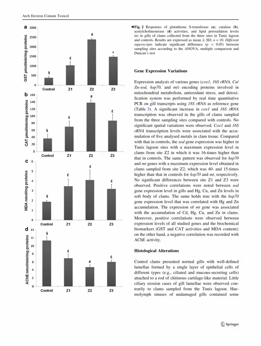
Biochemical Analysis
Biomarker responses in the gill of R. decussatus sampled
from the Tunis lagoon are shown in Fig. 2. The oxidative
stress was assessed using GST and CAT activities and
MDA content. Variations in GST activity in clam gills
were observed among the sampling sites in the Tunis
lagoon (Fig. 2a). GST activity ranged from 1036 to 2388
lmol min-1 mg-1 proteins. Clams sampled from site Z2
presented the highest value of GST level. Bivalves from
the three sampling sites presented a significantly greater
GST activity with 2.7-, 6.3-, and 3.9-fold increases for sites
Z1, Z2, and Z3, respectively, compared with controls.
The sites Z1, Z2, and Z3 exhibited an increase in CAT
level with, respectively, 1.9-, 3.7-, and 2.4-fold increases
compared with controls (Fig. 2b). A statistically significant
difference in CAT activity was also measured in bivalves
collected at the three sampling sites. The highest value
(138 lmol min-1 mg-1 proteins) was recorded at site Z2.
Lipid oxidative alteration was investigated through the
evaluation of the gill MDA accumulation (Fig. 2c). MDA
level in the gills of clams was significantly higher at Z1,
Z2, and Z3 in comparison with that in controls. Results
showed the absence of a significant difference of MDA
levels between sampling sites. MDA accumulation values
in clams from the Tunis lagoon ranged from 3.2 to 4.1
nmol mg-1 proteins.
AChE was significantly inhibited in clams from all the
sampling sites compared with controls (Fig. 2d). AChE
level at sites Z1, Z2, and Z3 displayed 1.6-, 2.4-, and 1.9-
fold decreases, respectively, compared with controls.
AChE activity differed significantly between the clams
from site Z2 and those from sites Z1 and Z3. The lowest
AChE activity (4.75 nmol min-1 mg-1 proteins) was
detected in clams sampled from site Z2.
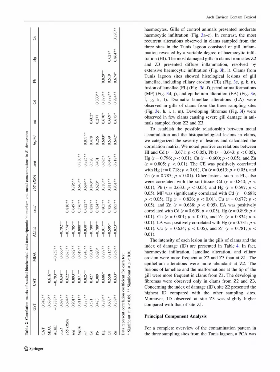
Table 2 displays the correlation obtained amongst the
investigated biochemical and transcriptomic biomarkers in
gills and metal levels in soft tissue of clams. Significant
correlations were recorded between the GST activity in
gills and Hg, Cu, and Zn levels in clam soft body. Gills
CAT activity was correlated with Hg and Zn accumulation
in the tissue of clams and GST activity in gills. Lipid
peroxidation level was positively correlated with GST and
CAT activities in gills and metal concentrations (Cd, Pb,
Hg, Cu, and Zn) in soft bodies of clams. Negative corre-
lations were recorded between AChE level in gills, on the
one hand, and the other biochemical parameters (GST and
CAT activities and LPO level) in gills and chemical
parameters (Cd, Pb, Hg, Cu and Zn) in soft body of clams,
on the other hand.
Table 1 Metal concentrations (lg g-1 dw) in sediments and tissue of R. decussatus collected from the three sites in Tunis lagoon and controls
Site Sediments Clam
Cd Pb Hg Cu Zn Cd Pb Hg Cu Zn
Control \DLa 3.6 ± 0.8a (28.1 ±
1.7)
0.10-3a
2.2 ± 0.5a 38 ± 7a \DLa 1.0 ± 0.1a (41.3 ± 0.7) 0.10-3a 5.4 ± 0.1a 84 ± 2a
Z1 0.19 ±
0.05b44 ± 2b (336 ± 7)
0.10-3b13 ± 1b 108 ± 15b 0.24 ±
0.07b3.7 ± 0.7b (146 ± 2) 0.10-3b 6.8 ± 0.9b 102 ± 7b
Z2 0.90 ±
0.06c170 ± 14c (163 ± 8)
0.10-3c16 ± 1c 368 ± 19c 0.19 ±
0.07b2.9 ± 0.5b (150 ± 1) 0.10-3c 7.4 ± 0.6b 107 ± 7b
Z3 0.33 ±
0.03d99 ± 13d (106 ± 6)
0.10-3d9.67 ± 0.04d 106 ± 2b 0.19 ±
0.06b4 ± 1b (139 ± 3) 0.10-3d 6 ± 1a 104 ± 6b
The above values represent mean ± SD (n = 10) in samples of sediments and pooled tissues of clams. Concentrations are expressed as
micrograms per gram dry weight.\DL below detection limit. Different letters indicate significant difference (p\ 0.05) among sampling sites
using one-way analysis of variance (ANOVA), multiple comparison and Duncan’s test. DL for Cd: 0.46 lg L-1
Arch Environ Contam Toxicol
123
Gene Expression Variations
Expression analysis of various genes (cox1, 16S rRNA, Cu/
Zn-sod, hsp70, and mt) encoding proteins involved in
mitochondrial metabolism, antioxidant stress, and detoxi-
fication system was performed by real time quantitative
PCR on gill transcripts using 18S rRNA as reference gene
(Table 3). A significant increase in cox1 and 16S rRNA
transcription was observed in the gills of clams sampled
from the three sampling sites compared with controls. No
significant spatial variations were observed. Cox1 and 16S
rRNA transcription levels were associated with the accu-
mulation of five analysed metals in clam tissue. Compared
with that in controls, the sod gene expression was higher in
Tunis lagoon sites with a maximum expression level in
clams from site Z2 in which it was 16-times higher than
that in controls. The same pattern was observed for hsp70
and mt genes with a maximum expression level obtained in
clams sampled from site Z2, which was 40- and 15-times
higher than that in controls for hsp70 and mt, respectively.
No significant differences between site Z1 and Z3 were
observed. Positive correlations were noted between sod
gene expression level in gills and Hg, Cu, and Zn levels in
soft body of clams. The same holds true with the hsp70
gene expression level that was correlated with Hg and Zn
accumulation. The expression of mt gene was associated
with the accumulation of Cd, Hg, Cu, and Zn in clams.
Moreover, positive correlations were observed between
expression levels of all studied genes and the biochemical
biomarkers (GST and CAT activities and MDA content);
on the other hand, a negative correlation was recorded with
AChE activity.
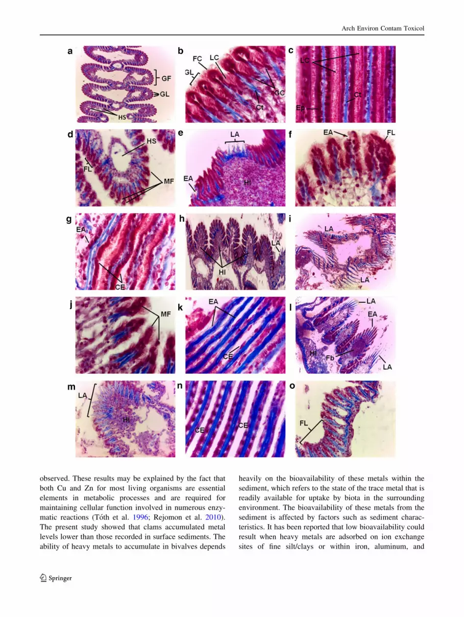
Histological Alterations
Control clams presented normal gills with well-defined
lamellae formed by a single layer of epithelial cells of
different types (e.g., ciliated and mucous-secreting cells)
attached to a rod of chitinous cartilage-like material. Little
ciliary erosion cases of gill lamellae were observed con-
trarily to clams sampled from the Tunis lagoon. Hae-
molymph sinuses of undamaged gills contained some
bFig. 2 Responses of glutathione S-transferase (a), catalase (b),acetylcholinesterase (d) activities, and lipid peroxidation levels
(c) in gills of clams collected from the three sites in Tunis lagoon
and controls. Results are expressed as mean ± SD; n = 10. Different
superscripts indicate significant difference (p \ 0.05) between
sampling sites according to the ANOVA, multiple comparison and
Duncan’s test
Arch Environ Contam Toxicol
123
haemocytes. Gills of control animals presented moderate
haemocytic infiltration (Fig. 3a–c). In contrast, the most
recurrent alterations observed in clams sampled from the
three sites in the Tunis lagoon consisted of gill inflam-
mation revealed by a variable degree of haemocytic infil-
tration (HI). The most damaged gills in clams from sites Z2
and Z3 presented diffuse inflammation, resolved by
extensive haemocytic infiltration (Fig. 3h, l). Clams from
Tunis lagoon sites showed histological lesions of gill
lamellae, including ciliary erosion (CE) (Fig. 3e, g, k, n),
fusion of lamellae (FL) (Fig. 3d–f), peculiar malformations
(MF) (Fig. 3d, j), and epithelium alteration (EA) (Fig. 3e,
f, g, k, l). Dramatic lamellae alterations (LA) were
observed in gills of clams from the three sampling sites
(Fig. 3e, h, i, l, m). Developing fibromas (Fig. 3l) were
observed in few clams causing severe gill damage in ani-
mals sampled from Z2 and Z3.
To establish the possible relationship between metal
accumulation and the histopathological lesions in clams,
we categorized the severity of lesions and calculated the
correlation matrix. We noted positive correlations between
HI and Cd (r = 0.671; p\0.05), Pb (r = 0.643; p\0.05),
Hg (r = 0.796; p\0.01), Cu (r = 0.600; p\0.05), and Zn
(r = 0.805; p\ 0.01). The CE was positively correlated
with Hg (r = 0.718; p\0.01), Cu (r = 0.613; p\0.05), and
Zn (r = 0.785; p\ 0.01). Other lesions, such as FL, also
were correlated with the soft-tissue Cd (r = 0.840; p \0.01), Pb (r = 0.633; p\ 0.05), and Hg (r = 0.597; p\0.05). MF was significantly correlated with Cd (r = 0.688;
p\ 0.05), Hg (r = 0.826; p\ 0.01), Cu (r = 0,677; p\0.05), and Zn (r = 0.638; p \ 0.05). EA was positively
correlated with Cd (r = 0.609; p\0.05), Hg (r = 0.895; p\0.01), Cu (r = 0.801; p\ 0.01), and Zn (r = 0.834; p\0.01). LA was positively correlated with Hg (r = 0.731; p\0.01), Cu (r = 0.634; p\ 0.05), and Zn (r = 0.781; p\0.01).
The intensity of each lesion in the gills of clams and the
index of damage (ID) are presented in Table 4. In fact,
haemocytic infiltration, lamellae alteration, and ciliary
erosion were more frequent at Z2 and Z3 than at Z1. The
epithelium alterations were more abundant at Z2. The
fusions of lamellae and the malformations at the tip of the
gill were more frequent in clams from Z1. The developing
fibromas were observed only in clams from Z2 and Z3.
Concerning the index of damage (ID), site Z2 presented the
highest ID compared with the other sampling sites.
Moreover, ID observed at site Z3 was slightly higher
compared with that of site Z1.
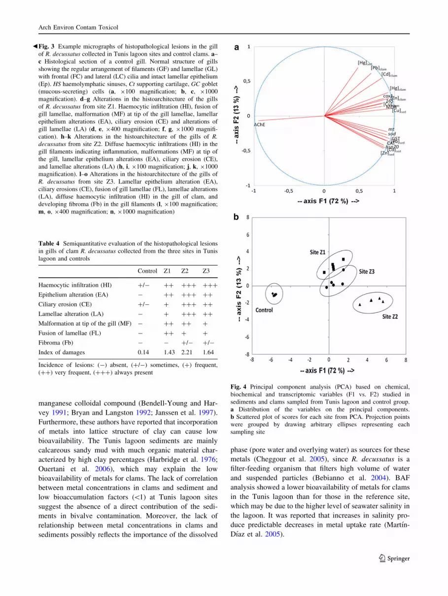
Principal Component Analysis
For a complete overview of the contamination pattern in
the three sampling sites from the Tunis lagoon, a PCA wasTable
2Correlationmatrixofstudiedbiochem
ical
andtranscriptomic
biomarkersandmetal
concentrationsin
R.decussatus
GST
CAT
MDA
AChE
cox1
16SrRNA
sod
hsp70
mt
Cd
Pb
Hg
Cu
CAT
0.942**
MDA
0.686**
0.616**
AChE
-0.888**
-0.793**
-0.753**
cox1
0.697**
0.634**
0.666**
-0.792**
16SrRNA
0.694**
0.622**
0.673**
-0.774**
0.810**
sod
0.901**
0.893**
0.672**
-0.785**
0.649**
0.795**
hsp70
0.911**
0.871**
0.619**
-0.800**
0.576**
0.643**
0.830**
mt
0.878**
0.825**
0.743**
-0.830**
0.622**
0.684**
0.825**
0.871**
Cd
0.571
0.427
0.901**
-0.790**
0.828**
0.840**
0.520
0.478
0.602*
Pb
0.473
0.309
0.620*
-0.709**
0.734**
0.620*
0.401
0.256
0.277
0.800**
Hg
0.789**
0.686*
0.797**
-0.917**
0.805**
0.783**
0.695*
0.600*
0.670*
0.879**
0.829**
Cu
0.608*
0.558
0.715**
-0.595*
0.726**
0.811**
0.647*
0.539
0.669*
0.772**
0.519
0.627*
Zn
0.739**
0.633*
0.869**
-0.823**
0.895**
0.931**
0.718**
0.642*
0.675*
0.924**
0.674*
0.864**
0.793**
Datarepresentcorrelationcoefficientforeach
test
*Significantat
p\
0.05,**Significantat
p\
0.01
Arch Environ Contam Toxicol
123

performed using all chemical data, biochemical responses,
and transcriptomic biomarkers (19 variables) (Fig. 4;
Table S4). The two principal components accounted for
86.5 % of the total variance. The first axis (F1), explaining
72 % of the total variance, was characterized by high
positive contributions of 17 variables: Cd, Pb, Hg, Cu, and
Zn levels in clams, Cd, Pb, Cu, and Zn levels in sediments,
GST and CAT activities, MDA accumulation and cox1,
16S rRNA, sod, hsp70, and mt genes expression (Fig. 4a).
Moreover, F1 presented a strong negative contribution due
to AChE activity. On the other hand, the variance
explained by the second axis (F2) (13 % of the total vari-
ance), represented a positive contribution of Hg level in
sediments. Axis F1 discriminated between Tunis lagoon
sites and the control group, whereas axis F2 discriminated
between the three sampling sites in the Tunis lagoon
(Fig. 4b). The scores plot showed that the control group
was clearly separated from the other sampling sites for
featuring lower values of metals in tissue and sediments,
lower levels of biochemical and genetic biomarkers in gills
and higher AChE activity. Both Z1 and Z3 sites shared the
same pollution pattern. Site Z2 was clearly differed from
the other sampling sites, because clams from that site
presented the highest values of sod, hsp70, and mt gene
expressions, CAT and GST activities and Cu, Cd, Pb, and
Zn levels in sediments, and the lowest AChE activity.
Discussion
Metal Accumulation in Sediments and Soft Tissue
of Clams
In coastal ecosystems, sediments are an important sink of
heavy metals and also may serve as an enriched source of
metal for benthic organisms (Pan and Wang 2012). Metal
levels in surface sediments collected from Tunis lagoon
sites were higher than those in the control sediments (site
of Louza). This is related to the presence of different
contamination sources around the lagoon. It is continually
submitted to various anthropogenic inputs (sewage,
industrial effluents, and agriculture runoff), as well as naval
and commercial shipping harbours (La Goulette Harbour
and Rades Harbour) (Ennouri et al. 2010). Concentrations
of heavy metals in sediments collected from the sampling
sites are relatively similar compared with some other
studied lagoon systems. The concentrations of Cd, Pb, Cu,
and Zn found in sediments of Bizerte lagoon (Tunisia)
ranged between 0–1.6, 0–94, 0–51, and 0–485 lg g-1 dw,
respectively (Ben Khedher et al. 2013). The sediments of
Berre lagoon (France) presented metal concentrations
ranging between 0.2–1.6, 0.15–0.4, 18–82, 11–48, and
50–151 lg g-1 dw for Cd, Hg, Pb, Cu, and Zn, respectively
(Accornero et al. 2008). Surface sediments from site Z2
contained the highest levels of Cd, Pb, Cu, and Zn due to
its proximity to industries and navigation canal. Further-
more, Z2 is the closest sampling site to the La Goulette
Harbour. However, sediments from site Z1 presented the
highest level of Hg. The increase in Hg load at site Z1 is
due primarily to the existence of an electric power plant
near this sampling site, in addition to the presence of an
industrial area. The results showed a decrease in Hg con-
centrations in sediments as we move away from the electric
power plant (Z1[Z2[Z3). Sediments sampled from site
Z3 presented higher Cd and Pb levels than those from site
Z1. The contamination at site Z3 is mainly due to the Rades
Harbour activities.
Analyses of metal loads in R. decussatus showed higher
levels in animals from the Tunis lagoon compared with the
control group. This is attributed to the contamination of
Tunis lagoon environment by heavy metals. A higher tis-
sular level of Zn and Cu than those of Cd, Pb, and Hg was
Table 3 Relative expression and expression factors of selected genes in gills of clams sampled from the three sites in Tunis lagoon and controls
Sampling site Mitochondrial metabolism Oxidative stress response Protein reparation and protection Detoxification system
cox1 16S rRNA sod hsp70 mt
Control RGE 0.46 ± 0.08a 2.9 ± 0.6a (0.45 ± 0.09) 0.10-3a (0.20 ± 0.04) 0.10-5a (0.6 ± 0.2) 0.10-6a
Z1 RGE 8.0 ± 0.9b 24 ± 4b (2.9 ± 0.6) 0.10-3b (2.0 ± 0.4) 0.10-5b (4.5 ± 0.5) 0.10-6b
DEF 17 8 6 10 7
Z2 RGE 10 ± 1b 32 ± 6b (7.4 ± 0.5) 0.10-3c (8.0 ± 0.9) 0.10-5c (9.3 ± 0.8) 0.10-6c
DEF 22 11 16 41 15
Z3 RGE 9 ± 2b 29 ± 5b (4.2 ± 0.8) 0.10-3b (3.5 ± 0.8) 0.10-5b (3.6 ± 0.9) 0.10-6b
DEF 19 10 9 18 6
Gene expression was normalized against 18S rRNA (mean ± SEM, n = 5). RGE relative gene expression, DEF differential expression factor.
Differential expression factors were calculated between each sampling site and control. Different letters indicate significant difference (p\0.05)
between sampling sites using one-way analysis of variance (ANOVA), multiple comparison and Duncan’s test
Arch Environ Contam Toxicol
123
observed. These results may be explained by the fact that
both Cu and Zn for most living organisms are essential
elements in metabolic processes and are required for
maintaining cellular function involved in numerous enzy-
matic reactions (Toth et al. 1996; Rejomon et al. 2010).
The present study showed that clams accumulated metal
levels lower than those recorded in surface sediments. The
ability of heavy metals to accumulate in bivalves depends
heavily on the bioavailability of these metals within the
sediment, which refers to the state of the trace metal that is
readily available for uptake by biota in the surrounding
environment. The bioavailability of these metals from the
sediment is affected by factors such as sediment charac-
teristics. It has been reported that low bioavailability could
result when heavy metals are adsorbed on ion exchange
sites of fine silt/clays or within iron, aluminum, and
Arch Environ Contam Toxicol
123
manganese colloidal compound (Bendell-Young and Har-
vey 1991; Bryan and Langston 1992; Janssen et al. 1997).
Furthermore, these authors have reported that incorporation
of metals into lattice structure of clay can cause low
bioavailability. The Tunis lagoon sediments are mainly
calcareous sandy mud with much organic material char-
acterized by high clay percentages (Harbridge et al. 1976;
Ouertani et al. 2006), which may explain the low
bioavailability of metals for clams. The lack of correlation
between metal concentrations in clams and sediment and
low bioaccumulation factors (\1) at Tunis lagoon sites
suggest the absence of a direct contribution of the sedi-
ments in bivalve contamination. Moreover, the lack of
relationship between metal concentrations in clams and
sediments possibly reflects the importance of the dissolved
phase (pore water and overlying water) as sources for these
metals (Cheggour et al. 2005), since R. decussatus is a
filter-feeding organism that filters high volume of water
and suspended particles (Bebianno et al. 2004). BAF
analysis showed a lower bioavailability of metals for clams
in the Tunis lagoon than for those in the reference site,
which may be due to the higher level of seawater salinity in
the lagoon. It was reported that increases in salinity pro-
duce predictable decreases in metal uptake rate (Martın-
Dıaz et al. 2005).
bFig. 3 Example micrographs of histopathological lesions in the gill
of R. decussatus collected in Tunis lagoon sites and control clams. a–c Histological section of a control gill. Normal structure of gills
showing the regular arrangement of filaments (GF) and lamellae (GL)
with frontal (FC) and lateral (LC) cilia and intact lamellar epithelium
(Ep). HS haemolymphatic sinuses, Ct supporting cartilage, GC goblet
(mucous-secreting) cells (a, 9100 magnification; b, c, 91000
magnification). d–g Alterations in the histoarchitecture of the gills
of R. decussatus from site Z1. Haemocytic infiltration (HI), fusion of
gill lamellae, malformation (MF) at tip of the gill lamellae, lamellar
epithelium alterations (EA), ciliary erosion (CE) and alterations of
gill lamellae (LA) (d, e, 9400 magnification; f, g, 91000 magnifi-
cation). h–k Alterations in the histoarchitecture of the gills of R.
decussatus from site Z2. Diffuse haemocytic infiltrations (HI) in the
gill filaments indicating inflammation, malformations (MF) at tip of
the gill, lamellar epithelium alterations (EA), ciliary erosion (CE),
and lamellae alterations (LA) (h, i, 9100 magnification; j, k, 91000
magnification). l–o Alterations in the histoarchitecture of the gills of
R. decussatus from site Z3. Lamellar epithelium alteration (EA),
ciliary erosions (CE), fusion of gill lamellae (FL), lamellae alterations
(LA), diffuse haemocytic infiltration (HI) in the gill of clam, and
developing fibroma (Fb) in the gill filaments (l, 9100 magnification;
m, o, 9400 magnification; n, 91000 magnification)
Table 4 Semiquantitative evaluation of the histopathological lesions
in gills of clam R. decussatus collected from the three sites in Tunis
lagoon and controls
Control Z1 Z2 Z3
Haemocytic infiltration (HI) ?/- ?? ??? ???
Epithelium alteration (EA) - ?? ??? ??
Ciliary erosion (CE) ?/- ? ??? ??
Lamellae alteration (LA) - ? ??? ??
Malformation at tip of the gill (MF) - ?? ?? ?
Fusion of lamellae (FL) - ?? ? ?
Fibroma (Fb) - - ?/- ?/-
Index of damages 0.14 1.43 2.21 1.64
Incidence of lesions: (-) absent, (?/-) sometimes, (?) frequent,
(??) very frequent, (???) always present
Fig. 4 Principal component analysis (PCA) based on chemical,
biochemical and transcriptomic variables (F1 vs. F2) studied in
sediments and clams sampled from Tunis lagoon and control group.
a Distribution of the variables on the principal components.
b Scattered plot of scores for each site from PCA. Projection points
were grouped by drawing arbitrary ellipses representing each
sampling site
Arch Environ Contam Toxicol
123
Enzymatic Activities and Lipid Peroxidation Levels
The increased GST and CAT activities and MDA content
in clams from the Tunis lagoon sites compared with control
values indicate that these animals are facing an oxidative
challenge, essentially associated with the presence of heavy
metals in the environment. It is known that metals are
involved in reactive oxygen species (ROS) generation,
which can adversely affect cells by producing lipid per-
oxidation of intracellular membranes. The destruction of
these membranes results in loss of cellular enzymes and
dysfunction of cellular metabolism (Geret and Bebianno
2004). A spatial variation of GST and CAT activities was
observed. The highest activities were noted in clams from
site Z2 and seemed to be associated with Hg, Cu and Zn
levels in clam tissues. It appeared that methylmercury
toxicity was principally based on mitochondrial metabo-
lism impairments due to ROS production in zebrafish
exposed through diet (Cambier et al. 2010). The high
affinity of mercury for binding to thiols suggests that
depletion of intracellular thiols (especially glutathione)
either directly or indirectly causes oxidative stress (Valko
et al. 2005). Copper is a potent redox active metal able to
generate reactive ROS through Fenton reaction (Valko
et al. 2005). Stamler (1994) reported that the inhibition of
glutathione reductase activity in cells could be a central
aspect in zinc toxicity. Glutathione reductase is essential in
the maintenance of glutathione in its reduced form, and the
inhibition of this enzyme creates an imbalance in the GSH/
GSSG ratio, contributing to oxidative stress (Stamler
1994). The highest correlation found between Cd tissue
levels and lipid peroxidation suggests the involvement of
this metal in inducing oxidative stress. Indeed, cadmium
can indirectly contribute to oxidative cell stress by dis-
placing Fe and Cu ions from ferritin and other proteins
(Varotto et al. 2013). It also can displace Zn from metal-
lothioneins and from the active sites of enzymes (e.g.,
metalloproteinases, lyases, dehydrogenases, SOD),
impairing the catalytic, inhibitory, or accessory Zn func-
tions in kinases/phosphatases and zinc-finger proteins
(Moulis 2010). Increased CAT activity and MDA content
has also been reported in R. decussatus sampled from
contaminated Tunisian coastal areas (Bizerte lagoon and
Gulf of Gabes) (Banni et al. 2009).
Several studies have demonstrated the usefulness of
measuring AChE activity in evaluating the effects of
exposure to neurotoxic compounds in aquatic organisms.
AChE-inhibiting neurotoxic compounds can cause a seri-
ous dysfunction in aquatic organisms, e.g., behavioural
changes, paralysis, and death (Fulton and Key 2001; Banni
et al. 2010). AChE activity measured in the gills of clams
sampled from the investigated sites appeared inhibited
compared with controls. Heavy metals, and particularly Hg
and Cu, present a pronounced preference for sulphur donor
groups, and may therefore inhibit this enzyme by binding
to thiol residues of proteins (Viarengo 1989). Clams from
sampling site Z2 presented the more decreased AChE
activity, which seemed to be due to the increased Hg and
Cu levels in clams. Similarly, clam R. decussatus sampled
from Bizerte lagoon (Tunisia) showed a lower AChE
activity at the most affected site by anthropogenic con-
tamination (Dellali et al. 2004).
Transcriptional Response
Clams collected from the three sampling sites in the Tunis
lagoon displayed an increased expression of cox1 gene,
compatible with an impaired electron-transport chain, and
increased expression of the mitochondrial 16S rRNA gene,
indicating an increased synthesis of mitochondria. The
overexpression of cox1 gene could be a compensatory
mechanism to restore decreased mitochondrial activity.
Overexpression of cox1 gene has been shown already in
zebrafish Danio rerio contaminated with dietary
methylmercury (Gonzalez et al. 2005) and in freshwater
and marine bivalves exposed to Cd and Cd–Zn mixture
(Achard-Joris et al. 2006). The overexpression of the 16S
rRNA gene also fits well with a compensatory response,
suggesting that cells increased the numbers of mitochon-
dria to balance those that were not functioning correctly.
The 16S rRNA induction also could reflect a higher energy
demand in these organs likely due to detoxification
mechanisms. Similarly, cox1 and 16S rRNA genes were
found overexpressed in Ruditapes philippinarum exposed
to Cd, Hg and Pb metals mixture (Dedeh et al. 2014).
Genes involved in oxidative stress scavenging (sod), gen-
eral stress response (hsp70), and detoxification mecha-
nisms (mt) were upregulated in the gills of clams sampled
from Tunis lagoon sites compared with controls. Bivalves
from site Z2 showed the highest expression levels of sod,
hsp70, and mt genes compared with sites Z1 and Z3,
because it is exposed to several origins of pollution (in-
dustrial activities and shipping). The upregulation of sod,
hsp70, and mt genes at site Z2 seemed to be more associ-
ated with Hg level in clams. Gene expression analysis in
methylmercury-exposed zebrafish revealed that the
expression levels of mt and both the cytoplasmic and
mitochondrial sod genes were highly induced, suggesting
an impact of methylmercury on detoxification process and
the generation of oxidative stress (Gonzalez et al. 2005).
The transcription levels of hsp70 and mt genes in Hg
exposed fish (Gobiocypris rarus) increased in a dose-de-
pendent manner (Li et al. 2014). Gene expression changes
in bivalves have been used as effective early warning tools
in aquatic environment monitoring (Arini et al. 2014).
Transcriptional responses are typically rapid and thus
Arch Environ Contam Toxicol
123

considered highly sensitive indicators of stress, as the ini-
tial interaction takes place at the molecular level and builds
the mechanistic basis for subsequent consequences at higher
levels of biological organization (Schirmer et al. 2010). In
this study, a correlation was found between molecular
responses, on the one hand, and GST, CAT, and MDA
levels on the other hand. This may explain that the cell is
mobilized, involving various subcellular mechanisms, to
deal with the metal contamination. Furthermore, it has been
demonstrated thatmt gene expression levels are upregulated
not only by metal exposures but also by ROS generation
(Viarengo et al. 2000; Fang et al. 2010). Our results suggest
that the induction of investigated genes and antioxidant
enzyme activity indicates an adaptive cellular response of
the redox defense system and the mitochondrial function in
R. decussatus after in situ metal exposure. According to
multivariate analysis, Tunis lagoon sites seemed to be more
affected by metal contamination compared with the refer-
ence site. Moreover, Z2 appeared to be the most disturbed
site, indicating a higher anthropogenic impact.
Histopathological Lesions in Clams
In aquatic organisms, the gills represent a vital organ,
because they play an important role in the transport of
respiratory gases and regulate the osmotic and ionic bal-
ance. Toxic substances, such as heavy metals, may cause
damage to gill tissues, thereby reducing the oxygen con-
sumption and disrupting the osmoregulatory function of
aquatic organisms (Ghate and Mulherkar 1979). In the
present study, detailed histopathology of the clam gills
from the Tunis lagoon revealed several structural alter-
ations. Clams sampled from sites Z2 and Z3, which are
localized near the Rades harbour and the navigation canal,
displayed gills with greater inflammatory alterations
(haemocytic infiltration, ciliary erosion, epithelium, and
lamellae alterations) and fibroma development compared
with those from site Z1, the latter being the furthest site
from sources of pollution. However, lamellae fusion
alteration was more frequent at site Z1 compared with Z2
and Z3 and seemed to be mainly related to the Cd level in
clam tissues. The use of ID suggests that gill tissues of
clams from site Z2 were the most altered. The metal-in-
duced physical damages in gills may result from micro-
tubules disassembly in ciliated epithelium. Metals, such as
Cu, may affect the microtubule structures of the gills either
by directing binding to tubulin thiol groups, or indirectly,
by inducing alterations of redox balance and oxidative
stress conditions in the tissue (Viarengo et al. 1994). Fur-
thermore, it was considered that metal-induced degenera-
tive alterations in tissues are related to autolytic processes
as a consequence of lysosomal membrane destabilization
(Krishnakumar et al. 1990).
According to previous works, inflammatory changes in
gills tend to be nonspecific and reflect a physiological
adaptation to stress (Mallatt 1985). These changes might be
considered as a protective mechanism, because the vul-
nerable surface area of the gills is decreased to maintain the
osmoregulatory functions (Saravana Bhavan and Geraldine
2000). In the present study, the inflammatory changes
(haemocytic infiltration, malformation and fusion of
lamellae) observed with other severe alterations (lamellae
alteration and fibroma) were more likely to represent a
progressive loss of gills’ biological functions (respiration,
osmotic, and ionic regulations) rather than protective
mechanisms and conceivably could lead to dysfunctional
or even nonfunctional gills. Histopathological damage in
aquatic organisms may decrease individual fitness through
disturbing the homeostasis and proper functioning of vital
biological processes (e.g., detoxification, endocrine func-
tioning, respiration, osmoregulation, nutrient absorption).
Thereby, these histopathological responses are highly
ecologically relevant (Au 2004). Krishnakumar et al.
(1990) reported that toxic effects of metals observed at the
tissue level were in agreement with those observed at the
organismic level. Indeed, these authors showed that filtra-
tion rate, scope for growth, and growth efficiency of metals
exposed mussels decreased significantly as a consequence
of tissue alterations. Studies in P. viridis exposed to Cu
indicated that impaired clearance rates were likely caused
by structural damage of the cilia (Nicholson 2003). It is
likely that histopathological damage to the gill interferes
with feeding and ultimately growth; thus, they may have a
potentially ecological impact on population (Nicholson and
Lam 2005). Histopathological lesions of gills have been
reported previously in clam R. philippinarum and crab
Carcinus maenas exposed to a mixture of metals (Martın-
Dıaz et al. 2008; Ben Khedher et al. 2014). Our results are
similar to those found in R. decussatus collected from
contaminated areas in Southern Portuguese coast (Costa
et al. 2013) and in R. philippinarum exposed in laboratory
to different metals (Cd, Cu, and Zn) (Martın-Dıaz et al.
2005).
Conclusions
The present study is the first that investigated both the
chemical and the multimarker approaches for Tunis lagoon
biomonitoring. Our work demonstrated a crucial contami-
nation of surface sediments by heavy metals with a spatial
variation. The clams R. decussatus sampled from the Tunis
lagoon accumulated high metal levels. The battery of
biomarker responses measured in gill tissues allowed the
discrimination among sites and was correlated with metal
contaminations. A marked inhibition of AChE activity and
Arch Environ Contam Toxicol
123
a higher induction of MDA level, CAT, and GST activities
were observed in clams sampled from site Z2. Moreover,
results revealed an overexpression of genes involved in
mitochondrial metabolism (cox1 and 16S rRNA), antioxi-
dant defense (sod), protein reparation (hsp70), and detox-
ification (mt) in gills of clams collected from the Tunis
lagoon. An occurrence of various histopathological alter-
ations in gills confirmed a metal contamination impact on
clams. The multivariate analysis for the overall data
revealed that clams from site Z2 were the most affected by
metal contamination, whereas in sites Z1 and Z3 clams
were less impacted. Overall, this study demonstrated the
potential of transcriptional, biochemical, and histopatho-
logical responses as early-warning biomarkers in moni-
toring programs and recommends their development to
complement traditional chemical analyses.
Acknowledgments This study was supported by the Ministry of
Scientific Research and Technology, the University of Monastir,
Tunisia, the French Institute of Tunisia, and the University of Bor-
deaux, France.
Conflict of interest The authors declare that they have no conflict
of interest.
References
Accornero A, Gnerre R, Manfra L (2008) Sediment concentrations of
trace metals in the Berre lagoon (France): an assessment of
contamination. Arch Environ Contam Toxicol 54:372–385
Achard-Joris M, Gonzalez P, Marie V, Baudrimont M, Bourdineaud
JP (2006) Cytochrome c oxydase subunit I gene is up-regulated
by cadmium in freshwater and marine bivalves. Biometals
19:237–244
Arini A, Daffe G, Gonzalez P, Feurtet-Mazel A, Baudrimont M
(2014) What are the outcomes of an industrial remediation on a
metal-impacted hydrosystem? A 2-year field biomonitoring of
the filter-feeding bivalve Corbicula fluminea. Chemosphere
108:214–224
Au DW (2004) The application of histo-cytopathological biomarkers
in marine pollution monitoring: a review. Mar Pollut Bull
48:817–834
Banni M, Bouraoui Z, Ghedira J, Clearandeau C, Jebali J, Boussetta H
(2009) Seasonal variation of oxidative stress biomarkers in clams
Ruditapes decussatus sampled from Tunisian coastal areas.
Environ Monit Assess 155:119–128
Banni M, Negri A, Dagnino A, Jebali J, Ameur S, Boussetta H (2010)
Acute effects of benzo[a]pyrene on digestive gland enzymatic
biomarkers and DNA damage on mussel Mytilus galloprovin-
cialis. Ecotoxicol Environ Saf 73:842–848
Banni M, Attig H, Sforzini S, Oliveri C, Mignone F, Boussetta H,
Viarengo A (2014) Transcriptomic responses to heat stress and
nickel in the mussel Mytilus galloprovincialis. Aquat Toxicol
148:104–112
Bebianno MJ, Geret F, Hoarau P, Serafim MA, Coelho MR, Gnassia-
Barelli M, Romeo M (2004) Biomarkers in Ruditapes decussa-
tus: a potential bioindicator species. Biomarkers 9:305–330
Ben Khedher S, Jebali J, Kamel N, Banni M, Rameh M, Jrad A,
Boussetta H (2013) Biochemical effects in crabs (Carcinus
maenas) and contamination levels in the Bizerta Lagoon: an
integrated approach in biomonitoring of marine complex pollu-
tion. Environ Sci Pollut Res 20:2616–2631
Ben Khedher S, Jebali J, Houas Z, Naweli H, Jrad A, Banni M,
Boussetta H (2014) Metals bioaccumulation and histopatholog-
ical biomarkers in Carcinus maenas crab from Bizerta lagoon,
Tunisia. Environ Sci Pollut Res 21:4343–4357
Bendell-Young L, Harvey HH (1991) Metal concentrations chromids
in relation to the geochemical characteristics of surficial
sediments. Arch Environ Contam Toxicol 21:202–211
Bradford MM (1976) A rapid and sensitive method for the
quantification of microgram of protein utilizing the principal of
protein-dye binding. Anal Biochem 72:248–254
Bryan GW, Langton WJ (1992) Bioavailability, accumulation and
effects of heavy metals in sediments with special reference to
United Kingdom estuaries: a review. Environ Pollut 76:89–131
Buege JA, Aust SD (1978) Microsomal lipid peroxidation. Method
Enzymol 52:302–310
Bustin SA, Benes V, Garson JA, Hellemans J, Huggett J, Kubista M,
Mueller R, Nolan T, Pfaffl MW, Shipley GL, Vandesompele J,
Wittwer CT (2009) The MIQE guidelines: minimum information
for publication of quantitative real-time PCR experiments. Clin
Chem 55:611–622
Cambier S, Gonzalez P, Durrieu G, Maury-Brachet R, Boudou A,
Bourdineaud JP (2010) Serial analysis of gene expression in the
skeletal muscles of zebrafish fed with a methylmercury-contam-
inated diet. Environ Sci Technol 44:469–475
Cheggour M, Chafik A, Fisher NS, Benbrahim S (2005) Metal
concentrations in sediments and clams in four Moroccan
estuaries. Mar Environ Res 59:119–137
Claiborne A (1985) Catalase activity. In: Greenwald RA (ed) CRC
handbook of methods for oxygen radical research. CRC Press
Inc., Boca Raton, pp 283–284
Costa PM, Carreira S, Costa MH, Caeiro S (2013) Development of
histopathological indices in a commercial marine bivalve
(Ruditapes decussatus) to determine environmental quality.
Aquat Toxicol 126:442–454
Cravo A, Pereira C, Gomes T, Cardoso C, Serafim A, Almeida C,
Rocha T, Lopes B, Company R, Medeiros A, Norberto R, Pereira
R, Araujo O, Bebianno MJ (2012) A multibiomarker approach in
the clam Ruditapes decussates to assess the impact of pollution
in the Ria Farmosa lagoon, South Coast of Portugal. Mar
Environ Res 75:23–24
De la Torre FR, Ferrari L, Salibian A (2005) Biomarkers of a native
fish species (Cnesterodon decemmaculatus) application to the
water toxicity assessment of a peri-urban polluted river of
Argentina. Chemosphere 59:577–583
Dedeh A, Ciutat A, Tran D, Bourdineaud JP (2014) DNA alterations
triggered by environmentally relevant polymetallic concentra-
tions in marine clams Ruditapes philippinarum and polychaete
worms Hediste diversicolor. Arch Environ Contam Toxicol
67:651–658
Dedeh A, Ciutat A, Treguer-Delapierre M, Bourdineaud JP (2015)
Impact of gold nanoparticles on zebrafish exposed to a spiked
sediment. Nanotoxicology 9:71–80
Dellali M, Romeo M, Gnassia-Barelli M, Aıssa P (2004) A
multivariate data analysis of the clam Ruditapes decussatus as
sentinel organism of the Bizerta lagoon (Tunisia). Water Air Soil
Pollut 156:131–144
Demayo A, Taylor MC, Taylor KW, Hodson PV, Hammond PB
(1982) Toxic effects of lead and lead compounds on human
health, aquatic life, wildlife plants, and livestock. CRC Crit Rev
Environ Control 12:257–305
Ellman GL, Courtney KO, Andres V, Featherstone RM (1961) A new
and rapid colorimetric determination of acetylcholinesterase
activity. Biochem Pharmacol 7:88–95
Arch Environ Contam Toxicol
123
Ennouri R, Chouba L, Magni P, Kraiem MM (2010) Spatial
distribution of trace metals (Cd, Pb, Hg, Cu, Zn, Fe, and Mn)
and oligo-elements (Mg, Ca, Na and K) in surface sediments of
the Gulf of Tunis (Northern Tunisia). Environ Monit Assess
163:229–239
Fang Y, Yang H, Wang T, Liu B, Zhao H, Chen M (2010)
Metallothionein and superoxide dismutase responses to sublethal
cadmium exposure in the clam Mactra veneriformis. Comp
Biochem Physiol C 151:325–333
Fu J, Wang H, Billah SM, Yu H, Zhang X (2014) Heavy metals in
seawater, sediments, and biota from the coastal area of
Yancheng City, China. Environ Toxicol Chem 33:1697–1704
Fulton MH, Key PB (2001) Acetylcholinesterase inhibition in
estuarine fish and invertebrates as an indicator of organophos-
phorus insecticide exposure and effects. Environ Toxicol Chem
20:37–45
Geret F, Bebianno MJ (2004) Does zinc produce reactive oxygen
species in Ruditapes decussatus? Ecotoxicol Environ Saf
57:399–409
Geret F, Serafim A, Bebianno MJ (2003) Antioxidant enzyme
activities, metallothioneins and lipid peroxidation as biomarkers
in Ruditapes decussates. Ecotoxicology 12:417–426
Ghate HV, Mulherkar L (1979) Histological changes in the gills of
two freshwater prawn species exposed to copper sulphate. Indian
J Exp Biol 17:838–840
Gonzalez P, Dominique Y, Massabuau JC, Boudou A, Bourdineaud
JP (2005) Comparative effects of dietary methylmercury on gene
expression in liver, skeletal muscle, and brain of the zebrafish
(Danio rerio). Environ Sci Technol 39:3972–3980
Gonzalez P, Baudrimont M, Boudou A, Bourdineaud JP (2006)
Comparative effects of direct cadmium contamination on gene
expression in gills, liver, skeletal muscles and brain of the
zebrafish (Danio rerio). Biometals 19:225–235
Habig W, Pabst M, Jakoby W (1974) Glutathione S-transferases. The
first enzymatic step in mercapturic acid formation. J Biol Chem
249:7130–7139
Hamida L, Medhiouband MN, Cochard JC, Romdhane, Le Pennec M
(2004) Etude comparative du cycle de reproduction de la
palourde Ruditapes decussatus en milieu naturel (sud Tunisie) et
controle (ecloserie). Cah Biol Mar 45:291–303
Harbridge W, Pilkey OH, Whaling P, Swetland P (1976) Sedimen-
tation in the lake of Tunis: a lagoon strongly influenced by man.
Environ Geol 1:215–225
Hellal MEA, Hellal F, El Khemissi Z, Jebali R, Dachraoui M (2011)
Trace metals in algae and sediments from the north-eastern
Tunisian lagoons. Bull Environ Contam Toxicol 86:194–198
Janssen RPT, Peijnenburg WJGM, Posthuma L, Van Den Hoop
MAGT (1997) Equilibrium partitioning of heavy metals in Dutch
soils. I. Relationship between metal partition coefficients and
soil characteristics. Environ Toxicol Chem 16:2470–2478
Kamel N, Burgeot T, Banni M, Chalghaf M, Devin S, Minier C,
Boussetta H (2014) Effects of increasing temperatures on
biomarker responses and accumulation of hazardous substances
in rope mussels (Mytilus galloprovincialis) from Bizerte lagoon.
Environ Sci Pollut Res 21:6108–6123
Krishnakumar PK, Asokan PK, Pillai VK (1990) Physiological and
cellular responses to copper and mercury in the green mussel
Perna viridis (Linnaeus). Aquat Toxicol 18:163–173
Kucuksezgin F, Kontas A, Altay O, Uluturhan E, Darilmaz E (2006)
Assessment of marine pollution in Izmir Bay: nutrient, heavy
metal and total hydrocarbon concentrations. Environ Int
32:41–51
Li ZH, Chen L, Wu YH, Li P, Li YF, Ni ZH (2014) Effects of
mercury on oxidative stress and gene expression of potential
biomarkers in larvae of the Chinese rare minnow Gobiocypris
rarus. Arch Environ Contam Toxicol 67:245–251
Mallatt J (1985) Fish gill structural changes induced by toxicants and
other irritants: a statistical review. Can J Fish Aquat Sci
42:630–648
Martın-Dıaz ML, Blasco J, Gonzalez de Canales M, Sales D, DelValls
TA (2005) Bioaccumulation and toxicity of dissolved heavy
metals from the Guadalquivir Estuary after the Aznalcollar
mining spill using Ruditapes philippinarum. Arch Environ
Contam Toxicol 48:233–241
Martın-Dıaz ML, Jimenez-Tenorio N, Sales D, Delvalls TA (2008)
Accumulation and histopathological damage in the clam Rudi-
tapes philippinarum and the crab Carcinus maenas to assess
sediment toxicity in Spanish ports. Chemosphere 71:1916–1927
Matozzo V, Binelli A, Parolini M, Locatello L, Marin MG (2010)
Biomarker responses and contamination levels in the clam
Ruditapes philippinarum for biomonitoring the Lagoon of
Venice (Italy). J Environ Monit 12:776–786
Morales-Caselles C, Martın-Dıaz ML, Riba I, Sarasquete C, Del Valls
TA (2008) Sublethal responses in caged organisms exposed to
sediments affected by oil spills. Chemosphere 72:819–825
Moulis JM (2010) Cellular mechanisms of cadmium toxicity
related to the homeostasis of essential metals. Biometals
23:877–896
Mzoughi N, Chouba L (2011) Distribution and partitioning of
aliphatic hydrocarbons and polycyclic aromatic hydrocarbons
between water, suspended particulate matter, and sediment in
harbours of the West coastal of the Gulf of Tunis (Tunisia).
J Environ Monit 13:689–698
Nasci C, Ros L, Nesto N, Sperni L, Passarini F, Pavoni B (2000)
Biochemical and histochemical responses to environmental
contaminants in clams, Tapes philippinarum, transplanted to
different polluted areas of Venice Lagoon, Italy. Mar Environ
Res 50:425–430
Negri A, Oliveri C, Sforzini S, Mignione F, Viarengo A, Banni M
(2013) Transcriptional response of the mussel Mytilus gallo-
provincialis (Lam.) following exposure to heat stress and copper.
PLoS One 8(6):e66802. doi:10.1371/journal.pone.0066802
Nicholson S (2003) Cardiac and branchial physiology associated with
copper accumulation and detoxication in the mytilid mussel
Perna viridis (L.). J Exp Mar Biol Ecol 295:157–171
Nicholson S, Lam PKS (2005) Pollution monitoring in Southeast Asia
using biomarkers in the mytilid mussel Perna viridis (Mytilidae:
Bivalvia). Environ Int 31:121–132
O’Connor TP (2002) National distribution of chemical concentrations
in mussels and oysters in the USA. Mar Environ Res 53:117–143
Oros DR, Ross JRM (2005) Polycyclic aromatic hydrocarbons in
bivalves from the San Francisco estuary: spatial distributions,
temporal trends, and sources. Mar Environ Res 60:466–488
Ouertani N, Hamouda R, Belayouni H (2006) Study of the organic
matter buried in recent sediments of an increasing anoxic
environment surrounded by an urban area: the ‘‘Lac sud de
Tunis’’. Geo Eco Trop 30:21–34
Pan K, Wang WX (2012) Trace metal contamination in estuarine and
coastal environments in China. Sci Total Environ 421–422:3–16
Pfaffl MW (2001) A new mathematical model for relative quantifi-
cation in real-time RT-PCR. Nucleic Acids Res 29:e45
Porter JS, Dyrynda PEJ, Ryland JS, Carvalho GR (2001) Morpho-
logical and genetic adaptation to a lagoon environment: a case
study in the bryozoan genus Alcyonidium. Mar Biol 139:
575–585
Rejomon G, Nair M, Joseph T (2010) Trace metal dynamics in fishes
from the southwest coast of India. Environ Monit Assess
167:243–255
Riba I, Gonzalez de Canales M, Forja JM, DelValls TA (2004)
Sediment quality in the Guadalquivir estuary: sublethal effects
associated with the Aznalcollar mining spill. Mar Pollut Bull
48:153–163
Arch Environ Contam Toxicol
123

Saravana Bhavan P, Geraldine P (2000) Histopathology of the
hepatopancreas and gills of the prawn Macrobrachium mal-
colmsonii exposed to endosulfan. Aquat Toxicol 50:331–339
Schirmer K, Fischer BB, Madureira DJ, Pillai S (2010) Transcrip-
tomics in ecotoxicology. Anal Bioanal Chem 397:917–923
Sfriso A, Argese E, Bettiol C, Facc C (2008) Tapes philippinarum
seed exposure to metals in polluted areas of the Venice lagoon.
Estuar Coast Shelf Sci 79:581–590
Stamler JS (1994) Redox signaling: nitrosylation and related target
interactions of nitric oxide. Cell 78:931–936
Toth L, Juhasz M, Varga T, Csikkel-Szolnoki A, Nemcsok KJ (1996)
Some effect of CuSO4 on carp. J Environ Sci Health B
31:627–635
Valko M, Morris H, Cronin MTD (2005) Metals, toxicity and
oxidative stress. Curr Med Chem 12:1161–1208
Varotto L, Domeneghetti S, Rosani U, Manfrin C, Cajaraville MP,
Raccanelli S, Pallavicini A, Venier P (2013) DNA damage and
transcriptional changes in the gills of Mytilus galloprovincialis
exposed to nanomolar doses of combined metal salts (Cd, Cu,
Hg). PLoS One 8(1):e54602. doi:10.1371/journal.pone.0054602
Viarengo A (1989) Heavy metals in marine invertebrates: mecha-
nisms of regulation and toxicity at the cellular level. CRC Crit
Rev Aquat Sci 1:295–317
Viarengo A, Nott JA (1993) Mechanisms of heavy metals homeosta-
sis in marine invertebrates. Comp Biochem Physiol C
104:355–372
Viarengo A, Arena N, Canesi L, Alia FA, Orunesu M (1994)
Structural and biochemical alterations in the gills of copper-
exposed mussels. In: Renzoni A, Mattei N, Lari L (eds)
Contaminants in the environment. Lewis Publishers, Boca
Raton, pp 135–144
Viarengo A, Burlando B, Ceratto N, Panfoli I (2000) Antioxidant role
of metallothioneins: a comparative overview. Cell Mol Biol
46:407–417
Viarengo A, Lowe D, Bolognesi C, Fabbri E, Koehler A (2007) The
use of biomarkers in biomonitoring: a 2-tier approach assessing
the level of pollutant-induced stress syndrome in sentinel
organisms. Comp Biochem Physiol C 146:281–300
Wetzel DL, Van Vleet ES (2004) Accumulation and distribution of
petroleum hydrocarbons found in mussels (Mytilus galloprovin-
cialis) in the canals of Venice, Italy. Mar Pollut Bull 48:927–936
Zhu XB, Xu WH, Wang XT, Huang XP, Deng LP, Kang XL, Jiang
ZG, Ma XL (2005) Research on heavy metals in Ruditapes
philippinarum and soda industry wastes. Chin J Oceanol Limnol
23:39–42
Arch Environ Contam Toxicol
123