transcriptional control in embryonic drosophila midline guidance assessed through a whole genome...
TRANSCRIPT
BioMed CentralBMC Neuroscience
ss
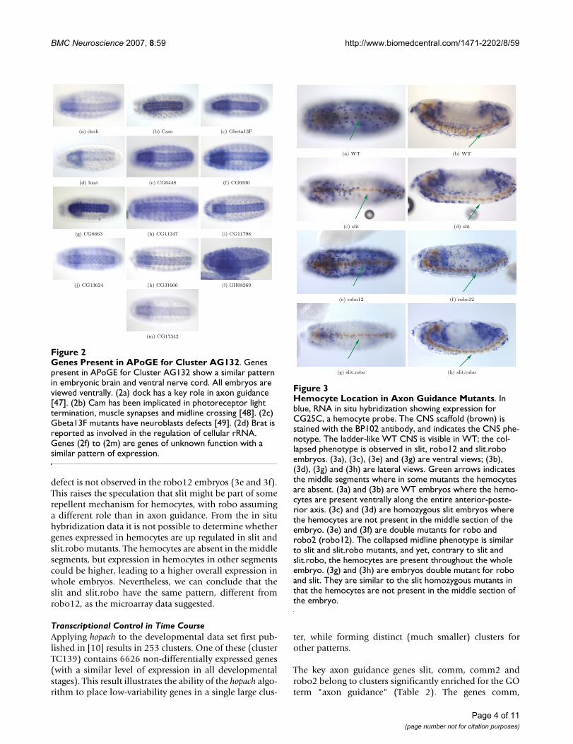
Open AcceResearch articleTranscriptional control in embryonic Drosophila midline guidance assessed through a whole genome approachTiago R Magalhães*1, Jessica Palmer2, Pavel Tomancak3 and Katherine S Pollard*4Address: 1Programa Gulbenkian Doutoramento Biologia e Medicina, Centro Neurociências, Universidade de Coimbra, 3000 – Coimbra, Portugal, 2Lewis-Clark State College, 500 8th Avenue, Lewiston, ID 83501, USA, 3Max-Planck-Institute of Molecular Cell Biology and Genetics, Dresden, Germany and 4UC Davis Genome Center & Department of Statistics, University of California, Davis, CA, 95616, USA
Email: Tiago R Magalhães* - [email protected]; Jessica Palmer - [email protected]; Pavel Tomancak - [email protected]; Katherine S Pollard* - [email protected]
* Corresponding authors
AbstractBackground: During the development of the Drosophila central nervous system the process ofmidline crossing is orchestrated by a number of guidance receptors and ligands. Many key axonguidance molecules have been identified in both invertebrates and vertebrates, but thetranscriptional regulation of growth cone guidance remains largely unknown. It is established thattranslational regulation plays a role in midline crossing, and there are indications that transcriptionalregulation is also involved. To investigate this issue, we conducted a genome-wide study oftranscription in Drosophila embryos using wild type and a number of well-characterizedDrosophila guidance mutants and transgenics. We also analyzed a previously published microarraytime course of Drosophila embryonic development with an axon guidance focus.
Results: Using hopach, a novel clustering method which is well suited to microarray data analysis,we identified groups of genes with similar expression patterns across guidance mutants andtransgenics. We then systematically characterized the resulting clusters with respect to theirrelevance to axon guidance using two complementary controlled vocabularies: the Gene Ontology(GO) and anatomical annotations of the Atlas of Pattern of Gene Expression (APoGE) in situhybridization database. The analysis indicates that regulation of gene expression does play a role inthe process of axon guidance in Drosophila. We also find a strong link between axon guidance andhemocyte migration, a result that agrees with mounting evidence that axon guidance molecules areco-opted in vertebrate vascularization. Cell cyclin activity in the context of axon guidance is alsosuggested from our array data. RNA and protein expression patterns of cell cyclins in axonguidance mutants and transgenics support this possible link.
Conclusion: This study provides important insights into the regulation of axon guidance in vivo.
BackgroundThe process by which axons cross the midline duringdevelopment of the Central Nervous System (CNS) in
Drosophila is of great interest and has been the focus ofmuch scientific research [1,2]. After neuroblast delamina-tion and cell fate decisions, axons undergo a journey that
Published: 31 July 2007
BMC Neuroscience 2007, 8:59 doi:10.1186/1471-2202-8-59
Received: 22 December 2006Accepted: 31 July 2007
This article is available from: http://www.biomedcentral.com/1471-2202/8/59
© 2007 Magalhães et al; licensee BioMed Central Ltd. This is an Open Access article distributed under the terms of the Creative Commons Attribution License (http://creativecommons.org/licenses/by/2.0), which permits unrestricted use, distribution, and reproduction in any medium, provided the original work is properly cited.
Page 1 of 11(page number not for citation purposes)
BMC Neuroscience 2007, 8:59 http://www.biomedcentral.com/1471-2202/8/59
will eventually wire them to the appropriate targets. Thisjourney includes a series of steps, one of which is the deci-sion to cross (or not cross) the midline. Four key regula-tors of midline crossing are known: slit, robo, robo2 andcomm. Growth cones in which the robo and robo2 recep-tors are present at the membrane sense the presence of theslit ligand and do not cross the midline [3,4]. Conversely,growth cones without robo and robo2 at the membraneare not able to respond to the repulsive cue of slit and docross the midline. robo levels at the growth cone mem-brane are regulated by comm.
A major question in midline crossing is how regulationoccurs through gene expression, i.e., to what extent is tran-scription relevant in the control of growth cone pathways.It is well established that rapid local translation doesoccur [5,6]. Some literature also indicates that transcrip-tion is relevant [7-9]. We designed this study of transcrip-tion during CNS development to i) find relevant genes inthe axon guidance process, ii) elucidate the role of tran-scription regulation during midline crossing in Dro-sophila, and iii) determine whether any upstream changesare visible in axon guidance mutants. Our analysis usestwo whole-genome microarray data sets: (1) axon guidance– Drosophila embryos, stage 15, of axon guidance GainOf Function (GOF) and Loss Of Function (LOF) mutants,and (2) time course – a developmental wild type timecourse, consisting of 12 non-overlapping 1 hour collec-tion points (previously published [10]). The axon guid-ance (AG) data was collected as part of this study and ispublished here for the first time. We have thoroughlyreanalyzed the previously published time course (TC)data.
To identify clusters of co-expressed genes in each data set,we employ the hopach clustering methodology [11]. Clus-tering algorithms have been successfully applied to micro-array studies, but the systematic analysis of clusteringoutput remains a major challenge. A significant obstacle isthe lack of reproducibility, or even assessment of variabil-ity, of clustering results. Clustering output can vary dra-matically from one repetition of an experiment toanother, particularly with the small sample sizes typical inmicroarray experiments. The R statistical package hopach[11] uses a bootstrap resampling method to assess clustervariability, producing a measure of membership of eachgene in every cluster. For this study, we adapted hopach touse these cluster membership values to assign genes toclusters, while allowing genes to belong to more than onecluster – a type of fuzzy clustering. This modificationimproves reproducibility and results in cluster output thatis suitable for functional characterization. hopach is freelyavailable and is easily used by biologists, providing a sta-tistically sound framework for analysis of microarray data.
We characterize gene clusters with two complementarysources of annotation, the Gene Ontology (GO) [12], andthe Atlas of Pattern of Gene Expression (APoGE), a com-ponent of the Berkeley Drosophila Genome Project [10].APoGE provides pictures of the expression pattern of eachgene in the Drosophila embryo and a description of theanatomical structures in which the gene is expressed. Acontrolled vocabulary is used by individual curators (AmyBeaton and Volker Hartenstein), experts who assure con-sistency in each call. The goals and scope of APoGE aredescribed in [10]. Its use for characterization of microar-ray data has never been explored. Together, GO andAPoGE provide a rich, multi-dimensional vocabulary forannotating hopach clusters. In this study, we analyze clus-ters in which genes with terms related to axon guidanceare significantly over represented. Querying the APoGE insitu database for genes expressed with specific spatial andtemporal patterns provides a means to further explore theaxon guidance clusters.
Applying this approach, we identify striking changes ingene expression across GOF/LOF, as well as groups ofaxon guidance genes co-expressed during wild type devel-opment. These findings suggest that regulation of geneexpression does in fact play a role in midline crossing. Inaddition, our analysis indicates that axon guidance genesmay be involved in the process of hemocyte migration,and that cell cyclins are active during midline crossing.These observations are confirmed through independentRNA and protein expression studies.
Results and discussionTranscriptional Control in Axon Guidance MutantsOur hopach analysis of gene expression profiles from 17Drosophila axon guidance mutants identified 517 clustersof median size 27 (range 1 to 821). Figure 1 shows theprofiles of these clusters ordered by the hopach algorithmso that clusters close to each other contain genes with sim-ilar expression profiles. If regulation during the course ofaxon guidance occurs mainly through protein translation,the changes observed in a microarray study will not yieldcoherent groups of genes with known function in axonguidance. Yet, we found several clusters significantlyenriched for axon guidance genes.
We ranked clusters based on their association with rele-vant terms from GO ("axon guidance") and APoGE ("ven-tral nerve cord", "embryonic brain", and "ventralmidline") as described in the Methods. The top four clus-ters for each term are shown in Table 1. Cluster AG132ranks first for "axon guidance", "embryonic brain" and"ventral midline". It is the cluster with the highest ranks inboth the GO and the APoGE vocabularies. This clustercontains a number of genes well known from the axonguidance literature, such as dock, plexB, Cam, CadN, caps,
Page 2 of 11(page number not for citation purposes)
BMC Neuroscience 2007, 8:59 http://www.biomedcentral.com/1471-2202/8/59
spen, Fs(2)Ket, Con, RhoGAP19D, Rac2, and shot. AG164is another cluster that also contains relevant genes in axonguidance mechanisms, such as trio, WASp, Nedd4, robo3,MICAL, RhoGAPp190, rhoGAP88C, RhoGAP18B, lola,side, and Fmr1 [2]. A full list of genes in all of the topranking clusters and a more detailed exploration of each
cluster are available in the Supplementary Materials(Addi-tional files 1234).
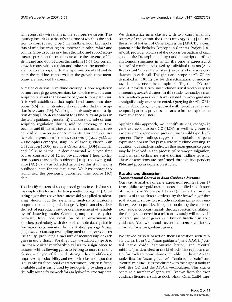
Querying APoGE, we identified a large subset of genes inCluster AG132 which are expressed in the commissuresand midline (Figure 2). Several genes of unknown func-tion (CG6448, CG6930, nrv3, CG11347, CG11798,CG13624, and CG31666) share this distinctive pattern ofexpression with the known genes dock, gBeta, brat andCam. This similarity provides some insight into the func-tions of the less well characterized genes and suggests theirinvolvement in axon guidance.
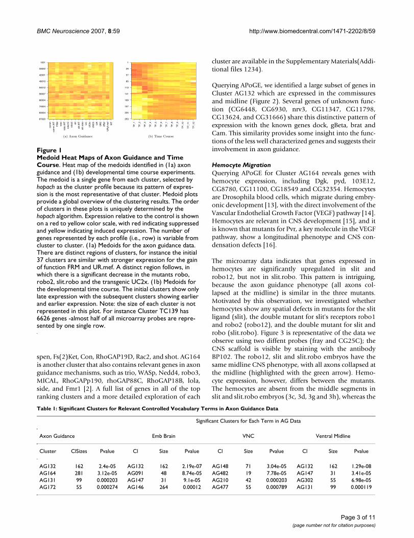
Hemocyte MigrationQuerying APoGE for Cluster AG164 reveals genes withhemocyte expression, including Dgk, pyd, 103E12,CG8780, CG11100, CG18549 and CG32354. Hemocytesare Drosophila blood cells, which migrate during embry-onic development [13], with the direct involvement of theVascular Endothelial Growth Factor (VEGF) pathway [14].Hemocytes are relevant in CNS development [15], and itis known that mutants for Pvr, a key molecule in the VEGFpathway, show a longitudinal phenotype and CNS con-densation defects [16].
The microarray data indicates that genes expressed inhemocytes are significantly upregulated in slit androbo12, but not in slit.robo. This pattern is intriguing,because the axon guidance phenotype (all axons col-lapsed at the midline) is similar in the three mutants.Motivated by this observation, we investigated whetherhemocytes show any spatial defects in mutants for the slitligand (slit), the double mutant for slit's receptors robo1and robo2 (robo12), and the double mutant for slit androbo (slit.robo). Figure 3 is representative of the data weobserve using two diffent probes (fray and CG25C); theCNS scaffold is visible by staining with the antibodyBP102. The robo12, slit and slit.robo embryos have thesame midline CNS phenotype, with all axons collapsed atthe midline (highlighted with the green arrow). Hemo-cyte expression, however, differs between the mutants.The hemocytes are absent from the middle segments inslit and slit.robo embryos (3c, 3d, 3g and 3h), whereas the
Table 1: Significant Clusters for Relevant Controlled Vocabulary Terms in Axon Guidance Data
Significant Clusters for Each Term in AG Data
Axon Guidance Emb Brain VNC Ventral Midline
Cluster ClSizes Pvalue Cl Size Pvalue Cl Size Pvalue Cl Size Pvalue
AG132 162 2.4e-05 AG132 162 2.19e-07 AG148 71 3.04e-05 AG132 162 1.29e-08AG164 281 3.12e-05 AG091 48 8.74e-05 AG482 19 7.78e-05 AG147 31 3.41e-05AG131 99 0.000203 AG147 31 9.1e-05 AG210 42 0.000203 AG302 55 6.98e-05AG172 55 0.000274 AG146 264 0.00012 AG477 55 0.000789 AG131 99 0.000119
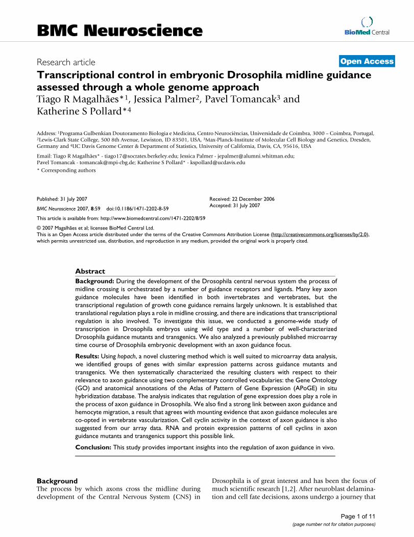
Medoid Heat Maps of Axon Guidance and Time CourseFigure 1Medoid Heat Maps of Axon Guidance and Time Course. Heat map of the medoids identified in (1a) axon guidance and (1b) developmental time course experiments. The medoid is a single gene from each cluster, selected by hopach as the cluster profile because its pattern of expres-sion is the most representative of that cluster. Medoid plots provide a global overview of the clustering results. The order of clusters in these plots is uniquely determined by the hopach algorithm. Expression relative to the control is shown on a red to yellow color scale, with red indicating suppressed and yellow indicating induced expression. The number of genes represented by each profile (i.e., row) is variable from cluster to cluster. (1a) Medoids for the axon guidance data. There are distinct regions of clusters, for instance the initial 37 clusters are similar with stronger expression for the gain of function FRM and UR.mef. A distinct region follows, in which there is a significant decrease in the mutants robo, robo2, slit.robo and the transgenic UC2x. (1b) Medoids for the developmental time course. The initial clusters show only late expression with the subsequent clusters showing earlier and earlier expression. Note: the size of each cluster is not represented in this plot. For instance Cluster TC139 has 6626 genes -almost half of all microarray probes are repre-sented by one single row.
comm
comm.WT
FRM
robo
robo.WT
robo12
robo2
slit
slit.WT
slit.robo
UC
UC2x
UNC5
UR
UR2
URyf
URyf.mef
WT
87020
83004
74004
66004
56007
54012
49010
42001
33002
1001
(a) Axon GuidanceTP_1
TP_2
TP_3
TP_4
TP_5
TP_6
TP_7
TP_8
TP_9
TP_10
TP_11
TP_12
253
225
197
169
141
113
85
57
29
1
(b) Time Course
Page 3 of 11(page number not for citation purposes)
BMC Neuroscience 2007, 8:59 http://www.biomedcentral.com/1471-2202/8/59
defect is not observed in the robo12 embryos (3e and 3f).This raises the speculation that slit might be part of somerepellent mechanism for hemocytes, with robo assuminga different role than in axon guidance. From the in situhybridization data it is not possible to determine whethergenes expressed in hemocytes are up regulated in slit andslit.robo mutants. The hemocytes are absent in the middlesegments, but expression in hemocytes in other segmentscould be higher, leading to a higher overall expression inwhole embryos. Nevertheless, we can conclude that theslit and slit.robo have the same pattern, different fromrobo12, as the microarray data suggested.
Transcriptional Control in Time CourseApplying hopach to the developmental data set first pub-lished in [10] results in 253 clusters. One of these (clusterTC139) contains 6626 non-differentially expressed genes(with a similar level of expression in all developmentalstages). This result illustrates the ability of the hopach algo-rithm to place low-variability genes in a single large clus-
ter, while forming distinct (much smaller) clusters forother patterns.
The key axon guidance genes slit, comm, comm2 androbo2 belong to clusters significantly enriched for the GOterm "axon guidance" (Table 2). The genes comm,
Hemocyte Location in Axon Guidance MutantsFigure 3Hemocyte Location in Axon Guidance Mutants. In blue, RNA in situ hybridization showing expression for CG25C, a hemocyte probe. The CNS scaffold (brown) is stained with the BP102 antibody, and indicates the CNS phe-notype. The ladder-like WT CNS is visible in WT; the col-lapsed phenotype is observed in slit, robo12 and slit.robo embryos. (3a), (3c), (3e) and (3g) are ventral views; (3b), (3d), (3g) and (3h) are lateral views. Green arrows indicates the middle segments where in some mutants the hemocytes are absent. (3a) and (3b) are WT embryos where the hemo-cytes are present ventrally along the entire anterior-poste-rior axis. (3c) and (3d) are homozygous slit embryos where the hemocytes are not present in the middle section of the embryo. (3e) and (3f) are double mutants for robo and robo2 (robo12). The collapsed midline phenotype is similar to slit and slit.robo mutants, and yet, contrary to slit and slit.robo, the hemocytes are present throughout the whole embryo. (3g) and (3h) are embryos double mutant for robo and slit. They are similar to the slit homozygous mutants in that the hemocytes are not present in the middle section of the embryo.
(a) WT (b) WT
(c) slit (d) slit
(e) robo12 (f) robo12
(g) slit.robo (h) slit.robo
Genes Present in APoGE for Cluster AG132Figure 2Genes Present in APoGE for Cluster AG132. Genes present in APoGE for Cluster AG132 show a similar pattern in embryonic brain and ventral nerve cord. All embryos are viewed ventrally. (2a) dock has a key role in axon guidance [47]. (2b) Cam has been implicated in photoreceptor light termination, muscle synapses and midline crossing [48]. (2c) Gbeta13F mutants have neuroblasts defects [49]. (2d) Brat is reported as involved in the regulation of cellular rRNA. Genes (2f) to (2m) are genes of unknown function with a similar pattern of expression.
(a) dock (b) Cam (c) Gbeta13F
(d) brat (e) CG6448 (f) CG6930
(g) CG8663 (h) CG11347 (i) CG11798
(j) CG13624 (k) CG31666 (l) GH08269
(m) CG17342
Page 4 of 11(page number not for citation purposes)
BMC Neuroscience 2007, 8:59 http://www.biomedcentral.com/1471-2202/8/59
comm2 and robo2 are all present in Cluster TC91, whichis a small cluster (15 members) with a pattern of expres-sion characterized by a peak around stages 4 to 9. Commis known to be transiently expressed in the commissuralaxons and midline glia, simultaneously with robo[17,18]. However, the similarity in expression for comm,comm2, and robo2 throughout the whole 18 hours ofdevelopment has never been reported. Given the excep-tional similarity between the expression of these threegenes it would be interesting to explore if they are control-led by the same upstream mechanism, or whether theycontrol a common downstream mechanism. Included inthe adjacent clusters (TC90 to TC97), hence with a verysimilar pattern of expression, there are other importantaxon guidance genes (RhoGEF4, neur, and RhoGEF3) andmany genes involved in Notch signaling (E(spl), neur,Brd, m4, HLHm5, HLHmgamma, Dl, bib and malpha).Notch involvement in axon guidance has been previouslyreported [19]. The genes spi, hh, argos, gsb, Dl, slp2, siz,and ems are also in these clusters and have documentedphenotypes in axon guidance or the CNS scaffold.
A query of APoGE for genes in Clusters TC90 to TC97 withmidline expression similar to comm, comm2 and robo2,yielded argos. The gene argos has been shown to be rele-vant in axon guidance in the visual system [20], which fur-ther strengthens the possibility of a role for argos inmidline axon guidance. Sulfated (Sulf1) is expressed incells that appear to be adjacent to comm2 and robo2expressing cells. Sulf1 has not been previously implicatedin axon guidance. It is our future project to investigatewhether either argos or Sulf1 has a genetic interactionwith comm, comm2 and robo2.
Cell CyclinsTime course clusters TC140, TC141 and TC142 are themost relevant clusters for the APoGE terms ("embryonicbrain", "ventral nerve cord", "ventral midline"). They donot contain axon guidance genes, but they have many cyc-lins, including CycA, polo, cdc2, CycB3 CycE and ago.Cell cyclins are involved in cell division and in CNS devel-opment, playing an important role in neuroblast division.However, a role for cyclins has not been reported in post-
mitotic neurons, the type of neurons involved in axonguidance. It is known that CycA and CycE mutants havesevere defects in the commissures and longitudinal axontracts of the CNS [21,22], and Cdk5 controls some aspectsof axon patterning in vivo [23]. The cyclins CycA, cdc2and CycB3 genetically interact with each other [24,25]and are important in the earlier stages of intense cell divi-sion [26]. Agreeing with this role, the TC data shows highexpression early in development, but suggests other func-tions at later stages because i) there is sustained expressionwell past the intense initial cell divisions, and ii) theAPoGE images show a similar pattern of expression for allcyclins for later stages.
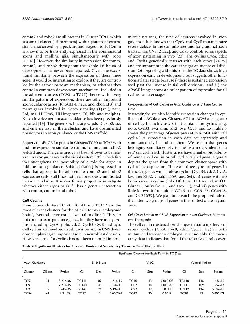
Co-expression of Cell Cyclins in Axon Guidance and Time Course DataInterestingly, we also identify expression changes in cyc-lins in the AG data set. Clusters AG1 to AG93 are a groupof cell cyclin rich clusters that contain the cyclins smg,polo, CycB3, swa, pim, cdc2, twe, CycB, and fzy. Table 3shows the percentage of genes present in APoGE with cellcyclin-like expression in each data set separately andsimultaneously in both of them. We reason that genesbelonging simultaneously to the two independent datasets' cell cyclin rich cluster space have a higher probabilityof being a cell cyclin or cell cyclin related gene. Figure 4depicts the genes from this common cluster space withcyclin-like expression. There are three types of genes inthis set: i) genes with a role as cyclins (CybB3, cdc2, CycA,fzy, mei-S332, G-ialpha65A, and Set), ii) genes with noknown role as cyclins (lola, Df31, Set, UTPase, Sd, msf11,Chrac16, Su(var)2–10, and Uch-L3), and iii) genes withlittle known information (CG15141, CG5175, CG8478,and CG31639). We plan to research the proposed role ofthe latter two groups of genes in the context of axon guid-ance.
Cell Cyclin Protein and RNA Expression in Axon Guidance Mutants and TransgenicsThe cell cyclin clusters show changes in transcript levels ofseveral cyclins (CycA, CycB, cdc2, CycB3, fzy) in bothmutant and transgenic embryos. Most notably, the micro-array data indicates that for all the robo GOF, robo over-
Table 2: Significant Clusters for Relevant Controlled Vocabulary Terms in Time Course Data
Significant Clusters for Each Term in TC Data
Axon Guidance Emb Brain VNC Ventral Midline
Cluster ClSizes Pvalue Cl Size Pvalue Cl Size Pvalue Cl Size Pvalue
TC52 21 5.22e-06 TC141 109 1.21e-15 TC10 13 0.000303 TC140 146 1.43e-16TC91 15 2.77e-05 TC140 146 1.14e-11 TC07 14 0.000545 TC141 109 1.99e-12TC27 12 3.68e-05 TC142 126 5.49e-11 TC97 17 0.00133 TC142 126 5.59e-11TC54 41 4.3e-05 TC97 17 0.000267 TC47 20 0.0016 TC10 13 0.000171
Page 5 of 11(page number not for citation purposes)
BMC Neuroscience 2007, 8:59 http://www.biomedcentral.com/1471-2202/8/59
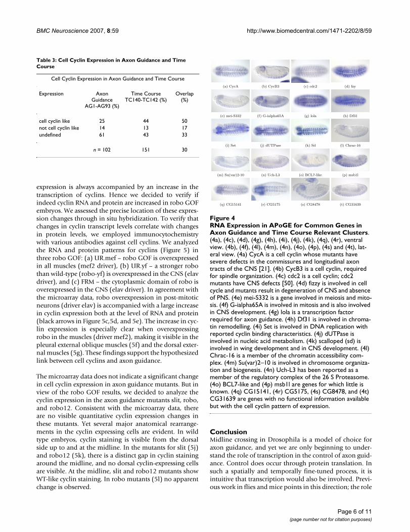
expression is always accompanied by an increase in thetranscription of cyclins. Hence we decided to verify ifindeed cyclin RNA and protein are increased in robo GOFembryos. We assessed the precise location of these expres-sion changes through in situ hybridization. To verify thatchanges in cyclin transcript levels correlate with changesin protein levels, we employed immunocytochemistrywith various antibodies against cell cyclins. We analyzedthe RNA and protein patterns for cyclins (Figure 5) inthree robo GOF: (a) UR.mef – robo GOF is overexpressedin all muscles (mef2 driver), (b) UR.yf – a stronger robothan wild-type (robo-yf) is overexpressed in the CNS (elavdriver), and (c) FRM – the cytoplasmic domain of robo isoverexpressed in the CNS (elav driver). In agreement withthe microarray data, robo overexpression in post-mitoticneurons (driver elav) is accompanied with a large increasein cyclin expression both at the level of RNA and protein(black arrows in Figure 5c,5d, and 5e). The increase in cyc-lin expression is especially clear when overexpressingrobo in the muscles (driver mef2), making it visible in thepleural external oblique muscles (5f) and the dorsal exter-nal muscles (5g). These findings support the hypothesizedlink between cell cyclins and axon guidance.
The microarray data does not indicate a significant changein cell cyclin expression in axon guidance mutants. But inview of the robo GOF results, we decided to analyze thecyclin expression in the axon guidance mutants slit, robo,and robo12. Consistent with the microarray data, thereare no visible quantitative cyclin expression changes inthese mutants. Yet several major anatomical rearrange-ments in the cyclin expressing cells are evident. In wildtype embryos, cyclin staining is visible from the dorsalside up to and at the midline. In the mutants for slit (5j)and robo12 (5k), there is a distinct gap in cyclin stainingaround the midline, and no dorsal cyclin-expressing cellsare visible. At the midline, slit and robo12 mutants showWT-like cyclin staining. In robo mutants (5l) no apparentchange is observed.
ConclusionMidline crossing in Drosophila is a model of choice foraxon guidance, and yet we are only beginning to under-stand the role of transcription in the control of axon guid-ance. Control does occur through protein translation. Insuch a spatially and temporally fine-tuned process, it isintuitive that transcription would also be involved. Previ-ous work in flies and mice points in this direction; the role
RNA Expression in APoGE for Common Genes in Axon Guidance and Time Course Relevant ClustersFigure 4RNA Expression in APoGE for Common Genes in Axon Guidance and Time Course Relevant Clusters. (4a), (4c), (4d), (4g), (4h), (4i), (4j), (4k), (4q), (4r), ventral view. (4b), (4f), (4l), (4m), (4n), (4o), (4p), (4s) and (4t), lat-eral view. (4a) CycA is a cell cyclin whose mutants have severe defects in the commissures and longitudinal axon tracts of the CNS [21]. (4b) CycB3 is a cell cyclin, required for spindle organization. (4c) cdc2 is a cell cyclin; cdc2 mutants have CNS defects [50]. (4d) fizzy is involved in cell cycle and mutants result in degeneration of CNS and absence of PNS. (4e) mei-S332 is a gene involved in meiosis and mito-sis. (4f) G-ialpha65A is involved in mitosis and is also involved in CNS development. (4g) lola is a transcription factor required for axon guidance. (4h) Df31 is involved in chroma-tin remodelling. (4i) Set is involved in DNA replication with reported cyclin binding characteristics. (4j) dUTPase is involved in nucleic acid metabolism. (4k) scalloped (sd) is involved in wing development and in CNS development. (4l) Chrac-16 is a member of the chromatin accessibility com-plex. (4m) Su(var)2–10 is involved in chromosome organiza-tion and biogenesis. (4n) Uch-L3 has been reported as a member of the regulatory complex of the 26 S Proteasome. (4o) BCL7-like and (4p) msb1l are genes for which little is known. (4q) CG15141, (4r) CG5175, (4s) CG8478, and (4t) CG31639 are genes with no functional information available but with the cell cyclin pattern of expression.
(a) CycA (b) CycB3 (c) cdc2 (d) fzy
(e) mei-S332 (f) G-ialpha65A (g) lola (h) Df31
(i) Set (j) dUTPase (k) Sd (l) Chrac-16
(m) Su(var)2-10 (n) Uch-L3 (o) BCL7-like (p) msb1l
(q) CG15141 (r) CG5175 (s) CG8478 (t) CG31639
Table 3: Cell Cyclin Expression in Axon Guidance and Time Course
Cell Cyclin Expression in Axon Guidance and Time Course
Expression Axon Guidance
AG1-AG93 (%)
Time Course TC140-TC142 (%)
Overlap (%)
cell cyclin like 25 44 50not cell cyclin like 14 13 17undefined 61 43 33
n = 102 151 30
Page 6 of 11(page number not for citation purposes)
BMC Neuroscience 2007, 8:59 http://www.biomedcentral.com/1471-2202/8/59
of transcription factors is also emerging as very relevant, asreviewed in [27]. To investigate this topic we have under-taken a whole-genome expression study of mutants andtransgenics of the key axon guidance genes. If control ofmidline crossing proceeds through transcription, thegenetic perturbations induced in the mutants and trans-genics should reveal clusters enriched for genes known tobe involved in axon guidance. We also investigated theexpression patterns of known axon guidance genes duringnormal development in a wild type microarray timecourse, using a previously published microarray data set.
We have found that the hopach package provides a robust,yet flexible, approach to clustering gene expression data.hopach is one of several hybrid algorithms [28] which aregaining popularity in the microarray community. Theextension of hopach proposed here has the advantage thatall genes can be assigned to a cluster, even when all pair-wise distances cannot be stored in computer memory.Also, the assignment of genes to clusters are based onbootstrap membership values, accounting directly for var-iability and making results reproducible [11].
To characterize clusters, we recommend integrating geneannotations from multiple complementary controlledvocabularies. In this study we used GO and APoGE, whichcombine the bulk of literature knowledge with nascentefforts towards a complete genetic expression characteri-zation in Drosophila. When co-expression in microarraydata is coupled with a shared RNA expression pattern and/or shared terms in the GO vocabulary, gene annotationvia the "guilt by association" paradigm utilized so often infunctional genomics becomes a much less tenuousmethod. For a specific cluster space, we used two inde-pendent data sets – a mutant/transgenic data set and atime course data set – and focused on genes which are co-expressed on both data sets. This approach is conceptuallysimilar to techniques where two different axes are used,such as 2-D electrophoresis protein separation.
Our analysis revealed clear co-expression of axon guid-ance genes in both the GOF/LOF data and the develop-mental time course. This finding indicates thattranscription does in fact play a role in control of midlinecrossing. We identified several clusters containing manygenes known to be relevant in midline crossing, but alsonumerous less well characterized genes. Cluster AG132 isone of the most interesting clusters; genes such as dock,plexB, Cam, CadN, caps, spen, Fs(2)Ket, Con,RhoGAP19D, Rac2, and shot belong to this cluster. Byquerying APoGE for spatial/temporal expression patternsof interest, we were able to immediately identify severaldozen candidates for further studies. We have selected apool of candidates meeting both of the following criteria:1) membership in a selected axon guidance cluster (as dis-
RNA and Protein Cell cyclin Expression in the Axon Guid-ance Mutants and TransgenicsFigure 5RNA and Protein Cell cyclin Expression in the Axon Guidance Mutants and Transgenics. All embryos are shown ventrally. (5a), (5c), (5e), (5f), (5g), (5i), and (5l) are mRNA expression patterns. (5b), (5d), (5h), (5j), and (5k) are protein expression patterns. (5a) CycA RNA expression (blue) in WT embryos, stage 13. The scaffold of the CNS is stained with antibody BP102 (brown). Cells expressing cyclins are visible on the ventral surface in the middle of the segmental commissures of the VNC. (5b) CycA protein staining in WT embryo, showing a similar pattern to the CycA RNA expression. (5c) CycB RNA expression in UR.yf embryos. There is a large increase in cyclin expression and cyclin-express-ing cells are away from the midline (black arrow). There are no cyclin expressing cells at the midline. (5d) CycB protein staining in UR.yf embryos (black arrow). The pattern is similar to the RNA expression. (5e). CycB3 RNA expression in FRM embryos. There is an increase of cyc-lin expression. (5f) and (5g) CycB RNA expression for embryos with robo overexpressed in the muscles (UR.mef); an increase is seen in the muscles (red arrow), with the pleural external oblique muscles clearly visible in (5f). (5h) Cdc2 protein staining in UR.mef embryos. The staining is very clear in the muscles (red arrow) showing cyclin expression on the mus-cles, whereas in the WT embryos there is no such expression. (5i) CycA RNA expression in a slit embryo. The cyclin expressing cells are present on a narrow region outside of the VNC, compared to their central and lat-eral location in the WT (compare regions highlighted with the green arrow); staining at the midline cells does not change. (5j) The midline cells express CycB. In a wt embryo, additional cyclin-expressing cells are also visible in the area abutting the midline. However, in the slit embryo, cyclin-expressing cells are not visible in this area, instead appearing pushed out-side the VNC. Also the more lateral cells are absent in the slit embryo. (5k) CycB protein staining of a robo12 embryo. As in the slit embryo (5j), cyclin expressing cells are absent from the area abutting the midline. (5l) CycA RNA expression in robo embryos. The cells expressing cyclins are located as in WT.
(a) WT (b) WT
(c) WT (d) slit
(e) slit (f) robo12
(g) URyf (h) URyf
(i) URmef (j) URmef
(k) URmef (l) URmef
(m) robo (n) FRM
Page 7 of 11(page number not for citation purposes)
BMC Neuroscience 2007, 8:59 http://www.biomedcentral.com/1471-2202/8/59
cussed in this paper), and 2) a known pattern of expres-sion in the wild-type CNS, according to APoGE. In short,these genes are present in the right time and place to beinvolved in axon guidance, and further, their expressionpatterns are perturbed when the guidance process is dis-rupted. This candidate pool is thus a very attractive targetfor genetic analysis, the more so because many candidategenes are already represented by mutant stocks in publi-cally available collections. Sensitized genetic back-grounds, such as the slit and robo heterozygotes, havebeen successfully used in the past to identify additionalgenes involved in pathfinding [29]. We suggest usingtrans-heterozygote analysis to search for interactionsbetween (1) candidate genes within the same cluster(such as AG132, AG164, and TC91), and (2) candidategenes and key axon guidance genes (slit, robo, etc.)
Our analysis suggests that known axon guidance genesmay also be involved in hemocyte migration. ClusterAG164 contains a significant number of genes expressedin hemocytes, the Drosophila blood cells. Hemocytesmigrate during development, with the VEGF pathwayinvolved in the guidance process. We observed throughRNA expression analysis that hemocyte migration doesnot proceed normally in slit and slit.robo – hemocytes areabsent in the central segments of the embryo. In robo12,a double mutant for genes robo and robo2 (the knownreceptors for slit), hemocyte migration is normal, eventhough the CNS phenotype is the same as in slit andslit.robo. Hence, we speculate that slit may function inde-pendently of its robo receptors in this context. Blood ves-sel migration has been linked in mammals to the samemolecular processes as axon guidance during recent stud-ies [30]. Both slit and robo have been implicated in sev-eral ways in the vascularization system of vertebrates[31,32]. Since vascularization is a more recent evolution-ary development than axon guidance [33,30], it appearsthat some of the molecules involved in axon guidancemay have been co-opted by vascularization mechanismsfairly early during evolution. Prompted by the observa-tion that numerous cell cyclin genes are present in a fewclusters in our microarray data, we analyzed RNA and pro-tein patterns of cyclins in axon guidance mutants andtransgenics. We examined several robo GOF (UR.mef,UR.yf, FRM), because they show the strongest overexpres-sion in the cyclin cluster of the axon guidance microarraydata. We also studied the LOF mutants of each of the keyaxon guidance genes slit, robo and robo12. The overex-pression of robo is accompanied with an increase in RNAand protein levels of several cyclins. We also observe thatcyclin expressing cells are dislocated in slit and in the dou-ble robo12 mutants. The cyclin expressing cells are visibleaway from the midline, exactly the opposite direction ofthe axons that are stalled at the midline in these twomutants. Our results suggest that coordinated expression
of cyclins may play a role in timing the midline crossingprocess. The role of cell cyclins might be to adequatelytime the developmental state of the neurons and to assurethat all the multiple signaling pathways for axon guidanceare working in the proper time and location, similar to therole cyclins play in the checkpoints in cell division [23].
In conclusion, this study has shown that axon guidance isunder transcriptional control. We have also observed thatgenes transcriptionally regulated in axon guidancemutants and transgenics are expressed in hemocytes, indi-cating that blood vessel migration may employ a mecha-nism involving axon guidance molecules. Furthermore,our results suggest that coordinated expression of cell cyc-lins may play a role in timing the midline crossing proc-ess. Together, these results provide new insights into theroles of axon guidance genes in vivo and demonstrate thevalidity of our whole genome approach as a method tostudy transcriptional networks in other biological sys-tems.
MethodsDrosophila StocksWe used 17 mutants and transgenic animals in this exper-iment. Flies were placed in cages at room temperature andplates were changed every hour. We collected embryos atlate stage 15, and visually assessed before RNA extraction.For lethal mutations, the CyO-Kruppel GFP balancer wasused [34] and the homozygous mutants were selectedagainst fluorescence under a UV microscope. All mutantsare available from Bloomington.
WT flies are Canton-S flies. comm embryos result fromcrossing Comm∆e39[35] with commP [36]. comm∆e39 is anull allele. commP is a 900 bp deletion of the 5' UTR andthe transcription start site. comm/WT was obtained bycrossing comm∆e39 with WT flies. The robo LOF is robo4
and is a point mutation [35]. robo.WT was obtained bycrossing robo4with WT flies. robo2x123 results from anexcision of EP 2582 [4]. robo12 is the robo, robo2 doublemutant resulting from the recombination of robo4 withrobo2x123 [4]. slit2 comes from an EMS screen and is apoint mutation [37]. slit.WT results from crossing slit2
with WT flies. slit.robo was obtained by recombination ofslit1 with robo4[29]. UC is the comm GOF, and corre-sponds to a single copy of the UAS-comm constructcrossed with the driver elavGal4 [3]. UC2x is a strongercomm GOF and was obtained using two copies of UAS-comm crossed with the driver elavGal4 (this work). UR isa robo GOF that results from crossing UAS-robo flies withthe postmitotic neural driver elavGal4 flies [38]. UR.yf isa phenotypically stronger robo GOF obtained from thecross of UAS-robo-yf flies with the postmitotic driverelavGal4 [39]. UR.mef designates embryos with robooverexpression in the muscles, produced by crossing UAS-
Page 8 of 11(page number not for citation purposes)
BMC Neuroscience 2007, 8:59 http://www.biomedcentral.com/1471-2202/8/59
robo-yf flies with the mesodermal driver mef2Gal4 [40].UR2 is a robo2 GOF obtained by crossing the constructUAS-robo2 with the elavGal4 driver [4]. FRM is a roboGOF, obtained by crossing the driver elavGal4 with a UASchimeric construct, which combines the robo cytoplasmicdomain with the frazzled extracellular domain [41].UNC5 is the unc-5 GOF, obtained by crossing, a GS-ele-ment insert located upstream of unc-5, with the elavGal4(GSunc5 kindly provided by John Thomas).
Developmental Time Course StocksThe time course data set was published and detailsreported in [10]. There are 12 samples of 3 replicates eachof non-overlapping 1 hour collection, starting from 30 to90 minutes and ending at 11.5 to 12.5 hours post egg lay-ing.
DNA Microarrays and Target PreparationRNA was extracted from the embryos using QIAGEN col-umns according to manufacturer recommendations.Embryonic RNA was hybridized to AffymetrixDrosGenome1 microarrays according to the standardAffymetrix protocols.
SoftwareAll analysis was performed in the R statistical program-ming language (v2.0.0), available at [42], with the Bio-conductor (release 1.5) add-on packages rma, siggenes, andhopach, available at [43].
Data ProcessingExpression measures were calculated using the RMA algo-rithm in the package rma, applied as indicated in [44],with the recommended quantile-quantile normalizationprocedure. The siggenes package was used to rank all genesbased on differential expression (log2 ratio) relative to acontrol, which was RNA from WT flies. The top rankedmost differentially expressed genes in each condition(mutant or transgenic) were selected until their numbertotaled 2000 for each experiment. These provided the ini-tial seed data set for hierarchical clustering.
Clustering with hopachhopach is a hybrid hierarchical clustering method specifi-cally designed for analysis of microarray data. The algo-rithm builds a non-binary, hierarchical tree, but alsoassigns genes to clusters [11]. The final level of the treeprovides a meaningful ordered list in which nearby genesare similarly expressed. The first level of the tree with max-imal average cluster homogeneity is identified and clusterlabels are assigned based on the partitioning of genes inthis level. Then, each gene's membership in every clusteris assessed using a non-parametric bootstrap method thatfixes the cluster profiles, and for each of many resampleddata sets reassigns every gene to the cluster whose profile
is closest. The proportion in which a gene appears in eachcluster is a measure of its cluster membership. Thus, thevariability of the estimated cluster labels can be directlyassessed from a single data set through this resamplingapproach.
We adapted the hopach methodology as follows:
1. Initial clustersApply the standard hopach algorithm with an appropriatechoice of distance to a set of several thousand pre-selectedgenes. This produces initial clusters, each represented by amedoid gene.
2. Bootstrap extended clustersRun the non-parametric bootstrap with the fixed medoids(from the previous step) as described in [11] to obtain apercentage membership value (between 0 and 1, with 1meaning that a gene only belongs to that cluster) for eachgene in every cluster. Reassign genes to clusters based onan appropriate threshold for bootstrap membership. Pos-sible values include > 0.8 (genes belong to only one clus-ter, very homogenous clusters), > 0.5 (genes belong toonly one cluster, less homogeneous) and > 0.3 (genes canbe present in more than one cluster – fuzzy clustering).Note that this step is performed using all genes in the dataset (not just selected genes), so that every gene is nowassigned to a cluster.
3. Final clustersOptionally, apply hopach again to every extended clusterto produce a new set of sub-clusters. In general, large clus-ters will be further divided at this step, whereas smallerones will not. When the membership threshold in Step 2is large enough that genes belong to only one cluster, thissecond application of hopach can be used to create a fullhierarchical tree and a unique, final ordering of all genes.If this step is skipped, the bootstrap extended clusters arethe final clusters.
4. Order within clustersReorder the genes in each cluster based on distance to themedoid – the medoid for each cluster is thus ranked one,genes with low ranks form the core of the cluster, andgenes with high ranks are more peripheral.
This modified hopach method was applied to each data setseparately, beginning with 2000 selected genes andincluding all genes on the Affymetrix DrosGenome1microarray in Step 2. We used cosine-angle (uncenteredcorrelation) distance (Step 1) and a membership thresh-old of 0.3 (Step 2).
Page 9 of 11(page number not for citation purposes)
BMC Neuroscience 2007, 8:59 http://www.biomedcentral.com/1471-2202/8/59
Controlled VocabulariesThe vocabularies for GO were downloaded from [45].APoGE vocabulary can be obtained upon request. Weselected terms relevant to axon guidance: "axon guidance"(GO:0007411) in GO; "ventral nerve cord", "embryonicbrain" and "ventral midline" in APoGE. Over representa-tion in each cluster was evaluated using the hypergeomet-ric distribution. Clusters of size 10 or larger were orderedbased on hypergeometric p-value, which gives the proba-bility of finding by chance alone the observed number ofgenes or more in that cluster annotated with the term(conditional on the total number of genes, the size of thecluster, and the frequency of the annotation over allgenes). For each term, this produces one p-value for eachcluster. For the most interesting clusters, we queried all theavailable pictures in APoGE, searching for patterns con-sistent with relevance to axon guidance mechanisms.
In Situ Hybridization and Antibody StainingIn situ hybridization were done to assess RNA expressionof several specific genes. The experiment was conducted in96 well plates and followed the protocol published in[10]. Antibody staining of whole mount embryos fol-lowed the standard procedure outlined in [46,29]. BP102(a gift from Corey Goodman) is a specific antibody for theDrosophila embryo central nervous system. B102 wasused at concentrations of 1:10. The antibodies against thecyclins CycA, CycB, CycB3 and Cdc2 were kindly providedby Patrick O'Farrel and were used at concentrations of 1:5,1:5, 1:5 and 1:4. The HRP-conjugated secondary antibod-ies [Jackson Labs] were used at a 1:250 dilution.
Authors' contributionsTRM designed the experiment, carried out the microarrayhybridizations and analysis, and performed the in situand protein hybridizations. JP participated in experimentdesign and in the in situ and protein hybridizations. PTparticipated in the design of the study and is the responsi-ble for the APoGE database. KSP contributed to design ofthe study, microarray analysis and writing the manuscript.All authors read and approved the final manuscript.
Additional material
AcknowledgementsWe thank Corey S. Goodman, Beth Blankemeier, Eileen Li, António Coutinho, and Carlos Duarte. Tiago R. Magalhães was supported by a fel-lowship from the Fundação para a Ciência e a Tecnologia.
References1. Tessier-Lavigne M, Goodman CS: The molecular biology of axon
guidance. Science 1996, 274(5290):1123-33.2. Huber AB, Kolodkin AL, Ginty DD, Cloutier JF: Signaling at the
growth cone: ligand-receptor complexes and the control ofaxon growth and guidance. Annu Rev Neurosci 2003, 26:509-63.
3. Kidd T, Russell C, Goodman CS, Tear G: Dosage-sensitive andcomplementary functions of roundabout and commissure-less control axon crossing of the CNS midline. Neuron 1998,20:25-33.
4. Simpson JH, Kidd T, Bland KS, Goodman CS: Short-range andlong-range guidance by slit and its Robo receptors. Robo andRobo2 play distinct roles in midline guidance. Neuron 2000,28(3):753-66.
5. Campbell DS, Holt CE: Chemotropic responses of retinalgrowth cones mediated by rapid local protein synthesis anddegradation. Neuron 2001, 32(6):1013-26.
6. Brittis PA, Lu Q, Flanagan JG: Axonal protein synthesis providesa mechanism for localized regulation at an intermediate tar-get. Cell 110(2):223-35.
7. Condron B: Gene expression is required for correct axonguidance. Curr Biol 12(19):1665-9.
8. Crowner D, Madden K, Goeke S, Giniger E: Lola regulates midlinecrossing of CNS axons in Drosophila. Development129(6):1317-25.
9. Pak W, Hindges R, Lim YS, Pfaff SL, O'Leary DD: Magnitude of bin-ocular vision controlled by islet-2 repression of a genetic pro-gram that specifies laterality of retinal axon pathfinding. Cell119(4):567-78.
10. Tomancak P, Beaton A, Weiszmann R, Kwan E, Shu S, Lewis SE, Rich-ards S, Ashburner M, Hartenstein V, Celniker SE, Rubin GM: Sys-tematic determination of patterns of gene expression duringDrosophila embryogenesis. Genome Biol 2002,3(12):RESEARCH0088.
11. van der Laan MJ, Pollard KS: A new algorithm for hybrid hierar-chical clustering with visualization and the bootstrap. Journalof Statistical Planning and Inference 2003, 117:275-303.
12. Ashburner M, Ball CA, Blake JA, Botstein D, Butler H, Cherry JM,Davis AP, Dolinski K, Dwight SS, Eppig JT, Harris MA, Hill DP, Issel-Tarver L, Kasarskis A, Lewis S, Matese JC, Richardson JE, Ringwald M,Rubin GM, Sherlock G: Gene ontology: tool for the unificationof biology. The Gene Ontology Consortium. Nat Genet 2000,25:25-9 [http://www.geneontology.org].
13. Holz A, Bossinger B, Strasser T, Janning W, Klapper R: The two ori-gins of hemocytes in Drosophila. Development 2003,130(20):4955-62.
14. Cho NK, Keyes L, Johnson E, Heller J, Ryner L, Karim F, Krasnow MA:Developmental control of blood cell migration by the Dro-sophila VEGF pathway. Cell 2002, 108(6):865-76.
Additional file 1Axon Guidance Cluster 132. Annotations and In situ images for genes in Cluster AG132.Click here for file[http://www.biomedcentral.com/content/supplementary/1471-2202-8-59-S1.pdf]
Additional file 2Axon Guidance Cluster 164. Annotations and In situ images for genes in Cluster AG164.Click here for file[http://www.biomedcentral.com/content/supplementary/1471-2202-8-59-S2.pdf]
Additional file 3Time Course Cluster 91. Annotations and In situ images for genes in Cluster TC91.Click here for file[http://www.biomedcentral.com/content/supplementary/1471-2202-8-59-S3.pdf]
Additional file 4Hopach Clustering Results. Tables of gene cluster assignments for axon guidance and time course microarray data sets.Click here for file[http://www.biomedcentral.com/content/supplementary/1471-2202-8-59-S4.xls]
Page 10 of 11(page number not for citation purposes)
BMC Neuroscience 2007, 8:59 http://www.biomedcentral.com/1471-2202/8/59
Publish with BioMed Central and every scientist can read your work free of charge
"BioMed Central will be the most significant development for disseminating the results of biomedical research in our lifetime."
Sir Paul Nurse, Cancer Research UK
Your research papers will be:
available free of charge to the entire biomedical community
peer reviewed and published immediately upon acceptance
cited in PubMed and archived on PubMed Central
yours — you keep the copyright
Submit your manuscript here:http://www.biomedcentral.com/info/publishing_adv.asp
BioMedcentral
15. Sears HC, Kennedy CJ, Garrity PA: Macrophage-mediatedcorpse engulfment is required for normal Drosophila CNSmorphogenesis. Development 2003, 130(15):3557-65.
16. Olofsson B, Page DT: Condensation of the central nervous sys-tem in embryonic Drosophila is inhibited by blocking hemo-cyte migration or neural activity. Dev Biol 2005, 279:233-43.
17. Georgiou M, Tear G: Commissureless is required both in com-missural neurones and midline cells for axon guidance acrossthe midline. Development 129(12):2947-56.
18. Keleman K, Rajagopalan S, Cleppien D, Teis D, Paiha K, Huber LA,Technau GM, Dickson BJ: Comm sorts robo to control axonguidance at the Drosophila midline. Cell 2002, 110(4):415-27.
19. Crowner D, Le Gall M, Gates MA, Giniger E: Notch steers Dro-sophila ISNb motor axons by regulating the Abl signalingpathway. Curr Biol 2003, 13(11):967-72.
20. Brunner A, Twardzik T, Schneuwly S: The Drosophila giant lensgene plays a dual role in eye and optic lobe development:inhibition of differentiation of ommatidial cells and interfer-ence in photoreceptor axon guidance. Mech Dev 1994,48(3):175-85.
21. Hummel T, Schimmelpfeng K, Klambt C: Commissure formationin the embryonic CNS of Drosophila. Dev Biol 1999,209(2):381-98.
22. McGovern VL, Pacak CA, Sewell ST, Turski ML, Seeger MA: A tar-geted gain of function screen in the embryonic CNS of Dro-sophila. Mech Dev 2003, 120(10):1193-207.
23. Connell-Crowley L, Le Gall M, Vo DJ, Giniger E: The cyclin-dependent kinase Cdk5 controls multiple aspects of axonpatterning in vivo. Curr Biol 2000, 10(10):599-602.
24. Jacobs HW, Knoblich JA, Lehner CF: Drosophila Cyclin B3 isrequired for female fertility and is dispensable for mitosislike Cyclin B. Genes Dev 1998, 12(23):3741-51.
25. Parry DH, O'Farrell PH: The schedule of destruction of threemitotic cyclins can dictate the timing of events during exitfrom mitosis. Curr Biol 2001, 11(9):671-83.
26. Edgar BA, Orr-Weaver TL: Endoreplication cell cycles: more forless. Cell 2001, 105(3):297-306.
27. Butler SJ, Tear G: Getting axons onto the right path: the roleof transcription factors in axon guidance. Development134(3):439-48.
28. Chipman H, Tibshirani R: Hybrid hierarchical clustering withapplications to microarray data. Biostatistics 2006, 7:286-301.
29. Kidd T, Bland KS, Goodman CS: Slit is the midline repellent forthe robo receptor in Drosophila. Cell 1999, 96(6):785-94.
30. Carmeliet P, Tessier-Lavigne M: Common mechanisms of nerveand blood vessel wiring. Nature 2005, 436(7048):193-200.
31. Park KW, Morrison CM, Sorensen LK, Jones CA, Rao Y, Chien CB,Wu JY, Urness LD, Li DY: Robo4 is a vascular-specific receptorthat inhibits endothelial migration. Dev Biol 2003, 261:251-67.
32. Bedell VM, Yeo SY, Park KW, Chung J, Seth P, Shivalingappa V, ZhaoJ, Obara T, Sukhatme VP, Drummond IA, Li DY, Ramchandran R:roundabout4 is essential for angiogenesis in vivo. Proc NatlAcad Sci USA 2005, 102(18):6373-8.
33. Suchting S, Bicknell R, Eichmann A: Neuronal clues to vascularguidance. Exp Cell Res 2006, 312(5):668-75.
34. Casso D, Ramirez-Weber F, Kornberg TB: GFP-tagged balancerchromosomes for Drosophila melanogaster. Mech Dev 2000,91(1–2):451-4.
35. Seeger M, Tear G, Ferres-Marco D, Goodman CS: Mutationsaffecting growth cone guidance in Drosophila: genes neces-sary for guidance toward or away from the midline. Neuron1993, 10(3):409-26.
36. Tear G, Harris R, Sutaria S, Kilomanski K, Goodman CS, Seeger MA:commissureless controls growth cone guidance across theCNS midline in Drosophila and encodes a novel membraneprotein. Neuron 1996, 16(3):501-14.
37. Rothberg JM, Hartley DA, Walther Z, Artavanis-Tsakonas S: slit: anEGF-homologous locus of D. melanogaster involved in thedevelopment of the embryonic central nervous system. Cell1988, 55(6):1047-59.
38. Kidd T, Brose K, Mitchell KJ, Fetter RD, Tessier-Lavigne M, GoodmanCS, Tear G: Roundabout controls axon crossing of the CNSmidline and defines a novel subfamily of evolutionarily con-served guidance receptors. Cell 1998, 92(2):205-15.
39. Bashaw GJ, Kidd T, Murray D, Pawson T, Goodman CS: Repulsiveaxon guidance: Abelson and Enabled play opposing roles
downstream of the roundabout receptor. Cell 2000,101(7):703-15.
40. Kramer SG, Kidd T, Simpson JH, Goodman CS: Switching repul-sion to attraction: changing responses to slit during transi-tion in mesoderm migration. Science 2001, 292(5517):737-40.
41. Bashaw GJ, Goodman CS: Chimeric axon guidance receptors:the cytoplasmic domains of slit and netrin receptors specifyattraction versus repulsion. Cell 97(7):917-26.
42. R Project [http://www.r-project.org]43. Bioconductor [http://www.bioconductor.org]44. Irizarry RA, Bolstad BM, Collin F, Cope LM, Hobbs B, Speed TP:
Summaries of Affymetrix GeneChip probe level data. NucleicAcids Res 2003, 31(4):e15.
45. Gene Ontology [http://www.geneontology.org]46. Patel NH: Imaging neuronal subsets and other cell types in
whole mount Drosophila embryos and larvae using antibodyprobes. In Drosophila melanogaster: Practical Uses in Cell Biology, Vol-ume 44 of Methods in Cell Biology Edited by: Goldstein LSB, Fryberg E.New York: Academic Press; 1994:7-25.
47. Garrity PA, Rao Y, Salecker I, McGlade J, Pawson T, Zipursky SL:Drosophila photoreceptor axon guidance and targetingrequires the dreadlocks SH2/SH3 adapter protein. Cell 1996,85(5):639-50.
48. VanBerkum MF, Goodman CS: Targeted disruption of Ca(2+)-calmodulin signaling in Drosophila growth cones leads tostalls in axon extension and errors in axon guidance. Neuron1995, 14:43-56.
49. Schaefer M, Petronczki M, Dorner D, Forte M, Knoblich JA: Heter-otrimeric G proteins direct two modes of asymmetric celldivision in the Drosophila nervous system. Cell 2001,107(2):183-94.
50. Hayashi S: A Cdc2 dependent checkpoint maintains diploidy inDrosophila. Development 1996, 122(4):1051-8.
Page 11 of 11(page number not for citation purposes)