through a microscope expand your world into the wide expanse
TRANSCRIPT

20
20
Through a microscope
Expand your world
Into the wide expanse
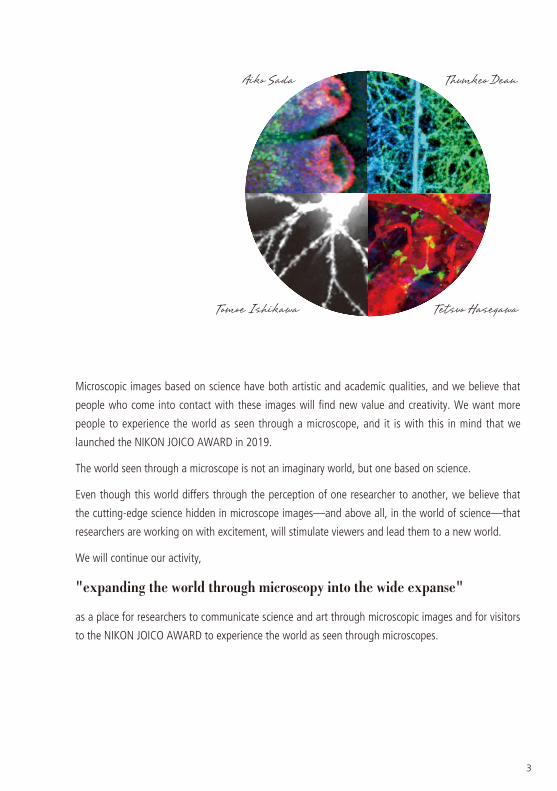
Microscopic images based on science have both artistic and academic qualities, and we believe that
people who come into contact with these images will find new value and creativity. We want more
people to experience the world as seen through a microscope, and it is with this in mind that we
launched the NIKON JOICO AWARD in 2019.
The world seen through a microscope is not an imaginary world, but one based on science.
Even though this world differs through the perception of one researcher to another, we believe that
the cutting-edge science hidden in microscope images—and above all, in the world of science—that
researchers are working on with excitement, will stimulate viewers and lead them to a new world.
We will continue our activity,
"expanding the world through microscopy into the wide expanse"
as a place for researchers to communicate science and art through microscopic images and for visitors
to the NIKON JOICO AWARD to experience the world as seen through microscopes.
Aiko Sada Thumkeo Dean
Tomoe Ishikawa Tetsuo Hasegawa
3
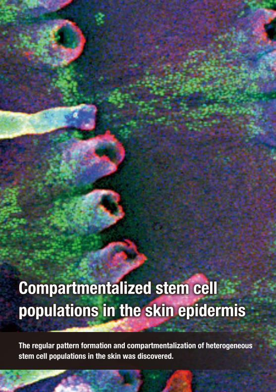
Compartmentalized stem cell populations in the skin epidermis
The regular pattern formation and compartmentalization of heterogeneous stem cell populations in the skin was discovered.

Comments from the award recipient:
Thank you very much for choosing me for the first prize JOICO Award of the NIKON JOICO AWARD.
One of the most important things I do in my research is to look carefully under the microscope. I still remember a "wow" moment when I saw this picture for the first time. A "small notice" based on microscopic observation led to an unexpected discovery through imagination and patiently repeated experiments.
I would like to further explore these beautiful and mysterious skin stem cells.
Mouse skinDetailed description : A tail skin derived from the H2B-GFP tet-off mouse
Green: H2B-GFP (cells with a low division frequency)Red: K14 (an epidermal cell marker)Blue: Stained nuclei
Observation method : Confocal microscopy, inverted, fluorescence
Magnification : 10x
Year : 2014
Microscopic data : Z-stack image
Compartmentalized stem cell populations in the skin epidermis
The regular pattern formation and compartmentalization of heterogeneous stem cell populations in the skin was discovered.
F i rst Pr ize JOICO Award
Aiko SadaAssociated Professor Laboratory of Skin Regeneration and Aging International Research Center for Medical Sciences, Kumamoto University
PaperSada A, Jacob F, Leung E, Wang S, White BS, Shalloway D and Tumbar T
Defining the cellular lineage hierarchy in the interfollicular epidermis of adult skin.
Nature Cell Biology. 2016, 18(6), doi: 10.1038/ncb3359
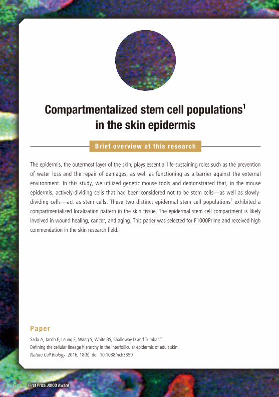
Compartmentalized stem cell populations1 in the skin epidermis
The epidermis, the outermost layer of the skin, plays essential life-sustaining roles such as the prevention
of water loss and the repair of damages, as well as functioning as a barrier against the external
environment. In this study, we utilized genetic mouse tools and demonstrated that, in the mouse
epidermis, actively-dividing cells that had been considered not to be stem cells—as well as slowly-
dividing cells—act as stem cells. These two distinct epidermal stem cell populations2 exhibited a
compartmentalized localization pattern in the skin tissue. The epidermal stem cell compartment is likely
involved in wound healing, cancer, and aging. This paper was selected for F1000Prime and received high
commendation in the skin research field.
Brief overview of th is research
6 First Prize JOICO Award
IntroductionTissue stem cells are special types of cells capable of generating mature, differentiating cells while they
maintaining themselves by self-renewal for the entire lifetime. They have attracted attention as an
excellent cell source for regenerative medicine owing to their high self-renewal capacity. Abnormity of
tissue stem cells leads to the impairment of functions of organs and tissues, which may cause disease
development, tumorigenesis and aging. Thus, accurate identification of tissue stem cells to understand
their properties and control mechanisms is an important first step toward the application of them for
regenerative medicine and the treatment of cancers and aging.
Here, we would like to introduce our research relating to microscope images for this award, focusing on
the mystery of stem cells. We successfully visualized the dynamics of stem cell divisions within skin tissues.
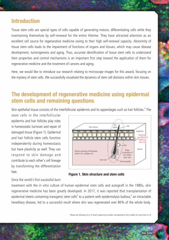
The development of regenerative medicine using epidermal stem cells and remaining questionsSkin epithelial tissue consists of the interfollicular epidermis and its appendages such as hair follicles.3 The
stem cells in the interfol l icular
epidermis and hair follicles play roles
in homeostatic turnover and repair of
damaged tissue (Figure 1). Epidermal
and hair follicle stem cells function
independently during homeostasis
but have plasticity as well: They can
r e spond to s k i n damage and
contribute to each other's cell lineage
by transforming the differentiation
fate.
Since the world's first successful burn
treatment with the in vitro culture of human epidermal stem cells and autograft in the 1980s, skin
regenerative medicine has been greatly developed. In 2017, it was reported that transplantation of
epidermal sheets containing transgenic stem cells5 to a patient with epidermolysis bullosa,4 an intractable
hereditary disease, led to a successful result where skin was regenerated over 80% of the whole body.
Please see Glossary on p.10 (each superscript number corresponds to the number for each term in it)
Figure 1. Skin structure and stem cells
Skin surface
Epidermis
Dermis
Hair follicle
Hair
Hair
Bulge Localization of
hair follicle stem cells
Basal layer Localization of
epidermal stem cells
Dermal papilla
Mainly consisting of fibroblasts Elastin-rich, collagen-rich
Hair root
7Aiko Sada
However, the already proved clinical usability so far is only for the regeneration of epidermis, the most
superficial layer of the tissue, and it is still technically difficult to completely regenerate complicated skin
structure including connective tissue.6 In addition, the location where epidermal stem cells
localize in the skin tissue of living bodies and how they behave remained unclear. It is vital
to understand the basic characteristics of epidermal stem cells in order to achieve stable and succsessful
regeneration of skin.
The discovery of compartmentalization of epidermal stem cells with distinct division frequenciesIt had been considered that tissue stem cells prevent aging and tumorigenesis through the suppression of
the division frequency to minimize the effects of DNA's damage and telomere7 shortening. On the other
hand, actively dividing cells had been regarded as progenitor cells8 without the stem cell ability.
In this study, Sada et al. reported that slowly- and actively-dividing cells function as two distinct,
independent stem cell populations in the mouse interfollicular epidermis (Nat Cell Biol, 2016). In the H2B-
GFP tet-off system11 that visualizes
c e l l s w i t h v a r i o u s d i v i s i o n
frequencies, the transcription of the
histone H2B-GFP takes place under
the control of the epidermal cell-
specific K5 promotor in the absence
of doxycycline,12 resulting in the
accumulation of GFP proteins in the
c e l l s . W h e n d o x y c y c l i n e i s
a d m i n i s t e r e d t o m i c e , t h e
transcription is suppressed and the
a m o u n t o f t h e G F P p r o t e i n s
decreases by half every cell division.
Thus, actively dividing cells gradually
l o s e t h e G F P s i g n a l , w h i l e
infrequently dividing ones retain the
high-level GFP signal for a prolonged
period and are detected as label-Figure 2. A system for visualizing epidermal cells with
distinct division frequencies
In the mouse epidermis, infrequently dividing cells (green) and frequently dividing ones (black) form regularly localized compartments in spatially distinct skin territories.
All epithelial cells are fluorescently labelled with the histone H2B-GFP.
Doxycycline (–)
Doxycycline (–)
Doxycycline (+)
Doxycycline (+)
Cell populations with distinct division frequencies can be identified.
B A localization pattern in mice epidermis
A The principle of H2B-GFP Tet-off
GFP proteins decrease by one half in every
cell division.
Tracing
GFP decreases by half every
division.
Tracing
8 First Prize JOICO Award
retaining cells (LRC) (Figure 2). Interestingly, they found that the two distinct epidermal stem cells with
distinct division frequencies exhibited a regularly localized, compartmentalized pattern in the skin tissue.
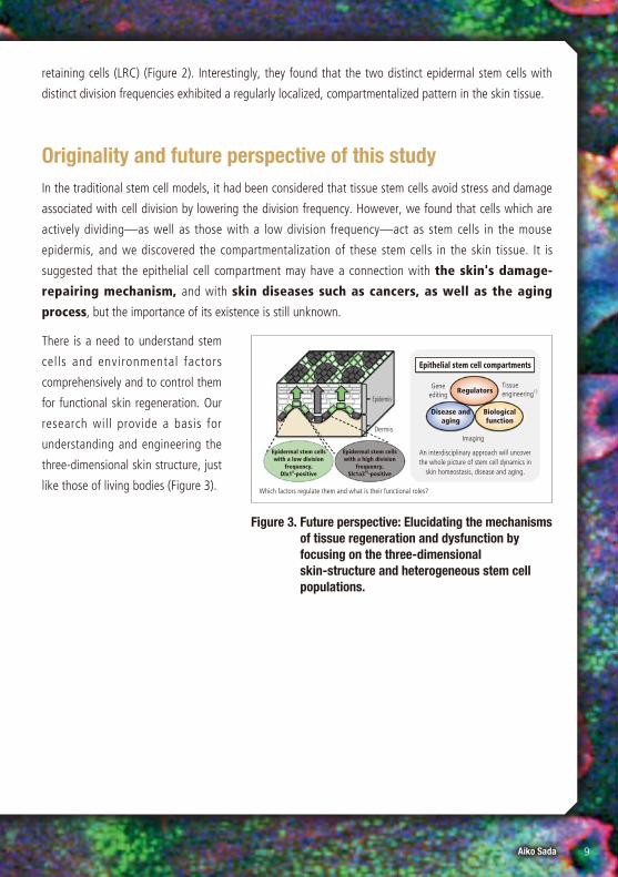
Originality and future perspective of this studyIn the traditional stem cell models, it had been considered that tissue stem cells avoid stress and damage
associated with cell division by lowering the division frequency. However, we found that cells which are
actively dividing—as well as those with a low division frequency—act as stem cells in the mouse
epidermis, and we discovered the compartmentalization of these stem cells in the skin tissue. It is
suggested that the epithelial cell compartment may have a connection with the skin's damage-
repairing mechanism, and with skin diseases such as cancers, as well as the aging
process, but the importance of its existence is still unknown.
There is a need to understand stem
cells and environmental factors
comprehensively and to control them
for functional skin regeneration. Our
research will provide a basis for
understanding and engineering the
three-dimensional skin structure, just
like those of living bodies (Figure 3).
Figure 3. Future perspective: Elucidating the mechanisms of tissue regeneration and dysfunction by focusing on the three-dimensional skin-structure and heterogeneous stem cell populations.
Epithelial stem cell compartments
Tissue engineering13Regulators
Disease and aging
Biological function
Imaging
Epidermis
Dermis
An interdisciplinary approach will uncover the whole picture of stem cell dynamics in
skin homeostasis, disease and aging.
Which factors regulate them and what is their functional roles?
Epidermal stem cells with a low division
frequency, Dlx19-positive
Epidermal stem cells with a high division
frequency, Slc1a310-positive
Gene editing
9Aiko SadaFirst Prize JOICO Award
1. Epithelial stem cellEpithelium is a layer of cells covering the surfaces of the body or
tissue. Skin consists of epidermis (epithelium tissue) and dermis
(connective tissue). Intestinal epithelium, corneal epithelium, and
oral mucosal epithelium are all categorized as epitheliumal tissues.
Stem cells existing in the epithelium tissue are collectively called
epithelial stem cells.
2. Epidermal stem cellStem cells which generate skin epidermis. They play roles in
epidermal turnover and damage-repairing. Epidermal stem cells
localize at the basal layer and move to the upper layers with
differentiation.
3. Hair follicleA hair-surrounding organ. A hair follicle has a hair, an inner root
sheath, a companion layer, and an outer root sheath structured in
layers from its center outwards. Hair follicle stem cells (hair-
generating stem cells) localize at the region called the bulge in the
outer root sheath.
4. Epidermolysis bullosaA skin disease in which epidermis and dermis dissociate. It
generates blisters and erosions due to weak irritation. It is caused
by genes defective in hemidesmosome-related proteins, which
function for adhesion between epidermis and dermis.
5. Transgenic stem cellIt was reported by M De Luca et al. in Nature in 2017 that the
treatment of epidermolysis bullosa was successful through the
transplantation of cultured epidermal stem cells into which laminin
genes were introduced by using genome-editing technique.
6. Connective tissueA supporting tissue infilling the inter-tissue region. For example,
skin dermis is a connective tissue, consisting of fibroblasts and an
extracellular matrix such as collagen and elastin.
7. TelomereA structure located at the end of chromosomes. It is known that its
length shortens with cell divisions. The length of telomeres is
considered important for the control of aging and tumorigenesis.
8. Progenitor cellIt is a relatively undifferentiated cell which is generated from stem
cells but does not have the self-renewal ability like them.
Progenitor cells divide only a limited number of times and are
comitted to terminal differentiation.
9. Dlx1A gene identified by the microarray analysis as a marker of
epidermal stem cells with a low division frequency. It belongs to
the Dlx family of transcriptional factor.
10. Slc1a3A gene identified by the microarray analysis as a marker of
epidermal stem cells with a high division frequency. It functions as
a glutamate transporter and has important functions for the
metabolic control of stem cells and cancer cells.
11. H2B-GFP tet-off systemTransgenic mice in which cell division frequencies can be visualized.
Cell nuclei are stably labeled by the green fluorescent proteins
(GFP) fused with histone H2B. The number of cell divisions taking
place during the suppression of transcription by doxycycline can be
detected as the GFP fluorescence intensity.
12. DoxycyclineA derivative of tetracycline and an agent to terminate the
transcription in the tet-off system. It prevents a tetracycline-
dependent transcription activation factor tTA from the binding to
the promotor sequence TRE (tetracycline responsive element).
13. Regenerative engineeringA collaborative field of medicine and engineering aiming at
regeneration of tissue damage using biomaterials such as cells and
the extracellular matrix. It attracts attention as the next generation
medicine.
G l o s s a r y
10 First Prize JOICO Award

Q1 Why did you focus on the difference in the cell division frequency?It had been known that cells with low division frequencies localize at the bulge region and behave as stem cells functioning for a long period
in hair follicles in the skin. In addition, it had been suggested that cells with low division frequencies act as cells with special functions in other
tissues. However, in the skin epidermis, there were many unclear points about the true nature of the tissue stem cells, and there were very few
studies that focused on the difference in division frequency, so I thought that something interesting might be found.
Q2 What happens to the balance and compartmentalization of two types of epithelial stem cells with differing division frequencies upon skin damage repair and skin diseases such as cancers?
That is exactly our current research target. It is known that the two types of epidermal stem cells have potential to contribute to each other's
cell lineage upon skin damage. Epidermal stem cells actively divide in the cases of cancers and skin inflammation, but the cell division ability
decreases with aging. I think that change of the balance and compartments of the two distinct stem cells in various situations will lead to the
understanding of the biological significance of the existence of two types of stem cell populations in the skin epidermis.
Q3 Are there any known mechanisms controlling the compartmentalization of epithelial stem cells?
It has been reported that signals from the dermis are one of the important regulators for the compartmentalization of epidermal stem cells. For
example, the size of compartments changes depending on the Wnt signal in the dermis. Moreover, autonomous factors of epidermal stem
cells, blood vessel patterns, the extracellular matrix, and the mechanical environment are considered to function for controlling the stem cell
compartments and function, but detailed mechanisms are still unknown.
Q & A
From award panel members
An amazing depiction of creepy structures that seem to be
crawling out of the ground. It is of great academic value.
The concept of visualizing the division frequency of stem
cells by the fluorescence intensity of GFP deserves
particular mention. The regular pattern of localization and
compartmentalization of two types of stem cells with
different division frequencies is not only scientifically
valuable but also beautiful and artistic.
It is a groundbreaking research that defies the existing
theories and reveals the compartment of epithelial stem
cells. The work is fantastic and evokes a sense of life.
In addition to being beautiful, it is also valuable from an
academic point of view.
From an academic point of view, the use of GFP-labeled
probes for histone proteins, which stably bind to
chromatin, to elucidate the dynamics of stem cells in the
skin—a fact that was previously unknown—is highly
commendable.
11Aiko Sada
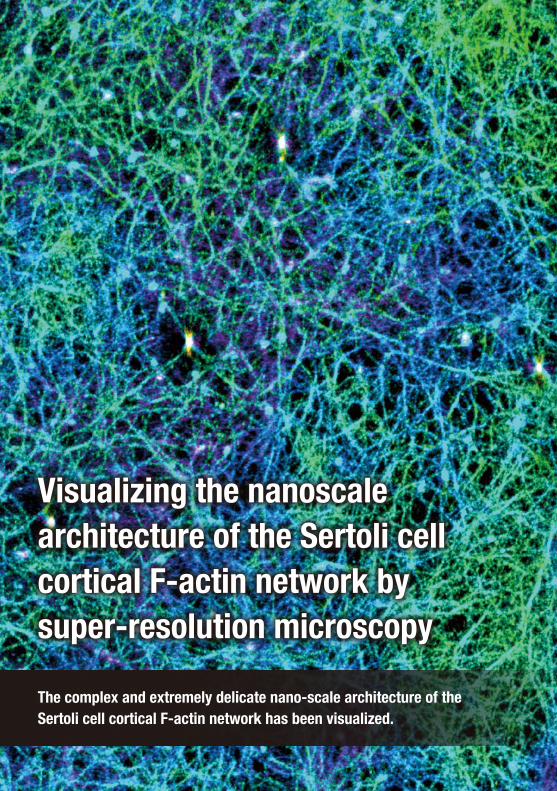
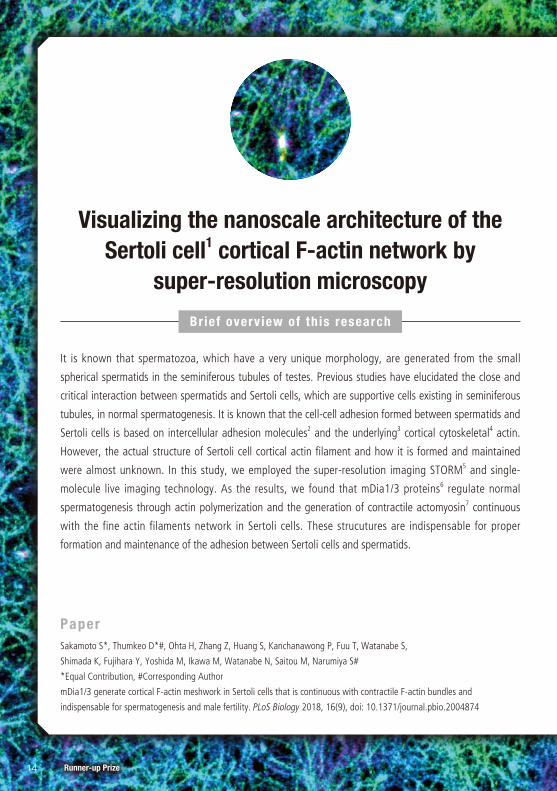
Visualizing the nanoscale architecture of the Sertoli cell cortical F-actin network by super-resolution microscopy
The complex and extremely delicate nano-scale architecture of the Sertoli cell cortical F-actin network has been visualized.
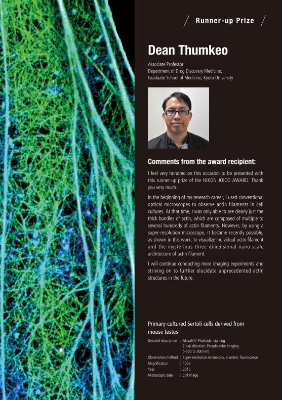
Primary-cultured Sertoli cells derived from mouse testesDetailed description : Alexa647-Phalloidin staining
Z-axis direction: Pseudo-color imaging (–300 to 300 nm)
Observation method : Super-resolution microscopy, inverted, fluorescence
Magnification : 100x
Year : 2015
Microscopic data : Still image
Comments from the award recipient:
I feel very honored on this occasion to be presented with this runner-up prize of the NIKON JOICO AWARD. Thank you very much.
In the beginning of my research career, I used conventional optical microscopes to observe actin filaments in cell cultures. At that time, I was only able to see clearly just the thick bundles of actin, which are composed of multiple to several hundreds of actin filaments. However, by using a super-resolution microscope, it became recently possible, as shown in this work, to visualize individual actin filament and the mysterious three dimensional nano-scale architecture of actin filament.
I will continue conducting more imaging experiments and striving on to further elucidate unprecedented actin structures in the future.
Visualizing the nanoscale architecture of the Sertoli cell cortical F-actin network by super-resolution microscopy
The complex and extremely delicate nano-scale architecture of the Sertoli cell cortical F-actin network has been visualized.
Runner-up Pr ize
Dean ThumkeoAssociate Professor Department of Drug Discovery Medicine, Graduate School of Medicine, Kyoto University
PaperSakamoto S*, Thumkeo D*#, Ohta H, Zhang Z, Huang S, Kanchanawong P, Fuu T, Watanabe S,
Shimada K, Fujihara Y, Yoshida M, Ikawa M, Watanabe N, Saitou M, Narumiya S#
*Equal Contribution, #Corresponding Author
mDia1/3 generate cortical F-actin meshwork in Sertoli cells that is continuous with contractile F-actin bundles and
indispensable for spermatogenesis and male fertility. PLoS Biology 2018, 16(9), doi: 10.1371/journal.pbio.2004874
Visualizing the nanoscale architecture of the Sertoli cell1 cortical F-actin network by
super-resolution microscopy
It is known that spermatozoa, which have a very unique morphology, are generated from the small
spherical spermatids in the seminiferous tubules of testes. Previous studies have elucidated the close and
critical interaction between spermatids and Sertoli cells, which are supportive cells existing in seminiferous
tubules, in normal spermatogenesis. It is known that the cell-cell adhesion formed between spermatids and
Sertoli cells is based on intercellular adhesion molecules2 and the underlying3 cortical cytoskeletal4 actin.
However, the actual structure of Sertoli cell cortical actin filament and how it is formed and maintained
were almost unknown. In this study, we employed the super-resolution imaging STORM5 and single-
molecule live imaging technology. As the results, we found that mDia1/3 proteins6 regulate normal
spermatogenesis through actin polymerization and the generation of contractile actomyosin7 continuous
with the fine actin filaments network in Sertoli cells. These strucutures are indispensable for proper
formation and maintenance of the adhesion between Sertoli cells and spermatids.
Brief overview of th is research
14 Runner-up Prize
Please see Glossary on p.18 (each superscript number corresponds to the number for each term in it)
It is known that spermatozoa, which have a unique shape, are produced from small, spherical spermatids
in the seminiferous tubules of testes.It was previously shown that close interaction between spermatids
and Sertoli cells, which are supportive cells existing in seminiferous tubules, is critical for normal
spermatogenesis. The cell-cell adhesion formed between spermatids and Sertoli cells is based on
intercellular adhesion molecules and the underlying cortical cytoskeletal actin. However, the details of the
structure of the underlying cortical actin on the Sertoli cells side and how this structure is formed and
maintained were largely unknown. Moreover, the physiological importance of such cortical actin was also
unclear.
In this study, We've found that the mDia1/3 double knockout male mice were infertile. Further
observation of spermatozoa and seminiferous tubules revealed that impaired spermatogenesis was the
cuase of male infertility. mDia1 and mDia3 expression were both found in Sertoli cells,suggesting that the
cause of male infertility of the mDia1/3 double knockout mice is due to abnormalities in Sertoli cells, but
not spermatids. Subsequently, in order to clarify the functions of mDia1/3 in Sertoli cells, filamentous
actin in primary-cultured Sertoli cells was observed using a super-resolution microscope based on
stochastic optical reconstruction microscopy (STORM) at the XY resolution of ~20 nm and the Z resolution
of ~50 nm. This revealed that filamentous actin form a meshwork structure with a mesh size of ~ 100 nm
that exists beneath the Sertoli cell membrane and the density of this actin meshwork structure was
significantly reduced in the double knockout of mDia1/3. Furthermore, to observe the dynamics of the
actin meshwork, high-speed and high-resolution live imaging with spinning disk confocal microscopy for
fluorescently labeled filamentous actin in primary-cultured Sertoli cells was conducted. As a result, two
components were found to exist in the meshwork and they polymerized at the different rate (0.4 μm/s
and 1.3 μm/s on average). Moreover, filamentous actin with the faster polymerization rate (1.3 μm/s)
specifically impaired in the mDia1/3 double knockout cells. The higher polymerization rate was similar to
the rate of actin polymerization8 by mDia3, calculated from the single-molecule imaging for mDia3. These
findings together indicate that mDia1/3 are involved in the formation and maintenance of meshwork
actin structure.
15Dean Thumkeo
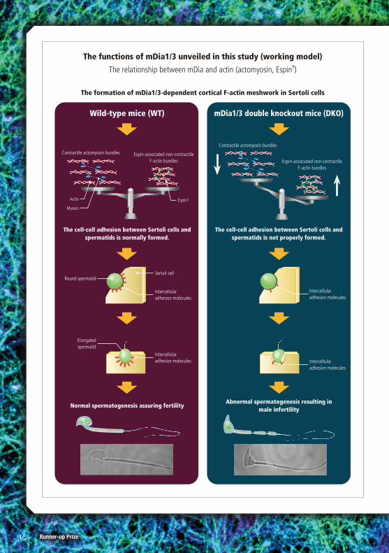
The functions of mDia1/3 unveiled in this study (working model)
The relationship between mDia and actin (actomyosin, Espin9)
The formation of mDia1/3-dependent cortical F-actin meshwork in Sertoli cells
mDia1/3 double knockout mice (DKO)
The cell-cell adhesion between Sertoli cells and spermatids is not properly formed.
Abnormal spermatogenesis resulting in male infertility
Wild-type mice (WT)
Contractile actomyosin bundlesContractile actomyosin bundles
Espin-associated non-contractile F-actin bundles Espin-associated non-contractile
F-actin bundles
The cell-cell adhesion between Sertoli cells and spermatids is normally formed.
Normal spermatogenesis assuring fertility
Actin
Myosin
Espin1
Round spermatid
Elongated spermatid
Sertoli cell
Intercellular adhesion molecules Intercellular
adhesion molecules
Intercellular adhesion molecules
Intercellular adhesion molecules
16 Runner-up Prize
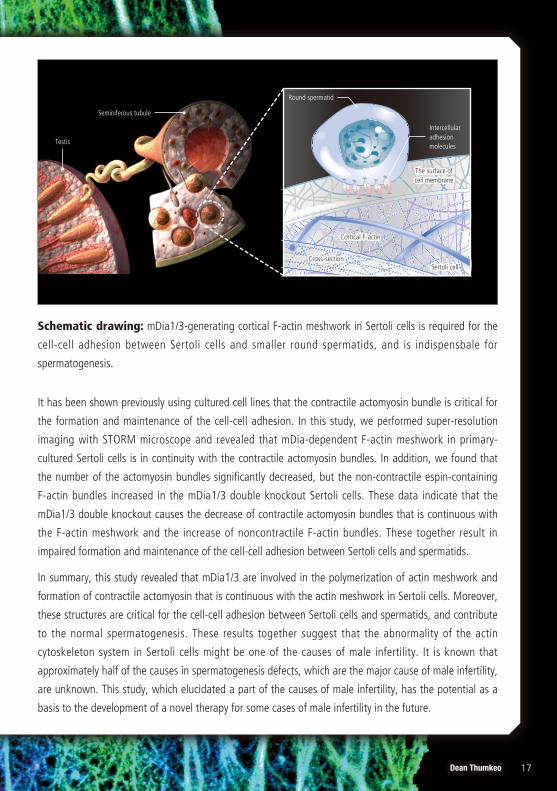
It has been shown previously using cultured cell lines that the contractile actomyosin bundle is critical for
the formation and maintenance of the cell-cell adhesion. In this study, we performed super-resolution
imaging with STORM microscope and revealed that mDia-dependent F-actin meshwork in primary-
cultured Sertoli cells is in continuity with the contractile actomyosin bundles. In addition, we found that
the number of the actomyosin bundles significantly decreased, but the non-contractile espin-containing
F-actin bundles increased in the mDia1/3 double knockout Sertoli cells. These data indicate that the
mDia1/3 double knockout causes the decrease of contractile actomyosin bundles that is continuous with
the F-actin meshwork and the increase of noncontractile F-actin bundles. These together result in
impaired formation and maintenance of the cell-cell adhesion between Sertoli cells and spermatids.
In summary, this study revealed that mDia1/3 are involved in the polymerization of actin meshwork and
formation of contractile actomyosin that is continuous with the actin meshwork in Sertoli cells. Moreover,
these structures are critical for the cell-cell adhesion between Sertoli cells and spermatids, and contribute
to the normal spermatogenesis. These results together suggest that the abnormality of the actin
cytoskeleton system in Sertoli cells might be one of the causes of male infertility. It is known that
approximately half of the causes in spermatogenesis defects, which are the major cause of male infertility,
are unknown. This study, which elucidated a part of the causes of male infertility, has the potential as a
basis to the development of a novel therapy for some cases of male infertility in the future.
Schematic drawing: mDia1/3-generating cortical F-actin meshwork in Sertoli cells is required for the
cell-cell adhesion between Sertoli cells and smaller round spermatids, and is indispensbale for
spermatogenesis.
Testis
Seminiferous tubule
Round spermatid
Intercellular adhesion molecules
The surface of cell membrane
Sertoli cellCross-section
Cortical F-actin
17Dean ThumkeoRunner-up Prize
1. Sertoli cellA large somatic cell that interacts with spermatids in the
seminiferous tubule to support the spermatogenesis. It is known
that Sertoli cell supplies nutrients to spermatids and forms the
blood-testis barrier. These 2 functions have been long considered
as its major functions, but it is likely that other unknown functions
are also exist.
2. Intercellular adhesion moleculeMolecules that physically connect two different cells. Claudin,
cadherin, and nectin are known as representative molecules.
3. Underlying (cortical structure)A structure consisting of thick actomyosin bundles and thin F-actin,
which regulates the adhesion strength of intercellular adhesion
molecules and is considered to connect with intercellular adhesion
molecules through catenin.
4. CytoskeletonThere are three kinds of cytoskeletons, that is, microtubules,
intermediate filaments, and filamentous actin. They are involved in
various cell functions such as cell migration and division as well as
physically supporting the cell morphology.
5. STORMAcronym for stochastic optical reconstruction microscopy. The
process utilizes the photoswitchable property of fluorophores and
activates only a portion of them to a fluorescent state to precisely
determine the positional information of a particular molecule on
the nm scale. By repeating this, eventually the positional
information of almost all molecules is obtained, and a high-
resolution image is constructed based on the information and
calculation. Generally, it is said that an approximately 10-times-
higher XY resolution image can be obtained with STORM than with
conventional optical microscopes.
6. mDia1/3 proteinsBelonging to the formin family of proteins, mDia is a protein with
actin polymerization activity. There are three isoforms in mammals,
mDia1, mDia2, and mDia3. Previous studies have shown that
mDia2 is related to the actin polymerization during cell division. On
the other hand, the functions of mDia1 and mDia3 overlap, and it
has been shown that they redundantly function in some cases such
as cell migartion and intercellular adhesion regulation, but not for
the actin polymerization during cell division.
7. ActomyosinOne of the actin bundle structures formed by the connection of
myosin, a molecular motor, to filamentous actin. Hydrolysis of ATP
by myosin causes the generation of the contractile force.
8. Actin polymerization rateIf we add actin monomers with appropriate amounts of ATP and
MgCl2 into a test tube, actin will spontaneously polymerize at a
certain polymerization rate to form a filamentous structure. In the
presence of profilin, one of the actin monomer-binding proteins,
the polymerization rate drastically increases with the addition of
proteins of the formin family such as mDia1.
9. EspinOne of the actin-binding proteins with the filamentous actin-
bundling activity. Unlike actomyosin, the F-actin bundled by espin
is not contractile.
Memo
G l o s s a r y
18 Runner-up Prize
Q & A
In efforts to elucidate the functions of mDia, an actin
polymerization regulator in the testis, the results of the
localization analysis of actin filaments in Sertoli cells using
localization microscopy are of high academic value. The
quality of the images obtained is also extremely high.
The image is very beautiful.
This is an impactful study that fully utilizes the potential of
super-resolution microscopy. It gives us a sense of the
fantastic nano-world.
The fine and beautiful structure of the actin cytoskeleton is
beautifully captured using super-resolution microscopy.
The green and blue colors and the fine linear expression
are beautiful, and the expression of delicacy is inspiring.
The delicacy and dynamism of the structure are captured
in a well-balanced manner.
The beauty of the nano-structure of the actin cytoskeleton
conveys the inspiration of the entrant.
The overlapping of the green and blue lines reminiscent of
the Sea of Trees is beautiful. A unique color tone that is
neither blue nor green has been created.
Q1 Do the mDia1/3 proteins investigated in this study also have important functions for spermatogonia and spermatocytes, precursors of spermatids?
In this study, we clarified that the functions of mDia1/3 in Sertoli cells are indispensable for differentiation of round spermatids to elongated
spermatids and spermatozoa. But in addition, we also found that they are involved in the blood-testis barrier that is formed due to the
intercellular adhesion between Sertoli cells. The blood-testis barrier is considered to be involved in the maintenance of the micro-environment
appropriate for spermatogonia and spermatocytes. Thus, it is possible that mDia1/3 proteins also affect the function of these cells.
Q2 Why does the abnormal spermatogenesis happen due to the differences in actin polymerization rates and F-actin mesh sizes in the case of mDia1/3 double knockout?
The connection of spermatids and Sertoli cells is vital for spermatogenesis; the adhesion force regulators for intercellular adhesion molecules
are intercellular adhesion molecule-underlying actomyosin bundles and the thin actin meshwork that is continuous with the actomyosin
bundles. These actins repeat polymerization and depolymerization for homeostatic remodeling. However, in the case of mDia1/3 double
knockout, the actin polymerization does not catch up with the depolymerization, resulting in a decrease in total mass of actin filaments, which
subsequently weakens cortical actin and intercellular adhesion , and thus adhesion detachment.
Q3 The declining birthrate in Japan is a current social problem. How do you think the new findings about mDia1/3 obtained in this study will lead to diagnosis of male infertility and therapeutic development?
Almost half of the cuases in spermatogenesis defects, which are the major cause of male infertility, are unknown. This study indicates that
abnormality of the actin cytoskeleton system in Sertoli cells may be one of the causes of male infertility. Therefore, mDia1/3 genetic variation
might be useful for diagnosis. In addition, it was found in this study that at least part of the spermatogenesis defects are caused from the
Sertoli cell side. Thus, we would like to suggest the possibility of therapeutic development for infertility by targeting the Sertoli cells (such as
transgenesis or drug delivery).
From award panel members
19Dean Thumkeo
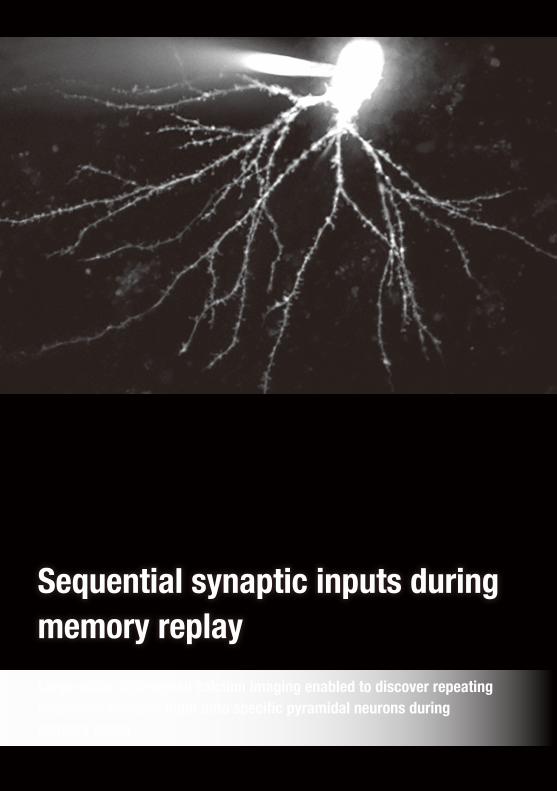
Sequential synaptic inputs during memory replay
Large-scale, high-speed calcium imaging enabled to discover repeating sequential synaptic input onto specific pyramidal neurons during memory replay.
Rat brain (the hippocampal subfield CA1)Detailed description : A fluorescent calcium indicator (Fluo-4)
Observation method : Confocal microscopy, upright, fluorescence
Magnification : 60x
Year : 2019
Microscopic data : Video
Comments from the award recipient:
I am extremely honored to have been chosen to receive this special award.
It is an honor for me to receive the wonderful praise of my work as "sparklers shining in the dark". Calcium activity of synaptic inputs across dendritic arbor is so beutiful that I never tire of seeing them. Day after day, I am amazed to find that seemingly chaotic synaptic inputs are precisely governed to the order of a few micrometers. I will be giving my all to push forward to understanding the rules that govern the the brain function.
It would bring me great joy if this movie enables you to experience a glimpse into the brain's beauty and elaborate nature.
Tomoe IshikawaAssistant Professor Department of Pharmacology, School of Medicine Keio University
Sequential synaptic inputs during memory replay
Large-scale, high-speed calcium imaging enabled to discover repeating sequential synaptic input onto specific pyramidal neurons during memory replay.
Specia l Pr ize

PaperIshikawa, T., Ikegaya, Y.
Locally sequential synaptic reactivation during hippocampal ripples.
Science Advances. 2020, 6(7), doi: 10.1126/sciadv.aay1492
Ishikawa, T., Kobayashi, C., Takahashi, N., Ikegaya, Y.
Functional multiple-spine calcium imaging from brain slices.
STAR Protoc. 2020, doi: 10.1016/j.xpro.2020.10012
Sequential synaptic inputs2 during memory replay1
We discovered that neighboring spines4 are activated serially along dendrites toward or away from cell
bodies during specific brain waves, called sharp-wave ripples3, which are frequently observed during
memory reply. This result suggests that the neuronal firing patterns of upstream neuron5 populations are
converge on the adjacent dendritic spines of a portion of downstream neurons in precisely wired neuronal
circuits. Squential synaptic inputs could be a novel algorithm in the brain6 to explain the generation of a
specific neuronal firing during memory replay.
Brief overview of th is research
22 Special Prize

Background #2 The spatiotemporal pattern of synaptic inputs7 during memory replay (a hypothesis)
Neurons fires an action potential by receiving synaptic inputs from other (upstream) neurons. The neuronal firing patterns depend on the spatiotemporal patterns in addition to the number of number of synaptic inputs. Then, what types of synaptic inputs do CA1 pyramidal neurons receive during SWs in which memory replay is frequently observed?
2. Memory consolidation (when sleeping or resting)
Please see Glossary on p.26 (each superscript number corresponds to the number for each term in it)
Background #1 Two stage model for the long-term memory
To understand the mechanism of long-term storage of a new memory, two stage (1) memory acquisition and 2) memory consolidation) model was proposed. However, how neurons activated during memory acquisition spontaneously fire during memory consolidation, i.e., the mechanism of memory replay had remained unclear.
To understand the mechanism of memory replay in subcellular level, we imaged the spatiotemporal patterns of synaptic inputs during SWs using the Nipkow-type confocal microscopy with the world's fastest scanning speed and largest number of recorded spines.
Our research purpose
Downstream decoder
Downstream decoder
Upstream circuit
Upstream circuit
Synaptic inputs
1. Memory acquisition (when exploring new environments)
Hippocampal neurons
Brain waves
SW
sharp wave ripple
The neuronal firing pattern when exploring a new environment
Cell
#
Cell
#
Time Time
A moving direction of a mouse
Memory replay (during SWs)
Hypothesis #1: Dispersed synaptic input Hypothesis #2: Clustered synaptic input
23Tomoe Ishikawa
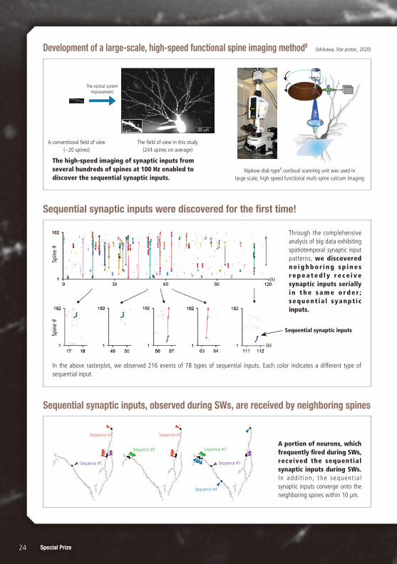
Development of a large-scale, high-speed functional spine imaging method8
The high-speed imaging of synaptic inputs from several hundreds of spines at 100 Hz enabled to discover the sequential synaptic inputs.
(Ishikawa, Star protoc, 2020)
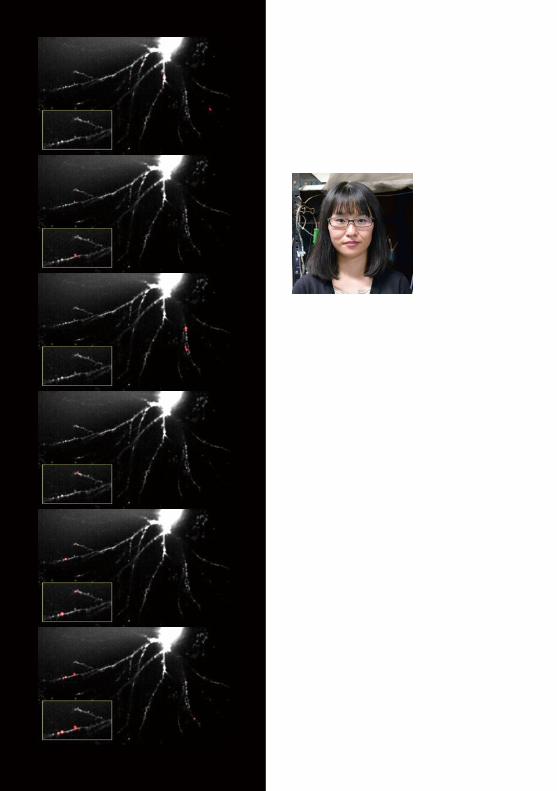
Sequential synaptic inputs, observed during SWs, are received by neighboring spines
A portion of neurons, which frequently fired during SWs, received the sequential synaptic inputs during SWs. I n add i t i on , the sequent ia l synaptic inputs converge onto the neighboring spines within 10 μm.
Sequence #2
Sequence #1 Sequence #1
Sequence #3
Sequence #4
Sequence #3
Sequence #2
Sequential synaptic inputs were discovered for the first time!
Through the complehensive analysis of big data exhibiting spatiotemporal synaptic input patterns, we discovered n e i g h b o r i n g s p i n e s r e p e a t e d l y r e c e i v e synaptic inputs serially i n t h e s a m e o r d e r ; sequent ia l syanpt ic inputs.
In the above rasterplot, we observed 216 events of 78 types of sequential inputs. Each color indicates a different type of sequential input.
Sequential synaptic inputs
Spin
e #
Spin
e #
The optical system improvement
A conventional field of view (−20 spines)
The field of view in this study (244 spines on average)
Nipkow disk-type9 confocal scanning unit was used in large-scale, high-speed functional multi-spine calcium imaging
24 Special Prize
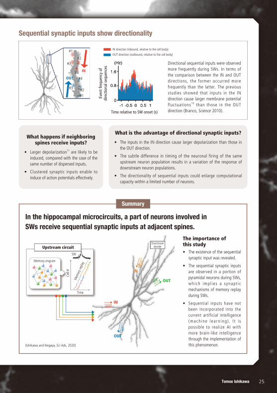
Sequential synaptic inputs show directionality
Directional sequential inputs were observed more frequently during SWs. In terms of the comparison between the IN and OUT directions, the former occurred more frequently than the latter. The previous studies showed that inputs in the IN direction cause larger membrane potential fluctuations10 than those in the OUT direction (Branco, Science 2010).
What happens if neighboring spines receive inputs?
• Larger depolarization11 are likely to be induced, compared with the case of the same number of dispersed inputs.
• Clustered synaptic inputs enable to induce of action potentials effectively.
What is the advantage of directional synaptic inputs?
• The inputs in the IN direction cause larger depolarization than those in the OUT direction.
• The subtle difference in timing of the neuronal firing of the same upstream neuron population results in a variation of the response of downstream neuron populations.
• The directionality of sequential inputs could enlarge computational capacity within a limited number of neurons.
Summary
Even
t fre
quen
cy o
f di
rect
iona
l seq
uenc
es
Time relative to SW onset (s)
IN direction (inbound, relative to the cell body)
OUT direction (outbound, relative to the cell body)
IN
OUT
#1#2#3
#4#4
#2
#1
#3
The importance of this study• The existence of the sequential
synaptic input was revealed.
• The sequential synaptic inputs are observed in a portion of pyramidal neurons during SWs, which impl ies a synapt i c mechanisms of memory replay during SWs.
• Sequential inputs have not been incorporated into the current artificial intelligence (mach ine l ea rn ing ) . I t i s possible to realize AI with more brain-like intelligence through the implementation of this phenomenon.
In the hippocampal microcircuits, a part of neurons involved in SWs receive sequential synaptic inputs at adjacent spines.
Upstream circuitDownstream
decoder
Memory engram
Time
SW
Cell
#
Mem
ory r
epla
y
IN
IN
OUT
OUT
(Ishikawa and Ikegaya, Sci Adv, 2020)
25Tomoe Ishikawa
1. Memory replayThe firing patterns observed during memory formation are likely to
be repeated during sleep and resting state, which we call memory
replay. Memory replay and sharp wave ripples3 are considered to
play an important role in the long-term memory preservation.
2. Sequential synaptic inputSpecific spine assemblies repeatedly receive synaptic inputs serially
in the same order. Our results suggest that memory replays of
multineuronal spikes are distributed across dendritic spines of a
postsynaptic neuron, with their spatiotemporal features preserved
(sequential synaptic inputs).
3. Sharp wave (SW) rippleOscillatory patterns observed during sleeping or resting. It consists
of the combination of SWs (2–30 Hz) and ripples (125–250 Hz). It
is closely related to memory replay and considered to have
important roles in long-term memory preservation.
4. Dendritic spineA small membrane structure present on dendrites where majority of
excitatory synaptic inputs are received. When neurotransmitters are
emitted from the presynaptic region, they connect to the receptors
on spines, leading to the inflow of sodium and calcium ions, which
contributes to the downstream neuronal firing.
5. Upstream and downstream neuronsNeurons are connected with each other to organize neuronal
circuits in the brain. Upstream neurons send information to down
stream neurons in the neuronal circuits.
6. Algorithm in the brainA rule of information processing among neurons. The sequential
synaptic input discovered in this research is considered to activate
cell bodies efficiently. Thus, for example, the incorporation of the
algorithm into a neural network is possible to construct a more
brain-like system.
7. Synaptic inputWhen an upstream neuron fired, neurotransmitters are emitted
from the presynapses. The neurotransmitters bind to the receptors
on the postsynpses of a downstream neuron and induce an ion
influx. This is called the synaptic input. In this study, we observed
calcium ion inflows caused by the excitatory transmission as
fluorescence intensity fluctuations.
8. Large-scale, high-speed spine imaging method
A method to observe spatiotemporal pattern of synaptic inputs at a
high speed and on a large scale through the combination of a
Nipkow disk confocal unit and a CMOS camera. Two hundred or
more spines on average can be imaged at 100 Hz—currently the
world's largest number and fastest speed.
9. Nipkow disk-typeSpinning disk with a spiral of holes. The high-speed rotation of the
disk can divide a laser, enabling simultaneous recording from
multiple points. Nipkow disk-type confocal microscope enables
high-speed, wide-field imaging with low photobleaching.
10. Membrane potential fluctuationIt is a fluctuation of somatic membrane potential caused by
convergence of synaptic inputs from other neurons. A shift towards
excitation is called depolarization and leads to neuronal firing.
11. DepolarizationIn a normal state, a cell membrane is negatively charged. When
exicitatory syanptic inputs are received, the membrane potential
becomes less negative, which is called depolarization. When the
degree of depolarization reaches a certain threshold, action
potential occurs, which transmits an output to further downstream
neurons.
Memo
G l o s s a r y
26 Special Prize

Q1 Could you describe the experimental system in this study?In this study, we used hippocampal slice culture preparations (300 μm thick, days in vitro 10–20). First, cell-attached recording was performed
using an electrode from a CA1 pyramidal cells. After that, negative pressure was applied to achieve whole-cell configuration to load the
fluorescent calcium indicator molecules—which are originally contained in the electrodes—into the cells, enabling the measurement of
calcium influx caused by synaptic inputs. Another electrode was placed to record local field potentials around the target neurons (approxinately
50 μm apart from the target neurons). After recording the synaptic inputs, regions of interest were set manually on the obtained image to
calculate the fluorescence intensity change.
Q2 What is the mechanism of controlling the cell-selective sequential synaptic input discovered in this study?
This is an important question, but we do not have a good answer so far. Although it may be related to how upstream and downstream
neurons connect with each other, many facets are still unclear in terms of the types of cells that form the synapses between CA3-CA1 circuits.
In addition, neurons receive inhibitory inputs (working as a brake in the brain) as well as excitatory inputs (as an accelerator), and thus we
need to consider the effect of inhibitory inputs.
Q3 Do you know anything about the relationship between the findings in this study and diseases such as memory disorders?
Since the sequential synaptic inputs were observed in only a portion of cells, we consider that the existence of the sequential inputs plays an
important role in the formation of cell-selective firing patterns. Thus, if the sequential inputs are selectively inhibited, diseases such as memory
disorders could be induced. But, it is challenging to inhibit specific sequential inputs. In the future, we would like to introduce new
technologies to explore the functions of sequential synaptic inputs.
Q & A
This work is scientifically valuable.
It has succeeded in capturing sequential synaptic inputs
during memory replay at a high spatial and temporal
resolution, and its scientific merit is very significant.
Furthermore, the video capturing this phenomenon is
reminiscent of sparklers in the dark, so it also has artistic
merit.
This is an important discovery which demonstrates
the existence of sequential synaptic inputs in a slice
preparation.
The images, which capture the work of the spine synapses
(where neurons transmit information) in high temporal
resolution, hold great value.
From award panel members
27Tomoe Ishikawa
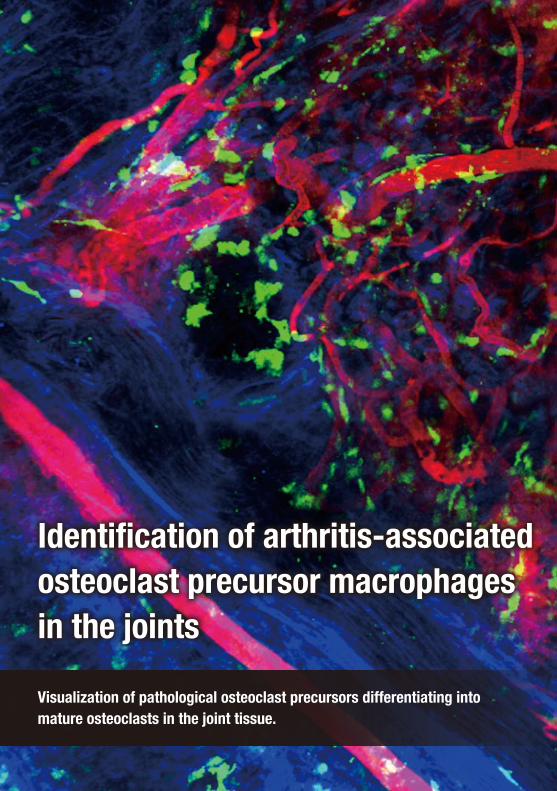
Identification of arthritis-associated osteoclast precursor macrophages in the joints
Visualization of pathological osteoclast precursors differentiating into mature osteoclasts in the joint tissue.
Comments from the award recipient:
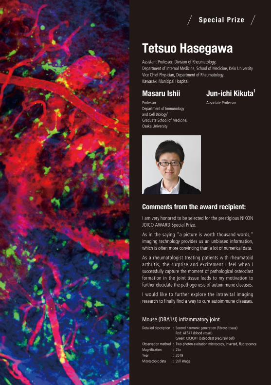
I am very honored to be selected for the prestigious NIKON JOICO AWARD Special Prize.
As in the saying "a picture is worth thousand words," imaging technology provides us an unbiased information, which is often more convincing than a lot of numerical data.
As a rheumatologist treating patients with rheumatoid arthritis, the surprise and excitement I feel when I successfully capture the moment of pathological osteoclast formation in the joint tissue leads to my motivation to further elucidate the pathogenesis of autoimmune diseases.
I would like to further explore the intravital imaging research to finally find a way to cure autoimmune diseases.
Tetsuo HasegawaAssistant Professor, Division of Rheumatology, Department of Internal Medicine, School of Medicine, Keio University Vice Chief Physician, Department of Rheumatology, Kawasaki Municipal Hospital
Masaru IshiiProfessor Department of Immunology and Cell Biology1 Graduate School of Medicine, Osaka University
Jun-ichi Kikuta1
Associate Professor
Identification of arthritis-associated osteoclast precursor macrophages in the joints
Visualization of pathological osteoclast precursors differentiating into mature osteoclasts in the joint tissue.
Mouse (DBA1/J) inflammatory jointDetailed description : Second harmonic generation (fibrous tissue)
Red: AF647 (blood vessel)Green: CX3CR1 (osteoclast precursor cell)
Observation method : Two-photon excitation microscopy, inverted, fluorescence
Magnification : 25x
Year : 2019
Microscopic data : Still image
Specia l Pr ize

Paper1. Tetsuo Hasegawa, Junichi Kikuta, Takao Sudo, Yoshinobu Matsuura, Takahiro Matsui,
Szandor Simmons, Kosuke Ebina, Makoto Hirao, Daisuke Okuzaki, Yuichi Yoshida,
Atsushi Hirao, Vladimir V. Kalinichenko, Kunihiro Yamaoka, Tsutomu Takeuchi, Masaru Ishii
Identification of a novel arthritis-associated osteoclast precursor macrophage regulated by FoxM1.
Nature Immunology. 2019, 20(12), doi: 1038/s41590-019-0526-7
2. Tetsuo Hasegawa, Junichi Kikuta, Takao Sudo, Erika Yamashita, Shigeto Seno,
Tsutomu Takeuchi, Masaru Ishii
Development of an intravital imaging system for the synovial tissue reveals the dynamics of CTLA-4 Ig in vivo.
Scientific Reports. 2020, 10(1), doi: 1038/s41598-020-70488-y
Identification of arthritis-associated osteoclast precursor macrophages in the joints
Osteoclasts1 are multinucleated cells with its unique bone-resorbing capacity. They play key roles in
homeostatic bone remodeling, while they are also involved in pathological bone destruction in rheumatoid
arthritis (RA). This study is aimed to elucidate where and how the pathological osteoclasts are developed
in the joint tissue through single-cell RNA sequencing analysis2 and intravital imaging technologies.
Brief overview of th is research
30 Special Prize
Please see Glossary on p.34 (each superscript number corresponds to the number for each term in it)
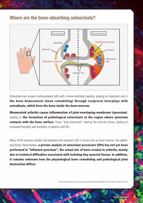
Osteoclasts are unique multinucleated cells with a bone-resorbing capacity, playing an important role in
the bone homeostasis (bone remodeling) through reciprocal interplays with
osteoblasts, which form the bone inside the bone marrow.
Rheumatoid arthritis causes inflammation of joint-enveloping membrane (synovium),
leading to the formation of pathological osteoclasts at the region where synovium
contacts with the bone surface. These "bad osteoclasts" destroy the articular bones, leading to
increased mortality and morbidity in patients with RA.
Many of the previous studies had explored and analyzed cells in tissue such as bone marrow, the spleen,
and blood. Nevertheless, a precise analysis of osteoclast precursors (OPs) has not yet been
performed in “inflamed synovium”, the actual site of bone erosion in arthritis, mainly
due to technical difficulties associated with isolating tiny synovial tissues. In addition,
it remains unknown how the physiological bone remodeling and pahtological joint
destruction differs.
Where are the bone-absorbing osteoclasts?
Normal joints Rheumatoid arthritis
Synovium3
Osteoblasts
Pannus4
Inflammatory cells
Cartilage5 loss
Physiological osteoclasts
Pathological osteoclasts
31Tetsuo HasegawaSpecial Prize
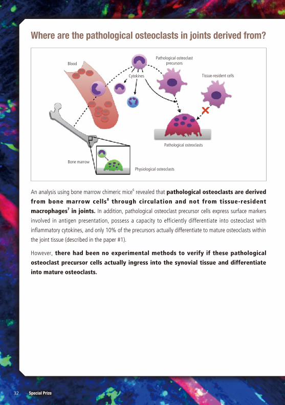
An analysis using bone marrow chimeric mice6 revealed that pathological osteoclasts are derived
from bone marrow cells8 through circulation and not from tissue-resident
macrophages7 in joints. In addition, pathological osteoclast precursor cells express surface markers
involved in antigen presentation, possess a capacity to efficiently differentiate into osteoclast with
inflammatory cytokines, and only 10% of the precursors actually differentiate to mature osteoclasts within
the joint tissue (described in the paper #1).
However, there had been no experimental methods to verify if these pathological
osteoclast precursor cells actually ingress into the synovial tissue and differentiate
into mature osteoclasts.
Pathological osteoclast precursors
Tissue-resident cells
Physiological osteoclasts
Pathological osteoclasts
Cytokines
Blood
Bone marrow
Where are the pathological osteoclasts in joints derived from?
32 Special Prize
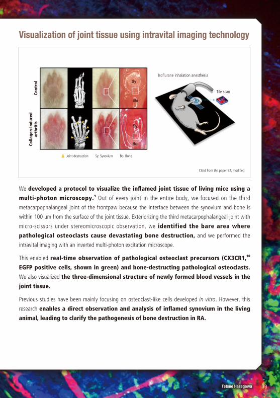
We developed a protocol to visualize the inflamed joint tissue of living mice using a
multi-photon microscopy.9 Out of every joint in the entire body, we focused on the third
metacarpophalangeal joint of the frontpaw because the interface between the synovium and bone is
within 100 μm from the surface of the joint tissue. Exteriorizing the third metacarpophalangeal joint with
micro-scissors under stereomicroscopic observation, we identified the bare area where
pathological osteoclasts cause devastating bone destruction, and we performed the
intravital imaging with an inverted multi-photon excitation microscope.
This enabled real-time observation of pathological osteoclast precursors (CX3CR1,10
EGFP positive cells, shown in green) and bone-destructing pathological osteoclasts.
We also visualized the three-dimensional structure of newly formed blood vessels in the
joint tissue.
Previous studies have been mainly focusing on osteoclast-like cells developed in vitro. However, this
research enables a direct observation and analysis of inflamed synovium in the living
animal, leading to clarify the pathogenesis of bone destruction in RA.
Visualization of joint tissue using intravital imaging technology
Cited from the paper #2, modified
Isoflurane inhalation anesthesia
Tile scan
Colla
gen-
indu
ced
arth
riti
sCo
ntro
l
Joint destruction Sy: Synovium Bo: Bone
33Tetsuo HasegawaSpecial Prize
1. OsteoclastOsteoclasts are multinucleated cell with a unique bone-resorbing
ability. They are derived from monocyte/macrophage-lineage
precursors. They reside inside of bone (bone marrow) and are
involved in the bone remodeling under physiological condition,
while they cause devastating joint destruction in patients with
rheumatoid arthritis.
2. Single-cell gene expression analysisThis technology provides transcriptional profiling of individual cells
and helps to understand what genes are expressed in what
quantities at the single-cell level.
3. SynoviumMembrane existing inside the joint-enveloping articular capsule. It
supports smooth movements of joints through synovial fluid
production. In rheumatoid arthritis, this tissue is significantly
inflamed and enlarged, leading to pathological bone destruction.
4. PannusRheumatoid arthritis causes an inflammatory response in joints that
leads to the formation of abnormal granulomatous tissue called
pannus. This tissue invades the bone and cartilage, leading to
articular bone erosion.
5. CartilageAn elastic tissue covering the articular surface, which functions as a
joint pad.
6. Bone marrow chimeric mouseA mouse whose bone marrow cells are replaced by bone-marrow
transplantation using those from another mouse after radiation and
chemotherapy. It is utilized to evaluate whether specific cells or
pathological conditions are derived from or induced by bone
marrow cells.
7. MacrophageOne type of leukocytes that widely distributes throughout the
whole body. They are involved mainly in phagocytosis, digestion of
dead cells, and invading bacteria in the body. It acquires various
functions depending on the surrounding microenvironment. They
have different names according to the tissue they reside in, such as
Kupffer cells in the liver, microglias in the brain, and osteoclasts in
the bone marrow.
8. Bone marrow cellCells reside within the bone marrow cavity. It includes bloods cells
originating from hematopoietic stem cells, and mesenchymal cells.
9. Multi-photon excitation microscopyTwo-photon microscopy is a fluorescence imaging technique that
allows imaging of living tissue. Unlike traditional fluorescence
microscopy, two-photon excitation requires simultaneous excitation
by two photons with longer wavelength than photons used in
traditional fluorescence microscopy. Two-photon excitation is useful
for intravital imaging due to its deeper tissue penetration, efficient
light detection, and reduced photobleaching.
10. CX3CR1A receptor of a chemokine, CX3CL1. It is utilized as a surface
marker of monocyte/macrophage-lineage cells and of some types of
lymphocytes.
Memo
G l o s s a r y
34 Special Prize

Q1 What determines the capacity to differentiate into mature osteoclasts?Receptor activator of nuclear factor-kappa B ligand (RANKL) and macrophage colony stimulating factor (M-CSF) are the two essential cytokines
involved in osteoclastogenesis. Although pathological osteolast precursor macrophages express receptors for both of the cytokines, synovium-
resident macrophages are reported to not express the receptor for M-CSF. Therefore, the difference of expression pattern for the receptor of
key cytokines involved in osteoclastogenesis may be involved in determining the capacity to differentiate into osteoclasts in the synovium.
Q2 What aspects did you have the hardest time in bio-imaging?Visualization of pathological osteoclasts in the joint tissue has been difficult for several reasons. First, the abundant red blood cells in the
inflamed synovial tissue scatter light and impede the deep tissue imaging. Second, the inflammatory synovium is composed of multiple layers
with various cell types, limiting the observation depth. Third, the skeletal system is directly connected throughout the body and respiratory
movement causes the visual field of the joint tissue to drift. Hence, it was hard to establish a protocol to overcome these obstacles.
Q3 How do you expect to utilize the research outcome of this study in the development of a new therapy of rheumatoid arthritis?
Future applications of novel technologies, including new fluorescent probes and optogenetic techniques, will further reveal the intravital
behaviour and function of immune cells in the synovium, leading to the development of novel therapeutic approaches to rheumatoid arthritis.
Q & A
In relation to the synovial membrane, which is the site of
major lesions in rheumatoid arthritis, the development of a
technique to visualize the dynamics of osteoclast precursors
and the process of synovial tissue reorganization at the
individual level is highly commendable from an academic
perspective.
The image is beautiful with high resolution.
It is powerful, dynamic, and has a strong impact.
The dynamism and the pressure of destruction are
exquisitely expressed, and the excitement of the
researchers can be felt.
It just feels powerful.
The description is beautiful, like green sparks of fire
scattered among the burning red flames. It is also
appropriate for imaging joint inflammation.
From award panel members
35Tetsuo Hasegawa

Masaru IshiiProfessor Department of Immunology and Cell Biology Graduate School of Medicine, Osaka University
Shigeo OkabeProfessor Department of Cellular Neurobiology Graduate School of Medicine and Faculty of Medicine, The University of Tokyo
Keigo KoharaPrincipal Investigator, Assistant Professor Department of Cellular and Functional Biology Institute of Biomedical Science, Kansai Medical University
Tomomi NemotoProfessor Biophotonics Research Group, Exploratory Research Center on Life and Living Systems, National Institutes of Natural Sciences Division of Biophotonics, National Institute for Physiological Science
Hirokazu HiraiProfessor Department of Neurophysiology & Neural Repair, Graduate School of Medicine, Gunma University
Nobuo HashimotoCenter Manager
Kenji BabaGroup Manager Industrial Design Group
Nobuhiko IwamuraManager of Life Imaging Lab
Jun KonnoGroup Manager Experience Design Group
Akiya MaekawaGroup Manager UI & Interaction Design Group
Kumiko SaitoGroup Manager Corporate Branding Group
Design Center, Nikon Corporation
Award panel members
36
First Pr ize JOICO Award
Long-Distance, Rapid Calcium Signaling in Plants
Masatsugu ToyotaAssociate Professor Graduate School of Science & Engineering Saitama University
Runner-up Pr ize
A Secret Inside Flowers
Yoko MizutaDesignated Assistant Professor Institute for Advanced Research/Institute for Transformative Bio-Molecules Nagoya University
Specia l Pr ize
Auditory brainstem circuits detecting interaural time difference
Ryo EgawaDesignated Assistant Professor Laboratory of Cell Physiology, Graduate School of Medicine Nagoya University
Specia l Pr ize
A super-resolution image of the inner mitochondrial membrane developed in starved cells
Masayasu TakiDesignated Associate Professor Institute of Transformative Bio-Molecules Nagoya University
Jun KonnoGroup Manager Experience Design Group
Kumiko SaitoGroup Manager Corporate Branding Group
Award winners in 2019
37

38

Responsible publishing organizer: Sales Strategy Division,
Sales Planning Department,
Biosciences Sales Headquarters,
Nikon Solutions Co., Ltd.
1-6-3 Nishioi, Shinagawa-ku, Tokyo 140-0015, Japan
TEL: +81-3-3773-8138
E-mail: [email protected]
Website: https://www.healthcare.nikon.com/ja/ss/joicoaward/
Website