the ultrastructure of the spermatozoon of the lizard iguana iguana (reptilia, squamata, iguanidae)...
TRANSCRIPT

J. Anat.
(2004)
204
, pp451–464
© Anatomical Society of Great Britain and Ireland 2004
Blackwell Publishing, Ltd.
The ultrastructure of the spermatozoon of the lizard
Iguana iguana
(Reptilia, Squamata, Iguanidae) and the variability of sperm morphology among iguanian lizards
Gustavo H. C. Vieira,
1
Guarino R. Colli
2
and Sônia N. Báo
1
1
Departamento de Biologia Celular, and
2
Departamento de Zoologia, Universidade de Brasília, Brazil
Abstract
The spermatozoon of
Iguana iguana
is filiform and resembles that of other iguanian lizards, being most similar to
Tropidurus
. All sperm synapomorphies of Tetrapoda, Amniota and Squamata are present in the sperm of
Iguana
iguana
. By reconstructing the evolution of 30 sperm characters we identified a novel synapomorphy of Iguania:
the presence of a well-developed acrosomal ridge at the level of the epinuclear lucent zone. Because of the
poor topological resolution among iguanian clades we could not discount the possibility of convergence or neutral
selection as determinant of the variability in characteristics of the sperm cell. In agreement with previous studies,
we identified heterogeneous rates of evolution among the three main regions of the sperm cell, namely the head,
midpiece and tail.
Key words
convergence; evolution; phylogeny; polymorphism; reproduction.
Introduction
Iguanians comprise a diverse array of lizards with
poorly understood phylogenetic relationships (Frost
& Etheridge, 1989; Macey et al. 1997; Schulte et al. 1998;
Frost et al. 2001). Despite the use of different data sets
( i.e. morphological, molecular or mixed data sets), the
results suffer from low resolution and disagreement
among phylogenies derived from each data set. More
recently, Schulte et al. (2003) analysed a combination
of molecular and morphological data and obtained
statistical support for the monophyly of many iguanian
clades, but, like all previous studies, arrived at a lack of
topological resolution among the major lineages. These
results probably stem from character incongruence, as
a result of heterogeneity in evolutionary rates among
different data sets or data sets with different phyloge-
netic histories, sampling error, lack of sufficient infor-
mation among the internal branches of the tree, or lack
of phylogenetic structure in the data (Swofford, 1991;
Miyamoto & Fitch, 1995; Wiens & Hollingsworth, 2000;
Schulte et al. 2003).
The ultrastructure of sperm has been used recently in
phylogenetic analysis of squamates (Jamieson, 1995b,
1999; Jamieson et al. 1996; Oliver et al. 1996; Teixeira
et al. 1999b). These studies demonstrated that sperm
ultrastructure characters contain significant phyloge-
netic signal. However, sperm ultrastructure phylogenies
suffer from limited number of characters and a limited rep-
resentation of taxonomic groups (Teixeira et al. 1999c).
The ultrastructure of sperm has been adequately
described in six families of Iguania: Agamidae (
Pogona
barbata
, Oliver et al. 1996), Chamaleonidae (
Bradypodion
karrooicum
, Jamieson, 1995b); Crotaphytidae (
Crota-
phytus bicinctores
and
Gambelia wislizenii
, Scheltinga
et al. 2001), Phrynosomatidae (
Urosaurus ornatus
and
Uta stansburiana
, Scheltinga et al. 2000), Polychrotidae
(
Anolis carolinensis
and
Polychrus acutirostris
, Teixeira
et al. 1999a; Scheltinga et al. 2001) and Tropiduridae
(
Liolaemus austromendocinus
,
Tropidurus semitaeniatus
and
T. torquatus
, Furieri, 1974; Teixeira et al. 1999d).
Saita et al. (1988) provided the first description of the
ultrastructure of reproductive cells of
Iguana iguana
(misidentified as
I. delicatissima
), but their account was
Correspondence
Gustavo H. C. Vieira, Departamento de Biologia Celular, Universidade de Brasília, DF, CEP 70910–900, Brazil. T: 55 61 307 24 24; F: 55 61 347 65 33; E: [email protected]
Accepted for publication
29 March 2004
The ultrastructure of the spermatozoa of
Iguana iguana
, G. H. C. Vieira et al.
© Anatomical Society of Great Britain and Ireland 2004
452
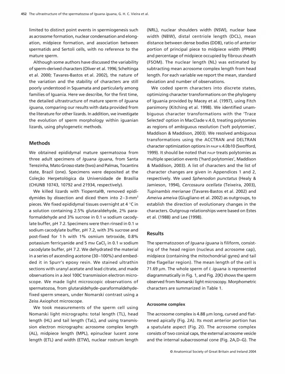
limited to distinct point events in spermiogenesis such
as acrosome formation, nuclear condensation and elong-
ation, midpiece formation, and association between
spermatids and Sertoli cells, with no reference to the
mature sperm.
Although some authors have discussed the variability
of sperm-derived characters (Oliver et al. 1996; Scheltinga
et al. 2000; Tavares-Bastos et al. 2002), the nature of
the variation and the stability of characters are still
poorly understood in Squamata and particularly among
families of Iguania. Here we describe, for the first time,
the detailed ultrastructure of mature sperm of
Iguana
iguana
, comparing our results with data provided from
the literature for other lizards. In addition, we investigate
the evolution of sperm morphology within iguanian
lizards, using phylogenetic methods.
Methods
We obtained epididymal mature spermatozoa from
three adult specimens of
Iguana iguana
, from Santa
Terezinha, Mato Grosso state (two) and Palmas, Tocantins
state, Brazil (one). Specimens were deposited at the
Coleção Herpetológica da Universidade de Brasília
(CHUNB 10743, 10792 and 21934, respectively).
We killed lizards with Tiopental®, removed epidi-
dymides by dissection and diced them into 2–3-mm
3
pieces. We fixed epididymal tissues overnight at 4
°
C in
a solution containing 2.5% glutaraldehyde, 2% para-
formaldehyde and 3% sucrose in 0.1
M
sodium cacody-
late buffer, pH 7.2. Specimens were then rinsed in 0.1
M
sodium cacodylate buffer, pH 7.2, with 3% sucrose and
post-fixed for 1 h with 1% osmium tetroxide, 0.8%
potassium ferricyanide and 5 m
M
CaCl
2
in 0.1
M
sodium
cacodylate buffer, pH 7.2. We dehydrated the material
in a series of ascending acetone (30–100%) and embed-
ded it in Spurr’s epoxy resin. We stained ultrathin
sections with uranyl acetate and lead citrate, and made
observations in a Jeol 100C transmission electron micro-
scope. We made light microscopic observations of
spermatozoa, from glutaraldehyde–paraformaldehyde-
fixed sperm smears, under Nomarski contrast using a
Zeiss Axiophot microscope.
We took measurements of the sperm cell using
Nomarski light micrographs: total length (TL), head
length (HL) and tail length (TaL), and using transmis-
sion electron micrographs: acrosome complex length
(AL), midpiece length (MPL), epinuclear lucent zone
length (ETL) and width (ETW), nuclear rostrum length
(NRL), nuclear shoulders width (NSW), nuclear base
width (NBW), distal centriole length (DCL), mean
distance between dense bodies (DDB), ratio of anterior
portion of principal piece to midpiece width (PPMR)
and percentage of midpiece occupied by fibrous sheath
(FSOM). The nuclear length (NL) was estimated by
subtracting mean acrosome complex length from head
length. For each variable we report the mean, standard
deviation and number of observations.
We coded sperm characters into discrete states,
optimizing character transformations on the phylogeny
of Iguania provided by Macey et al. (1997), using Fitch
parsimony (Kitching et al. 1998). We identified unam-
biguous character transformations with the ‘Trace
Selected’ option in MacClade v.4.0, treating polytomies
as regions of ambiguous resolution (‘soft polytomies’,
Maddison & Maddison, 2003). We resolved ambiguous
transformations using the ACCTRAN and DELTRAN
character optimization options in
PAUP
v.4.0b10 (Swofford,
1999). It should be noted that
PAUP
treats polytomies as
multiple speciation events (‘hard polytomies’, Maddison
& Maddison, 2003). A list of characters and the list of
character changes are given in Appendices 1 and 2,
respectively. We used
Sphenodon punctatus
(Healy &
Jamieson, 1994),
Cercosaura ocellata
(Teixeira, 2003),
Tupinambis merianae
(Tavares-Bastos et al. 2002) and
Ameiva ameiva
(Giugliano et al. 2002) as outgroups, to
establish the direction of evolutionary changes in the
characters. Outgroup relationships were based on Estes
et al. (1988) and Lee (1998).
Results
The spermatozoon of
Iguana iguana
is filiform, consist-
ing of the head region (nucleus and acrosome cap),
midpiece (containing the mitochondrial gyres) and tail
(the flagellar region). The mean length of the cell is
71.69
µ
m. The whole sperm of
I. iguana
is represented
diagrammatically in Fig. 1, and Fig. 2(K) shows the sperm
observed from Nomarski light microscopy. Morphometric
characters are summarized in Table 1.
Acrosome complex
The acrosome complex is 4.88
µ
m long, curved and flat-
tened apically (Fig. 2A). Its most anterior portion has
a spatulate aspect (Fig. 2I). The acrosome complex
consists of two conical caps, the external acrosome vesicle
and the internal subacrosomal cone (Fig. 2A,D–G). The

The ultrastructure of the spermatozoa of
Iguana iguana
, G. H. C. Vieira et al.
© Anatomical Society of Great Britain and Ireland 2004
453
acrosome vesicle caps the subacrosomal cone and,
anteriorly, is divided into two portions (Fig. 2C,J): the
internal, moderately electron-dense medulla and the
external, electron-denser and thinner cortex. A light
band joins the two portions (Fig. 2C). Running posteri-
orly through the acrosome complex, the acrosome
vesicle is more homogeneous and presents a unilateral
ridge (Fig. 2D,E). The ridge becomes less evident (Fig. 2F)
and finally disappears at the posterior end of the
acrosome complex (Fig. 2G), conferring on it a circular
shape. Within the medulla, the perforatorium is an
elongate, inclined and narrow rod, with a pointed tip
(Fig. 2I–J). It has a basal modification, immersed into
the subacrosomal cone and that is knob shaped. This
basal modification is clearly distinct, being electron-
denser than the subacrosomal cone (Fig. 2A). The
subacrosomal cone covers the anterior portion of the
nucleus, the nuclear rostrum (Fig. 2A). Anteriorly, it
is clearly separated from the acrosome vesicle by an
electron-lucent region, the subacrosomal space (Fig. 2A,J).
The subacrosomal cone appears paracrystalline and
homogeneous in longitudinal sections (Fig. 2A,D–F),
but in transverse sections it has a radial arrangement
(Fig. 2G). An epinuclear electron-lucent zone is present
from the anterior portion of the nuclear rostrum to the
perforatorium base plate (Fig. 2A,D). A flange of the
subacrosomal cone projects laterally, behind the acrosome
Fig. 1 Schematic drawing of mature spermatozoon of Iguana iguana in longitudinal section and each corresponding transverse section. All structures are proportionally drawn. Drawn from TEM micrographs.
Table 1 Mean, standard deviation (SD) and number of observations (n) of each morphometric character from Iguana iguana sperm. All values are in micrometres, except the percentage of fibrous sheath occupancy into the midpiece (FSOM) and the ratio of the anterior portion of the principal piece and the midpiece width (PPMR)
Character Mean ± SD n
AL 4.88 0.35 15DCL 1.23 0.27 12DDB 0.68 0.09 14ETL 0.47 0.07 16ETW 0.17 0.03 16FSOM 0.57 0.04 10HL 18.22 1.39 12MPL 3.36 0.38 16NBW 0.53 0.04 14NL 13.34 1.39 12NRL 2.62 0.25 10NSW 0.34 0.06 10PPMR 0.49 0.08 10TaL 53.47 2.97 12TL 71.69 2.43 12
Abbreviations: acrosome complex length (AL), distal centriole length (DCL), mean distance between dense bodies (DDB), epinuclear lucent zone length (ETL), epinuclear lucent zone width (ETW), percentage of fibrous sheath occupancy into the midpiece (FSOM), head length (HL), midpiece length (MPL), nuclear base width (NBW), nuclear length (NL), nuclear rostrum length (NRL), nuclear shoulders width (NSW), ratio of the anterior portion of the principal piece and the midpiece width (PPMR), tail length (TaL), total length (TL).

The ultrastructure of the spermatozoa of
Iguana iguana
, G. H. C. Vieira et al.
© Anatomical Society of Great Britain and Ireland 2004
454

The ultrastructure of the spermatozoa of
Iguana iguana
, G. H. C. Vieira et al.
© Anatomical Society of Great Britain and Ireland 2004
455
vesicle, and delimits the most posterior portion of the
acrosome complex (Fig. 2A).
Nucleus
The nucleus is cylindrical, elongate (13.34
µ
m) and
slightly curved. It consists of a homogeneous, electron-
dense and highly compacted chromatin (Fig. 2A,H).
The nuclear rostrum is cone shaped and invades a
substantial portion of the subacrosomal cone, from the
subacrosomal flange to the epinuclear electron-lucent
zone (Fig. 2A,E–G). The electron-lucent space above the
nuclear rostrum and delimited by the nuclear mem-
brane is part of the epinuclear electron-lucent zone.
Nuclear shoulders, which hold the acrosome complex,
are round and mark the beginning of the nuclear
rostrum (Fig. 2A). The posterior region of the nucleus is
shaped like a narrow conical hollow, the nuclear fossa,
into which the neck elements rest (Fig. 3A,K,L).
Neck region
The neck region connects the head with the midpiece
and tail. It has two centrioles, the first ring of dense
bodies and the pericentriolar material (Fig. 3A,K,L).
The proximal centriole is closely fitted and centrally
located at the nuclear fossa (Fig. 3A,K,L). It has a
rounded, centrally located, electron-dense structure in
its interior (Fig. 3K). Immediately posterior and with
perpendicular orientation to the proximal centriole,
the distal centriole represents the basal body of the
axoneme and is the first axial component of the
midpiece (Fig. 3A). The distal centriole extends deeply into
the midpiece (approximately one-third of the midpiece
length, Table 1, Fig. 3A). It consists of nine triplets of
microtubules, nine peripheral fibres that partially cover
the triplets and the two central singlets of the axoneme
(Fig. 3B). Associated with the nine triplets of the distal
centriole, there are nine peripheral coarse (dense) fibres
(Fig. 3B). Both centrioles are encircled by a homogeneously
electron-dense material, the pericentriolar material,
that conforms in shape to the nuclear fossa and is
connected with the anterior portion of the distal
centriole as the dense peripheral fibres (Fig. 3A,K). A
discrete laminar structure projects bilaterally from the
pericentriolar material (Fig. 3A).
Midpiece
The midpiece is approximately five times shorter than
the head (Table 1). It begins at the nuclear fossa, incor-
porating the neck elements, and terminates at the most
posterior electron-dense ring, the annulus (Fig. 3A).
The midpiece consists of the neck and flagellar compo-
nents (axoneme and dense fibres), surrounded by mito-
chondrial gyres and dense body rings. The axial element
of the midpiece changes from centriolar to axonemal
at the anteriormost third of the midpiece (Fig. 3A). This
transition coincides with the beginning of the fibrous
sheath, which extends into approximately 57% of the
midpiece (Table 1). The axoneme is characteristically
arranged in a 9 + 2 pattern of double microtubules and
is surrounded by the fibrous sheath (Fig. 3C–E). The
peripheral dense fibres extend from the pericentriolar
material and decrease in size with the exception of
the fibres 3 and 8, which remain conspicuous through
the midpiece. They are apparently double and detached
from their doublets, being closely associated with the
Fig. 2
Head region of mature spermatozoon of
Iguana iguana
. (A–H, I, J) Transmission electron micrographs. (A)
Longitudinal section through the anterior portion of the nucleus and through the acrosome complex, showing the nuclear rostrum, the subacrosomal cone, the acrosome vesicle, the epinuclear electron-lucent zone and the knob-like perforatorium base plate. The arrows indicate the nuclear shoulders and the flange of the subacrosomal cone; the asterisk indicates the subacrosomal space. (B–G) Corresponding transverse sections of the acrosome complex. Note that the acrosome complex becomes highly depressed, from its base (G) to its apex (B). (B) Most anterior portion of the acrosome complex. (C)
Acrosome vesicle at the perforatorium level showing its subdivision into cortex and medulla; the asterisk shows a less electron-dense region joining the cortex and medulla. (D)
Transverse section through the epinuclear electron-lucent zone; note that the acrosome complex is unilaterally ridged. (E, F) Transverse sections through the nuclear rostrum; the acrosome complex is still unilaterally ridged most anteriorly, but shifts to a circular shape most posteriorly. (G)
Transverse section of the most posterior portion of the acrosome complex. The subacrosomal cone is clearly radially paracrystalline and the acrosome vesicle is very discrete. (H)
Transverse section of the nucleus. (I)
Longitudinal section of the acrosome complex showing the whole perforatorium; the arrow indicates the funnel-shaping of the acrosome vesicle. (J)
Longitudinal section through the acrosome complex showing highly electron-dense cortex and moderate electron-dense medulla. (K)
Nomarski light micrograph of the entire sperm cell. Abbreviations: av, acrosome vesicle; bp, perforatorium base plate; c, cortex of the acrosome vesicle; et, epinuclear electron-lucent zone; h, head; me, medulla of the acrosome vesicle; mp, midpiece; n, nucleus; nr, nuclear rostrum; p, perforatorium; pm, plasma membrane; sc, subacrosomal cone; t, tail; ur, unilateral ridge.

The ultrastructure of the spermatozoa of
Iguana iguana
, G. H. C. Vieira et al.
© Anatomical Society of Great Britain and Ireland 2004
456

The ultrastructure of the spermatozoa of
Iguana iguana
, G. H. C. Vieira et al.
© Anatomical Society of Great Britain and Ireland 2004
457
fibrous sheath (Fig. 3C–E). The fibrous sheath encircles
the axoneme, forming a complete and electron-dense
ring in transverse sections (Fig. 3C–E). The fibrous
sheath lies just posterior to the distal centriole and
longitudinal sections indicate that it is formed by regularly
spaced, dense, squared blocks (Fig. 3A). Mitochondria
are sinuous tubules (Fig. 3L) that form regular tiers in
sections that have a perfect longitudinal orientation
(Fig. 3A). They surround the distal centriole and axoneme
and have linear cristae (Fig. 3A). In transverse sections,
they appear trapezoidal, usually establishing 5–6
elements around the axoneme (Fig. 3D) and being
eventually separated by dense body remains. Dense
bodies are complete or interrupted rings (ring struc-
tures) interposed among mitochondrial tiers (Fig. 3A–C).
There are four regularly spaced rings (0.68
µ
m apart)
in longitudinal sections, the first being located in the
vicinity of the proximal centriole, in the neck region
(Fig. 3A,K,L). The rings are formed by granular and
dense structures, not delimited by membranes, and
they lie juxtaposed to the fibrous sheath. Associated
with each dense body ring is a posterior ring of mito-
chondria, which gives the midpiece an aspect of four
identical sets of mitochondria/dense bodies, represented
as rs1/m1, rs2/m2, rs3/m3 and rs4/m4 (Fig. 3A). In trans-
verse sections, dense bodies can form regular and
complete rings or incomplete rings interrupted by
mitochondria (Fig. 3C). Finally, the midpiece ends at a
small dense ring, the annulus, with triangular aspect in
longitudinal section and irregular aspect in transverse
section (Fig. 3A,E).
Principal piece
The principal piece starts posteriorly to the annulus and
is composed of the plasma membrane encircling the
fibrous sheath and the axoneme (Fig. 3F–H). Along with
the endpiece, the principal piece forms the sperm tail.
In its anterior region, a large mass of finely granular
cytoplasm is observed between the membrane and the
fibrous sheath (Fig. 3F), which decreases the diameter
of the transition between the midpiece and the princi-
pal piece. Within this transition, fibres 3 and 8 are still
present, but are not as evident (Fig. 3F). Posteriorly, the
principal piece is solely composed of the plasma mem-
brane juxtaposed to the fibrous sheath, with fibres 3
and 8 absent (Fig. 3G). The fibrous sheath becomes
slender within the posterior portion of the principal
piece (Fig. 3H).
Endpiece
The endpiece is characteristically marked by the absence
of the fibrous sheath. This region of the tail has a reduced
diameter, with its anterior portion maintaining the
9 + 2 axonemal microtubule arrangement (Fig. 3I) and
the posterior portion with disordered microtubules,
the doublets being separated (Fig. 3J).
Evolution of characters
Characters and character states used in analyses are
listed in Table 2. Considering that the phylogeny of
Iguania examined contains a polytomy and that dif-
ferent resolutions of the polytomy may yield different
tree lengths, we produced 10 000 random resolutions
of the polytomy using MacClade. Because the polytomy
involves five elements (Figs 4 and 5), it represents 105
possible dichotomous resolutions that should have been
sampled in approximately homogeneous proportions,
considering the large number of random solutions
examined. Mapping of sperm ultrastructure characters
resulted in tree lengths that ranged from 71 to 74
(mean = 72.97
±
1.10). In what follows, we describe the
Fig. 3
Midpiece and tail region of mature spermatozoon of
Iguana iguana
. (A–L) Transmission electron micrographs. (A)
Longitudinal section through the midpiece showing the arrangements of mitochondria and dense bodies. Note the rs1/m1, rs2/m2, rs3/m3, rs4/m4 arrangement of mitochondria and dense bodies. The arrows indicate the stratified laminar structure. (B–J) Series of transverse sections of the tail. (B)
Neck region showing the distal centriole. The arrowheads show the peripheral fibres. (C)
Through a dense body ring (ring structure) surrounding the axoneme, showing fibres 3 and 8 enlarged (arrowheads). (D)
Through a mitochondrial ring. Asterisk indicates the fibrous sheath. (E) Through the annulus. (F)
Anterior portion of the principal piece, with a large portion of cytoplasm. Asterisk indicates the fibrous sheath. (G)
Medial portion of the principal piece. The plasma membrane is closely associated to the fibrous sheath. (H)
Posterior portion of the principal piece, with a discrete fibrous sheath (asterisk). (I)
Anterior region of the endpiece. The axoneme is still organized. (J)
Posterior portion of the endpiece, with no axonemal organization of microtubules. (K, L) Longitudinal sections of the midpiece. (K)
Through the neck region, showing the central electron-dense element of the proximal centriole (arrow). (L) Oblique longitudinal section of the midpiece showing the columnar mitochondria and the dense body rings. Abbreviations: an, annulus; ax, axoneme; cy, cytoplasm; db, dense body; dc, distal centriole; m, mitochondria; pc, proximal centriole; rs, ring structure.

The ultrastructure of the spermatozoa of
Iguana iguana
, G. H. C. Vieira et al.
© Anatomical Society of Great Britain and Ireland 2004
458
reconstructions that involve Iguania or its clades. Six
characters changed unambiguously on the phylogeny
(nos. 3, 4, 5, 7, 9 and 11; Figs 4 and 5). Among them, the
presence of a unilateral electron-lucent space in the
acrosome complex (character 3) is a possible synapo-
morphy of Tropiduridae (evolved independently in
Ameiva
); a subacrosomal cone with radial aspect in tran-
sverse section (character 4) is a possible synapomorphy
Table 2 Matrix showing the distribution of states of 30 sperm ultrastructure characters among iguanian families and the outgroups
Species 10 20 30 Source
Sphenodon punctatus 0000000000 0000000001 0022000010 (Healy & Jamieson, 1994)Cercosaura ocellata 0000010010 0111012110 2300011111 (Teixeira, 2003)Ameiva ameiva 1111011201 0122114110 2300022201 (Giugliano et al. 2002)Tupinambis merianae 1101011101 0122114111 2400012201 (Tavares-Bastos et al. 2002)Pogona barbata 1100111200 0121012111 2311011201 (Oliver et al. 1996)Bradypodion karrooicum 1100110000 1121012111 1011011201 (Jamieson, 1995b)Iguana iguana 1101111100 0121112111 2211021211 Present workTropidurus semitaeniatus 1110110001 0121112111 2111021211 (Teixeira et al. 1999b)Tropidurus torquatus 1110110001 0121112111 2111021211 (Teixeira et al. 1999b)Anolis carolinensis 1200111210 0121114111 2211011201 (Scheltinga et al. 2001)Polychrus acutirostris 1100111101 0121014111 2211111201 (Teixeira et al. 1999a)Urosaurus ornatus 1100111200 0121112111 2211011201 (Scheltinga et al. 2000)Uta stansburiana 1100111200 0121113111 2311011201 (Scheltinga et al. 2000)Crotaphytus bicinctores 1100111200 0121115111 2311111201 (Scheltinga et al. 2001)Gambelia wislizenii 1100111200 0121115111 2311111201 (Scheltinga et al. 2001)
Fig. 4 Cladogram depicting the evolution of sperm-derived characters among iguanian lizards (according to Macey et al. 1997), according to the ACCTRAN optimization. Sphenodon punctatus, Cercosaura ocellata, Ameiva ameiva and Tupinambis merianae are used as outgroups. Numbers indicate characters that change along branches. Underlined numbers represent unambiguous character transformations. See Appendix 2 and text for details.
The ultrastructure of the spermatozoa of Iguana iguana, G. H. C. Vieira et al.
© Anatomical Society of Great Britain and Ireland 2004
459
of Iguanidae (evolved independently in Teiidae);
a ridge at the epinuclear electron-lucent zone level in
the acrosome complex (character 5) is a synapomorphy
of Iguania; the presence of a perforatorial base plate
(character 7) apparently has multiple origins within
Iguania (evolved independently in Teiidae); a rounded
perforatorium tip (character 9) is a possible synapomor-
phy of Anolis (evolved independently in Cercosaura);
and the presence of a deep nuclear fossa (character 11) is
a possible synapomorphy of Bradypodion (Figs 4 and 5).
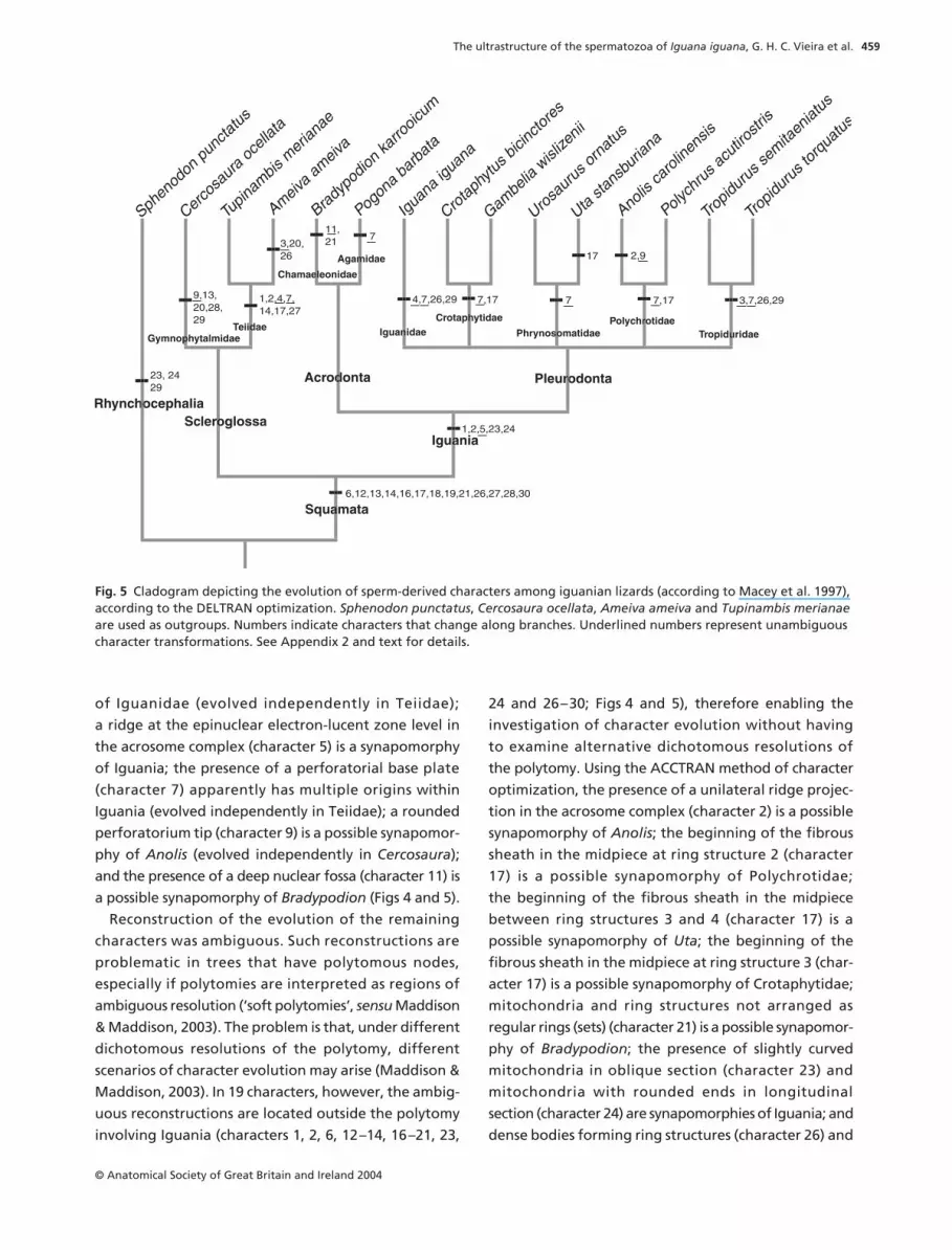
Reconstruction of the evolution of the remaining
characters was ambiguous. Such reconstructions are
problematic in trees that have polytomous nodes,
especially if polytomies are interpreted as regions of
ambiguous resolution (‘soft polytomies’, sensu Maddison
& Maddison, 2003). The problem is that, under different
dichotomous resolutions of the polytomy, different
scenarios of character evolution may arise (Maddison &
Maddison, 2003). In 19 characters, however, the ambig-
uous reconstructions are located outside the polytomy
involving Iguania (characters 1, 2, 6, 12–14, 16–21, 23,
24 and 26–30; Figs 4 and 5), therefore enabling the
investigation of character evolution without having
to examine alternative dichotomous resolutions of
the polytomy. Using the ACCTRAN method of character
optimization, the presence of a unilateral ridge projec-
tion in the acrosome complex (character 2) is a possible
synapomorphy of Anolis; the beginning of the fibrous
sheath in the midpiece at ring structure 2 (character
17) is a possible synapomorphy of Polychrotidae;
the beginning of the fibrous sheath in the midpiece
between ring structures 3 and 4 (character 17) is a
possible synapomorphy of Uta; the beginning of the
fibrous sheath in the midpiece at ring structure 3 (char-
acter 17) is a possible synapomorphy of Crotaphytidae;
mitochondria and ring structures not arranged as
regular rings (sets) (character 21) is a possible synapomor-
phy of Bradypodion; the presence of slightly curved
mitochondria in oblique section (character 23) and
mitochondria with rounded ends in longitudinal
section (character 24) are synapomorphies of Iguania; and
dense bodies forming ring structures (character 26) and
Fig. 5 Cladogram depicting the evolution of sperm-derived characters among iguanian lizards (according to Macey et al. 1997), according to the DELTRAN optimization. Sphenodon punctatus, Cercosaura ocellata, Ameiva ameiva and Tupinambis merianae are used as outgroups. Numbers indicate characters that change along branches. Underlined numbers represent unambiguous character transformations. See Appendix 2 and text for details.

The ultrastructure of the spermatozoa of Iguana iguana, G. H. C. Vieira et al.
© Anatomical Society of Great Britain and Ireland 2004
460
the presence of fibres 3 and 8 at the anteriormost
region of the principal piece (character 29) are possible
synapomorphies of Iguanidae and Tropiduridae (Fig. 4).
Using the DELTRAN tracing produced the same evolu-
tionary reconstructions (regarding Iguania and its
clades), with the exception of characters 1 and 2, which
are synapomorphies of Iguania (Fig. 5). Reconstruction
of the evolution of characters 8, 10, 15, 22 and 25
involves the resolution of ambiguities within the
polytomy of Iguania. Considering the lack of resolution
of the relationships within Iguania, we avoided recon-
structing the evolution of those characters and they are
not displayed in Figs 4 and 5.
We examined the number of character-state changes
in different regions of the spermatozoon based on
10 000 random solutions of the polytomy of Iguania.
Changes were distributed approximately in equal
proportions (∼2.5×) among the sperm head (11 characters,
27.64 ± 0.72 changes), midpiece (17, 40.52 ± 0.59) and
tail (two, 4.80 ± 0.40). However, there were significant
differences in the average number of tree steps or the
average consistency index (averaged across 10 000
random solutions) among characters from different
regions of the spermatozoon (Table 3). Our results
suggest higher rates of evolutionary change (and
homoplasy) in the head, followed by the tail and the
midpiece (Table 3), but should be interpreted with
caution given the very (artificially) large number of
degrees of freedom.
Discussion
The general aspect of the sperm of Iguana iguana
resembles those of other iguanian lizards, being more
similar to tropidurids (among iguanians) and Ameiva
ameiva (regarding Squamata as a whole). Nevertheless,
I. iguana also shares some traits with Cercosaura
ocellata, Polychrus acutirostris, Sphenodon punctatus
and Tupinambis merianae, as depicted in Fig. 4 (see
also Appendix 2).
Because lists of derived characters in the sperm of
Iguania (Teixeira et al. 1999a,d; Scheltinga et al. 2000,
2001) and Squamata (Jamieson, 1995b; Jamieson et al.
1996; Oliver et al. 1996) are available in previous works,
we avoid repeating them here. All synapomorphies of
Tetrapoda and Amniota (Jamieson, 1995a, 1999) and
Squamata (Jamieson, 1995b) were also observed in
the sperm of I. iguana. A presumed synapomorphy of
Iguania, the arrangement of intermitochondrial dense
bodies as regular incomplete rings (Scheltinga et al.
2001), was also present in I. iguana. However, this trait is
also present in some members of Autarchoglossa used
in this study and the gymnophtalmid Micrablepharus
maximiliani (Teixeira et al. 1999b). Conversely, species
of the tropidurid genus Liolaemus (Furieri, 1974) show
dense bodies organized as regular complete rings.
The mapping of 30 sperm morphology characters
revealed a novel synapomorphy of Iguania: the deve-
lopment of a well-developed acrosomal ridge at the
level of the epinuclear lucent zone. Nevertheless, as the
relationships among the clades of Iguania become
resolved, it is likely that more unambiguous evolutionary
changes will emerge.
The agreement between mapped characters and the
proposed phylogeny of a group indicates phylogenetic
inertia (Cheverud et al. 1986; Brown et al. 2000) or, in
other words, that the studied traits are in some way
conserved through the evolutionary history of the
group. We show that, at a more exclusive level (e.g. using
lower taxonomical ranks), the greatest agreement
between the mapped characters and the topology
is achieved (as candidate synapomorphies). As more
inclusive ranks are taken into account, this agreement
is reduced. The increasing divergence (variability) seen
with increasing taxonomic rank seems to hold in general
(Foote, 1997). However, variability of the lower rank taxa,
particularly within the ingroup, is of concern. Why does
it consistently not fit the topology? The variability of
characteristics can show a consistent pattern or a
random pattern. Consistency between the variability and
Region of spermatozoon No. of tree steps Consistency index
Head (11) 2.51 ± 0.07 0.59 ± 0.01Midpiece (17) 2.38 ± 0.03 0.84 ± 0.004Tail (2) 2.40 ± 0.20 0.64 ± 0.02ANOVA F 3219.19 1608 786.00Degrees of freedom 2,29997 2,29997P < 0.0001 < 0.0001
Table 3 Statistics indicative of rates of change in characters across three regions of the spermatozoon of Iguana iguana. Values are based on 10 000 random solutions of polytomy within Iguania. See text for details. Number of characters in parentheses
The ultrastructure of the spermatozoa of Iguana iguana, G. H. C. Vieira et al.
© Anatomical Society of Great Britain and Ireland 2004
461
the evolutionary history of the group suggests adapta-
tion as the driving force, and it can be explained by
historical inheritance (Brooks & McLennan, 1991; Harvey
& Pagel, 1991). The random pattern can result from con-
vergence (Wiens, 2000) or neutral selection, without an
adaptive basis. Tavares-Bastos et al. (2002) found a
poor fit between seven polymorphic sperm characters
and the proposed phylogeny of Tupinambis (i.e. no
phylogenetic inertia), discarding the possibility of
convergence as a result of similar reproductive ecologies.
The variability in sperm morphology of Iguania (and
Squamata) can be associated with reproductive charac-
teristics. Among squamates, there is ample variation in
reproductive characteristics, including: enzymes associ-
ated with the transport of sperm, places of sperm
storage in the female reproductive tract (Olsson et al. 1994;
Blackburn, 1998; Server & Hamlett, 2002), fertilization
process, place of fertilization (Blackburn, 1998), oviduct
or sperm secretions for sperm maintenance in the
female reproductive tract (Girling, 2002), and the mech-
anism of sperm release from storage regions (Girling,
2002). However, detailed data regarding female repro-
ductive tract and fertilization among iguanians are still
lacking. There are two plausible mechanisms for female
discrimination of sperm cell: selection by the reproduc-
tive tract or choice of sperm by the ova (Olsson et al.
1997). One could imagine that sperm characteristics
evolved through sperm competition rules but, again,
studies for sperm competition in reptiles are rare. We
propose that the variability in the sperm cell of Iguania
is a product of historical inheritance, even though this
conclusion is superficial owing to the lack of resolution
for relationships among major lineages (Macey et al.
1997; Schulte et al. 2003). Hence, we should not dis-
count the possibility that sperm-derived characters are
evolutionarily labile and selectively neutral in Iguania,
or a product of convergence due to female reproduc-
tive tract characteristics, particularities of fertilization
and/or sperm competition.
The more characters a region contains, the more
likely that it will contain polymorphism (or variability),
the rates of evolutionary change being the same for
all characters or the changes being selectively neutral
(Ridley, 1993; Tavares-Bastos et al. 2002). Our results
suggest that different regions of the spermatozoon
experience heterogeneous rates of evolution (see
Scheltinga et al. 2000). However, the paucity of data
for female reproductive tract characteristics, particular-
ities of fertilization and/or sperm competition precludes
the correlation of sperm variability with any of these
characteristics.
Acknowledgements
We thank Ruscaia D. Teixeira for her valuable theoret-
ical and technical help and for providing the data from
Cercosaura ocellata, Daniel O. Mesquita for field assist-
ance, Ayrton K. Pérez Jr for providing the specimens
used on the present study, and two anonymous reviewers
for their insightful comments and suggestions. Some
characters and character-states used here are derived
from observations by David M. Scheltinga and Ruscaia
D. Teixeira. This study was supported by FINATEC and
a fellowship from PIBIC-UnB-CNPq and a doctorate
fellowship from CNPq to G.H.C.V.
References
Blackburn DG (1998) Structure, function, and evolution ofthe oviducts of squamate reptiles, with special referenceto viviparity and placentation. J. Exp. Zool. 282, 560–617.
Brooks DR, McLennan DA (1991) Phylogeny, Ecology, andBehavior, a Research Program in Comparative Biology.Chicago: The University of Chicago Press.
Brown B, Emberson RM, Paterson AM (2000) Morphologicalcharacter evolution in hepialid moths (Lepidoptera: Hepial-idae) from New Zealand. Biol. J. Linn. Soc. 69, 383–397.
Cheverud JM, Dow MM, Leutenegger W (1986) The quantita-tive assessment of phylogenetic constraints in comparativeanalysis: sexual dimorphism in body weight among primates.Evolution 39, 1335–1351.
Estes R, de Queiroz K, Gauthier J (1988) Phylogenetic relation-ships within Squamata. In Phylogenetic Relationships ofthe Lizard Families. Essays Commemorating Charles L. Camp(eds Estes R, Pregill G), pp. 119–281. Stanford, CA: StanfordUniversity Press.
Foote M (1997) The evolution of morphological diversity. Ann.Rev. Ecol. Syst. 28, 129–152.
Frost DR, Etheridge R (1989) A phylogenetic analysis andtaxonomy of iguanian lizards (Reptilia: Squamata). Misc.Publ. Mus. Nat. Hist. Univ.Kansas 81, 1–65.
Frost DR, Etheridge R, Janies D, Titus TA (2001) Total evidence,sequence alignmemt, evolution of Polychrotid lizards, anda reclassification of the Iguania (Squamata: Iguania). Am.Mus. Novit. 3343, 1–38.
Furieri P (1974) Spermi e spermatogenesi in alcuni iguanidiargentini. Riv. Biol. 67, 233–279.
Girling JE (2002) The reptilian oviduct: a review of structureand function and directions for future research. J. Exp. Zool.293, 141–170.
Giugliano LG, Teixeira RD, Colli GR, Báo SN (2002) Ultrastruc-ture of spermatozoa of the lizard Ameiva ameiva, withconsiderations on polymorphism within the family Teiidae(Squamata). J. Morph. 253, 264–271.

The ultrastructure of the spermatozoa of Iguana iguana, G. H. C. Vieira et al.
© Anatomical Society of Great Britain and Ireland 2004
462
Harvey PH, Pagel MD (1991) The Comparative Method inEvolutionary Biology. New York: Oxford University Press.
Healy JM, Jamieson BGM (1994) The ultrastructure of sperma-togenesis and epididymal spermatozoa of the tuataraSphenodon punctatus (Sphenodontia, Amniota). Philos.Trans. Roy. Soc. B 344, 187–199.
Jamieson BGM (1995a) Evolution of tetrapod spermatozoawith particular reference to amniotes. In Advances inSpermatozoal Phylogeny and Taxonomy (eds JamiesonBGM, Ausio J, Justine J), pp. 343–358. Paris: Muséum Nationald’Histoire Naturelle.
Jamieson BGM (1995b) The ultrastructure of spermatozoa ofthe Squamata (Reptilia) with phylogenetic considerations.In Advances in Spermatozoal Phylogeny and Taxonomy(eds Jamieson BGM, Ausio J, Justine J), pp. 359–383. Paris:Muséum National d’Histoire Naturelle.
Jamieson BGM, Oliver SC, Scheltinga DM (1996) The ultra-structure of spermatozoa of Squamata. I. Scincidae, Gekko-nidae and Pygopodidae (Reptilia). Acta Zool. – Stockholm77, 85–100.
Jamieson BGM (1999) Spermatozoal phylogeny of the Verte-brata. In The Male Gamete: from Basic Science to ClinicalApplications (ed. Gagnon C), pp. 303–331. Vienna, IL: CacheRiver Press.
Kitching IJ, Forey PL, Humphries CJ, Williams DM (1998)Cladistics. The Theory and Practice of Parsimony Analysis.Oxford: Oxford University Press.
Lee MSY (1998) Convergent evolution and character correla-tion in burrowing reptiles: towards a resolution of squamaterelationships. Biol. J. Linn. Soc. 65, 369–453.
Macey JR, Larson A, Ananjeva NB, Papenfuss TJ (1997) Evolu-tionary shifts in three major structural features of themitochondrial genome among iguanian lizards. J. Mol. Evol.44, 660–674.
Maddison WP, Maddison DR (2003) MacClade: Analysis ofPhylogeny and Character Evolution v.4.06. Sunderland,MA: Sinauer Associates, Inc.
Miyamoto MM, Fitch WM (1995) Testing species phylogeniesand phylogenetic methods with congruence. Syst. Biol. 44,64–76.
Oliver SC, Jamieson BGM, Scheltinga DM (1996) Theultrastructure of spermatozoa of Squamata. II. Agamidae,Varanidae, Colubridae, Elapidae, and Boidae (Reptilia).Herpetologica 52, 216–241.
Olsson M, Gullberg A, Tegelstrom H (1994) Sperm competitionin the sand lizard, Lacerta agilis. Anim. Behav. 48, 193–200.
Olsson M, Shine R, Madsen T, Gullberg A, Tegelstrom H (1997)Sperm choice by females. Trends Ecol. Evol. 12, 445–446.
Ridley M (1993) Evolution. Boston: Blackwell ScientificPublications.
Saita A, Comazzi M, Perrotta E (1988) New data at the E.M. onthe spermiogenesis of Iguana delicatissima (Laurent) involvingcomparative significance. Acta Embryol. Morph. Exper. 9,105–114.
Scheltinga DM, Jamieson BGM, Trauth SE, McAllister CT(2000) Morphology of the spermatozoa of the iguanianlizards Uta stansburiana and Urosaurus ornatus (Squamata,Phrynosomatidae). J. Submicr. Cytol. Path. 32, 261–271.
Scheltinga DM, Jamieson BGM, Espinoza RE, Orrel KS (2001)Descriptions of the mature spermatozoa of the lizards
Crotaphytus bicinctores, Gambelia wislizenii (Crotaphytidae),and Anolis carolinensis (Polychrotidae) (Reptilia, Squamata,Iguania). J. Morph. 247, 160–171.
Schulte II JA, Macey JR, Larson A, Papenfuss TJ (1998) Molec-ular test of phylogenetic taxonomies: a general procedureand example using four subfamilies of the lizard familyiguanidae. Mol. Phylogenet. Evol. 10, 367–376.
Schulte II JA, Valladares JP, Larson A (2003) Phylogeneticrelationships within Iguanidae inferred using molecularand morphological data and a phylogenetic taxonomy ofiguanian lizards. Herpetologica 59, 399–419.
Server DM, Hamlett WC (2002) Female sperm storage inreptiles. J. Exp. Zool. 292, 187–199.
Swofford DL (1991) When are phylogeny estimates frommolecular and morphological data sets incongruent? In Phy-logenetic Analysis of DNA Sequences (eds Miyamoto MM,Cracraft J), pp. 295–333. Oxford: Oxford University Press.
Swofford DL (1999) PAUP*. Phylogenetic Analysis Using Parsimony(*and Other Methods). Sunderland, MA: Sinauer Associates.
Tavares-Bastos L, Teixeira RD, Colli GR, Báo SN (2002) Poly-morphism in the sperm ultrastructure among four species oflizards in the genus Tupinambis (Squamata: Teiidae). ActaZool. – Stockholm 83, 297–307.
Teixeira RD, Colli GR, Báo SN (1999a) The ultrastructure ofspermatozoa of the lizard Polychrus acutirostris (Squamata,Polychrotidae). J. Submicr. Cytol. Path. 31, 387–395.
Teixeira RD, Colli GR, Báo SN (1999b) The ultrastructure of thespermatozoa of the lizard Micrablepharus maximiliani(Squamata, Gymnophthalmidae), with considerations onthe use of sperm ultrastructure characters in phylogeneticreconstruction. Acta Zool. – Stockholm 80, 47–59.
Teixeira RD, Colli GR, Báo SN (1999c) The ultrastructure of thespermatozoa of the worm-lizard Amphisbaena alba (Squa-mata, Amphisbaenidae), and the phylogenetic relationshipsof amphisbaenians. Can. J. Zool. 77, 1254–1264.
Teixeira RD, Vieira GHC, Colli GR, Báo SN (1999d) Ultrastruc-tural study of spermatozoa of the neotropical lizards, Tropi-durus semitaeniatus and Tropidurus torquatus (Squamata,Tropiduridae). Tissue Cell 31, 308–317.
Teixeira RD (2003) Análise filogenética da família Teiidae(Squamata, Reptilia), a ultra-estrutura de espermatozóide ea sua utilidade filogenética. Doctorate thesis, UniversidadeEstadual de Campinas.
Wiens JJ (2000) Decoupled evolution of display morphologyand display behaviour in phrynosomatid lizards. Biol. J. Linn.Soc. 70, 597–612.
Wiens JJ, Hollingsworth BD (2000) War of the Iguanas: con-flicting molecular and morphological phylogenies and long-branch attraction in iguanid lizards. Syst. Biol. 49, 143–159.
Appendix 1
Sperm-derived characters and character-states used
(consistency index in parenthesis).
1. Acrosome complex, ridge (0.500): (0) absent; (1)
present.
2. Acrosome complex, ridge projection (0.667): (0) not
applicable; (1) unilateral; (2) bilateral.

The ultrastructure of the spermatozoa of Iguana iguana, G. H. C. Vieira et al.
© Anatomical Society of Great Britain and Ireland 2004
463
3. Acrosome complex, unilateral electron-lucent space
(0.500): (0) absent; (1) present.
4. Acrosome complex, radial aspect of subacro-
somal cone in transverse section (0.500): (0) absent;
(1) present.
5. Acrosome complex, ridge at the epinuclear
electron-lucent zone level (1.000): (0) poorly developed;
(1) well developed.
6. Perforatorium, number (1.000): (0) two; (1) one.
7. Perforatorium, base plate (0.250): (0) absent;
(1) present.
8. Perforatorium, shape of the base plate (0.286):
(0) not applicable; (1) knob-like; (2) stopper-like.
9. Perforatorium, tip (0.500): (0) pointed; (1) rounded.
10. Nucleus, lacunae (0.333): (0) absent; (1) present.
11. Nucleus, nuclear fossa (1.000): (0) shallow; (1) deep.
12. Neck region, stratified laminar structure (1.000):
(0) absent; (1) present.
13. Neck region, stratified laminar structure projection
(1.000): (0) not applicable; (1) unilateral; (2) bilateral.
14. Neck region, stratified laminar structure (1.000):
(0) not applicable; (1) poorly developed; (2) well
developed.
15. Neck region, electron-dense structure inside the
proximal centriole (0.333): (0) absent; (1) present.
16. Midpiece, fibrous sheath (1.000): (0) absent;
(1) present.
17. Midpiece, beginning of the fibrous sheath in the
midpiece (0.800): (0) not applicable; (1) level of mito-
chondria tier 1–2; (2) tier 2–3; (3) tier 3–4; (4) at ring
structure 2; (5) at ring structure 3.
18. Midpiece, mitochondrial cristae (1.000): (0) concen-
tric; (1) linear.
19. Midpiece, dense bodies outside mitochondria
(1.000): (0) absent; (1) present.
20. Midpiece, fibres 3 and 8 (0.500): (0) grossly
enlarged; (1) not grossly enlarged.
21. Midpiece, mitochondria and ring structures as
regular rings (sets) (1.000): (0) not applicable; (1) absent;
(2) present.
22. Midpiece, number of sets of mitochondria and ring
structures (0.571): (0) not applicable; (1) three tiers;
(2) four tiers; (3) five tiers; (4) six tiers.
23. Midpiece, mitochondria in oblique section (1.000):
(0) columnar; (1) slightly curved; (2) rounded.
24. Midpiece, mitochondrial shape in longitudinal
section (1.000): (0) trapezoidal; (1) round ends; (2) totally
round.
25. Midpiece, dense bodies (0.500): (0) solid; (1) granular.
26. Midpiece, dense bodies forming ring structures
(0.500): (0) not applicable; (1) absent; (2) present.
27. Midpiece, appearance of dense bodies in the
ring structures in oblique section (1.000): (0) not appli-
cable; (1) fused, forming compact structures; (2) not
fused.
28. Midpiece, intermitochondrial dense bodies in
transverse section (1.000): (0) not applicable; (1) sepa-
rated from fibrous sheath by mitochondria; (2) juxta-
posed to fibrous sheath.
29. Principal piece, fibres 3 and 8 at the anteriormost
region (0.250): (0) absent; (1) present.
30. Principal piece, wide band of cytoplasm in the
anteriormost region (1.000): (0) absent; (1) present.
Appendix 2
List of changes for the 30 characters of the sperm
derived-characters (see Figs 4 and 5 for reference of
labelled stems). Underlined numbers represent unam-
biguous character transformations.
ACCTRAN optimization
Squamata: 1: 0 → 1, 2: 0 → 1, 6: 0 → 1, 12: 0 → 1, 13:
0 → 2, 14: 0 → 1, 16: 0 → 1, 17: 0 → 2, 18: 0 → 1, 19:
0 → 1, 21: 0 → 2, 26: 0 → 1, 27: 0 → 1, 28: 0 → 2, 30:
0 → 1;
Iguania: 5: 0 → 1, 23: 0 → 1, 24: 0 → 1;
Pleurodonta: no changes;
Acrodonta: no changes;
Scleroglossa: 20: 1 → 0;
Teiidae: 4: 0 → 1, 7: 0 → 1, 14: 1 → 2, 17: 2 → 4, 27:
1 → 2;
Crotaphytidae: 7: 0 → 1, 17: 2 → 5;
Phrynosomatidae: 7: 0 → 1;
Polychrotidae: 7: 0 → 1, 17: 2 → 4;
Tropiduridae: 3: 0 → 1, 7: 1 → 0, 26: 1 → 2, 29: 0 → 1;
B. karrooicum (voucher of Chamaeleonidae): 11: 0 → 1,
21: 2 → 1;
P. barbata (voucher of Agamidae): 7: 1 → 0;
I. iguana (voucher of Iguanidae): 4: 0 → 1, 7: 1 → 0, 26:
1 → 2, 29: 0 → 1;
U. stansburiana: 17: 2 → 3;
A. carolinensis: 2: 1 → 2, 9: 0 → 1;
S. punctatus (voucher of Rhynconcephalia): 23: 0 → 2,
24: 0 → 2, 29: 0 → 1;
C. ocellata (voucher of Gymnophthalmidae): 1: 1 → 0,
2: 1 → 0, 9: 0 → 1, 13: 2 → 1, 28: 2 → 1, 29: 0 → 1;

The ultrastructure of the spermatozoa of Iguana iguana, G. H. C. Vieira et al.
© Anatomical Society of Great Britain and Ireland 2004
464
T. merianae: 20: 0 → 1;
A. ameiva: 3: 0 → 1, 26: 1 → 2;
DELTRAN optimization
Squamata: 6: 0 → 1, 12: 0 → 1, 13: 0 → 2, 14: 0 → 1, 16:
0 → 1, 17: 0 → 2, 18: 0 → 1, 19: 0 → 1, 21: 0 → 2, 26:
0 → 1, 27: 0 → 1, 28: 0 → 2, 30: 0 → 1;
Iguania: 1: 0 → 1, 2: 0 → 1, 5: 0 → 1, 23: 0 → 1, 24:
0 → 1;
Pleurodonta: no changes;
Acrodonta: no changes;
Scleroglossa: no changes;
Teiidae: 1: 0 → 1, 2: 0 → 1, 4: 0 → 1, 7: 0 → 1, 14: 1 → 2,
17: 2 → 4, 27: 1 → 2;
Crotaphytidae: 7: 0 → 1, 17: 2 → 5;
Phrynosomatidae: 7: 0 → 1;
Polychrotidae: 7: 0 → 1, 17: 2 → 4;
Tropiduridae: 3: 0 → 1, 7: 1 → 0, 26: 1 → 2, 29: 0 → 1;
B. karrooicum (voucher of Chamaeleonidae): 11: 0 → 1,
21: 2 → 1;
P. barbata (voucher of Agamidae): 7: 0 → 1;
I. iguana (voucher of Iguanidae): 4: 0 → 1, 7: 0 → 1, 26:
1 → 2, 29: 0 → 1;
U. stansburiana: 17: 2 → 3;
A. carolinensis: 2: 1 → 2, 9: 0 → 1;
S. punctatus (voucher of Rhynconcephalia): 23: 0 → 2,
24: 0 → 2, 29: 0 → 1;
C. ocellata (voucher of Gymnophthalmidae): 9: 0 → 1,
13: 2 → 1, 20: 1 → 0, 28: 2 → 1, 29: 0 → 1;
A. ameiva: 3: 0 → 1, 20: 1 → 0, 26: 1 → 2;