the superior paraolivary nucleus of the rat is a gabaergic nucleus
TRANSCRIPT

JARO 01: 255–269 (2000)DOI: 10.1007/s101620010054
Superior Paraolivary Nucleus of the Rat Is a GABAergicNucleus
RANDY J. KULESZA, JR., AND ALBERT S. BERREBI
Departments of Otolaryngology and Anatomy, Neuroscience Graduate Program and Sensory Neuroscience Research Center,West Virginia University School of Medicine, Morgantown, WV 26506–9200, USA
Received: 30 June 2000; Accepted: 8 September 2000; Online publication: 23 November 2000
ABSTRACT Keywords: superior olive, dorsomedial periolivarynucleus, glycine, glutamic acid decarboxylase
The presence of the inhibitory neurotransmitters gly-cine and GABA (gamma-amino butyric acid) and GAD(glutamic acid decarboxylase), the synthesizing INTRODUCTIONenzyme for GABA, was examined by immunocyto-
Inhibition plays a crucial role in the vertebrate nervouschemistry in the superior paraolivary nucleus (SPON)system, and glycine and gamma-aminobutyric acidof the rat. Only rarely were SPON neurons observed(GABA) are the major inhibitory neurotransmitters into be glycine-immunoreactive, but the majority werethe CNS (Nicholls 1994). Although glycine is a com-GABA-immunoreactive. Using unbiased stereologicalmon amino acid present in all proteins and cells,counting methods, we estimated that this nucleus con-GABA is synthesized from the amino acid glutamatetains approximately 2500 neurons. Moreover, 90% ofby the enzyme glutamic acid decarboxylase (GAD)SPON neurons were immunolabeled by antisera(Roberts and Frankel 1950; Nicholls 1994). Becausedirected against either the 65- or 67-kD isoform ofGABA is a small labile molecule that can be difficultGAD, or a third antiserum that recognizes both GADto localize with immunocytochemistry, GAD is widelyisoforms. Morphometric analysis of GAD-immunola-considered a reliable marker for GABAergic neuronsbeled neurons indicated that SPON neurons possess(Wu et al. 1973; Oertel et al. 1981). Two isoforms ofcell bodies and dendritic arbors that are elongatedGAD, named according to their molecular weights,rostrocaudally and relatively flattened parasagittally.have been isolated (Bayon et al. 1977; Spink et al.Abundant glycine-, GABA-, and GAD-immunoreactive1983; Denner and Wu 1985; Legay et al. 1987; Kaufmanpunctate profiles—presumed to represent, for theet al. 1991). Although both GAD isoforms are presentmost part, presynaptic axon terminals—were observedin most GABAergic neurons, GAD 65 and GAD 67in apposition to SPON neurons. We conclude that(65 and 67 kDa, respectively) are transcribed fromthe rat SPON contains a homogeneous population ofseparate genes and have different affinities for themultipolar GABAergic neurons that receive abundantcofactor pyridoxal 58-phosphate (Erlander et al. 1991;GABAergic and glycinergic innervation. The vastMartin et al. 1991; Kaufman et al. 1991). Recent immu-majority of glycinergic inputs to SPON are presumednocytochemical studies suggest that the two isoformsto originate in the ipsilateral medial nucleus of themay have different intracellular distributions, withtrapezoid body, but the source(s) of its GABAergicGAD 67 being widely distributed in somata, dendrites,innervation remains to be determined.and axon terminals, while GAD 65 is located predomi-nantly in axon terminals (Esclapez et al. 1994). GAD67, which is present as the active holoenzyme, is pre-
Correspondence to: Dr. Albert S. Berrebi, Ph.D. • Department of Otola- sumably responsible for utilizing glutamate, throughryngology • West Virginia University School of Medicine • P.O. Box
the GABA shunt, for metabolic needs or supplying9200, Health Sciences Center • Morgantown, WV 26506. Telephone:(304) 293-2357; fax: (304) 293-2902; email: [email protected] basal levels of GABA for neurotransmission (Denner
255

256 KULESZA AND BERREBI: Inhibition in the Rat SPON
and Wu 1985; Erlander and Tobin 1991). In contrast, purely ipsilateral ascending projection to the IC (Ber-rebi et al. 1997; Saldana and Berrebi 2000). UsingGAD 65 is active only in the presence of cofactor and
synthesizes GABA in terminals when there is increased antibodies directed against GABA or an antiserum thatrecognizes both isoforms of GAD, other investigatorsdemand (Martin and Rimwall 1993).
The neurotransmitters GABA and glycine are have suggested that the rat SPON contains a substantialproportion of GABAergic neurons (Oertel et al. 1981;found in abundance in central auditory nuclei, and
inhibitory neurotransmission plays a fundamentally Mugnaini and Oertel 1985; Roberts and Ribak 1987;Moore and Moore 1987; Gonzalez-Hernandez et al.important role in the function of the mammalian audi-
tory system (Helfert and Aschoff 1997). Inhibition has 1996). Glycine-IR has been described in the rat SOC(Campistron et al. 1986; Aoki et al. 1988; Pourcho etbeen shown to sharpen tuning curves and modulate
discharge properties of auditory neurons, and it is al. 1992) but has not been systematically examined inthe SPON or in other periolivary cell groups.crucial to the ability to localize high-frequency sounds
(Finlayson and Caspary 1989; Yang et al. 1992; Caspary In order to gain a better understanding of the func-tional role of SPON, we used immunocytochemicalet al. 1994; Park and Pollak 1993; Klug et al. 1995; Le
Beau et al. 1996; Koch and Grothe 1998). GABAergic methods to delineate the distribution of GABAergicand glycinergic neurons and axon terminals withinand glycinergic neurons are especially prominent in
the superior olivary complex (SOC) (Helfert et al. the nucleus. We also obtained unbiased stereologicalestimates of the percentage of SPON neurons that are1989; Spirou and Berrebi 1997; Saint Marie and Baker
1990; Ostapoff et al. 1990, 1997). This auditory brain- GAD IR and performed a quantitative morphometricassessment of SPON neuronal morphologies. Some ofstem center contains three principal nuclei with identi-
fied roles in sound localization, the medial nucleus of the data contained herein have been presented inabstract form (Kulesza et al. 1998; Kulesza and Ber-the trapezoid body (MNTB), the medial superior olive
(MSO) and the lateral superior olive (LSO), and sev- rebi 1999).eral accessory or periolivary cell groups whose rolesin audition are poorly understood (Schwartz 1992;Helfert and Aschoff 1997).
METHODSThe superior paraolivary nucleus (SPON) is a con-spicuous periolivary nucleus in the SOC of rodentsand guinea pigs. Situated medial to the LSO and dorso-
Animalslateral to the MNTB, this nucleus receives ascendinginputs from the contralateral cochlear nucleus (Friauf Adult female Sprague–Dawley rats, weighing 230–285and Ostwald 1988; Kuwabara et al. 1991; Thompson g, were deeply anesthetized by an intramuscular injec-and Thompson 1991a,b; Schofield 1995), as well as a tion of a xylazine and ketamine mixture (42 mg xylaz-substantial local inhibitory input from the ipsilateral ine and 57 mg ketamine per 100 g body weight) priorMNTB (Kuwabara and Zook 1991, 1992a,b; Banks and to vascular perfusion. A total of 36 animals were usedSmith 1992; Sommer et al. 1993). It also sends a promi- for this study. The West Virginia University Institu-nent projection to the ipsilateral inferior colliculus tional Animal Care and Use Committee approved all(IC) (Beyerl 1978; Adams 1983; Coleman and Clerici animal protocols used for this project.1987; Moore 1988; Saint Marie and Baker 1990; Gonza-lez-Hernandez et al. 1996; Fuentes et al. 1999; Saldanaand Berrebi 2000). Notably, the SPON of the rat does Postembedding immunocytochemistry fornot receive a descending projection from the IC nor glycine and GABAdoes it project to the cochlear nucleus or cochlea(White and Warr 1983; Osen et al. 1984; Faye-Lund Twelve animals were perfused through the ascending
aorta with a rinse of Ca21-free Ringer’s variant followed1986; Aschoff and Ostwald 1987; Vetter et al. 1991,1993; Vetter and Mugnaini 1992; Caicedo and Her- by 1 l of fixative composed of 2% paraformaldehyde
and 2.5% glutaraldehyde in 0.1M sodium phosphatebert 1993).A number of immunohistochemical and retrograde buffer. Animals were placed on ice and remained
undisturbed for 30 minutes before their brains weretransport studies have revealed populations of GABA-immunoreactive (IR) and glycine-IR neurons in the dissected and immersed in fixative overnight. Using
a Vibratome, the brains were cut into 100-mm-thickguinea pig SPON, suggesting that these two neuro-chemically defined cell groups have different projec- sections in the coronal plane. The sections were post-
fixed in 1% osmium tetroxide, stained with 2% aque-tion targets (Helfert et al. 1989; Thompson et al. 1985;Saint Marie and Baker 1990; Ostapoff et al. 1985, ous uranyl acetate, dehydrated, and flat embedded in
Epon (Polybed 812, Polysciences, Warrington, PA).1990). Recent tract-tracing data in rats, however, dem-onstrate a homogeneous neuronal population with a After polymerization, sections containing the SOC

KULESZA AND BERREBI: Inhibition in the Rat SPON 257
were cut into a series of 1-mm semithin sections, heat- regions within the neuronal cytoplasm did not includethe pale staining cell nuclei. The grey value intensitydried onto glass slides, and prepared for immunocyto-
chemistry (Spirou and Berrebi 1997). Briefly, Epon scale was calibrated for each pair of sections so thatthe darkest staining soma (i.e., a glycine-IR MNTBwas etched from the sections using sodium ethoxide
(absolute ethanol saturated with sodium hydroxide). neuron) was assigned a value of 255 and the lumenof a blood vessel was assigned a value of 0. Data wereThe tissues were then rehydrated, first in ethanols and
then in water. Osmium was subsequently removed with entered into JMP (SAS Institute Inc., Cary, NC) andStatview (Abacus Concepts, Inc., Berkeley, CA) data1% aqueous sodium periodate.
Adjacent pairs of 1-mm sections were incubated in analysis software packages for statistical comparisonsand graphical output.5% normal donkey serum in 0.5M Trizma base–HCl
(Tris, pH 7.6) for 1 h followed by overnight incubation In one pair of sections, the glycine antiserum waspreadsorbed with 5mM GABA and the GABA antise-in a primary antiserum as follows: rabbit anti-glycine
(Chemicon, Temecula, CA; diluted 1:1200), rabbit rum was preadsorbed with 10mM glycine before incu-bation with the tissue. The optical density dataanti-GABA (gift from Dr. D. Pow, University of Queens-
land, Brisbane, Australia: diluted 1:8000; or purchased obtained from this pair of sections were statisticallyindistinguishable from that of the remaining five pairsfrom HTI Bioproducts, Inc., Ramona, CA, now Strate-
gic BioSolutions: diluted 1:1000). Sections were then of sections which were not incubated in preadsorbedantisera (t 5 1.819, p . 0.07 for GABA antiserum; andincubated in biotinylated secondary antiserum and
further processed using the ABC method (Vector, Burl- t 5 1.056, p . 0.29 for glycine antiserum). Therefore,the effect of preadsorption will not be considered fur-ingame, CA). The chromogenic reaction took place in
0.05% diaminobenzidine (DAB) with 0.01% hydrogen ther. We also compared the immunostaining patternsobtained with the GABA antiserum provided by Dr.peroxide in 0.5M Tris.
The specificity of the Chemicon glycine and Pow Pow with the commercially available (HTI Biopro-ducts) antiserum. Within the range of dilutions weGABA antisera was previously established in our labo-
ratory with preadsorption control experiments using tested, the latter resulted in somewhat weaker stainingintensities throughout the brain, but the pattern ofan amino acid inhibitor complex according to Storm–
Mathisen and Ottersen (1990) (see Fig. 1 of Spirou immunostaining obtained within nuclei of the SOCwere judged to be qualitatively equivalent. Therefore,and Berrebi 1997). The affinity-purified anti-GABA
serum from HTI Bioproducts was reported to display the data obtained from both GABA antisera werepooled for all analyses.approximately 2% cross-reactivity with glycine. In the
present experiments, we tested the effects of antiserumpreadsorption on intensity of immunoreactivity using GAD immunocytochemistry on frozen sectionsan optical densitometric procedure (see below).
A total of 24 animals were perfused through theascending aorta with a vascular rinse of physiologicalOptical densitysaline, followed by a fixative composed of 4% commer-cial formaldehyde and a zinc salt (0.5% zinc dichro-To classify individual cells according to their glycine
and/or GABA immunoreactivities, we performed opti- mate, pH4.0, or 0.5% zinc salicylate, pH 6.5) in 0.9%saline (Mugnaini and Dahl 1983; Berrebi and Mug-cal densitometry in adjacent semithin (1 mm thick)
sections. A total of six pairs of sections (sections in naini 1991). The animals were placed on ice for 30minutes before brains were dissected and immersedeach pair were within 2–3 mm of each other) from the
SOC of six animals were used in this analysis. Semithin in cryoprotectant (30% sucrose in saline) for at least24 h. Brainstems were sectioned in series order, eithersections were used because individual neurons could
be identified in sequential sections immunostained coronally or parasagittally, at a thickness of 30 mm ona freezing microtome.with either glycine or GABA antiserum. We evaluated
every SPON neuron encountered in each section, as Free-floating sections were rinsed in 0.5M Tris,blocked in 5% normal donkey serum, and incubatedwell as randomly selected MNTB and MSO cells. The
immunostaining intensities of MNTB neurons served for 48 h at 48C in primary antiserum with gentle agita-tion. To optimize the immunostaining, different proto-as a reference standard for glycine immunopositivity
and those of MSO cells as an indication of background cols were used depending on the primary antiserum.GAD-1440 antiserum (Oertel et al. 1981; provided byimmunostaining levels.
Images were captured with a SONY video camera Judith Harvey–White, NIH, Bethesda, MD, and usedat a dilution of 1:1200) is a sheep polyclonal antiserummounted on an Olympus BH-2 microscope (40 times
objective, final magnification 980 times). Average pixel that recognizes both GAD isoforms. GAD 6 (Changand Gottlieb 1988; purchased from Developmentalgrey values were measured from uniform regions of
neuronal cytoplasm. Care was taken so that sampling Studies Hybridoma Bank, University of Iowa, and used
258 KULESZA AND BERREBI: Inhibition in the Rat SPON
at a dilution of 1:1000) is a mouse monoclonal antise- Unbiased stereologyrum directed against the GAD-65 isoform. Both of We estimated the number of Nissl-stained and GAD-these antibodies were revealed using the PAP method IR neurons in the SPON using methods of unbiased(Sternberger 1979), although the best results with the stereology (Gundersen 1988; Gundersen et al. 1988).GAD-6 antiserum were obtained with the double PAP The Computer Assisted Stereological Toolbox systemmethod (Ordronneau et al. 1981). K2 antiserum (C.A.S.T.-Grid, Olympus, Denmark) was used to imple-(Kaufman et al. 1991, sold by Chemicon, Temecula, ment the optical fractionator technique (GundersenCA, and used at a dilution of 1:3000) is a rabbit poly- 1986), which incorporates the optical dissector toolclonal antiserum that primarily recognizes GAD 67 (Sterio 1984). We randomly selected a slide caudal toand was revealed according to the ABC method. The the SPON as a starting point and sampled uniformlychromogenic reaction for each antiserum took place spaced sections (every fourth or fifth section) throughin 0.05% DAB with 0.01% hydrogen peroxide in 0.5M the nucleus using the optical dissector tool. A countingTris. For reference, alternating sections were not volume (approximately 33,600 mm3) was definedimmunoreacted but were stained for Nissl substance within the thickness of the tissue section, and onlyusing cresyl violet according to standard protocols. All cells that were contained and in focus within the appro-sections were mounted onto glass slides from gelatin- priate boundaries of the counting frame were marked.alcohol, air-dried, and coverslipped using Accumount. Our final estimates were calculated using the equation:
Specificity assays (preabsorption controls and West-N 5 number of cells counted 3 area fractionern blots) using the GAD 6 and K2 antisera were per-
formed previously by Esclapez et al. (1994). They 3 section fraction 3 dissector fractionindicated that the GAD-67-specific K2 antiserum hasa slight cross-reactivity with GAD 65 and that GAD- Quantitative analysis of puncta in plastic6 antiserum does not cross-react with GAD 67. The sectionsspecificity of the GAD-1440 antiserum for GAD has The number of perisomatic puncta was quantified atbeen established (Oertel et al. 1981). This antiserum 100 times under oil immersion (final magnificationlocalizes to both isoforms of the enzyme (Kaufman et 1100 times). Measurements of puncta perimeter andal. 1991). long and short axes were made on the computer moni-
tor with the C.A.S.T.-Grid software package using thedistance/boundary function. The area of punctateprofiles was calculated by modeling them as ellipsesMorphometric analysisusing the equation:
Frozen sections immunoreacted with GAD-1440 or Areapuncta 5 0.5 major axis 3 0.5 minor axis 3 pGAD-6 antiserum were sampled at 90-mm intervals forfurther analysis. All labeled neurons containing nuclei
RESULTSwere traced using a camera lucida with a 100 times oilimmersion objective (final magnification of tissue 1100
The subset of SOC sections stained with cresyl violettimes). Care was taken to estimate the cell body con-
was used to assist in delineating the boundaries of thetour so that dendrites extending from the soma were SPON and other SOC nuclei. A Nissl-stained coronalnot included. The drawings were scanned into a Power section of the rat pons at a middle rostrocaudal levelMacintosh computer, and NIH Image software (ver- through the SOC is shown for reference in Figure 1.sion 1.61, NIH, Bethesda, MD) was used to measure At this level, all three principal nuclei (LSO, MSO,the area, perimeter, and major and minor diameters and MNTB) can be identified. Several periolivary cellof each cell body. An index of circularity (Yin et al. groups, including the ventral (VNTB) and lateral1990) was then calculated for each soma using the nuclei of the trapezoid body (LNTB) and the SPON,equation: are also present. The SPON is identified as a promi-
nently sized ovoid-shaped nucleus interposed betweencircularity 5 4p area/perimeter2
the LSO, MSO, and MNTB. In this material, the SPONappears to possess a low density of neurons relative towhich yields an estimate of cell shape that is indepen-the MNTB and LSO.dent of size. Using this formula, a perfectly circular
cell body profile would be assigned a circularity valueOverview of glycine and GABAof 1.0, with increasingly elliptical profiles resulting inimmunoreactivities in the SOCdecreasing values. Statistical comparisons were per-
formed using the Statview data analysis software Postembedding immunocytochemistry for glycine andGABA revealed immunolabeled somata, dendrites,package.

KULESZA AND BERREBI: Inhibition in the Rat SPON 259
FIG. 1. Organization of the rat superior olivary complex. Cresylviolet-stained coronal section through the SOC at the level of theseventh cranial nerve (7n) root. The three principal nuclei of theSOC—the lateral superior olive (LSO), medial superior olive (MSO),and the medial nucleus of the trapezoid body (MNTB)—can all beseen in this section, along with several periolivary nuclei. It is evidentthat the superior paraolivary nucleus (SPON) has a relatively lowdensity of neurons. D-dorsal, LNTB-lateral nucleus of the trapezoidbody, M-medial, rf-pontine reticular formation, tb-trapezoid body,VNTB-ventral nucleus of the trapezoid body. Scale bar 5 200 mm.
fibers, and punctate profiles, presumed to representfor the most part axon terminals, throughout the SOC(Fig. 2). Qualitatively, neurons of the MNTB appearedmost intensely glycine IR, and the VNTB contained
FIG. 2. Overview of glycine IR and GABA IR in the rat SOC. Postem-most of the intensely GABA-IR neurons. Glycine- andbedding immunocytochemistry performed on plastic-embedded sem-GABA-IR cell bodies of varying staining intensitiesithin sections demonstrates glycine IR and GABA IR within SOCwere also found scattered in the LSO and LNTB, as nuclei. A. In glycine material, MNTB neurons are easily recognized
well as other nuclei. Cell bodies of the MSO appeared by their darkly immunostained somata. The SPON contains only rareimmunonegative after incubation with either anti- glycine-IR cell bodies but the neuropil reveals a high density of
glycine-IR fibers and punctate profiles. B. In sections processed toserum.reveal GABA, cell bodies and neuropil in VNTB are the most promi-The boundary between the SPON and LSO wasnently immunolabeled. SPON contains numerous GABAergic cellclearly identified by an immunonegative fiber bundle bodies, most of which are lightly to moderately immunolabeled.
coursing between these nuclei. The medial border of GABA-IR punctate profiles are distributed throughout the neuropilthe SPON was somewhat more difficult to define pre- of SPON. Abbreviations as in Fig. 1. Scale bar 5 100 mm.cisely. In some sections, we noted a thin wedge of tissuelocated dorsolateral to the MNTB that contained asmall number of neurons and a densely immunoreac- SPON, although at a lower density than glycine-IR
profiles (Fig. 3).tiveneuropil (Fig. 2). The morphological appearanceof these cells suggested that they belong to SPON,but we cannot exclude the possibility that displaced Quantitative analysis of glycine and GABAneurons from the MNTB or the reticular formation immunoreactivitieswere occasionally included with the limits of SPON.Nonetheless, inclusive of these few peripherally A quantitative densitometric method was used to clas-
sify the glycine and GABA immunoreactivities of SOClocated cell bodies, the SPON contained only raresomata qualitatively judged to be glycine IR, and such neurons (Fig. 4). The IR intensity values representing
MNTB neurons were localized to the upper portioncells were lightly immunostained. Most SPON neuronsappeared lightly to moderately GABA IR, while a small of the glycine-IR intensity scale and the lower portion
of the GABA-IR intensity scale, while values of MSOpercentage were intensely immunolabeled (Figs. 2, 3).Glycine-IR and GABA-IR puncta were observed neurons were clustered at the lower extreme of both
intensity scales. The IR intensity values associated withthroughout the SOC. Glycine-IR punctate profileswere densely distributed in the SPON, the middle and SPON neurons were widely dispersed across nearlythe
entire GABA-IR intensity scale.lateral limbs of the LSO, the LNTB, and the VNTB.GABA-IR punctate profiles were also abundant in the The densitometric data are summarized in Table 1.
260 KULESZA AND BERREBI: Inhibition in the Rat SPON
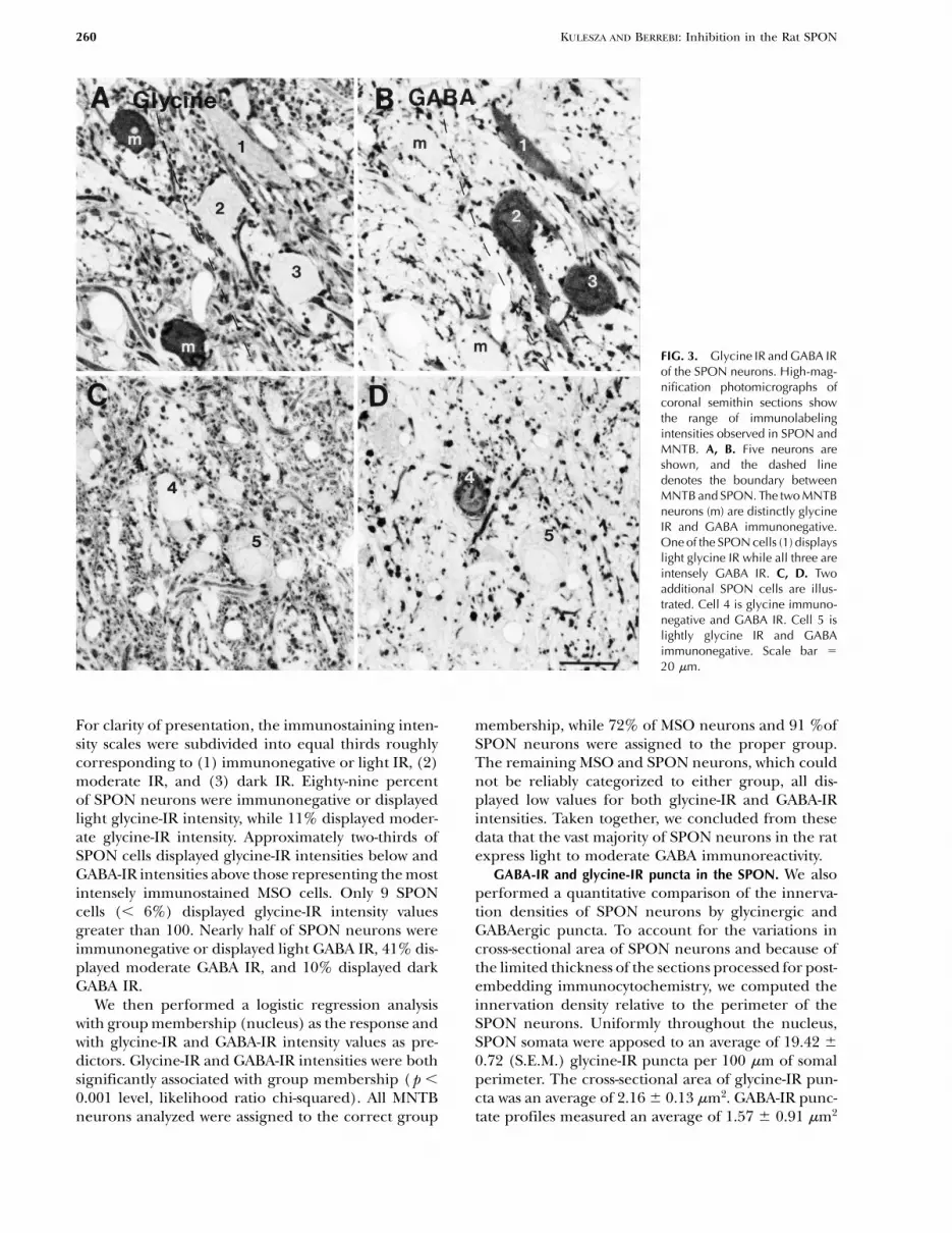
FIG. 3. Glycine IR and GABA IRof the SPON neurons. High-mag-nification photomicrographs ofcoronal semithin sections showthe range of immunolabelingintensities observed in SPON andMNTB. A, B. Five neurons areshown, and the dashed linedenotes the boundary betweenMNTB and SPON. The two MNTBneurons (m) are distinctly glycineIR and GABA immunonegative.One of the SPON cells (1) displayslight glycine IR while all three areintensely GABA IR. C, D. Twoadditional SPON cells are illus-trated. Cell 4 is glycine immuno-negative and GABA IR. Cell 5 islightly glycine IR and GABAimmunonegative. Scale bar 5
20 mm.
For clarity of presentation, the immunostaining inten- membership, while 72% of MSO neurons and 91 %ofSPON neurons were assigned to the proper group.sity scales were subdivided into equal thirds roughly
corresponding to (1) immunonegative or light IR, (2) The remaining MSO and SPON neurons, which couldnot be reliably categorized to either group, all dis-moderate IR, and (3) dark IR. Eighty-nine percent
of SPON neurons were immunonegative or displayed played low values for both glycine-IR and GABA-IRintensities. Taken together, we concluded from theselight glycine-IR intensity, while 11% displayed moder-
ate glycine-IR intensity. Approximately two-thirds of data that the vast majority of SPON neurons in the ratexpress light to moderate GABA immunoreactivity.SPON cells displayed glycine-IR intensities below and
GABA-IR intensities above those representing the most GABA-IR and glycine-IR puncta in the SPON. We alsoperformed a quantitative comparison of the innerva-intensely immunostained MSO cells. Only 9 SPON
cells (, 6%) displayed glycine-IR intensity values tion densities of SPON neurons by glycinergic andGABAergic puncta. To account for the variations ingreater than 100. Nearly half of SPON neurons were
immunonegative or displayed light GABA IR, 41% dis- cross-sectional area of SPON neurons and because ofthe limited thickness of the sections processed for post-played moderate GABA IR, and 10% displayed dark
GABA IR. embedding immunocytochemistry, we computed theinnervation density relative to the perimeter of theWe then performed a logistic regression analysis
with group membership (nucleus) as the response and SPON neurons. Uniformly throughout the nucleus,SPON somata were apposed to an average of 19.42 6with glycine-IR and GABA-IR intensity values as pre-
dictors. Glycine-IR and GABA-IR intensities were both 0.72 (S.E.M.) glycine-IR puncta per 100 mm of somalperimeter. The cross-sectional area of glycine-IR pun-significantly associated with group membership ( p ,
0.001 level, likelihood ratio chi-squared). All MNTB cta was an average of 2.16 6 0.13 mm2. GABA-IR punc-tate profiles measured an average of 1.57 6 0.91 mm2neurons analyzed were assigned to the correct group

KULESZA AND BERREBI: Inhibition in the Rat SPON 261
FIG. 4. Assessment of glycine-IR and GABA-IR intensities in somata IR values of SPON neurons are distributed mostly within the lowerof the SOC. The same neurons were immunolabeled with antiserum third of the glycine-IR intensity scale. The number at the top left ofdirected against glycine or GABA in adjacent semithin sections from each panel indicates the experimental animal identification number.six animals. IR intensity values for MSO cells (squares) cluster at the The sections from all animals were incubated with the commerciallower end of both intensity scales. In contrast, the IR intensity values glycine antiserum (Chemicon) and the GABA antiserum provided byrepresenting MNTB neurons (circles) cluster at the high end of the Dr. David Pow, except those from animal 054/97 (top left panel)glycine-IR and the low end of the GABA-IR scales. SPON neurons which were incubated with the commercial GABA antiserum (HTI(triangles) show a wide range of GABA-IR intensities and are the most Bioproducts). In animals 054/97 and 031/96, but not the remainingintensely immunoreactive for GABA among the three nuclei. Glycine- cases, the antisera were preadsorbed as described in Methods.
262 KULESZA AND BERREBI: Inhibition in the Rat SPON
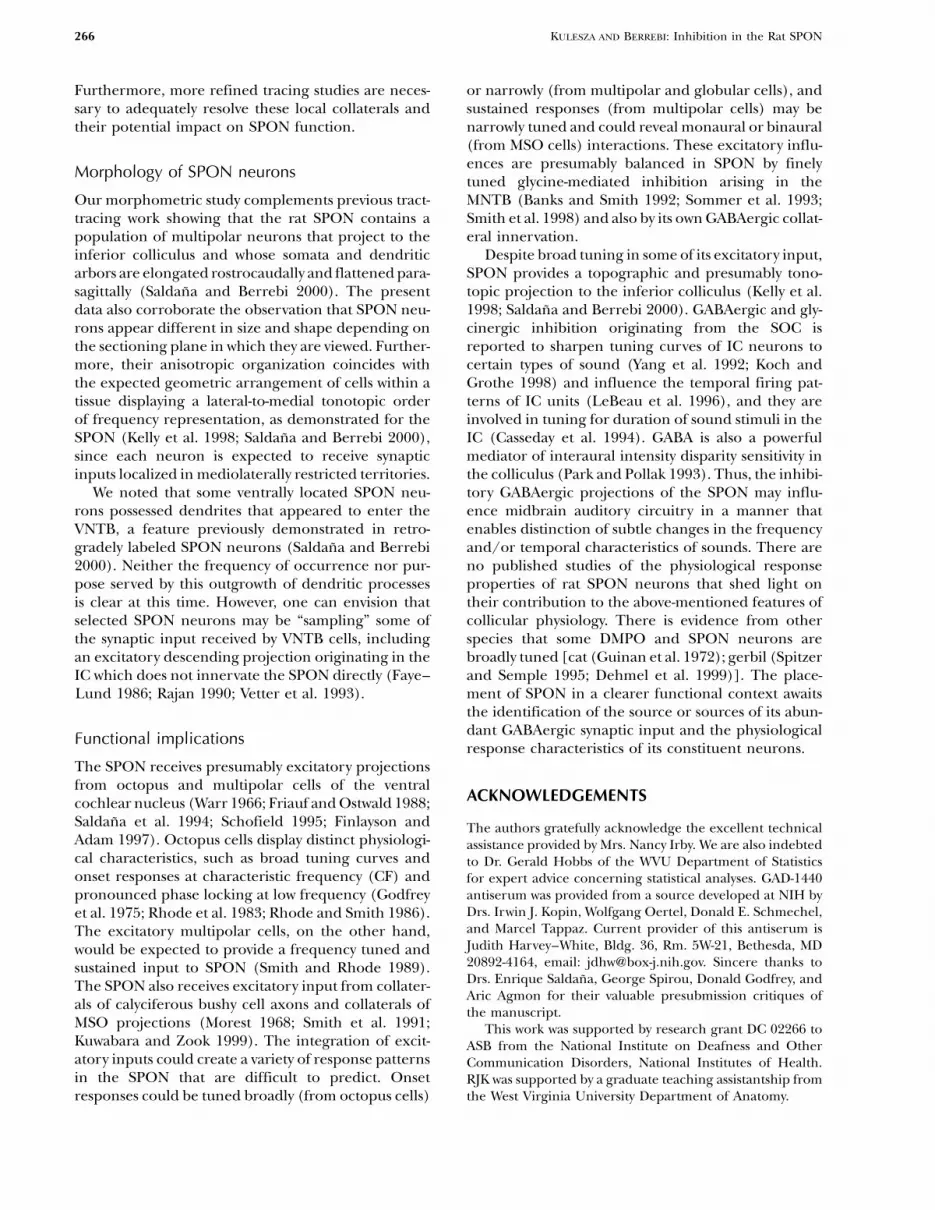
TABLE 1
Classification of glycine and GABA immunoreactivities in SOC neurons
Glycine-IR intensity proportion of cells GABA-IR intensity proportion of cells
Neg. - Light Moderate Dark Neg. - Light Moderate DarkNucleus (OD 5 0–85) (OD 5 85–170) (OD 5 170–255) (OD 5 0–85) (OD 5 85–170) (OD 5 170–255)
MNTB 0% 43% 57% 95% 5% 0%(n 5 60)MSO 100% 0% 0% 100% 0% 0%(n 5 60)SPON 89% 11% 0% 49% 41% 10%(n 5 154)
and were found in apposition to somata throughoutthe SPON. However, somata in the ventro lateral SPONwere apposed to an average of 15.04 6 0.76 GABA-IRpuncta, whileneurons in the dorsomedial portion ofthe nucleus were apposed to an average of 23.37 60.96 puncta per 100 mm of somal perimeter. This dif-ference in innervation density was statistically signifi-cant by ANOVA ( p , 0.0001).
Overview of GAD IR in the SOC
GABA is a small labile molecule that can be difficultto localize with immunocytochemistry and may not
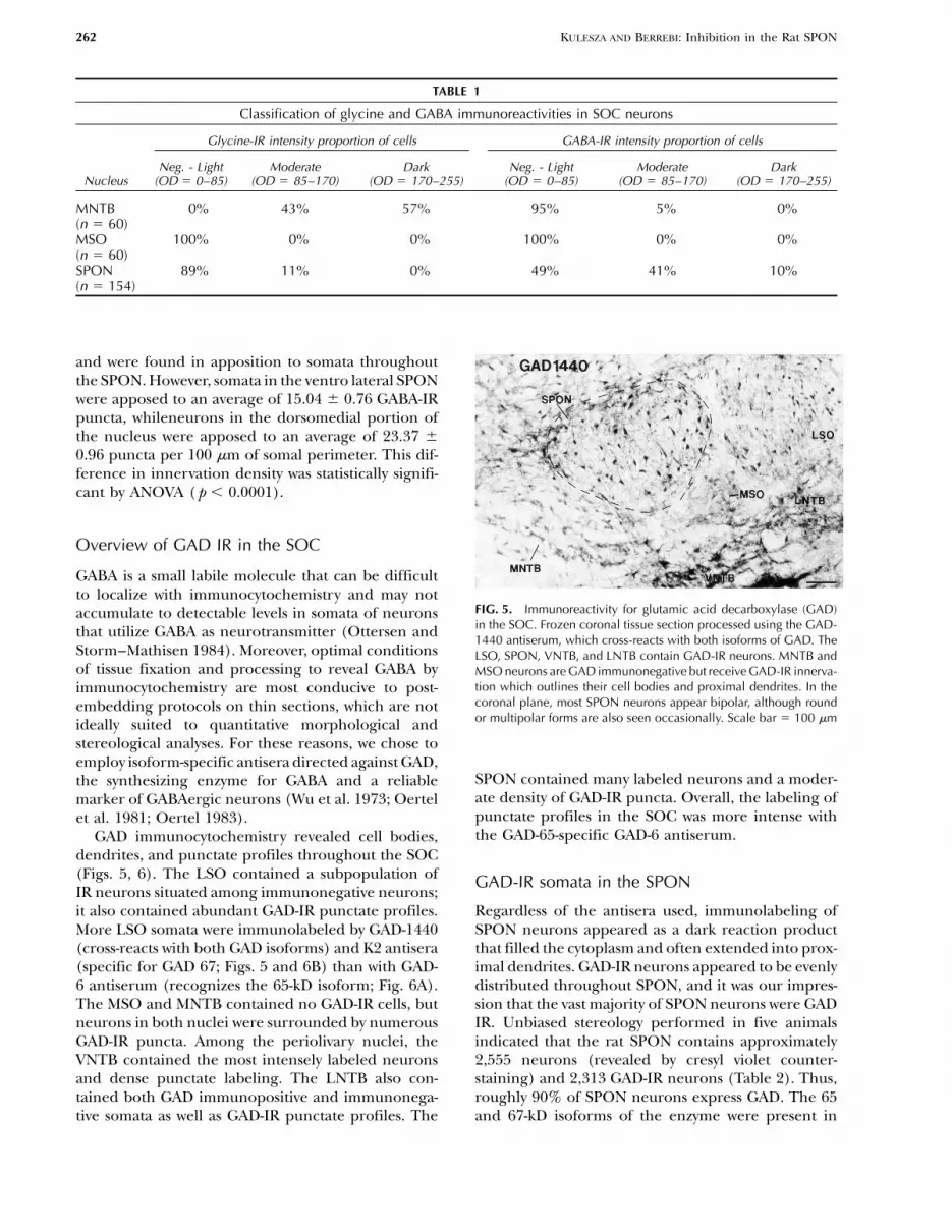
FIG. 5. Immunoreactivity for glutamic acid decarboxylase (GAD)accumulate to detectable levels in somata of neuronsin the SOC. Frozen coronal tissue section processed using the GAD-that utilize GABA as neurotransmitter (Ottersen and1440 antiserum, which cross-reacts with both isoforms of GAD. The
Storm–Mathisen 1984). Moreover, optimal conditions LSO, SPON, VNTB, and LNTB contain GAD-IR neurons. MNTB andof tissue fixation and processing to reveal GABA by MSO neurons are GAD immunonegative but receive GAD-IR innerva-
tion which outlines their cell bodies and proximal dendrites. In theimmunocytochemistry are most conducive to post-coronal plane, most SPON neurons appear bipolar, although roundembedding protocols on thin sections, which are notor multipolar forms are also seen occasionally. Scale bar 5 100 mmideally suited to quantitative morphological and
stereological analyses. For these reasons, we chose toemploy isoform-specific antisera directed against GAD,
SPON contained many labeled neurons and a moder-the synthesizing enzyme for GABA and a reliableate density of GAD-IR puncta. Overall, the labeling ofmarker of GABAergic neurons (Wu et al. 1973; Oertelpunctate profiles in the SOC was more intense withet al. 1981; Oertel 1983).the GAD-65-specific GAD-6 antiserum.GAD immunocytochemistry revealed cell bodies,
dendrites, and punctate profiles throughout the SOC(Figs. 5, 6). The LSO contained a subpopulation of GAD-IR somata in the SPONIR neurons situated among immunonegative neurons;it also contained abundant GAD-IR punctate profiles. Regardless of the antisera used, immunolabeling of
SPON neurons appeared as a dark reaction productMore LSO somata were immunolabeled by GAD-1440(cross-reacts with both GAD isoforms) and K2 antisera that filled the cytoplasm and often extended into prox-
imal dendrites. GAD-IR neurons appeared to be evenly(specific for GAD 67; Figs. 5 and 6B) than with GAD-6 antiserum (recognizes the 65-kD isoform; Fig. 6A). distributed throughout SPON, and it was our impres-
sion that the vast majority of SPON neurons were GADThe MSO and MNTB contained no GAD-IR cells, butneurons in both nuclei were surrounded by numerous IR. Unbiased stereology performed in five animals
indicated that the rat SPON contains approximatelyGAD-IR puncta. Among the periolivary nuclei, theVNTB contained the most intensely labeled neurons 2,555 neurons (revealed by cresyl violet counter-
staining) and 2,313 GAD-IR neurons (Table 2). Thus,and dense punctate labeling. The LNTB also con-tained both GAD immunopositive and immunonega- roughly 90% of SPON neurons express GAD. The 65
and 67-kD isoforms of the enzyme were present intive somata as well as GAD-IR punctate profiles. The

KULESZA AND BERREBI: Inhibition in the Rat SPON 263
TABLE 2
Unbiased estimates of Nissl-stained and GAD-IR neuronsin the rat SPON
Estimated number of neurons
Animal No. Isoform GAD Nissl
002/97aa 2,398002/97b GAD65&67 2,158 2,278037/98 GAD65 2,314024/98 GAD67 2,346005/94 — 2,499035/94 — 2,611
Overall estimates 2,313 2,555
aTo verify the realibility of the unbiased stereology method in our material,two separate estimates of GAD-IR neurons were obtained from aniaml 002/97 using the GAD-1440 antiserum. The number of GAD-IR neurons were thenestimated from two other animals using either the GAD-6 or the K2 antiserum.We also estimated the number of Nissl-stained cells in the SPON of twoadditional animals. Approximately 90% of SPON neurons are GAD-IR, andeach antiserum reveals essentially the same number of cells.
FIG. 6. GAD-65 IR and GAD-67 IR in the SOC. Coronal sectionsillustrate the distribution of GAD isoforms in the SOC as revealedby antisera specifically directed against either A GAD 65 or B GAD67. Immunoreactive neurons are present in SPON, VNTB, and LNTB.Fewer LSO neurons were immunolabeled for GAD 65 than for GAD67. GAD-IR dendrites (arrowheads) appear to extend between theventral SPON and the VNTB. Scale bar 5 100 mm.
equal numbers of neurons (2,314 and 2,346 neurons,respectively), indicating a great degree of coex-pression.
Neuronal morphology
SPON neurons exhibited some variability in size andshape (Figs. 5, 6). In the coronal plane of section, themajority of GAD-IR SPON neurons were either bipolaror oval with a vertical orientation, while a small fractionappeared to have a multipolar morphology. Whenviewed in the parasagittal plane, SPON neurons typi-
FIG. 7. GAD IR in a parasagittal section through the SOC. A. Frozencally displayed a more homogeneous multipolarparasagittal tissue section processed according to the PAP methodappearance, with multiple dendrites extending fromusing GAD-65 antiserum. At this level through the SOC, portions ofthe soma in various directions (Fig. 7). Parasagittallythe VNTB, MNTB, and SPON (outlined by dashed border) are visible.
sectioned profiles of SPON somata (traced separately Bundles of GAD-IR dendrites (arrowheads) are separated by immuno-from their dendrites) were significantly larger and less negative fascicles of trapezoid body fibers. B. Higher-magnification
photomicrograph of the bracketed region in A. SPON neurons appearcircular than coronally sectioned profiles (Table 3).distinctly multipolar when sectioned parasagittally. Note also theThese morphometric data, taken together, indicatedense axosomatic and axodendritic GAD-IR innervation of thesethat typical GAD-IR SPON neurons are large (335.9neurons. In some cases, the ventrally directed GAD-IR dendrites
6 10.64 mm2 average cross-sectional area in the para- (arrowheads) could be traced to their parent cell bodies in the SPON.sagittal plane), multipolar cells whose somata and den- C-caudal, D-dorsal, rf-pontine reticular formation. Scale bar 5 100
mm in A, 50 mm in B.dritic trees are flattened parasagittally.

264 KULESZA AND BERREBI: Inhibition in the Rat SPON
TABLE 3
Morphometric analysis of GAD-labeled SPON neuronsa
Mean MeanMean maximum minimum Mean
Plane of Mean area perimeter diameter diameter circularitysection (mm2 6 S.E.) (mm 6 S.E.) (mm 1 S.E.) (mm 6 S.E.) (6 S.E.)
Coronal (n 5 138) 184.89 6 5.74 63.69 6 1.22 21.51 6 0.43 10.90 6 0.24 0.578 6 0.01b
Sagittal (n 5 108) 335.90 6 10.64b 113.81 6 2.42b 29.64 6 0.71b 14.61 6 0.39b 0.336 6 0.01
aGAD-immunoreactive cell bodies were larger and less circular when sectioned parasagitally than when sectioned coronally, supporting the conclusion thatSPON cell bodies are multipolar, elongated rostrocaudally and flattened parasagitally.
bDifference is significantly different ( p , 0.05) by ANOVA.
In both planes of section, GAD-IR dendrites wereseen extending between the SPON and the neuropilof the nearby VNTB (Figs. 6, 7). In coronal sections,these dendrites appeared to form a single narrow bun-dle. In parasagittal sections it was possible to view aconsiderable portion of the rostrocaudal extent of theSPON, and GAD-IR dendrites appeared as severalsmall bundles separated by distinct immunonegativefascicles of trapezoid body fibers (Fig. 7). In some casesit was evident that the GAD-IR dendrites belonged toSPON neurons (Fig. 7B).
GAD-IR puncta density in the SPON
Each of the three antisera used revealed myriad GAD-IR punctate profiles in SPON, which we interpret aslargely representing axon terminals. These punctawere apposed to GAD-IR somata throughout theSPON, forming characteristic perisomatic and peride-ndritic arrays (Fig. 8). Some of the puncta were con-nected by delicate fibers, suggesting that they were enpassant boutons. GAD-IR puncta were uniform in cross-sectional area throughout the nucleus, measuring anaverage of 1.58 6 0.54 mm2.
FIG. 8. GAD-IR puncta apposed to a GAD-IR neuron in the SPON.SPON neuron displaying a distinct multipolar morphology andDISCUSSIONstrongly expressing the 65-kD isoform of GAD. Numerous GAD-65-IR punctate profiles can be seen apposed to the soma (arrowheads)and dendrites (arrows) of this cell. Scale bar 5 10 mm.Constituent neurons of the SPON
Previous studies of the rat SOC utilizing antisera toGABA or GAD have revealed a GABAergic populationof neurons within the SPON (Mugnaini and Oertel distinct GABAergic and glycinergic neuronal popula-
tions (Helfert et al. 1989; Saint Marie and Baker 1990;1985; Li et al. 1995; Gonazalez–Hernandez et al.1996). Mugnaini and Oertel (1985) did not focus on Ostapoff et al. 1990, 1997). These findings, considered
in combination with tract-tracing studies in guinea pig,the SPON per se but, using the same non-isoform-spe-cific GAD-1440 antiserum used in this study, estimated led Schofield (1991) to suggest that somal morphology
and neurochemical phenotype are correlated withthat between 50% and 90% of the neurons in the ratSPON were GAD immunoreactive. It has also been efferent projection target of SPON neurons. Our data
demonstrate remarkable homogeneity of the SPON ofreported that aspartate-IR neurons dominate in therat SPON (Kumoi et al. 1993), but this isolated finding the rat in that the vast majority of rat SPON neurons
are immunoreactive for GAD. Moreover, it has beenhas not been confirmed. In contrast, immunocyto-chemical studies of guinea pig SPON have described reported recently that virtually all SPON neurons in

KULESZA AND BERREBI: Inhibition in the Rat SPON 265
the rat project to the ipsilateral central nucleus of the Glycinergic and GABAergic inputs to SPONIC (Saldana and Berrebi 2000). Taken together, these neuronsstudies suggest that the rat SPON represents a relatively
Rat SPON neurons express GABA and glycine recep-simply organized nucleus with virtually all of its cellstors on their membranes (Friauf et al. 1997, 1998),providing a purely GABAergic innervation of the ipsi-and we observed abundant glycine IR and GABA IRlateral IC. For these reasons, we propose that the ratin the neuropil of the nucleus. Our data and previousis an ideal species in which to study the physiology
and pharmacology of GABAergic projections. reports suggest an extremely dense glycinergic synap-tic input to SPON which arises, in large part, fromMNTB (Morest 1968; Helfert et al. 1989; Kuwabaraand Zook 1991, 1992b; Banks and Smith 1992; SommerColocalization of GAD isoforms in SPONet al. 1993). Our results also coincide with the highneuronsconcentration of glycine reported in the rat SPON byhigh-performance liquid chromatography (Godfrey etThe fact that both the 65- and 67-kD isoforms of GADal. 2000).colocalize in SPON somata is noteworthy. Esclapez and
The prominent punctate labeling revealed by GABAcoworkers (1994) reported that in many neurons theand GAD immunocytochemistry indicates that SPONtwo GAD isoforms occupy different intracellular com-also receives GABAergic synaptic input, but the sourcepartments, with GAD 65 highly concentrated in termi-of this innervation is not known. One candidate is thenals and GAD 67 distributed in both somata andnearby VNTB, which contains a population of GABAer-terminals. However, there is molecular evidence thatgic neurons (Mugnaini and Oertel 1985; Moore andGAD 65 and 67 can form heterodimers (Sheikh and
Martin 1996), and biochemical data suggest that GAD Moore 1987). In rats, VNTB and SPON display higher67 is targeted to the perikaryal Golgi membrane via GABA concentrations than other SOC nuclei (Godfreyan interaction with GAD 65 (Dirkx et al. 1995). The et al. 2000). However, tract-tracing studies in the ratmembrane-bound GAD 65–67 heterodimer is then have failed to show such a projection (Warr and Beckpresumably shipped from the Golgi apparatus to nerve 1996), and we consider it unlikely that the VNTB repre-terminals, possibly resulting in the cell body and axon sents a significant source of GABAergic puncta interminal colocalization of GAD isoforms in our study. the SPON.The production and possible dimerization of GAD 65 The lemniscal nuclei are also possible sources ofin somata may explain our localization of this isoform GAD-IR puncta in the SPON. Both the dorsal andin SPON perikarya. The precise role of these isoforms ventral nuclei of the lateral lemniscus (DNLL andin GABAergic neurotransmission and cellular metabo- VNLL) contain GABAergic neurons (Thompson et al.lism is not yet understood. Based on counts of GAD- 1985; Moore and Moore 1987; Roberts and Ribak 1987;IR neurons we obtained by unbiased stereology, we Gonzalez–Hernandez et al. 1996; Riquelme et al.report that both isoforms were present in the vast 1998). Tract-tracing data provide evidence that themajority of SPON neurons. Even though it is difficult DNLL innervates the SOC in rats, although it is uncer-to quantify immunolabeling intensity in our material,
tain if the SPON receives any of this input (Bajo et al.the intense labeling found with both the GAD-6 and
1993). The VNLL in cats has been shown to project toK2 antisera suggests that both isoforms are located inthe dorsomedial periolivary nucleus (DMPO) (Whitleysomata at relatively high levels. GAD 65 is inactive inand Henkel 1984), the presumed homolog of thethe absence of cofactor (pyridoxal 58 phosphate, PLP)SPON of rodents, but we are not aware of any reportsand cannot synthesize GABA in cell somata since theof a similar projection in the rat.cofactor is specifically localized to nerve terminals
It is also quite possible, in fact likely, that a propor-(Nicholls 1994). Therefore, if GAD 67 is responsibletion of GAD-IR puncta within the SPON arises fromfor metabolic processes, such as the GABA shunt whichbranches of SPON axons. Preliminary intracellularmoves glutamate into the Krebs cycle (Baxter 1970),labeling experiments from our laboratory indicate thatthen pools of GABA made by this enzyme are likelySPON axons collateralize before leaving the nucleusto be quickly converted to another metabolite (e.g.,and contact other SPON neurons (Kulesza et al. 2000).succinic semialdehyde) and not be involved in neuro-Such an arrangement suggests that SPON neuronstransmission. The inactivity of GAD 65 and the possiblemay provide modulatory feedback to local targets inmetabolic role of GAD 67 might contribute to theand around the nucleus. We are particularly interestedcommon difficulties in revealing GABAergic cellin this possibility, especially given the recent demon-bodies using antisera directed against GABA (e.g.,strations that the SPON of the rat is tonotopically orga-Gonzalez–Hernandez et al. 1996; see also Ottersen
and Storm–Mathisen 1994). nized (Kelly et al. 1998; Saldana and Berrebi 2000).
266 KULESZA AND BERREBI: Inhibition in the Rat SPON
Furthermore, more refined tracing studies are neces- or narrowly (from multipolar and globular cells), andsustained responses (from multipolar cells) may besary to adequately resolve these local collaterals and
their potential impact on SPON function. narrowly tuned and could reveal monaural or binaural(from MSO cells) interactions. These excitatory influ-ences are presumably balanced in SPON by finelyMorphology of SPON neuronstuned glycine-mediated inhibition arising in theMNTB (Banks and Smith 1992; Sommer et al. 1993;Our morphometric study complements previous tract-
tracing work showing that the rat SPON contains a Smith et al. 1998) and also by its own GABAergic collat-eral innervation.population of multipolar neurons that project to the
inferior colliculus and whose somata and dendritic Despite broad tuning in some of its excitatory input,SPON provides a topographic and presumably tono-arbors are elongated rostrocaudally and flattened para-
sagittally (Saldana and Berrebi 2000). The present topic projection to the inferior colliculus (Kelly et al.1998; Saldana and Berrebi 2000). GABAergic and gly-data also corroborate the observation that SPON neu-
rons appear different in size and shape depending on cinergic inhibition originating from the SOC isreported to sharpen tuning curves of IC neurons tothe sectioning plane in which they are viewed. Further-
more, their anisotropic organization coincides with certain types of sound (Yang et al. 1992; Koch andGrothe 1998) and influence the temporal firing pat-the expected geometric arrangement of cells within a
tissue displaying a lateral-to-medial tonotopic order terns of IC units (LeBeau et al. 1996), and they areinvolved in tuning for duration of sound stimuli in theof frequency representation, as demonstrated for the
SPON (Kelly et al. 1998; Saldana and Berrebi 2000), IC (Casseday et al. 1994). GABA is also a powerfulmediator of interaural intensity disparity sensitivity insince each neuron is expected to receive synaptic
inputs localized in mediolaterally restricted territories. the colliculus (Park and Pollak 1993). Thus, the inhibi-tory GABAergic projections of the SPON may influ-We noted that some ventrally located SPON neu-
rons possessed dendrites that appeared to enter the ence midbrain auditory circuitry in a manner thatenables distinction of subtle changes in the frequencyVNTB, a feature previously demonstrated in retro-
gradely labeled SPON neurons (Saldana and Berrebi and/or temporal characteristics of sounds. There areno published studies of the physiological response2000). Neither the frequency of occurrence nor pur-
pose served by this outgrowth of dendritic processes properties of rat SPON neurons that shed light ontheir contribution to the above-mentioned features ofis clear at this time. However, one can envision that
selected SPON neurons may be “sampling” some of collicular physiology. There is evidence from otherspecies that some DMPO and SPON neurons arethe synaptic input received by VNTB cells, including
an excitatory descending projection originating in the broadly tuned [cat (Guinan et al. 1972); gerbil (Spitzerand Semple 1995; Dehmel et al. 1999)]. The place-IC which does not innervate the SPON directly (Faye–
Lund 1986; Rajan 1990; Vetter et al. 1993). ment of SPON in a clearer functional context awaitsthe identification of the source or sources of its abun-dant GABAergic synaptic input and the physiologicalFunctional implicationsresponse characteristics of its constituent neurons.
The SPON receives presumably excitatory projectionsfrom octopus and multipolar cells of the ventral
ACKNOWLEDGEMENTScochlear nucleus (Warr 1966; Friauf and Ostwald 1988;Saldana et al. 1994; Schofield 1995; Finlayson and The authors gratefully acknowledge the excellent technicalAdam 1997). Octopus cells display distinct physiologi- assistance provided by Mrs. Nancy Irby. We are also indebtedcal characteristics, such as broad tuning curves and to Dr. Gerald Hobbs of the WVU Department of Statisticsonset responses at characteristic frequency (CF) and for expert advice concerning statistical analyses. GAD-1440
antiserum was provided from a source developed at NIH bypronounced phase locking at low frequency (GodfreyDrs. Irwin J. Kopin, Wolfgang Oertel, Donald E. Schmechel,et al. 1975; Rhode et al. 1983; Rhode and Smith 1986).and Marcel Tappaz. Current provider of this antiserum isThe excitatory multipolar cells, on the other hand,Judith Harvey–White, Bldg. 36, Rm. 5W-21, Bethesda, MDwould be expected to provide a frequency tuned and20892-4164, email: [email protected]. Sincere thanks tosustained input to SPON (Smith and Rhode 1989).Drs. Enrique Saldana, George Spirou, Donald Godfrey, andThe SPON also receives excitatory input from collater-Aric Agmon for their valuable presubmission critiques of
als of calyciferous bushy cell axons and collaterals of the manuscript.MSO projections (Morest 1968; Smith et al. 1991; This work was supported by research grant DC 02266 toKuwabara and Zook 1999). The integration of excit- ASB from the National Institute on Deafness and Otheratory inputs could create a variety of response patterns Communication Disorders, National Institutes of Health.in the SPON that are difficult to predict. Onset RJK was supported by a graduate teaching assistantship from
the West Virginia University Department of Anatomy.responses could be tuned broadly (from octopus cells)

KULESZA AND BERREBI: Inhibition in the Rat SPON 267
ESCLAPEZ M, TILLAKARATNE NJ, KAUFMAN DL, TOBIN AJ, HOUSERREFERENCESCR. Comparative localization of two forms of glutamic acid decar-boxylase. J. Neurosci. 14:1834–1855, 1994.ADAMS JC. Cytology of periolivary cells and the organization of their
FAYE–LUND H. Projection from the inferior colliculus to the superiorprojections in the cat. J. Comp. Neurol. 215:275–289, 1983.olivary complex in the albino rat. Anat. Embryol. 175:35–52, 1986.AOKI E, SEMBA R, KEINO H, KATO K, KASHIWAMATA S. Glycine-like
FINLAYSON PG, ADAM TJ. Excitatory and inhibitory response adapta-immunoreactivity in the rat auditory pathway. Brain Res. 442:tion in the superior olive complex affects binaural acoustic pro-63–71, 1988.cessing. Hear. Res. 103:1–18, 1997.ASCHOFF A, OSTWALD J. Different origins of cochlear efferents in
FINLAYSON PG, CASPARY DM. Synaptic potentials of chinchilla lateralsome bat species, rats, and guinea pigs. J. Comp. Neurol. 264:superior olivary neurons. Hear. Res. 38:221–228, 1989.56–72, 1987.
FRIAUF E, OSTWALD J. Divergent projections of physiologically char-BAJO VM, MERCHAN MA, LOPEZ DE, ROUILLER EM. Neuronal mor-acterized rat ventral cochlear nucleus neurons as shown by intra-phology and efferent projections of the dorsal nucleus of theaxonal injection of horseradish peroxidase. Exp. Brain Res.lateral lemniscus in the rat. J. Comp. Neurol. 334:241–262, 1993.73:263–284, 1988.BANKS MI, SMITH PH. Intracellular recordings from neurobiotin-
FRIAUF F, HAMMERSCHMIDT B, KIRSCH J. Development of adult typelabeled cells in brain slices of the rat medial nucleus of the trape-zoid body. J. Neurosci. 12:2819–2837, 1992. inhibitory glycine receptors in the central auditory system of rats.
BAXTER CF. The nature of GABA. In: A Lajtha (ed) Handbook of J. Comp. Neurol. 385:117–134, 1997.Neurochemistry. Plenum Press, New York, pp 289–353, 1970. FRIAUF E, FRITSCHY JM, KUCHENBROD T. Differential distribution of
BAYON A, POSSANI LD, TAPIA M, TAPIA R. Kinetics of brain glutamate GABA-A receptor subunits in the auditory brainstem of adult rats.decarboxylase. Interactions with glutamate, pyridoxal 58-phos- Assoc. Res. Otolaryngol. 21:457, 1998.phate and glutamate-pyridoxal 58-phosphate Schiff base. J. Neuro- FUENTES V, BERREBI AS, SALDANA E. Trajectory, morphology andchem. 29:519–525, 1977. distribution of axons of the superior paraolivary nucleus that
BERREBI AS, MUGNAINI E. Distribution and targets of the cartwheel innervate the inferior colliculus in the rat. Assoc. Res. Otolaryn-cell axon in the dorsal cochlear nucleus of the guinea pig. Anat. gol. 22:885, 1999.Embryol. 183:427–454, 1991. GODFREY DA, KIANG NY, NORRIS BE. Single unit activity in the pos-
BERREBI AS, MALMIERCA MS, SALDANA E. Features of organization teroventral cochlear nucleus of the cat. J. Comp. Neurol. 162:of the superior paraolivary nucleus of the rat, guinea pig and 247–268, 1975.chinchilla. Assoc. Res. Otolaryngol. 20:570, 1997. GODFREY DA, FARMS WB, GODFREY TG, MIKESELL NL, LIU J. Amino
BEYERL BD. Afferent projections to the central nucleus of the inferior acid concentrations in rat cochlear nucleus and superior olivecolliculus in the rat. Brain Res. 145:209–223, 1978. measured by high performance liquid chromatography. Hear.
CAICEDO A, HERBERT H. Topography of descending projections from Res. in press. 2000.the inferior colliculus to auditory brainstem nuclei in the rat. J. GONZALEZ–HERNANDEZ T, MANTOLAN–SARMIENTO B, GONZALEZ–Comp. Neurol. 328:377–392, 1993. GONZALEZ B, PEREZ–GONZALEZ H. Sources of GABAergic input
CAMPISTRON G, BUIJS RM, GEFFARD M. Glycine neurons in the brain to the inferior colliculus of the rat. J. Comp. Neurol. 372:and spinal cord. Antibody production and immunocytochemical 309–326, 1996.localization. Brain Res. 376:400–405, 1986. GUINAN JJ, NORRIS BE, GUINAN SS. Single auditory units in the
CASSEDAY JH, EHRLICH D, COVEY E. Neural tuning for sound dura- superior olivary complex II: Locations of unit categories andtion: Role of inhibitory mechanisms in the inferior colliculus. tonotopic organization. Int. J. Neurosci. 4:147–166, 1972.Science. 264:847–850, 1994. GUNDERSEN HJ. Stereology of arbitrary particles. A review of unbi-
CASPARY DM, BACKOFF PM, FINLAYSON PG, PALOMBI PS. Inhibitoryased number and size estimators and the presentation of some
inputs modulate discharge rate within frequency receptive fieldsnew ones, in memory of William R. Thompson. J. Microsc. 143:
of anteroventral cochlear nucleus neurons. J. Neurophysiol.3–45, 1986.
72:2124–2133, 1994.GUNDERSEN HJ. The nucleator. J. Microsc. 151:3–21, 1988.CHANG YC, GOTTLIEB DI. Characterization of the proteins purifiedGUNDERSEN HJ, BAGGER P, BENDTSEN TF, EVANS SM, KORBO L, MAR-with monoclonal antibodies to glutamic acid decarboxylase. J.
CUSSEN N, MOLLER A, NIELSEN K, NYENGAARD JR, PAKKENBERG B.Neurosci. 8:2123–2130, 1988.The new stereological tools: disector, fractionator, nucleator andCOLEMAN JR, CLERICI WJ. Sources of projections to subdivisions ofpoint sampled intercepts and their use in pathological researchthe inferior colliculus in the rat. J. Comp. Neurol. 262:and diagnosis. Acta. Pathol. Microbiol. Immunol. Scand. 96:215–226, 1987.857–881, 1988.DEHMEL S, DOERRSCHEIDT GJ, REUBSAMEN R. Electrophysiological
HELFERT RH, BONNEAU JM, WENTHOLD RJ, ALTSCHULER RA. GABAcharacterization of neurons in the superior paraolivary nucleusand glycine immunoreactivity in the guinea pig superior olivaryof the gerbil (Meriones unguiculatus). Assoc. Res. Otolaryngol.complex. Brain Res. 6:269–286, 1989.22:279, 1999.
HELFERT RH, ASCHOFF A. Superior olivary complex and nuclei ofDENNER LA, WU JY. Two forms of rat brain glutamic acid decarboxyl-the lateral lemniscus. In: G Ehret, R Romand (eds) The Centralase differ in their dependence on free pyridoxal phosphate. J.Auditory System. Oxford University Press, New York, ppNeurochem. 44:957–965, 1985.193–258, 1997.DIRKX R, JR, THOMAS A, LI L, LERNMARK A, SHERWIN RS, DE CAMILLI
KAUFMAN DL, HOUSER CR, TOBIN AJ. Two forms of the gamma-P, SOLIMENA M. Targeting of the 67-kDa isoform of glutamic acidaminobutyric acid synthetic enzyme glutamate decarboxylase havedecarboxylase to intracellular organelles is mediated by its interac-distinct intraneuronal distributions and cofactor interactions. J.tion with the NH2- terminal region of the 65-kDa isoform ofNeurochem. 56:720–723, 1991.glutamic acid decarboxylase. J. Biol. Chem. 270:2241–2246, 1995.
KELLY JB, LISCUM A, VAN ADEL B, ITO M. Projections from theERLANDER M, TILLAKARATNE N, FELDBLUM N, PATEL N, TOBIN A.superior olive and lateral lemniscus to tonotopic regions of theTwo genes encode distinct glutamate decarboxylases. Neuron.rat’s inferior colliculus. Hear. Res. 116:43–54, 1998.7:91–100, 1991.
KLUG A, PARK TJ, POLLAK GD. Glycine and GABA influence binauralERLANDER MG, TOBIN AJ. The structural and functional heterogene-processing in the inferior colliculus of the mustache bat. J. Neuro-ity of glutamic acid decarboxylase:a review. Neurochem. Res.
16:215–226, 1991. physiol. 74:1701–1713, 1995.

268 KULESZA AND BERREBI: Inhibition in the Rat SPON
KOCH U, GROTHE B. GABAergic and glycinergic inhibition sharpens OERTEL W, SCHMECHEL D, MUGNAINI E, TAPPAZ M, KOPIN I. Immuno-tuning for frequency modulations in the inferior colliculus of the cytochemical localization of glutamate decarboxylase in rat cere-big brown bat. J. Neurophysiol. 80:71–82, 1998. bellum with a new antiserum. Neuroscience. 6:2715–2735, 1981.
KULESZA RJ, SALDANA E, BERREBI AS. Distribution of inhibitory neu- OERTEL W. Synaptic responses and electrical properties of cells inrotransmitters in the superior paraolivary nucleus of the rat. Assoc. brain slices of the mouse anteroventral cochlear nucleus. J. Neu-Res. Otolaryngol. 21:367, 1998. rosci. 3:2043–2053, 1983.
KULESZA R, BERREBI AS. Distribution of GAD isoforms in the superior ORDRONNEAU P, LINDSTROM PB, PETRUSZ P. Four unlabeled antibodyparaolivary nucleus (SPON) of the rat. Assoc. Res. Otolaryngol. bridge techniques: a comparison. J. Histochem. Cytochem.22:278, 1999. 29:1397–1404, 1981.
KULESZA R, HOLT A, SPIROU G, BERREBI AS. Intracellular labeling OSEN KK, MUGNAINI E, DAHL A-L, CHRISTIANSEN AH. Histochemicalof axonal collaterals of SPON neurons. Assoc. Res. Otolaryngol. localization of acetylcholinesterase in the cochlear and superior23:132, 2000. olivary nuclei. A reappraisal with emphasis on the cochlear gran-
KUMOI K, SAITO N, TANAKA C. Immunohistochemical localization ule cell system. Arch. Ital. Biol. 122:169–212, 1984.of gamma-aminobutyric acid- and aspartate-containing neurons OSTAPOFF EM, MOREST DK, POTASHER SJ. Retrograde transport ofin the guinea pig superior olivary complex. Hear. Res. 68: 3H-GABA from the cochlear nucleus to the superior olive in guinea173–179, 1993. pig. Soc. Neurosci. Abstr. 11:1051, 1985.
KUWABARA N, DICAPRIO RA, ZOOK JM. Afferents to the medial OSTAPOFF EM, MOREST DK, POTASHNER SJ. Uptake and retrogradenucleus of the trapezoid body and their collateral projections. J. transport of [3H]GABA from the cochlear nucleus to the superiorComp. Neurol. 314:684–706, 1991. olive in the guinea pig. J. Chem. Neuroanat. 3:285–295, 1990.
KUWABARA N, ZOOK JM. Classification of the principal cells of the OSTAPOFF EM, BENSON CG, SAINT MARIE RL. GABA- and glycine-medial nucleus of the trapezoid body. J. Comp. Neurol. 314: immunoreactive projections from the superior olivary complex707–720, 1991. to the cochlear nucleus in guinea pig. J. Comp. Neurol. 381:
KUWABARA N, ZOOK JM. Projections to the medial superior olive 500–512, 1997.from the medial and lateral nuclei of the trapezoid body in rodents OTTERSEN OP, STORM–MATHISEN J. Glutamate- and GABA-con-and bats. J. Comp. Neurol. 324:522–538, 1992a. taining neurons in the mouse and rat brain, as demonstrated
KUWABARA N, ZOOK JM. Inputs to the superior paraolivary nucleus. with a new immunocytochemical technique. J. Comp. Neurol.Soc. Neurosci. Abstr. 18:1193, 1992b. 229:374–392, 1984.
KUWABARA N, ZOOK JM. Local collateral projections from the medial PARK TJ, POLLAK GD. GABA shapes sensitivity to interaural intensitysuperior olive to the superior paraolivary nucleus in the gerbil. disparities in the mustache bat’s inferior colliculus: implicationsBrain Res. 846:59–71, 1999. for encoding sound location. J. Neurosci. 13:2050–2067, 1993.
LEBEAU FE, REES A, MALMIERCA MS. Contribution of GABA- and POURCHO RG, GOEBEL DJ, JOJICH L, HAZLETT JC. Immunocytochem-glycine-mediated inhibition to the monaural temporal response ical evidence for the involvement of glycine in sensory centers ofproperties of neurons in the inferior colliculus. J. Neurophysiol. the rat brain. Neuroscience. 46:643–656, 1992.75:902–919, 1996. RAJAN R. Electrical stimulation of the inferior colliculus at low rates
LEGAY F, HENRY S, TAPPAZ M. Evidence for two distinct forms of protects the cochlea from auditory desensitization. Brain Res.native glutamic acid decarboxylase in rat brain soluble extract: 506:192–204, 1990.an immunoblotting study. J. Neurochem. 48:1022–1026, 1987. RHODE WS, OERTEL D, SMITH PH. Physiological response properties
LI L, WU SH, ZHANG DX, KELLY JB. GABAergic projections to theof cells labeled intracellularly with horseradish peroxidase in cat
central nucleus of the inferior colliculus of the rat: a combinationventral cochlear nucleus. J. Comp. Neurol. 213:448–463, 1983.
of fluorogold retrograde tracing with immunocytochemistry. Soc.RHODE WS, SMITH PH. Encoding timing and intensity in the ventral
Neurosci. Abstr. 25:166.13, 1995.cochlear nucleus of the cat. J. Neurophysiol. 56:261–286, 1986.
MARTIN DL, MARTIN SB, WU SJ, ESPINA N. Regulatory properties ofRIQUELME R, MERCHAN MA, OTTERSEN OP. GABA and glycine inbrain glutamate decarboxylase GAD: the apoenzyme of GAD is
the ventral nucleus of the lateral lemniscus: An inmunocytochemi-present principally as the smaller of two molecular forms of GADcal and in situ hybridization study in rat. Assoc. Res. Otolaryngol.in brain. J. Neurosci. 11:2725–2731, 1991.21:370, 1998.MARTIN DL, RIMVALL K. Regulation of gamma-aminobutyric acid
ROBERTS E, FRANKEL S. Gamma-butyric acid in brain: Its formationsynthesis in the brain. J. Neurochem. 60:395–407, 1993.from glutamic acid. J. Biol. Chem. 187:55–63, 1950.MOORE DR. Auditory brainstem of the ferret: sources of projections
ROBERTS RC, RIBAK CE. GABAergic neurons and axon terminals into the inferior colliculus. J. Comp. Neurol. 269:342–354, 1988.the brainstem auditory nuclei of the gerbil. J. Comp. Neurol.MOORE JK, MOORE RY. Glutamic acid decarboxylase-like immunore-258:267–280, 1987.activity in brainstem auditory nuclei of the rat. J. Comp. Neurol.
SAINT MARIE RL, BAKER RA. Neurotransmitter-specific uptake and260:157–174, 1987.retrograde transport of [3H]glycine from the inferior colliculusMOREST DK. The growth of synaptic endings in the mammalianby ipsilateral projections of the superior olivary complex andbrain: a study of the calyces of the trapezoid body. Z. Anat.nuclei of the lateral lemniscus. Brain Res. 524:244–253, 1990.Entwicklungsgesch. 127:201–220, 1968.
SALDANA E, LOUGH DR, BERREBI AS. Superior Paraolivary NucleusMUGNAINI E, DAHL AL. Zinc-aldehyde fixation for light-microscopicSPON: Connectivity revealed by transport of dextran amine. Soc.immunocytochemistry of nervous tissues. J. Histochem. Cyto-Neurosci. Abstr. 20:975, 1994.chem. 31:1435–1438, 1983.
SALDANA E, BERREBI AS. Anisotropic organization of the rat superiorMUGNAINI E, OERTEL WH. An atlas of the distribution of GABAergicparaolivary nucleus. Anat. Embryol. in press. 202:265–279, 2000.neurons and terminals in the rat CNS as revealed by GAD immuno-
SCHOFIELD BR. Superior paraolivary nucleus in the pigmentedhistochemistry. In: A Bjorklund, T Hokfelt (eds) Handbook ofguinea pig:separate classes of neurons project to the inferior colli-Chemical Neuroanatomy. Vol. 4: GABA and Neuropeptides in theculus and the cochlear nucleus. J. Comp. Neurol. 312:68–76, 1991.CNS, Part I. Elsevier Science Publishers, Amsterdam, pp
SCHOFIELD BR. Projections from the cochlear nucleus to the superior436–608, 1985.paraolivary nucleus in guinea pigs. J. Comp. Neurol. 360:NICHOLLS D. Blackwell Science Ltd., Oxford Proteins, Transmitters
and Synapses. 1994. 135–149, 1995.

KULESZA AND BERREBI: Inhibition in the Rat SPON 269
SCHWARTZ IR. The superior olivary complex and the lateral lemniscal THOMPSON GC, CORTEZ AM, LAM DM. Localization of GABA immu-noreactivity in the auditory brainstem of guinea pigs. Brain Res.nuclei. In: DB Webster, AN Popper, RR Fay (eds) Mammalian339:119–122, 1985.Auditory Pathways: Neuroanatomy. Springer-Verlag, New York, pp
THOMPSON AM, THOMPSON GC. Projections from the posteroventral117–167, 1992.cochlear nucleus to the superior olivary complex in guinea pig:SHEIKH SN, MARTIN DL. Heteromers of glutamate decarboxylaselight and EM observations with the PHA-L method. J. Comp.isoforms occur in rat cerebellum. J. Neurochem. 66:2082–2090,Neurol. 311:495–508, 1991a.1996.
THOMPSON AM, THOMPSON GC. Posteroventral cochlear nucleusSMITH PH, RHODE WS. Structural and functional properties distin-projections to olivocochlear neurons. J. Comp. Neurol. 303:guish two types of multipolar cells in the ventral cochlear nucleus.267–285, 1991b.
J. Comp. Neurol. 4:595–616, 1989.VETTER DE, ADAMS JC, MUGNAINI E. Chemically distinct rat olivo-
SMITH PH, JORIS PX, CARNEY LH, YIN TC. Projections of physiologi- cochlear neurons. Synapse. 7:21–43, 1991.cally characterized globular bushy cell axons from the cochlear VETTER DE, MUGNAINI E. Distribution and dendritic features of threenucleus of the cat. J. Comp. Neurol. 304:387–407, 1991. groups of rat olivocochlear neurons. A study with two retrograde
SMITH PH, JORIS PX, YIN TC. Anatomy and physiology of principal cholera toxin tracers. Anat. Embryol. 185:1–16, 1992.cells of the medial nucleus of the trapezoid body MNTB of the VETTER DE, SALDANA E, MUGNAINI E. Input from the inferior collicu-cat. J. Neurophysiol. 79:3127–3142, 1998. lus to medial olivocochlear neurons in the rat: a double label
SOMMER I, LINGENHOHL K, FRIAUF E. Principal cells of the rat medial study with PHA-L and cholera toxin. Hear. Res. 70:173–186, 1993.WARR WB. Fiber degeneration following lesions in the anterior ven-nucleus of the trapezoid body: an intracellular in vivo study of their
tral cochlear nucleus of the cat. Exp. Neurol. 14:453–474, 1966.physiology and morphology. Exp. Brain Res. 95:223–239, 1993.WARR WB, BECK JE. Multiple projections from the ventral nucleusSPINK DC, WU SJ, MARTIN DL. Multiple forms of glutamate decarbox-
of the trapezoid body in the rat. Hear. Res. 93:83–101, 1996.ylase in porcine brain. J. Neurochem. 40:1113–1119, 1983.WHITE JS, WARR WB. The dual origins of the olivocochlear bundleSPIROU GA, BERREBI AS. Glycine immunoreactivity in the lateral
in the albino rat. J. Comp. Neurol. 219:203–214, 1983.nucleus of the trapezoid body of the cat. J. Comp. Neurol.WHITLEY JM, HENKEL CK. Topographical organization of the in-383:473–488, 1997.
ferior collicular projection and other connections of the ventralSPITZER MW, SEMPLE MN. Neurons sensitive to interaural phasenucleus of the lateral lemniscus in the cat. J. Comp. Neurol. 229:
disparity in gerbil superior olive: diverse monaural and temporal257–270, 1984.
response properties. J. Neurophysiol. 73:1668–1690, 1995. WU JY, MATSUDA T, ROBERTS E. Purification and characterizationSTERIO DC. The unbiased estimation of number and sizes of arbitrary of glutamate decarboxylase from mouse brain. J. Biol. Chem.
particles using the disector. J. Microsc. 134:127–136, 1984. 248:3029–3034, 1973.STERNBERGER LA. Immunocytochemistry. Wiley, New York 1979. YIN TCT, CHAN JCK. Interaural time sensitivity in the medial superiorSTORM–MATHISEN J, OTTERSEN OP. Antibodies and fixatives for the olive of the cat. J. Neurophysiol. 65:465–488, 1990.
immunocytochemical localization of glycine. In: OP Ottersen, J YANG L, POLLAK GD, RESLER C. GABAergic circuits sharpen tuningStorm–Mathisen (eds) Glycine Neurotransmission. John Wiley curves and modify response properties in the mustache bat infe-
rior colliculus. J. Neurophysiol. 68:1760–1774, 1992.and Sons, New York pp 281–302, 1990.