the role of casein in supporting the operation of surface bound kinesin
TRANSCRIPT
BioMed CentralJournal of Biological Engineering
ss
Open AcceResearchThe role of casein in supporting the operation of surface bound kinesinVivek Verma1, William O Hancock2 and Jeffrey M Catchmark*3Address: 1Engineering Science and Mechanics, University Park, PA 16802, USA, 2Department of Bioengineering, University Park, PA 16802, USA and 3Agricultural and Biological Engineering, University Park, PA 16802, USA
Email: Vivek Verma - [email protected]; William O Hancock - [email protected]; Jeffrey M Catchmark* - [email protected]
* Corresponding author
AbstractMicrotubules and associated motor proteins such as kinesin are envisioned for applications such asbioseparation and molecular sorting to powering hybrid synthetic mechanical devices. One of thechallenges in realizing such systems is retaining motor functionality on device surfaces. Kinesinmotors adsorbed onto glass surfaces lose their functionality or ability to interact with microtubulesif not adsorbed with other supporting proteins. Casein, a milk protein, is commonly used inmicrotubule motility assays to preserve kinesin functionality. However, the mechanism responsiblefor this preservation of motor function is unknown. To study casein and kinesin interaction, a seriesof microtubule motility assays were performed where whole milk casein, or its αs1 and αs2, β or κsubunits, were introduced or omitted at various steps of the motility assay. In addition, a series ofepifluorescence and total internal reflection microscopy (TIRF) experiments were conductedwhere fluorescently labeled casein was introduced at various steps of the motility assay to assesscasein-casein and casein-glass binding dynamics. From these experiments it is concluded that caseinforms a bi-layer which supports the operation of kinesin. The first tightly bound layer of caseinmainly performs the function of anchoring the kinesin while the second more loosely bound layerof casein positions the head domain of the kinesin to more optimally interact with microtubules.Studies on individual casein subunits indicate that β casein was most effective in supporting kinesinfunctionality while κ casein was found to be least effective.
BackgroundBiological molecular motors are a unique class of pro-teins, which exist in eukaryotic cells and function as nanoscale vehicles that drive a range of fundamental biologicalprocesses. Kinesin motor proteins transport intracellularcargo, move proteins and mRNA in neurons, and play avital role in cell division. These motor proteins move uni-directionally on protein tracks known as microtubuleswhich assemble and disassemble creating a uniquedynamically reconfigurable transportation system. Kines-
ins are powered by the hydrolysis of adenosine triphos-phate (ATP) and exert a maximal force of ~6 pN andexhibit a maximal efficiency of ~50% [1-3]. Biologicalmotors are the subject of intense research in part becauseof their potential to be used in-vitro as 'nano-engines' forseveral future applications ranging from bioseparation topowering hybrid micro and nano scale electromechanicalsystems (MEMS/NEMS) [4-8]. Surface interactions andadsorption of molecular motors have also been studied[9,10].
Published: 20 October 2008
Journal of Biological Engineering 2008, 2:14 doi:10.1186/1754-1611-2-14
Received: 23 October 2007Accepted: 20 October 2008
This article is available from: http://www.jbioleng.org/content/2/1/14
© 2008 Verma et al; licensee BioMed Central Ltd. This is an Open Access article distributed under the terms of the Creative Commons Attribution License (http://creativecommons.org/licenses/by/2.0), which permits unrestricted use, distribution, and reproduction in any medium, provided the original work is properly cited.
Page 1 of 10(page number not for citation purposes)
Journal of Biological Engineering 2008, 2:14 http://www.jbioleng.org/content/2/1/14
An important hurdle that must be overcome before realiz-ing these applications is developing approaches for pre-serving motor protein function when integrated intoengineered devices and surfaces. Motor protein function isstudied using gliding assays or bead assays [11,12]. In thebead assay, microtubules are immobilized on a surfaceand biological motors attached to microscale beads movealong these immobilized microtubules. In the glidingassay, the motor proteins are immobilized on a surfaceand microtubules are transported across the surface by theimmobilized motors. In either case, these motor proteinsare attached to a surface in a manner that is intended topreserve their functionality. This is typically accomplishedvia the use of blocker proteins that prevent motor dena-turation on the untreated surface [11,13]. We studiedmicrotubule motility through gliding assay.
The most common proteins used for creating kinesincompatible surface interfaces are caseins extracted frombovine milk [12-14]. These proteins have been extensivelyused in the surface immobilization of motor proteinssuch as kinesin, and they are also used in Western Blots forblocking nonspecific adsorption of antibodies to nitrocel-lulose membranes. A typical process for performing a glid-ing assay consists of: 1) constructing a flow cell using aglass slide (Fisher Finest Premium microscope slides) andglass cover slip (Corning 1 1/2, 18 mm2); 2) blocking theglass surfaces by flowing in a solution of 0.5 mg/ml caseinprotein; 3) adsorbing kinesin to the glass surface by flow-ing in a solution of kinesin containing ATP and 0.2 mg/mlcasein; 4) flowing in a microtubule solution containingATP, antifade reagents and casein, and 5) observingmicrotubule movements by epi-fluorescence microscopy.Microtubule motility is quantified by examining severalparameters including the density of microtubulesobserved, their landing rate on the surface, the microtu-bule transport velocity, and the distances microtubulesmove before detaching from the surface and diffusingaway.
The initial blocking step is implemented to form a layer ofthe protein casein on the surface to prevent the kinesindenaturation that would occur if the motors proteins weredirectly adsorbed to the glass surface [11]. Casein isincluded in the motor and microtubule solutions basedon a qualitative observation that it improves microtubulemotility. However, although casein is used to optimizemotor activity in a number of different in vitro motilityassays on surfaces, no studies have been conducted tounderstand the mechanism by which casein creates com-patible interfaces. Understanding the role of casein in sup-porting biomotor function is important for developingdesign rules for engineering optimal surfaces for use inadvanced hybrid devices that incorporate biologicalmotor proteins for actuation and transport.
Caseins are phosphoproteins that bind calcium and formlarge aggregates in milk. Casein subunits range from20–30 kDa and are classified into 4 types: αs1, αs2, β, andκ. The relative concentrations of these caseins varydepends upon the milk producing species. Casein frombovine milk, which is commonly used in motor proteinexperiments, contains the following subunit composi-tions in skim milk (mg/ml): αs1 (12%–15%), αs2(3%–4%), β (9%–11%), and κ (2%–4%) [15,16]. The dif-ferent forms of casein have been extensively studied dueto their importance in milk, as food additives and asemulsifiers and stabilizers for glue, paint, and other mate-rials [17]. Although there are no crystal structures of anyof the subunits, every one of the subunits contains one ormore clearly defined hydrophobic and hydrophilicdomains, resulting in the formation of micelles with dif-fering geometries [18-21].
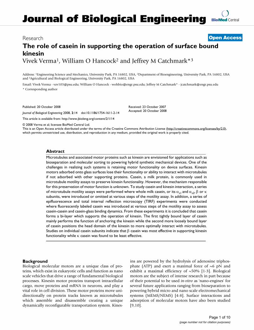
The dynamics of casein adsorption onto surfaces has beenstudied using various approaches. Specifically, adsorptionof β casein onto hydrophilic and hydrophobic surfaceshas been studied by Nylander, et al. [22] and Kull, et. al[23]. The β casein protein has one distinct hydrophilicand hydrophobic domain, which gives the molecule astrong amphiphilic character [15]. It has been found thaton hydrophobic surfaces β casein forms a monolayerwhere its hydrophobic region is positioned next to thehydrophobic surface and its hydrophilic region is protrud-ing outward into the aqueous solution. However, in thecase of a hydrophilic surface, the hydrophilic domain ofthe β casein protein adsorbs into the surface resulting in atightly packed monolayer producing a hydrophobic sur-face. A second more loosely bound layer of β casein isthen adsorbed onto the first layer where its hydrophobicregion is aligned with the hydrophobic region of the firstadsorbed casein layer and its hydrophilic region is posi-tioned outward into the aqueous solution. The thicknessof the bi-layer has been measured to be ~15 nm [22]. Thisis illustrated in figure 1.
In the present work, we have examined the role of bothwhole casein (consisting of mixture of αs1, αs2, β, and κcasein) and the various casein subunits in modulatingkinesin function in the microtubule gliding assay. In par-ticular, microtubule motility assays were performedwhere casein was either included or excluded in the sur-face blocking, kinesin adsorption, and microtubule solu-tions. The inclusion of casein in various incubation stepsand the specific casein subunits used had dramaticimpacts on the functionality of surface adsorbed kinesin,as measured by counting the number of microtubules onthe surface. Most notably, samples including κ caseinexhibited significant reductions in the number of micro-tubules observed. Studies on fluorescently labeled caseinwere also performed where the labeled casein was intro-
Page 2 of 10(page number not for citation purposes)
Journal of Biological Engineering 2008, 2:14 http://www.jbioleng.org/content/2/1/14
duced into the flow-cell surface either as a first or afterunlabeled casein had been adsorbed. By measuring thebinding of fluorescent casein to the surface, we hypothe-size that casein forms a bi-layer on the glass surface andthat optimum kinesin-microtubule binding is achievedwhen kinesin proteins co-assemble with the casein bi-layer.
MethodsCasein was prepared by dissolving the protein powder(Sigma C-7078, St Louis, MO) in BRB80 buffer (80 mMPIPES, 1 mM MgCl2, 1 mM EGTA, pH 6.9), centrifuged toremove un-dissolved casein. The solution was then fil-tered using a 220 nm filter (Fisher brand, Ireland). Fulllength Drosophila conventional kinesin heavy chain andlacking light chains was bacterially expressed and purifiedaccording to established procedures [24]. Tubulin wasprepared from bovine brain and rhodamine-labeledaccording to standard procedures [25,26]. Microtubuleswere polymerized by incubating a mixture of 32 μM tubu-lin, 4 mM MgCl2, 1 mM GTP, and 5% DMSO in BRB80buffer at 37°C for 20 minutes, and were stabilized bydiluting to a final concentration of 0.32 μM in a solutioncontaining 10 μM paclitaxel. Kinesin was labeled withRhodamine-NHS using the standard protocol for tubulinlabeling [25] to examine relative density of kinesinabsorbing to the surface in the presence of differentcasein.
All the motility experiments were performed in a closedvolume flow cell as previously described [13,27]. Flowcells consisted of a microscope slide (Fisher Finest Pre-mium microscope slides) and a glass cover slip (Corning1 1/2, 18 mm2) separated by double side tape, and hadvolumes of ~20 μl. The protein solutions were exchangedin these flow cells and motility of microtubules wasassessed. In a flow cell, solutions were exchanged byinjecting fresh buffer from one end and wicking out theprevious solution from the other. In control experimentsthe first step is surface blocking where 0.5 mg/ml casein is
incubated in the flow cell for 10 minutes followed by asolution of kinesin motors (0.05–5 μg/ml) including 0.2mg/ml casein and 100 μM Mg-ATP in BRB80 buffer.Finally, microtubule motility solution containing 0.032μM microtubules, 10 μM paclitaxel, 0.2 mg/ml casein, 1mM Mg-ATP, 20 mM D-glucose, 20 μg/ml glucose oxi-dase, 8 μg/ml catalase and 0.5% β-merceptoethanol inBRB80 buffer is introduced. Microtubules were observedby epifluorescence microscopy (Nikon E600, 100×, 1.3 N.A. oil immersion Plan Fluor objective) and captured tovideotape using a Genwac GW-902H camera. Microtu-bule surface binding was quantified by counting thenumber of microtubules present in a given video screen(65 μm × 48 μm) after 15 minute incubation time. Micro-tubules with length longer than 1 μm and moving formore than 2 μm were counted. Immobilized microtu-bules or those smaller than 1 μm were not counted. In allcases microtubule binding refers to microtubule bindingto and moved by kinesin.
Epifluorescence and total internal reflection microscopy(TIRF) using a Nikon TE2000 inverted microscope (60×,1.45 NA, CFI Plan Apo TIRF oil objective) was used tomeasure fluorescent intensities to study bi-layer forma-tion of casein, and co-assembly between kinesin andcasein proteins. Meta-Vue (Universal Imaging, PA) soft-ware was used to acquire images for bi-layer and co-assembly studies using rhodamine casein and rhodaminekinesin. Fluorescent intensities from the surface from therecorded images were measured in arbitrary units.
Results and discussionTo assess the role that casein is playing in supporting theactivity of surface-adsorbed kinesin, we first examinedkinesin function under a range of casein conditions andthen quantified the binding of both casein and kinesinmotors to the surfaces using fluorescence assays. Whilesurface-adsorbed motors are difficult to visualize micro-scopically, it is relatively straightforward to visualize andquantify the interaction of microtubules with these
Schematic illustration of the adsorption of β casein on hydrophilic and hydrophobic surfacesFigure 1Schematic illustration of the adsorption of β casein on hydrophilic and hydrophobic surfaces. In the case of a hydrophilic surface, a bi-layer structure forms with a loosely bound second layer which measures ~15 nm thick [22].
������������ ���
hydrophobic region
hydrophilic region �����
�������������� ����
Page 3 of 10(page number not for citation purposes)
Journal of Biological Engineering 2008, 2:14 http://www.jbioleng.org/content/2/1/14
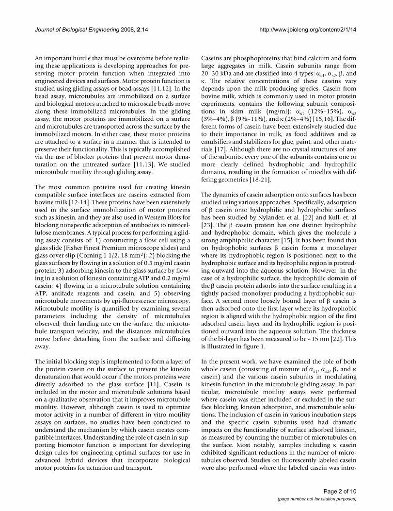
motors. It has been shown previously that because indi-vidual kinesin motors are sufficient to bind microtubulesand transport them across the surface, below maximalmotor densities, the rate that microtubules land and moveover the surface is proportional to the density of activekinesins on the surface [11,24]. Hence, the surface densityof functional motors can be estimated by allowing micro-tubules to land for a defined time and then counting thenumber of microtubules on the surface. We systematicallyremoved casein from either the blocking solution, themotor solution, or the microtubule solution and countedthe average number of microtubules propagating over akinesin functionalized glass surface after 15 minutes.Whole bovine milk casein, containing all four casein sub-units, was used, and both high (8 μg/ml) and low (0.8 μg/ml) concentrations of kinesin protein were examined. Ithas been shown previously that at low kinesin concentra-tions, very few or no motors are functionally adsorbed tothe surface if the surfaces are not pretreated with a block-ing protein, but at high motor concentrations some frac-
tion of the motors presumably bind and denature on thesurface and replace the role of blocking proteins [11].However, to date, the role of casein in the motor andmicrotubule solutions has not been systematically inves-tigated.
Table 1 shows the measured number of microtubulespropagating in a 65 μm × 48 μm video screen area undervarious casein loading conditions using either high or lowkinesin concentrations. The numbers of microtubuleswere counted from five screen shots and their mean andstandard deviation was calculated. From this data threeclear observations can be made. First, if there is no caseinblocking step and no casein is included in the motor solu-tion then no microtubules are observed. This result, whichis consistent with previous work [11] suggests that in theabsence of a casein treatment to block the surface, thekinesin motors denature on the glass surface or bind suchthat their motor domains cannot interact with microtu-bules. Second, the initial casein blocking step does not sig-
Table 1: Measured average number of microtubules observed under different casein and kinesin concentration conditions.
Casein Blocking Kinesin concentration Casein in Kinesin Casein in MT Average MT per screen (N = 5)
No Low (0.8 μg/ml) No No 0 ± 0
Yes 0 ± 0
Yes No 1.4 ± 1.3
Yes 4.4 ± 1.5
High (8 μg/ml) No No 0 ± 0
Yes 0 ± 0
Yes No 9.4 ± 1.1
Yes 60 ± 4.4
Yes Low (0.8 μg/ml) No No 0 ± 0
Yes 0 ± 0
Yes No 3.6 ± 1.5
Yes 9.2 ± 2.4
High (8 μg/ml) No No 13.6 ± 2.4
Yes 48 ± 5.7
Yes No 20 ± 3
Yes 80 ± 9.6
N: Number of screen shots.
Page 4 of 10(page number not for citation purposes)
Journal of Biological Engineering 2008, 2:14 http://www.jbioleng.org/content/2/1/14
nificantly impact the microtubule motility when casein isincluded in the subsequent kinesin adsorption solution.The inclusion of a casein blocking step improved thenumber of observed microtubules from 60 ± 4.4 (Mean ±standard deviation, N = 5) to 80 ± 9.6 (33% improve-ment) for high kinesin concentrations, and from 4.4 ± 1.5to 9.2 ± 2.4 (109% improvement) for low kinesin concen-trations. Presumably, because the casein concentration(200 μg/ml) is considerably higher than the motor con-centration (8 or 0.8 μg/ml), the casein binds rapidly to thesurface and is able to carry out its blocking role if it isincluded in the motor solution. Third, the inclusion ofcasein in the microtubule solution always increases theobserved number of microtubules, with an averageimprovement of 3-fold over all conditions, and a maxi-mum observed improvement of 5.4-fold in the case wherecasein is included in both the blocking and kinesinadsorption steps and a high kinesin concentration wasused.
The observed variation in the number of microtubulespresent on the surface under differing experimental condi-tions must then result from the impact of casein on eitherthe availability of surface adsorbed kinesin to participatein microtubule binding, or the density of kinesinadsorbed. From table 1 it can be concluded that surfaceadsorbed kinesin retain their functionality except in thecase of low kinesin concentration and where no casein hasbeen included in any incubation solution. The loss ofkinesin functionality can either be due to denaturing ofthe kinesin or binding of the kinesin head domain to thesurface, which may result in no observed microtubulebinding. Hence, the inclusion of casein is not impactingthe ability of the kinesin to operate normally on an avail-able microtubule if contact with a microtubule was possi-ble. This hypothesis is based on the observation ofincreased observed microtubule density when casein isadded to the final microtubule solution. Moreover, sinceno additional kinesin had been added in the microtubulesolution with casein, the density of kinesin on the surfacecould not have been increased, but rather more of theexisting kinesin on the surface were available to partici-pate in microtubule binding when casein was included inthe microtubule solution. It is also apparent that oncekinesin are adsorbed in the absence of casein, they cannotbe revived by subsequent inclusion of casein in the micro-tubule solution. This may result from kinesin having astronger affinity for the surface than casein or that, in thecase of kinesin denaturing on the glass surface, that dena-tured kinesin cannot regain their functionality if casein issubsequently introduced and is able to displace thekinesin.
Based on these observations, we hypothesize that casein isforming a bi-layer on the surface of the glass substrate in
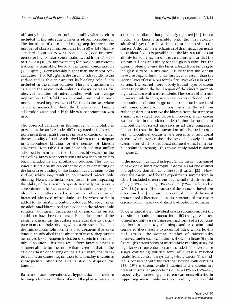
a manner similar to that previously reported [22]. In ourmodel, the kinesin assemble onto the first stronglyadsorbed layer of casein which anchor the kinesin to thesurface. Although the mechanism of this interaction needsto be identified, it is possible that the kinesin tail has anaffinity for some region on the casein protein or that thekinesin tail has an affinity for the glass surface but thecasein protein prevents the kinesin head from binding tothe glass surface. In any case, it is clear that the kinesinhave a stronger affinity to the first layer of casein than thesecond layer of casein has for the first layer of casein or thekinesin. The second more loosely bound layer of caseinserves to position the head region of the kinesin promot-ing interaction with a microtubule. The observed increasein microtubule binding when casein was included in themicrotubule solution suggests that the kinesin are fixedwith some affinity to their position since the solutionexchange does not remove the kinesin from the surface toa significant extent (see below). However, when caseinwas included in the microtubule solution the number ofmicrotubules observed increases in all cases suggestingthat an increase in the interaction of adsorbed motorswith microtubules occurs in the presence of additionalcasein, which replenishes the second loosely boundcasein layer which is disrupted during the final microtu-bule solution exchange. This co-assembly model is shownin figure 2.
In the model illustrated in figure 2, the casein is assumedto have one distinct hydrophilic domain and one distincthydrophobic domain, as is true for β-casein [22]. How-ever, the casein used for the experiments summarized intable 1 included casein from bovine milk which consistsof αs1(12%–15%), αs2(3%–4%), β (9%–11%), and κ(2%–4%) caseins. The structure of these caseins have beendetermined [21] and are not identical. One of the mostpronounced differences is in the structure of the two αcaseins, which have two distinct hydrophobic domains.
To determine if the individual casein subunits impact thekinesin-microtubule interaction differently, we per-formed motility assays using purified forms of α (contain-ing both αs1 and αs2 subunits),β and κ caseins, andcompared these results to a control using whole bovinemilk casein. The average number of microtubulesobserved under each condition is shown in figure 3(a). Infigure 3(b) screen shots of microtubule motility assay forhigh kinesin concentration are included. The results forassays containing purified form of α casein matchedresults from control assays using whole casein. This find-ing is consistent with the fact that bovine milk contains15%–19% α casein, while β caseins and κ caseins arepresent in smaller proportions of 9%–11% and 2%–4%,respectively. Interestingly, β casein was most effective insupporting microtubule motility, leading to a 1.6-fold
Page 5 of 10(page number not for citation purposes)
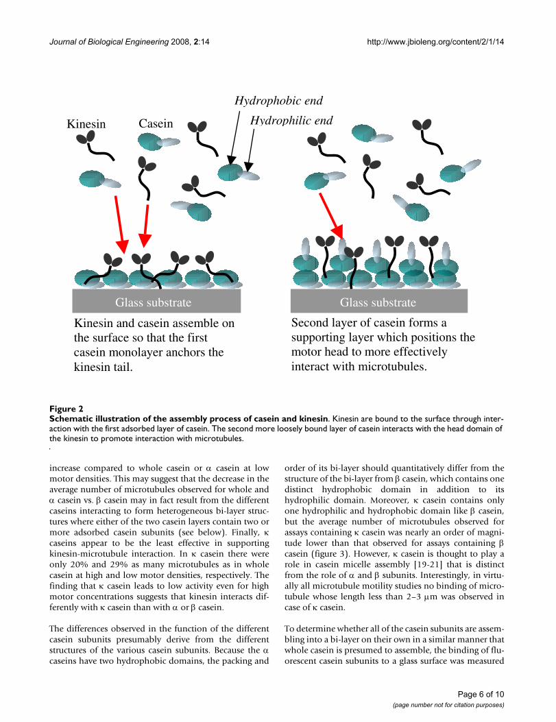
Journal of Biological Engineering 2008, 2:14 http://www.jbioleng.org/content/2/1/14
increase compared to whole casein or α casein at lowmotor densities. This may suggest that the decrease in theaverage number of microtubules observed for whole andα casein vs. β casein may in fact result from the differentcaseins interacting to form heterogeneous bi-layer struc-tures where either of the two casein layers contain two ormore adsorbed casein subunits (see below). Finally, κcaseins appear to be the least effective in supportingkinesin-microtubule interaction. In κ casein there wereonly 20% and 29% as many microtubules as in wholecasein at high and low motor densities, respectively. Thefinding that κ casein leads to low activity even for highmotor concentrations suggests that kinesin interacts dif-ferently with κ casein than with α or β casein.
The differences observed in the function of the differentcasein subunits presumably derive from the differentstructures of the various casein subunits. Because the αcaseins have two hydrophobic domains, the packing and
order of its bi-layer should quantitatively differ from thestructure of the bi-layer from β casein, which contains onedistinct hydrophobic domain in addition to itshydrophilic domain. Moreover, κ casein contains onlyone hydrophilic and hydrophobic domain like β casein,but the average number of microtubules observed forassays containing κ casein was nearly an order of magni-tude lower than that observed for assays containing βcasein (figure 3). However, κ casein is thought to play arole in casein micelle assembly [19-21] that is distinctfrom the role of α and β subunits. Interestingly, in virtu-ally all microtubule motility studies no binding of micro-tubule whose length less than 2–3 μm was observed incase of κ casein.
To determine whether all of the casein subunits are assem-bling into a bi-layer on their own in a similar manner thatwhole casein is presumed to assemble, the binding of flu-orescent casein subunits to a glass surface was measured
Schematic illustration of the assembly process of casein and kinesinFigure 2Schematic illustration of the assembly process of casein and kinesin. Kinesin are bound to the surface through inter-action with the first adsorbed layer of casein. The second more loosely bound layer of casein interacts with the head domain of the kinesin to promote interaction with microtubules.
Kinesin and casein assemble on the surface so that the first casein monolayer anchors the kinesin tail.
Hydrophobic end
Hydrophilic endKinesin Casein
Glass substrate
Second layer of casein forms a supporting layer which positions the motor head to more effectively interact with microtubules.
Glass substrate
Page 6 of 10(page number not for citation purposes)
Journal of Biological Engineering 2008, 2:14 http://www.jbioleng.org/content/2/1/14
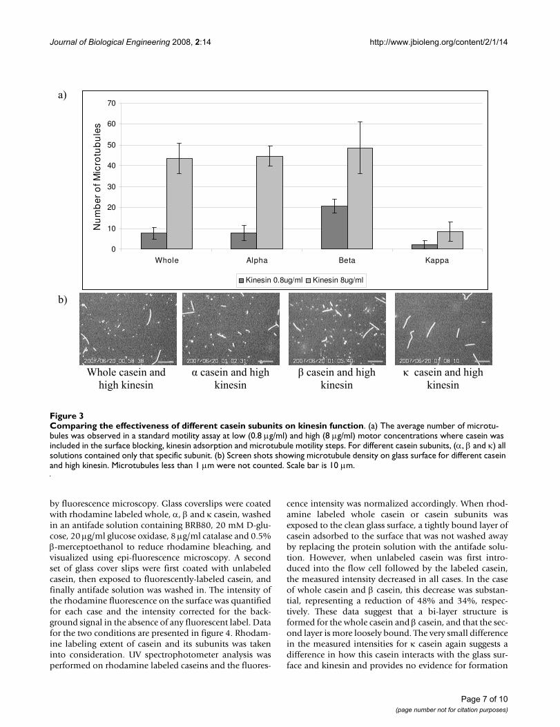
by fluorescence microscopy. Glass coverslips were coatedwith rhodamine labeled whole, α, β and κ casein, washedin an antifade solution containing BRB80, 20 mM D-glu-cose, 20 μg/ml glucose oxidase, 8 μg/ml catalase and 0.5%β-merceptoethanol to reduce rhodamine bleaching, andvisualized using epi-fluorescence microscopy. A secondset of glass cover slips were first coated with unlabeledcasein, then exposed to fluorescently-labeled casein, andfinally antifade solution was washed in. The intensity ofthe rhodamine fluorescence on the surface was quantifiedfor each case and the intensity corrected for the back-ground signal in the absence of any fluorescent label. Datafor the two conditions are presented in figure 4. Rhodam-ine labeling extent of casein and its subunits was takeninto consideration. UV spectrophotometer analysis wasperformed on rhodamine labeled caseins and the fluores-
cence intensity was normalized accordingly. When rhod-amine labeled whole casein or casein subunits wasexposed to the clean glass surface, a tightly bound layer ofcasein adsorbed to the surface that was not washed awayby replacing the protein solution with the antifade solu-tion. However, when unlabeled casein was first intro-duced into the flow cell followed by the labeled casein,the measured intensity decreased in all cases. In the caseof whole casein and β casein, this decrease was substan-tial, representing a reduction of 48% and 34%, respec-tively. These data suggest that a bi-layer structure isformed for the whole casein and β casein, and that the sec-ond layer is more loosely bound. The very small differencein the measured intensities for κ casein again suggests adifference in how this casein interacts with the glass sur-face and kinesin and provides no evidence for formation
Comparing the effectiveness of different casein subunits on kinesin functionFigure 3Comparing the effectiveness of different casein subunits on kinesin function. (a) The average number of microtu-bules was observed in a standard motility assay at low (0.8 μg/ml) and high (8 μg/ml) motor concentrations where casein was included in the surface blocking, kinesin adsorption and microtubule motility steps. For different casein subunits, (α, β and κ) all solutions contained only that specific subunit. (b) Screen shots showing microtubule density on glass surface for different casein and high kinesin. Microtubules less than 1 μm were not counted. Scale bar is 10 μm.
��
0
10
20
30
40
50
60
70
Whole Alpha Beta Kappa
Nu
mb
er o
f Mic
rotu
bu
les
Kinesin 0.8ug/ml Kinesin 8ug/ml
��
�� ������� ���� ���
������������ ���� �������������
�� ���� �������������
��� ���� �������������
�
Page 7 of 10(page number not for citation purposes)
Journal of Biological Engineering 2008, 2:14 http://www.jbioleng.org/content/2/1/14
of a bi-layer. For α casein there was less overall bindingand the pretreatment with unlabeled casein had a mini-mal effect. The microtubule binding data from figure 3suggest that whole casein and α casein interact similarlywith surface-adsorbed kinesin, but the binding data in fig-ure 4 imply the opposite and instead suggest similaritiesbetween whole casein and β casein. It is possible that thedifference rests in the specific interactions between the dif-ferent αs1 and αs2 caseins, which are not examined in iso-lation here. Alternatively, it is possible that in the case ofthe whole casein, the individual caseins form a heteroge-neous bi-layer that integrates different casein subunits.For example, the first adsorbed layer could be an α caseinwhose hydrophobic region interacts with the hydropho-bic region of a β casein, etc. Importantly, the maximumobserved number of microtubules in figure 3 wasobserved for pure β casein, and both figure 4 and previouswork suggest that β casein forms a bi-layer on the surface[22], suggesting that a casein bi-layer is an important com-ponent in promoting effective kinesin-microtubule inter-action.
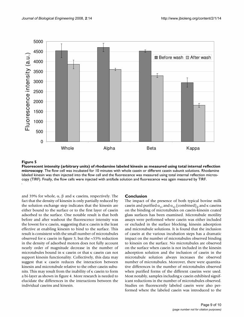
While measuring microtubule binding is a good readoutfor the density of active kinesin motors on the surface, itcannot differentiate between changes in the concentrationof motors adsorbed to the surface and the relative activity
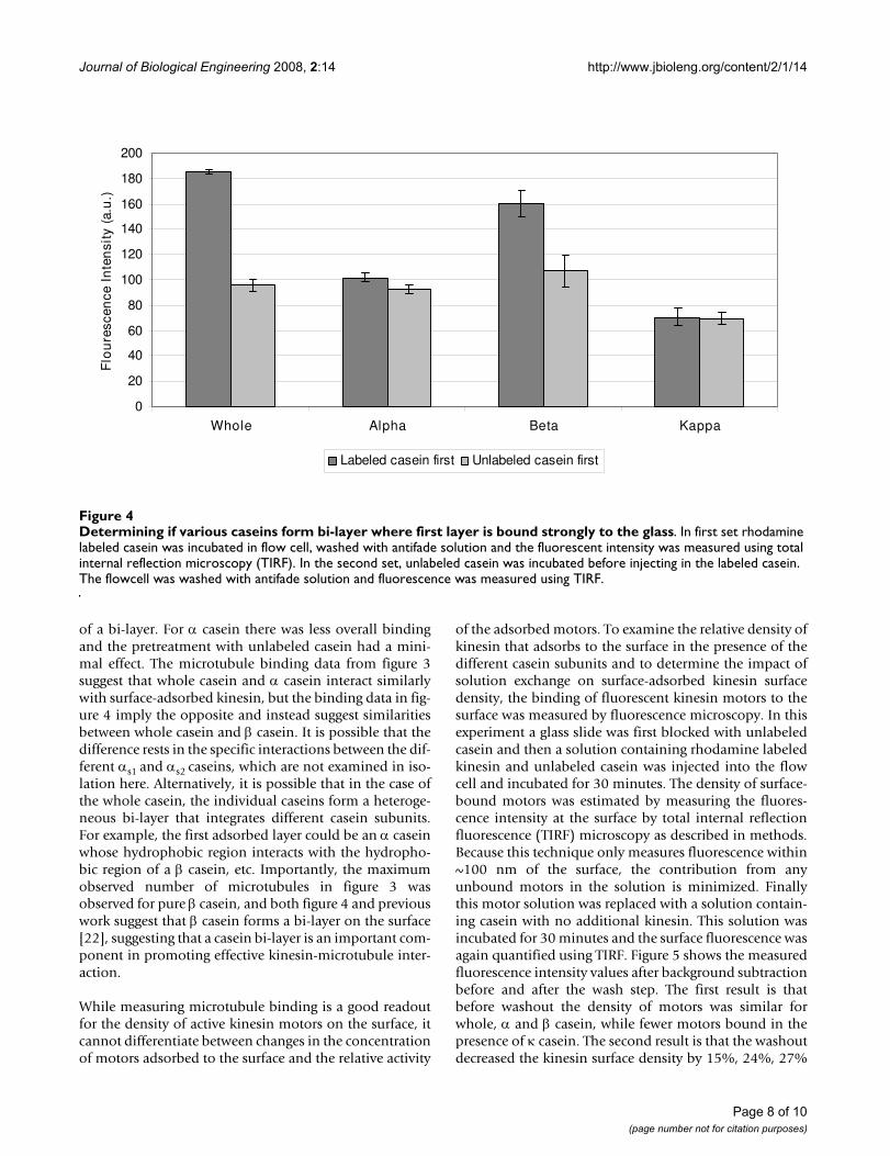
of the adsorbed motors. To examine the relative density ofkinesin that adsorbs to the surface in the presence of thedifferent casein subunits and to determine the impact ofsolution exchange on surface-adsorbed kinesin surfacedensity, the binding of fluorescent kinesin motors to thesurface was measured by fluorescence microscopy. In thisexperiment a glass slide was first blocked with unlabeledcasein and then a solution containing rhodamine labeledkinesin and unlabeled casein was injected into the flowcell and incubated for 30 minutes. The density of surface-bound motors was estimated by measuring the fluores-cence intensity at the surface by total internal reflectionfluorescence (TIRF) microscopy as described in methods.Because this technique only measures fluorescence within~100 nm of the surface, the contribution from anyunbound motors in the solution is minimized. Finallythis motor solution was replaced with a solution contain-ing casein with no additional kinesin. This solution wasincubated for 30 minutes and the surface fluorescence wasagain quantified using TIRF. Figure 5 shows the measuredfluorescence intensity values after background subtractionbefore and after the wash step. The first result is thatbefore washout the density of motors was similar forwhole, α and β casein, while fewer motors bound in thepresence of κ casein. The second result is that the washoutdecreased the kinesin surface density by 15%, 24%, 27%
Determining if various caseins form bi-layer where first layer is bound strongly to the glassFigure 4Determining if various caseins form bi-layer where first layer is bound strongly to the glass. In first set rhodamine labeled casein was incubated in flow cell, washed with antifade solution and the fluorescent intensity was measured using total internal reflection microscopy (TIRF). In the second set, unlabeled casein was incubated before injecting in the labeled casein. The flowcell was washed with antifade solution and fluorescence was measured using TIRF.
0
20
40
60
80
100
120
140
160
180
200
Whole Alpha Beta Kappa
Flo
ure
scen
ce I
nte
nsi
ty (
a.u
.)
Labeled casein first Unlabeled casein first
Page 8 of 10(page number not for citation purposes)
Journal of Biological Engineering 2008, 2:14 http://www.jbioleng.org/content/2/1/14
and 39% for whole, α, β and κ caseins, respectively. Thefact that the density of kinesin is only partially reduced bythe solution exchange step indicates that the kinesin areeither bound to the surface or to the first layer of caseinadsorbed to the surface. One notable result is that bothbefore and after washout the fluorescence intensity wasthe lowest for κ casein, suggesting that κ casein is the leasteffective at enabling kinesin to bind to the surface. Thisresult is consistent with the small number of microtubulesobserved for κ casein in figure 3, but the ~55% reductionin the density of adsorbed motors does not fully accountnearly order of magnitude decrease in the number ofmicrotubules bound in κ casein or that κ casein can notsupport kinesin functionality. Collectively, this data maysuggest that κ casein reduces the interaction betweenkinesin and microtubule relative to the other casein subu-nits. This may result from the inability of κ casein to forma bi-layer as shown in figure 4. More research is needed toelucidate the differences in the interactions between theindividual caseins and kinesin.
ConclusionThe impact of the presence of both typical bovine milkcasein and purified αs1 and αs2 (combined),β and κ caseinson the binding of microtubules on casein-kinesin coatedglass surfaces has been examined. Microtubule motilityassays were performed where casein was either includedor excluded in the surface blocking, kinesin adsorptionand microtubule solutions. It is found that the inclusionof casein at the various incubation steps has a dramaticimpact on the number of microtubules observed bindingto kinesin on the surface. No microtubules are observedon the surface when casein is not included in the kinesinadsorption solution and the inclusion of casein in themicrotubule solution always increases the observednumber of microtubules. Moreover, there were quantita-tive differences in the number of microtubules observedwhen purified forms of the different caseins were used.Most notably, samples including κ casein exhibited signif-icant reductions in the number of microtubules observed.Studies on fluorescently labeled casein were also per-formed where the labeled casein was introduced to the
Fluorescent intensity (arbitrary units) of rhodamine labeled kinesin as measured using total internal reflection microscopyFigure 5Fluorescent intensity (arbitrary units) of rhodamine labeled kinesin as measured using total internal reflection microscopy. The flow cell was incubated for 10 minutes with whole casein or different casein subunit solutions. Rhodamine labeled kinesin was then injected into the flow cell and the fluorescence was measured using total internal reflection micros-copy (TIRF). Finally, the flow cells were injected with antifade solution and fluorescence was again measured by TIRF.
0
500
1000
1500
2000
2500
3000
3500
4000
4500
5000
Whole Alpha Beta Kappa
Flu
ore
scen
ce in
ten
sity
(a.
u.)
Before wash After wash
Page 9 of 10(page number not for citation purposes)
Journal of Biological Engineering 2008, 2:14 http://www.jbioleng.org/content/2/1/14
Publish with BioMed Central and every scientist can read your work free of charge
"BioMed Central will be the most significant development for disseminating the results of biomedical research in our lifetime."
Sir Paul Nurse, Cancer Research UK
Your research papers will be:
available free of charge to the entire biomedical community
peer reviewed and published immediately upon acceptance
cited in PubMed and archived on PubMed Central
yours — you keep the copyright
Submit your manuscript here:http://www.biomedcentral.com/info/publishing_adv.asp
BioMedcentral
surface either first or after an unlabeled casein had beenadsorbed. Fluorescence intensities were notably higher inthe case where the labeled casein was introduced to thesurface first, except in the case of α and κ casein.
From this data a bi-layer casein adsorption model is pro-posed where the first tightly bound layer of casein mainlyperforms the function of anchoring the kinesin while thesecond more loosely bound layer of casein positions thehead domain of the kinesin to more optimally interactwith microtubules. Studies on adsorption of fluorescentlylabeled caseins in the first and second layers support thebi-layer model of casein assembly. Results using wholecasein from bovine milk are also compared to thatobtained when using purified αs1 and αs2 (combined),βand κ caseins and significant variations in observednumber of microtubules are found. β casein is found to bethe best at promoting kinesin-microtubule interactionwhile κ casein is found to be the worst, and may behavecompletely different than the other caseins including thelack of a bi-layer assembly.
Kinesin and microtubule system is studied extensively todesign hybrid synthetic devices for detection and trans-port. Motility assays are also used in studying the role ofkinesin, in vitro, which may help get an insight on how tocontrol cell division. Some of the assays, however, requiremore specific engineered surfaces than others and it isimportant to understand the mechanism which supportsoperation of surface bound kinesin. In this paper, we pro-pose a bi-layer model based on our studies. However,more studies are needed to understand the interaction ofkinesin with the various caseins. A more detailed under-standing may provide new insights enabling the design ofengineered surfaces for optimally supporting kinesinactivity, a key parameter in the development of hybridbiological-synthetic devices incorporating biologicalmolecular motors.
Competing interestsThe authors declare that they have no competing interests.
Authors' contributionsVV carried out all the experiments. JMC, VV and WOHcontributed to the manuscript preparation. VV and JMCconceived the studies and designed the experiments. JMC,VV and WOH contributed to the discussion and bi-layermodel. All authors have read and approved the final man-uscript.
AcknowledgementsThis work was supported by The Pennsylvania State University Center for Nanoscale Science, a NSF Materials Research Science and Engineering Center (DMR0213623). It was also supported by the National Nanotech-nology Infrastructure Network (NSF Cooperative Agreement No. 0335765 with Cornell University) and The Pennsylvania State University
Materials Research Institute. Vivek Verma wishes to thank the Haythorn-thwaite Foundation for their Founder's Prize and grant for year 2005–06.
We thank Maruti Uppalapati for purifying kinesin and Gayatri Muth-ukrishnan for purifying and labeling tubulin.
References1. Coy DL, Wagenbach M, Howard J: J Biol Chem 1999, 274:3667.2. Meyhofer E, Howard J: Proc Natl Acad Sci USA 1995, 92:574.3. Svoboda K, et al.: Nature 1993, 365:721.4. Mavroidis C, Dubey A, Yarmush ML: Annual Review of Biomedical Engi-
neering 2004, 6:363.5. Hancock WO: Nanodevices for the Life Sciences Edited by: Kumar
CSSR. Wiley-VCH, Weinheim, Germany; 2006:241. 6. Hess H, Bachand GD, Vogel H: Chemistry – A European Journal 2004,
10:2110.7. Hess H, Vogel V: Reviews in Molecular Biotechnology 2001, 82:67.8. Schmidt JJ, Montemagno CD: Annual Review of Materials Research 2004,
34:315.9. Katira P, et al.: Advanced Materials 2007, 19:3171.10. Mansson A, et al.: Frontiers in bioscience 2008, 13:5732.11. Howard J, Hudspeth AJ, Vale RD: Nature 1989, 342:154.12. Block SM, Goldstein LS, Schnapp BJ: Nature 1990, 348:348.13. Howard J, Hunt AJ, Baek S: Methods Cell Biol 1993, 39:137.14. Romberg L, Vale RD: Nature 1993, 361:168.15. Eigel WN, et al.: J Dairy Sci 1984, 67:1599.16. Modler HW: J Dairy Sci 1985, 68:2195.17. Plank J: Applied Microbiology and Biotechnology 2004, 66:1.18. Ginger MR, Grigor MR: Comparative Biochemistry and Physiology Part B:
Biochemistry and Molecular Biology 1999, 124:133.19. McMahon DJ, McManus WR: J Dairy Sci 1998, 81:2985.20. Ruettimann KW, Ladisch MR: Enzyme and Microbial Technology 1987,
9:578.21. Phadungath C, Songklanakarin : J Sci Technol 2005, 27:201.22. Nylander T, Wahlgren NM: Langmuir 1997, 13:6219.23. Kullm T, et al.: Langmuir 1997, 13:5141.24. Hancock WO, Howard J: J Cell Biol 1998, 140:1395.25. Hyman AA: J Cell Sci Suppl 1991, 14:125.26. Williams RC Jr, Lee JC: Methods Enzymol 1982, 85(Pt B):376.27. Hunt AJ, Howard J: Proc Natl Acad Sci USA 1993, 90:11653.
Page 10 of 10(page number not for citation purposes)