the relationship between candida species cultured from the respiratory tract and systemic...
TRANSCRIPT

REPORTS OF ORIGINAL INVESTIGATIONS
The relationship between Candida species culturedfrom the respiratory tract and systemic inflammation in criticallyill patients with ventilator-associated pneumonia
La relation entre la mise en culture de Candida prelevee dans lesvoies respiratoires et l’inflammation systemique chez les patientsgravement malades atteints de pneumonie sous ventilation assistee
David R. Williamson, MSc • Martin Albert, MD • Marc M. Perreault, PharmD •
Marie-Soleil Delisle, MSc • John Muscedere, MD • Coleman Rotstein, MD •
Xuran Jiang, MSc • Daren K. Heyland, MD
Received: 29 July 2010 / Accepted: 1 December 2010 / Published online: 14 December 2010
� Canadian Anesthesiologists’ Society 2010
Abstract
Purpose In patients with ventilator-associated pneumo-
nia (VAP), the isolation of Candida species (spp.) in
respiratory secretions has been associated with worse
outcomes. It is unclear whether Candida colonization is
causally related or is a marker of disease severity. The
objective of this study was to compare systemic inflam-
matory markers in patients with a clinical suspicion of VAP
with Candida in respiratory tract (RT) cultures vs patients
who have bacteria and those with no pathogens.
Methods This was a prospective observational study in
adults with a clinical suspicion of VAP who were enrolled
within 24 hr of intensive care unit (ICU) admission.
Patients were divided into four groups according to RT
cultures, i.e., bacterial pathogens only, Candida spp. only,
culture negative, and a control group with no clinical
suspicion of VAP. Clinical outcomes were collected and
compared as were systemic inflammatory and coagulation
markers, including procalcitonin (PCT), C-reactive protein
(CRP) and interleukin (IL)-6.
Results The PCT, CRP, and IL-6 levels were similar in
the Candida, bacterial pathogen, and culture negative
groups but were significantly increased between the Can-
dida group and the control group (P \ 0.05). In the first
28 days, the number of ICU free days was significantly
lower in the Candida group compared with the other
groups, and mortality at 28 days was greater (Candida
42.9%, bacterial pathogen 25.0%, culture negative 19.8%,
control 0.0%; P \ 0.05).
Conclusions In patients with a clinical suspicion of VAP,
the presence of Candida spp. only in the RT is associated
with similar levels of inflammation and worse clinical
outcomes compared with patients without Candida in RT
secretions.
D. R. Williamson, MSc
Department of Pharmacy Services, Hopital du Sacre-Cœur de
Montreal, Montreal, QC, Canada
D. R. Williamson, MSc (&) � M. M. Perreault, PharmD
Faculte de Pharmacie, Universite de Montreal, C.P. 6128,
Succursale Centre-ville, Montreal, QC H3C 3J7, Canada
M. Albert, MD
Department of Critical Care, Hopital du Sacre-Cœur de
Montreal, Montreal, QC, Canada
M. Albert, MD
Faculte de Medecine, Universite de Montreal, Montreal,
QC, Canada
M. M. Perreault, PharmD � M.-S. Delisle, MSc
Department of Pharmacy Services, McGill University Health
Centre, Montreal General Hospital, Montreal, QC, Canada
J. Muscedere, MD � X. Jiang, MSc � D. K. Heyland, MD
Clinical Evaluation Research Unit, Queen’s University,
Kingston General Hospital, Kingston, ON, Canada
J. Muscedere, MD � D. K. Heyland, MD
Department of Medicine, Queen’s University, Kingston General
Hospital, Kingston, ON, Canada
C. Rotstein, MD
Division of Infectious Diseases, University of Toronto, University
Health Network, Toronto General Hospital, Toronto, ON, Canada
123
Can J Anesth/J Can Anesth (2011) 58:275–284
DOI 10.1007/s12630-010-9439-5

Resume
Objectif Chez les patients atteints de pneumonie sous
ventilation assistee (PVA), l’isolation d’une espece de
Candida (spp.) dans les secretions respiratoires a ete
associee a un pronostic defavorable. Nous ne savons pas
si la colonisation de Candida est la cause de la gravite
de la maladie ou si elle en est un marqueur. L’objectif
de cette etude etait de comparer des marqueurs de
l’inflammation systemique chez des patients pour lesquels
on soupconnait cliniquement une PVA avec Candida
dans les cultures des voies respiratoires (VR) a des
patients avec bacteries et a d’autres ne presentant pas
de pathogenes.
Methode Cette etude observationnelle prospective a ete
realisee chez des adultes chez lesquels on soupconnait
cliniquement une PVA et qui ont ete recrutes dans les 24
heures suivant leur admission a l’unite des soins intensifs
(USI). Les patients ont ete repartis en quatre groupes selon
les cultures de leurs VR, soit pathogenes bacteriens
seulement, Candida spp. seulement, culture negative et
un groupe temoin sans suspicion clinique de PVA. Les
devenirs cliniques ont ete colliges et compares, tout
comme les marqueurs d’inflammation systemique et de
coagulation, y compris la procalcitonine (PCT), la proteine
C-reactive (CRP) et l’interleukine (IL)-6.
Resultats Les niveaux de PCT, de CRP et d’IL-6 etaient
semblables dans les groupes Candida, pathogenes
bacteriens et culture negative, mais etaient significativement
plus eleves dans le groupe Candida par rapport au groupe
temoin (P \ 0,05). Au cours des premiers 28 jours, le
nombre de jours hors de l’USI etait significativement plus
bas dans le groupe Candida par rapport aux autres
groupes, et la mortalite a 28 jours etait plus elevee
(Candida 42,9 %, pathogenes bacteriens 25,0 %, culture
negative 19,8 %, temoin 0,0 %; P \ 0,05).
Conclusion Chez les patients chez lesquels on soupconne
cliniquement une PVA, la presence de Candida spp.
seulement dans les VR est associee a des niveaux
d’inflammation semblables et des devenirs cliniques moins
bons que chez les patients qui n’ont pas de Candida dans
leurs secretions des VR.
Nosocomial infections are a major complication in inten-
sive care unit (ICU) patients.1,2 Among these, hospital-
acquired pneumonia is encountered the most frequently,
and ventilator-associated pneumonia (VAP) accounts for
the majority of these infections. Increased morbidity,
mortality, and health care costs have been attributed to
VAP.3-8 The pathogens most commonly recovered in VAP
are Pseudomonas aeruginosa, Enterobacteriaceae, and
Staphylococcus aureus.9
However, recent studies in the ICU have demonstrated
that Candida species (spp.) are also consistently recovered
from the lungs of critically ill patients.10,11 In the recent
Canadian VAP trial, Candida spp. were identified in the
pulmonary cultures of 16% of patients with suspected VAP
upon enrolment.10 Historically, the presence of Candida
spp. in the respiratory tract (RT) has been considered
colonization, and Candida pneumonia, as defined by evi-
dence of invasive disease, is rare in critically ill
patients.12-14 However, Candida colonization at one or
multiple sites, including the RT, has been identified as an
independent risk factor for candidemia.15,16
In a recent analysis of the Canadian VAP study, the
presence of Candida isolated solely from RT secretions
was associated with a significant increase in median hos-
pital stay (59.9 vs 38.6 days; P = 0.006) and hospital
mortality (34.2% vs 21.0%; P = 0.003), while being
independently associated with hospital mortality in a
multivariable regression model (odds ratio [OR] 2.47, 95%
confidence intervals [CI] 1.39-4.37).17 In addition, an
association between the presence of Candida in the RT
secretions, prolonged mechanical ventilation, and
increased length of hospitalization has also been repor-
ted.18 It is unclear whether Candida colonization is
causally related or is a marker of disease severity.
Pneumocystis jiroveci, another b-glucan containing
organism, has shown an association with systemic inflam-
mation with colonization.19 In addition, procalcitonin
(PCT), tumour necrosis factor alpha (TNF-a), and inter-
leukin (IL)-6 are elevated in infection and have been
associated with worse outcomes in critically ill patients.
Hence, evaluating the relationship between markers of
systemic inflammation and the presence of Candida in RT
secretions may provide insight as to the impact of Candida
on patient outcomes.20,21 Hypothetically, a low level of
systemic inflammation would be inconsistent with Candida
playing a central pathogenic role, whereas, increased levels
could mean that either Candida presence in the lungs is
causing systemic inflammation or that increased inflam-
mation enhances patients’ predilection to harbour Candida.
The null hypothesis is that inflammatory markers are sig-
nificantly lower in patients harbouring Candida than in
those growing bacteria. Thus, the primary objective of this
study was to compare the profile of key markers of sys-
temic inflammation in patients with clinically suspected
VAP who had only Candida isolated in RT secretions with
that of patients who had only bacteria isolated, patients
without isolated pathogens, and a group of control patients
without clinically suspected VAP.
276 D. R. Williamson et al.
123

Methods
This was a prospective observational study conducted in
three Canadian tertiary care ICUs from October 2002 to
October 2003. All patients 18 yrs-of-age and older were
enrolled within 24 hr of ICU admission. Exclusion criteria
included patients with elective surgery, overdoses, and those
whose expected stay was less than 24 hr. Of all study patients
enrolled, we focused on a subset of patients with a clinical
suspicion of VAP that evolved more than 48 hr after
admission to ICU. We defined clinically suspected VAP as
the presence of a pulmonary infiltrate on chest x-ray with
clinical systemic inflammatory response syndrome (i.e., at
least two of the following four criteria: fever [ 38�C, leu-
kocytosis [[ 11.0 9 109�L-1] or neutropenia [\ 3.5 9
109�L-1], tachycardia [ 100 beats� min-1, and tachyp-
nea [ 20 breaths�min-1), along with a sampling of RT
secretions for culture, and a prescription for new antibiotics.
For the sampling of RT secretions, bronchoalveolar lavage
and/or endotracheal aspiration were performed according to
local standards. Based on the culture results, these patients
were then separated into three groups: Candida only, iso-
lation of bacterial pathogens only, and those with cultures
negative for Candida or bacterial pathogens. As the overall
objective of this study was to assess the relationship between
systemic inflammation and Candida from the RT, we
excluded patients with both bacteria and Candida in the RT
secretions and patients with Candida from other non-pul-
monary sites. So as not to obfuscate the signal, patients with
organisms with uncertain pathogenicity were also excluded.
Patients with organisms consistent with bacterial commen-
sal flora were included in the no pathogen group. This
categorization of the larger study population into homoge-
nous groups was carried out to obtain the most robust
evaluation of the effects of isolated RT cultured pathogens
on systemic inflammation. Two investigators reviewed the
clinical records of the study patients and further excluded
patients with a non-respiratory source of infection within 72
hr before or after the index RT cultures were taken. Finally,
patients who did not receive antibiotics and had no RT
cultures during the study period were included in the anal-
ysis as a control group (i.e., no suspicion of VAP).
Local institutional research ethics boards approved the
protocol, and informed consent was obtained prior to
enrolment. The clinical management of patients was
determined by the clinical team caring for the patients as per
the clinical protocols operational in each respective ICU.
Data collection
We obtained baseline demographics, pertinent clinical
data, and medications either from the patients or from their
medical records. Necessary variables were recorded in
order to calculate the Acute Physiological and Chronic
Health Assessment (APACHE) II22 on admission and the
daily Sequential Organ Failure Assessment (SOFA)
scores23 until day ten, discharge from the ICU, or death.
Blood samples were collected for analysis in the
morning following enrolment and each subsequent ICU
day until discharge, death, or a maximum of ten days.
Plasma was analyzed for inflammatory and coagulation
markers using the following assays: protein C (MDA�Protein C assay kit, Organon Teknika Corporation, Dur-
ham, NC, USA), antithrombin (MDA� Antithombin III
assay kit, BioMerieux, Inc. Durham, NC, USA), D-Dimer
(MDA� D-Dimer assay kit, Organon Teknika Corporation,
Durham, NC, USA), IL-6 (Bender Medsystems ELISA kit-
Cat BMS-213 [Bender Med systems Inc, Burlingame, CA,
USA]), and PCT (BRAHMS PCT LIA assay, Hennigsdorf,
Germany). C-reactive protein (CRP), fibrinogen, and cho-
lesterol levels were analyzed at local institutions according
to standard laboratory operating procedures.
The clinical outcomes for this study included 14-day,
28-day, and hospital mortality; ICU and ventilator free
days in the first 28 days; and maximum and delta SOFA
scores. The SOFA score was calculated using a revised
rule: regardless of their creatinine levels, patients on dial-
ysis were assigned a score of 4 for renal domain on that
day. Physicians were blinded to the results of blood tests
measured specifically for this study, but not to routine tests.
Statistical analysis
Categorical variables were described as counts and percent-
ages and compared using Chi square tests, whereas continuous
variables were described as means with standard deviations
and compared using one-way analysis of variance. Pair-wise
comparisons were performed between the Candida only group
and all other groups for all variables reaching an overall level
of significance of 0.05. Biomarkers were described as medians
with quartiles and compared using Kruskal-Wallis testing.
A linear mixed-effects model for repeated measures was used
to compare the means of the four groups across all study days.
This model was estimated by restricted maximum likelihood.
An additional regression model was built to describe variables
associated with 28-day mortality; those variables significant in
univariate analysis (P \ 0.05) were further evaluated in the
multivariable logistic regression analysis. The SAS (version
9.1; SAS Institute, Cary, NC, USA) statistical software
package was used for all statistical analyses.
Results
Of the 598 patients enrolled in the initial study, 28 patients
had no antibiotics prescribed and no cultures sent for
Candida in Lungs and Inflammation 277
123

laboratory analysis during their ICU stay. Three of these
patients were excluded from analysis because of death
within 72 hr (two patients) and bacterial infection before
admission (one patient). The remaining 25 patients were
assessed as not having suspected VAP and served as a
control group (Fig. 1). Of the remaining 570 patients, 359
patients were excluded because their respiratory cultures
were not sent for laboratory analysis following 48 hr of
admission. Thus, there were 211 patients with suspected
VAP. An additional 41 patients were excluded for the
following reasons: ten patients had both bacteria and
Candida spp. in the respiratory specimens; 14 patients had
a non-respiratory source of infection; 14 patients had an
uncertain pathogenic organism; one patient received
fluconazole for more than a week; and two patients had
positive Candida spp. cultures in both respiratory and
blood cultures. Of the remaining 170 patients, 81 patients
had no growth or commensal flora in their respiratory
specimens; 68 patients had pathogenic bacteria only, and
21 patients had Candida spp. only. No patients in the
Candida group received antifungal therapy. In the culture
negative group, commensal flora was recovered in 29
patients (35.8%), and Aspergillus spp. was considered to be
a contaminant in one patient (1.2%). The organisms
recovered most frequently in the bacterial pathogen only
group were methicillin-sensitive Staphylococcus aureus
(26.5%), Pseudomonas species (16.2%), methicillin-resis-
tant Staphylococcus aureus (10.3%), and Streptococcus
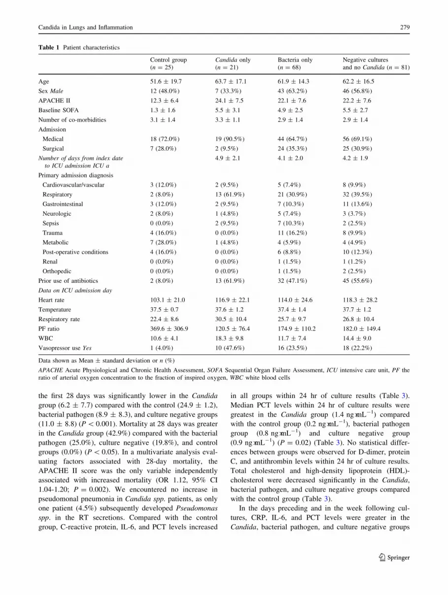
species (5.9%). Patient characteristics from all four groups
are compared in Table 1. There were significant differ-
ences in baseline characteristics across the four groups with
respect to APACHE II and SOFA scores, admission diag-
noses, PaO2/FIO2 ratio, white blood cell count, and
vasopressor use (see Table 1). The numbers of patients
with cancer, metastatic cancer, leukemia, or lymphoma
were similar across the groups; control group: 2/25 (8%),
Candida group: 2/22 (9%), bacterial pathogen group 7/68
(10%), and culture negative group 10/81 (12%); (P = NS).
Patients in the Candida group were admitted mostly for
respiratory illnesses, and they had a higher rate of prior use
of antibiotics (61.9%) than the bacterial pathogen (47.1%)
and culture negative (55.6%) groups.
Patient outcomes (i.e., maximum SOFA score, delta
SOFA score, number of days on mechanical ventilation,
and number of ICU days) were significantly worse in the
Candida group than in the control group, but they were
comparable with the bacterial pathogen and culture nega-
tive groups (Table 2). The number of ICU free days within
Patients in study (n=598)
Control Group
Patients with no antibiotics prescribed and no culture sent during their ICU stay (n=25).
N=570 after exclusion of the Control Group
Patients with Candida only in their respiratory specimens
(n=21)
Patients with bacteria in their respiratory
specimens
(n=68)
Patients with no growth (negative cultures and no
Candida) in their respiratory specimens
(n=81)
Patients with respiratory specimens (n=211)
(i.e. ETA, BAL, Bronchial, Sputum) 48 hours after ICU admission
41 patients excluded
• One patient received fluconazole for more than a week
• One patient had Candida in blood
• One patient had previous respiratory cultures that grew yeast
• 10 patients have both bacteria and Candida in their respiratory specimens
• 14 patients with non respiratory source of infection
• 14 due to uncertain pathogenecity
3 patients were excluded from control:
2 died within 72 hours of admission and 1 was infected before admission
Fig. 1 Patient flowchart
278 D. R. Williamson et al.
123
the first 28 days was significantly lower in the Candida
group (6.2 ± 7.7) compared with the control (24.9 ± 1.2),
bacterial pathogen (8.9 ± 8.3), and culture negative groups
(11.0 ± 8.8) (P \ 0.001). Mortality at 28 days was greater
in the Candida group (42.9%) compared with the bacterial
pathogen (25.0%), culture negative (19.8%), and control
groups (0.0%) (P \ 0.05). In a multivariate analysis eval-
uating factors associated with 28-day mortality, the
APACHE II score was the only variable independently
associated with increased mortality (OR 1.12, 95% CI
1.04-1.20; P = 0.002). We encountered no increase in
pseudomonal pneumonia in Candida spp. patients, as only
one patient (4.5%) subsequently developed Pseudomonas
spp. in the RT secretions. Compared with the control
group, C-reactive protein, IL-6, and PCT levels increased
in all groups within 24 hr of culture results (Table 3).
Median PCT levels within 24 hr of culture results were
greatest in the Candida group (1.4 ng�mL-1) compared
with the control group (0.2 ng�mL-1), bacterial pathogen
group (0.8 ng�mL-1) and culture negative group
(0.9 ng�mL-1) (P = 0.02) (Table 3). No statistical differ-
ences between groups were observed for D-dimer, protein
C, and antithrombin levels within 24 hr of culture results.
Total cholesterol and high-density lipoprotein (HDL)-
cholesterol were decreased significantly in the Candida,
bacterial pathogen, and culture negative groups compared
with the control group (Table 3).
In the days preceding and in the week following cul-
tures, CRP, IL-6, and PCT levels were greater in the
Candida, bacterial pathogen, and culture negative groups
Table 1 Patient characteristics
Control group
(n = 25)
Candida only
(n = 21)
Bacteria only
(n = 68)
Negative cultures
and no Candida (n = 81)
Age 51.6 ± 19.7 63.7 ± 17.1 61.9 ± 14.3 62.2 ± 16.5
Sex Male 12 (48.0%) 7 (33.3%) 43 (63.2%) 46 (56.8%)
APACHE II 12.3 ± 6.4 24.1 ± 7.5 22.1 ± 7.6 22.2 ± 7.6
Baseline SOFA 1.3 ± 1.6 5.5 ± 3.1 4.9 ± 2.5 5.5 ± 2.7
Number of co-morbidities 3.1 ± 1.4 3.3 ± 1.1 2.9 ± 1.4 2.9 ± 1.4
Admission
Medical 18 (72.0%) 19 (90.5%) 44 (64.7%) 56 (69.1%)
Surgical 7 (28.0%) 2 (9.5%) 24 (35.3%) 25 (30.9%)
Number of days from index dateto ICU admission ICU a
4.9 ± 2.1 4.1 ± 2.0 4.2 ± 1.9
Primary admission diagnosis
Cardiovascular/vascular 3 (12.0%) 2 (9.5%) 5 (7.4%) 8 (9.9%)
Respiratory 2 (8.0%) 13 (61.9%) 21 (30.9%) 32 (39.5%)
Gastrointestinal 3 (12.0%) 2 (9.5%) 7 (10.3%) 11 (13.6%)
Neurologic 2 (8.0%) 1 (4.8%) 5 (7.4%) 3 (3.7%)
Sepsis 0 (0.0%) 2 (9.5%) 7 (10.3%) 2 (2.5%)
Trauma 4 (16.0%) 0 (0.0%) 11 (16.2%) 8 (9.9%)
Metabolic 7 (28.0%) 1 (4.8%) 4 (5.9%) 4 (4.9%)
Post-operative conditions 4 (16.0%) 0 (0.0%) 6 (8.8%) 10 (12.3%)
Renal 0 (0.0%) 0 (0.0%) 1 (1.5%) 1 (1.2%)
Orthopedic 0 (0.0%) 0 (0.0%) 1 (1.5%) 2 (2.5%)
Prior use of antibiotics 2 (8.0%) 13 (61.9%) 32 (47.1%) 45 (55.6%)
Data on ICU admission day
Heart rate 103.1 ± 21.0 116.9 ± 22.1 114.0 ± 24.6 118.3 ± 28.2
Temperature 37.5 ± 0.7 37.6 ± 1.2 37.4 ± 1.4 37.7 ± 1.2
Respiratory rate 22.4 ± 8.6 30.5 ± 10.4 25.7 ± 9.7 26.8 ± 10.4
PF ratio 369.6 ± 306.9 120.5 ± 76.4 174.9 ± 110.2 182.0 ± 149.4
WBC 10.6 ± 4.1 18.3 ± 9.8 11.7 ± 7.4 14.4 ± 9.0
Vasopressor use Yes 1 (4.0%) 10 (47.6%) 16 (23.5%) 18 (22.2%)
Data shown as Mean ± standard deviation or n (%)
APACHE Acute Physiological and Chronic Health Assessment, SOFA Sequential Organ Failure Assessment, ICU intensive care unit, PF the
ratio of arterial oxygen concentration to the fraction of inspired oxygen, WBC white blood cells
Candida in Lungs and Inflammation 279
123

compared with the control group without reaching statis-
tical significance (Figs 2, 3, and 4).
Discussion
In order to assess whether Candida.isolated in RT secre-
tions is associated with systemic inflammation, we
conducted an analysis of the systemic inflammatory
markers of patients who developed a clinical suspicion of
VAP from a large multicentre observational study in a
heterogeneous ICU population. Our main finding was that
Candida in the RT secretions in patients with suspected
VAP was associated with increased levels of PCT, IL-6,
and CRP. These increased levels were similar to the
increased levels in patients with both positive bacterial
cultures and negative culture results, and they were sta-
tistically increased compared with our control patients.
Moreover, 28-day mortality was significantly increased in
the Candida group compared with the bacterial pathogen
and culture negative groups. However, in our multivariate
analysis, only the APACHE II score was independently
associated with increased mortality. Presence of Candida
in the RT was not significantly associated with mortality.
The purpose of evaluating these inflammatory markers
associated with the presence of Candida in the RT for
patients with suspected VAP was to assess whether Can-
dida may predispose such patients to a negative outcome.
Based on our findings, we reject our null hypothesis that
Candida is associated with low levels of systemic inflam-
mation. The question remaining is whether Candida in the
airways is directly responsible for worse clinical outcomes
or whether the presence of Candida is merely a marker for
severity of disease.
Candida spp. are normal commensals of the human body.
They are commonly found on the skin, gastrointestinal tract,
genitourinary tract, and RT, and they occupy a unique eco-
logical niche. However, in critically ill patients, there may
be perturbations in patients’ homeostasis under the influence
of risk factors, such as broad spectrum antibacterial therapy,
leading to the proliferation of Candida organisms usually
kept in check by bacterial organisms beyond their normal
niche. At such times, invasive Candida infection may arise,
particularly in the presence of mucosal barrier damage. This
concept of invasion based on organism density has been
demonstrated previously by the translocation of Candida
from the gastrointestinal tract in the presence of high
organism loads.24,25 Such invasion could easily enhance the
propensity to produce systemic inflammation. In critically ill
patients, maintenance of normal microbial flora and an intact
immune system is rare, as antibiotics and relative immu-
nosuppression (corticosteroids, late sepsis) are common,
thus promoting Candida colonization and perhaps infection.
Yet, Candida pneumonia is uncommon, and its diagnosis
must be secured by demonstrating the presence of yeast or
pseudohyphae in lung tissue.9 Perhaps this paradigm is
shifting, as the presence of Candida spp. in the RT speci-
mens of patients on mechanical ventilation in the ICU,
independent of the histological diagnosis of VAP, may have
more significance than previously considered.
Candida possesses a ß-glucan-rich cell wall that has
been shown to activate immune responses.26-28 Studies
with Pneumocystis jiroveci and fungi have demonstrated
that ß-glucans can stimulate the release of inflammatory
Table 2 Clinical outcomes
Control group
(n = 25)
Candida only
(n = 21)
Bacteria only
(n = 68)
Negative cultures
and no Candida(n = 81)
P values
Maximum SOFA 3.2 ± 2.0 10.4 ± 3.3 9.7 ± 4.2 9.8 ± 4.0 \ 0.001*
Delta SOFA 2.3 ± 2.2 5.0 ± 2.9 4.9 ± 3.5 4.4 ± 3.0 0.03*
Number of days on MV 0.6 ± 0.8 11.0 ± 4.7 10.3 ± 8.0 9.0 ± 5.2 \ 0.001*
Number of days in ICU 3.1 ± 1.2 19.3 ± 17.6 18.5 ± 17.5 16.7 ± 19.3 0.001*
ICU free days in the first 28 days 24.9 ± 1.2 6.2 ± 7.7 8.9 ± 8.3 11.0 ± 8.8 \ 0.001*�
Mortality at day 14 0 (0.0%) 4 (19.0%) 12 (17.6%) 12 (14.8%) 0.16
Mortality at day 28 0 (0.0%) 9 (42.9%) 17 (25.0%) 16 (19.8%) 0.004
Duration of antibiotics (days) 0.0 ± 0.0 11.2 ± 8.8 17.5 ± 9.4 16.9 ± 10.4 \ 0.001*��
Days alive and off all antibiotics in
the first 28 days (from ICU admission)
28.0 ± 0.0 11.2 ± 10.1 6.8 ± 8.6 8.6 ± 10.3 \ 0.001*
Pair-wise comparisons were performed between the Candida only group vs the other groups for variables reaching an overall level of significance
of 0.05. * Candida vs control group; � Candida vs bacteria group; � Candida vs negative group. SOFA = Sequential Organ Failure Assessment;
MV = mechanical ventilation; ICU = intensive care unit
280 D. R. Williamson et al.
123

Ta
ble
3B
iom
ark
ers
wit
hin
24
hr
of
the
firs
td
ate
of
resp
irat
ory
cult
ure
s
Co
ntr
ol
gro
up�
(n=
25
)
Ca
nd
ida
on
ly
(n=
21
)
Bac
teri
ao
nly
(n=
68
)
Neg
ativ
ecu
ltu
res
and
no
Ca
nd
ida
(n=
81
)
Pv
alu
es
d
Med
ian
[q1
,q3
]n
Med
ian
[q1
,q3
]n
Med
ian
[q1
,q3
]n
Med
ian
[q1
,q3
]n
Pri
mar
yo
utc
om
em
easu
res
Pro
calc
itio
nin
(ng�m
L-
1)
0.2
[0.2
to0
.5]
81
.4[0
.4to
2.9
]2
00
.8[0
.4to
5.6
]6
40
.9[0
.4to
6.7
]8
00
.02
*
Pro
tein
C(%
)8
8.5
[79
.5to
10
2.0
]9
90
.9[5
7.1
to1
17
.7]
20
64
.0[4
9.0
to8
7.0
]6
56
9.0
[52
.3to
88
.4]
81
0.0
9
CR
P(m
g�L
-1)
80
.0[2
1.0
to1
13
.0]
91
30
.0[6
9.0
to2
13
.0]
21
15
7.0
[89
.0to
20
3.0
]6
31
16
.0[7
6.0
to1
87
.0]
78
0.0
3*
Oth
erm
easu
rem
ents
D-d
imer
(ug�m
L-
1)
1.7
[1.0
to6
.8]
96
.5[
3.3
to1
0.8
]1
75
.8[
1.8
to9
.4]
64
4.9
[1.9
to1
1.3
]8
00
.25
An
tith
rom
bin
(%)
86
.7[6
7.3
to9
3.1
]9
71
.3[5
4.8
to8
7.1
]2
06
4.0
[44
.0to
79
.8]
65
64
.7[4
8.9
to7
2.5
]8
10
.07
Fib
rin
og
en(m
g�d
L-
1)
44
5.4
[26
1.2
to5
15
.7]
94
57
.4[3
77
.9to
61
0.2
]2
05
21
.6[3
90
.0to
69
3.9
]6
64
80
.5[3
90
.0to
61
0.0
]8
10
.26
Inte
rleu
kin
-6(p
g�m
L-
1)
15
.3[7
.4to
36
.7]
85
3.7
[16
.5to
12
0.5
]2
06
6.3
[32
.4to
13
9.2
]6
44
3.1
[22
.5to
77
.3]
80
0.0
09
*
Tri
gly
ceri
des
(mm
oL�L
-1)
1.3
[1.1
to1
.5]
91
.3[
1.0
to1
.8]
21
1.2
[0
.8to
1.9
]6
41
.3[0
.9to
1.8
]8
00
.85
Ch
ole
ster
ol
(mm
oL�L
-1)
3.3
[3.2
to3
.8]
92
.2[1
.7to
3.0
]2
12
.2[1
.5to
3.0
]6
42
.3[1
.7to
3.0
]8
00
.02
HD
L-C
ho
lest
ero
l(m
mo
L�L
-1)
1.0
[0.8
to1
.3]
90
.5[0
.4to
0.8
]2
00
.6[0
.4to
0.8
]6
40
.6[0
.4to
0.8
]7
90
.00
5*
LD
L-C
ho
lest
ero
l(m
mo
L�L
-1)
1.8
[1.2
to2
.2]
91
.3[0
.9to
1.6
]2
00
.9[0
.6to
1.5
]6
21
.0[0
.6to
1.5
]7
80
.03
d=
Pv
alu
efr
om
Kru
skal
-Wal
lis
test
�A
ver
age
bio
mar
ker
val
ues
wer
eu
sed
for
the
con
tro
lg
rou
po
nd
ayth
ree
and
afte
rwar
d
Pai
r-w
ise
com
par
iso
ns
wer
ep
erfo
rmed
bet
wee
nth
eC
an
did
ao
nly
gro
up
vsth
eo
ther
gro
up
sfo
ral
lv
aria
ble
sre
ach
ing
ano
ver
all
lev
elo
fsi
gn
ifica
nce
of
0.0
5.
*C
an
did
ag
rou
pvs
con
tro
lg
rou
p
ata
0.0
5le
vel
of
sig
nifi
can
ce.
CR
P=
C-r
eact
ive
pro
tein
;H
DL
=h
igh
-den
sity
lip
op
rote
in;
LD
L=
low
-den
sity
lip
op
rote
in
Candida in Lungs and Inflammation 281
123

markers, such as IL-1 and TNF-a, through NF-jB activa-
tion, trigger fungicidal responses, and generate reactive
oxygen intermediates.29-32 These observations provide a
mechanism by which Candida may play a causal role in the
increased inflammatory markers and may potentiate worse
clinical outcomes.33 In a recent study, rats instilled with
live Candida albicans developed increased pulmonary
TNF-a and increased rates of pseudomonal pneumonia;
whereas, rats instilled with normal saline or ethanol
destroyed the Candida albicans. In critically ill patients,
tracheal colonization with Candida spp. (C 103�L-1)
within the first four days of mechanical ventilation was
associated with greater tracheal IL-8 and IL-6 levels than
patients whose tracheas were colonized with bacteria.34
The results of the current study may imply that Candida in
the RT secretions can play a greater pathophysiological
role, as suggested by the increased inflammation observed
in the Candida group.
Although our study included a significant number of
patients from multiple centres in a prospective fashion and
employed standardized inflammatory biomarker analysis,
there are a number of limitations to our observations. First,
Days from First Airway Sample Day (ICU Day 3 was Day 0 for Control Group)
CR
P
-3 -2 -1 0 1 2 3 4 5 6 7 8 9 10
020
4060
8010
012
014
016
018
020
022
024
0 Comparing the four groups from day 0 to day 10: p=0.25Control GroupCandida Only GroupBacteria Only GroupNegative Group
Fig. 2 CRP (Mean ± SE)
Days from First Airway Sample Day (ICU Day 3 was Day 0 for Control Group)
log(
IL-6
)
-3 -2 -1 0 1 2 3 4 5 6 7 8 9 10
01
23
45
6 Comparing the four groups from day 0 to day 10: p=0.30Control GroupCandida Only GroupBacteria Only GroupNegative Group
Fig. 3 Log(IL-6) (Mean ± SE)
282 D. R. Williamson et al.
123
this study included only a small number of patients in the
Candida and control groups and excluded a large number of
patients from the initial study because no cultures were sent
for laboratory analysis. This selection was justified by our
goal of obtaining the most robust data possible with regard
to the relation between Candida spp. and systemic inflam-
mation. Second, the definition of suspected VAP was
applied retrospectively to our data set and may have
resulted in some misclassification. Third, bacteria or viruses
that could have been detected using molecular techniques
may have been missed with the traditional culture tech-
niques.35 Fourth, the group described as having no bacteria
included 29 patients (35.8%) with commensal bacterial
flora. These patients could have artificially increased the
median levels of inflammatory markers in this group, thus
obscuring differences between it and the Candida only
group. Fifth, patients in the Candida group had the highest
proportion of prior use of antibiotics; thus, bacterial cultures
may have been negative at the time of sampling in some
patients. Finally, the Candida cultures were qualitative and
were not speciated or quantified. Perhaps greater quantities
of Candida organisms or a specific threshold of organisms
would enhance the level of systemic inflammation. Also, we
did not systematically screen for Candida from all sites,
including the gastrointestinal tract, so we cannot be certain
as to the determination of widespread Candida coloniza-
tion. However, blood and urine cultures were drawn from
all study subjects at the same time, and we did exclude
patients with documented presence of Candida spp. from
other sources; therefore, the probability of disseminated
candidiasis is very low.
We have demonstrated that the presence of Candida
spp. in the RT, in comparison with the presence of bacterial
pathogens or no pathogens, is associated with comparable
systemic inflammation and may be associated with worse
clinical outcomes. Future studies should target the patho-
genic role that Candida plays to affect outcomes in this
patient population. Based on these results and the results of
previous studies, a placebo-controlled trial evaluating the
role of antifungal treatment on the inflammatory cytokine
profiles and clinical outcomes is warranted and is now
underway. (ClinicalTrials.gov number, NCT00934934).
Financial disclosure and competing interests None declared.
References
1. Vincent JL, Bihari DJ, Suter PM, et al. The prevalence of nos-
ocomial infection in intensive care units in Europe. Results of the
European Prevalence of Infection in Intensive Care (EPIC) Study.
EPIC International Advisory Committee. JAMA 1995; 274: 639-
44.
2. Richards MJ, Edwards JR, Culver DH, Gaynes RP. Nosocomial
infections in combined medical-surgical intensive care units in
the United States. Infect Control Hosp Epidemiol 2000; 21:
510-5.
3. Heyland DK, Cook DJ, Griffith L, Keenan SP, Brun-Buisson C.
The attributable morbidity and mortality of ventilator-associated
pneumonia in the critically ill patient. The Canadian Critical
Trials Group. Am J Respir Crit Care Med 1999; 159: 1249-56.
4. National Nosocomial Infections Surveillance System. National
Nosocomial Infections Surveillance (NNIS) System Report, data
summary from January 1992 through June 2004, issued October
2004. Am J Infect Control 2004; 32: 470-85.
Days from First Airway Sample Day (ICU Day 3 was Day 0 for Control Group)
Log
(Pro
calc
ition
in)
-3 -2 -1 0 1 2 3 4 5 6 7 8 9 10
-2-1
01
23 Comparing the four groups from day 0 to day 10: p=0.23
Control GroupCandida Only GroupBacteria Only GroupNegative Group
Fig. 4 Log(Procalcitionin)
(Mean ± SE)
Candida in Lungs and Inflammation 283
123

5. Bueno-Cavanillas A, Delgado-Rodriguez M, Lopez-Luque A,
Schaffino-Cano S, Galvez-Vargas R. Influence of nosocomial
infection on mortality rate in an intensive care unit. Crit Care
Med 1994; 22: 55-60.
6. Safdar N, Dezfulian C, Collard HR, Saint S. Clinical and eco-
nomic consequences of ventilator-associated pneumonia: a
systematic review. Crit Care Med 2005; 33: 2184-93.
7. Warren DK, Shukla SJ, Olsen MA, et al. Outcome and attribut-
able cost of ventilator-associated pneumonia among intensive
care unit patients in a suburban medical center. Crit Care Med
2003; 31: 1312-7.
8. Muscedere JG, Martin CM, Heyland DK. The impact of venti-
lator-associated pneumonia on the Canadian health care system.
J Crit Care 2008; 23: 5-10.
9. Chastre J, Fagon JY. Ventilator-associated pneumonia. Am J
Respir Crit Care Med 2002; 165: 867-903.
10. The Canadian Critical Care Trials Group. A randomized trial of
diagnostic techniques for ventilator-associated pneumonia.
N Engl J Med 2006; 355: 2619-30.
11. Kollef MH, Morrow LE, Niederman MS, et al. Clinical charac-
teristics and treatment patterns among patients with ventilator-
associated pneumonia. Chest 2006; 129: 1210-8.
12. Meersseman W, Lagrou K, Spriet I, et al. Significance of the
isolation of Candida species from airway samples in critically ill
patients: a prospective, autopsy study. Intensive Care Med 2009;
35: 1526-31.
13. el-Ebiary M, Torres A, Fabregas N, et al. Significance of the
isolation of Candida species from respiratory samples in critically
ill, non-neutropenic patients. An immediate postmortem histo-
logic study. Am J Respir Crit Care Med 1997; 156: 583-90.
14. Rello J, Esandi ME, Diaz E, Mariscal D, Gallego M, Valles J.
The role of Candida sp isolated from bronchoscopic samples in
nonneutropenic patients. Chest 1998; 114: 146-9.
15. Wey SB, Mori M, Pfaller MA, Woolson RF, Wenzel RP. Risk
factors for hospital-acquired candidemia. A matched case-control
study. Arch Intern Med 1989; 149: 2349-53.
16. Magill SS, Swoboda SM, Johnson EA, et al. The association
between anatomic site of Candida colonization, invasive candi-
diasis, and mortality in critically ill surgical patients. Diagn
Microbiol Infect Dis 2006; 55: 293-301.
17. Delisle MS, Williamson DR, Perreault MM, Albert M, Jiang X,
Heyland DK. The clinical significance of Candida colonization of
respiratory tract secretions in critically ill patients. J Crit Care
2008; 23: 11-7.
18. Azoulay E, Timsit JF, Tafflet M, et al. Candida colonization of the
respiratory tract and subsequent pseudomonas ventilator-associ-
ated pneumonia. Chest 2006; 129: 110-7.
19. Calderon EJ, Rivero L, Respaldiza N, et al. Systemic inflam-
mation in patients with chronic obstructive pulmonary disease
who are colonized with Pneumocystis jiroveci. Clin Infect Dis
2007; 45: e17-9.
20. Kellum JA, Kong L, Fink MP, et al. Understanding the inflam-
matory cytokine response in pneumonia and sepsis: results of the
Genetic and Inflammatory Markers of Sepsis (GenIMS) Study.
Arch Intern Med 2007; 167: 1655-63.
21. Jensen JU, Heslet L, Jensen TH, Espersen K, Steffensen P, TvedeM. Procalcitonin increase in early identification of critically ill
patients at high risk of mortality. Crit Care Med 2006; 34: 2596-
602.
22. Knaus WA, Draper EA, Wagner DP, Zimmerman JE. APACHE
II: a severity of disease classification system. Crit Care Med
1985; 13: 818-29.
23. Vincent JL, Moreno R, Takala J, et al. The SOFA (Sepsis-related
Organ Failure Assessment) score to describe organ dysfunction/
failure. On behalf of the Working Group on Sepsis-Related
Problems of the European Society of Intensive Care Medicine.
Intensive Care Med 1996; 22: 707-10.
24. Krause W, Matheis H, Wulf K. Fungaemia and funguria after oral
administration of Candida albicans. Lancet 1969; 1: 598-9.
25. Pittet D, Monod M, Suter PM, Frenk E, Auckenthaler R. Candida
colonization and subsequent infections in critically ill surgical
patients. Ann Surg 1994; 220: 751-8.
26. Young SH, Ostroff GR, Zeidler-Erdely PC, Roberts JR, AntoniniJM, Castranova V. A comparison of the pulmonary inflammatory
potential of different components of yeast cell wall. J Toxicol
Environ Health A 2007; 70: 1116-24.
27. Muller V, Viemann D, Schmidt M, et al. Candida albicans triggers
activation of distinct signaling pathways to establish a proin-
flammatory gene expression program in primary human
endothelial cells. J Immunol 2007; 179: 8435-45.
28. Inoue K, Takano H, Oda T, et al. Candida soluble cell wall beta-
D-glucan induces lung inflammation in mice. Int J Immunopathol
Pharmacol 2007; 20: 499-508.
29. Lebron F, Vassallo R, Puri V, Limper AH. Pneumocystis carinii
cell wall beta-glucans initiate macrophage inflammatory respon-
ses through NF-kappaB activation. J Biol Chem 2003; 278:
25001-8.
30. Hahn PY, Evans SE, Kottom TJ, Standing JE, Pagano RE, LimperAH. Pneumocystis carinii cell wall beta-glucan induces release of
macrophage inflammatory protein-2 from alveolar epithelial cells
via a lactosylceramide-mediated mechanism. J Biol Chem 2003;
278: 2043-50.
31. McCann F, Carmona E, Puri V, Pagano RE, Limper AH. Mac-
rophage internalization of fungal beta-glucans is not necessary for
initiation of related inflammatory responses. Infect Immun 2005;
73: 6340-9.
32. Wheeler RT, Fink GR. A drug-sensitive genetic network masks
fungi from the immune system. PLoS Pathog 2006; 2: e35.
33. Varela JM, Respaldiza N, Sanchez B, et al. Lymphocyte response
in subjects with chronic pulmonary disease colonized by Pneu-
mocystis jirovecii. J Eukaryot Microbiol 2003; 50(Suppl): 672-3.
34. Durairaj L, Mohamad Z, Launspach JL, et al. Patterns and
density of early tracheal colonization in intensive care unit
patients. J Crit Care 2009; 24: 114-21.
35. Chiche L, Forel JM, Roch A, et al. Active cytomegalovirus
infection is common in mechanically ventilated medical intensive
care unit patients. Crit Care Med 2009; 37: 1850-7.
284 D. R. Williamson et al.
123