the microtubule plus-end tracking protein armadillo-repeat kinesin1 promotes microtubule catastrophe...
TRANSCRIPT
The Microtubule Plus-End Tracking ProteinARMADILLO-REPEAT KINESIN1 PromotesMicrotubule Catastrophe in ArabidopsisW OPEN
Ryan Christopher Eng and Geoffrey O. Wasteneys1
Department of Botany, University of British Columbia, Vancouver, British Columbia V6T 1Z4, Canada
ORCID ID: 0000-0002-8746-7994 (G.O.W.)
Microtubule dynamics are critically important for plant cell development. Here, we show that Arabidopsis thaliana ARMADILLO-REPEAT KINESIN1 (ARK1) plays a key role in root hair tip growth by promoting microtubule catastrophe events. This destabilizingactivity appears to maintain adequate free tubulin concentrations in order to permit rapid microtubule growth, which in turn iscorrelated with uniform tip growth. Microtubules in ark1-1 root hairs exhibited reduced catastrophe frequency and slower growthvelocities, both of which were restored by low concentrations of the microtubule-destabilizing drug oryzalin. An ARK1-GFP (greenfluorescent protein) fusion protein expressed under its endogenous promoter localized to growing microtubule plus ends andrescued the ark1-1 root hair phenotype. Transient overexpression of ARK1-RFP (red fluorescent protein) increased microtubulecatastrophe frequency. ARK1-fusion protein constructs lacking the N-terminal motor domain still labeled microtubules,suggesting the existence of a second microtubule binding domain at the C terminus of ARK1. ARK1-GFP was broadlyexpressed in seedlings, but mutant phenotypes were restricted to root hairs, indicating that ARK1’s function is redundant in cellsother than those forming root hairs.
INTRODUCTION
Precise coordination of microtubule dynamics and organizationis necessary for normal plant cell development, growth, and shape.Microtubules contribute to root hair morphogenesis by establish-ing and maintaining the polar growth process (Sieberer et al.,2002). Root hairs are tubular extensions of epidermal cells thatprotrude perpendicularly from the root and grow through a highlypolarized process called tip growth in which the plasma mem-brane and cell wall components are asymmetrically secreted atthe root hair apex (Carol and Dolan, 2002). Microtubule de-polymerization or hyperstabilization by treatment with the drugsoryzalin and taxol, respectively, can lead to wavy and branchedroot hairs, stressing the importance of microtubules in focusingtip growth (Bibikova et al., 1999). Root hairs of Arabidopsisthaliana possess two types of microtubule populations: corticalmicrotubules found at the cortex and endoplasmic microtubuleslocated within the transvacuolar strands and cytoplasmic-richzone near the root hair apex (Sieberer and Timmers, 2009). Live-cell imaging of the green fluorescent protein fused to a microtu-bule binding domain (GFP-MBD) reporter protein in Arabidopsisplants showed that cortical microtubules are present throughoutall developmental stages of the root hair, while endoplasmicmicrotubules are only present in elongating hairs (Van Bruaeneet al., 2004).
Microtubules are dynamic polymers made up of heterodimerictubulin subunits that self-assemble into a polar cylindrical polymerwith a less dynamic minus end and a more dynamic plus end. Thestochastic addition or removal of tubulin subunits from the mi-crotubule ends causes the microtubule to either grow/polymerizeor shrink/depolymerize, respectively (Desai and Mitchison, 1997).Microtubule dynamics and organization are achieved through theactivity of microtubule-associated proteins (MAPs) such as theArabidopsis MICROTUBULE ORGANIZATION1 protein that pro-motes microtubule dynamics by aiding in microtubule polymeri-zation and depolymerization (Kawamura andWasteneys, 2008). Ineukaryotes, the highly conserved microtubule-based motor pro-teins called kinesins can, in addition to transporting cargo alongmicrotubules, also modulate microtubule dynamics and organi-zation. Thus far for Arabidopsis, in which 61 putative kinesinshave been identified, the majority of those characterized havebeen shown to organize interphase and mitotic microtubulearrays through bundling, cross-linking, and translocation ofother microtubules. A smaller proportion of Arabidopsis kinesinsare related to organelle and vesicle motility along microtubules(Reddy and Day, 2011; Zhu and Dixit, 2012). To date, Kinesin-13A is the only kinesin shown to be a catastrophe factor. It actsspecifically to promote microtubule depolymerization in thesecondary cell wall pits of developing xylem cells (Oda andFukuda, 2013). The depolymerizing function of Kinesin-13Ais similar to that of its homologs in other organisms (Desaiet al., 1999; Oda and Fukuda, 2013). Kinesins can also organizemicrotubules in tip-growing cells such as caulonemal cells ofthe moss Physcomitrella patens. The KINID1a and KINID1bkinesins in these cells are responsible for the bundling of mi-crotubule plus ends. Loss of both these kinesins leads to re-duced microtubule bundling and aberrant tip growth (Hiwatashiet al., 2014)
1 Address correspondence to [email protected] author responsible for distribution of materials integral to the findingspresented in this article in accordance with the policy described in theInstructions for Authors (www.plantcell.org) is: Geoffrey O. Wasteneys([email protected]).W Online version contains Web-only data.OPENArticles can be viewed online without a subscription.www.plantcell.org/cgi/doi/10.1105/tpc.114.126789
This article is a Plant Cell Advance Online Publication. The date of its first appearance online is the official date of publication. The article has been
edited and the authors have corrected proofs, but minor changes could be made before the final version is published. Posting this version online
reduces the time to publication by several weeks.
The Plant Cell Preview, www.aspb.org ã 2014 American Society of Plant Biologists. All rights reserved. 1 of 15
The ARMADILLO-REPEAT KINESIN1 (ARK1), originally namedMORPHOGENESIS OF ROOT HAIR2 (MRH2) (Jones et al., 2006),is thought to participate in microtubule dynamics. Because ARK1is an uncharacterized kinesin belonging to an ungrouped plant-specific family of kinesins, its function cannot be predicted basedon phylogeny (Reddy and Day, 2011; Zhu and Dixit, 2012).However, with previous evidence showing that ark1mutants haveroot hairs with increased endoplasmic microtubule abundanceand wavy/branched morphologies, it has been suggested thatARK1 specifically promotes microtubule depolymerization in roothairs (Sakai et al., 2008; Yoo et al., 2008).
A single ARK1 polypeptide has an N-terminal catalytic motordomain that interacts with microtubules through the binding andhydrolysis of ATP (Sakai et al., 2008). Much like most conven-tional kinesins, ARK1 is predicted to function as a homodimer, asevidenced by its internal coiled-coil domain. The C terminus ofARK1 is composed of three tandem Armadillo (ARM) repeat do-mains that gives rise to the kinesin’s name (Sakai et al., 2008). AnARM repeat forms three a-helices and, when in tandem, forms asuperhelix that allows for its interaction with other proteins. ARMrepeats have been shown to function in essential cellular pro-cesses related to signaling, the cytoskeleton, and protein-proteininteractions in a variety of eukaryotes (Tewari et al., 2010), buttheir function in ARK1 is still unclear. Interestingly, previousstudies using in vitro analyses have shown that the ARM domainof ARK1 is able to bind to actin (Yang et al., 2007) as well as theNever in Mitosis, Gene A (NIMA)-related kinase, NEK6 (Sakaiet al., 2008). ARK1 is also thought to play a pivotal role in thetightly coordinated tip growth signaling pathway as a potentiallinker among the cytoskeletal, endomembrane, and GTPasesignaling components (Yoo et al., 2008; Yoo and Blancaflor,2013).
Although previous studies have confirmed ARK1’s ability tobind microtubules through fluorescent protein tagging (Yoo et al.,2008) and in vitro microtubule binding (Yang et al., 2007), itsfunction in modulating microtubule dynamics and/or organiza-tion has not been confirmed. In this study, we use genetic,chemical, and overexpression analyses to show that ARK1 is akinesin with plus-end tracking properties that promotes microtu-bule depolymerization.
RESULTS
Microtubule Catastrophe Frequency and Growth Rates AreReduced in ark1-1 Mutants
Based on the fact that microtubules are more abundant in theT-DNA insertional mutant ark1-1 (SALK_035063) (Sakai et al.,2008), we hypothesized that microtubule dynamics would be al-tered. In order to test this hypothesis, we quantified microtubuleplus-end growth rates as well as the frequency at which micro-tubules undergo catastrophe by tracking the microtubule plus-end tracking protein END BINDING 1b (EB1b). This reporter wastagged with green fluorescent protein (GFP) and expressed usingthe 35S cauliflower mosaic virus promoter (35Spro:EB1b-GFP);thus, we were able to visualize microtubule plus-ends. EB1b-GFPmovement was measured in wild-type and ark1-1 elongating root
hairs by spinning-disc confocal microscopy. Both endoplasmicand cortical microtubule mean growth velocities were significantlyreduced in ark1-1 (cortical microtubule [CMT], 3.5 6 1.1 mm/min;endoplasmic microtubule [EMT], 5.7 6 1.3 mm/min) relative to thewild type (CMT, 6.5 6 1.7 mm/min; EMT, 7.1 6 1.9 mm/min; two-sample unequal variance t test: CMT, P < 10277; EMT, P < 10205)(Figures 1A, 1D, and 1E; Table 1; Supplemental Figure 1 andSupplemental Movies 1 and 2). The frequency of cortical micro-tubule catastrophe was significantly reduced by 31% in ark1-1(0.020 6 0.011 events/second) relative to wild-type root hairs(0.029 6 0.015 events/second; two-sample unequal variancet test: P < 10209) (Figure 1B, Table 1). In contrast, we found nosignificant difference in catastrophe frequency between wild-type(0.048 6 0.025 events/second) and ark1-1 endoplasmic micro-tubules (0.055 6 0.034 events/second) (Figure 1B, Table 1) de-spite a greater abundance of endoplasmic microtubules in ark1-1root hairs (Supplemental Figures 2A to 2C).The decreased frequency of microtubule catastrophe in ark1-1
mutants is consistent with the increased microtubule polymermass and the occurrence of microtubule bundling. The slowerrate of microtubule polymerization, however, is more difficult toexplain because decreased polymerization rates should theoret-ically reduce the polymer mass. Given that microtubule poly-merization rates are proportional to free tubulin concentrations,we considered the possibility that the reduced polymerizationrates in ark1-1 are the consequence of a reduced free tubulinconcentration, which is likely to result from the reduced catas-trophe frequency. We hypothesized that increasing the avail-ability of free tubulin should restore normal microtubule growthrates in ark1-1. To achieve this, we applied low concentrationsof the microtubule destabilizing drug oryzalin and measuredmicrotubule growth and catastrophe rates using the EB1b-GFPreporter.Exposure to 100 nM oryzalin increased the ark1-1 plus-end
growth rates (as measured by EB1b-GFP) such that they wereequivalent to the oryzalin-free wild-type growth rate in bothcortical and endoplasmic microtubule populations (Figures 1Aand 1C, Table 1). Microtubules in wild-type root hairs, incontrast, showed reduced plus-end growth rates when ex-posed to 100 nM oryzalin (Figures 1A and 1C, Table 1), whichis consistent with previously published data (Nakamura et al.,2004).The cortical microtubule catastrophe frequency in 100 nM
oryzalin-treated ark1-1 root hairs was increased relative to that ofuntreated wild-type and ark1-1 cells. The dynamics of endo-plasmic microtubules in oryzalin-treated ark1-1 root hairs anduntreated wild-type root hairs were not significantly different(Figure 1B, Table 1).Despite a clear difference in plus-end growth velocities of
oryzalin-treated wild-type and ark1-1 microtubules as assessedwith EB1b-GFP, both genotypes exposed to the same oryzalinconcentrations had fragmented microtubules that looked in-distinguishable from each other when using the 35Spro:GFP-MBD (MBD is the microtubule binding domain of MAP4) marker(Figure 1F). This observation possibly resulted from increasedresistance to microtubule destabilizing drugs and bundling typi-cally associated with the 35Spro:GFP-MBD marker (Marc et al.,1998; Lechner et al., 2012).
2 of 15 The Plant Cell
Figure 1. Cortical Microtubule Plus-End Velocities and Catastrophe Frequencies Are Reduced in ark1-1 Root Hairs but Can Be Rescued by OryzalinTreatment.
(A) Plus-end velocities of EB1b-GFP of CMTs and EMTs were measured in wild-type and ark1-1 elongating root hairs upon imaging with spinning-discconfocal microscopy. The mean ark1-1 microtubule plus-end velocities (CMT, 3.5 6 1.1 mm/min, n = 111; EMT, 5.7 6 1.3 mm/min, n = 85) aresignificantly reduced relative to wild-type microtubules (CMT, 6.56 1.7 mm/min, n = 209; EMT, 7.16 1.9, n = 149). Exposure to 100 nM oryzalin rescuedthe microtubule plus-end velocities of ark1-1 root hairs (CMT, 6.1 6 1.6 mm/min, n = 47; EMT, 6.7 6 2.0 mm/min, n = 106) so that there was nosignificant difference from oryzalin-free wild-type root hairs (CMT, 6.5 6 1.7 mm/min, n = 209; EMT, 7.1 6 1.9 mm/min, n = 149). Oryzalin treatmentsignificantly reduced wild-type velocities (CMT, 5.6 6 1.6 mm/min, n = 134; EMT, 5.9 6 1.7 mm/min, n = 76). A minimum of five roots (for untreated) andthree roots (for oryzalin-treated) (five root hairs per root) from each genetic background were imaged.
ARK1 Is a Microtubule Destabilizing Kinesin 3 of 15

Low Concentrations of Oryzalin Partially Rescue the ark1-1Root Hair Morphology Phenotype
Based on the restoration of wild-type microtubule growth rates inthe ark1-1 mutant by the oryzalin treatments, we hypothesizedthat these treatments would ameliorate the ark1-1 root hair mor-phological defects. It was previously shown that wavy andbranched root hair phenotypes were indicative of a less severeand more severe loss of root hair polarity, respectively, and thatthe severity of polarity loss was positively correlated with in-creasing oryzalin and taxol concentrations (Bibikova et al., 1999).Exposure to 100 nM, 1 mM, and 5 mM oryzalin partially rescuedthe ark1-1 root hair phenotype by significantly increasing the roothair length relative to untreated ark1-1 root hairs (Figures 2A and2B). Moreover, the frequency of the most severe root hair phe-notype (branched) appeared to decrease with the less severephenotype (wavy and bulbous) increasing in frequency at 1 and5 mM oryzalin concentrations (Figures 2A and 2C). In the wildtype, these same oryzalin concentrations decreased root hairlength and led to wavy and branched root hairs that resembledthe ark1-1 phenotype (Figures 2A and 2C). Oryzalin (10 mM) wasunable to partially rescue the ark1-1 phenotype, suggesting asaturation effect of oryzalin on microtubules.
In contrast to the oryzalin treatments, taxol, which promotesmicrotubule polymerization, further impaired root hair elongation
in both wild-type and ark1-1 root hairs (Figures 2A and 2B) andincreased root hair waving/branching (Figure 2A).
ARK1 Accumulates on Plus Ends of Growing Microtubules
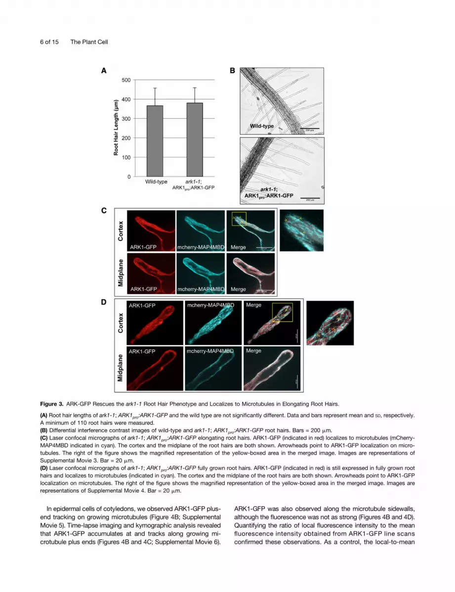
In order to see where ARK1 is localized on the microtubule, wemade an ARK1pro:ARK1-GFP construct using the entire ARK1genomic sequence including the putative 59-end promoter re-gion (888 bp upstream of the ARK1 start codon). ark1-1 plantsexpressing 35Spro:mCherry-MAP4MBD were transformed withthe ARK1pro:ARK-GFP construct and a T3 homozygous line forthe ARK1-GFP transgene was used for further analysis. ARK1pro:ARK1-GFP expression rescued the ark1-1 root hair phenotype(Figures 3A and 3B), indicating that the GFP-tagged ARK1 proteinwas fully functional.ARK1-GFP was associated with microtubules and expressed
in elongating and fully grown root hairs (Figures 3C and 3D; seeSupplemental Movies 3 and 4 for z-stacks of ARK1-GFP in roothairs), although characterization of ARK1-GFP on microtubules wasdifficult to analyze in root hairs due to high cytoplasmic fluores-cence of ARK1-GFP. However, we were able to observe ARK1-GFPexpression in other cell types, including root atrichoblasts as well asepidermal cells of the hypocotyl, petiole, cotyledons, and root tip,which is consistent with previous ARK1 gene expression analysis(Figure 4A) (Yang et al., 2007; Sakai et al., 2008).
Figure 1. (continued).
(B) Catastrophe frequencies of cortical and endoplasmic microtubules were measured in the same elongating root hairs as in (A). Cortical microtubulecatastrophe frequencies in ark1-1 (0.020 6 0.011 events/second) are significantly reduced relative to wild-type microtubules (0.029 6 0.015 events/second). There is no significant difference for endoplasmic microtubules (wild type, 0.048 6 0.025 events/second; ark1-1, 0.055 6 0.034 events/second). Exposure to 100 nM oryzalin increased the catastrophe frequency of cortical microtubules in ark1-1 root hairs (0.041 6 0.03 events/second)relative to untreated wild-type root hairs (0.029 6 0.015 events/second). The same microtubules in (A) were used to calculate the catastrophefrequency.(C) Exposure to 100 nM oryzalin reduced microtubule plus-end velocities in wild-type root hairs but increased velocities in ark1-1 root hairs.(D) and (E) Confocal micrographs of EB1b-GFP in wild-type (D) and ark1-1 (E) root hairs showing both cortical and endoplasmic microtubule plus ends.The colored lines represent the EB1b-GFP trajectories after 40 s with the wild type showing faster EB1b-GFP movement than ark1-1. Images representthe medial plane of the root hair. Magnified images of the trajectories are seen below and the yellow boxes indicate the area where the magnified imageswere selected. For the full montage, refer to Supplemental Figure 1. Images are representations of Supplemental Movies 1 and 2 online. Bars =10 mm.(F) Spinning-disc laser confocal micrographs of root hairs expressing the 35Spro:GFP-MBD microtubule marker in the wild type and ark1-1. Nodifferences were noticeable between the microtubules of wild-type and ark1-1 root hairs treated with 100 nM oryzalin. Images are merged Z-projectionsof the entire root hair stack.Data and bars are represented as means 6 SD, respectively. Asterisk shows a significant reduction, while NS indicates no significant difference invelocity relative to untreated wild-type root hairs using a two-sample t test with unequal variance (a = 0.01).
Table 1. Plus-End Growth Velocities and Catastrophe Frequencies of the Wild Type and ark1-1 in Untreated and 100 nM Oryzalin-Treated Root Hairs
CMTs EMTs
Wild Type ark1-1 Wild Type ark1-1
Plus-end velocity (mm min21) 6.54 6 1.66 3.39 6 1.09a 7.11 6 1.90 5.74 6 1.29b
With oryzalin 5.63 6 1.58a 6.14 6 1.64a 5.93 6 1.66b 6.74 6 2.03d
Catastrophe frequency (events s21) 0.029 6 0.015 0.020 6 0.0118a 0.048 6 0.024 0.055 6 0.034d
With oryzalin 0.034 6 0.022c 0.041 6 0.030a 0.041 6 0.028d 0.043 6 0.024d
Data represent mean 6 SD; a = 0.01.aSignificant difference from wild-type CMTs.bSignificant difference from wild-type EMTs.cNo significant difference from wild-type CMTs.dNo significant difference from wild-type EMTs.
4 of 15 The Plant Cell
Figure 2. Oryzalin Partially Rescues the ark1-1 Root Hair Morphology.
(A) Bright-field images of wild-type and ark1-1 root hairs treated with either 5 mM taxol or oryzalin. The root hair phenotype of ark1-1 is partially rescuedwith oryzalin but not with taxol. Bars = 50 mm.(B) Mean length of wild-type and ark1-1 root hairs treated with various concentrations of taxol and oryzalin. ark1-1 exposed to 100 nM, 1 mM,and 5 mM oryzalin partially rescues the root hair length phenotype. Data and bars represent mean and SD, respectively. Asterisks representa significant increase in mean length relative to untreated ark1-1 root hairs. A minimum of 25 root hairs were measured for each genotype andtreatment.(C) Frequency distribution histograms comparing root hair morphology types in the wild type and ark1-1 grown on varying oryzalin concentrations.Oryzalin (1 and 5 mM) partially mitigates the ark1-1 root hair defect by shifting the distribution of the most severe branched phenotype to a milder wavy/bulbous phenotype. As a control, wild-type root hairs show an increase loss of root hair polarity upon exposure to increasing oryzalin concentrations.A minimum of 10 roots and 150 root hairs were measured for each treatment.
ARK1 Is a Microtubule Destabilizing Kinesin 5 of 15
In epidermal cells of cotyledons, we observed ARK1-GFP plus-end tracking on growing microtubules (Figure 4B; SupplementalMovie 5). Time-lapse imaging and kymographic analysis revealedthat ARK1-GFP accumulates at and tracks along growing mi-crotubule plus ends (Figures 4B and 4C; Supplemental Movie 6).
ARK1-GFP was also observed along the microtubule sidewalls,although the fluorescence was not as strong (Figures 4B and 4D).Quantifying the ratio of local fluorescence intensity to the meanfluorescence intensity obtained from ARK1-GFP line scansconfirmed these observations. As a control, the local-to-mean
Figure 3. ARK-GFP Rescues the ark1-1 Root Hair Phenotype and Localizes to Microtubules in Elongating Root Hairs.
(A) Root hair lengths of ark1-1; ARK1pro:ARK1-GFP and the wild type are not significantly different. Data and bars represent mean and SD, respectively.A minimum of 110 root hairs were measured.(B) Differential interference contrast images of wild-type and ark1-1; ARK1pro:ARK1-GFP root hairs. Bars = 200 mm.(C) Laser confocal micrographs of ark1-1; ARK1pro:ARK1-GFP elongating root hairs. ARK1-GFP (indicated in red) localizes to microtubules (mCherry-MAP4MBD indicated in cyan). The cortex and the midplane of the root hairs are both shown. Arrowheads point to ARK1-GFP localization on micro-tubules. The right of the figure shows the magnified representation of the yellow-boxed area in the merged image. Images are representations ofSupplemental Movie 3. Bar = 20 mm.(D) Laser confocal micrographs of ark1-1; ARK1pro:ARK1-GFP fully grown root hairs. ARK1-GFP (indicated in red) is still expressed in fully grown roothairs and localizes to microtubules (indicated in cyan). The cortex and the midplane of the root hairs are both shown. Arrowheads point to ARK1-GFPlocalization on microtubules. The right of the figure shows the magnified representation of the yellow-boxed area in the merged image. Images arerepresentations of Supplemental Movie 4. Bar = 20 mm.
6 of 15 The Plant Cell

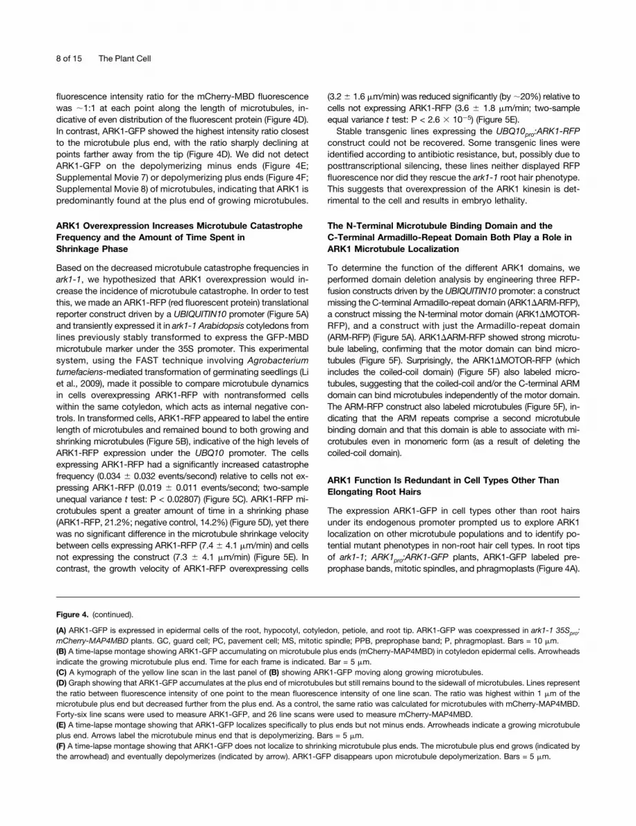
Figure 4. ARK1-GFP Is Expressed in Non-Root Hair Cells and Accumulates on Growing Microtubule Plus Ends.
ARK1 Is a Microtubule Destabilizing Kinesin 7 of 15
fluorescence intensity ratio for the mCherry-MBD fluorescencewas ;1:1 at each point along the length of microtubules, in-dicative of even distribution of the fluorescent protein (Figure 4D).In contrast, ARK1-GFP showed the highest intensity ratio closestto the microtubule plus end, with the ratio sharply declining atpoints farther away from the tip (Figure 4D). We did not detectARK1-GFP on the depolymerizing minus ends (Figure 4E;Supplemental Movie 7) or depolymerizing plus ends (Figure 4F;Supplemental Movie 8) of microtubules, indicating that ARK1 ispredominantly found at the plus end of growing microtubules.
ARK1 Overexpression Increases Microtubule CatastropheFrequency and the Amount of Time Spent inShrinkage Phase
Based on the decreased microtubule catastrophe frequencies inark1-1, we hypothesized that ARK1 overexpression would in-crease the incidence of microtubule catastrophe. In order to testthis, we made an ARK1-RFP (red fluorescent protein) translationalreporter construct driven by a UBIQUITIN10 promoter (Figure 5A)and transiently expressed it in ark1-1 Arabidopsis cotyledons fromlines previously stably transformed to express the GFP-MBDmicrotubule marker under the 35S promoter. This experimentalsystem, using the FAST technique involving Agrobacteriumtumefaciens-mediated transformation of germinating seedlings (Liet al., 2009), made it possible to compare microtubule dynamicsin cells overexpressing ARK1-RFP with nontransformed cellswithin the same cotyledon, which acts as internal negative con-trols. In transformed cells, ARK1-RFP appeared to label the entirelength of microtubules and remained bound to both growing andshrinking microtubules (Figure 5B), indicative of the high levels ofARK1-RFP expression under the UBQ10 promoter. The cellsexpressing ARK1-RFP had a significantly increased catastrophefrequency (0.034 6 0.032 events/second) relative to cells not ex-pressing ARK1-RFP (0.019 6 0.011 events/second; two-sampleunequal variance t test: P < 0.02807) (Figure 5C). ARK1-RFP mi-crotubules spent a greater amount of time in a shrinking phase(ARK1-RFP, 21.2%; negative control, 14.2%) (Figure 5D), yet therewas no significant difference in the microtubule shrinkage velocitybetween cells expressing ARK1-RFP (7.4 6 4.1 mm/min) and cellsnot expressing the construct (7.3 6 4.1 mm/min) (Figure 5E). Incontrast, the growth velocity of ARK1-RFP overexpressing cells
(3.26 1.6 mm/min) was reduced significantly (by;20%) relative tocells not expressing ARK1-RFP (3.6 6 1.8 mm/min; two-sampleequal variance t test: P < 2.6 3 1025) (Figure 5E).Stable transgenic lines expressing the UBQ10pro:ARK1-RFP
construct could not be recovered. Some transgenic lines wereidentified according to antibiotic resistance, but, possibly due toposttranscriptional silencing, these lines neither displayed RFPfluorescence nor did they rescue the ark1-1 root hair phenotype.This suggests that overexpression of the ARK1 kinesin is det-rimental to the cell and results in embryo lethality.
The N-Terminal Microtubule Binding Domain and theC-Terminal Armadillo-Repeat Domain Both Play a Role inARK1 Microtubule Localization
To determine the function of the different ARK1 domains, weperformed domain deletion analysis by engineering three RFP-fusion constructs driven by the UBIQUITIN10 promoter: a constructmissing the C-terminal Armadillo-repeat domain (ARK1DARM-RFP),a construct missing the N-terminal motor domain (ARK1DMOTOR-RFP), and a construct with just the Armadillo-repeat domain(ARM-RFP) (Figure 5A). ARK1DARM-RFP showed strong microtu-bule labeling, confirming that the motor domain can bind micro-tubules (Figure 5F). Surprisingly, the ARK1DMOTOR-RFP (whichincludes the coiled-coil domain) (Figure 5F) also labeled micro-tubules, suggesting that the coiled-coil and/or the C-terminal ARMdomain can bind microtubules independently of the motor domain.The ARM-RFP construct also labeled microtubules (Figure 5F), in-dicating that the ARM repeats comprise a second microtubulebinding domain and that this domain is able to associate with mi-crotubules even in monomeric form (as a result of deleting thecoiled-coil domain).
ARK1 Function Is Redundant in Cell Types Other ThanElongating Root Hairs
The expression ARK1-GFP in cell types other than root hairsunder its endogenous promoter prompted us to explore ARK1localization on other microtubule populations and to identify po-tential mutant phenotypes in non-root hair cell types. In root tipsof ark1-1; ARK1pro:ARK1-GFP plants, ARK1-GFP labeled pre-prophase bands, mitotic spindles, and phragmoplasts (Figure 4A).
Figure 4. (continued).
(A) ARK1-GFP is expressed in epidermal cells of the root, hypocotyl, cotyledon, petiole, and root tip. ARK1-GFP was coexpressed in ark1-1 35Spro:mCherry-MAP4MBD plants. GC, guard cell; PC, pavement cell; MS, mitotic spindle; PPB, preprophase band; P, phragmoplast. Bars = 10 mm.(B) A time-lapse montage showing ARK1-GFP accumulating on microtubule plus ends (mCherry-MAP4MBD) in cotyledon epidermal cells. Arrowheadsindicate the growing microtubule plus end. Time for each frame is indicated. Bar = 5 mm.(C) A kymograph of the yellow line scan in the last panel of (B) showing ARK1-GFP moving along growing microtubules.(D) Graph showing that ARK1-GFP accumulates at the plus end of microtubules but still remains bound to the sidewall of microtubules. Lines representthe ratio between fluorescence intensity of one point to the mean fluorescence intensity of one line scan. The ratio was highest within 1 mm of themicrotubule plus end but decreased further from the plus end. As a control, the same ratio was calculated for microtubules with mCherry-MAP4MBD.Forty-six line scans were used to measure ARK1-GFP, and 26 line scans were used to measure mCherry-MAP4MBD.(E) A time-lapse montage showing that ARK1-GFP localizes specifically to plus ends but not minus ends. Arrowheads indicate a growing microtubuleplus end. Arrows label the microtubule minus end that is depolymerizing. Bars = 5 mm.(F) A time-lapse montage showing that ARK1-GFP does not localize to shrinking microtubule plus ends. The microtubule plus end grows (indicated bythe arrowhead) and eventually depolymerizes (indicated by arrow). ARK1-GFP disappears upon microtubule depolymerization. Bars = 5 mm.
8 of 15 The Plant Cell
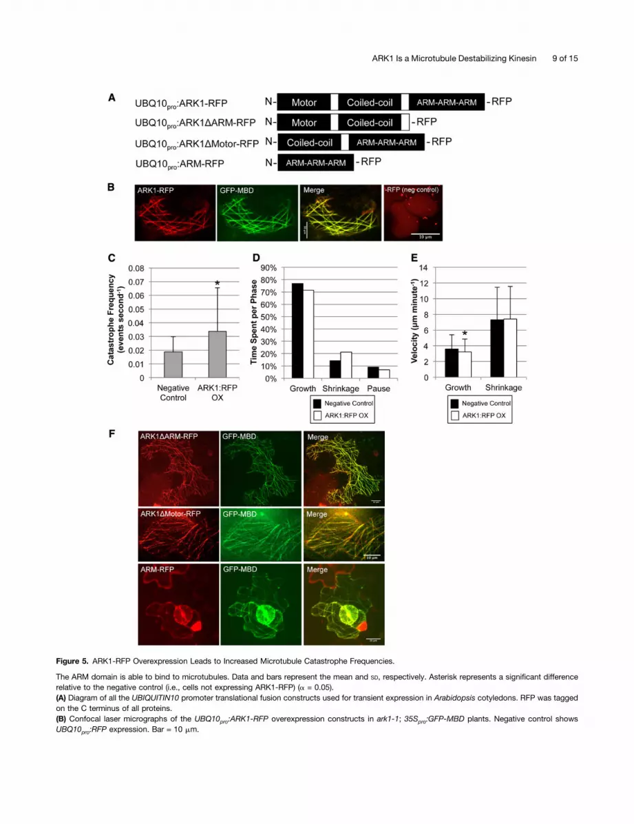
Figure 5. ARK1-RFP Overexpression Leads to Increased Microtubule Catastrophe Frequencies.
The ARM domain is able to bind to microtubules. Data and bars represent the mean and SD, respectively. Asterisk represents a significant differencerelative to the negative control (i.e., cells not expressing ARK1-RFP) (a = 0.05).(A) Diagram of all the UBIQUITIN10 promoter translational fusion constructs used for transient expression in Arabidopsis cotyledons. RFP was taggedon the C terminus of all proteins.(B) Confocal laser micrographs of the UBQ10pro:ARK1-RFP overexpression constructs in ark1-1; 35Spro:GFP-MBD plants. Negative control showsUBQ10pro:RFP expression. Bar = 10 mm.
ARK1 Is a Microtubule Destabilizing Kinesin 9 of 15
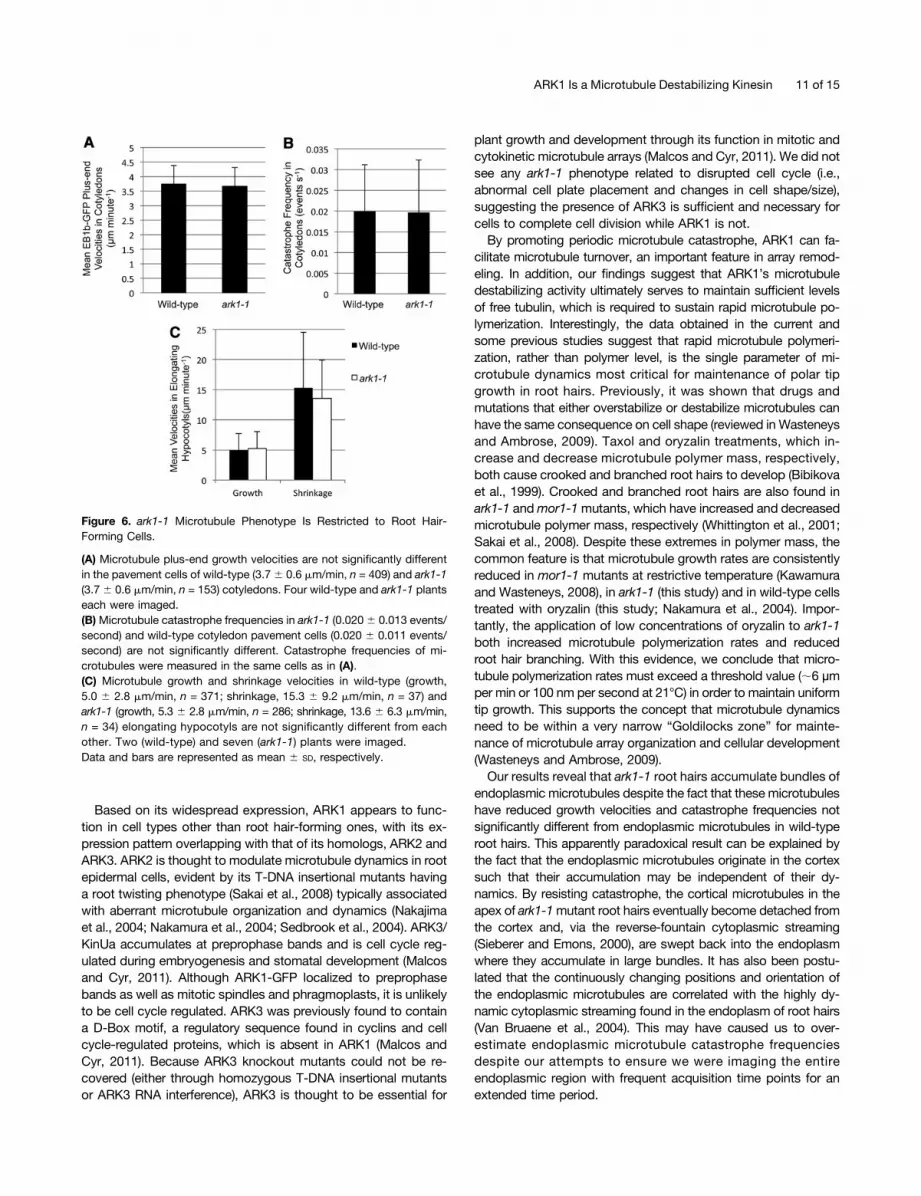
Despite ARK1’s association with microtubules in mitotic and di-viding cells, we did not detect any phenotypes related to cell sizeor cell plate positioning in ark1-1 mutants (Supplemental Figure 3).Moreover, cotyledon pavement cells, atrichoblasts, and hypocotylepidermal cells in ark1-1 mutants showed no morphological dif-ferences from wild-type equivalents as previously shown (Yanget al., 2007; Sakai et al., 2008). No differences in microtubule dy-namic parameters were detected in these cells (Figures 6A to 6C).
DISCUSSION
ARK1 has not been grouped into any of the 14 conventionalkinesin families, which means that its function could not easilybe predicted based on sequence homology. Using translationalreporters, overexpression and loss-of-function analysis, we de-termined that ARK1 is a microtubule plus-end tracking catastrophefactor. The exact mechanism by which it promotes catastropheremains to be determined. Specifically, it is unknown if the motor/ATP catalytic domain is used for kinesin movement along themicrotubule and/or for physically removing tubulin heterodimersfrom the protofilament.
ARK1’s activity is potentially similar to that of the yeast(Saccharomyces cerevisiae) kinesin, Kip3p. A member of theKinesin-8 family, Kip3p, was previously shown to be a plus-endtracking protein as well as a microtubule catastrophe factor (Guptaet al., 2006). Much like ark1-1 mutants, kip3p mutants have in-creased polymer mass (i.e., longer microtubules) (Cottingham andHoyt, 1997) and reduced catastrophe frequency (Gupta et al.,2006). Movement of Kip3p along microtubules is ATP dependent(Gupta et al., 2006; Varga et al., 2006) and highly processive, whichallows the kinesin to maintain plus-end association when de-polymerization occurs. ATP hydrolysis in the motor domain wasalso shown to physically remove tubulin from the plus end of themicrotubule protofilament (Gupta et al., 2006; Varga et al., 2006). Invitro experiments using purified ARK1 and tubulin are currentlybeing pursued to determine ARK1’s exact mechanism of action.Nevertheless, we can conclude that ARK1 promotes catastrophespecifically at the plus ends of microtubules in vivo.
Although previous reports have supported the idea that ARK1acts as a catastrophe factor (Yoo et al., 2008; Sakai et al., 2008),one study has hypothesized that ARK1 promotes microtubule
stability and orientation (Yang et al., 2007). This hypothesis wasbased on confocal micrographs showing randomly oriented,fragmented microtubules in the root hairs of the cae1-1/mrh2-3mutant allele of ark1, which, through a G-to-A substitution, has apremature stop codon and reduced transcript. Moreover, it wasreported that upon taxol exposure, the mrh2-3 root hair mor-phological defects were partially rescued (Yang et al., 2007). Theconfocal images of microtubules in the Yang et al. (2007) study,however, were from root hairs that appeared to have completedgrowth. In wild-type fully elongated root hairs, endoplasmicmicrotubules are absent and cortical microtubules generallyappear fragmented and sparse (Van Bruaene et al., 2004). Themrh2-3 phenotype rescue by taxol reported by Yang et al.(2007), however, remains puzzling and is clearly in contrast toour finding that taxol enhances the ark1-1 root hair phenotype.The ability of the C-terminal ARM domain to independently
associate with microtubules suggests that this domain plays arole in localizing ARK1 to microtubules. This interaction couldpotentially aid in ARK1’s ability to remain at the growing plusends. The idea that a kinesin has two independent microtubulebinding domains is not entirely new. The Arabidopsis ATK5,for example, possesses two microtubule binding domains: theC-terminal motor domain and an additional N-terminal domainthat enables microtubule plus-end tracking (Ambrose et al., 2005).Members of the kinesin-8 family (S. cerevisiae Kip3p and humanKif18A) also contain secondary microtubule binding domains thatallow these kinesins to remain bound to microtubules, resulting inhigh processivity and plus-end accumulations (Stumpff et al.,2011; Su et al., 2011; Mayr et al., 2011; Weaver et al., 2011). Thesecond microtubule binding domain of Kif18A also functions in-dependently of ATP (Mayr et al., 2011; Weaver et al., 2011) muchlike the Armadillo-repeats, which do not have an ATP catalytic site(Tewari et al., 2010).Whether the ARM domain of ARK1 enables direct or indirect
binding to microtubules remains to be determined. A previousstudy demonstrated that this domain, while showing some affinityfor actin filaments in vitro, had little or no affinity for microtubules(Yang et al., 2007). One possibility is that the ARM domain in-teracts with microtubules via the NIMA-related kinase, NEK6. Thisis based on previous evidence that showed NEK6’s ability to bindto the ARM domain (Sakai et al., 2008) and NEK6-GFP’s ability tolocalize to microtubules (Motose et al., 2008, 2011).
Figure 5. (continued).
(C) Cells transiently overexpressing ARK1-RFP have a significant increase in microtubule catastrophe frequency (0.034 6 0.032 events/second) relativeto cells not expressing ARK1-RFP (0.019 6 0.011 events/second). For ARK1-RFP overexpression and the negative control, 26 and 42 microtubuleswere measured over a time period of 1700 and 2500 s, respectively.(D) Distribution of time spent in each phase (growth, shrinkage, and pause) for cells overexpressing and not overexpressing ARK1-RFP in 35Spro:GFP-MBD plants. Microtubules in cells overexpressing ARK1-RFP spent a greater amount of time shrinking as a result of increased catastrophe frequencyshown in (C). For ARK1-RFP overexpression and the negative control, 86 and 82 microtubules were visualized over a time period of 8000 and 5800 s,respectively.(E) The mean microtubule growth and shrinkage velocities in cells overexpressing and not overexpressing ARK1-RFP. There is a significant reduction ingrowth velocities in ARK1-RFP overexpression cells (3.2 6 0.1.6 mm/min) relative to the negative control (3.6 6 1.8 mm/min). There is no significantdifference in shrinkage velocities between the two treatments (ARK1-RFP, 7.4 6 4.1 mm/min; negative control, 7.3 6 4.2 mm/min). For each treatment,a minimum of 700 growth events and 150 shrinkage events were measured.(F) Confocal laser micrographs of the various UBQ10pro overexpression constructs seen in (A) expressed in ark1-1; 35Spro:GFP-MBD plants. Micro-tubule labeling still occurs in constructs lacking the motor or ARM domain. Bars = 10 mm.
10 of 15 The Plant Cell
Based on its widespread expression, ARK1 appears to func-tion in cell types other than root hair-forming ones, with its ex-pression pattern overlapping with that of its homologs, ARK2 andARK3. ARK2 is thought to modulate microtubule dynamics in rootepidermal cells, evident by its T-DNA insertional mutants havinga root twisting phenotype (Sakai et al., 2008) typically associatedwith aberrant microtubule organization and dynamics (Nakajimaet al., 2004; Nakamura et al., 2004; Sedbrook et al., 2004). ARK3/KinUa accumulates at preprophase bands and is cell cycle reg-ulated during embryogenesis and stomatal development (Malcosand Cyr, 2011). Although ARK1-GFP localized to preprophasebands as well as mitotic spindles and phragmoplasts, it is unlikelyto be cell cycle regulated. ARK3 was previously found to containa D-Box motif, a regulatory sequence found in cyclins and cellcycle-regulated proteins, which is absent in ARK1 (Malcos andCyr, 2011). Because ARK3 knockout mutants could not be re-covered (either through homozygous T-DNA insertional mutantsor ARK3 RNA interference), ARK3 is thought to be essential for
plant growth and development through its function in mitotic andcytokinetic microtubule arrays (Malcos and Cyr, 2011). We did notsee any ark1-1 phenotype related to disrupted cell cycle (i.e.,abnormal cell plate placement and changes in cell shape/size),suggesting the presence of ARK3 is sufficient and necessary forcells to complete cell division while ARK1 is not.By promoting periodic microtubule catastrophe, ARK1 can fa-
cilitate microtubule turnover, an important feature in array remod-eling. In addition, our findings suggest that ARK1’s microtubuledestabilizing activity ultimately serves to maintain sufficient levelsof free tubulin, which is required to sustain rapid microtubule po-lymerization. Interestingly, the data obtained in the current andsome previous studies suggest that rapid microtubule polymeri-zation, rather than polymer level, is the single parameter of mi-crotubule dynamics most critical for maintenance of polar tipgrowth in root hairs. Previously, it was shown that drugs andmutations that either overstabilize or destabilize microtubules canhave the same consequence on cell shape (reviewed in Wasteneysand Ambrose, 2009). Taxol and oryzalin treatments, which in-crease and decrease microtubule polymer mass, respectively,both cause crooked and branched root hairs to develop (Bibikovaet al., 1999). Crooked and branched root hairs are also found inark1-1 andmor1-1mutants, which have increased and decreasedmicrotubule polymer mass, respectively (Whittington et al., 2001;Sakai et al., 2008). Despite these extremes in polymer mass, thecommon feature is that microtubule growth rates are consistentlyreduced in mor1-1 mutants at restrictive temperature (Kawamuraand Wasteneys, 2008), in ark1-1 (this study) and in wild-type cellstreated with oryzalin (this study; Nakamura et al., 2004). Impor-tantly, the application of low concentrations of oryzalin to ark1-1both increased microtubule polymerization rates and reducedroot hair branching. With this evidence, we conclude that micro-tubule polymerization rates must exceed a threshold value (;6 µmper min or 100 nm per second at 21°C) in order to maintain uniformtip growth. This supports the concept that microtubule dynamicsneed to be within a very narrow “Goldilocks zone” for mainte-nance of microtubule array organization and cellular development(Wasteneys and Ambrose, 2009).Our results reveal that ark1-1 root hairs accumulate bundles of
endoplasmic microtubules despite the fact that these microtubuleshave reduced growth velocities and catastrophe frequencies notsignificantly different from endoplasmic microtubules in wild-typeroot hairs. This apparently paradoxical result can be explained bythe fact that the endoplasmic microtubules originate in the cortexsuch that their accumulation may be independent of their dy-namics. By resisting catastrophe, the cortical microtubules in theapex of ark1-1mutant root hairs eventually become detached fromthe cortex and, via the reverse-fountain cytoplasmic streaming(Sieberer and Emons, 2000), are swept back into the endoplasmwhere they accumulate in large bundles. It has also been postu-lated that the continuously changing positions and orientation ofthe endoplasmic microtubules are correlated with the highly dy-namic cytoplasmic streaming found in the endoplasm of root hairs(Van Bruaene et al., 2004). This may have caused us to over-estimate endoplasmic microtubule catastrophe frequenciesdespite our attempts to ensure we were imaging the entireendoplasmic region with frequent acquisition time points for anextended time period.
Figure 6. ark1-1 Microtubule Phenotype Is Restricted to Root Hair-Forming Cells.
(A) Microtubule plus-end growth velocities are not significantly differentin the pavement cells of wild-type (3.7 6 0.6 mm/min, n = 409) and ark1-1(3.7 6 0.6 mm/min, n = 153) cotyledons. Four wild-type and ark1-1 plantseach were imaged.(B)Microtubule catastrophe frequencies in ark1-1 (0.0206 0.013 events/second) and wild-type cotyledon pavement cells (0.020 6 0.011 events/second) are not significantly different. Catastrophe frequencies of mi-crotubules were measured in the same cells as in (A).(C) Microtubule growth and shrinkage velocities in wild-type (growth,5.0 6 2.8 mm/min, n = 371; shrinkage, 15.3 6 9.2 mm/min, n = 37) andark1-1 (growth, 5.3 6 2.8 mm/min, n = 286; shrinkage, 13.6 6 6.3 mm/min,n = 34) elongating hypocotyls are not significantly different from eachother. Two (wild-type) and seven (ark1-1) plants were imaged.Data and bars are represented as mean 6 SD, respectively.
ARK1 Is a Microtubule Destabilizing Kinesin 11 of 15
From our mutant, drug, and overexpression analysis, it is clearthat the correct modulation of microtubule dynamics encom-passes a broad range of cellular factors, including free tubulinconcentration and the expression and activation of differentmicrotubule-associated proteins. Although it was expected thatARK1-RFP overexpression would result in an increase in catas-trophe frequency, we were surprised to observe a reduced mi-crotubule growth rate. It is possible that overaccumulation ofARK1-RFP, particularly at the microtubule plus end, inhibits thebinding of other microtubule-associated proteins or affects itsown function. In Arabidopsis, several MAPs have been reported tointeract with microtubule plus ends to orchestrate dynamics (e.g.,EB1 [Chan et al., 2003], SKU6/SPIRAL1 [Nakajima et al., 2004;Sedbrook et al., 2004], ATK5 [Ambrose et al., 2005], and CLASP1[Ambrose et al., 2007]), the activity of which might be altered byARK1-RFP overexpression. In vitro analysis has previously shownthat increasing concentrations of Kip3p and other MAPs candecrease Kip3p’s velocity along microtubules as a result ofmacromolecular crowding (Leduc et al., 2012). Given that the plus-end microtubule-associated proteins interact at the nanoscalelevel, an appropriate ratio of these proteins is required for preciseregulation of microtubule dynamics. Based on our study, completeremoval of ARK1 from microtubule plus ends or its overexpressionis detrimental to proper microtubule dynamics.
METHODS
Plant Material and Culture
Arabidopsis thaliana wild-type (Columbia-0 ecotype) and ark1-1 plantswith the 35Spro:GFP-MBD transgene were used, as previously described(Sakai et al., 2008). The 35Spro:EB1b-GFP reporter line (Mathur et al., 2003)and the 35Spro:mCherry-MAP4MBD construct (Gutierrez et al., 2009) werekindly provided by Jaideep Mathur (University of Guelph, Canada) andDavid Erhardt (Carnegie Institute of Science, Stanford, CA), respectively.
All seeds were sterilized in 70% ethanol, rinsed three times with doubledistilled water, and plated onto Petri dishes with Hoagland media (1.2%Bacto-agar [BD Diagnostics], no sucrose). Plates with seeds were storedin the dark at 4°C for 2 to 3 d and transferred to a 21°C growth cabinet(24 h light) where they were grown vertically until imaging. For the drugstudies, seeds were initially vertically grown on Hoagland media sup-plementedwith DMSO (Fisher Scientific) and then transferred to Hoaglandmedia containing various concentrations of taxol (Sigma-Aldrich) or oryzalin(Sigma-Aldrich) for 2 d prior to imaging.
ARK1 Construct Design and Cloning Strategies
Gateway cloning technology (Invitrogen) was used for the ARK1 genomicand coding sequences. To generate the ARK1pro:ARK1-GFP construct,the ARK1 genomic sequence (between 888 bp upstream of the ATG/startcodon and the TGA/stop codon) was amplified from the F28P10 BAC(from the ABRC, Ohio State University) using the full-length ARK1 ge-nomic sequence primer set (Supplemental Table 1). A second PCR withthe attB-adapter primers was performed according to the manufacturer’sprotocol (see Invitrogen for attB-adapter sequence). For the amplificationof the ARK1 coding sequences, different primers were used to amplify thecDNA templates for the ARK1, ARK1DARM, ARK1DMotor, and ARMconstructs (see Supplemental Table 1 for primer list). A second PCR withthe attB-adapter primers was performed according to the manufacturer’sprotocol (Invitrogen). The mRNA extraction and cDNA synthesis protocolwere performed as described (Galway et al., 2011). Following the BP
reaction with the various attB-PCR products and the pDONR221 vector(Invitrogen), an LR reaction was performed with the pMDC107 vector(Curtis and Grossniklaus, 2003) for the ARK1 genomic sequence and thepUBC-RFP DEST vector (Grefen et al., 2010) for the various ARK1 codingsequences.
Generation of Transgenic Plant Materials
For stable transgenic lines, the ARK1pro:ARK1-GFP construct was firsttransformed into Agrobacterium tumefaciens (GV3101 strain) and thentransformed into an Arabidopsis ark1-1 35Spro:mCherry-MAP4MBD lineusing the floral dip method (Clough and Bent, 1998). T3 lines homozygousfor the ARK1pro:ARK1-GFP transgene were segregated and used forfurther experiments. Transient expression in cotyledons of the four dif-ferent UBQpro-driven ARK1 fragment-RFP constructs was performed inark1-1; 35Spro:GFP-MBD plants using the FAST technique (Li et al., 2009).ark1-1 (SALK_035063) plants were crossed into the 35Spro:EB1b-GFPreporter line (Mathur et al., 2003), and homozygous F3 ark1-1 and wild-type plants were segregated for imaging.
Live-Cell Imaging
For observing root hair morphology, whole seedlings were mounted onslides with cover slips and bright-field images of root hairs were thencollected with a 203 (air) objective lens on a Leica DMR light microscopewith a Q-CAM digital camera (Leica).
Live imaging of the various microtubule reporter proteins, the ARK1pro-driven ARK1-GFP, and the UBQ10pro-driven ARK1-RFP was done usinga Perkin-Elmer Ultraview VoX Spinning Disc Confocal system (Perkin-Elmer) mounted on a Leica DMI6000 B invertedmicroscope and equippedwith a Hamamatsu 9100-02 electron multiplier CCD camera (Hamamatsu).An argon 488-nm laser line with a complementary GFP (525/36) emissionband-pass filter or a 561-nm laser with a complementary RFP (595/50)emission band-pass filter was used. Images were acquired with a 633(water) objective lens every 8 s for 3 to 5 min with 0.3- to 0.5-mm opticalz-slices. For imaging of microtubule dynamics during drug treatments,seedlings grown on Hoagland medium with 100 nM oryzalin were mountedin 100 nMoryzalin. The imaging temperature of the sampleswasmaintainedat 21°C using a Bionomic Controller BC-110 with a HEC-400 Heat Ex-changer, a Bionomic Controller BC-100 (20-20 Technology) temperature-controlled stage, and an objective heater (Bioptechs).
Image and Data Analysis
All images were processed and analyzed using ImageJ (http://rsbweb.nih.gov/ij/). For root hair length analysis, line selections were superimposedon root hairs and then measured. Measuring the microtubule velocities ofthe EB1b-GFP and GFP-MBD microtubule marker lines were done usingthe Manual Tracking plug-in (http://rsbweb.nih.gov/ij/plugins/track/track.html). EB1b-GFP particles were considered endoplasmic when imagesfrom the medial confocal plane of the root hair were analyzed in theendoplasmic region of the root hair. Cortical EB1b-GFP particles weremeasured and analyzed from cortical plane of the root hairs or along thecortex/plasmamembrane in themedial confocal plane of the root hair. Fordetermining the catastrophe frequency of EB1b-GFP, the inverse of theduration of time spent tracking one EB1b-GFP comet was taken. This isbased on the assumption that the disappearance of the EB1b-GFP signalmeant the microtubule was undergoing catastrophe since EB1b-GFP hashighest affinity for the growing plus end of microtubules. The samemethod for determining catastrophe frequency was used with the GFP-MBD marker in the ARK1 overexpression analysis. For determining theamount of time spent in each phase, the amount of time spent in onephase (i.e., growth, shrinkage, or pause) was divided by the total measured
12 of 15 The Plant Cell
time of a microtubule lifespan (using the GFP-MBD marker). The mean, SD,F-tests, and t tests were calculated using Excel (Microsoft).
We also sought to measure potential changes in shrinkage velocitiesand rescue frequencies by imaging the microtubule markers 35Spro:GFP-MBD and UBQ1pro:mRFP-TUB6 in root hairs. However, we were unable toaccurately observe and quantify plus-end dynamics of single microtubuleswith thesemarkers in root hairs because of increasedmicrotubule bundling,dense microtubule populations, or high background fluorescence.
For quantifying the ratio of fluorescence intensity tomean fluorescenceintensity, the equations below were used. The procedure involved taking3-mm line scans on microtubule plus ends from ARK1-GFP images andthen measuring the fluorescence intensities/gray values (xn) every 0.108mm along the line scan using the Plot Profile function in ImageJ. Thefluorescence intensities were then averaged (�x:) by the total number ofpoints (N ) measured in the line scan (Equation 1). Each fluorescenceintensity (xn) value was then divided by the mean fluorescence intensity (�x)to get the desired ratio (Rn) at each point along the 3-mm line scan(Equation 2). All ratios are reported as mean values. The same mea-surements were done with the mCherry-MAP4 MBD images as a control.
�x ¼ x1 þ x2 þ x3.þ xnN
ð1Þ
Rn ¼ xn�x
ð2Þ
Quantification of Endoplasmic and Cortical Microtubules
Confocal images of wild-type and ark1-1; 35Spro:GFP-MBD root hairswere analyzed in Image J according to Sakai et al. (2008).
Propidium Iodide Staining and Imaging of Wild-Type and ark1-1Root Tips
Labeling of the cell wall in the root tips was done by incubating whole wild-type and ark1-1 seedlings in 10 mg/mL propidium iodide (Calbiochem) for1 min, rinsing them with water, and mounting them on slides and coverslips. Laser confocal images of the stained root tips were done on a ZeissAxioImager M1 microscope with a Zeiss PASCAL Excite two-channelLSM 780 system (Carl Zeiss). A helium-neon 543-nm laser line and a 560-nmemission long-pass filter were used. Images were acquired in 2-mm opticalz-slices with a 633 (oil) objective lens.
Accession Numbers
Sequence data from this article can be found in the Arabidopsis GenomeInitiative or EMBL/GenBank databases under the following ArabidopsisGenome Initiative identifier: At3g54870 (ARK1/MRH2).
Supplemental Data
The following materials are available in the online version of this article.
Supplemental Figure 1. EB1b-GFP Velocity Is Slower in ark1-1 RootHairs Than in Wild-Type Root Hairs.
Supplemental Figure 2. Endoplasmic Microtubules Are More Abun-dant in ark1-1 Than in Wild-Type Root Hairs.
Supplemental Figure 3. Cell and Tissue Patterns Are Not Affected inark1-1 Root Tips.
Supplemental Table 1. Primer Sets Used for Cloning the VariousARK1 Constructs.
Supplemental Movie 1. EB1b-GFP Trajectories in Wild-Type Elon-gating Root Hairs Show That Microtubules Undergo Rapid Elongationand Frequent Catastrophe.
Supplemental Movie 2. EB1b-GFP Trajectories in ark1-1 ElongatingRoot Hairs Show That Microtubule Growth Rates and CatastropheFrequencies Are Reduced.
Supplemental Movie 3. ARK1-GFP Localizes to Microtubules inElongating Root Hairs.
Supplemental Movie 4. ARK1-GFP Localizes to Microtubules in FullyGrown Root Hairs.
Supplemental Movie 5. ARK1-GFP Is Concentrated at MicrotubulePlus Ends in Cotyledon Pavement and Guard Cells.
Supplemental Movie 6. High-Magnification Time-Lapse ImagesDemonstrate the ARK1-GFP Plus-End Tracking.
Supplemental Movie 7. Time-Lapse Images of a Treadmilling Micro-tubule Demonstrates that ARK1-GFP Is Distributed Specifically to theGrowing Microtubule Plus Ends and Not the Minus Ends.
Supplemental Movie 8. ARK1-GFP Is Not Associated with ShrinkingMicrotubule Plus Ends.
ACKNOWLEDGMENTS
This research was funded by a Natural Sciences and Engineering ResearchCouncil of Canada (NSERC) Discovery grant (298264-09) to G.O.W., anNSERC postgraduate scholarship to R.C.E., and the Canada Foundationfor Innovation. We thank Jaideep Mathur (University of Guelph, Canada)and David Ehrhardt (Carnegie Institute of Science, Stanford, CA) forreporter lines, Chris Ambrose (University of British Columbia [UBC]) fortechnical insight and careful reading of the article, Lacey Samuels (UBC)for use of her Leica light microscope, and the UBC Bioimaging Facility forconfocal microscope access and assistance.
AUTHOR CONTRIBUTIONS
R.C.E. performed experiments. G.O.W. and R.C.E. designed the re-search, analyzed the data, and wrote the article.
Received April 21, 2014; revised June 30, 2014; accepted August 5,2014; published August 26, 2014.
REFERENCES
Ambrose, J.C., Li, W., Marcus, A., Ma, H., and Cyr, R. (2005). Aminus-end-directed kinesin with plus-end tracking protein activity isinvolved in spindle morphogenesis. Mol. Biol. Cell 16: 1584–1592.
Ambrose, J.C., Shoji, T., Kotzer, A.M., Pighin, J.A., and Wasteneys,G.O. (2007). The Arabidopsis CLASP gene encodes a microtubule-associated protein involved in cell expansion and division. PlantCell 19: 2763–2775.
Bibikova, T.N., Blancaflor, E.B., and Gilroy, S. (1999). Microtubulesregulate tip growth and orientation in root hairs of Arabidopsisthaliana. Plant J. 17: 657–665.
Carol, R.J., and Dolan, L. (2002). Building a hair: tip growth inArabidopsis thaliana root hairs. Philos. Trans. R. Soc. Lond. B Biol.Sci. 357: 815–821.
Chan, J., Calder, G.M., Doonan, J.H., and Lloyd, C.W. (2003). EB1reveals mobile microtubule nucleation sites in Arabidopsis. Nat. CellBiol. 5: 967–971.
ARK1 Is a Microtubule Destabilizing Kinesin 13 of 15

Clough, S.J., and Bent, A.F. (1998). Floral dip: a simplified method forAgrobacterium-mediated transformation of Arabidopsis thaliana.Plant J. 16: 735–743.
Cottingham, F.R., and Hoyt, M.A. (1997). Mitotic spindle positioningin Saccharomyces cerevisiae is accomplished by antagonisticallyacting microtubule motor proteins. J. Cell Biol. 138: 1041–1053.
Curtis, M.D., and Grossniklaus, U. (2003). A Gateway cloning vectorset for high-throughput functional analysis of genes in planta. PlantPhysiol. 133: 462–469.
Desai, A., and Mitchison, T.J. (1997). Microtubule polymerizationdynamics. Annu. Rev. Cell Dev. Biol. 13: 83–117.
Desai, A., Verma, S., Mitchison, T.J., and Walczak, C.E. (1999). Kin Ikinesins are microtubule-destabilizing enzymes. Cell 96: 69–78.
Galway, M.E., Eng, R.C., Schiefelbein, J.W., and Wasteneys, G.O.(2011). Root hair-specific disruption of cellulose and xyloglucan inAtCSLD3 mutants, and factors affecting the post-rupture resumptionof mutant root hair growth. Planta 233: 985–999.
Grefen, C., Donald, N., Hashimoto, K., Kudla, J., Schumacher, K.,and Blatt, M.R. (2010). A ubiquitin-10 promoter-based vector setfor fluorescent protein tagging facilitates temporal stability andnative protein distribution in transient and stable expression studies.Plant J. 64: 355–365.
Gupta, M.L., Jr., Carvalho, P., Roof, D.M., and Pellman, D. (2006).Plus end-specific depolymerase activity of Kip3, a kinesin-8 protein,explains its role in positioning the yeast mitotic spindle. Nat. CellBiol. 8: 913–923.
Gutierrez, R., Lindeboom, J.J., Paredez, A.R., Emons, A.M., andEhrhardt, D.W. (2009). Arabidopsis cortical microtubules positioncellulose synthase delivery to the plasma membrane and interact withcellulose synthase trafficking compartments. Nat. Cell Biol. 11: 797–806.
Hiwatashi, Y., Sato, Y., and Doonan, J.H. (2014). Kinesins havea dual function in organizing microtubules during both tip growthand cytokinesis in Physcomitrella patens. Plant Cell 26: 1256–1266.
Jones, M.A., Raymond, M.J., and Smirnoff, N. (2006). Analysis of theroot-hair morphogenesis transcriptome reveals the molecular identityof six genes with roles in root-hair development in Arabidopsis. PlantJ. 45: 83–100.
Kawamura, E., and Wasteneys, G.O. (2008). MOR1, the Arabidopsisthaliana homologue of Xenopus MAP215, promotes rapid growthand shrinkage, and suppresses the pausing of microtubules in vivo.J. Cell Sci. 121: 4114–4123.
Lechner, B., Rashbrooke, M.C., Collings, D.A., Eng, R.C., Kawamura, E.,Whittington, A.T., and Wasteneys, G.O. (2012). The N-terminal TOGdomain of Arabidopsis MOR1 modulates affinity for microtubulepolymers. J. Cell Sci. 125: 4812–4821.
Leduc, C., Padberg-Gehle, K., Varga, V., Helbing, D., Diez, S., andHoward, J. (2012). Molecular crowding creates traffic jams of kinesinmotors on microtubules. Proc. Natl. Acad. Sci. USA 109: 6100–6105.
Li, J.F., Park, E., von Arnim, A.G., and Nebenführ, A. (2009). TheFAST technique: a simplified Agrobacterium-based transformationmethod for transient gene expression analysis in seedlings ofArabidopsis and other plant species. Plant Methods 5: 6.
Malcos, J.L., and Cyr, R.J. (2011). An ungrouped plant kinesinaccumulates at the preprophase band in a cell cycle-dependentmanner. Cytoskeleton (Hoboken) 68: 247–258.
Marc, J., Granger, C.L., Brincat, J., Fisher, D.D., Kao, Th.,McCubbin, A.G., and Cyr, R.J. (1998). A GFP-MAP4 reporter genefor visualizing cortical microtubule rearrangements in living epidermalcells. Plant Cell 10: 1927–1940.
Mathur, J., Mathur, N., Kernebeck, B., Srinivas, B.P., andHülskamp, M. (2003). A novel localization pattern for an EB1-likeprotein links microtubule dynamics to endomembrane organization.Curr. Biol. 13: 1991–1997.
Mayr, M.I., Storch, M., Howard, J., and Mayer, T.U. (2011). A non-motor microtubule binding site is essential for the high processivityand mitotic function of kinesin-8 Kif18A. PLoS ONE 6: e27471.
Motose, H., Tominaga, R., Wada, T., Sugiyama, M., and Watanabe,Y. (2008). A NIMA-related protein kinase suppresses ectopicoutgrowth of epidermal cells through its kinase activity and theassociation with microtubules. Plant J. 54: 829–844.
Motose, H., Hamada, T., Yoshimoto, K., Murata, T., Hasebe, M.,Watanabe, Y., Hashimoto, T., Sakai, T., and Takahashi, T. (2011).NIMA-related kinases 6, 4, and 5 interact with each other to regulatemicrotubule organization during epidermal cell expansion in Arabidopsisthaliana. Plant J. 67: 993–1005.
Nakajima, K., Furutani, I., Tachimoto, H., Matsubara, H., andHashimoto, T. (2004). SPIRAL1 encodes a plant-specific microtubule-localized protein required for directional control of rapidly expandingArabidopsis cells. Plant Cell 16: 1178–1190.
Nakamura, M., Naoi, K., Shoji, T., and Hashimoto, T. (2004). Lowconcentrations of propyzamide and oryzalin alter microtubuledynamics in Arabidopsis epidermal cells. Plant Cell Physiol. 45:1330–1334.
Oda, Y., and Fukuda, H. (2013). Rho of plant GTPase signalingregulates the behavior of Arabidopsis kinesin-13A to establishsecondary cell wall patterns. Plant Cell 25: 4439–4450.
Reddy, A.S.N., and Day, I.S. (2011). Microtubule motor proteins in theeukaryotic green lineage: Functions and regulation. In The PlantCytoskeleon, Advances in Plant Biology, B. Liu, ed (New York: Springer),pp. 119–141.
Sakai, T., et al. (2008). Armadillo repeat-containing kinesins and aNIMA-related kinase are required for epidermal-cell morphogenesisin Arabidopsis. Plant J. 53: 157–171.
Sedbrook, J.C., Ehrhardt, D.W., Fisher, S.E., Scheible, W.R., andSomerville, C.R. (2004). The Arabidopsis sku6/spiral1 gene encodesa plus end-localized microtubule-interacting protein involved in directionalcell expansion. Plant Cell 16: 1506–1520.
Sieberer, B., and Timmers, A. (2009). Microtubules in plant root hairsand their role in cell polarity and tip growth in root hairs. In Plant CellMonograph, Vol. 12, A.M. Emons and T. Ketelaar, eds (Berlin: Springer),pp. 233–248.
Sieberer, B., and Emons, A.M.C. (2000). Cytoarchitecture and pattern ofcytoplasmic streaming in root hairs of Medicago truncatula duringdevelopment and deformation by nodulation factors. Protoplasma214: 118–127.
Sieberer, B.J., Timmers, A.C., Lhuissier, F.G., and Emons, A.M.(2002). Endoplasmic microtubules configure the subapical cytoplasmand are required for fast growth of Medicago truncatula root hairs. PlantPhysiol. 130: 977–988.
Stumpff, J., Du, Y., English, C.A., Maliga, Z., Wagenbach, M.,Asbury, C.L., Wordeman, L., and Ohi, R. (2011). A tetheringmechanism controls the processivity and kinetochore-microtubuleplus-end enrichment of the kinesin-8 Kif18A. Mol. Cell 43: 764–775.
Su, X., Qiu, W., Gupta, M.L., Jr., Pereira-Leal, J.B., Reck-Peterson,S.L., and Pellman, D. (2011). Mechanisms underlying the dual-mode regulation of microtubule dynamics by Kip3/kinesin-8. Mol.Cell 43: 751–763.
Tewari, R., Bailes, E., Bunting, K.A., and Coates, J.C. (2010).Armadillo-repeat protein functions: questions for little creatures.Trends Cell Biol. 20: 470–481.
Van Bruaene, N., Joss, G., and Van Oostveldt, P. (2004). Reorganizationand in vivo dynamics of microtubules during Arabidopsis root hairdevelopment. Plant Physiol. 136: 3905–3919.
Varga, V., Helenius, J., Tanaka, K., Hyman, A.A., Tanaka, T.U., andHoward, J. (2006). Yeast kinesin-8 depolymerizes microtubules ina length-dependent manner. Nat. Cell Biol. 8: 957–962.
14 of 15 The Plant Cell

Wasteneys, G.O., and Ambrose, J.C. (2009). Spatial organization ofplant cortical microtubules: close encounters of the 2D kind. TrendsCell Biol. 19: 62–71.
Weaver, L.N., Ems-McClung, S.C., Stout, J.R., LeBlanc, C., Shaw,S.L., Gardner, M.K., and Walczak, C.E. (2011). Kif18A usesa microtubule binding site in the tail for plus-end localization andspindle length regulation. Curr. Biol. 21: 1500–1506.
Whittington, A.T., Vugrek, O., Wei, K.J., Hasenbein, N.G., Sugimoto, K.,Rashbrooke, M.C., and Wasteneys, G.O. (2001). MOR1 is essential fororganizing cortical microtubules in plants. Nature 411: 610–613.
Yang, G., Gao, P., Zhang, H., Huang, S., and Zheng, Z.L. (2007). Amutation in MRH2 kinesin enhances the root hair tip growth defect
caused by constitutively activated ROP2 small GTPase in Arabidopsis.PLoS ONE 2: e1074.
Yoo, C.M., and Blancaflor, E.B. (2013). Overlapping and divergentsignaling pathways for ARK1 and AGD1 in the control of root hairpolarity in Arabidopsis thaliana. Front. Plant Sci. 4: 528.
Yoo, C.M., Wen, J., Motes, C.M., Sparks, J.A., and Blancaflor, E.B.(2008). A class I ADP-ribosylation factor GTPase-activating proteinis critical for maintaining directional root hair growth in Arabidopsis.Plant Physiol. 147: 1659–1674.
Zhu, C., and Dixit, R. (2012). Functions of the Arabidopsis kinesinsuperfamily of microtubule-based motor proteins. Protoplasma 249:887–899.
ARK1 Is a Microtubule Destabilizing Kinesin 15 of 15

DOI 10.1105/tpc.114.126789; originally published online August 26, 2014;Plant Cell
Ryan Christopher Eng and Geoffrey O. WasteneysArabidopsisMicrotubule Catastrophe in
The Microtubule Plus-End Tracking Protein ARMADILLO-REPEAT KINESIN1 Promotes
This information is current as of August 26, 2014
Supplemental Data http://www.plantcell.org/content/suppl/2014/08/13/tpc.114.126789.DC1.html
Permissions https://www.copyright.com/ccc/openurl.do?sid=pd_hw1532298X&issn=1532298X&WT.mc_id=pd_hw1532298X
eTOCs http://www.plantcell.org/cgi/alerts/ctmain
Sign up for eTOCs at:
CiteTrack Alerts http://www.plantcell.org/cgi/alerts/ctmain
Sign up for CiteTrack Alerts at:
Subscription Information http://www.aspb.org/publications/subscriptions.cfm
is available at:Plant Physiology and The Plant CellSubscription Information for
ADVANCING THE SCIENCE OF PLANT BIOLOGY © American Society of Plant Biologists