the exosome associates cotranscriptionally with the nascent pre-mrnp through interactions with hnrnp...
TRANSCRIPT
Ms E09-01-0079 Revised The exosome associates cotranscriptionally with the nascent pre-mRNP through interactions with hnRNP proteins Viktoria Hessle1, Petra Björk1, Marcus Sokolowski1,2, Ernesto González de Valdivia1, Rebecca Silverstein1, Konstantin Artemenko3, Anu Tyagi1, Gianluca Maddalo4, Leopold Ilag4, Roger Helbig1, Roman A. Zubarev3 and Neus Visa1
1 Department of Molecular Biology & Functional Genomics, Stockholm University, SE-10 691 Stockholm, Sweden
2 Present address: Department of Public Health Sciences, Karolinska institutet, SE-17 177 Stockholm, Sweden
3 Division of Molecular Biometry, Institute for Cell and Molecular Biology, Uppsala University, Box 596, SE-75 124 Uppsala, Sweden
4 Department of Analytical Chemistry, Stockholm University, SE-10 691 Stockholm, Sweden
Running head: Cotranscriptional mRNA surveillance Corresponding author: Neus Visa address as above fax +48 8 16 64 88 e-mail [email protected]
1 http://www.molbiolcell.org/content/suppl/2009/06/02/E09-01-0079.DC1.html
Supplemental Material can be found at:
ABSTRACT
Eukaryotic cells have evolved quality control mechanisms to degrade aberrant messenger
RNA molecules and prevent the synthesis of defective proteins that could be deleterious
for the cell. The exosome, a protein complex with ribonuclease activity, is a key player in
quality control. An early quality checkpoint takes place cotranscriptionally but little is known
about the molecular mechanisms by which the exosome is recruited to the transcribed
genes. Here we study the core exosome subunit Rrp4 in two insect model systems,
Chironomus and Drosophila. We show that a significant fraction of Rrp4 is associated with
the nascent pre-mRNPs and that a specific mRNA-binding protein, Hrp59/hnRNP M,
interacts in vivo with multiple exosome subunits. Depletion of Hrp59 by RNA interference
reduces the levels of Rrp4 at transcription sites, which suggests that Hrp59 is needed for
the exosome to stably interact with nascent pre-mRNPs. Our results lead to a revised
mechanistic model for cotranscriptional quality control in which the exosome is constantly
recruited to newly synthesized RNAs through direct interactions with specific hnRNP
proteins.
2
INTRODUCTION
Expression of protein-coding genes is a complex multistep process. Precursor
messenger RNAs (pre-mRNAs) are synthesized by RNA polymerase II (Pol-II) and
assembled into ribonucleoprotein (RNP) complexes during transcription. The pre-mRNPs
must be processed to become mature mRNPs, which are exported to the cytoplasm and
translated into protein. Mutations in the DNA or errors in the transcription and processing
reactions can lead to the synthesis of aberrant mRNPs. Eukaryotic cells have evolved
quality control mechanisms that identify aberrant mRNPs and prevent their expression into
protein. In the yeast Saccharomyces cerevisiae there are at least three nuclear steps of
mRNP biogenesis that are under surveillance: the cleavage and polyadenylation of the 3'
end (Hilleren et al., 2001), the assembly of the mRNA-protein complexes (Zenklusen et al.,
2002; Luna et al., 2005), and the removal of introns (Galy et al., 2004). In all cases,
mRNPs with assembly and/or processing defects are detected by nuclear surveillance
mechanisms, retained in the nucleus and degraded (reviewed by Vinciguerra and Stutz,
2004; Sommer and Nehrbass, 2005; Saguez et al., 2005). Genetic studies in S. cerevisiae
have identified the nuclear exosome as a key player in the recognition and retention of
defective transcripts (reviewed by Jensen et al., 2003; Houseley et al., 2006; Vanacova
and Stefl, 2007; Schmid and Jensen, 2008).
The exosome is a protein complex with ribonuclease activity (Mitchell et al., 1997;
Allmang et al., 1999; Mitchell and Tollervey, 2000). The core of the exosome has a barrel-
like architecture (reviewed by Lorentzen et al., 2008). The barrel is made of nine protein
subunits organized into two rings, a hexameric ring and a trimeric ring, and the structure of
the barrel is conserved throughout evolution (Lorentzen et al., 2005; Liu et al., 2006; Wang
et al., 2007). Two additional proteins, Dis3/Rrp44 and Rrp6, are associated with the barrel
and provide the ribonucleolytic activity (Dziembowski et al., 2007; Liu et al., 2006;
Schneider et al., 2007).
The exosome has several roles. It is involved in the surveillance of mRNA biogenesis,
the cytoplasmic turnover of mRNA and the degradation of unstable transcripts derived
from intergenic regions in the genome of the S. cerevisiae (Wyers et al., 2005; Preker et
al., 2008). The exosome also plays an essential role in the processing of non-coding RNA
precursors, including pre-rRNA, pre-snRNA, pre-snoRNA and pre-tRNA (reviewed by
Butler, 2002). The specificity and the regulation of the activity of the exosome require co-
factors or auxiliary proteins. For cytoplasmic mRNA turnover, the exosome requires Ski7
3
and the Ski complex (van Hoof et al., 2002). At least three auxiliary factors are involved in
nuclear surveillance in yeast. Rrp6 is required for cotranscriptional surveillance of mRNA
biogenesis and for many of the functions of the exosome in RNA processing (see, for
example, Hilleren et al., 2001; Zenklusen et al., 2002; Galy et al., 2004). The nucleic acid-
binding protein Rrp47 is necessary for the processing of rRNA, snoRNA and snRNA
precursors (Mitchell et al., 2003). The TRAMP polyadenylation complex, furthermore, acts
as a cofactor for the degradation of cryptic transcripts and structured RNA substrates
(LaCava et al., 2005; Vanacova et al., 2005), and for mRNA surveillance (Rougemaille et
al., 2007).
Much of our knowledge about mRNA surveillance comes from studies in yeast. The
composition and structure of the exosome are highly conserved from yeast to humans,
which suggests that the functions of the exosome are also conserved in complex
eukaryotes. However, little is known about the mechanisms by which the exosome is
recruited to the nascent pre-mRNP in metazoans. In Drosophila melanogaster, the
exosome interacts physically with the transcription elongation factors Spt5/6, which
provides a means for cotranscriptional recruitment of the exosome to transcribed genes
(Andrulis et al., 2002). To get further insight into the mechanisms of recruitment of the
nuclear exosome to protein-coding genes, we have studied the core exosome subunit
Rrp4 in Drosophila and Chironomus, two insect model systems that offer different
advantages for studies of gene expression. We first studied the association of Rrp4 with
the Balbiani ring (BR) genes in the polytene chromosomes of Chironomus tentans (C.
tentans). The BR genes code for large secretory proteins of the larval salivary glands, and
the nucleotide sequences of the BR genes have been determined (reviewed by
Wieslander, 1994). Large chromosomal puffs known as "BR puffs" form in the polytene
chromosomes when the BR genes are transcribed. The BR pre-mRNAs have all the
features of typical protein-coding transcripts and undergo normal pre-mRNA processing
(reviewed by Wieslander et al., 1996). The BR genes can be easily identified in polytene
chromosome preparations, and the cotranscriptional association of specific proteins with
the newly synthesized BR pre-mRNAs can be studied by immunoelectron microscopy
(immuno-EM). Thanks to these features, the BR genes are a useful experimental system
for in situ studies of mRNA biogenesis (reviewed by Daneholt, 2001). We have also
analyzed the association of Rrp4 with several genes of Drosophila melanogater by ChIP
and we present evidence that, both in Drosophila and Chironomus, a fraction of Rrp4
4
interacts with the nascent pre-mRNP. We also show that this interaction can be mediated
by the hnRNP protein Hrp59 and that Hrp59 interacts not only with Rrp4 but also with
other exosome subunits. Our results suggest that hnRNP proteins play an important role in
mediating the interactions between the surveillance machinery and nascent transcripts.
5
MATERIALS AND METHODS
Culturing conditions
Chironomus tentans was cultured as described by Meyer et al. (1983). Salivary glands
were isolated from fourth instar larvae. C. tentans tissue culture cells were cultivated at
24 °C as previously described (Wyss, 1982). D. melanogaster S2 cells were cultured at
28 °C following the Drosophila Expression System manual from Invitrogen.
Amplification and cloning of Rrp4 cDNA
Primers were designed to match conserved amino acid regions in the Rrp4 sequence.
Sequence conservation was based on alignment of the D. melanogaster Rrp4 (CG3931)
and A. gambiae Rrp4 (BWB5561) amino acid sequences using the Align program at
http://workbench.sdsc.edu.
C. tentans total RNA was isolated, reverse transcribed and used for PCR amplification
following standard procedures. The PCR product was ligated into the TOPO vector
(Invitrogen) and the resulting plasmid was called TOPO-Rrp4-471. Detailed procedures
and primer sequences are provided as Supplemental Data.
SDS-PAGE and Western blotting
Proteins were separated by SDS-PAGE using the Mini-Protean II system (BioRad) and
transferred to polyvinylidenefluoride (PVDF) membranes (Millipore) in Tris-glycine buffer
with 0.02% SDS and 4 M urea using a semi-dry electrophoretic transfer cell (BioRad). The
membranes were probed with antibodies following standard procedures.
Antibodies
A synthetic peptide spanning amino acids 143 to156 (LIKRRKNHFHNLPC) of Ct-Rrp4 was
conjugated to keyhole limpet hemocyanin and used to immunize rabbits following standard
procedures. The antibody was puified by affinity chromatography. The anti-V5 antibody
was from Invitrogen. The anti Pol-II CTD antibody 4H8 (ab5408) was from Abcam, and the
negative control anti-IgG antibody (Z0456) was from DakoCytomation. The following
antibodies were used for Western blotting: polyconal anti-Hrp59 (Kiesler et al., 2005),
polyclonal against Rrp6 (kindly provided by E. Andrulis), mAb 10:3G1 against Chironomus
Hrp36/sqd (Kiseleva et al., 1997), mAb Bj6 against NonA (kindly provided by H.
Saumweber), and mAB against PEP (kindly provided by S. Amero). The mAb Y12 was
6
used for the detection of snRNP proteins (Lerner et al., 1981). Gold-conjugated secondary
antibodies were from Jackson Immunoresearch Laboratories.
Immunofluorescence
C. tentans polytene chromosomes were isolated from the salivary glands of fourth instar
larvae and fixed essentially as described by Björkroth et al. (1988). The isolated
chromosomes were incubated with primary antibody, anti-Rrp4, mAb Y12 or mAb 4H8, in
0.5% BSA in TKM buffer (10 mM triethanolamine-HCl, 100 mM KCl and 1 mM MgCl2) at
4 ºC overnight. The working concentrations of the antibodies were 5-10 µg/ml. The
chromosomes were washed, incubated with fluorescent secondary antibodies, mounted
with Vectashield mounting medium (Vector Laboratories Inc.) and examined in an LSM
510 laser confocal microscope (Carl Zeiss). For DRB treatments, fourth instar larvae were
treated with 0.4 mM DRB for 45 min before dissection of the glands and chromosome
isolation. For experiments of RNA digestion, the chromosomes were incubated with 100
µg/ml RNase A for 30 min and washed with TKM before being fixed and processed as
above. Immunofluorescent staining of salivary glands was carried out as described by
Kiesler et al. (2005).
Immunoelectron microscopy
Salivary glands were pre-fixed for 30-60 sec with 2% paraformaldehyde in TKM buffer,
washed with TKM and subsequently permeabilized and fixed as described by Björkroth et
al. (1988). The isolated chromosomes were blocked in 2% BSA in TKM for 30 min and
incubated with 10 µg/ml purified anti-Rrp4 antibody diluted in TKM containing 0.5% BSA.
The secondary antibody was an anti-rabbit IgG conjugated to 6 nm gold particles. The
chromosomes were postfixed with 2% glutaraldehyde and embedded in Agar 100. Thin
sections were examined and photographed in a FEI 120 kV TECNAI electron microscope
at 80 kV using a Gatan US 1000P CCD camera. Quantitative analysis was carried out
using images recorded from random fields. The numbers of gold particles in the proximal,
middle and distal regions of the BR genes were counted and presented as the
concentration of immunogold labelling in each region relative to the proximal region. A
more detailed description of the experimental procedure is provided as Supplemental
Data. For double labeling experiments, the isolated chromosomes were incubated with a
mixture of anti-Rrp4 and anti-Pol-II (mAb 4H8) antibodies. Secondary antibodies
7
conjugated to 6 and 12 nm colloidal gold were used to detect the binding of anti-Pol-II and
anti-Rrp4 antibodies, respectively.
Stably transfected S2-Rrp4, S2-Rrp6 and S2-Ski6 cells
cDNAs comprising the complete ORFs of the Rrp4, Rrp6 and Ski6 genes, respectively,
were amplified using specific primers (Table S1) and cloned into the pMT/V5-His B
plasmid (Invitrogen). The pMT-Rrp4, pMT-Rrp6 and pMT-Ski6 plasmids were sequenced
to confirm the accuracy of the cloning. Stable cells were generated by cotransfecting S2
cells with pCoHygro (Invitrogen) and either pMT-Rrp4, pMT-Rrp6 or pMT-Ski6 using the
Calcium Phosphate Transfection Kit (Invitrogen). Stable mock-transfected cells were
generated by transfection with the pCoHygro plasmid alone. Stable transfectants were
selected in the presence of 300 µg/ml hygomycin B following the procedure recommended
by Invitrogen. The details of the cloning are provided as Supplemental Data. Expression of
the V5-tagged proteins was induced with 400 µM CuSO4 for 24 hours.
Fractionation of S2 cells
The cells were washed with PBS, resuspended in lysis buffer (PBS, 2 mM MgCl2, 0.2%
Nonident P-40, Complete protease inhibitors (Roche)), and homogenized using a glass
homogenizer (tight pestle). The homogenate was centrifuged at 4,000 g for 10 min at 4 C.
The pellet containing the nuclei was resuspended in PBS containing 2 mM MgCl2 and
Complete protease inhibitors, sonicated three times for 4-5 seconds each time, and
centrifuged at 16,000 g for 10 min at 4 C. The resulting supernatant was the soluble
nuclear extract referred to as soluble proteins in Figure 6. The pellet was resuspended in
PBS, digested with 0.1 mg/ml RNase A at room temperature for 20 min, and centrifuged
as above. The supernatant contained chromosomal RNA-binding proteins (chromosomal
RNP in Figure 6).
Chromatin immunoprecipitation (ChIP)
ChIP was performed essentially as described by Takahashi et al. (2000). Approximately 1
x 107 cells were used for each immunoprecipitation. Expression of Rrp4-V5 in S2-Rrp4
cells was induced with 400 µM CuSO4 for 24 hours. The cells were fixed at room
temperature by the addition of a fixing solution containing formaldehyde to give a final
concentration of 2%. The chromatin was sheared by sonication to give a DNA size of 250–
900 bp. Immunoprecipitation was performed for 90 min at 4 °C using 10 µg/ml of primary
8
antibody. A mixture of protein A and protein G coupled to Sepharose (Sigma) was added,
and the incubation was continued for an additional 60 min. The specimens were washed
with RIPA buffer. The immunoprecipitated DNA was purified and amplified by quantitative
real-time PCR in an ABI7000 system using SYBR Green (Applied Biosystems). The
sequences of primer sets for the different parts of the five genes analyzed are provided as
Supplemental Data. At least two independent ChIP experiments were quantified for each
gene region analyzed, and each experiment was quantified in triplicate. The relative
amounts of coimmunoprecipitated sequences for each PCR product were determined on
the basis of the threshold cycle (Ct). Within each experiment, the values obtained were
expressed relative to the input, and the values obtained with the negative control antibody
were subtracted. The results presented in the histograms are averages relative to the input
after subtraction of the negative control values. Error bars show the standard deviations.
ChIP experiments performed with Chironomus salivary glands were carried out as
described in Botelho et al. (2008) using the anti-Rrp4 antibody.
Coimmunoprecipitation assays
The cells were washed with PBS, resuspended in lysis buffer (PBS, 2 mM MgCl2, 0.2%
Nonident P40, Complete protease inhibitors (Roche)), and homogenized using a glass
homogenizer (tight pestle). The homogenate was centrifuged at 4,000 g for 10 min at 4 C.
The supernatant was the cytoplasmic fraction. The pellet containing the nuclei was
resuspended in PBS containing 2 mM MgCl2 and Complete protease inhibitors, sonicated
three times for 4-5 seconds each, and centrifuged. The resulting supernatant was the
soluble nuclear extract referred to as soluble RNPs in Figure 4. The pellet was
resuspended in PBS, digested with 0.1 mg/ml RNase A, and centrifuged. The supernatant
contained chromosomal RNA-binding proteins. These three fractions were pre-cleared and
used for coimmunoprecipitation assays using the anti-V5 (Invitrogen) antibody at 5 µg/ml
and a mixture of protein A and protein G coupled to Sepharose (Sigma). The bound
proteins were eluted with 1% SDS and analyzed by SDS-PAGE and Western blotting.
Immunoprecipitation and HPLC/MS/MS
S2 cells expressing V5-tagged Rrp4, Rrp6 or Ski6 were homogenized in a lysis buffer
containing 50mM Tris pH 7.5, 150 mM NaCl, 1 mM MgCl2, 0.1 % Nonidet P-40, 10%
Glycerol and protease inhibitors. Mock-transfected S2 cells were used in parallel as a
negative control. The homogenates were centrifuged at 7100 g for 10 min at 4°C. The
9
nuclear pellets were resuspended in lysis buffer without detergent, sonicated,
supplemented with Nonidet P-40 to a final concentration of 0.1% and digested with 0.1
mg/ml RNase A. The RNase-treated samples were centrifuged at 15000 g for 20 min at
4°C and used for immunoprecipitation as described above. The immunoprecipitated
proteins were resolved by SDS-PAGE, and each lane was excised and digested with
sequence-grade trypsin (Roche). The peptides were extracted and purified essentially as
described by Shevchenko et al. (2006).
HPLC/MS/MS was carried out as described by Zubarev et al. (2008). Database searches
were carried out using Mascot (version 2.1.3, Matrix Science, UK), and the peptides were
identified by searching against the NCBInr (National Center for Biotechnology Information
non-redundant) database. The Mascot output files were used as input files for
quantification using an in-house written program package (C++) as described in
Supplemental Materials and Methods. Three immunoprecipitation experiments followed by
HPLC/MS/MS analysis were carried out for each exosome subunit (Rrp4, Rrp6 and Ski6).
Protein abundances were normalized by the abundance of the immunoglobulin heavy
chain variable region [Mus musculus] (gi|27728890).
RNA interference in Drosophila S2 cells
RNA interference was carried out essentially as described by Hase et al. (2006). DsRNAs
against Hrp59 and GFP were prepared by in vitro transcription from PCR products with T7
promoters on both ends of the amplimers, using the Megascript RNAi kit (Ambion). The
PCR primers are described in the Supplemental Data. 20 µg of dsRNA was applied to S2
cells every 48 h and the cells were harvested after 96 h.
10
RESULTS
Ct-Rrp4 is associated with transcribed genes
The sequences of the exosome proteins of C. tentans were not known when we
started the work described here. We cloned the cDNA for C. tentans Rrp4 (referred to as
Ct-Rrp4). We aligned the amino acid sequences of the Rrp4 proteins from D.
melanogaster and Anopheles gambiae, and we designed degenerate PCR primers
corresponding to three highly conserved regions. The primers were used in nested PCR
reactions using cDNA prepared from C. tentans as a template. A PCR product of length
471 nt was obtained and sequenced. A BLAST analysis showed that the sequence of the
PCR product was homologous to the central part of Rrp4. The flanking 5' and 3'
sequences of the Ct-Rrp4 cDNA were obtained by RACE.
The full-length cDNA for Ct-Rrp4 contains an open reading frame of 291 amino acids
that encodes a protein of 32.8 kDa. BLAST comparisons showed that Ct-Rrp4 was 61%
and 63% identical to the D. melanogaster and A. gambiae orthologs, respectively.
A search for conserved protein domains revealed the existence of two types of RNA-
binding domains in Ct-Rrp4. A conserved S1 domain was found between amino acids 81
and 156 (Figure S1). A putative KH domain was found downstream of the S1 domain but
was not as highly conserved. No obvious nuclear localization signals were identified.
An anti-peptide antibody against Ct-Rrp4 was raised in rabbit and purified by affinity
chromatography. The specificity of the purified anti-Rrp4 antibody was analyzed by
Western blotting on a total protein extract of C. tentans, and on recombinant Rrp4
expressed in E. coli. After affinity purification, the anti-Rrp4 antibody was monospecific
(Figure S2).
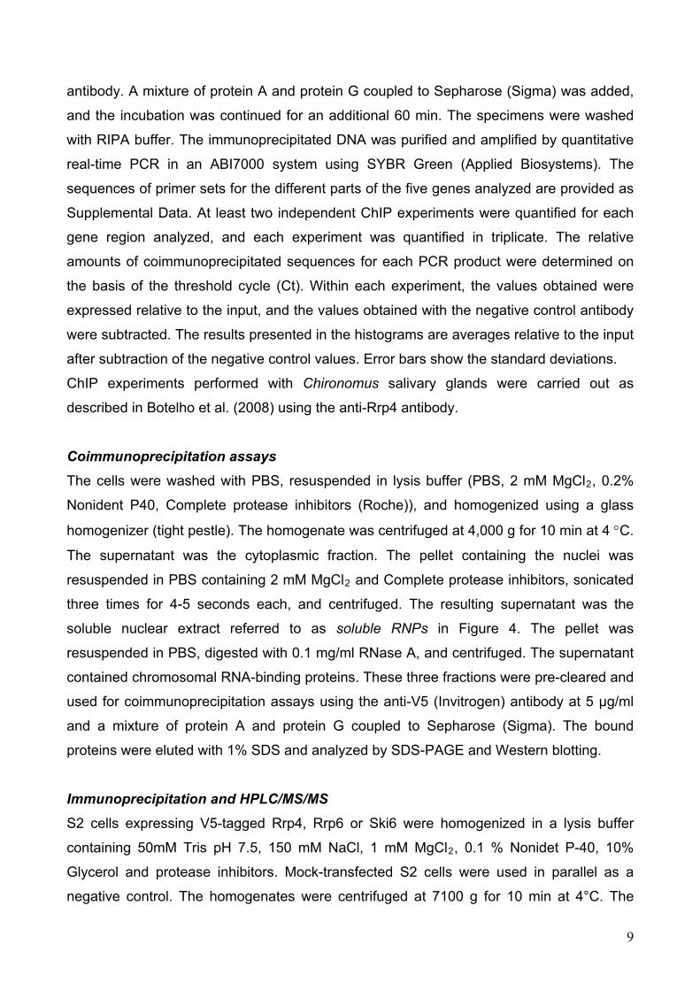
We used the anti-Rrp4 antibody for immunocytochemical studies. We first analyzed
the distribution of Rrp4 in the polytene chromosomes of C. tentans by
immunofluorescence (Figure 1A). Ct-Rrp4 was detected at numerous loci in all four
chromosomes, including the BR puffs in chromosome IV. The nucleolus was intensely
stained, as expected from the important role that the exosome plays in pre-rRNA
maturation.
We next asked whether Rrp4 was associated with the chromosomes only during
ongoing transcription (Figure 1B). We treated the larvae with the transcription inhibitor 5,6-
11
dichloro-1-beta-D-ribofuranosylbenzimidazole (DRB) and we isolated polytene
chromosomes for immunostaining. The effect of the DRB treatment was assessed by the
reduction of the diameter of the BR puffs (see for instance the reduced size of BR2 in the
DRB-treated sample shown in Figure 1B). Figure 1B shows that the immunofluorescent
staining was nearly abolished by the DRB treatment.
Preparations of polytene chromosomes were digested with RNase A before
immunostaining to determine whether the association of Rrp4 with the chromosomes was
mediated by RNA (Figure 1C). The chromosomes were co-stained with Y12, a mAb
against core snRNP proteins, to monitor the effect of the RNase treatment. Control
chromosomes were incubated in parallel in the absence of RNase A. The snRNP staining
(red in Figure 1C) was abolished by the RNase treatment, as expected. The intensity of
the Rrp4 staining (green in Figure 1C) was significantly reduced, and this reduction was
very pronounced at the BR puffs (asterisks in Figure 1C). However, a part of the Rrp4
staining was resistant to the RNase treatment. Double-staining of polytene chromosomes
with anti-Rrp4 (green in Figure 1D) and anti-Pol-II (red in Figure 1D) antibodies revealed
that the RNase-resistant fraction of Rrp4 overlaps with Pol-II. These results suggest that
there are two modes of interaction of Rrp4 with the chromosomes. One mode is
independent of RNA and may be explained by a direct association of the exosome with the
transcription machinery, as reported by Andrulis and coworkers (2002). The other mode is
predominant in highly transcribed genes such as the BR genes, and this mode requires
RNA.
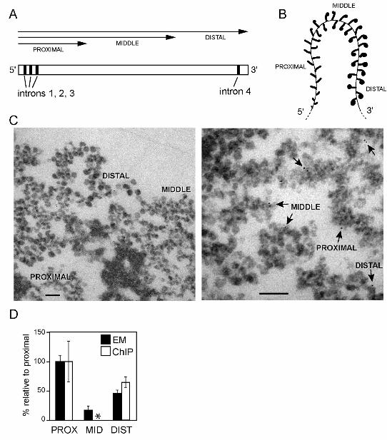
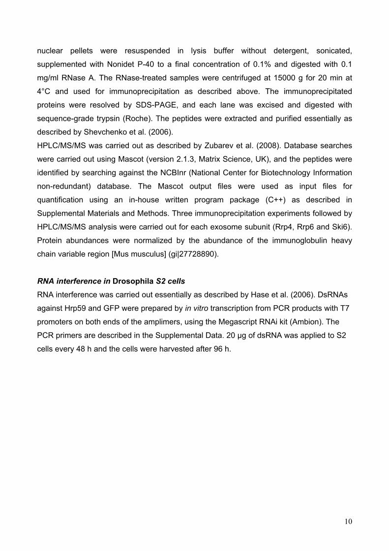
Ct-Rrp4 is present along the entire BR transcription unit
We analyzed the distribution of Ct-Rrp4 along the BR genes in the polytene
chromosomes of C. tentans by immuno-EM in order to test the hypothesis that the
exosome travels with the transcription machinery along transcribed genes. The exon-intron
structure of the BR genes is known (Wieslander and Paulsson, 1992; Figure 2A), and the
morphology of the active BR genes and BR mRNPs has been characterized in detail
(Skoglund et al., 1983). The active BR genes are loaded with multiple RNA polymerases
and, at any given time, each BR gene is being transcribed simultaneously by many RNA
polymerases that have reached different elongation stages. This gives the BR genes a
distinct polarity and the different regions of the gene show specific morphological features
due to the progressive growth of the nascent BR pre-mRNPs (Figure 2B). Full-length
12
genes are not available in the sections used for TEM, but partial gene segments are
observed (Figure 2C). The gene segments can be classified into proximal, middle and
distal segments, based on the morphology of the nascent pre-mRNPs. The pre-mRNPs
are seen as fibers of increasing length in the proximal portion of the BR gene. In the
middle and distal portions, the growing pre-mRNPs appear as stalked granules of
increasing diameter (Figures 2B and 2C).
For immuno-EM experiments, polytene chromosomes were manually isolated and
immunostained with the purified anti-Rrp4 antibody. The binding sites were revealed with a
gold-conjugated secondary antibody. Negative control samples were processed in parallel
without primary antibody (not shown). After the immunolabeling, the chromosomes were
embedded in plastic and sectioned for EM analysis. Examples of immunolabeling are
shown in Figure 2C. Samples from three independent experiments were used to quantify
the distribution of labeling in the different parts of the BR gene. For quantification, each
gold marker was classified according to its association with the proximal, middle or distal
part of the BR gene. The distribution of the labeling was not uniform along the gene, but
significant levels of Rrp4 were detected in all the parts of the BR gene (Figure 2D).
The association of Rrp4 with initial and distal segments of the BR gene was confirmed
by ChIP. The association of Rrp4 with the middle region could not be quantified by ChIP
because the sequence of the middle region is highly repetitive.
In summary, the results reported above show that Rrp4 is present along the entire
length of the BR gene.
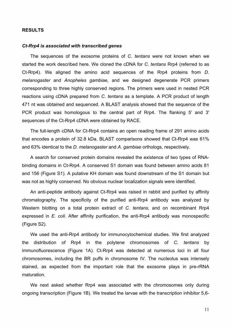
Ct-Rrp4 is associated with BR transcripts
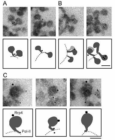
We next analyzed the precise location of Rrp4 in the active BR genes. The BR pre-
mRNPs in the distal part of the gene are large enough to allow us to discriminate between
labeling near the transcription machinery and labeling to the RNP. We selected immuno-
EM images in which the relative positions of the chromatin axis and the nascent pre-
mRNPs could be seen, and for each image we determined whether the gold markers were
close to the chromatin (within 25 nm from the axis) or distant from the chromatin (more
than 25 nm from the axis). Given the dimensions of the antibodies, this latter group
contains only gold markers associated with BR pre-mRNP. The markers close to the
chromatin axis, in contrast, may label Rrp4 molecules bound to the stalk of the growing
pre-mRNP particles, bound to the chromatin, or bound to the transcription machinery. The
13
gold markers were close to the axis in 9 out of 19 selected images (Figure 3A), while the
labeling was far from the axis in 10 out of 19 cases (Figure 3B). We conclude that a
significant fraction of Rrp4 is associated with the nascent BR pre-mRNPs without being in
contact with the transcription machinery. This conclusion was further strengthened by
double-labeling experiments in which Pol-II and Rrp4 were detected simultaneously. As
shown in Figure 3C, Rrp4 (large gold markers) is located far from the Pol-II (small gold
markers) on the nascent pre-mRNP.
The distribution of Rrp4 along transcribed genes is independent of exon-intron
organization
The concentration of Rrp4 along the BR gene was not constant (Figure 2D). This
observation suggests that the levels of Rrp4 that are present at a given gene region may
be determined by sequence-specific features, such as the presence of splice sites. This
prompted us to analyze the distribution of the exosome along other genes with different
structural features. It is possible to map proteins along only the large BR genes by
immuno-EM, and we were therefore compelled to use chromatin immunoprecipitation
(ChIP) instead. ChIP can detect proteins that are bound to the DNA as well as proteins
that interact with the nascent transcript (see for example Görnemann et al., 2005). We
used D. melanogaster for these experiments, since the genomic sequence information is
available. We constructed stable S2 cells that expressed a V5-tagged version of the
Drosophila Rrp4 protein (Rrp4-V5) under the control of an inducible promoter. This cell line
was called S2-Rrp4. The expressed Rrp4-V5 was present in both the nucleus and
cytoplasm, as expected (Figure S4A). The anti-V5 antibody specifically
immunoprecipitated Rrp4-V5 (Figure S4B and Table S2), and the exosome cofactor Rrp6
was coimmunoprecipitated (Figure S4C), which indicated that Rrp4-V5 was able to engage
in functional interactions.
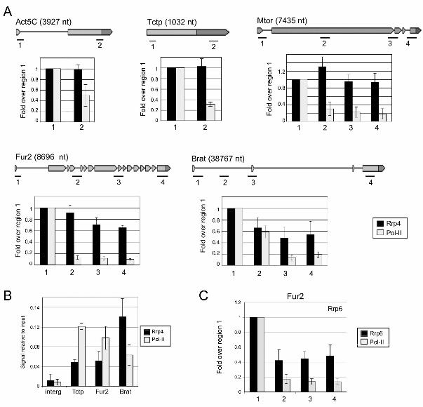
We used the S2-Rrp4 cells to study the association of Rrp4 with genes of D.
melanogaster by ChIP. We arbitrarily selected five genes that differ in length and exon-
intron organization: Act5C, Tctp, Mtor, Fur2 and Brat. The expression of these five
selected genes in S2 cells was documented in the FLIGHT database (http://flight.licr.org/)
and was validated by RT-PCR (Figure S5). The expression of Rrp4-V5 was induced for 24
h, chromatin was prepared, and immunoprecipitation was carried out using the anti-V5
antibody. Quantification of the immunoprecipitated DNA was based on real-time PCR
14
assays using primer-pairs specific for different regions of each gene. Negative control
ChIP reactions were carried out in parallel using an unrelated antibody. For each gene, the
background values obtained in the negative control were substracted, and the relative
density of Rrp4 was expressed relative to the input. In order to illustrate variations along
each gene, the relative abundance of Rrp4 in each region was expressed relative to the
most upstream region situated near the transcription start (black bars in Figure 4A). Rrp4
was present in all the regions of all the genes analyzed. For Act5C, Tctp and Mtor, the
relative abundance of Rrp4 was relatively constant along the genes. For Fur2 and Brat, the
levels of Rrp4 were significantly higher in the 5' region than at the 3' end, as immuno-EM
had shown for the BR genes.
We could not establish any obvious correlation between the levels of Rrp4 associated
with the genes and the presence of splice sites. The patterns of Rrp4 distribution were
similar for intronless and intron-containing genes and, within a given gene, the distribution
of Rrp4 could not be correlated with intron-exon features. For instance, region 2 of Brat,
located in the middle of a very long intron, had levels of Rrp4 similar to those in regions 3
and 4, located in exonic sequences (Figure 4A).
The specificity of the ChIP results was tested using the parental S2 cells that did not
express V5-tagged Rrp4. No significant signals were obtained for any of the analyzed
genes (data not shown).
We also determined the distribution of Pol-II along the same genes (white bars in
Figure 4). The rationale of this experiment was that if the exosome was tethered to
transcribed genes through interactions with the transcription machinery, the levels of Rrp4
would parallel those of Pol-II. However, in all the genes analyzed, the levels of Pol-II
decreased in a very pronounced manner towards the 3' end, and the Pol-II levels in the
downstream region of the genes were much lower than those obtained for Rrp4. Similar
profiles have been reported for Pol-II in mammalian cells (see, for example, Listerman et
al., 2006), and the decrease can be attributed to the fact that many of the Pol-II complexes
that are recruited to the gene fail to proceed to productive elongation. In summary, we
have shown that the pattern of distribution of Rrp4 does not parallel that of Pol-II. The
difference observed between the Rrp4 and Pol-II distributions could be explained if
multiple Rrp4 molecules become associated with each transcript during transcription (see
Discussion).
15
Finally, we asked whether the levels of Rrp4 recruited to the upstream region of the
genes were similar for all the genes analyzed and proportional to the density of Pol-II. For
this purpose, we compared the ChIP signals obtained in the upstream regions of each
gene (regions 1). Figure 4B shows that the levels of Rrp4 were different from gene to
gene, and did not correlate with the levels of Pol-II in the same region.
We conclude that the amount of Rrp4 and its distribution are to some extent
determined by gene-specific features, but are not directly correlated with the exon-intron
structure of the gene nor with the levels of Pol-II. The density of Rrp4 decreased in the 5'
to 3' direction along long genes and this decrease was much less pronounced than that
observed for Pol-II, which can be explained if each growing mRNP associates with multiple
Rrp4 molecules. The presence of multiple copies of Rrp4 associated with the nascent
transcripts in a given region of the gene would result in increased ChIP signals for that
particular region.
An important question was whether the results obtained for Rrp4 reflect the behavior
of the entire exosome. To address this point, we carried out ChIP experiments using S2
cells that express a V5-tagged version of Rrp6, one of the catalytic subunits of the
exosome, and we analyzed the distribution of Rrp6 along the Fur2 gene. As shown in
Figure 4C, the density of Rrp6 also decreased in the 5' to 3' direction but remained higher
than that of Pol-II, as observed for Rrp4.
The exosome interacts physically with the hnRNP protein Hrp59
The immuno-EM and ChIP results reported above suggest that additional interactions
contribute to the localization of the exosome to transcribed genes, in addition to the
binding of the exosome to the transcription machinery. We have examined whether Rrp4
interacts with proteins that are associated with the nascent pre-mRNA using a co-
immunoprecipitation assay and we have identified co-immunoprecipitated proteins by
mass spectrometry. We prepared nuclear protein extracts from mock-transfected S2 cells
and from S2 cells expressing the V5-tagged Rrp4, and we treated the extracts with RNase
A to disrupt RNA-mediated interactions. These RNase-treated nuclear extracts were used
in immunoprecipitation experiments using an anti-V5 antibody (lane 2 in Figure 5A).
Negative control immunoprecipitations were run in parallel using the mock-transfected S2
cells (lane 3 in Figure 5A). The immunoprecipitated proteins were resolved by SDS-PAGE,
digested in-gel with trypsin and analyzed by high-performance liquid
16

chromatography/tandem mass spectrometry (HPLC/MS/MS). Biological triplicates were
carried out for statistical purposes (not shown). In all the experiments, Mascot searches
identified Hrp59, the product of the CG9373 gene, as a protein significantly enriched in the
immunoprecipitates (Figure 5B, Table S3 and Table S4), which indicates that Hrp59 and
Rrp4 interact physically in vivo. Hrp59, also known as Rumpelstiltskin, is the hnRNP M
protein of D. melanogaster (Kiesler et al. 2005; Hase et al., 2006; Jain and Gavis, 2008).
We also carried out co-immunoprecipitation experiments using S2 cells that express
either Rrp6-V5 or Ski6/Rrp41-V5 (lanes 4 and 6, respectively, in Figure 5A). In both cases,
Hrp59 was found significantly enriched in the immunoprecipitates (Figure 5B). These
results suggest that Hrp59 is not associated with the individual Rrp4 subunit but with the
exosome complex.
The exosome had previously been shown to interact with Pol-II and with the
transcription elongation factors Spt5 and Spt6 (Andrulis et al., 2002). These interactions
were also detected in our HPLC/MS/MS study (Table S4).
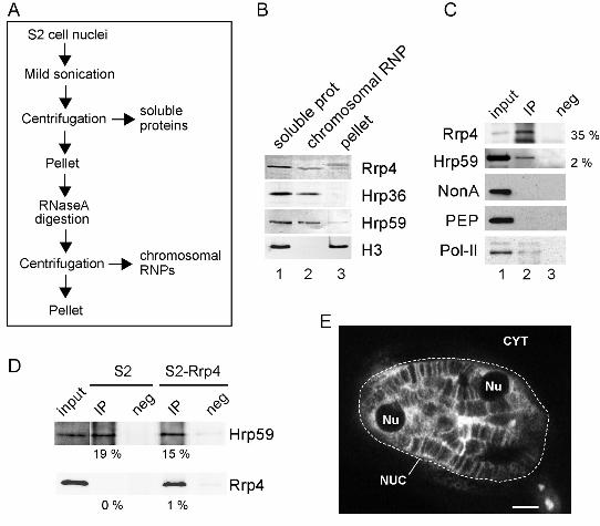
We used cell fractionation and immunoprecipitation methods to further characterize
the interaction between Rrp4 and Hrp59, and to determine whether this interaction takes
place cotranscriptionally. We isolated nuclei from S2 cells and prepared two types of
protein extracts (Figure 6A). One of the extracts contained soluble nuclear proteins and
RNPs (soluble proteins) and the other extract contained proteins bound to the
chromosomes via RNA (chromosomal RNPs). These proteins were released by RNase A
digestion. Rrp4 was present in the soluble nuclear fraction, in the chromosomal RNP
fraction and in the pellet (Figure 6B). Antibodies against Hrp36, an abundant hnRNP
protein of the A/B type, and against histone H3 were used as controls to assess the quality
of the fractions.
We used a chromosomal RNP extract prepared from cells expressing Rrp4-V5 in
immunoprecipitation experiments using the V5 antibody. Rrp4 itself was efficiently
precipitated as expected (Figure 6C). A significant fraction of Hrp59 (2 %) was also
detected in the immunoprecipitate (lane 2 in Figure 6C). Other abundant mRNA-binding
proteins such as NonA and PEP were analyzed in parallel but were not detected in the
immunoprecipitate. Pol-II, which is known to interact with the exosome (Andrulis et al.,
2002), was co-immunoprecipitated with Rrp4 (Figure 6C).
We also carried out the reciprocal immunoprecipitation: we immunoprecipitated Hrp59
from S2 cells expressing Rrp4-V5, and we detected co-immunoprecipitated Rrp4 by
Western blot using the anti-V5 antibody (Figure 6D). Mock-transfected S2 cells were used
17
in parallel as negative control. It is worth noticing that the immunoprecipitations were
carried out using a chromosomal RNP extract that was digested with RNase A (Figure 6A).
This suggestst that the association of Rrp4 with Hrp59 takes place on the chromosome
and that the Rrp4-Hrp59 interaction is not mediated by RNA. From these experiments we
conclude that Rrp4 interacts, directly or indirectly, with Hrp59 in vivo.
Hrp59 associates cotranscriptionally with transcripts from many different genes, as
shown by the fact that an anti-Hrp59 antibody stains many bands in the polytene
chromosomes (Figure 6E and Kiesler et al., 2005). The nucleoli appear as dark spots in
the Hrp59-stained cells (Nu in Figure 6E), which indicates that Hrp59 is not present in the
nucleoli.
The results presented above indicate that Rrp4 interacts with the hnRNP M protein of
Drosophila, Hrp59, and suggest that this interaction does not take place in the nucleolus
but on the chromosomes. The HPLC/MS/MS data also suggest that the exosome complex,
not Rrp4 alone, is associated with Hrp59. Which of the exosome subunits interacts directly
with Hrp59 remains to be elucidated.
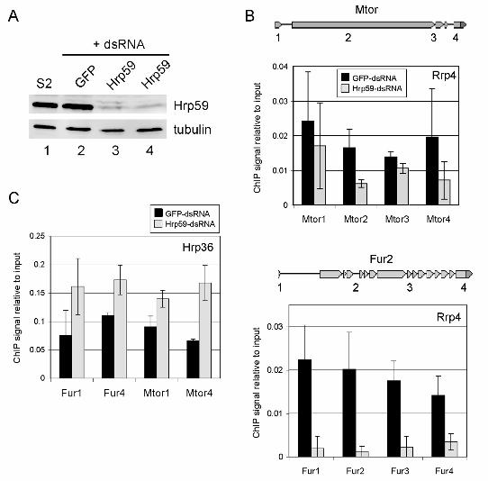
Hrp59 depletion inhibits the association of Rrp4 with transcribed genes
We next explored the functional significance of the interaction between the exosome
and Hrp59. If Hrp59 had a role in the recruitment and/or tethering of the exosome to
nascent transcripts, depletion of Hrp59 by RNA interference would lead to reduced levels
of Rrp4 at the transcription sites. We silenced the expression of Hrp59 by RNA
interference (RNAi) in S2 cells expressing Rrp4-V5, and we analyzed the effects of the
silencing on the association of Rrp4 with transcribed genes by ChIP. The efficiency of the
RNA interference was checked by Western blotting (Figure 7A). Control cells were treated
in parallel with GFP-dsRNA. The association of Rrp4 with two genes, Mtor and Fur2, was
analyzed by ChIP (Figure 7B). For both genes, depletion of Hrp59 resulted in decreased
levels of Rrp4. We asked whether this decrease was a general effect due to limited
accessibility of proteins to the transcribed genes in the absence of Hrp59, and we
analyzed the association of Hrp36, an abundant hnRNP protein, with the Mtor and Fur2
genes (regions 1 and 4 for each gene). Hrp36 signals were found not significant in an
intergenic region (data not shown). As shown in Figures 7C and S6, the levels of Hrp36
associated with Mtor and Fur2 increased in response to Hrp59 depletion. This observation
rules out a general concealing of the RNP structure and can be explained by the fact that
hnRNP proteins have overlapping binding sites on the mRNAs (Dreyfuss et al., 2002).
18
Thus in the absence of Hrp59, other abundant hnRNP proteins are likely to gain access to
the transcripts.
In summary, our results show that depletion of Hrp59 interferes with the association
of Rrp4 with the transcribed genes, which suggestst that Hrp59 is involved in the
interaction between the exosome and the pre-mRNP. Our results also suggest that other
hnRNP proteins bind to the nascent pre-mRNA in the absence of Hrp59, and that the
resulting mRNP complexes have a reduced ability to interact with the surveillance
machinery.
19
DISCUSSION
We have studied the association of Rrp4 with protein-coding genes, and we have
shown that Rrp4 is recruited to transcriptionally active loci in the polytene chromosomes of
C. tentans. This agrees with previous observations in D. melanogaster, in which the
exosome is associated with the RNA polymerase II and with the transcription elongation
factors Spt5 and Spt6 (Andrulis et al., 2002). These interactions have led to the proposal
that the transcription machinery is responsible for recruiting the exosome to sites of pre-
mRNA synthesis, and that the exosome travels along the transcribed genes bound to the
transcription elongation complex (Andrulis et al., 2002). The results of our work on the BR
genes of C. tentans are compatible with the hypothesis that the transcription machinery is
involved in the recruitment and tethering of the exosome to transcribed genes. However,
our work has also revealed the existence of an additional mechanism that operates in the
cotranscriptional surveillance of nascent pre-mRNPs. This additional mechanism involves
an interaction between the exosome and the nascent pre-mRNP and requires the hnRNP
M protein Hrp59.
We have determined the precise location of Rrp4 in the BR1 and BR2 genes by
immuno-EM, and we have shown that a fraction of Rrp4 is directly associated with the
nascent BR pre-mRNP without any contact with the transcription machinery. Our results
from cell fractionation experiments and from experiments in which the chromosomes were
digested with RNase A before they were immunostained also point to the existence of a
fraction of Rrp4 that is bound to the chromosomes via RNA. We propose that a fraction of
Rrp4 binds directly to the nascent pre-mRNP, while another fraction is associated with the
chromosomes independently of RNA (Figure 8). This latter fraction is likely to be
associated with the transcription elongation factors Spt5 and Spt6, as reported by Andrulis
et al. (2002). Given that the number of Pol-II molecules present in a given gene region
equals the number of transcripts, the finding that the relative density of Rrp4 along the
genes remains higher than the density of Pol-II suggests that multiple Rrp4 molecules bind
to each nascent transcript during transcription. This would be difficult to explain by
interactions between the exosome and the transcription machinery, but is in full agreement
with the finding that at least three exosome subunits can also interact with hnRNP
proteins, which are usually associated with the pre-mRNA in multiple copies.
We have shown that three different exosome subunits can be coimmunoprecipitated
20
with the mRNA-binding protein Hrp59 in a RNA-independent manner. Given that Hrp59 is
an hnRNP protein that binds to nascent mRNPs cotranscriptionally (Kiesler et al., 2005)
and given that Hrp59 interacts with Rrp4, Ski6/Rrp41 and Rrp6, we propose that Hrp59
mediates the association of the exosome to the nascent pre-mRNPs. Several exosome
subunits, including Rrp4, contain RNA-binding domains and can, in principle, bind RNA
directly. However, the pronounced effect that Hrp59 depletion has on the association of
Rrp4 with transcribed genes indicates that direct RNA binding does not play a major role in
the association of the exosome with nascent transcripts. Alternatively, Hrp59 could act as
a cofactor necessary for the direct binding of Rrp4 -or the exosome- to RNA.
Hrp59 is evolutionarily conserved. The closest orthologs of Hrp59 in S. cerevisiae are
Gbp2 and Hrb1, two shuttling mRNA-binding proteins that are recruited to the pre-mRNA
co-transcriptionally (Hurt et al., 2004), and a genetic interaction between Gbp2 and Rrp6
has been reported (Wilmes et al., 2008). Further research is needed to determine whether
Gbp2 and/or Hrb1 are involved in the recruitment of the exosome to active genes.
It is unclear how the exosome discriminates between correct and defective transcripts.
We have found Rrp4 associated with many transcriptionally active loci under normal
growth conditions in C. tentans, which suggests that the discrimination between correct
and defective transcripts is not based on the selective recruitment of the exosome to
defective RNAs. Our results suggest that the exosome instead is recruited to expressed
genes by molecular interactions that are independent of expression defects. It has been
proposed that the packaging of the mRNA into an mRNP complex plays a crucial role in
the definition of aberrant transcripts. According to this proposal, the correct assembly of
the nascent mRNA into a mRNP complex protects the transcript from degradation by the
exosome. Instead, aberrant transcripts become assembled into abnormal mRNP
structures that render the mRNA unprotected and susceptible to degradation (Jensen et
al., 2003).
Two different strategies can be envisioned for the cotranscriptional recruitment of
surveillance factors to sites of mRNA synthesis. The exosome could be specifically
recruited to nascent transcripts as specific processing reactions take place. For example,
the exosome could be recruited to pre-mRNA splice sites by the spliceosome for
surveillance of intron removal. Alternatively, the recruitment of the exosome could be
constitutive and independent of specific processing events. For instance the degradation
of cryptic unstable transcripts in S. cerevisiae is closely coupled to the Nrd1p-Nab3p-
21
dependent transcription termination pathway (Arigo et al., 2006; Thiebaut et al., 2006). Our
results suggest, in contrast, that protein-coding genes in insects are monitored by a
constitutive mechanism. Our ChIP analysis shows that the exosome is present along the
complete length of protein-coding genes, even of very long exons and in intronless
transcripts, and the density of Rrp4 along the genes does not correlate with their exon-
intron structure. The interactions of the exosome with the Pol-II machinery and with the
hnRNP protein Hrp59 provide two general mechanisms for the constitutive recruitment of
the exosome to protein-coding transcripts.
Hrp59 is an abundant hnRNP protein that belongs to the hnRNP M family. Hrp59
associates cotranscriptionally with transcripts from many different genes, as shown by
immunostaining of polytene chromosomes, and it is probable that Hrp59 has a general role
in the packaging of mRNPs (Kiesler et al., 2005). Hrp59 has also a specific role in the
regulating the alternative splicing of some transcripts (Hase et al., 2006). We have now
shown that pre-mRNAs synthesized in the absence of Hrp59 are defective in the
recruitment of the surveillance machinery. Based on the fact that Hrp59 is associated with
the nascent mRNA and with Rrp4, we propose that Hrp59 is directly involved in the
recruitment of the exosome to expressed genes and works as an adaptor for mRNA
surveillance.
22
ACKNOWLEDGMENTS
We are grateful to B. Björkroth (CMB, Karolinska Institutet) for technical support and
George Farrants for language editing. We are also grateful to E. Andrulis, H. Saumweber
and S. Amero for the generous gift of antibodies against Rrp6, NonA and PEP,
respectively. Alexander R. Zubarev wrote the code for protein quantification. This work
was supported by the Swedish Research Council (grants 2006-5398 to NV and 621-2007-
4410 to RZ), the Carl Trygger Foundation, the Swedish Cancer Society, the Knut and Alice
Wallenberg Foundation (instrumental grant to RZ) and the European Science Foundation
(RNAQuality Programme to NV).
23
REFERENCES
Allmang C, Petfalski E, Podtelejnikov A, Mann M, Tollervey D, Mitchell P. (1999) The yeast
exosome and human PM-Scl are related complexes of 3' --> 5' exonucleases. Genes
Dev 13: 2148-2158.
Andrulis E, Werner J, Nazarian A, Erdjument-Bromage H, Tempst P, Lis JT. (2002) The
RNA processing exosome is linked to elongating RNA polymerase II in Drosophila.
Nature 420: 837-841.
Arigo JT, Eyler DE, Carroll KL, Corden JL. (2006) Termination of cryptic unstable
transcripts is directed by yeast RNA-binding proteins Nrd1 and Nab3. Mol Cell 23:
841-851.
Björkroth B, Ericsson C, Lamb MM, Daneholt B. (1988) Structure of the chromatin axis
during transcription. Chromosoma 96: 333-340.
Botelho SC, Tyagi A, Hessle V, Farrants AK, Visa N. (2008) The association of Brahma
with the Balbiani ring 1 gene of Chironomus tentans studied by immunoelectron
microscopy and chromatin immunoprecipitation. Insect Mol Biol. 17: 505-513.
Butler JS. (2002) The yin and yang of the exosome. Trends Cell Biol 12: 90-96.
Buttner K, Wenig K, Hopfner KP. (2005) Structural framework for the mechanism of
archaeal exosomes in RNA processing. Mol Cell 20: 461-471.
Daneholt, B. (2001) Assembly and transport of a pre-messenger RNP particle. Proc Natl
Acad Sci U S A. 98: 7012-7017.
Dreyfuss G, Narry KV, Kataoka N. (2002) Messenger-RNA-binding proteins and the
messages they carry. Nat Rev Mol Cell Biol 3: 195-205.
Dziembowski A, Lorentzen E, Conti E, Séraphin B. (2007) A single subunit, Dis3, is
essentially responsible for yeast exosome core activity. Nat Struct Mol Biol 14: 15-22.
Galy V, Gadal O, Fromont-Racine M, Romano A, Jaquier A, Nehrbass U. (2004) Nuclear
retention of unspliced mRNAs in yeast is mediated by perinuclear Mlp1. Cell 116: 63-
73.
Görnemann J, Kotovic KM, Hujer K, Neugebauer KM. (2005) Cotranscriptional
spliceosome assembly occurs in a stepwise fashion and requires the cap binding
complex. Mol Cell 19: 53-63.
Graham AC, Kiss DL, Andrulis ED. (2006) Differential distribution of exosome subunits at
the nuclear lamina and in cytoplasmic foci. Mol Biol Cell 17: 1399-1409.
Hase ME, Yalamanchili P, Visa N. (2006) The Drosophila heterogeneous nuclear
24
ribonucleoprotein M protein, HRP59, regulates alternative splicing and controls the
production of its own mRNA. J Biol Chem 281: 39135-39141.
Hilleren P, McCarthy T, Rosbash M, Parker R, Jensen TH. (2001) Quality control of mRNA
3'-end processing is linked to the nuclear exosome. Nature 413: 538-542.
Houseley J, LaCava J, Tollervey D. (2006) RNA-quality control by the exosome. Nat Rev
Mol Cell Biol 7: 529-539.
Hurt E, Luo MJ, Röther S, Reed R, Strässer K. (2004) Cotranscriptional recruitment of the
serine-arginine-rich (SR)-like proteins Gbp2 and Hrb1 to nascent mRNA via the TREX
complex. Proc Natl Acad Sci U S A. 101: 1858-1862.
Jain RA, Gavis ER. (2008) The Drosophila hnRNP M homolog Rumpelstiltskin regulates
nanos mRNA localization. Dev 135: 973-982.
Jensen TH, Dower K, Libri D, Rosbash M. (2003) Early formation of mRNP: license for
export or quality control? Mol Cell 11: 1129-1133.
Kiesler E, Hase ME, Brodin D, Visa N. (2005) Hrp59, an hnRNP M protein in Chironomus
and Drosophila, binds to exonic splicing enhancers and is required for expression of a
subset of mRNAs. J Cell Biol 168:1013-1025.
Kiseleva E, Visa N, Wurtz T, Daneholt B. (1997) Immunocytochemical evidence for a
stepwise assembly of Balbiani ring premessenger ribonucleoprotein particles. Eur J
Cell Biol 74: 407-416.
LaCava J, Houseley J, Saveanu C, Petfalski E, Thompson E, Jacquier A, Tollervey D.
(2005) RNA degradation by the exosome is promoted by a nuclear polyadenylation
complex. Cell 121: 713-724.
Lerner EA, Lerner MR, Janeway CA Jr, Steitz JA. (1981) Monoclonal antibodies to nucleic
acid-containing cellular constituents: probes for molecular biology and autoimmune
disease. Proc Natl Acad Sci U S A 78: 2737-2741.
Lorentzen E, Basquin J, Conti E. (2008) structural organization of the RNA-degrading
exosome. Curr Opin Cell Biol 18: 709-713.
Liu Q, Greimann JC, Lima CD. (2006) Reconstitution, activities, and structure of the
eukaryotic RNA exosome. Cell 127:1223-1237. Erratum in: Cell 131:188-189.
Listerman I, Sapra AK, Neugebauer KM. (2006) Cotranscriptional coupling of splicing
factor recruitment and precursor messenger RNA splicing in mammalian cells. Nat
Struct Mol Biol 13: 815-822.
Lorentzen E, Walter P, Fribourg S, Evguenieva-Hackenberg E, Klug G, Conti E. (2005)
The archaeal exosome core is a hexameric ring structure with three catalytic subunits.
25
Nat Struct Mol Biol 12: 575-581.
Lorentzen E, Conti E. (2006) The exosome and the proteasome: nano-compartments for
degradation. Cell 125: 651-654.
Luna R, Jimeno S, Martin M, Huertas P, Garcia-Rubio M, Aguilera A. (2005)
Interdependence between transcription and mRNP processing and export, and its
impact on genetic stability. Mol Cell 18: 711-722.
Meyer B, Mähr R, Eppenberger HM, Lezzi M. (1983) The activity of Balbiani rings 1 and 2
in salivary glands of Chironomus tentans larvae under different modes of development
and after pilocarpine treatment. Dev Biol 98: 265-277.
Mitchell P, Petfalski E, Shevchenko A, Mann M, Tollervey D. (1997) The exosome: a
conserved eukaryotic RNA processing complex containing multiple 3'-->5'
exoribonucleases. Cell 91: 457-466.
Mitchell P, Tollervey D. (2000) Musing on the structural organization of the exosome
complex. Nat Struct Biol 7: 843-846.
Mitchell P, Petfalski E, Houalla R, Podtelejnikov A, Mann M, Tollervey D. (2003) Rrp47p is
an exosome-associated protein required for the 3' processing of stable RNAs. Mol Cell
Biol 23: 6982-6992.
Preker P, Nielsen J, Kammler S, Lykke-Andersen S, Christensen MS, Mapendano CK,
Schierup MH, Jensen TH. (2008) RNA Exosome Depletion Reveals Transcription
Upstream of Active Human Promoters. Science Dec 4 . [Epub ahead of print].
Rougemaille M, Gudipati RK, Olesen JR, Thomsen R, Seraphin B, Libri D, Jensen TH.
(2007) Dissecting mechanisms of nuclear mRNA surveillance in THO/sub2 complex
mutants. EMBO J. 26: 2317-2326.
Saguez C, Olesen JR, Jensen TH. (2005) Formation of export-competent mRNP:
escaping nuclear destruction. Curr Opin Cell Biol 17: 287-293.
Schmid M, Jensen TH. (2008) The exosome: a multipurpose RNA-decay machine. Trends
Biochem Sci 33: 501-510.
Schneider C, Anderson JT, Tollervey D. (2007) The exosome subunit Rrp44 plays a direct
role in RNA substrate recognition. Mol Cell 27: 324-331.
Skoglund U, Andersson K, Björkroth B, Lamb MM, Daneholt B. (1983) Visualization of the
formation and transport of a specific hnRNP particle. Cell 34: 847-855.
Sommer P, Nehrbass U. (2005) Quality control of messenger ribonucleoprotein particles in
the nucleus and at the pore. Curr Opin Cell Biol 17: 294-301.
Takahashi Y, Rayman JB, Dynlacht BD (2000) Analysis of promoter binding by the E2F
26
and pRB families in vivo: distinct E2F proteins mediate activation and repression.
Genes Dev 14: 804-816.
Thiebaut M, Kisseleva-Romanova E, Rougemaille M, Boulay J, Libri D. (2006)
Transcription termination and nuclear degradation of cryptic unstable transcripts: a role
for the nrd1-nab3 pathway in genome surveillance. Mol Cell 23: 853-864.
Vanacova S, Wolf J, Martin G, Blank D, Dettwiler S, Friedlein A, Langen H, Keith G, Keller
W. (2005) A new yeast poly(A) polymerase complex involved in RNA quality control.
PLoS Biol. 3: e189.
Vanacova S, Stefl R. (2007) The exosome and RNA quality control in the nucleus. EMBO
Rep. 8: 651-657.
van Hoof A, Staples RR, Baker RE, Parker R. (2002) Function of the ski4p (Csl4p) and
Ski7p proteins in 3'-to-5' degradation of mRNA. Mol Cell Biol 20: 8230-8243.
Vinciguerra P, Stutz F. (2004) mRNA export: an assembly line from genes to nuclear
pores. Curr Opin Cell Biol 16: 285-292.
Wang HW, Wang J, Ding F, Callahan K, Bratkowski MA, Butler JS, Nogales E, Ke A.
(2007) Architecture of the yeast Rrp44 exosome complex suggests routes of RNA
recruitment for 3' end processing. Proc Natl Acad Sci U S A 104: 16844-16849.
Wieslander, L. (1994) The Balbiani ring multigene family: coding repetitive sequences and
evolution of a tissue-specific cell function. Prog Nucleic Acid Res Mol Biol 48: 275-313.
Wieslander L, Bauren G, Bernholm K, Jiang WQ, Wetterberg I. (1996) Processing of pre-
mRNA in polytene nuclei of Chironomus tentans salivary gland cells. Exp Cell Res
229: 240-246.
Wieslander L, Paulsson G. (1992) Sequence organization of the Balbiani ring 2.1 gene in
Chironomus tentans. Proc Natl Acad Sci U S A 89: 4578-4582.
Wilmes GM, Bergkessel M, Bandyopadhyay S, Shales M, Braberg H, Cagney G, Collins
SR, Whitworth GB, Kress TL, Weissman JS, Ideker T, Guthrie C, Krogan NJ. (2008) A
genetic interaction map of RNA-processing factors reveals links between Sem1/Dss1-
containing complexes and mRNA export and splicing. Mol Cell 32: 735-746.
Wyers F, Rougemaille M, Badis G, Rousselle JC, Dufour ME, Boulay J, Régnault B,
Devaux F, Namane A, Séraphin B, Libri D, Jacquier A. (2005) Cryptic Pol II transcripts
are degraded by a nuclear quality control pathway involving a new poly(A)
polymerase. Cell 121: 725–737.
Wyss C. (1982) Chironomus tentans epithelial cell lines sensitive to ecdysteroids, juvenile
hormone, insulin and heat shock. Exp Cell Res 139: 297-307.
27
Zenklusen D, Vinciguerra P, Wyss JC, Stutz F. (2002) Stable mRNP formation and export
require cotranscriptional recruitment of the mRNA export factors Yra1p and Sub2p by
Hpr1p. Mol Cell Biol 22: 8241-8253.
Zubarev RA, Nielsen ML, Fung EM, Savitski MM, Kel-Margoulis O, Winbender E, Kel A.
(2008) Identification of dominant signaling pathways from proteomics expression data.
J Proteomics 71: 89-96.
28
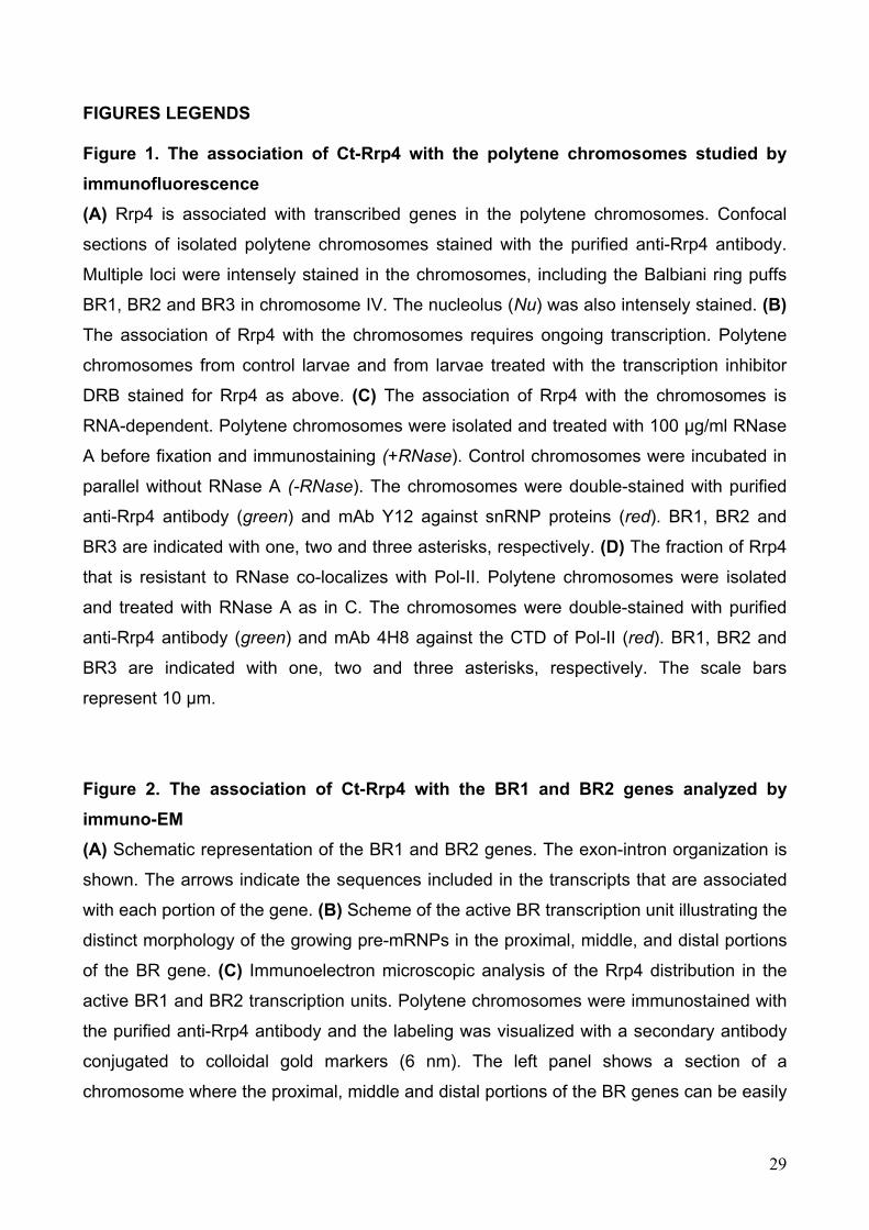
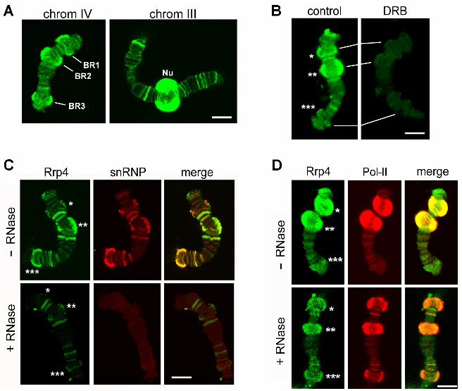
FIGURES LEGENDS Figure 1. The association of Ct-Rrp4 with the polytene chromosomes studied by
immunofluorescence
(A) Rrp4 is associated with transcribed genes in the polytene chromosomes. Confocal
sections of isolated polytene chromosomes stained with the purified anti-Rrp4 antibody.
Multiple loci were intensely stained in the chromosomes, including the Balbiani ring puffs
BR1, BR2 and BR3 in chromosome IV. The nucleolus (Nu) was also intensely stained. (B)
The association of Rrp4 with the chromosomes requires ongoing transcription. Polytene
chromosomes from control larvae and from larvae treated with the transcription inhibitor
DRB stained for Rrp4 as above. (C) The association of Rrp4 with the chromosomes is
RNA-dependent. Polytene chromosomes were isolated and treated with 100 µg/ml RNase
A before fixation and immunostaining (+RNase). Control chromosomes were incubated in
parallel without RNase A (-RNase). The chromosomes were double-stained with purified
anti-Rrp4 antibody (green) and mAb Y12 against snRNP proteins (red). BR1, BR2 and
BR3 are indicated with one, two and three asterisks, respectively. (D) The fraction of Rrp4
that is resistant to RNase co-localizes with Pol-II. Polytene chromosomes were isolated
and treated with RNase A as in C. The chromosomes were double-stained with purified
anti-Rrp4 antibody (green) and mAb 4H8 against the CTD of Pol-II (red). BR1, BR2 and
BR3 are indicated with one, two and three asterisks, respectively. The scale bars
represent 10 µm.
Figure 2. The association of Ct-Rrp4 with the BR1 and BR2 genes analyzed by
immuno-EM
(A) Schematic representation of the BR1 and BR2 genes. The exon-intron organization is
shown. The arrows indicate the sequences included in the transcripts that are associated
with each portion of the gene. (B) Scheme of the active BR transcription unit illustrating the
distinct morphology of the growing pre-mRNPs in the proximal, middle, and distal portions
of the BR gene. (C) Immunoelectron microscopic analysis of the Rrp4 distribution in the
active BR1 and BR2 transcription units. Polytene chromosomes were immunostained with
the purified anti-Rrp4 antibody and the labeling was visualized with a secondary antibody
conjugated to colloidal gold markers (6 nm). The left panel shows a section of a
chromosome where the proximal, middle and distal portions of the BR genes can be easily
29
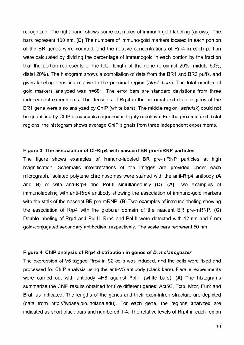
recognized. The right panel shows some examples of immuno-gold labeling (arrows). The
bars represent 100 nm. (D) The numbers of immuno-gold markers located in each portion
of the BR genes were counted, and the relative concentrations of Rrp4 in each portion
were calculated by dividing the percentage of immunogold in each portion by the fraction
that the portion represents of the total length of the gene (proximal 20%, middle 60%,
distal 20%). The histogram shows a compilation of data from the BR1 and BR2 puffs, and
gives labeling densities relative to the proximal region (black bars). The total number of
gold markers analyzed was n=681. The error bars are standard deviations from three
independent experiments. The densities of Rrp4 in the proximal and distal regions of the
BR1 gene were also analyzed by ChIP (white bars). The middle region (asterisk) could not
be quantified by ChIP because its sequence is highly repetitive. For the proximal and distal
regions, the histogram shows average ChIP signals from three independent experiments.
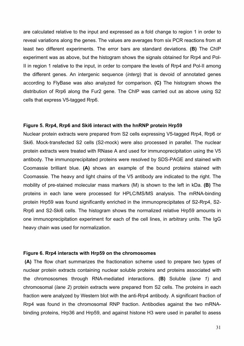
Figure 3. The association of Ct-Rrp4 with nascent BR pre-mRNP particles
The figure shows examples of immuno-labeled BR pre-mRNP particles at high
magnification. Schematic interpretations of the images are provided under each
micrograph. Isolated polytene chromosomes were stained with the anti-Rrp4 antibody (A
and B) or with anti-Rrp4 and Pol-II simultaneously (C). (A) Two examples of
immunolabeling with anti-Rrp4 antibody showing the association of immuno-gold markers
with the stalk of the nascent BR pre-mRNP. (B) Two examples of immunolabeling showing
the association of Rrp4 with the globular domain of the nascent BR pre-mRNP. (C)
Double-labeling of Rrp4 and Pol-II. Rrp4 and Pol-II were detected with 12-nm and 6-nm
gold-conjugated secondary antibodies, respectively. The scale bars represent 50 nm.
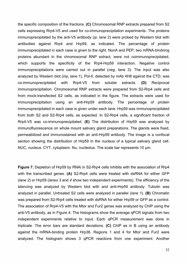
Figure 4. ChIP analysis of Rrp4 distribution in genes of D. melanogaster
The expression of V5-tagged Rrp4 in S2 cells was induced, and the cells were fixed and
processed for ChIP analysis using the anti-V5 antibody (black bars). Parallel experiments
were carried out with antibody 4H8 against Pol-II (white bars). (A) The histograms
summarize the ChIP results obtained for five different genes: Act5C, Tctp, Mtor, Fur2 and
Brat, as indicated. The lengths of the genes and their exon-intron structure are depicted
(data from http://flybase.bio.indiana.edu). For each gene, the regions analyzed are
indicated as short black bars and numbered 1-4. The relative levels of Rrp4 in each region
30
are calculated relative to the input and expressed as a fold change to region 1 in order to
reveal variations along the genes. The values are averages from six PCR reactions from at
least two different experiments. The error bars are standard deviations. (B) The ChIP
experiment was as above, but the histogram shows the signals obtained for Rrp4 and Pol-
II in region 1 relative to the input, in order to compare the levels of Rrp4 and Pol-II among
the different genes. An intergenic sequence (interg) that is devoid of annotated genes
according to FlyBase was also analyzed for comparison. (C) The histogram shows the
distribution of Rrp6 along the Fur2 gene. The ChIP was carried out as above using S2
cells that express V5-tagged Rrp6.
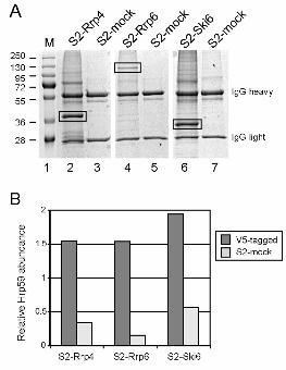
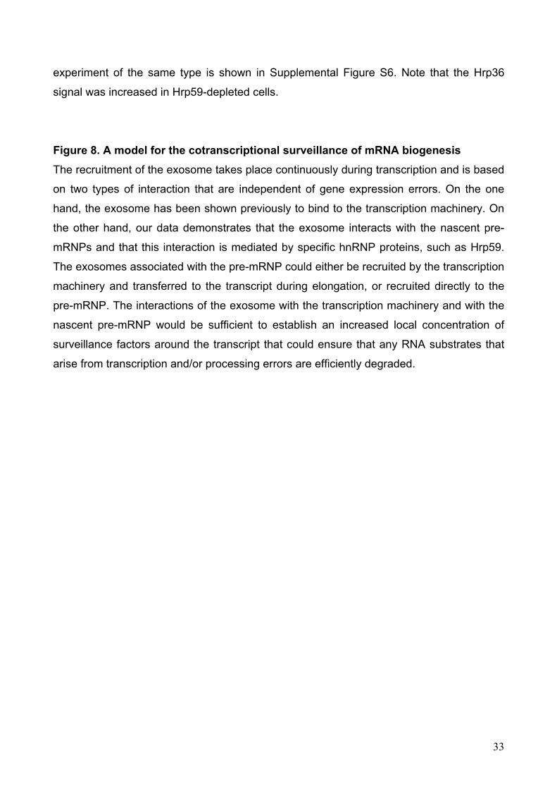
Figure 5. Rrp4, Rrp6 and Ski6 interact with the hnRNP protein Hrp59
Nuclear protein extracts were prepared from S2 cells expressing V5-tagged Rrp4, Rrp6 or
Ski6. Mock-transfected S2 cells (S2-mock) were also processed in parallel. The nuclear
protein extracts were treated with RNase A and used for immunoprecipitation using the V5
antibody. The immunoprecipitated proteins were resolved by SDS-PAGE and stained with
Coomassie brilliant blue. (A) shows an example of the bound proteins stained with
Coomassie. The heavy and light chains of the V5 antibody are indicated to the right. The
mobility of pre-stained molecular mass markers (M) is shown to the left in kDa. (B) The
proteins in each lane were processed for HPLC/MS/MS analysis. The mRNA-binding
protein Hrp59 was found significantly enriched in the immunoprecipitates of S2-Rrp4, S2-
Rrp6 and S2-Ski6 cells. The histogram shows the normalized relative Hrp59 amounts in
one immunoprecipitation experiment for each of the cell lines, in arbitrary units. The IgG
heavy chain was used for normalization.
Figure 6. Rrp4 interacts with Hrp59 on the chromosomes
(A) The flow chart summarizes the fractionation scheme used to prepare two types of
nuclear protein extracts containing nuclear soluble proteins and proteins associated with
the chromososmes through RNA-mediated interactions. (B) Soluble (lane 1) and
chromosomal (lane 2) protein extracts were prepared from S2 cells. The proteins in each
fraction were analyzed by Western blot with the anti-Rrp4 antibody. A significant fraction of
Rrp4 was found in the chromosomal RNP fraction. Antibodies against the two mRNA-
binding proteins, Hrp36 and Hrp59, and against histone H3 were used in parallel to asess
31
the specific composition of the fractions. (C) Chromosomal RNP extracts prepared from S2
cells expressing Rrp4-V5 and used for co-immunoprecipitation experiments. The proteins
immunoprecipitated by the anti-V5 antibody (ip, lane 2) were probed by Western blot with
antibodies against Rrp4 and Hrp59, as indicated. The percentage of protein
immunoprecipitated in each case is given to the right. NonA and PEP, two mRNA-bindning
proteins abundant in the chromosomal RNP extract, were not coimmunoprecipitated,
which supports the specificity of the Rrp4-Hrp59 interaction. Negative control
immunoprecipitations were carried out in parallel (neg, lane 3). The input was also
analyzed by Western blot (inp, lane 1). Pol-II, detected by mAb 4H8 against the CTD, was
co-immunoprecipitated with Rrp4-V5 from soluble extracts. (D) Reciprocal
immunoprecipitation. Chromosomal RNP extracts were prepared from S2-Rrp4 cells and
from mock-transfected S2 cells, as indicated in the figure. The extracts were used for
immunoprecipitation using an anti-Hrp59 antibody. The percentage of protein
immunoprecipitated in each case is given under each lane. Hrp59 was immunoprecipitated
from both S2 and S2-Rrp4 cells, as expected. In S2-Rrp4 cells, a significant fraction of
Rrp4-V5 was co-immunoprecipitated. (E) The distribution of Hrp59 was analyzed by
immunofluorescence on whole mount salivary gland preparations. The glands were fixed,
permeabilized and immunostained with an anti-Hrp59 antibody. The image is a confocal
section showing the distribution of Hrp59 in the nucleus of a typical salivary gland cell.
NUC, nucleus. CYT, cytoplasm. Nu, nucleolus. The scale bar represents 10 µm.
Figure 7. Depletion of Hrp59 by RNAi in S2-Rrp4 cells inhibits with the association of Rrp4
with the transcribed genes. (A) S2-Rrp4 cells were treated with dsRNA for either GFP
(lane 2) or Hrp59 (lanes 3 and 4 show two independent experiments). The efficiency of the
silencing was analyzed by Western blot with and anti-Hrp59 antibody. Tubulin was
analyzed in parallel. Untreated S2 cells were analyzed in parallel (lane 1). (B) Chromatin
was prepared from S2-Rrp4 cells treated with dsRNA for either Hrp59 or GFP as a control.
The association of Rrp4-V5 with the Mtor and Fur2 genes was analyzed by ChIP using the
anti-V5 antibody, as in Figure 4. The histograms show the average qPCR signals from two
independent experiments relative to input. Each qPCR measurement was done in
triplicate. The error bars are standard deviations. (C) ChIP as in B using an antibody
against the mRNA-binding protein Hrp36. Regions 1 and 4 for Mtor and Fur2 were
analyzed. The histogram shows 3 qPCR reactions from one experiment. Another
32
33
experiment of the same type is shown in Supplemental Figure S6. Note that the Hrp36
signal was increased in Hrp59-depleted cells.
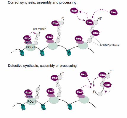
Figure 8. A model for the cotranscriptional surveillance of mRNA biogenesis
The recruitment of the exosome takes place continuously during transcription and is based
on two types of interaction that are independent of gene expression errors. On the one
hand, the exosome has been shown previously to bind to the transcription machinery. On
the other hand, our data demonstrates that the exosome interacts with the nascent pre-
mRNPs and that this interaction is mediated by specific hnRNP proteins, such as Hrp59.
The exosomes associated with the pre-mRNP could either be recruited by the transcription
machinery and transferred to the transcript during elongation, or recruited directly to the
pre-mRNP. The interactions of the exosome with the transcription machinery and with the
nascent pre-mRNP would be sufficient to establish an increased local concentration of
surveillance factors around the transcript that could ensure that any RNA substrates that
arise from transcription and/or processing errors are efficiently degraded.