the efficiency of using silver nanoparticles singly and in combination with traditional...
TRANSCRIPT

+
THE EFFICIENCY OF USING SILVER NANOPARTICLES SINGLY AND IN COMBINATION WITH TRADITIONAL ANTIMICROBIAL AGENTS IN CONTROL OF SOME FUNGAL AND
BACTERIAL AFFECTION OF BUFFALOES
*,1Hassan, A. A., 2Mogda K. Mansour
1Department of Mycology and Mycotoxins, Animal Health Research Institute, Agriculture Research Center,
2Department Biochemistry, Animal Health Research Institute, Agriculture Research Center, Dokki, Cairo, Egypt3Buffalo Diseases Research Department, Animal Health Research Institu
ARTICLE INFO ABSTRACT
Chemical synthesis of silver nanoparticles (Agfungal and bacterial causes of diseases in buffaloes were investigated. A total of 75 animal cases of dairy buffaloes were selected from a private farm at Giza governorates in which animals suffered from diarrrespiratory symptoms. Seventy five samples (25from each of nasal swabs, pharyngeal swabs from dairy buffaloes with respiratory disorders, fecal swabs of diarrheic animals and milk samples of mastitis animals)bacterial isolates and Klebsiella spp. samples of buffaloes that suffered from respiratory12 % and8% .On the other hand, tfumigates of (41.33%) and was recovered from 68%, 40% and 16% of samples of buffaloes with mastitis, diarrhrespiratory disorders respectivelymorphology of Agby transmission electron microscope (TEM) and scanning electron microscope (SEM) for detection of their particle size and the C.albicans, A.flavus, S.aureas NPs increased up to 300 ug/ml, the optical density of treatedtransmittance and clear medium. The minimum inhibitory concentration (MIC) of AgSalmonella sp., S.aureasbetween AgNPs and traditional antibioticrevealed that the requirement of lower concentrations from both to obtain the antimicrobial effects (200, 150, 200 and 200ug/ml) for respectivelywall was detected or membrane damage and some pits and adheredbeen caused leakage in inter cellular componentstherapy of Agof nanoparticles, overcome the microbial resistant to traditional antibiotiantimicrobial activity for the treatment of human and animal diseases.
Copyright © 2016, Hassan et al. This is an open access article distributed under the Creative Commons Attdistribution, and reproduction in any medium, provided the original work is properly cited.
INTRODUCTION
The animal wealth in developing countries represents the major role in food security for human consumption. One of essential animals in our country included buffalo (Bubalusbubalis) *Corresponding author: Hassan, A. A. Department of Mycology and Mycotoxins, Animal Health Research Institute, Agriculture Research Center, Dokki, Cairo, Egypt.
ISSN: 0975-833X
Article History:
Received xxxxxxxxxx, 2016 Received in revised form xxxxxxxxxxxxxxxxx, 2016 Accepted xxxxxxxxxx, 2016 Published online xxxxxxx, 2016 Key words: Silver nanoparticles (Ag-NPs), Antimicrobial potential, Buffaloes, Staphylococcus aureus, Salmonella spp., A.flavus sp., C.albicans, Traditional antibiotic
Citation: Hassan, A. A., Mogda K. Mansour, Noha H. Oraby andcombination with traditional antimicrobial agents in control of some fungal and bacterial affection of buffaloes8, (04), 29758-29770.
Article History:
Received 19th January, 2016 Received in revised form 18th February, 2016 Accepted 28th March, 2016 Published online 26th April, 2016 Key words: Silver nanoparticles (Ag-NPs), Antimicrobial potential, Buffaloes, Staphylococcus aureus, Salmonella spp., A.flavus sp., C.albicans, Traditional antibiotic
RESEARCH ARTICLE
THE EFFICIENCY OF USING SILVER NANOPARTICLES SINGLY AND IN COMBINATION WITH TRADITIONAL ANTIMICROBIAL AGENTS IN CONTROL OF SOME FUNGAL AND
BACTERIAL AFFECTION OF BUFFALOES
Mogda K. Mansour, 1Noha H. Oraby and 3Aliaa A
Department of Mycology and Mycotoxins, Animal Health Research Institute, Agriculture Research Center, Dokki, Cairo, Egypt
Department Biochemistry, Animal Health Research Institute, Agriculture Research Center, Dokki, Cairo, EgyptResearch Department, Animal Health Research Institute, Agriculture Research Center
Dokki, Cairo, Egypt
ABSTRACT
Chemical synthesis of silver nanoparticles (Ag-NPs) and evaluation of their antimicrobial potential against some fungal and bacterial causes of diseases in buffaloes were investigated. A total of 75 animal cases of dairy buffaloes were selected from a private farm at Giza governorates in which animals suffered from diarrrespiratory symptoms. Seventy five samples (25from each of nasal swabs, pharyngeal swabs from dairy buffaloes with respiratory disorders, fecal swabs of diarrheic animals and milk samples of mastitis animals)bacterial isolates were Staphylococcus aureus, Sreptococcus spp, Salmonella spp
Klebsiella spp. The species of Staphylococcus are considered the most predominant isolates from different samples of buffaloes that suffered from respiratory disorders, diarrhea or mastitis at rates of incidence of (32%,
and 36%) respectively. While, S.typhimurium was recovered from diarrheic buffaloes at incidence rate of On the other hand, the most predominant members of Aspergillus species in samples
fumigates (54.6%), A.niger (53.3%), followed by A. ochraceus (41.3%). While,41.33%) and was recovered from 68%, 40% and 16% of samples of buffaloes with mastitis, diarrh
piratory disorders respectively. The silver nanoparticles was synthesized by chemical methodmorphology of Ag-NPs were characterized by visual inspection; in a UVby transmission electron microscope (TEM) and scanning electron microscope (SEM) for detection of their particle size and the purity of the prepared powder. The antimicrobial potential of prepared AgC.albicans, A.flavus, S.aureas and Salmonella sp. was concentration dependent, when the concentrations of AgNPs increased up to 300 ug/ml, the optical density of treated spore suspension were decreased till reach 100% transmittance and clear medium. The minimum inhibitory concentration (MIC) of AgSalmonella sp., S.aureas sp. was (250-, 300,300 and 250ug/ml), respectively. Whereas, the resultsbetween AgNPs and traditional antibioticrevealed that the requirement of lower concentrations from both to obtain the antimicrobial effects (200, 150, 200 and 200ug/ml) for C.albican, A.flavus, Salmonella sp., S.aureas sp.respectively. The treated fungal and bacterial cells were subjected to SEM, the damage and rupture of their cell wall was detected or membrane damage and some pits and adhered to respiratory sequence of cytoplasm been caused leakage in inter cellular components and finally cell death. Therefore, ttherapy of Ag-NPs with other traditional antibiotics drugs was urgently required to decrease the used concentration of nanoparticles, overcome the microbial resistant to traditional antibiotiantimicrobial activity for the treatment of human and animal diseases.
is an open access article distributed under the Creative Commons Attribution License, which distribution, and reproduction in any medium, provided the original work is properly cited.
countries represents the major role in food security for human consumption. One of essential animals in our country included buffalo (Bubalusbubalis)
Department of Mycology and Mycotoxins, Animal Health Research Institute, Agriculture Research Center, Dokki, Cairo, Egypt.
which are considered as “black gold”. The products of these animals are of huge economic countries (milk, meat, hides and draft power for various agricultural operations (Patel microbial infections resulted from opportunistic bacteria and fungi have been common especially in human and animals which being affected by special conditions like immune weakness and the candidates infection to animals. However, the
Available online at http://www.journalcra.com
International Journal of Current Research Vol. 8, Issue, 04, pp. 29758-29770, April, 2016
INTERNATIONAL
Hassan, A. A., Mogda K. Mansour, Noha H. Oraby and Aliaa A. E. Mohamed, 2016. “The efficiency of using silver nanoparticles singly and in combination with traditional antimicrobial agents in control of some fungal and bacterial affection of buffaloes”, International Journal of Current Research
z
THE EFFICIENCY OF USING SILVER NANOPARTICLES SINGLY AND IN COMBINATION WITH TRADITIONAL ANTIMICROBIAL AGENTS IN CONTROL OF SOME FUNGAL AND
Aliaa A. E. Mohamed
Department of Mycology and Mycotoxins, Animal Health Research Institute, Agriculture Research Center,
Department Biochemistry, Animal Health Research Institute, Agriculture Research Center, Dokki, Cairo, Egypt
te, Agriculture Research Center,
of their antimicrobial potential against some fungal and bacterial causes of diseases in buffaloes were investigated. A total of 75 animal cases of dairy buffaloes were selected from a private farm at Giza governorates in which animals suffered from diarrhea, mastitis and respiratory symptoms. Seventy five samples (25from each of nasal swabs, pharyngeal swabs from dairy buffaloes with respiratory disorders, fecal swabs of diarrheic animals and milk samples of mastitis animals). The main
Salmonella spp., Escherichia coli, Ps.aeruginosa are considered the most predominant isolates from different disorders, diarrhea or mastitis at rates of incidence of (32%, was recovered from diarrheic buffaloes at incidence rate of
species in samples were A.flavus (60%), A. 41.3%). While, C.albicans was isolated at the rates
41.33%) and was recovered from 68%, 40% and 16% of samples of buffaloes with mastitis, diarrhea and The silver nanoparticles was synthesized by chemical method and the sizes and
NPs were characterized by visual inspection; in a UV-visible spectrophotometer and scanning by transmission electron microscope (TEM) and scanning electron microscope (SEM) for detection of their
purity of the prepared powder. The antimicrobial potential of prepared Ag-NPs against was concentration dependent, when the concentrations of Ag-
spore suspension were decreased till reach 100% transmittance and clear medium. The minimum inhibitory concentration (MIC) of Ag-NPs for C.albican, A.flavus,
, 300,300 and 250ug/ml), respectively. Whereas, the results of combination between AgNPs and traditional antibioticrevealed that the requirement of lower concentrations from both to obtain
C.albican, A.flavus, Salmonella sp., S.aureas sp., The treated fungal and bacterial cells were subjected to SEM, the damage and rupture of their cell
to respiratory sequence of cytoplasm that had and finally cell death. Therefore, the synergistic, combination
NPs with other traditional antibiotics drugs was urgently required to decrease the used concentration of nanoparticles, overcome the microbial resistant to traditional antibiotics and resulted more efficient
ribution License, which permits unrestricted use,
which are considered as “black gold”. The products of these animals are of huge economic importance in developing countries (milk, meat, hides and draft power for various agricultural operations (Patel et al., 2010). Nowadays,
from opportunistic bacteria and fungi have been common especially in human and animals
fected by special conditions like immune weakness and the candidates infection to animals. However, the
INTERNATIONAL JOURNAL OF CURRENT RESEARCH
The efficiency of using silver nanoparticles singly and in International Journal of Current Research,
fungal infections particularly by C.albicans and mycotoxigenicmoulds represent the widest spread causes of mycotic diseases of man and animals (Hassan et al., 2007, 2012a, 2014, 2015a and 2016). In spite of progressive advances in harvesting, storage and processing technologies, fungal spoilage still has a major economic impact on world food supplies. The most common and destructive food spoilage fungi belong to the genera Aspergillus, Penicillium and Fusarium, although other genera are of significance in particular foods and feed (Refai and Hassan, 2013). Moreover, Candida albicansand other fungal infection are probably one of the most successful opportunistic pathogens in humans. Under conditions of a weakened immune system, colonizing C. albicansandmouldcan become opportunistic, causing recurrent mucosal infections and life-threatening contagious infections with high mortality rates (Refai et al., 2014a). In addition, there are a number of fungal and bacterial diseases which adversely affect the health of this animal, as mastitis, diarrhea and respiratory tract infections which are the major production-limiting disease causing staggering economic losses to the animal industry. The most important effects are related to economic losses due to decrease in milk yield (McDowell et al., 1995), diminished meat production due to diarrhea (Fagiolo et al., 2005) and respiratory disorders which are stress factors resulted a bad production of animal (Quinn et al., 2002). Several studies recovered various bacterial and fungal causes of these diseases in man and animals as Staphylococcus species Streptococcus species and Escherichia coli species, C. albicans, Aspergillussp. And Penicillium sp. which are the dominant microbial isolates in cases of mastitis (Yuan et al., 2012 and Hassan et al., 2014 and 2015). However, in cases of calve diarrhea, the most important bacterial are enterotoxigenic E.coli (ETEC) which producing directly detectable toxin, Salmonellae sp. and Y. enteroclotica (Milnes et al., 2008). While, in water buffalo, S. typhimuriumcan induce a variety of clinical syndromes with different pathologicallesions (Fagiolo et al., 2005). Some mold as members of Aspergillus sp., Penicillium sp. and Fusarium sp. caused mycosis and mycotoxicosis in buffalo (Hassan et al.,2008, 2010 and 2014) in poultry (Hassan et al., 2007) and in rabbits (Hassan et al., 2016)and aflatoxicosis in cattle (Hassan et al., 2012a). Recently, a rapid increase in microbial infections that are resistant to conventional antibiotics has been observed, especially, the frequency of infections provoked by opportunistic fungal and bacterial strains has increased dramatically (Goffeau, 2008; Hassan et al., 2015a and 2016 and Nabawy et al., 2014). Furthermore, the number of known multidrug resistant bacteria and fungi is increasing rapidly. Thus, the development of more effective antifungal therapies is of great importance. Understanding the mechanisms and decisions of cell death in fungi may provide new developments in the search for diverse novel antifungal nanoparticles (Whitesides, 2003 and Hwang et al., 2012). Many factors appear to be involved in its emergence, the excessive and improper use and abuse of antibiotics has been shown to play a major role. The continued evolution of drug resistance, which has already invalidated many routinely used antibiotics, has reached a fevered pitch and is a serious public health threat, with some even warning of the possibility of the 21st century becoming the “post-antibiotic” era (Kåhrström
et al., 2013). In the ongoing race between the emergence of drug resistance and the development of novel antimicrobial agents, microbes appear to be pulling ahead (Yunis, 1988). Therefore, new, safe antimicrobial agents are needed to prevent and overcome severe fungal and bacterial infections. In the recent times, the advances in the fieldof nanotechnology has brought to form thenanosizedin organic and organic particles which are used formany applications as amendments in industrial, medicine and therapeutics, synthetic textiles and food packaging products (Gajjar et al., 2009). Silver nanoparticles (AgNPs) are the most intensely studied metal nanomaterial. They are capable of killing fungal and bacterial cells and are effective against many drug-resistant microbes, such as C.albicans, T. verrucosum and T. mentagrophytes (Hassan et al., 2013) and Pseudomonas aeruginosa (P. aeruginosa), ampicillin-resistant E. coli O157:H7 and erythromycin-resistant Streptococcus pyogenes (Lara et al., 2010). In addition, the combination antibiotic therapy appears to hold a great deal of potential not only in tackling existing mechanisms of drug resistance but in preventing its development in the first place and combining multiple drugs can result in higher potency and higher antimicrobial efficacy by additive or synergistic effects (Chow and Yu, 1999). Therefore, the present study was designated to survey the fungal and bacterial diseases of buffaloes in Egypt. Synthize and characterize silver nanoparticles and evaluation its antibacterial and antifungal potentials against isolates recovered from field buffalo diseases. The antimicrobial potential of AgNPs was evaluated singly and in combination with traditional antimicrobial agents.
MATERIALS AND METHODS Samples A total of 75 animal cases of dairy buffaloes were selected from a private farm at Giza governorates in which animals suffered from diarrhea, mastitis and respiratory symptoms. Seventy five samples (25each of nasal swabs, pharyngeal swabs from dairy buffaloes with respiratory disorders, fecal swabs from diarrheic animal and milk samples from mastitis animals) were aseptically collected in sterile swabs and McCartney bottles. Each sample was divided into two parts, one was incubated at37ºC for 24 h for bacteriological examination, while the second part were subjected to mycological examination Control antibacterial and antifungal A known antifungal as Fluconazol (20 ug) and antibacterial asFloricol (12.5 mg) were purchased from Sigma Chemical Company (USA) and used as a comparable control. Bacteriological Examination of samples It was carried out according to the standard methods recommended by (Quinn et al., 2002). Milk samples were incubated aerobically at 37ºC for 24 hrs then centrifuged at 3000 rpm for 20 minutes, the supernatant fluid was discarded and a sterile loopful from the sediment was streaked onto the surface of following media: Blood, MacConkey, Baird parker,
29759 Hassan et al. The efficiency of using silver nanoparticles singly and in combination with traditional antimicrobial agents in control of some fungal and bacterial affection of buffaloes

EMB, Salmonella Shigella agar and Eduard agars., while swabs (nasal, pharyngeal and fecal) Samples were streaked directly on the same media. Inoculated plates were incubated at 37ºC for 24-48 hrs. Presumptive identification of bacterial isolates was made based on colony morphologic features, Gram-stain reaction, hemolytic characteristics and biochemical testes. Identification of isolated bacteria from samples Confirmation of S.aureuscoagulase positive isolates by Staphylococci Latex agglutination test: Staphylococci were tested using dry spot kit (Oxoid). A fresh culture grown over night 18-36 hrs incubation was used. A positive result showed agglutination of the latex particle within 20 seconds this indicates the presence of S.aureus (Finegold and Baron, 1986). Serological identification for Escherichia coli speciesby agglutination test Serological identification of the isolates was carried out as described by Lee et al. (2009) using polyvalent and monovalent antisera (DENKA SEIKEN CO., LTD). Isolation and Serological identification of Salmonella isolates Fecal samples were routinely grown in selenite F broth, after incubation at 37°C for 18 h; a loopful was inoculated on MacConkey agar and SalmonellaShigella agar then serological identification was carried out (SIFIN Institute FürImmun-praparate und NährmedienGmb H Berlin Berliner Allee 317/321,13o88 Berlin, Germany) according to (Edwards and Ewing, 1972). Mycological examination of samples The collected samples of nasal swabs, pharyngeal swabs ,fecal swabs and milk samples were prepared and examined for isolation of fungi according to the technique recommended by Refai and Hassan (2013) and Refai et al. (2014 a&b). After addition of medium, the plates were left to solidify at room temperature then incubated at 25oc for 5 -10 days. Identification of mold and yeasts, which isolated from samples The identification of moulds was basedon the morphology of the colony, the rate of growth and microscopic morphology of the isolates in a direct culture mount and micro-slide culture technique. The identification was made according to the morphological description in textbooks dealing with mould (Koneman et al., 1992 and Refai and Hassan, 2013 and Refai et al., 2014 a).While, yeast isolates were identified according to Kriger van Rij, (1984) and (Refai et al., 2014 b). Synthesis and characterization of silver nanoparticles (Kim et al., 2008) One hundred grams of solid silver were dissolved in100 ml of 100% nitric acid at 900C, and then 1 liter of distilled water was
added. By adding sodium chloride to the silver solution, the Ag ions were precipitated and then clustered together to form monodispersed nanoparticles in the aqueous medium. The Particle sizes and morphology of Nano-Ag distributions of these samples were also obtained using scanning electron microscope (SEM) (Joe, JSM-5600LV, Japan). The prepared Ag-NPs were identified and characterized by visual inspection; in a UV-visible spectrophotometer and Scanning electron microscope (SEM) for detection of their particle size and the purity of the prepared powder. Preparation of spore suspension of isolates (Gupta and kohli, 2003) The tested isolates of C.albicans, A.flavus, S.aureasandS. typhimuriumwere cultivated on SDA (for fungi) or nutrient agar (for bacteria) for 1-3 days at 30-37°C. At the end of incubation period the fungal mycelia / spore mat and bacterial colonies were washed off with a 6 ml of sterile distilled water by sterile loop, the outer most layer of growth (fungal spores and bacterial colonies) was scraped. The mycelia were removed by filtration through a 500mm pore sieve. This spore suspension was counted in haemocytometer slide considering the dilution factor and the spores count was adjusted to 105spores /ml. Measurement of MIC of prepared AgNPs singly and in combination with traditional antimicrobial agents against fungi and bacteria isolated from diseased cases of buffalos (CLSI 2008) The minimum inhibitory concentration (MIC) of Ag-NPs for the tested isolated was determined by a broth microdilution method based on the National Committee for Clinical Laboratory Standards (NCCLS ) for bacteria (Balachandran et al., 2015), for filamentous fungi (CLSI 2008) and for yeasts (NCCLS,M27-A2 2002). In sterile 12- x 75-mm plastic test tubes, 900 ul of RPMI 1640 broth medium or SD broth medium (for fungi) or nutrient broth (for bacteria) was inoculated separately, then, 100 ul of spore suspension added to adjust the inoculum of, S. aureus, Salmonella and A.flavus (2.5 х 103cells/ml) and Candida albicansto (5 х 104 cells/ml). 100 ul of silver nanoparticles concentrations (50, 100, 150, 200, 250, 300ug/mL) for bacteria and fungi , were added. Thetraditional antifungal agent Fluconazole (20 ug) and antibacterial agent Florficol (12.5 mg)were included in the separate assays as positive controls. A.combination effects ofAgNPs and traditional antimicrobial agents was performed as above mentioned testsbut100ul was added from each. All the test tubes were incubated for 48 hr–5days at 28-30o C (for fungi) and for 24-48 h at 37o C (for bacteria). The experiment was repeated twice. The MIC for fungi and bacteria was defined as the lowest silver nanoparticles concentration that showing no visible fungal or bacterial growth after incubation time. After end of incubation period, 5 uL of tested broth were inoculated on the sterile nutrient agar plates for bacteria and SDA plate for fungi and incubated at 37o C for 24 hr- 2 weeks. The MIC was determined as the lowest concentration of AgNPs inhibiting the visual growth of the test cultures on the agar plate. The turbidity of the growth in tubes was observed every 24 hrs. The growth was assayed by measurement of optical density and
29760 International Journal of Current Research, Vol. 08, Issue, 04, pp. 29758-29770, April, 2016

transmittance % of each tubes content at 405 nm using spectrophotometer. Scanning Electron Microscopy of the treated microbial cells (SEM) (Gong et al., 2007) The morphological changes of Candidaalbicans, A. flavus, S. aureusand Salmonella typhimurium which were treated by Ag–NPs singly or in combination with traditional antimicrobials were observed with a scanning electron microscope (SEM). All the content of tubes were centrifuged and the sediments of each were dehydrated separately through a graded series of ethanol (30, 50, 60, 70, 80, 90, 95, and 100%), each level was applied twice for 15 min each time, then the ethanol:isoamyl acetate (3:1, 1:1, 1:3) and 100% isoamyl acetate applied twice for 30 min). The solutions in wells were dried with a critical-point drier using liquid CO2 and coated with gold-coater for 5 min. The coated samples were observed under SEM, JSM-5600LV with accelerating voltage of 10 kV.
RESULTS AND DISSCUSSION Buffaloes are an economically important source of milk and meat, there are about 3.98 million head in Egypt and several serious problem facing animals health including respiratory disorders, mastitis and diarrhea(Arab agriculture statistics yearbook (A.A.S., 2011).The respiratory disorders have continuous effect on animal productivity and value (Ali et al., 2009).It occurred when the immune defenses of animal was failed and the lung can be affected by bacterial pathogens resulting in the development of pneumonia (Caswell, 2014). The different types of microorganisms (fungi, viruses and bacteria) can reach the alveoli of the calf’s lung, cause irritation and inflammatory reactions (Garcia and Daly, 2010). The most common agents of bronchopneumonia, particularly in buffalo, are P. multocida, A.pyogenes, streptococcus spp., staphylococcus spp., E. coli, Proteus spp. and Mycoplasma spp., which were the predominant isolated bacterial pathogens (McGavin and Zachary, 2007; Sayed and Zaitoun, 2009). Also, diarrhea remains a major public health problem. Surveillance for a broad range of enteric pathogens is necessary to accurately predict the frequency of pathogens and potential changes in antibiotic resistance patterns (Shirley et al., 2013). Infectious diarrhea is a significant contributor to high morbidity and mortality worldwide. It is caused by enteric pathogens (Salmonella spp., Shigella spp., Escherichia coli) and it is among the most important disorders in calves (Svenssion et al.,2003). On the other hand, mastitis is a frustrating, costly and extremely complex disease resulted in a marked reduction in the quality and quantity of milk (Akhtar et al., 2012). The pathogens that cause mastitis can be divided into different groups of organisms depending on the source of the organism involved; these include contagious and, environmental pathogens (Philpot and Nickerson, 1999). The causes of environmental mastitis episodes in dairy cows and buffaloes are S. aureus (62.9%), Streptococcus species (15.5%) (Aguilar, 2001), E. coli (12.4%) (Yuan et al., 2012), Salmonella, Klebsiella, Corynebacterium and Mycoplasma species (Fox et al., 2005 and Khan and Khan, 2006). In the present study, the current results in (Table, 1) revealed that (88%) of samples were positive for bacteriological examination. The main
isolates which are more pathogenic and causes different diseases in animals and human are Staphylococcus aureus, streptococcus spp, Salmonella spp., E.coli, Ps.aeruginosa and Klebsiellaspp. The species of Staphylococcus is considered the most predominant isolates from different samples of buffaloes that suffered from respiratory disorders, diarrhea or mastitis at rates of incidence of (32%, 12% and 36%), respectively. Moreover, Table (2) showed that coagulase positive Staphylococcus aureus (S.aureus) was isolated with an incidence of (65%), while coagulase negative Staphylococci were isolated with an incidence of (35%). S. aureusis an opportunistic pathogen in dairy ruminant where it is found in healthy carriage and can be a major cause of mastitis (Seyffert et al., 2012). It is classified among the most serious pathogens causing clinical symptoms of various diseases not only in animals, but also in human (VASIĽ, 2007) and the coagulase negative staphylococci (CNS) are the most prevalent important pathogen (Pradiee et al., 2012; Gebrewahid et al., 2012). S. aureus is the main inhabitant of the mucous membranes of the upper respiratory tract and opportunistically involves them in pathologic role following stress conditions, such as infection by influenza virus and can be become a serious cause of infection in immune-suppressed hosts(Quinn et al., 2002). Currently, in (Table 1) E.coli isolates from different samples of buffaloes suffered from respiratory disorder, mastitis or diarrhea showed the incidence rates of (8%, 28% and 60%) respectively. According to virulence properties and the clinical symptoms of the host, pathogenic E. coli strains are designated as entero-toxigenic E. coli (ETEC), attaching and effacing E. coli, entero-pathogenic E. coli, Shiga toxin producing E. coli (STEC) and necro-toxigenic E. Coli (DebRoy and Maddox, 2001). The ETEC can cause severe diarrhea in newborn calves via the production of heat-stable enterotoxin (STa). Even though both healthy and diarrheic calves harbor STEC in their intestine, natural outbreaks and experimental infections have documented the association of STEC with diarrhea and dysentery in young calves (Dean-Nystrom et al., 1997 and Sandhu and Gyles, 2002). Most outbreaks and sporadic cases of bloody and non-bloody diarrhea and HUS have been attributed to strains of the STEC serogroups including O157, O26, O111 and O128 (Erickson and Doyle, 2007 and Lin et al., 2011).In the present study, Salmonella Typhimuriumwas recovered from diarrheic buffaloes at incidence rate of 8%. Other study reported that Salmonella is one of the most extensively characterizedbacterial pathogens and is a leadingcause of bacterial gastroenteritis and a significant cause of morbidity and mortality in humans and animals, with multidrug-resistant Salmonella typhimurium being an emerging problem (Yan et al., 2004). Itcan survive in the environment and once established on a farm, contaminationcan be difficult to eradicate. Introduction of Salmonella onto a dairy farm can occurthrough a variety of routes, including purchased cattle, contaminated feed or water, wild animals such as rodents and birds, human traffic, and insects (Sanchez et al., 2002 and Nielsen et al., 2007). The risk of Salmonella infection has been heightened by the globalization of trade in food, feed and live animal and changes in production, processing and handling of foods (Hoelzer et al., 2010 and Scallan et al., 2011).
29761 Hassan et al. The efficiency of using silver nanoparticles singly and in combination with traditional antimicrobial agents in control of some fungal and bacterial affection of buffaloes

Table 1. Prevalence of bacteria in samples collected from diseased buffaloes with mastitis, diarrhea and respiratory symptoms
diseased buffaloes:
(75cases) Buffaloes with mastitis
(25 cases) Buffaloes with diarrhea
(25cases) Buffaloes with respiratory symptoms
(25 cases) Bacterial
Species % No. % No. % No. % No.
2.7 2 - - - - 8 2 Coryne bacterium 26.7 20 36 9 12 3 32 8 Staphaphylococcus.sp 10.7 8 20 5 - - 12 3 Streptcoccus sp. 2.7 2 - - 8 2 - - Salmonella spp. 32 24 28 7 60 15 8 2 Escherichia coli sp. 8 6 8 2 - - 16 4 Klebsiella spp.
5.3 4 4 1 - - 12 3 Ps.aeruginosa. 88% 66 96% 24 80% 20 88% 22 TOTAL
Table 2. Prevalence of S.aureus coagulase positive and coagulase negative staphylococcus spp
Types of examined Staphylococcus. sp No. %
S.aureus Coagulase positive 13 65 Staphylococcus. sp Coagulase negative 7 35 Total 20 100
Table 3. Serological identification of E. coli isolates recovered from samples of diseased buffalo
Serotype E. coli isolates
No % O126 6 25 O158 3 12.5 O26 1 4.2 O128 7 29 O114 4 16.7 O111 1 4.2 Untypable 2 8.4 Total 24 100
Table 4. Prevalence of fungi in samples collected from diseased buffaloes with mastitis, diarrhea and respiratory symptoms
Type of sample Identified spp
Buffaloes with respiratory symptoms (25 cases)
Buffaloes with diarrhea (25cases)
Buffaloes with mastitis (25 cases)
Total isolates (75cases)
No % No % No % No. % A.flavus 18 72 11 44 16 64 45 60
A. niger 12 48 16 64 12 48 40 53.3 A. fumigates 13 52 14 56 14 56 41 54.6
A. ochraceus 9 36 12 48 10 40 31 41.3 Penicillium sp. 9 36 7 28 5 20 21 28
Fusarium sp. 1 4 4 16 3 12 8 10.6 C. albicans 4 16 10 40 17 68 31 41.3 Rhodotorulasp. 2 44 8 32 11 44 21 28
Table 5. Optical density and transmittance (T%) of treated isolates recovered from diseased cases of buffaloes at gradual concentration of AgNPs OD : Optical density of treated spores at wave length 405 nm..- T%: Transmittance%
Gradual conc. of AgNps (ug/ml)
C.albicans A. flavus S. aurous S.typhimurium
O.D T % O.D T % OD T% O.D T%
0 0.52 30. 2.5 6.3 0.133 4.6 0.33 46.7 50 0.025 94.4 1.0 10.0 0.032 92.9 0.2 63.1
100 0.018 95.9 0.3 50.1 0.014 96.7 0.15 70.8 150 0.016 96.3 0.15 70.8 0.01 97.7 0.08 83.1
200 0.005 97.6 0.08 83.2 0.001 99.8 0.06 87.1 250 0 100 0.02 95.5 0 100 0.02 95.5
300 0 100 0 100 0 100 0 100
Table 6. Optical density and transmittance (T%) of treated isolates recovered from diseased cases of buffaloes at gradual concentration of AgNPs. in combination with traditional antimicrobial agents
Gradual conc. of AgNpsug/ml C.albicans A.flavus S. aurous S. typhimurium
O.D T % O.D T % O.D. T% O.D T%
0 0.52 30.2 2.56.3 6.3 1.33 4.6 0.33 46.7
50 0.034 92.4 0.5 31 0.424 37.7 0.23 58.8
100 0.025 94.4 0.2 63 0.23 58.9 0.21 61.6 150 0.012 97.2 0.00 100 0.132 73.7 0.1 79.4 200 0.0 100 0.00 100 0.0 100 0.0 100 250 0.0 100 0.0 100 0.0 100 0.0 100 300 0.0 100 0.0 100 0.0 100 0.0 100
The used traditional antimicrobial agents: Antifungal as Fluconazol50ug.Antibacterial as Florfenicol 12.5 mg.OD: Optical density of treated spores at wave length ; 405 nm. - T%: Transmittance
29762 International Journal of Current Research, Vol. 08, Issue, 04, pp. 29758-29770, April, 2016

On the other hand, moulds and yeasts are found on a wide variety of environmental factors such as feed, litters, air, soil, plants, water and animals discharges. The previous literatures recorded that the environmental pollution affect upon the growth rate and health of human being and animals and may cause several diseases as thrush, disseminated candidiasis, aspergillosis, dermatophytosis and mastitis, aneamia , stunted growth, carcinogenic, tremor-genic, hemorrhagic, dermatitis, pulmonary edema, immunosuppressive and hormonal effects (Hassan, 2003 and Hassan et al., 2009, 2014; 2015 a and 2016 and Asfour et al., 2009). While, the mycotoxigenic fungi can induce both toxicologic and immunotoxic effects in a variety of cell systems and animal species as cytotoxic effect to reticulocytes, fibroblasts and lymphocytes and the cellular toxicity appears to be mediated by the inhibition of protein synthesis as reported by (Mogeda et al., 2002; Hassan et al., 2015 b&c and 2016 a). Whereas, other fungi as C. albicansis considered a commensal organism for humans and animals and when host defenses are compromised C. albicans can transform into a tissue invasive pathogen (Palmer, 2008) resulting in several affections of the oral cavity, gastrointestinal tract, animal abortion and skin (Shawky et al., 2014; Hassan et al., 2015 and 2016 b). However Candida sp. is recognized as the fourth most common cause of bloodstream infection, with a high attributable mortality rate, while C. albicansremains the most common pathogen (Marr, 2004).The intestinal tract provides an important reservoir for many nosocomial pathogens, including Candida species and some bacterial species. Disruption of normal barriers, such as gastric acidity and endogenous microflora of the colon, facilitates the overgrowth of pathogens (Donskey, 2004). The reproductive tracts of different animals are the major reservoir of yeasts such as C. albicansandC. Neoformans (Radwan et al., 2008; Hassan et al., 2014 and Shawky et al., 2014). Recently, C.albicans anddermatophytes were recovered from cases of buffaloes ringworm in Egypt (Refai et al., 2014 a; Hassan et al., 2015 b&c). Nearly every food or feed commodity can be contaminated by fungal organisms and many of these fungi are capable of producing one or more mycotoxins, which are toxic metabolites of concern to human and animal health. It is estimated that 25 to 50% of the crops harvested worldwide are contaminated with mycotoxins. The percentage is highest in tropical regions, where, up to 80% of the crops are reported to contain significant amounts of mycotoxins (Smith et al., 1994; Hassan et al., 2012 and Refai and Hassan, 2013). Several studies indicated that there was significant prevalence of fungal infection in cattle and buffaloes that infested with ticks. This may be due to the fact that toxins present in saliva of ticks result in immunosuppression of the animals. In cattle, it causes high economic losses as body weight loss, decreased meat and milk yield and the acceptance of live animals in the market would also be reduced (Hassan et al., 2015 a and 2016 a & b). Currently, the present results in Table (4) illustrated that the most predominant members of Aspergillusspeciesin samples collected from diseased buffaloes were belonging to A.flavus (60%), A. fumigates (54.6%), A.niger (53.3%), followed by A. ochraceus (41.3%). While, Penicillium sp. and Fusarium sp. were recovered from 24% and 10.6 % of samples ,respectively. On the other hand, C.albicans and Rhodotorula sp. were isolated at the rates of 41.33% and 28%, respectively.
Candidaalbicans were recovered from 68%, 40% and 16% of diseased cases in buffaloes with mastitis, diarrhea and respiratory disorders respectively. Similar results were detected by Mosherf (2005), who reported that various Candida species particularly, C. albicans were the most common yeasts recovered from milk of clinically and sub-clinically mastitis milk. While, Hassan et al. (2012a) reported that yeast of C. albicans was yielded from 20% of cases of diarrhea (fecal samples), isolated from 32% and 24% of mastitis cases and recovered from nasal swabs of sheep and goat suffered from respiratory disorders (20% and 16 %), respectively. In other study, many mould isolates were recovered from mastitis milk of sheep and cattle (Hassan et al., 2010), who isolated Aspergillus flavus from animal feeds, mastitis milk and vaginal swabs at the rates of (80%, 50% and 50%), respectively, while the rates of isolation for A. parasiticus were (35%, 24% and 10%), respectively. The fungal diarrhea and respiratory affection were also detected during examination of fecal samples and nasal swabs of affected cattle which had included several members of genus Aspergillus at the rates of (47.0 %) (Hassan et al., 2011). However, Shawky et al.(2014) recovered C. albicans from cases of buffalo’s abortion, where it was isolated from 20 % of milk samples, 7.5%of placenta and 20% of fetal stomach contents, respectively.
Since microbes have progressively eroded the effectiveness of previously successful antibiotics by developing resistance, the emergence of resistant and more virulent strains of bacteria and fungi has outpaced the development of new antibiotics over the last few decades. Although antifungal drug resistance does not seem to be as much of a problem as resistance to antibacterial agents in bacteria, one long-term concern is that the number of fundamentally different types of antifungal agents that are available for treatment remains extremely limited. This is because fungi are eukaryotic organisms with a structure and metabolism that are similar to those of eukaryotic hosts. Therefore, there is an inevitable and urgent medical need for antibiotics with novel antimicrobial mechanisms (Whitesides, 2003). The application of nanotechnologies in medicine, or nanomedicine, which has already demonstrated its tremendous impact on the pharmaceutical and biotechnology industries, is rapidly becoming a major driving force behind ongoing changes in the antimicrobial field (Xi Zhu et al., 2014). One promising approach is the application of nanotechnology in the battle against microorganisms and is being applied not only to the treatment of infectious diseases but also to diagnostics of infections and to prophylaxis against its effect (Klabunde et al., 1996). Silver and its compounds have strong inhibitory and bactericidal effects as well as a broad spectrum of antimicrobial activities for bacteria, fungi and virus since ancient times (Aymonier et al., 2002; Shahverdi et al., 2007).
Compared with other metals, silver exhibits higher toxicity to microorganisms while it exhibits lower toxicity to mammalian cells (Kim et al., 2008). The recent advances in researches on metal nanoparticles appear to revive the use of Ag-Nps for antimicrobial applications. It has been shown that Ag-Nps prepared with a variety of synthetic methods have effective antimicrobial activity (Aymonier et al., 2002; Alt et al., 2004; Kim and Kim, 2006 and Thomas et al., 2007).
29763 Hassan et al. The efficiency of using silver nanoparticles singly and in combination with traditional antimicrobial agents in control of some fungal and bacterial affection of buffaloes

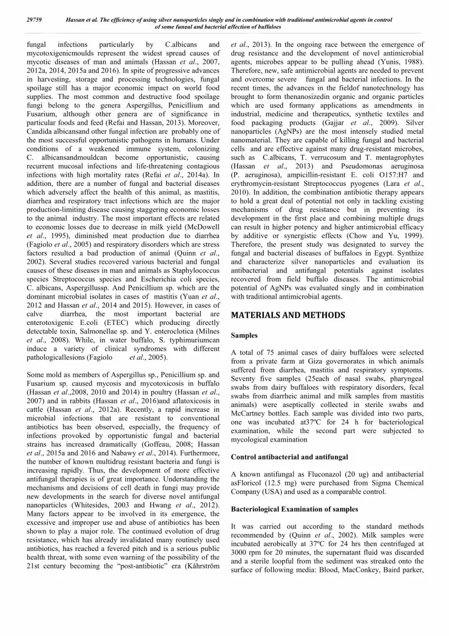
Fig.1.The TEM image of the of silver nanoparticles (50nm) (x 20 000)
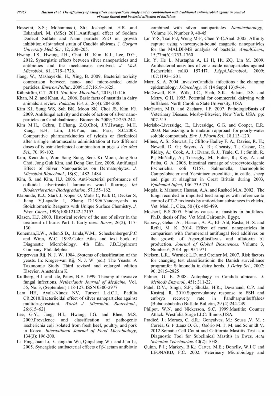
Fig.(4): O.D of treated A.flavus with gradual
concentration of Ag NPs.
Fig.(3): O.D of treated C. albicanswith gradual
concentration of Ag NPs.
Fig.(6) : O.D of treated of treated S.Typhimurium with
gradual concentration of Ag NPs.
Fig.(7): O.D of treated C.albicans at gradual
concentration of Ag NPs in combination with
floconazol.20 ug
Fig.(8): O.D of treated A.flavus at gradual
concentration of Ag NPs in combination
with floconazol.20 ug
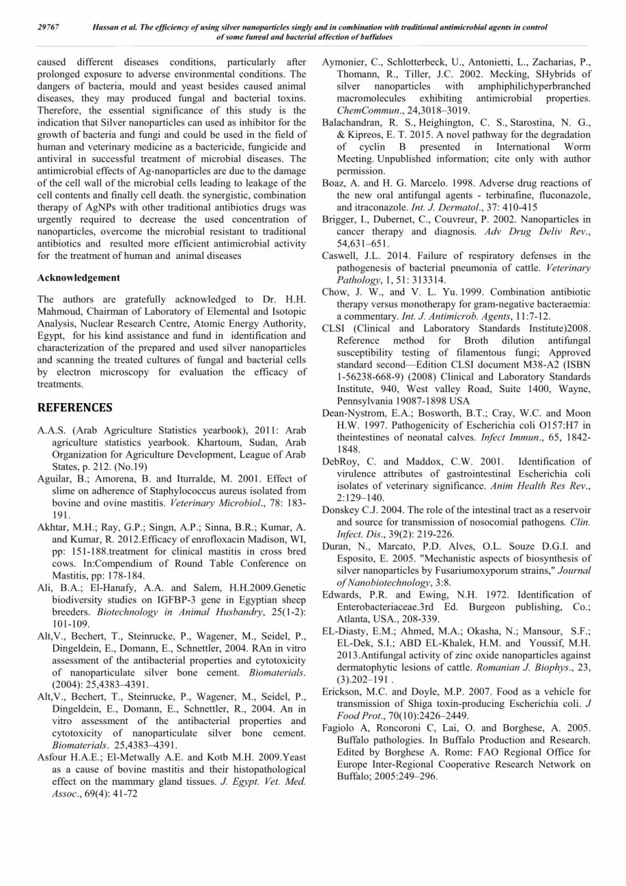
Fig.2. The UV-VIS absorbance spectra of Ag-NPS(50nm) at 405 nm wave length
Fig.(5) : O.D of treated of treated S. aureus with gradual concentration of Ag NPs
29764 International Journal of Current Research, Vol. 08, Issue, 04, pp. 29758-29770, April, 2016

Hence, Ag-Nps have been applied to a wide range of healthcare products, such as burn dressings, scaffold, water purification systems, and medical devices, (Kim et al., 2008).In the present study, silver nanoparticles were synthesized by chemical method by adding sodium chloride to the silver solution, Ag ions reduced and clustered together to form monodispersed nanoparticles in the aqueous medium. Because the final concentration of colloidal silver was 60,000 ppm, this solution was diluted, and then samples of different. concentrations were used to investigate the antimicrobial effects of Ag-NPs. The sizes and morphology of Ag-NPs were examined byvisual inspection; in a UV-visible spectrophotometer and scanning by transmission electron microscope (TEM) and scanning electron microscope (SEM) for detection of their particle size and the purity of the prepared powder. The particle size of prepared Ag-NPs was 50 nm and with spherical and granular morphology had uniform distribution(Fig.1). It was reported that the characterized absorption peak of AgNPs was detected at wave length of 400 nm due to electron transition from valence band to conduction band (Fig 2). Silver nanoparticles (Ag-NPs) are the most intensely studied metal nanomaterial. They are capable of killing both Gram-positive and Gram-negative bacteria and are effective against many drug-resistant microbes, such as Pseudomonas aeruginosa (P. aeruginosa), ampicillin-resistant E. coli O157:H7and erythromycin-resistant Streptococcus pyogenes (Lara et al., 2010). In addition, Several studies indicated the antifungal effect of AgNPs against C.albicans and Dermatophytes that responsible for ring worm in animals and human (Kim et al., 2008; Hassan et al., 2013 and Refai et al., 2014 a). Other studies had shown the antimicrobial effects of Ag-NPs (Klasen, 2000; Russell and Hugo, 1994 and Silver, 2003) and the effects of Ag-NPs against fungal pathogens of the skin including clinical isolates of T.mentagrophytes and Candida species are mostly illustrated by Kim et al. (2008) and Hassan et al.(2013). Currently, the antimicrobial potential of prepared AgNPs was evaluated by broth micro dilution method as recommended by (CLSI, 2008). The inoculum size, turbidity and transmittance of treated spore suspension of fungi and bacteria was determined spectrophotometrically to provides target percent of the treated spores of fungi or bacterial cells were determined spectrophotometrically to provide percent of transmittance (T %) readings. It is difficult to hold microtiter tests longer than 72 hrs due to the possibility of dehydration.
Many of the less frequently encountered fungi as dermatophytes may require as long as 120–144 hours before growth is detected in the drug-free growth control well. For this reason, isolates that are known to be slow growers should be tested via the macro broth method (CLSI, 2008). The endpoint determination depend on a reduction in turbidity which typically easy to visualize. Reading the MIC endpoint determined at the lowest concentration that prevents discernable growth, the first clear well (Ghannoum et al., 2004).In the present study, the tabulated results in Table (5), illustrated that the antimicrobial potential of Ag-NPs against C.albicans, A.flavus, S.aureas and Salmonella sp. was concentration dependent, when the concentrations of Ag-NPs increased up to 300 ug/ml, the optical density of treated spore suspension were decreased till reach 100% transmittance and clear medium. The inhibitory concentration of Ag-NPsthat inhibited the growth of each of C.albican S. aureas was 250 ug/ml and it was 300ug/ml for each of Salmonella sp. And A. flavus. The transmittance percentage and clearance of agent turbidity were confirmed by re-cultivation of inoculums from treated tubes on the specific medium SDA for fungi and NA for bacteria. The accordance of all previous parameters was repeated 2 times to pooled data (Table, 5 and Fig. 3-6). There are also reports of the application of Ag-NPs having antifungal activity in bio-stabilization of foot wear materials, wherein, 1% solution inhibited the growth of the majority of yeast-like fungal and mold strains (Falkiewicz and Macura, 2008). Similarly, Hassan et al. (2014), evaluatedthe antifungal activities of ZnO-NPs, the yeast of C. albicans was more sensitive for relatively lower concentrations (100 ug/ml), while, A. niger and A. flavus, A. ochraceus required higher concentration of ZnO-NPs to inhibit their growth (200-300 and 300 ug/ml), respectively. Other study by Hosseiniet al. (2011) reported that the MIC of ZnONPs against Aspergillus spp. and C. albicans was 1.013-296 μg/ml and for SDS and of Fluconazole were 0.001-0.56 and 0.062-128 μg/ml, respectively. Furthermore, different studies conducted in different laboratories showed that the antimicrobial activity is influenced by not only nanoparticles concentration but also by the size of the ZnO particles (Violeta et al., 2011 and Shawky et al., 2014). Recently, the antimicrobial potential of prepared ZnO-NPs was evaluated by broth microdilution methods against recovered microbial species from skin affection of
Fig.(9):O.D of treated of treated S. aureus at gradual concentration of
AgNPsin combination with Florfenicol 12.5 mg
Fig.(10):O.D of treated of treated S. typhimuriumat gradual concentration of
AgNPs in combination with Florfenicol 12.5 mg
29765 Hassan et al. The efficiency of using silver nanoparticles singly and in combination with traditional antimicrobial agents in control of some fungal and bacterial affection of buffaloes

buffaloes namely T.verucosum, T.mentagrophytes, D.congolenesis and S. aureus species. As the concentrations of ZnONPs increased, the optical density and turbidity of treated spore suspension decreased and reached 100% transmittance and clear medium at the MIC of ZnONPs (Hassan et al., 2015 c). In similar study, Hassan et al. (2015 b)investigated the antimicrobial effect of Fe2O3 NPs against isolated Dermatophytes and Dermatophilus species that recovered from skin affection of cattle and it had an inhibitory effect against the growth of T. verrucosum at concentrations of 3 mg /ml and 4 mg /ml, respectively (using well diffusion test). While, in case of T.mentagrophytes, iron oxide NPs revealed an inhibitory effect at concentration of 1, 2, 3, 4 and 5 mg/ml by well diffusion test. The treatment by Fe2O3 NPs had no effect on the growth of Dermatophilus sp. at the concentration ranged from 1- 3 mg/ml using disc diffusion test. While, the treatment by 4 mg/ml or more resulted in inhibition of bacterial growth. However, EL-Diasty et al. (2013) evaluated the antifungal activity of zinc oxide nanoparticles against species of Trichophytonmentagrophytes, Microsporumcanis, Candida albicans and Aspergillus fumigatus that were isolated from diseased cases. They detected that the largest inhibition of the germination of all the tested fungi was observed at largest ZnO nanoparticles concentration (40 mg/ml). There are also other studies confirming the strong antimicrobial activity of ZnO nanoparticles where, the nanoparticles could completely lyse the food-borne bacteria Salmonella typhimurium and Staphylococcus aureus (Liu et al., 2009 and Hassan et al., 2014). In another study, ZnO nanoparticles (12 nm) inhibited the growth of E. coli by disintegrating the cell membrane and increasing the membrane permeability (Jiang et al., 2009). The above findings suggest that ZnO nanoparticles can find applications in food systems and can be used to inhibit growth of pathogenic bacteria.Regarding the use of traditional antimicrobial agents, it has been reported that the in vitropotency of floricol against, pathogenic microorganisms are of higher effect than that of chloramphenicol and thiamphenicol (Yunis, 1988), and that its levels in bronchial secretions are higher than the minimum inhibitor concentrations necessary to affect secondary pathogens causing respiratory tract diseases (Varma et al.,1994). Also, Azoles that inhibit sterol formation and polyenes that bind to mature membrane sterols have been the mainstays regarding antifungal therapy for several decades (Kullberg and Pauw, 1999; Sheehan et al., 1999) On the other hand, Amphotericin B and fluconazole were used as a positive control toward fungi; amphotericin B is a fungicidal agent widely used in treating serious systemic infections (Hartsel and Bolard. 1996) and fluconazole is used in the treatment of superficial skin infections caused by dermatophytes and Candida species (Boaz and Marcelo. 1998). Whereas, Ag-NPs exhibited potent activity against clinical isolates and ATCC strains of Trichophytonmentagrophytes and Candida species (IC80, 1-7 μg/ml). The activity of Ag-NPs was comparable to that of amphotericin B and fluconazole. The antifungal activity of Ag-NPs is attributed to its effects on the fungal mycelia (Kim et al., 2009; Hassan et al., 2013).In the present study, the scanning by electron microscopy analysis observed the interaction between Ag-NPs and the membrane structure of bacterial or fungal cells by detection a significant
changes to their membranes, which are recognized by the formation of ‘‘pits’’ on their surfaces, and finally, result in the formation of pores and cell death due to Ag-Nps which release silver ion in cell and increased antimicrobial function. These results came in accord with Kim and Kim, (2006) who reported that SEM has been used for evaluating Ag-NPs capability in destroying surface membrane structure of the fungus. The Ag-Nps attached to cell membrane and penetrate it then produce a site witch little molecular weight in center of fungi, and then Ag-Nps attach to respiratory sequence and finally cell division stop lead to cell death, Ag-Nps release silver ion in fungal cell which increase its antifungal function. These results indicated that Ag-NPs have remarkable potential as an antifungal agent in treating fungal infectious diseases. Also,Kimet al.(2008), had reported that the inhibition of bud growth correlates with membrane damage. This report suggests that Ag-NPs inhibit the normal bud dingprocess, probably through the destruction of membrane integrity. Finally, Ag-NPs exhibited potent antifungal effects on fungi tested, probably through destruction of membrane integrity. On the other hand, the combination antibiotic therapy appears to hold a great deal of potential not only in tackling existing mechanisms of drug resistance but in preventing its development in the first place and combining multiple drugs can result in higher potency and higher antimicrobial efficacy by additive or synergistic effects (Chow et al., 1999). In this regard, a number of antibacterial drug combinations, including amoxicillin/clavulinic acid, ampicillin/sulbactam, trimethoprim/sulfonamide, trimetoprim/ sulfadimethoxine, and florfenicol/ tylosin have been used in veterinary area (Fernández-Varón et al., 2005; Kim et al., 2008).In recent study, Li et al. (2005) investigated the bactericidal action of AgNPs and amoxicillin on Escherichia coli. They revealed that the increasing concentration of both amoxicillin (0–0.525 mg ml 1) and silver nanoparticles (0–40 µg ml 1) showed a higher antibacterial effect on Escherichia coli cells. When amoxicillin and AgNPs were combined, it results in greater bactericidal efficiency on Escherichia coli cells than when they were applied separately. During dynamic tests on bacterial growth indicated that exponential and stationary phases are greatly decreased and delayed in the synergistic effect of amoxicillin and silver nanoparticles. Currently, the antibacterial potential of combination between AgNPs and traditional antibiotic were observed in the current data in Table (6) and Figures (7-10), Whereas, the results of combination between Ag-NPs and traditional antibiotic revealed that the requirement of lower concentrations from both to obtain the antimicrobial effects (200, 150, 200 and 200ug/ml) against C.albican, A.flavus, Salmonella sp. and S.aureas sp., respectively. The obtained MIC in combination was relatively lower than the single use of AgNPs and traditional antimicrobial agents. Therefore, the synergistic, combination therapy of AgNPs with other traditional antibiotics drugs was urgently required to decrease the used concentration of nanoparticles, overcome the microbial resistant to traditional antibiotics and resulted more efficient antimicrobial activity for the treatment of human and animal diseases.
Conclusion
Several bacterial and fungal isolates that recovered from dairy buffalo’s diseases were reported as potential pathogens and
29766 International Journal of Current Research, Vol. 08, Issue, 04, pp. 29758-29770, April, 2016
caused different diseases conditions, particularly after prolonged exposure to adverse environmental conditions. The dangers of bacteria, mould and yeast besides caused animal diseases, they may produced fungal and bacterial toxins. Therefore, the essential significance of this study is the indication that Silver nanoparticles can used as inhibitor for the growth of bacteria and fungi and could be used in the field of human and veterinary medicine as a bactericide, fungicide and antiviral in successful treatment of microbial diseases. The antimicrobial effects of Ag-nanoparticles are due to the damage of the cell wall of the microbial cells leading to leakage of the cell contents and finally cell death. the synergistic, combination therapy of AgNPs with other traditional antibiotics drugs was urgently required to decrease the used concentration of nanoparticles, overcome the microbial resistant to traditional antibiotics and resulted more efficient antimicrobial activity for the treatment of human and animal diseases
Acknowledgement
The authors are gratefully acknowledged to Dr. H.H. Mahmoud, Chairman of Laboratory of Elemental and Isotopic Analysis, Nuclear Research Centre, Atomic Energy Authority, Egypt, for his kind assistance and fund in identification and characterization of the prepared and used silver nanoparticles and scanning the treated cultures of fungal and bacterial cells by electron microscopy for evaluation the efficacy of treatments.
REFERENCES
A.A.S. (Arab Agriculture Statistics yearbook), 2011: Arab agriculture statistics yearbook. Khartoum, Sudan, Arab Organization for Agriculture Development, League of Arab States, p. 212. (No.19)
Aguilar, B.; Amorena, B. and Iturralde, M. 2001. Effect of slime on adherence of Staphylococcus aureus isolated from bovine and ovine mastitis. Veterinary Microbiol., 78: 183-191.
Akhtar, M.H.; Ray, G.P.; Singn, A.P.; Sinna, B.R.; Kumar, A. and Kumar, R. 2012.Efficacy of enrofloxacin Madison, WI, pp: 151-188.treatment for clinical mastitis in cross bred cows. In:Compendium of Round Table Conference on Mastitis, pp: 178-184.
Ali, B.A.; El-Hanafy, A.A. and Salem, H.H.2009.Genetic biodiversity studies on IGFBP-3 gene in Egyptian sheep breeders. Biotechnology in Animal Husbandry, 25(1-2): 101-109.
Alt,V., Bechert, T., Steinrucke, P., Wagener, M., Seidel, P., Dingeldein, E., Domann, E., Schnettler, 2004. RAn in vitro assessment of the antibacterial properties and cytotoxicity of nanoparticulate silver bone cement. Biomaterials. (2004): 25,4383–4391.
Alt,V., Bechert, T., Steinrucke, P., Wagener, M., Seidel, P., Dingeldein, E., Domann, E., Schnettler, R., 2004. An in vitro assessment of the antibacterial properties and cytotoxicity of nanoparticulate silver bone cement. Biomaterials. 25,4383–4391.
Asfour H.A.E.; El-Metwally A.E. and Kotb M.H. 2009.Yeast as a cause of bovine mastitis and their histopathological effect on the mammary gland tissues. J. Egypt. Vet. Med. Assoc., 69(4): 41-72
Aymonier, C., Schlotterbeck, U., Antonietti, L., Zacharias, P., Thomann, R., Tiller, J.C. 2002. Mecking, SHybrids of silver nanoparticles with amphiphilichyperbranched macromolecules exhibiting antimicrobial properties. ChemCommun., 24,3018–3019.
Balachandran, R. S., Heighington, C. S., Starostina, N. G., & Kipreos, E. T. 2015. A novel pathway for the degradation of cyclin B presented in International Worm Meeting. Unpublished information; cite only with author permission.
Boaz, A. and H. G. Marcelo. 1998. Adverse drug reactions of the new oral antifungal agents - terbinafine, fluconazole, and itraconazole. Int. J. Dermatol., 37: 410-415
Brigger, I., Dubernet, C., Couvreur, P. 2002. Nanoparticles in cancer therapy and diagnosis. Adv Drug Deliv Rev., 54,631–651.
Caswell, J.L. 2014. Failure of respiratory defenses in the pathogenesis of bacterial pneumonia of cattle. Veterinary Pathology, 1, 51: 313314.
Chow, J. W., and V. L. Yu. 1999. Combination antibiotic therapy versus monotherapy for gram-negative bacteraemia: a commentary. Int. J. Antimicrob. Agents, 11:7-12.
CLSI (Clinical and Laboratory Standards Institute)2008. Reference method for Broth dilution antifungal susceptibility testing of filamentous fungi; Approved standard second—Edition CLSI document M38-A2 (ISBN 1-56238-668-9) (2008) Clinical and Laboratory Standards Institute, 940, West valley Road, Suite 1400, Wayne, Pennsylvania 19087-1898 USA
Dean-Nystrom, E.A.; Bosworth, B.T.; Cray, W.C. and Moon H.W. 1997. Pathogenicity of Escherichia coli O157:H7 in theintestines of neonatal calves. Infect Immun., 65, 1842-1848.
DebRoy, C. and Maddox, C.W. 2001. Identification of virulence attributes of gastrointestinal Escherichia coli isolates of veterinary significance. Anim Health Res Rev., 2:129–140.
Donskey C.J. 2004. The role of the intestinal tract as a reservoir and source for transmission of nosocomial pathogens. Clin. Infect. Dis., 39(2): 219-226.
Duran, N., Marcato, P.D. Alves, O.L. Souze D.G.I. and Esposito, E. 2005. "Mechanistic aspects of biosynthesis of silver nanoparticles by Fusariumoxyporum strains," Journal of Nanobiotechnology, 3:8.
Edwards, P.R. and Ewing, N.H. 1972. Identification of Enterobacteriaceae.3rd Ed. Burgeon publishing, Co.; Atlanta, USA., 208-339.
EL-Diasty, E.M.; Ahmed, M.A.; Okasha, N.; Mansour, S.F.; EL-Dek, S.I.; ABD EL-Khalek, H.M. and Youssif, M.H. 2013.Antifungal activity of zinc oxide nanoparticles against dermatophytic lesions of cattle. Romanian J. Biophys., 23, (3). 191–202 .
Erickson, M.C. and Doyle, M.P. 2007. Food as a vehicle for transmission of Shiga toxin-producing Escherichia coli. J Food Prot., 70(10):2426–2449.
Fagiolo A, Roncoroni C, Lai, O. and Borghese, A. 2005. Buffalo pathologies. In Buffalo Production and Research. Edited by Borghese A. Rome: FAO Regional Office for Europe Inter-Regional Cooperative Research Network on Buffalo; 2005:249–296.
29767 Hassan et al. The efficiency of using silver nanoparticles singly and in combination with traditional antimicrobial agents in control of some fungal and bacterial affection of buffaloes

Falkiewicz-Dulik M, Macura A.B. 2008.Nanosilver as substance biostabilising footwear materials in the foot mycosis prophylaxis. Mikologia Lekarska. 15:145-150.
Fernández-Varón E, Escudero-Pastor E. and Cárceles- Rodríguez, C.M. 2005. Pharmacok.inetics of an ampicillin-sulbactam combination after intravenous and intramuscular
Finegold, S.M., Baron, E.J. 1986. “Diagnostic Microbiology” 7th Edition. The C.V. Mosby Co. St. Louis, London. The biochemical characterization and serological identification.
Fox, L.K.; Kirk, J.H. and Britten, A. 2005. Mycoplasma mastitis: A review of transmission and control. J.Veterinary Medical Sci., 52: 153- 160.
Gajjar, P., Pettee, B., Britt, D.W., Huang, W. Johnson, W.P. and Anderson, J. 2009. Antimicrobial activities of commercial nanoparticles against an environmental soil microbe, Pseudomonas putida KT2440. Journal of Biological Engineering, 3 :9-22.
Garcia, A. and Daly, M. 2010. Respiratory disease in young dairy calves. Dairy science; April 1-4.Access at http://agbiopubs.sdstate. edu/articles/ExEx4045.pdf.
Gebrewahid, T.T., Abera, B.H. and Menghistu, H.T. 2012. Prevalence and Etiology of Subclinical Mastitis in Small Ruminants of Tigray Regional State, North Ethiopia, Vet. World 5 (2): 103-109.
Ghannoum, M.A., Chaturvedi,V .and Espinel-Ingroff, A. 2004. Intra- and interlaboratory study of a method for testing the antifungal susceptibilities of dermatophytes. J ClinMicrobiol, 42(7):2977–2979
Goffeau, A. 2008. Drug resistance: the fight against fungi. Nature, Vol. 452, No. 7187, (April) 541-542, ISSN 0028-0836.
Gong, P., Li,H., HeX., Wang, K., Hu,J., Zhang, S. and Yang, X. 2007. Preparation and antibacterial activity of Fe3O4@Ag na-noparticles. Nanotechnology, 18, No. 28.
Gupta, A.k. and Kohli, Y. 2003. In-vitro susceptibility testing of ciclopirox, terbinafine, Ketoconazole and Itraconazolee against dermatophytes and nondermatophytes, and in vitro evaluation of combination antifungal activity. Br. J. Dermatol., 149 : 296-306
Hartsel, S. and J. Bolard. 1996. Amphotericin B: New life for an old drug. Trends Pharmacol. Sci., 17: 445-449.
Hassan , A.A. 2003. Detection of some mycotoxins and mycotoxins producing fungi in both macro- and microenvironment of diseased animals. 7th Sci. Cong. Egyptian Society for Cattle Diseases, pp. 112 – 119, 7-9 Dec., 2003, Assiut, Egypt
Hassan, A. A. , Mogda, K. M,, Ibrahim, E.M., Naglaa M. A., Flourage, A.M. A., Rady, M. and Darwish A.S. 2016. Aflatoxicosis in Rabbits With Particular Reference to Their Control by N. Acetyl Cysteine and Probiotic. International Journal of Current Research, Vol. 8, Issue, 1, pp.
Hassan, A. A. ,Manal, A. Hassan,Rasha, M.H. Sayed El Ahland Darwish, A.S. 2012a.Prevalence of yeast infections in small ruminants with particular references to their treatment by some natural herbal extracts. Bulletin of Environment, Pharmacology and Life Sciences, Volume 1, Issue 3, February 2012: 12-22.
Hassan, A. A., Hanan K. Mahmoud, Taha H. ,Rasha M.H. Sayed El-Ahl and Mahmoud, H. H.2015 c.Herbal biosynthesis of zinc Nanoparticles and evaluation of their antifungal and antibacterial effect for buffaloes skin
affections. International Journal of Current Research, Vol. 7, Issue, 12, pp.24338-24349.
Hassan, A. A., Noha H. Oraby, El-Dahshan, E.M. E. and Ali, M.A. 2015 a. Antimicrobial Potential of Iron Oxide Nanoparticles in Control of Some Causes of Microbial Skin Affection in Cattle. European Journal of Academic Essays, 2(6): 20-31,
Hassan, A. A.; Hammad, A.M. and Manal, A. Hassan, 2008.Prevalence of some dermatophytes and yeasts infections in cattle and their sensitivity to some antimycotics .. The 5 th Scientific Congress, Minufiya Vet. J.Vol.5 (1): 27-39.
Hassan, A. A.; Ramadan M. Khoudair and EL Sayed E. Youniss. 2009. The Effect of Some Mycotoxins on Immunity of Cattle Vaccinated against Brucellosis and Guinea Pigs Experimentally Vaccinated With S19 Vaccine Egypt. J. Appl. Sciences, Vol. 24 No. (2 A) 2009 (1-13).
Hassan, A., A.; Mogda, K. Mansour , Samira, A.M. Snousi and Randa, A. Hassan. 2010.Mycological, biochemical and histopathol studies on acute fusariotoxicosis in sheep. Life Science Journal, Vol 7, No 3, 49-57.
Hassan, A.A ; El Barawy, A.M and Manal, Hassan, 2007. Screening of meat and dairy byproducts for Candidaalbicance and effect of some antifungal on growth and germ tube formation of its isolates in Giza Governorate. Egypt. J. Comp. Path. & Clinic. Path., 20 (1):333-342.
Hassan, A.A.,Howayda, M. El Shafei,Noha, H. Oraby,Rasha, M.H. Sayed El Ahl and Mogeda, K. Mansour .2012b . Studies on mycosis and mycotoxicosis in cattle. 1st Conf. of An. Health Res. Inst. Assoc., December 2012. pp. 216 – 227
Hassan, A.A.,Noha, H. Oraby, Aliaa.A. E. Mohamed and Mahmoud H.H.2014.The possibility of using Zinc Oxide nanoparticles in controlling some fungal and bacterial strains isolated from buffaloes. Egypt. J. of Appl. Sci., 29 (3) 2014, 58-83.
Hassan, A.A.; El-Barawy, A.M. and El-Mokhtar, Nahed,M. 2011.Evaluation of biological compound of streptomyces species for control of some fungal diseases. J. of American Science, 7 (4),752-760.
Hassan, A.A.; El-Shafei, H.M. and Mahmoud, H.H. 2013. Effect of Zinc Oxid Nanoparticles on The Growth of Some MycotoxigenicMoulds. J. Studies in Chemical Process Technology (SCPT), Ameri. Soci. Sci. Engin., 1, (3), 16–25.
Hassan, A.A.; Rashid, M.A.,Minshawy M.M. and Noha, H. Oraby.2015b.Efficacy of chitinolyic enzyme produced by some soil fungi (candida albicans and aspergillusfumigatus) in biological control of cattle ticks. International Journal 2of Research Studies in Biosciences (IJRSB), Vol. 3, Issue 2, ISSN 2349-0365 (Online) under Academicians' Research Center (ARC).
Hoelzer, K. Soyer Y. Rodriguez-Rivera L.D., Cummings, K.J., McDonough P.L., Schoonmaker-Bopp DJ, Root TP, Dumas NB, Warnick LD, Grohn YT, Wiedmann M, Baker, K.N., Besser T.E., Hancock D.D. and Davis M.A. 2010. The prevalence of multidrug resistance is higher among bovine than human Salmonella enterica serotype Newport, Typhimurium, and 4,5,12:i:- isolates in the United States but differs by serotype and geographic region. Appl Environ Microbiol., 76, 5947-5959
29768 International Journal of Current Research, Vol. 08, Issue, 04, pp. 29758-29770, April, 2016
Hosseini, S.S.; Mohammadi, Sh.; Joshaghani, H.R. and Eskandari, M. (MSc) 2011.Antifungal effect of Sodium Dodecil Sulfate and Nano particle ZnO on growth inhibition of standard strain of Candida albicans. J. Gorgan University Med. Sci., 12, 200–205.
Hwang, I.S., Hwang, J.H., Choi, H., Kim, K.J., Lee, D.G., 2012. Synergistic effects between silver nanoparticles and antibiotics and the mechanisms involved. J. Med. Microbiol., 61, 1719–1726.
Jiang, W., Mashayekhi, H., Xing, B. 2009. Bacterial toxicity comparison between nano- and micro-scaled oxide particles. Environ.Pollut., 2009;157:1619–1625.
Kåhrström, C.T. 2013. Nat. Rev. Microbiol., 2013;11:146 Khan, M.Z. and Khan, A., 2006. Basic facts of mastitis in dairy
animals: a review. Pakistan Vet. J., 26(4): 204-208. Kim KJ, Sung WS, Suh BK, Moon SK, Choi JS, Kim JG.
2009. Antifungal activity and mode of action of silver nano-particles on Candidaalbicans. Biometals. 2009; 22:235-242.
Kim M.H., Gebru, E.Chang, Z.Q.Choi, J.Y.Hwang, M.H. Kang, E.H. Lim, J.H.Yun, and Park, S.C.2008. Comparative pharmacokinetics of tylosin or florfenicol after a single intramuscular administration at two different doses of tylosin-florfenicol combination in pigs. J Vet Med Sci., 70: 99-102.
Kim, Keuk-Jun, Woo Sang Sung, Seok-Ki Moon, Jong-Soo Choi, Jong Guk Kim, and Dong Gun Lee, 2008. Antifungal Effect of Silver Nanoparticles on Dermatophytes. J. Microbiol.Biotechnol., 18(8), 1482–1484
Kim, S. and Kim, H.J. 2006. Anti-bacterial performance of colloidal silvertreated laminates wood flooring. Int Biodeterioration Biodegradation, 57,155–162.
Klabunde, K.J., Stark J, Koper O, Mohs C, Park D, Decker S, Jiang Y,Lagadic I, Zhang D.1996.Nanocrystals as Stoichiometric Reagents with Unique Surface Chemistry. J. Phys. Chem., 1996;100:12142-12153.
Klasen, H.J. 2000. Historical review of the use of silver in the treatment of burns Part 1: Early uses. Burns, 26(2), 117-130.
Koneman,E.W., Allen,S.D., Janda,W.M., Scheckenberger,P.C and Winn, W.C. 1992.Color Atlas and text book of Diagnostic Microbiology. 4th Edn. J.B.Lippincott Company. Philadelphia.
Kreger-van Rij, N. J. W. 1984. Systems of classification of the yeasts. In: Kreger-van Rij, N. J. W. (ed.). The Yeasts: A Taxonomic Study Third revised and enlarged edition Elsevier. Amsterdam K
Kullberg, B.J. and de, Pauw, B.E. 1999. Therapy of invasive fungal infections. Netherlands Journal of Medicine, Vol. 55, No. 3, (September) 118-127, ISSN 0300-2977.
Lara HH, Ayala-Núnez NV, Turrent L.d.C.I., Padilla CR.2010.Bacteriicidal effect of silver nanoparticles against multidrug-resistant. World J. Microbiol. Biotechnol., 26:615–621
Lee, G.Y.; Jang, H.I.; Hwang, I.G. and Rhee, M.S. 2009.Prevalence and classification of pathogenic Escherichia coli isolated from fresh beef, poultry, and pork in Korea. International Journal of Food Microbiology, 134(3): 196-200.
Li Ping, Juan Li, Changzhu Wu, Qingsheng Wu and Jian Li, 2005. Synergistic antibacterial effects of β-lactam antibiotic
combined with silver nanoparticles. Nanotechnology, Volume 16, Number 9, 40-45.
Lin Y-S, Tsai P-J, Weng M-F, Chen Y-C.Anal. 2005. Affinity capture using vancomycin-bound magnetic nanoparticles for the MALDI-MS analysis of bacteria. AnnalChem., 15;77m(6):1753–1760.
Liu Y, He L, Mustapha A, Li H, Hu ZQ, Lin M. 2009. Antibacterial activities of zinc oxide nanoparticles against Escherichia coliO 157:H7. J.Appl.Microbiol., 2009; 107:1193–1201.
Marr, K. A. 2004. InvasiveCandida infections : the changing epidemiology. J.Oncology, 18 (14 Suppl 13):9-14.
McDowell, R.E., Wilk, J.C., Shah, S.K., Balain, D.S. and Metry, G.H. 1995. Potential for commercial dairying with buffaloes. North Carolina State University, USA
McGavin, M.D. and Zachary, J.F. 2007. PathologicBasis of Veterinary Disease. Mosby-Elsevier, New York. USA. pp: 507-515.
Merisko-Liversidge, E., Liversidge, G.G. and Cooper, E.R. 2003. Nanosizing: a formulation approach for poorly-water soluble compounds. Eur. J. Pharm Sci., 18,113–120.
Milnes, A. S.; Stewart, I.; Clifton-Hadley F. A.; Davies, R. H.; Newell, D. G.; Sayers, A. R.; Cheasty, T.; Cassar, C.; Ridley, A.; Cook, A. J.; Evans, S. J.; Teale, C. J.; Smith, R. P.; McNally, A.; Toszeghy, M.; Futter, R.; Kay, A. and Paiba, G. A. 2008. Intestinal carriage of verocytotoxigenic Escherichia coli O157, Salmonella, thermophilic Campylobacter and Yersiniaenterocolitica, in cattle, sheep and pigs at slaughter in Great Britain during 2003, Epidemiol Infect, 136: 739-751.
Mogda, k. Mansour; Hassan, A.A. and Rashed M.A. 2002. The fungi recorded in imported feed samples with reference to control of T-2 toxicosis by antioxidant substances in chicks. Vet. Med. J., Giza, 50 (4): 485-499.
Mosherf, B.S.2005. Studies causes of inastitis in buffaloes. Ph.D. thesis of Fac. Vet.Med.Cairouniv. Egypt.
Nabawy, Gehan A. ; Hassan, A. A.; El- Ahl, Rasha, H. S. and Refai, M. K. 2014. Effect of metal nanoparticles in comparison with Commercial antifungal feed additives on the growth of Aspergillusflavus and aflatoxin b1 production. Journal of Global Biosciences, Volume 3, Number 6, 2014, pp. 954-971
Nielsen, L.R., Warnick L.D. and Greiner M. 2007. Risk factors for changing test classificationin the Danish surveillance programfor Salmonella in dairy herds. J Dairy Sci., 2007; 90: 2815–2825
Palmer, G. E. 2008. Autophagy in Candida albicans. J. Methods Enzymol., 451: 311-22.
Patel, D.V.; Singh, S.P.; Shukla, H.R.; Devanand, C.P. and Kasiraj, R. 2010.Superovulatory response to FSH and embryo recovery rate in Pandharpuribuffaloes (Bubalusbubalis) Buffalo Bulletin, 29 (4):244-249.
Philpot, W.N. and Nickerson, S.C. 1999.Mastitis: Counter Attack. Westfalia Surge LLC: Illinois,USA.
Pradieé, J.; Moraes, C. d.R.; Gonçalves, M.; Sousa ,V. M. ; Corrêa, G. F.;Lauz O. G. ; Osório M. T. M. and Schmidt V. 2012.Somatic Cell Count and California Mastitis Test as a Diagnostic Tool for Subclinical Mastitis in Ewes. Acta Scientiae Veterinariae. 40(2): 1038.
Quinn, P.J.; Markey, B.K.; Carter, M.E.; Donelly, W.J.C and LEONARD, F.C. 2002. Veterinary Microbiology and
29769 Hassan et al. The efficiency of using silver nanoparticles singly and in combination with traditional antimicrobial agents in control of some fungal and bacterial affection of buffaloes

Microbiological Diseases. 1st Iowa State University Press Blackwell Science.
Radwan, M. 2008. Characterization of Candida albicans Strains by Biochemical and Molecular Biology Techniques Bull. Fac. Pharm. Cairo Univ., Vol. 46, No. 1 (Special Issue)
Rai, M., Yadav A., Gade, A. 2009.Silver nanoparticles as a new generation of antimicrobials. Biotechnology Advances, Vol. 27, No. 1, 76-83.
Refai, M.; Heidy Abo El-Yazid and El-Hariri, M. 2014 a.Monograph On Dermatophytes: A guide for isolation and identification of dermatophytes, diseases and treatment. Academia edu.egy.
Refai, M.K. and Hassan, A.A. 2013. Monograph OnMycotoxigenic Fungi and Mycotoxins in food and feeds with synopsis of the authours done on Mycotoxigenic Fungi and Mycotoxins in Foods and Feeds. http:// Cairo academic edu./ Egypt, Mohamed Refai/ Monograph.
Refai, M.K., Mona El- Enbawy and Hassan, A A.2014 b.Monograph On Candida albicans. https://www. academia.edu.eg./7688721-AtefHassan-https://www.researchgate.net/publication/AtefHassan
Russell, A. D. and W. B. Hugo. 1994. Antimicrobial activity and action of silver. Prog. Med. Chem., 31: 351-370.
Sanchez, A. ; Ysunza, F. ; Beltran-Garcia, M. J. ; Esqueda, M., 2002. Biodegradation of viticulture wastes by Pleurotus: a source of microbial and human food and its potential use in animal feeding. J. Agric. Food Chem., 50 (9): 2537-2542
Sandhu, K.S. and Gyles, C.L. 2002. Pathogenic Shiga toxin-producing Escherichia coli in the intestine of calves. Can. J. Vet. Res., 66:65-72.
Sayed, S.M. and Zaitoun, A.M.A. 2009. Aerobic bacterial pathogens of pneumonic feedlotbuffalo-calves, in Assiut Governorate, Egypt. Ass. Univ. Bull. Environ. Res., Vol. 12 No. 1, March
Scallan ,E.; Hoekstra, R. and Angulo, F. 2011. Foodborne illness acquired by Salmonella Typhimurium in the United States -- major pathogens. Emerg Infect Dis., 2011; 17:7-15
Seyffert, N. ;Le Marechel, C.; Jardin, J.; McCulloch, JA.; Rosado, FR.; Miyoshi, A.; Even, S.; Jan, G.;Berkova , N; Vauter, E.; Thiery, R.; Azevedo, V. and Le Loir, Y.2012. Staphylococcus aureus proteins differentially recognized by ovine immune response in mastitis or oral carriage. Vet. Microb. J., 15; 157(3-4).
Shahverdi, A.R., S. Minaeian, H.R. Shahverdi, H. Jamalifar and A. Nohi .2007."Rapid synthesis of silver nanoparticles using culture supernatants of Enterobacteria: A novel biological approach," Process Biochemistry, 42: 919-923.
ShawkyNahed, M.A. ; A.A. Hassan; Rasha M.H. Sayed El Ahl and H.H. Mahmoud, 2014. Evaluation of the antimicrobial effect of zinc oxide nanoparticles on Listeria monocytogenes and Candida albicans isolated from infected Egyptian buffalo suffering from abortion. 2nd Scientific Conference of Scientific Ass. of An. Health Res., 2-4: 110 – 119.
Sheehan, D.J ;Christopher, A. H. and Carol M. S. 1999. Current and Emerging Azole Antifungal Agents. ClinMicrobiol Rev., Jan; 12(1): 40–79.
Shirley K., Matiru, N, Michael, K., Joseph, O. 2013. Characterization and factors associated with diarrheal diseases caused by enteric bacterial pathogens. The pan African Medical Journal, 16: 37.
Silver, S. 2003. Bacterial silver resistance: Molecular biology and uses and misuses of silver compounds. FEMS Microbiol. Rev., 27: 341-353
Smith, E.E., Phillips, T.D., Ellis, J.A., Harvey, R.B., Kubena, L.F., Thompson, J., Newton G. 1994. Hydrated sodium calcium aluminosilicate reduction of AFM1 residues in dairy goat milk. J Anim Sci., 72:677–682.
Svensson C., Lundborg K., Emanuelson U., Olsson S.O. 2003. Morbidity in Swedish dairy calves from birth to 90 days of age and individual calf-level risk factors for infectious diseases. Preventive Veterinary Medicine, 58, 179-197.
Thomas, V., Yallapu, M.M., Sreedhar, B., Bajpai, S.K., 2007.A versatile strategy to fabricate hydrogel–silver nanocomposites and investigation of their antimicrobial activity. J Colloid Interface Sci., 315,389–395.
Varma KJ, Walker RD, Sams RA, and Holland RE. 1994. Pharmacokinetics and distribution of florfenicol in bronchial secretions and tissue cage fluid in cattle. XVIII World Buiatrics Congress, August 29-September2, Bologna, Italy, 1994; 547-50.
VASIĽ, M. 2007. Aetiology of mastitis and enterotoxin production by Staphylococcus sp. Isolated from milk of two sheep herds. Slovak J. Anim. Sci., vol 40, (4): 189 – 195.
Violeta, V.; Catalin, P.; Constantin, F.; Monica, A. and Marius, B. 2011. Nanoparticles applications for improving the food safety and food processing. 7th International Conference on Materials Science and Engineering, Bramat, Brasov, 24 – 26 February 2011, 77.
Viswanathan, V.K., Hodges K, Hecht, G. 2009. Enteric infection meets intestinal function: how bacterial pathogens cause diarrhoea. Nat Rev Microbiol., 7:110–119.
Whitesides, G.M., 2003. The right size in Nanobiotechnology. Nature Biotechnol., 21: 1161-1165.
Xi Zhu, Aleksandar F. Radovic-Moreno, JunWu, aRobert Langer, and Jinjun Shi. 2014. Nanomedicine in the Management of Microbial Infection – Overview and Perspectives, 2014 Aug 1; 9(4): 478–498
Yan SS, Pendrak ML, Abela-Ridder B, Punderson JW, Fedorka DP and Foley SL. 2004. An overview of Salmonella typing: public health perspectives. ClinApplImmunol. Rev., 2004; 4:189–204. Macro Fungi Australian Journal of Basic and Applied Sciences, 6(1): 222-226, 201
Yen SC, Mashitah MD. 2012. Characterization of Ag Nanoparticles Produced by White –Rot Fungi and Its in Vitro Antimicrobial Activities. Int. Arab. J. Antimicrob., Agents, 2:1-8
Yuan-Yuan Chen, Zheng-TaoYang, Wen-Bo Liu, Qiao-Cheng Chang, Li-Guo Wang and Nai-ShengZhang 2012. Prevalence and Major Pathogen Causes of Dairy Cows Subclinical Mastitis in Northeast China. Journal of Animal and Veterinary Advances, 11: 1278-1280
Yunis AA. 1988. Chloramphenicol: Relation structure to activity and toxicity. Annual Reviews of Pharmacology and Toxicology, 1988; 28: 83-100.
Zhao, G.J. and Stevens, S.E. 1998. Multiple parameters for the comprehensive evaluation of the susceptibility of Escherichia coli to the silver ion. Biometals, 11,27–32.
*******
29770 International Journal of Current Research, Vol. 08, Issue, 04, pp. 29758-29770, April, 2016