the early melting of closed duplex dna - citeseerx
TRANSCRIPT
volume 8 Number 51980 Nucleic Acids Research
The early melting of closed duplex DNA: analysis by banding in buoyant neutral rubidiumtrichloroacetate
Rae Lyn Burked and William R.Bauer
Department of Microbiology, Health Sciences Center, State University of New York at Stony Brook,Stony Brook, NY 11794, USA
Received 13 November 1979
ABSTRACT
Aqueous RbTCA permits the buoyant banding of both native and denaturedDNA at room temperature and neutral pH. A unique property of this solventis the buoyant resolution of closed circular, underwound DNA (I) from thecorresponded nicked (II) species. Conditions are reported here in which PM-2 DNA I is physically resolved from native PM-2 DNA II, the buoyant separa-tion being 1.27 ma/ml in 3.3 H RbTCA at 25°C. The separation between nickedand closed DNAs increases with temperature up to 35.5°C, at which PM-2 DNAII cooperatively melts and subsequently pellets. The isothermal buoyantdensity of a closed DNA increases linearly as the linking number (Lk) of theclosed DNA decreases. The early melting of closed DNA may be monitored withhigh precision by buoyant banding in RbTCA, it being possible to detect thedisruption of as few as 40 base pairs in PM-2 DNA (10,000 base pairs). Theconstraint that the linking number be conserved in closed DNA requires thata change in duplex windina be accompanied by a compensating change in suoer-coilina. We estimate the linking number deficiency of PM-2 DNA I to be0.094 turns per decibase pair. This result permits the estimation of theEtdBr unwinding angle, 0, by comparison with alternative determinations ofthe linking number deficiency which deDend upon the value of t>. The resultobtained here is that fi = 27.7° ± 0.5° and is approximately independent oftemperature over the range 15° - 35°.
INTRODUCTION
Aqueous solutions of RbTCA* possess the appropriate chemical and physi-
cal characteristics which permit the buoyant banding of DNA in either the
native or the denatured form at room temoerature and neutral nH (1). Native
DNA is buoyant at approximately 3.3 M salt at 25°C. As the concentration of
this chaotropic salt is increased the DNA eventually denatures and exhibits
a pronounced elevation of the buoyant density; denatured linear or nicked
circular DNAs are buoyant at a.5 M salt, also at 25°C. The density shift
for the complete denaturation of PM-2 DNA II under these conditions is 174
mq/ml (1). In the present communication we monitor the chaotropic salt-
induced helix-coil transition for both closed and nicked DNA in RbTCA by
buoyant equilibrium sedimentation. By comparing the RbTCA buoyant density
© IRL Press Limited, 1 Falconberg Court. London W 1 V 5FG. U.K. 1145
at Pennsylvania State University on Septem
ber 17, 2016http://nar.oxfordjournals.org/
Dow
nloaded from
Nucleic Acids Research
of a partially denatured DNA with that of the native species, it is possible
to detect a sinqle-stranded region comDrising as little as 0.4" of the
molecule.
We find that the buoyant density of cdDNAs depends linearly upon the
topological underwinding and is normally greater than that of ncDNA. The
tODological winding or linking number, Lk (2-4), is invariant 1n cdDNA; and
all naturally occurrinn cdDNAs are underwound (reduced Lk) with respect to
ncDNA. In an underwound cdDNA the duplex winding resists alternation from
the thermodynamicall.y preferred B form. Since Lk must remain constant, com-
pensating supercoils are generated. With the possible exceotion of highly
underwound DNAs, the number of supercoils is annroximately equal to the link-
ing deficiency.
Closed, underwound DNAs beqin to denature in aqueous RbTCA, with a
reduction in the number of supercoils, at temperatures and salt concentra-
tions well below those required to produce the helix-coil transition in nc-
DNA. A similar non-coooerative early melting of cdDNAs in aqueous NaClO^
and Hg(C10.)2 has been described (5,6). This loss of supercoiling is pri-
marily due to local base pair disruption. The reduction in supercoiling is
therefore proportional to the RbTCA-induced buoyant separation of cdDNA from
ncDNA. By selecting the appropriate buoyant salt and temperature conditions,
we are consequently able to calculate the linking number deficiency of any
closed DNA. This value is then compared to that determined by an EtdBr ti-
tration (which depends upon the EtdBr unwindino angle). By combining these
results we estimate that the intercalative unwinding angle of this dye is
27.2° ± 0.5°.
MATERIALS AND METHODS
The preparation and aqueous properties of RbTCA have been described (1).
A. Source of DMAs. Bacteriophage PM-2 DNA was isolated and purified as
described previously (1). SV40 DNA I was the qenerous gift of Robert Watson.
B. Preparation of HeLa Toooisomerase. A crude extract containing topoi-
somerase was isolated from HeLa cell nuclei (Fraction I of Vosberg et al.
(7)). The enzyme preparation was frozen in a buffer containing 0.2 M potas-
sium phosphate pH 7, 10% qlycerol, 1 nM dithiothreitol, and 0.1 mM Na3EDTA
and stored in small aliquots at -GOT.. Activity was assayed by agarose gel
electrophoresis (3).
C. Calculation of Linking Numbers. The value of Lk for cdDNA is the net
number of interstrand revolutions. For a closed relaxed DNA, Lk is approxi-
1146
at Pennsylvania State University on Septem
ber 17, 2016http://nar.oxfordjournals.org/
Dow
nloaded from
Nucleic Acids Research
mately equal to the duplex winding number of ncDNA, N/?. Here N is the
number of base pairs and c the duplex pitch, 10.0 bp/turn in drawn fibers
(9) and 10.4 bp/turn in solution (10). The linking deficiency, ALk, is Lk-
LkQ and is also invariant. In order to estimate ALk by a dye titration, v °
is measured and ALk = - ((5/18) t v ° , where t is the EtdBr unwinding angle
(taken to be positive). The standard critical binding ratio, v ° moles dye
per mole nucleotide, is that needed to remove all supercoils at 37° and 0.2
M NaCl. This relationship is exact for cdDNAs prepared enzymatically under
these conditions and is a good approximation otherwise. The binding ratio
determined under CsCl/EtdBr buoyant conditions, v , is greater than v ° by
the constant value 0.016.. (11). Finally, we define the specific linking
deficiency as ALk/N,_, where the normalizing factor N,Q is defined for con-
venience as N/10.
D. Preparation of PM-2 DNAs of altered Linkino Number. PH-2 DNA IL was
prepared by incubating up to 50 ug of PM-2 DNA I with a minimal volume of
the topoisomerase preparation in incubation buffer (0.2 H NaCl, 0.02 M Tris
pH 8.0, 2.5 mM MA3EPTA, 1 pg/ml BSA) for 30 min. at 37°C. The reaction was
stopped by rapidly coolinq to 4°, Na.EDTA added to a final concentration of
10 mfi, and NaCl added to a final concentration of 2.0 M. The DNA was
pelleted in a Beckon SW65 rotor at 60 Krnm for 4 hr at 4°C then resuspended
in NET buffer (0.1 H NaCl, 0.01 M Tris-HCl pH 7.5, 2.5 mM Na3EDTA). DNA IL
is completely relaxed under standard conditions defined as 0.2 M NaCl and
37° (11, 12) such that ALk = 0.
PM-2 DNAs of decreasing Lk were prepared by a similar incubation with
topoisomerase in the presence of increasing concentrations of EtdBr (13).
The DNA was isolated by buoyant banding in CsCl/EtdBr (14). Values of v
for the resulting cdDNAs were determined from the EtdBr-induced buoyant
separation from marker PM-2 ncDNA in CsCl (15, 49) and corrected to v °
as described (12). The closed, underwound PM-2 DNAs thus prepared are
listed in Table I.
E. Analytical IHtracentrifunation. Equilibrium buoyant density ultracen-
trifugation at 25° was performed as described previously (1). Equilibrium
buoyant temperature shift experiments were conducted by establishina equili-
brium at 15°C, then raising the temperature in 2.5° or 5° increments to
37.5°C. Generally 36-48 hr were required to reach equilibrium at each new
temperature. No evaporation occurred over the course of the experiment, the
radial position of the solvent meniscus having remained unchanged.
The practical buoyant densities (16) of all closed DNAs were determined
1147
at Pennsylvania State University on Septem
ber 17, 2016http://nar.oxfordjournals.org/
Dow
nloaded from
Nucleic Acids Research
using PM-2 ncDNA as a marker (17) and employing the RbTCA buoyant density
qradient (1). The apparent buoyant density, ea (the buoyant density at
temperatures other than 25*C), was determined for ncDNA by calculating the
solution density at band center employinq the compositional density gradient
(1), then applying the pressure correction (1, 17). The resulting value of
9a is not corrected for the minor effects of solvent expansion (18). Values
of 6a for cdDNA soecies were calculated using the RbTCA buoyant density
gradient (1) with PM-2 ncDNA as a marker. These calculations assume that
all physical density gradients are independent of temperature.
RESULTS
A. Buoyant Density of Nicked and Closed Virion PM-2 DNAs in RbTCA. The
practical buoyant density of native PM-2 ncDNA in 3.3 M RbTCA, 25°, is
1.480g ± 0.001 g/ml (1). Virion PM-2 DNA I , buoyant at 1.4935 ± 0.001 q/ml
under the same conditions, is completely resolved from the nicked species.
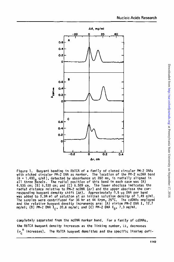
Equilibrium buoyant profi les of PM-2 DNA I and of the corresponding ncDNA
in RbTCA are presented in Finure 1, panel A. This separation of unt i t rated,
chemically unmodified DNA I from native ncDNA is a unique property of
aqueous neutral RbTCA density oradients. An analoqous buoyant separation,
but of much smaller mannitude, is obtained between nar t ia l ly t i t ra ted closed
DNA and untitrated nicked DNA in alkaline CsCl over the very narrow pH ranae
11.4-11.8 (19, 20). In neutral CsCl at 20°, a very small increase in buoy-
ant density (2 ma/ml) was reported with a closed PM-2 DNA sample of very
low Lk (20). The buoyant shi f t was ascribed to the possible existence of
denatured, untitrated renions in this hiohly stressed molecule. The chao-
troDic potency of RbTCA is suff ic ient to bring about the loss of most super-
coils in PM-2 DNA I at 25° by a combination of two effects: a salt-associated
reduction in duplex s tab i l i t y , and the concomitant release of the free energy
associated with supercoilinq.
B. Dependence of the Buoyant Density upon the Extent of Topolonical Under-
winding. To determine the buoyant density of a closed DNA as a function of
the extent of underwinding, a family of closed PM-2 DNAs was prepared as
described above. The buoyant banding of hiohly underwound DNA I £ (vc° =
0.098), with PM-2 ncDNA as a marker, is shown in Fiaure IB. DNA IE > banding
at 1.501 a/ml, is 20.6 mq/ml denser than nicked DNA and 7.8 ma/ml denser
than PM-2 DNA I . When a cdDNA with a relat ively small winding deficiency is
banded in RbTCA i t has a characteristically lower buoyant density, as shown
in Figure 1C. Here DNA Ig (vc° = 0.045), buoyant at 1.4881 g/ml, is not
1148
at Pennsylvania State University on Septem
ber 17, 2016http://nar.oxfordjournals.org/
Dow
nloaded from
Nucleic Acids Research
-20
AS, mg/ml
0 20 40
0.6
0.4
0.2
0
0.6 -
• 0.4o
0 -
0.6
0.4
0.2
0
1 1 'A
^ /
B
J
C
' • /
1
i | i | 1
-0.2 0 0.2
Ar, em0.4
Figure 1. Buoyant banding in RbTCA of a family of closed circular PM-2 DNAswith nicked circular PM-2 DNA as marker. The location of the PM-2 ncDNA band(6 = 1.480g g/ml), detected by absorbance at 280 nm, is radial ly aligned ina l l three panels. The radial position of this band in each case was (A)6.535 cm; (B) 6.530 cm; and (C) 6.589 cm. The lower abscissa indicates theradial distance relative to PM-2 ncDNA (Ar) and the upper abscissa the cor-responding buoyant density sh i f t (A8). Approximately 0.5 ug DNA per bandwas added to 0.3S ml of solution at an i n i t i a l solution density of 1.49 g/ml.The samples were centrifuged for 36 hr at 44 Krpm, 25°C. The cdDNAs employedand the relat ive buoyant density increments are: (A) v i r ion PM-2 DNA I , 12.7mg/ml; (B) PM-2 DNA IE > 20.6 mg/ml; and (C) PM-2 DNA Ig , 7.3 mg/ml.
completely separated from the ncDNA marker band. For a family of cdDNAs,
the RbTCA buoyant density increases as the l inking number, Lk, decreases
(v increases). The RbTCA buoyant densities and the specific l inking def i -
1149
at Pennsylvania State University on Septem
ber 17, 2016http://nar.oxfordjournals.org/
Dow
nloaded from
Nucleic Acids Research
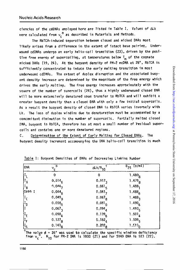
ciencies of the cdDNAs employed here are l is ted in Table I . Values of ALk
were calculated from v ° as described in Materials and Methods.
The RbTCA-induced separation between closed and nicked DNAs most
l i ke ly arises from a difference in the extent of intact base pair ing. Under-
wound cdDNAs undergo an early hel ix-coi l transit ion (23), driven by the posi-
t ive free energy of supercoiling, at temperatures below Tm of the cognate
nicked DNAs (19, 24). At the buoyant density of PM-2 ncDNA at 25°, RbTCA is
suf f ic ient ly concentrated to induce the early melting transit ion in most
underwound cdDNAs. The extent of duplex disruption and the associated buoy-
ant density increase are determined by the magnitude of the free energy which
drives the early melting. The free energy increases approximately with the
square of the number of sunercoils (24), thus a highly underwound closed DNA
w i l l be more extensively denatured upon transfer to RbTCA and w i l l exhibit a
qreater buoyant density than a closed DNA with only a few i n i t i a l supercoils.
As a result the buoyant density of closed DNA in RbTCA varies inversely with
Lk. The loss of duplex winding due to denaturation must be accompanied by a
concomitant diminution in the number of supercoils. Part ial ly melted closed
DNA, buoyant in RbTCA, therefore has at most a small number of residual super-
coils and contains one or more denatured regions.
C. Determination of the Extent of Early Melting for Closed DNAs. The
buoyant density increment accompanying the DNA hel ix-coi l transit ion is much
Table I : Buoyant Densities of DNAs of Decreasing Linking Number
DNA
I L
hrBSV40 I
hI
h
vc°
0
0.0146
0.0M7
0.044g
0.049Q
0.058Q
0.0673
0.0984
0.1278
0.1418
ALk/NlQ1-
0
0.0173
0.0615
0.0617
0.0678
0.081 Q
0.0944
0.1396
0.1824
0.202g
925 (p/ml)
1.4808
1.4798
1.4881
1.4886
1.4883
1.4906
1.493g
1.5014
1.5095
l.E130
The value i = 26° was used to calculate the specific windinq deficiencyfrom v c ° . N1Q for PM-2 DNA is 1000 (21) and for SV40 DNA is 523 (22).
1150
at Pennsylvania State University on Septem
ber 17, 2016http://nar.oxfordjournals.org/
Dow
nloaded from
Nucleic Acids Research
greater in RbTCA than in other buoyant salt systems (1). Buoyant banding in
RbTCA is therefore a sensitive monitor of the helix inteqrity in a molecule
containing both single-stranded and duplex reaions. For the present case of
cdDNA, the RbTCA buoyant density shift from marker native nicked DNA may be
quantitatively related to the extent of early melting.
The fraction of bases unpaired, f, for a cdDNA buoyant in RbTCA may be
calculated from the relative buoyant density shift, R, assuming that sequence
heterogeneity may be neglected (19).
0The parameter R is the ratio of the buoyant increment accompanying partial
denaturation to that accompanying complete denaturation, R = (6. - 8,. )/
(9jId - ejj )- Here 9j is the buoyant density of the closed DNA, e,j the
buoyant density of native ncDNA, and 9JJ. the buoyant density of denatured
ncDNA. In equation (1), the factor k is the ratio (M.. eJT /M,, .ejr.),
where M.. and M... are the solvated molecular weiqhts of the neutral nucleo-
tide subunits (including the counterion) in native and denatured DNA. The
value of M for the native case, includina the preferential hydration, isMIIn = M3 ^ + r n n ) • In peneral a subscript arabic numeral refers to a
parameter associated with component 1, water; comDonent 2, salt; or compo-
nent 3, neutral anhydrous macromolecule (25). For RbDNA, M, is 393.35 dal-
tons. The preferential hydration for native DNA, I",, (moles of water pre-
ferentially associated per mole of anhydrous neutral nucleotide), may be
calculated from the appropriate buoyant density, 9JJ , as (26)
M, (1-ev,)f ()
The partial specific volume of anhydrous RbDNA, v3, is 0.516 ml/g (27), and
the partial specific volume of water, v., is taken to be unity.
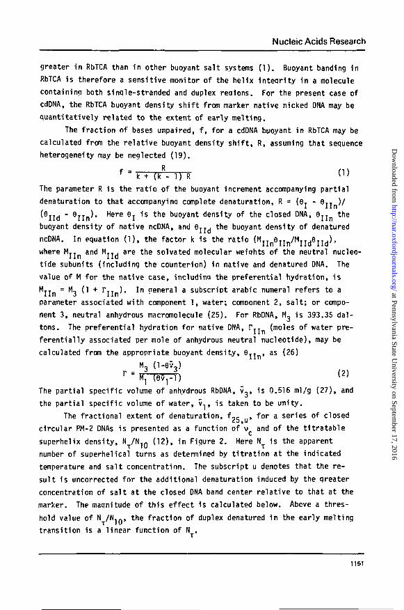
The fractional extent of denaturation, f-c . for a series of closed
circular PM-2 DNAs is presented as a function of v and of the titratable
superhelix density, N T / N 1 0 (12), in Figure 2. Here N^ is the apparent
number of superhelical turns as determined by titration at the indicated
temperature and salt concentration. The subscript u denotes that the re-
sult is uncorrected for the additional denaturation induced by the greater
concentration of salt at the closed DNA band center relative to that at the
marker. The maqnitude of this effect is calculated below. Above a thres-
hold value of N /Nlf., the fraction of duplex denatured in the early melting
transition is a linear function of N .
1151
at Pennsylvania State University on Septem
ber 17, 2016http://nar.oxfordjournals.org/
Dow
nloaded from
Nucleic Acids Research
NT
/ N . o0 0.05
0.2
0.1
n
-
0.10i
/
i i
0.15
/
/
i
0.201
P
/-
-
i
0.05 0.10 0.15
Figure 2. The fractional extent of denaturation at 25*C, fy, u. of cdDNAsplotted as a function of the critical dye binding ratio, v , (Tower abscissa),or the associated change in the titratable superhelix density, N /N-jn» whereN is the titratable number of superhelical turns under the given experimentalc5nditions (upper abscissa). The value of f~5 is calculated from the RbTCAbuoyant densities presented in Table I using Eq.'T and is uncorrected for thedenaturing effects of the salt gradient (see text). All DNAs banded were PM-2cdDNAs except for the darkened circle, which represents SV40 ONA I.
These experimental conditions, 3.3 M RbTCA and 25°C, are insufficiently
denaturina to remove all supercoils, as demonstrated by the absence of a
buoyant density shift for PM-2 DNA I.. In order to follow the early melting
transition of PM-2 DNA I to completion (defined bv removal of all supercoils),
more potent denaturing conditions are required. The salt concentration can-
not be varied due to the requirement of buoyancy. We therefore vary the
experimental temperature.
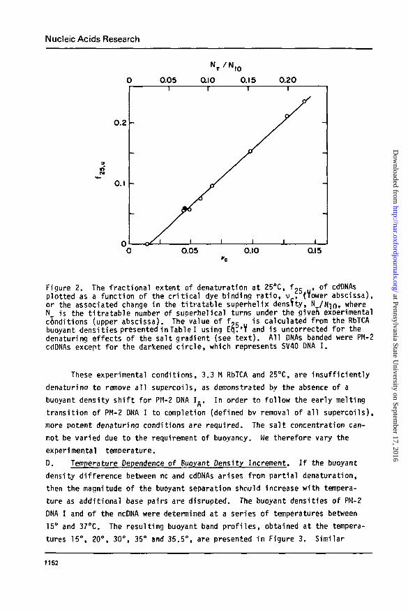
D. Temperature Dependence of Buoyant Density Increment. If the buoyant
density difference between nc and cdDNAs arises from partial denaturation,
then the magnitude of the buoyant separation should increase with tempera-
ture as additional base pairs are disrupted. The buoyant densities of PM-2
DNA I and of the ncDNA were determined at a series of temperatures between
15° and 37°C. The resulting buoyant band profiles, obtained at the tempera-
tures 15°, 20°, 30°, 35° and 35.5°, are presented in Figure 3. Similar
1152
at Pennsylvania State University on Septem
ber 17, 2016http://nar.oxfordjournals.org/
Dow
nloaded from
Nucleic Acids Research
0.8
0.4
0.8
0.4
0.4
0.4
0 •
15*
30"
• 35.5'
5.8 6.2 6.6, cm
7.0
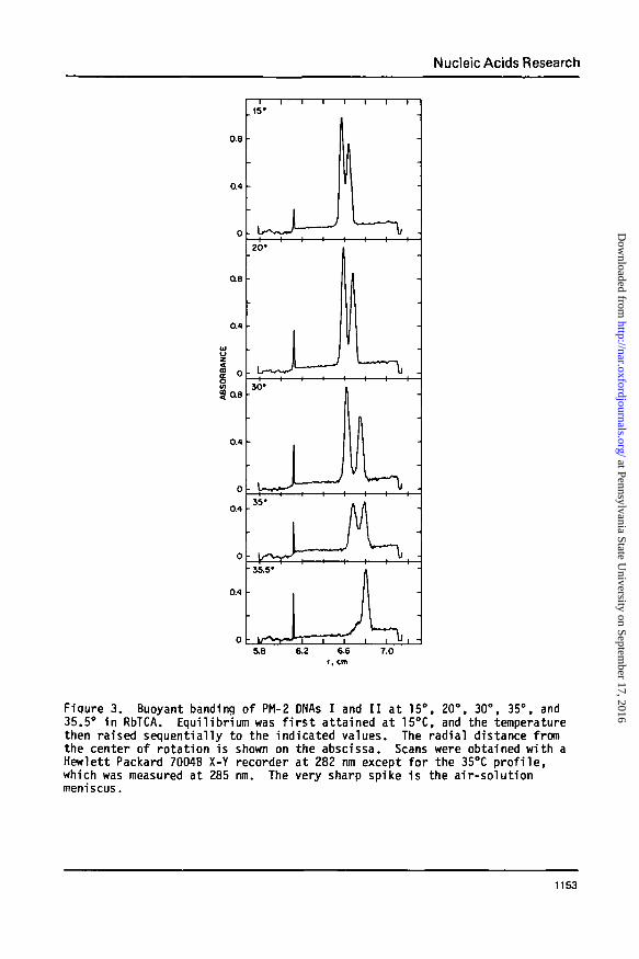
Fiqure 3. Buoyant banding of PM-2 DNAs I and I I at 15°, 20°, 30°, 35°, and35.5° In RbTCA. Equilibrium was f i r s t attained at 15°C, and the temperaturethen raised sequentially to the indicated values. The radial distance fromthe center of rotation is shown on the abscissa. Scans were obtained with aHewlett Packard 7004B X-Y recorder at 282 nm except for the 35°C p ro f i l e ,which was measured at 285 nm. The very sharp spike is the air-solut ionmeniscus.
1153
at Pennsylvania State University on Septem
ber 17, 2016http://nar.oxfordjournals.org/
Dow
nloaded from
Nucleic Acids Research
experiments were performed at 17.5° and 25°C (data not shown), with com-
parable results. Both DNA bands move to a region of greater solution den-
sity following a temperature increase. Below 35°C, the increment in buoy-
ant density accompanying a temperature increase is less for PM-2 ncDNA, the
lighter of the two species, than for PM-2 DNA I. Since d9jj/dT < d8j/dT,
the separation between native nicked and closed DNAs becomes greater with
increasing temperature. The maximum resolution between bands, 1.24 mm,
occurs at 30°C. At 35°C, the nicked SDecies undergoes pronounced band
broadening and a relatively greater density gain than at lower temperatures.
Both these changes are characteristic of the initiation of denaturation.
When the temperature is raised an additional 0.5°, PM-2 ncDNA completely
denatures and pellets, while PM-2 DNA I remains buoyant. The midpoint of
the melting transition for PM-2 ncDNA is 35.0°C in 3.29 M RbTCA at atmos-
pheric pressure, as determined by monitoring absorbance changes (28). In
contrast to the hypersharp melting of nicked DNA, the early region of the
helix-coil transition for the closed molecule is noncooperative.
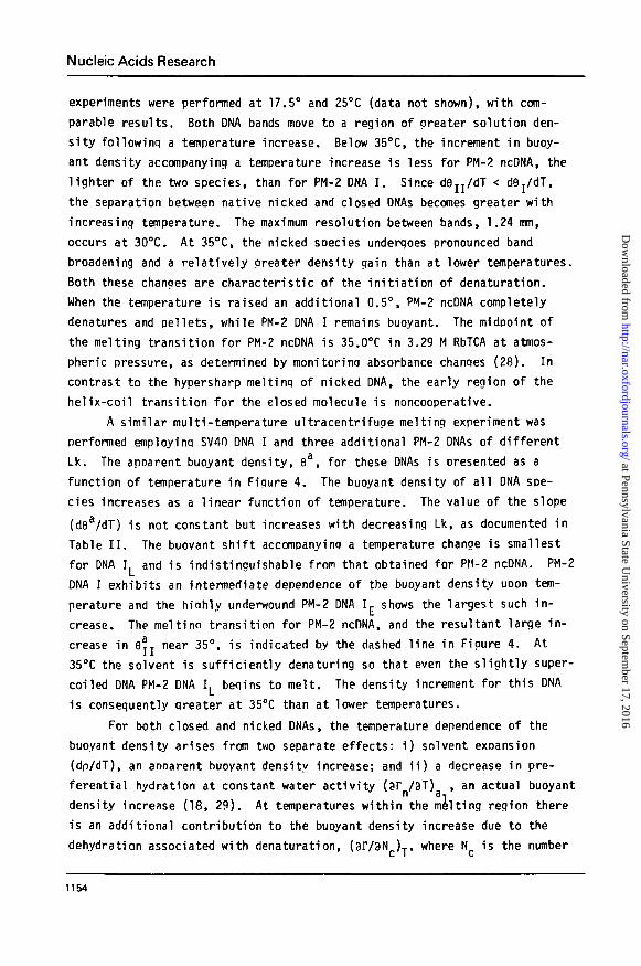
A similar multi-temperature ultracentrifuge melting experiment was
performed employing SV40 DNA I and three additional PM-2 DNAs of different
Lk. The apparent buoyant density, 9a, for these DNAs is presented as a
function of temperature in Figure 4. The buoyant density of all DNA spe-
cies increases as a linear function of temperature. The value of the slope
(dea/dT) is not constant but increases with decreasing Lk, as documented in
Table II. The buoyant shift accompanying a temperature change is smallest
for DNA I. and is indistinguishable from that obtained for PM-2 ncDNA. PM-2
DNA I exhibits an intermediate dependence of the buoyant density uoon tem-
perature and the hiahly underwound PM-2 DNA K shows the largest such in-
crease. The meltina transition for PM-2 ncDNA, and the resultant large in-
crease in 6jj near 35°, is indicated by the dashed line in Figure 4. At
35°C the solvent is sufficiently denaturing so that even the slightly super-
coiled DNA PM-2 DNA I. begins to melt. The density increment for this DNA
is consequently qreater at 35CC than at lower temperatures.
For both closed and nicked DNAs, the temperature dependence of the
buoyant density arises from two separate effects: i) solvent exoansion
(dp/dT), an annarent buoyant density increase; and ii) a decrease in pre-
ferential hydration at constant water activity (ar /3T) , an actual buoyantn a
density increase (18, 29). At temperatures within the melting region there
is an additional contribution to the buoyant density increase due to the
dehydration associated with denaturation, (3I73N ),, where N is the number
1154
at Pennsylvania State University on Septem
ber 17, 2016http://nar.oxfordjournals.org/
Dow
nloaded from
Nucleic Acids Research
1.51 -
i.so -
1.49 -
1.48 -
19
Figure 4. The apparent buoyant density (6a) for a series ofDNAsin RbTCAplotted as a function of temperature. In order of increasing buoyant denssitythe DNAs are PM-2 ONA I. (A), PM-2 DNA I I ( • ) , PM-2 DNA I . (O), SV40 DNA I(•) , PM-2 I (o) , and PM-2 DNA I £ ( A ) . The apparent buoyant densities wer
l l d d i A 2 ° C h b diwere£ pp
calculated as described in the text . At 25°C the apparent buoyant density isthe same as the practical buoyant density, 9. The dashed l ine indicates thecooperative denaturation of ncDNA.
of nucleotide bases transferred from helix to c o i l . The magnitude of this
dehydration is extraordinari ly large in RbTCA (1). At any temperature belowTm I I ' t h e b u ° y a n t density increment between a marker native ncDNA and a
cdDNA therefore arises only from the early melting of the la t te r . The
assumption is made that the loss of preferential hydration due to a change
1155
at Pennsylvania State University on Septem
ber 17, 2016http://nar.oxfordjournals.org/
Dow
nloaded from
Nucleic Acids Research
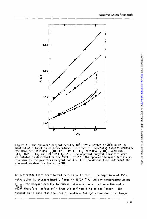
Table I I : The Temperature Dependence of RbTCA Buoyant Density for CircularPM-2 DNAs
DNA
II
ITE
Lk(Turns/DNA)*
(1000)
1000
982
906
860
dea/dT(g/ml°C)+
(x 10")
3.23 ± 0.11
3.07 ± 0.14
3.50 ± 0.19
7.46 + 0.25
ft.55 ± 0.15
*Calculated from the data in Table 1.The apDarent buoyant density uncorrected for the minor effect of solventexpansion. The slopes were determined for temperatures below 35°C.
in temperature is the same for native and for denatured DNA. Tunis and
Hearst (29) have shown (3IV3T) . is slightly less than (3r /3T) in KTFA.Q SI ' n ai
However in the present case the dominant contribution to the density in-
crease arises from, denaturation, and (3I73N ) » (ar/?T)M . In addition,
since the total fraction of base pairs denatured in the early melting of
closed DNA does not exceed 23%, the contribution of a small difference be-
tween (8r /3T) and (3r JdT), to the total hydration chanqe may ben a-i Q Om
neglected.E. Temperature Dependence of the Early Melting of Closed DNA in Buoyant
RbTCA. The extent of denaturation of a cdDNA buoyant in RbTCA depends upon
the temperature, salt concentration, and initial winding deficiency of the
cdDNA. It was shown above (see Fig. 2) that 1n Buoyant RbTCA f-5 is a
linear function of N . The quantity f,c ,, denotes the fraction of base
pairs disrupted at 25° and at the salt concentration at the center of the
buoyant band of the cdDNA. This salt concentration is, in general, greater
than that at the location of the marker PM-2 ncDNA. The salt concentration
aradient is of sufficient maanitude so that closed DNAs of different initial
suDerhelix density undemo an extent, of early melting which depends upon
their distance from the marker nicked DNA. This effect arises because, in
addition to a density gradient, a substantial denaturation gradient is
associated with the increase in chaotropic salt concentration. A closed DNA,
buoyant at a greater solution density than the nicked species, is melted to
a greater extent than would have occurred in the presence of the denatura-
tion gradient.
1156
at Pennsylvania State University on Septem
ber 17, 2016http://nar.oxfordjournals.org/
Dow
nloaded from
Nucleic Acids Research
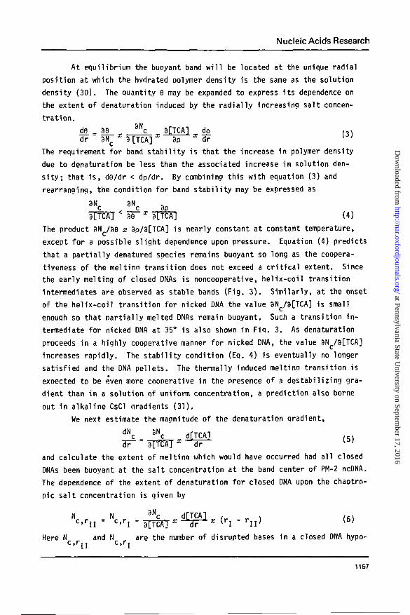
At equilibrium the buoyant band w i l l be located at the unique radial
position at which the hvdrated polymer density is the same as the solution
density (30). The quantity 9 may be expanded to express i ts dependence on
the extent of denaturation induced by the radially increasing salt concen-
trat ion.
d9_ _ 89_ 3Nc 3[TCA] d£ ,^)
dr 8Nc x 8 [TCA] x 8p x dr
The requirement for band stabi l i ty is that the increase in polymer density
due to denaturation be less than the associated increase in solution den-
s i t y ; that i s , d9/dr < dp/dr. By combining this with equation (3) and
rearranging, the condition for band stabi l i ty may be expressed as
•*3TTCA] (4)
The product 8N /89 x 8p/8[TCA] is nearly constant at constant temperature,
except for a possible slight dependence upon pressure. Equation (4) predicts
that a part ia l ly denatured species remains buoyant so long as the coopera-
tiveness of the meltinn transition does not exceed a cr i t i ca l extent. Since
the early melting of closed DNAs is noncooperative, helix-coil transition
intermediates are observed as stable bands (Fig. 3). Similarly, at the onset
of the helix-coil transition for nicked DNA the value 8Nc/8[TCA] is small
enough so that part ial ly melted DNAs remain buoyant. Such a transition in-
termediate for nicked DNA at 35° is also shown in Fiq. 3. As denaturation
proceeds in a highly cooperative manner for nicked DNA, the value 8N /8[TCA]
increases rapidly. The stabi l i ty condition (Eq. 4) is eventually no longer
satisfied and the DNA pellets. The thermally induced melting transition is
expected to be even more cooperative in the presence of a destabilizing gra-
dient than in a solution of uniform concentration, a prediction also borne
out in alkaline CsCl oradients (31).
We next estimate the magnitude of the denaturation gradient,
dN 3N.
and ca l cu la te the extent of mel t inq which would have occurred had a l l closed
DNAs been buoyant a t the s a l t concent ra t ion a t the band center of PM-2 ncDNA.
The dependence of the extent of denatura t ion f o r closed DNA upon the chaotro-
pic salt concentration is given by
M 3Nc d[TCA]c= " c ' r i " sTTEAT X " " 3 F " X (ri - r n
Here N and M are the number of d isrupted bases i n a closed DNA hypo-c,rn c,rT
1157
at Pennsylvania State University on Septem
ber 17, 2016http://nar.oxfordjournals.org/
Dow
nloaded from
Nucleic Acids Research
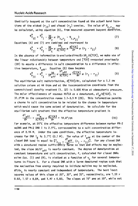
t h e t i c a l l y buoyant a t the s a l t concentrat ion found at the actual band loca-
t ions of the nicked ( r T I ) and closed ( r . ) sDecies. The value of N may11 l * II
be calculated, using equation (6), from measured apparent buoyant densities.
V ^ - I f * ("c.r, - Nc,rn> <"Equations (6) and (7) are combined and rearranged to
In the absence of information to evaluate directly 8N /3[TCA], we make use of
the linear relationship between temperature and [TCA] presented previously
(28) to equate a difference in salt concentration to a difference in effec-
tive temperature, T _f. Equation (8) then becomes
The equilibrium sal t redist r ibut ion, d[TCA]/dr, calculated for a 1.1 cm
solution column at 44 Krpm and at the isoconcentration coordinate from the
compositional density oradient ( 1 , 32) is 0.605 M/cm at atmospheric pressure.
The molar effectiveness of aqueous RbTCA as a denaturant, dT /dfTCA], is
31.0°/M in the concentration ranqe 3.1-3.9 M (28). This information permits
a change in salt concentration to be related to the change in temperature
which would cause the same extent of denaturation. We calculate for the
equilibrium sal t gradient that the effective temperature gradient is
For example, at 25°C the effective temperature difference between marker PM-2
ncDNA and PM-2 DMA I is 2.0°C, corresponding to a salt concentration d i f fe r -
ence of O.Ofi M. Under the same conditions, the effective temperature i n -
crease for DNA I £ is 3.1°C (0.1 M). The value of T f f at the center of the
closed DNA band is equal to [T + d Teff ( r . - r , T ) L F ° r any closed DNA
with a denatured reqion suf f ic ient ly Targe so that end effects may be neglec-
ted, the slope 38/3T f f is nearly constant. The degree of denaturation at
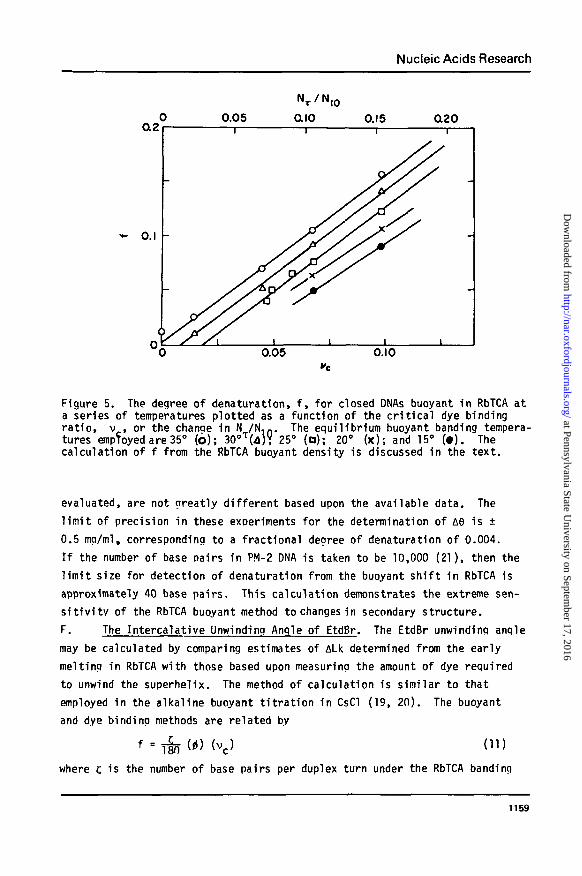
constant temperature and sal t concentration, f , calculated for closed DNAs
usino Eqs. (1) and (9), is olotted as a function of v for several tempera-
tures in Figure 5. For a closed DNA with a laroe denatured region such that
the nucleation free energy required to i n i t i a te meltina may be neglected,
df/dv is nearly constant and independent of temperature. The best least
squares values of this slope at 25°, 30°, and 35°, respectively, are 1.51 +
0.02, 1.57 ± 0.04, and 1.47 ± 0.03. The slopes at 15° and at 20°, while not
1158
at Pennsylvania State University on Septem
ber 17, 2016http://nar.oxfordjournals.org/
Dow
nloaded from
Nucleic Acids Research
0.2
0.05 0.10
Figure 5. The degree of denaturation, f, for closed DNAs buoyant in RbTCA ata series of temperatures plotted as a function of the critical dye bindingratio, v , or the change in N /N.n. The equilibrium buoyant banding tempera-tures empToyed are 35° (b); 30oT(d)? 25° (o); 20° (x); and 15° (•). Thecalculation of f from the RbTCA buoyant density is discussed in the text.
evaluated, are not greatly different based upon the available data. The
limit of precision in these experiments for the determination of A9 is +
0.5 mg/ml, corresponding to a fractional degree of denaturation of 0.004.
If the number of base Dairs in PM-2 DNA is taken to be 10,000 (21), then the
limit size for detection of denaturation from the buoyant shift in RbTCA is
approximately 40 base pairs. This calculation demonstrates the extreme sen-
sitivity of the RbTCA buoyant method to changes in secondary structure.
F. The Intercalative Unwinding Angle of EtdBr. The EtdBr unwinding angle
may be calculated by comparing estimates of ALk determined from the early
melting in RbTCA with those based upon measuring the amount of dye required
to unwind the superhelix. The method of calculation is similar to that
employed in the alkaline buoyant titration in CsCl (19, 20). The buoyant
and dye binding methods are related by
f = T§n (*) <vc> (11)where c is the number of base pairs per duplex turn under the RbTCA banding
1159
at Pennsylvania State University on Septem
ber 17, 2016http://nar.oxfordjournals.org/
Dow
nloaded from
Nucleic Acids Research
conditions. This assumes that the residual superhelix density for closed
DNAs in buoyant RbTCA is the same for a l l DNAs and is given by the abscissa
intercept where f = 0. Employing a straight-forward thermodynamic analysis,
Wang (20) has shown that this assumption is val id near the endpoint of the
early melting t ransi t ion. I t should be emphasized that the unwinding angle
refers to the conditions of the v - based assay, buoyant CsCl/EtdBr at
20°. Using the value of the slope (df/dv ) calculated from the data of
Fiqure 5 and Eq. (11), and taking c = 10.4 (10), we calculate that t =
27.2° ± 0.5°.
DISCUSSIONMelting of Closed DNA. A l inkina number deficiency in cdDNA dramati-
cal ly al ters many of the fundamental physical and chemical properties of this
molecule compared to both relaxed cdDNA and to ncDNA (12, 20, 23, 24, 33-35).
Al l naturally occurring superhelical DNAs behave as i f the duplex winding
were incomplete at the time of covalent ring closure. The restoration of
the optimal duDlex windina generates supercoiling. The associated positive
free energy may be released via a variety of pathways. For example, the
relat ive bindinq a f f i n i t y towards intercalating dyes is enhanced or inhibited
for a closed DNA dependinq on whether supercoils are removed or formed during
the reaction. In addit ion, closed underwound DNA is more susceptible to
cleavage by single-strand specific nucleases, has a greater i n i t i a l a f f in i t y
for single-strand specific reagents and binding proteins, and in i t ia tes
thermal or alkaline denaturation at a lower temperature or pH.
The difference in s tab i l i t y between closed and nicked duplex DNAs is
clearly demonstrated by comoaring their thermal or chaotrooic salt-induced
melting pro f i les . As demonstrated previously (28) the hel ix-coi l transit ion
for nicked DNAs in 4 M RbTCA is cooperative and occurs over a narrow tempera-
ture range of 5.6°C. The same transit ion for closed DNA is substantially
broadened, to greater than 45°C, and beains at a lower temperature. The spec-
troscopic technique employed in the earl ier work (28), while covering the
entire meltino transi t ion for both closed and open DNAs, reveals l i t t l e
detail of the early melting recfion. The method of band velocity sedimenta-
t ion permits a variat ion of salt concentration at constant temperature. I t
is clear from these results (28) that closed DNAs undergo an early structural
transit ion resulting in reduced supercoiling. The unique properties of buo-
yant RbTCA permit a more quantitative analysis of the early melting of a
closed DNA. F i r s t , the chaotropic potency of the solvent reduces the melting
1160
at Pennsylvania State University on Septem
ber 17, 2016http://nar.oxfordjournals.org/
Dow
nloaded from
Nucleic Acids Research
transit ion region to an experimentally accessible range at neutral pH.
Second, the large buoyant density increment upon denaturation allows de-
tection and precise determination of the extent of part ia l denaturation.
The melting transit ion for buoyant nicked DNA in RbTCA in the u l t ra-
centrifuge is hypersharp. As shown in Figure 3, the temperature span of the
melt is about 0.5°C compared to a value of 5.6°C measured spectrophotometri-
cal ly (28). The enhanced cooperativeness of the duplex disruption results
from the denaturation gradient present in the ultracentr i fuge, as pointed out
above.
Comparison with Other Estimates of i . The value of the EtdBr unwind-
ing angle was long taken to be 12° based on model building (36). Experimen-
tal support for this value came from the determination (19) that the number
of supercoils in polyoma DNA measured by buoyant alkaline t i t r a t i on (inde-
pendent of i>) is approximately the same as that of SV4O DNA measured by
t i t r a t i on in buoyant EtdBr/CsCl (37) taking (S to be 12°. Since these two
viruses are similar in size and in many other characterist ics, the experimen-
tal agreement was taken to be evidence for the 12° unwinding angle. This
experimental approach was subject to several potential errors, which the
authors carefully pointed out, and which in fact led to an underestimate of
the actual extent of supercoiling. More recently the buoyant t i t r a t i on in
alkaline CsCl was repeated (20) with the result i> = 26° + 2.6°. This larger
value is also supported by the EtdBr t i t r a t i on of closed DNAs of known under-
winding produced by carbodiimide binding (38), i> = 24° - 36°, and by electron
microscopic length measurements of PM-2 DNA I hybridized with restr ic t ion
fragments (39), i> = 28.5°. Keller (40) and Shure and Vinograd (41) have
independently counted the number of bands on agarose gels for intermediates
in the generation of relaxed, closed circles by topoisomerase to determine
the number of supercoils in SV40 DNA. Comparison of this value to that
obtained by EtdBr/CsCl buoyant banding results in estimates of 28° and 23°,
respectively. Recently, X-ray crystallograDhic studies of iodinated dinu-
cleoside monophosphate: dye complexes with ethidium (42, 43) yields a value
of 10° for the angular orientation between the dinucleoside base pairs in a
fragment of a DNA hel ix. Assuming that the intercalated dye: dinucleoside
complex is on rea l i s t i c model for an intercalated drug: DNA complex, this
leads to a prediction of about 26° for the drug-induced unwinding angle in
DNA (44). A similar estimate, 23°, was obtained by X-ray d i f f ract ion of a
2:1 complex of CpG and acridine orange (45). The magnitude of the intercala-
t ive unwinding angle determined here is 27.2° ± 0.5° and is consistent with
1161
at Pennsylvania State University on Septem
ber 17, 2016http://nar.oxfordjournals.org/
Dow
nloaded from
Nucleic Acids Research
these estimates. I t should be emphasized that these values are averages, and
no Information is yet available concerning whether or not j) varies with dye
occupancy or with base sequence.
Onset of the Salt-Induced Early Melting. The pitch of the DNA duplex
may be increased by two mechanisms: a general unwinding in which base pairing
remains intact but the number of base pairs per duplex turn, c, decreases; or
a local denaturation in which single-stranded, unpaired regions are formed.
For a closed DNA buoyant in RbTCA, the magnitudes of both the reduction in
supercoiling and the buoyant density increase relat ive to ncDNA suggest that
denaturation is the dominant effect. However, since the value of c in buoy-
ant concentrations of RbTCA enters into the calculation of t>, i t is necessary
to estimate the extent to which t, varies as conditions are changed from those
of the standard state. The transfer of PM-2 DNA I from standard conditions
(0.2 M NaCl, 37°C) to the experimental environment (3.4 M RbTCA, 35°C) may be
considered to occur through a series of intermediate, elementary steps. The
transfer of DNA I from 0.2 M NaCl to 0.2 M RbCl at constant temperature re-
moves 5.4 supercoils (11). The subsequent transfer from 0.2 M RbCl to 0.2 M
RbTCA removes one additional supercoil. This effect is not expected to arise
from local denaturation, since the transfer occurs well below the melting
region of ncDNA. The total duplex winding reduction of approximately 6.1
turns is probably due to a s l iqht overwinding of the duplex in 0.2 M RbTCA
relat ive to 0.2 M NaCl. In the next step the sal t concentration is increased
from 0.2 M to 3.4 M. For both NaCl and RbCl, non-denaturing solvents, the
number of supercoils is re la t ive ly Insensitive to changes in ionic strength
in the ranae beyond 0.26 M (11). An equivalent increase in the chaotropic
TCA" concentration i s , by contrast, expected to induce denaturation. The
f inal transfer step, involving a sh i f t in temperature from 37° to 35°C, in t ro-
duces approximately 1 supercoil (11). We then estimate that in the absence of
denaturation (TCA"effect) the supercoiling of closed PM-2 DNA is reduced by
7 turns relat ive to i t s extent under standard conditions. This reduction in
supercoiling would correspond to a change in duplex winding of less than 1%.
Since PM-2 DNA contains approximately 94 supercoils, local denaturation would
therefore appear to account for by far the greater part of the early melting
observed in buoyant RbTCA.
Implications for DNA Structure in Solution. I t has recently been sug-
gested (46, 47 and see 48 for other references) that the duplex structure of
DNA is not a double helix of the Watson-Crick type but that the two strands
are in an essentially side-by-side configuration. The evidence and arguments
1162
at Pennsylvania State University on Septem
ber 17, 2016http://nar.oxfordjournals.org/
Dow
nloaded from
Nucleic Acids Research
against this proposal are strong (10, 48) and, in our opinion, convincing.Fundamentally, all such alternative models require that Lk be either
small or zero. In the case of PM-2 DNA, experiments based ultimately uponband counting in gel electrophoresis demonstrate absolutely that ALk forthis molecule is close to 94.4 turns (12). Our present results have allowedus to calculate independetly the extent of duplex denaturation required toremove the associated superhelical turns. The value of f at which this occursfor PM-2 DNA I is readily calculated from Figure 5 by drawing a line throughthe origin with the same slope as the bracketing lines. This line representsthe value of f, denoted fQ, associated with ALk for each DNA. For PM-2 DNAI, f = 0.102. This value must, due to the nature of the buoyant experiments(see above), represent the fractional duplex winding associated with ALk. Ingeneral, then, ALk/Lk = f . Combination of the above measurements results inthe estimate Lk = 925. The value of Lk predicted by the original Watson-Crick model is 1000, assuming 10.0 base pairs per turn. In solution, however,the pitch appears to be about 10.4 base pairs per turn (10), leading to theexpectation that Lk = 960 for the double helix of PM-2 DNA in solution. Itis therefore clear that the two strands of this DNA are in fact linked nearlyexactly to the extent predicted by the classical double helical model. Allproposed alternative structures which predict that Lk is significantly dif-ferent from ten base pairs per turn are therefore ruled out.
ACKNOWLEDGEMENTSThis research was supported by grant GM-21176 from the USPHS. RLB
was the recipient of a Traineeship from the United States Public HealthService under Grant CA-09176. We wish to thank Mrs. Mary Fils-Aime for herinvaluable assistance in the preparation of the manuscript.
REFERENCESt Present address: Department of Biochemistry and Biophysics, University of
California Medical Center, San Francisco, California 94143.* Abbreviations used are: TCA, trichloroacetate; TFA, trifluoroacetate;
Na,EDTA, trisodium salt of ethylenediaminetetraacetic acid; Tris, tris(hydroxymethyl)aminomethane; EtdBr, ethidium bromide; DNA I, nativecovalently closed circular duplex DNA; cdDNA, closed duplex DNA; ncDNAor DNA II, nicked circular DNA.
1. Burke, R.L. and Bauer, W.R. (1977) Nucl. Acids Res. 4., 1891-1909.2. White, J.H. (1969) Am. J . Math. 41, 693-728.3. Ful ler, F.B. (1971) Proc. Nat l . Scad. Sci. USA 68, 815-819.4. Crick, F.H.C. (1976) Proc. Nat l . Acad. Sci. USA~73, 2639-2643.
1163
at Pennsylvania State University on Septem
ber 17, 2016http://nar.oxfordjournals.org/
Dow
nloaded from
Nucleic Acids Research
5. Bauer, W.R. (1972) Biochemistry H , 2915-2920.6. Bauer, W.R. (1972) J . Moi. Bio i . 67, 183-198.7. Vosberg, H.-P., Grossman, L. and VTnograd, J . (1975) Eur. J . Biochem. £5,
79-93.8. Sharp, P., Sudqin, B. and Sambrook, J . (1973) Biochemistry U, 3055-3063.9. Wilkins, M.H.F. (1961) J . Chem. Phys. 58, 891-898.
10. Wang, J.C. (1979) Proc. Natl. Acad. Sci. USA 76, 200-203.11. Anderson, P. and Bauer, W. (1978) Biochemistry V7, 594-601.12. Bauer, W.R. (1978) Ann. Rev. Biophys. Bioeng. ]_, 287-313.13. Keller, W. (1975) Proc. Natl. Acad. Sci. USA 72, 4876-4880.14. Radloff, R., Bauer, W. and Vinograd, J . (1967TProc. Natl . Acad. Sci. USA
57, 1514-1521.15. Burke, R.L. and Bauer, W. (1977) J . B io l . Chem. 252, 291-292.16. Bauer, W. and Vinograd, J . (1967) Ann. N.Y. Acad. Sci. 164, 192-225.17. Vinograd, J . and Hearst, J.E. (1962) Prog. Chem. Org. Nat. Prod. 20, 373-
420/18. Vinograd, J . and Greenwaid, R. (1965) Biopoiymers 2» 109-114.19. Vinograd, J . , Lebowitz, J . and Watson, R. (1968) J . Mol. Bio l . 33_j_ 173-
197.'20. Wang, J.C. (1974) J. Mol. Biol. 89_, 783-801.21. Stuber, D. and Bujard, H. (1977) Mol. Gen. Genet. ]5±, 299-303.22. Reddy, V.B. Timmappaya, B., Dhar, R., Subramanian, K.N., Zain, B.S., Pan,
J., Gosh, P.K., Ceima, M.L. and Weissman, S.M. (1978) Science 200, 494-502.
23. Vinograd, J. and Lebowitz, J. (1966) J. Gen. Physiol. 49, 103-125.24. Bauer, W. and Vinoarad, J. (1970) J. Mol. Biol. 47, 419-435.25. Hearst, J.E. and Vinograd, J. (1961) Proc. Natl. Sci. USA 47_, 999-1004.26. Hearst, J.E. and Vinograd, J. (1961) Proc. Natl. Acad. Sci. USA 47_, 825-
830.27. Hearst, J.E. (1962) J . Mol. Biol 4, 415-417.28. Burke, R.L. and Bauer, W.R. (1978T Nuc!. Acidr. P.es. 5., 4819-4836.29. Tunis, M.B. and Hearst, J.E. (1968) Biopoiymers £ , 1325-1344.30. Hearst, J .E. , I f f t , J.B. and Vinograd, J.E. (1961) Proc. Nat l . Acad. Sci.
USA 47^ 1015-1025.31. Weishahn, G., Cech, T.R. and Hearst, J.E. (1976) Biopolymers 15, 1591-1613.32. I f f t , J .B. , Voet, D.H. and Vinoarad, J . (1961) J . Phys. Chem. 6^, 1139-
1145.33. Hsieh, T.-S and Wanq, J.C. (1975) Biochemistry U_, 527-535.34. Pulleybiank, D.E., Shure, M., Tanq, D., Vinograd, J . and Vosberg, H.-P.,
(1975) Proc. Nat l . Acad. Sci. USA 72, 42R0-4284.35. Depew, R.E. and Wang, J.C. (1975) Proc. Nat l . Acad. Sci. USA 72_, 4275-
4279.36. Ful ler, W. and Waring, M.J. (1964) Ber. Bunsengesellschaft 68, 805-808.37. Bauer, W. and Vinoqrad, J. (1968) J . Mol. B io l . 33, 141-172.38. Puileyblank, D.E. and Morgan, A.R. (1975) J . Mol. B ic l . 91_, 1-13.39. L iu , L.F. and Wang, J.C. "(1975) Biochim. Biophys. Acta 395_, 405-416.40. Keller, W. (1975) Proc. Natl. Acad. Sci. USA ]2_, 4876-4880.41. Shure, M. and Vinoqrad, J. (1976) Cell 8, 215-226.42. Tsai, C . -C , Jain, S.C. and Sobell, H.M. (1977) J . Mol. Bio l . H i , 301-
315.43. Ja in, S .C , Tsai, C.-C. and Sobell, H.M. (1977) J . Bio l . V\±, 317-331.44. Sobell, H.M. Tsai, C.-C. and Gi lbert , S.G. (1977) J . Mol. Bio l . 114,
333-365.45. Wang, H.-J. , Quiqley, G.J. and Rich, A. (1978) Nuci. Acids Res. 6, 3879-
3890.46. Rodley, G.A., Scobie, R.S., Bates, R.H.T. and Lewitt, R.M. (1976) Proc.
Nat l . Acad. Sci. USA 73. 2959-2963.
1164
at Pennsylvania State University on Septem
ber 17, 2016http://nar.oxfordjournals.org/
Dow
nloaded from
Nucleic Acids Research
47. Sasisekharan, V . , Pattabiraman, N. and Gupta, G. (1978) Proc. N a t l . Acad.S c i . USA 75, 4092-4096.
48. C r i ck , F.H7C, Wang, J.C. and Bauer, W.R. (1979) J . Mol. B i o l . J29, 449-461.
49. Gray, H.B., J r . , Uphol t , W.B. and Vinograd, J . (1971) J . Mol . B i o l . 62, 1 -19.
1165
at Pennsylvania State University on Septem
ber 17, 2016http://nar.oxfordjournals.org/
Dow
nloaded from
Nucleic Acids Research
1166
at Pennsylvania State University on Septem
ber 17, 2016http://nar.oxfordjournals.org/
Dow
nloaded from