the appendicular skeleton of majungasaurus crenatissimus (theropoda: abelisauridae) from the late...
TRANSCRIPT
THE APPENDICULAR SKELETON OF MAJUNGASAURUS CRENATISSIMUS (THEROPODA:ABELISAURIDAE) FROM THE LATE CRETACEOUS OF MADAGASCAR
MATTHEW T. CARRANODepartment of Paleobiology, Smithsonian Institution, P.O. Box 37012, MRC 121, Washington, DC 20013-7012;
ABSTRACT—The appendicular skeleton of the abelisaurid theropod Majungasaurus crenatissimus (Depéret, 1896)Lavocat, 1955 is described for the first time. The available materials include an incomplete pectoral girdle and forelimb,along with the ilium and a nearly complete hind limb. These materials display a number of ceratosaur, abelisauroid, andabelisaurid synapomorphies, supporting the phylogenetic placement of Majungasaurus based previously on cranialanatomy. As in Ceratosaurus and Carnotaurus, the scapular blade is relatively wide and has a pronounced dorsal lip overthe glenoid. The humerus is short and bears a globular head, but is more slender than in Carnotaurus. The ilium has apreacetabular hook, a strong supraacetabular crest, a notched posterior margin, and peg-and-socket articulations withboth the pubis and ischium. Hind limb elements are proportionally stocky, as in some other abelisaurids. The femur lacksa trochanteric shelf, the tibia has a greatly enlarged cnemial crest, and the fibula bears a deep, posteriorly facing medialfossa. The abelisaurid astragalocalcaneum is described here in detail for the first time, and is more similar to that oftetanurans than to those of coelophysoids. Taken together, these materials illustrate that the appendicular skeleton ofabelisaurids was specialized over the typical condition in basal theropods, particularly through the development ofenlarged muscle attachment processes.
MALAGASY ABSTRACT (FAMINTINANA)—Sambany izao no namelabelabelarin’i Lavocat tamin’ny 1955 nymomban’ny taolan-drambon’ny abelisaurid theropod Majungasaurus crenatissimus (Depéret, 1896). Ireo karazan-taolananisy tamin’ireo dia maro ny ceratosaur sy abelisauroid ary abelisaurid synapomorphies, izay nanamarina ny toeranaphylogenetic –n’ny Majungasaurus izay tamin’ny bikan’ny karan-doha ny nametrahana azy. Toy ny an’ny Ceratosaurussy Carnotaurus dia azo ambara fa mivelatra ny taolan-tsoroka ary iny faritra ambonin’ny glenoid iny dia misy molonyaoriana mivoitra mazava tsara. Fohy ny taolan-tsandry ary borobory ny lohany, saingy marotsadrotsaka raha ampitahainany an’i Carnotaurus. Ny ilium dia ahitana faingoka alohan’ny acetabular, ny tampony supraacetabular matanjaka, misyfaingoka aoriana amin’ny sisiny, ary lavaka fitoerana miaro vohitra mahatazona ny fifanohizan’ny taolana izay miarakaamin’ny pubis sy ischium. Ny taolan-tongotra dia mitovitovy ny fahafohezany, toy ireo sasany amin’ny abelisaurids. Tsyahitana trochanteric self ny taolam-pe, ny tibia (taolan-dranjo iray) dia misy vohitra cnemial mivelatra be, ary ny fibula(taolan-dranjo iray hafa) dia mitondra lavaka lalina manatrika aoriana. Ny abesilaurid astragalocalcaneum dia novela-belarina volalohany tamin’ny antsipirihiny eto, izay toy ny natao tamin’ny tetanurans ka mihoatra ny natao tamin’nycoelophysoids. Rehefa nojerena miaraka dia tsapa fa ireo taolana ireo dia manazava fa ny taolan-damosin’ny abelisauridsdia voatokana manokana tamina fisehoan-javatra izay tsy mahazatra teo amin’ny faritra ambany amin’ny vatan’nytheropods, indrindra indrindra ny fisian’ny fivelaran’ny toerana fipetrahan’ny hozatra.
INTRODUCTION
The first theropod materials in Madagascar were discoveredby French military personnel in the 1890s and subsequentlydescribed by Charles Depéret (1896a, b). As was common in19th-century dinosaur paleontology, Depéret allocated thesefragmentary theropod elements from the Upper CretaceousMaevarano Formation to a species of Megalosaurus (M. cren-atissimus), and subsequently to the genus Dryptosaurus(Depéret and Savornin, 1928). Although Lavocat (1955) laterreferred a dentary to this form, separating it from Megalosaurusas Majungasaurus crenatissimus, its anatomy and broader phy-logenetic relationships remained obscure.
Two decades after Depéret described the Malagasy remains,Matley (1921) reported the presence of at least two ‘megalosau-rians’ from the Upper Cretaceous Lameta Formation of India.He later removed some of these materials—including ilia, tibiae,a sacrum, and a number of scutes—and placed them into anew stegosaur taxon, Lametasaurus indicus (Matley, 1924).Lametasaurus was later recognized as a theropod by Chakra-varti (1934, 1935) and Walker (1964), thus ‘re-associating’ it withthe original Lameta theropod materials. Huene and Matley(1933) named two additional theropods from the same deposit(Indosaurus matleyi and Indosuchus raptorius), along with Dryp-
tosauroides grandis. Like Depéret and Lavocat, however, Hueneand Matley did not appreciate the distinctive nature of their taxaat the suprageneric level, and referred them to the Allosauridae.Chatterjee (1978) later explicitly supported the notion of twodistinct large theropods in the Lameta Beds by suggesting thatIndosuchus was a tyrannosaurid.
The peculiar morphological specializations of the Indian andMalagasy theropods went unappreciated until the discovery anddescription of the more complete South American forms Abeli-saurus comahuensis (Bonaparte and Novas, 1985) and Carnotau-rus sastrei (Bonaparte, 1985; Bonaparte et al., 1990). These taxawere recognized as distinct from allosaurids, ‘megalosaurs,’ andtyrannosaurids, instead having shared a closer phylogenetic his-tory with the unusual North American theropod Ceratosaurusnasicornis (and possibly with Indosaurus and Indosuchus;Bonaparte and Novas, 1985). In particular, the abelisaurid skulland vertebral column were noted as being highly derived, differ-ing markedly from those of tetanurans. The appendicular skel-eton also appeared to be very specialized, but was comparativelyincompletely known.
With these South American examples in hand, several authors(Bonaparte et al., 1990; Molnar, 1990) suggested that the Mala-gasy and Indian theropods were probably also members of theAbelisauridae. Unfortunately, these remained fragmentary and
Society of Vertebrate Paleontology Memoir 8Journal of Vertebrate PaleontologyVolume 27, Supplement to Number 2: 163–179, June 2007© 2007 by the Society of Vertebrate Paleontology
163
therefore poorly understood. More recently, however, Sampsonand colleagues (1996, 1998) described new, more completetheropod materials from the Maevarano Formation of Madagas-car. They regarded M. crenatissimus as a nomen dubium becauseit was not distinguishable on morphological grounds from otherabelisaurids. Instead they referred the newly discovered materi-als to Majungatholus atopus, which originally had been describedas a pachycephalosaurid (Sues and Taquet, 1979). However, asdetailed elsewhere (Krause et al., this volume), new materialshave made it clear that: (1) Majungasaurus can be distinguishedfrom other abelisaurids; (2) only one large abelisaurid is presentin the Maevarano Formation; and (3) this taxon should be re-ferred to as Majungasaurus crenatissimus, with M. atopus as ajunior synonym.
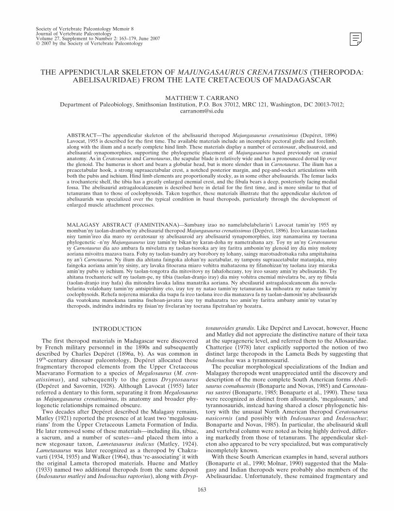
Majungasaurus is now known from numerous specimens thatpreserve nearly the entire skull and vertebral column, as well asmost of the appendicular skeleton, making it one of the best-known abelisaurids (Fig. 1). Although only portions of the fore-limb have been recovered, much of the hind limb skeleton ispreserved. This is in contrast to nearly all other abelisaurids,where this region of the skeleton is either incomplete (e.g., Car-notaurus, Xenotarsosaurus, Lametasaurus, Ekrixinatosaurus) orentirely unknown (e.g., Abelisaurus, Indosaurus, Indosuchus).Although the holotype of Aucasaurus garridoi includes nearlycomplete hind limb materials, these have been only preliminarilydescribed (Coria et al., 2002).
In this article, I describe the appendicular materials of Majun-gasaurus and discuss their relevance to phylogenetic and func-tional interpretations of this taxon and other abelisaurids. Be-cause many major theropod clades are diagnosed by features ofthe hind limb, understanding these structures in abelisaurids sig-nificantly affects assessments of their phylogenetic position. In abroader sense, these features also bear on the placement of abe-lisauroids and neoceratosaurs relative to coelophysoids and tet-anurans (i.e., the monophyly or paraphyly of Ceratosauria sensuGauthier, 1986). Finally, the apomorphic nature of the abelisau-rid appendicular skeleton highlights several potential functionalspecializations in this unusual theropod clade.
Institutional Abbreviations—DGM, Museu de Ciências daTerra, Rio de Janeiro, Brazil; FMNH, Field Museum of NaturalHistory, Chicago, IL; FSL, Faculté des Sciences, Université deLyon, France; GSI, Geological Society of India, Kolkata, India;HMN, Humboldt Museum für Naturkunde, Berlin, Germany;ISI, Indian Statistical Institute, Kolkata, India; MACN, MuseoArgentino de Ciencias Naturales “Bernardino Rivadavia,”Buenos Aires, Argentina; MCF-PVPH, Museo “Carmen
Funes,” Plaza Huincul, Argentina; MNHN, Muséum Nationald’Histoire Naturelle, Paris, France; MPCA-PV, Museo Provin-cial “Carlos Ameghino,” Cipoletti, Argentina; MWC, Museumof Western Colorado, Fruita, CO; UA, Université d’Antanana-rivo, Antananarivo, Madagascar; UMNH VP, Utah Museum ofNatural History, Salt Lake City, UT; UNPSJB-PV, UniversidadNacional de Patagonia “San Juan Bosco,” Comodoro Rivadavia,Argentina; USNM, National Museum of Natural History, Smith-sonian Institution, Washington, DC.
Comparative Taxa and Specimens—The following specimenswere examined for the comparisons mentioned in this paper.When literature illustrations were used, the appropriate refer-ence is given below. Abelisauridae indet. (GSI 296, K27/525, 539,558, 560, 568-570, 620, 653-654, 658-659, 671, Huene and Matley,1933; ISI R91/1); Aucasaurus garridoi (MCF-PVPH 236); Car-notaurus sastrei (MACN-CH 895); Ceratosauria indet. (HMN 37,69); Ceratosaurus nasicornis (MWC 1.1; UMNH VP 5278,USNM 4735); Elaphrosaurus bambergi (HMN Gr. S. 38-44); Ge-nusaurus sisteronis (MNHN Bev-1); Ilokelesia aguadagrandensis(MCF-PVPH 35); Lametasaurus indicus (GSI uncat., Matley,1924); Masiakasaurus knopfleri (FMNH PR 2112, 2114-2119,2129-2132, 2120-2123, 2134-2136, 2143, 2146-2155, 2158-2161,2167, 2169, 2171-2176, 2205-2106, 2208, 2214-2119, 2223-2225,2227, 2234, 2236; UA 8681, 8683-8684, 8686, 8693-8694, 8700,8710-8711, 8713-8714); Pycnonemosaurus nevesi (DGM 859-R,Kellner and Campos, 2002); Quilmesaurus curriei (MPCA-PV100); Rajasaurus narmadensis (GSI 21141/1-33, Wilson et al.,2003); Tarascosaurus salluvicus (FSL 330 201-3); Xenotarsosau-rus bonapartei (MACN 1468, cast of UNPSJB-PV 184/PVL 612).
SYSTEMATIC PALEONTOLOGY
DINOSAURIA Owen, 1842SAURISCHIA Seeley, 1888THEROPODA Marsh, 1881
CERATOSAURIA Marsh, 1884ABELISAUROIDEA (Bonaparte and Novas, 1985)
ABELISAURIDAE Bonaparte and Novas, 1985MAJUNGASAURUS Lavocat, 1955
MAJUNGASAURUS CRENATISSIMUS (Depéret, 1896)Lavocat, 1955
Type Specimen—MNHN MAJ-1, nearly complete right den-tary of subadult individual (Lavocat, 1955).
Referred Specimens—See complete listing in Krause et al.(this volume).
FIGURE 1. Skeletal reconstruction of Majungasaurus crenatissimus in left lateral view, showing known skeletal elements in white, unknownelements in gray.
SOCIETY OF VERTEBRATE PALEONTOLOGY, MEMOIR 8164
Revised Diagnosis—See Krause et al. (this volume).Age and Distribution—Appendicular materials of Majungas-
aurus were found at several localities (MAD93-01, 93-18, 93-19,93-20, 93-32, 93-33, 93-35, 93-73, 95-14, 95-16, 96-01, 96-07, 96-18,96-21, 99-26, 99-32, 99-33, and 01-05) near the village of Beriv-otra, in the Mahajanga Basin of northwestern Madagascar(Krause et al., 1997; Sampson et al., 1998). Most materials ofMajungasaurus derived from the Anembalemba Member, theuppermost white sandstone stratum of the Maevarano Forma-tion (Maastrichtian, Upper Cretaceous) (Rogers and Hartman,1998; Rogers et al., 2000, this volume).
Described Material—FMNH PR 2423 (right humerus), UA9031 (left humerus), UA 8678 (left and right ilia), UA 9032 (lefttibia), FMNH PR 2424 (left tibia), UA 9077 (left tibia andfibula), UA 9078 (right fibula), FMNH PR 2425 (left astragalo-calcaneum), UA 9033 (right astragalocalcaneum), UA 9082 (as-tragalus), UA 9034 (left metatarsal II), UA 9079 (left metatarsalIII), UA 9035 (left metatarsal IV), UA Bv 532 (left pedal pha-lanx I-2), UA Bv 1658 (left pedal phalanx I-2), UA 9036 (leftpedal phalanx II-1), FMNH PR 2426 (right pedal phalanx II-1),UA Bv 1260 (right pedal phalanx II-1), FMNH PR 2428 (rightpedal phalanx II-1, left pedal phalanx II-3 and III-2), FMNH PR2427 (right pedal phalanx II-2), UA 9037 (right pedal phalanxII-2), UA 9038 (left pedal phalanx II-3), UA Bv 1265 (left pedalphalanx III-1), FMNH PR 2429 (left pedal phalanx III-1), UA9039 (right pedal phalanx III-1), UA 9042 (left pedal phalanxIII-2), UA 9081 (right pedal phalanx III-1 or III-2), FMNH PR2430 (right pedal phalanx IV-1), UA 9040 (right pedal phalanxIV-1), FMNH PR 2431 (left pedal phalanx IV-3), UA 9041 (rightpedal phalanx IV-2), FMNH PR 2432 (left pedal phalanx IV-4),FMNH PR 2433 (right pedal phalanx IV-4), FMNH PR 2434(left pedal phalanx IV-5), UA 9043 (pedal phalanx IV-5), andFSL 92.290 (pedal ungual phalanx).
FMNH PR 2278 is an associated skeleton that includes cranial,axial, and appendicular elements from site MAD99-26. Amongthe latter have been identified a left scapulocoracoid, partial leftilium, left femur, left and fragmentary right tibiae, left and partialright fibulae, left astragalocalcaneum, left metatarsals II-IV,right pedal phalanges II-1 and IV-2, and left pedal phalangesIV-2 and IV-3.
Specimens UA 9031, 9033-9037, 9040-9041, 9077-9079, 9081-9082, and FMNH PR 2430 come from a single quarry horizon(site MAD99-33), along with a nearly complete skull. Althoughthere is some duplication of elements (notably two right pedalIV-2 phalanges and two right premaxillae), most of the materialsare consistent with having been derived from a single subadultindividual.
DESCRIPTION AND COMPARISONS
Pectoral Girdle
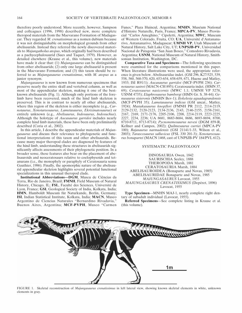
The pectoral girdle of Majungasaurus is represented only by asingle, incomplete scapulocoracoid (FMNH PR 2278). Theclavicles and sternum are not known.
Scapulocoracoid—FMNH PR 2278 includes an incomplete,coossified scapulocoracoid (FMNH PR 2278; Fig. 2). The scapu-lar blade is long, with nearly parallel anterodorsal and postero-ventral edges where they are complete (approximately the ven-tral one-third). Although the anterodorsal edge of the distal endis missing, the posteroventral edge does not flare appreciably,thus resembling the condition in tetanurans and Herrerasaurusmore than that in coelophysoids and Eoraptor. Unlike the con-dition in tetanurans, however, the blade is relatively wide an-teroposteriorly, as in abelisauroids (Masiakasaurus, Carnotau-rus, Aucasaurus), Ceratosaurus, and more primitive taxa. Onlythe base of the acromial process is preserved. The posteroven-trally-facing glenoid has prominent lips both dorsally and ven-
trally, with the former being the most pronounced. The postero-ventral process appears to have been relatively deep dorsoven-trally, with a moderately concave notch between it and theglenoid. In general shape and proportions, this element is mostsimilar to the scapulocoracoids of Masiakasaurus, Carnotaurus,and Aucasaurus.
On the lateral surface, the scapulocoracoid suture is faintlyvisible as a roughened ridge that passes anteriorly from the glen-oid along a slightly undulating line. A large, elliptical rugosity sitsventral to this, just anterior to the glenoid and dorsal to thecoracoid foramen (Fig. 2). The coracoid foramen faces ventrallyin lateral view, and opens into a dorsally directed channel thatpasses through the bone to the medial side. A small, faint tu-
FIGURE 2. Left scapulocoracoid of Majungasaurus crenatissimus(FMNH PR 2278) in lateral view. Abbreviations: cf, coracoid foramen;dl, dorsal lip; g, glenoid; pvp, posteroventral process; r, rugosity. Dashedlines indicate reconstructed outlines of element. Scale bar equals 10 cm.
CARRANO—APPENDICULAR SKELETON OF MAJUNGASAURUS 165
bercle is visible ventral to the glenoid, between it and the pos-teroventral process; this may have been associated with the origi-nation for M. biceps (Ostrom, 1974; Welles, 1984, Carpenter andSmith, 2001; Jasinoski et al., 2006) and/or M. coracobrachialisbrevis (Osmólska et al., 1972; Walker, 1977; Jasinoski et al.,2006). The lateral surface of the scapular blade is smooth.
The medial surface of the scapular blade is more poorly pre-served and illuminates few additional details. In posterior view,the blade can be seen to curve broadly, with its concave facedirected medially (against the ribcage). Its thickness decreasesfrom proximal to distal, as well as from posteroventral to an-terodorsal. The glenoid is approximately reniform, with a slightlylonger dorsal (scapular) component. The scapulocoracoid suturebetween these two components is unclear. Additionally, there isa deep fossa just dorsal to the glenoid that exaggerates theprominence of the dorsal lip, as in other ceratosaurs.
Forelimb
The forelimb of Majungasaurus is represented only by thehumerus. All remaining elements (radius, ulna, carpus, and ma-nus) are unknown and cannot be compared to the highly derived
corresponding elements of the abelisaurids Carnotaurus and Au-casaurus. These elements are also largely unknown in other abe-lisauroids (with the exception of individual manual phalanges inNoasaurus and Masiakasaurus; Carrano et al., 2004), and thusare currently of limited use in phylogenetic assessments of thesetaxa. The forelimb materials known for Ceratosaurus are rela-tively short compared to those of other theropods, but lack thepeculiar derived morphology seen in abelisaurids.
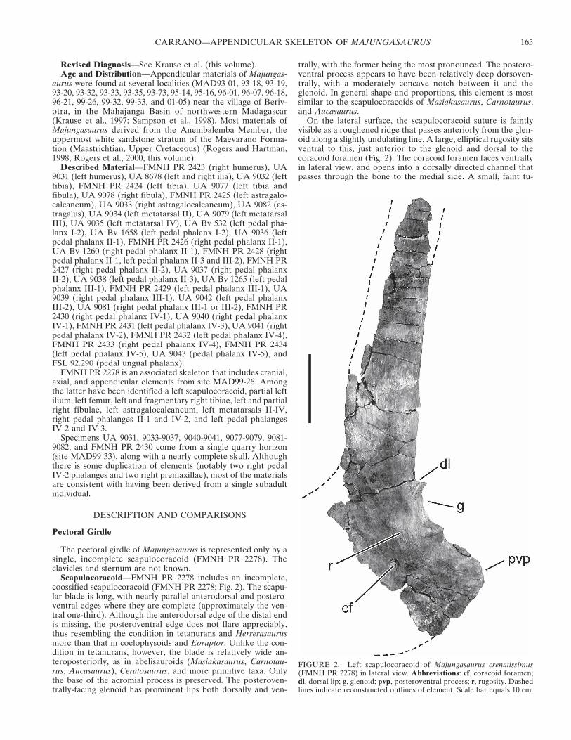
Humerus—The humerus is short but not particularly stocky(Fig. 3; Table 1), similar in proportions to the same bone in adulttyrannosaurids (Carpenter and Smith, 2001; Currie, 2003). Thelong axes of the proximal and distal ends are slightly twistedrelative to each other in Majungasaurus and other abelisaurids,more than in coelophysoids but less than in tetanurans. Thehumeral shaft is nearly straight in anteroposterior view (Fig. 3B,D), bowed anteroposteriorly, and flares as it approaches the dis-tal end. The proximal end is dominated by a large, rounded head,and the accompanying internal tuberosity and greater tubercleare comparatively small. The humeral head and internal tuber-osity both expand anteroposteriorly from the shaft.
The deltopectoral crest is a low, rugose ridge that curves fromthe lateral surface (proximally) onto the anterior midshaft. It
FIGURE 3. Right humerus of Majungasaurus crenatissimus (FMNH PR 2423). A, anteromedial view; B, anterolateral view; C, posterolateral view;D, posteromedial view; E, proximal view; F, distal view. Abbreviations: dpc, deltopectoral crest; ecc, ectepicondyle; enc, entepicondyle; gt, greatertubercle; hh, humeral head; it, internal tuberosity; lds, scar for M. latissimus dorsi; rc, radial condyle; uc, ulnar condyle. Scale bar equals 10 cm.
SOCIETY OF VERTEBRATE PALEONTOLOGY, MEMOIR 8166
likely served as the insertion point for Mm. pectorales (Dilkes,2000, 2001; Carpenter and Smith, 2001; Jasinoski et al., 2006).The crest is widest at its distal end, and approximately half againas high as the shaft is wide. A large fossa located just distal to thehumeral head on the anterior surface may mark the insertion ofM. coracobrachialis (Carpenter and Smith, 2001; Jasinoski et al.,2006).
The cross-section of the proximal shaft is elliptical, whereasthat of the distal half is nearly circular. At the distal end, a faintridge runs obliquely proximomedially away from the radial con-dyle, delineating the lateral border of the shallow intercondylardepression. The medial border is more pronounced, and runsmore directly proximally from the weakly convex ulnar condyle.The convexity of the radial condyle is particularly evident inposteromedial view (Fig. 3D).
In anteroposterior view, the humeral head appears moreasymmetrically rounded than in mediolateral view. A large, oval,rugose ridge extends roughly parallel to the distal deltopectoralcrest on the posterolateral side of the bone, with a small fossabetween them proximally. An additional fossa is positioned ad-jacent to this second ridge, separating it from a rounded, rugosebump more posteriorly; this probably represents the insertions ofM. latissimus dorsi and part of M. deltoideus (Fig. 3C; Dilkes,2000, 2001; Carpenter and Smith, 2001; Jasinoski et al., 2006).
In proximal view (Fig. 3E), the humeral head is rounded andbulbous, with no clear long axis. The internal tuberosity is visibleas a triangular medial projection, but the greater tubercle is in-distinct from the deltopectoral crest, which descends from thegreater tubercle as a ridge. The distal end is unusual in lackingprominent, convex condyles for the radius and ulna, insteadbearing nearly flat articular surfaces. In distal view (Fig. 3F), thisend is irregularly shaped, with two slightly concave swellings thatrepresent the articular surfaces for the radius and ulna. A smallcentral depression separates these indistinct condyles.
The humerus of Majungasaurus is strikingly similar to those ofCarnotaurus and Aucasaurus, the only other abelisaurids forwhich this bone is known. In all three taxa, this bone is propor-tionally short, lacks significant mediolateral curvature, and bearsa globular head and flattened distal condyles. Of the two, thehumerus of Majungasaurus more closely resembles that of Au-casaurus than the bulkier element of Carnotaurus. The humeri ofMasiakasaurus and Elaphrosaurus also share several featureswith that of Majungasaurus (Carrano et al., 2002), but are rela-tively longer and more slender.
Pelvic Girdle
The pelvic girdle of Majungasaurus is represented only by theilium; the pubis and ischium are not known.
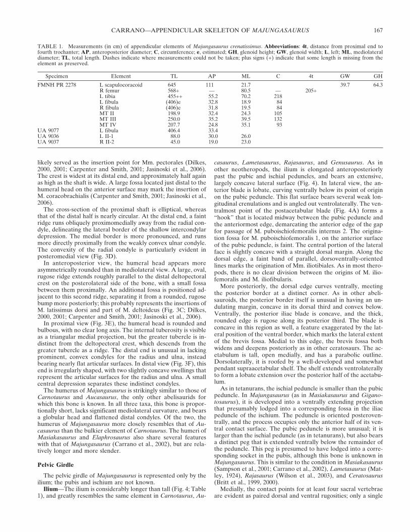
Ilium—The ilium is considerably longer than tall (Fig. 4; Table1), and greatly resembles the same element in Carnotaurus, Au-
casaurus, Lametasaurus, Rajasaurus, and Genusaurus. As inother neotheropods, the ilium is elongated anteroposteriorlypast the pubic and ischial peduncles, and bears an extensive,largely concave lateral surface (Fig. 4). In lateral view, the an-terior blade is lobate, curving ventrally below its point of originon the pubic peduncle. This flat surface bears several weak lon-gitudinal crenulations and is angled out ventrolaterally. The ven-tralmost point of the postacetabular blade (Fig. 4A) forms a“hook” that is located midway between the pubic peduncle andthe anteriormost edge, demarcating the anterior edge of the gapfor passage of M. puboischiofemoralis internus 2. The origina-tion fossa for M. puboischiofemoralis 1, on the anterior surfaceof the pubic peduncle, is faint. The central portion of the lateralface is slightly concave with a straight dorsal margin. Along thedorsal edge, a faint band of parallel, dorsoventrally-orientedlines marks the origination of Mm. iliotibiales. As in most thero-pods, there is no clear division between the origins of M. ilio-femoralis and M. iliofibularis.
More posteriorly, the dorsal edge curves ventrally, meetingthe posterior border at a distinct corner. As in other abeli-sauroids, the posterior border itself is unusual in having an un-dulating margin, concave in its dorsal third and convex below.Ventrally, the posterior iliac blade is concave, and the thick,rounded edge is rugose along its posterior third. The blade isconcave in this region as well, a feature exaggerated by the lat-eral position of the ventral border, which marks the lateral extentof the brevis fossa. Medial to this edge, the brevis fossa bothwidens and deepens posteriorly as in other ceratosaurs. The ac-etabulum is tall, open medially, and has a parabolic outline.Dorsolaterally, it is roofed by a well-developed and somewhatpendant supraacetabular shelf. The shelf extends ventrolaterallyto form a lobate extension over the posterior half of the acetabu-lum.
As in tetanurans, the ischial peduncle is smaller than the pubicpeduncle. In Majungasaurus (as in Masiakasaurus and Gigano-tosaurus), it is developed into a ventrally extending projectionthat presumably lodged into a corresponding fossa in the iliacpeduncle of the ischium. The peduncle is oriented posteroven-trally, and the process occupies only the anterior half of its ven-tral contact surface. The pubic peduncle is more unusual; it islarger than the ischial peduncle (as in tetanurans), but also bearsa distinct peg that is extended ventrally below the remainder ofthe peduncle. This peg is presumed to have lodged into a corre-sponding socket in the pubis, although this bone is unknown inMajungasaurus. This is similar to the condition in Masiakasaurus(Sampson et al., 2001; Carrano et al., 2002), Lametasaurus (Mat-ley, 1924), Rajasaurus (Wilson et al., 2003), and Ceratosaurus(Britt et al., 1999, 2000).
Medially, the contact points for at least four sacral vertebraeare evident as paired dorsal and ventral rugosities; only a single
TABLE 1. Measurements (in cm) of appendicular elements of Majungasaurus crenatissimus. Abbreviations: 4t, distance from proximal end tofourth trochanter; AP, anteroposterior diameter; C, circumference; e, estimated; GH, glenoid height; GW, glenoid width; L, left; ML, mediolateraldiameter; TL, total length. Dashes indicate where measurements could not be taken; plus signs (+) indicate that some length is missing from theelement as preserved.
Specimen Element TL AP ML C 4t GW GH
FMNH PR 2278 L scapulocoracoid 645 111 21.7 39.7 64.3R femur 568+ — 80.5 — 205+L tibia 455++ 55.2 70.2 218L fibula (406)e 32.8 18.9 84R fibula (406)e 31.8 19.5 84MT II 198.9 32.4 24.3 105MT III 250.0 35.2 39.5 132MT IV 207.7 24.8 35.1 93
UA 9077 L fibula 406.4 33.4UA 9036 L II-1 88.0 30.0 26.0UA 9037 R II-2 45.0 19.0 23.0
CARRANO—APPENDICULAR SKELETON OF MAJUNGASAURUS 167
contact is present for the first sacral (Fig. 4B). Along the dorsalmargin, long, parallel dorsoventral ridges run along nearly theentire length of the bone. The brevis fossa is inset medially to aconsiderable degree, forming a distinct medial shelf whose ven-tral border turns from convex to strongly concave as it ap-proaches the ischial peduncle. The pegs of both peduncles aremore prominent in medial view, appearing to extend directlyfrom the medial wall of the bone. The anterior blade divergeslaterally from the midline, beginning at a point just anterior tothe base of the pubic peduncle. The medial edge of the dorsalacetabulum is thin.
In posterior view (Fig. 4F), the brevis fossa can be seen toexpand both medially and laterally beneath the main iliac blade.
Longitudinal striations are visible on most of the internal surfaceof the fossa. Its lateral wall extends farther ventrally than medi-ally. In anterior view (Fig. 4E), the lateral deviation of the an-terior iliac blade is quite evident, which creates a distinct passagefor M. puboischiofemoralis internus 2. A well-defined fossa atthe junction of the blade and pubic peduncle marks the begin-ning of this passage. The anteriormost end of the iliac blade isthinner and rougher than the other edges.
Hind Limb
Most of our knowledge of the hind limb of Majungasaurus isderived from one specimen, FMNH PR 2278, a partial skeleton
FIGURE 4. Left ilium of Majungasaurus crenatissimus (UA 8678). A, lateral view; B, medial view; C, dorsal view; D, ventral view; E, anterior view;F, posterior view. Abbreviations: bf, brevis fossa; ifis, scar for M. iliofibularis; ip, ischial peduncle; its, scar for Mm. iliotibiales; lbf, lateral wall ofbrevis fossa; mbf, medial wall of brevis fossa; pp, pubic peduncle; sac, supraacetabular crest; sr2-5, attachment surfaces for sacral ribs 2-5; st1-5,attachment surfaces for transverse processes of sacral vertebrae 1-5; vph, ventral preacetabular hook. Scale bar equals 10 cm.
SOCIETY OF VERTEBRATE PALEONTOLOGY, MEMOIR 8168
that includes most of the hind limb elements. This specimenindicates that the hind limb is unusually short relative to otherskeletal elements (Fig. 1; Table 1). This stockiness is especiallyapparent in the tibia and metatarsals, which are robust for theirsize, but even the femur is relatively short (although not particu-larly robust).
Nearly the entire hind limb of Majungasaurus is known, withthe exception of metatarsals I and V, and pedal phalanges I-1and III-4. Among abelisaurids only Aucasaurus has a more com-pletely preserved hind limb (Coria et al., 2002).
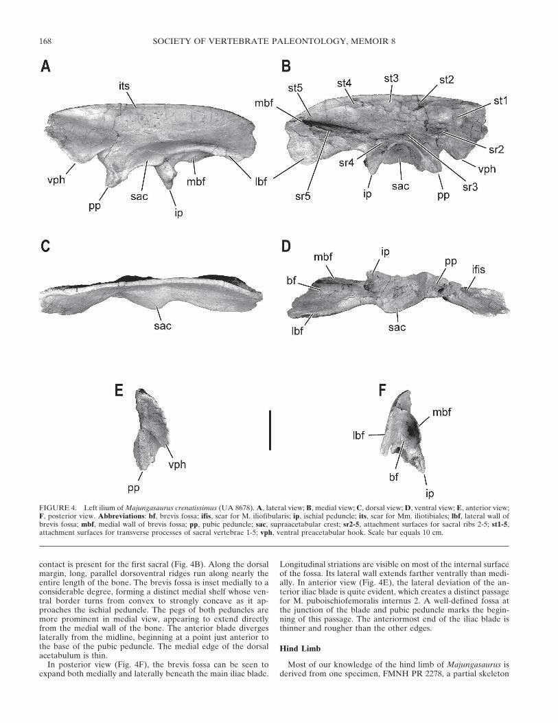
Femur—The femur is known only from parts of two damagedelements, both pertaining to FMNH PR 2278. The head, neck,and much of the anterolateral surface have been weathered awayin even the most complete specimen (Fig. 5). The exposed inter-nal structure reveals a large central cavity that permeated theentire femoral shaft, as is typical for theropods. Proximally, thebase of the lesser trochanter is preserved, as well as the deepestportion of the sulcus between it and the main femoral shaft. Thissulcus indicates that the lesser trochanter was elevated, probablycomparable to the condition in Carnotaurus, Xenotarsosaurus,Tarascosaurus, Genusaurus, and Ceratosaurus. The preservedportion of the lesser trochanter is extremely rugose. Near itsbase, but more distolaterally positioned, a rounded bump marksthe insertion of M. iliofemoralis externus. These two structuresresult from complete separation of the primitive trochantericshelf (Hutchinson, 2001), as in tetanurans but not coelophysoids.A flat, longitudinally striated facet marks the lateral edge of thegreater trochanter, and represents the insertion of Mm. pubois-chiofemorales externi (Hutchinson, 2001).
Still farther distally, the posteromedial surface shows a well-preserved, ridge-like fourth trochanter (for Mm. caudofemo-rales) situated approximately two-fifths of the way down theshaft. A rugose region extending proximally onto the posterior
shaft surface represents the additional insertion area for M. cau-dofemoralis brevis. Compared to the other prominent limbmuscle attachment sites, the fourth trochanter appears to besurprisingly small, although the relevance of this to actual musclesize is questionable (e.g., Bryant and Seymour, 1990). The inser-tion of M. adductor femoris 1 cannot be identified with certaintybecause the posteromedial side of the distal shaft is damaged.However, M. adductor femoris 2 insertion is complete and lo-cated about two-thirds down the shaft, along its lateral edge; theinsertion is flat and longitudinally elliptical.
The shaft exhibits bowing in both the anteroposterior and me-diolateral planes, being slightly concave posteriorly and medi-ally. Both distal condyles are present but poorly preserved. Thefibular condyle is approximately twice as wide mediolaterally asthe tibial, but the two extend the same distance distally (Fig. 5A,C). In distal view, the fibular condyle is expanded and bulbous,as in Masiakasaurus, Carnotaurus, and Xenotarsosaurus. Thedeep, broad posterior intercondylar sulcus contains no markedlongitudinal ridges for attachment of the knee flexors. A medio-laterally narrow tibiofibular crest is separated from the mainfibular condyle by a broad, shallow groove. The medial edge ofthe bone is battered, so the presence of a prominent entepicon-dylar crest (as in Masiakasaurus and Genusaurus; Carrano et al.,2002) cannot be ascertained.
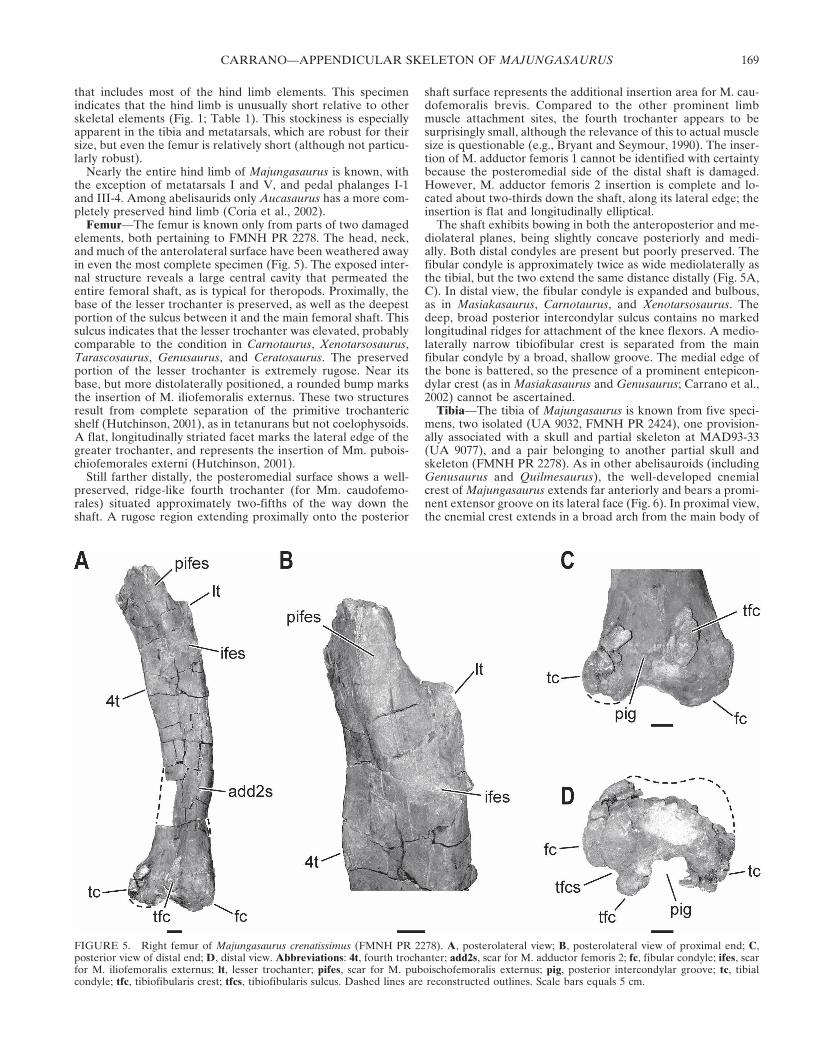
Tibia—The tibia of Majungasaurus is known from five speci-mens, two isolated (UA 9032, FMNH PR 2424), one provision-ally associated with a skull and partial skeleton at MAD93-33(UA 9077), and a pair belonging to another partial skull andskeleton (FMNH PR 2278). As in other abelisauroids (includingGenusaurus and Quilmesaurus), the well-developed cnemialcrest of Majungasaurus extends far anteriorly and bears a promi-nent extensor groove on its lateral face (Fig. 6). In proximal view,the cnemial crest extends in a broad arch from the main body of
FIGURE 5. Right femur of Majungasaurus crenatissimus (FMNH PR 2278). A, posterolateral view; B, posterolateral view of proximal end; C,posterior view of distal end; D, distal view. Abbreviations: 4t, fourth trochanter; add2s, scar for M. adductor femoris 2; fc, fibular condyle; ifes, scarfor M. iliofemoralis externus; lt, lesser trochanter; pifes, scar for M. puboischofemoralis externus; pig, posterior intercondylar groove; tc, tibialcondyle; tfc, tibiofibularis crest; tfcs, tibiofibularis sulcus. Dashed lines are reconstructed outlines. Scale bars equals 5 cm.
CARRANO—APPENDICULAR SKELETON OF MAJUNGASAURUS 169
the bone, with a wide lateral concavity that does not resemblethe distinct notch of forms such as Allosaurus, Sinraptor, andmany coelurosaurs. In lateral view (Fig. 6A), the cnemial crestexhibits a strong dorsal curvature such that a substantial portionof this structure occurs dorsal to the lateral and medial condyles.The anteriormost portion of the cnemial crest is slightly ex-panded mediolaterally, with a distinct sulcus (presumably for theknee extensor tendons) visible along the lateral edge. The twoproximal condyles are similarly sized and separated by a weaknotch. In addition, the posterior edge of the tibia is angledobliquely relative to the mediolateral axis, as evident in proximalview (Fig. 6B).
The lateral fossa between the cnemial crest and the tibial shaftis also correspondingly well-developed. In addition, the terminusof the cnemial crest is expanded both proximally and distally,and does not taper to a rounded point as in most theropods. Thisstructure is thin and delicate, and is completely preserved (ornearly so) only in UA 9077. More posteriorly, the lateral tibiabears a stout ridge, the fibular crest, which runs for approxi-mately one-fourth of the bone’s length and articulates with thefibula. A short gap sits distal to the crest, and a large foramen islocated posterior to it. Farther distally, the long, flat fibular facetruns to a point just proximal to the expansion of the lateralmalleolus. This facet is exceptionally rugose and wide, as in otherabelisaurids, contrasting with the thinner, fainter structure ofmost other theropods.
The anterior aspect of the tibia is relatively smooth and flat-tened, with a rounded anteromedial edge. At the distal end (Fig.6C), a triangular facet for the astragalar ascending process can beobserved, bounded proximally by an obliquely oriented buttress.In this view, the lateral malleolus is clearly larger and descendsfarther distally than the medial. The medial malleolus bears ashort proximal ridge. Medially, the tibial surface is smooth andlargely featureless, with a broadly rounded cross-sectionthroughout most of its length. The distal end shows the same‘torsion’ relative to the proximal end as do most theropod tibiae;that is, the long axis of the distal end is oriented perpendicular tothat of the proximal end. The posterior shaft is also rounded andfairly smooth, with a modest distal ridge that terminates at thesulcus between the two malleoli.
In distal view (Fig. 6D), the tibia of Majungasaurus moreclosely resembles those of tetanurans than those of coelophy-soids or Herrerasaurus. The expanded lateral malleolus and dis-tal ridge lend an elongate shape to the distal tibia, in contrast tothe rectangular or circular form of more primitive taxa. Theastragalar facet invaginates the anterior profile opposite the pos-terior distal ridge. The outer ends of both malleoli are tapered.
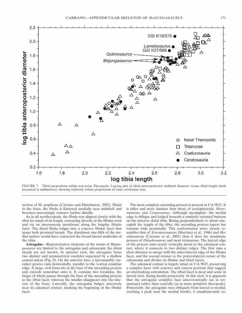
The tibia of Majungasaurus is relatively short and stocky(Fig. 7; Table 1), as are those of Lametasaurus (Matley, 1924),other Indian theropod tibiae (GSI K19/579, K27/568; Huene andMatley, 1933), Quilmesaurus, and a large ceratosaurian from theTendaguru Formation of Tanzania (HMN 37, 69). In contrast,the tibiae of Rajasaurus, Xenotarsosaurus and Aucasaurus aremore slender than that of Majungasaurus. The tibia of Carno-taurus is incomplete, but given its general similarity to those ofthe latter two taxa, it was probably proportionally longer thanthat of Majungasaurus but still shorter than originally recon-structed (Bonaparte et al., 1990).
Fibula—The fibula is correspondingly short but not propor-tionally stocky (Fig. 8; Table 1). Its lateral face is generallysmooth and rounded. The anterolateral edge below the proximalend bears a roughened, bulbous expansion that is elevated abovethe rest of the concave proximal surface. This tubercle, the in-sertion of M. iliofibularis (Romer, 1923; Carrano and Hutchin-son, 2002), is more prominent than in most other theropods, butsimilar to those of Ceratosaurus, Xenotarsosaurus, Genusaurus,Carnotaurus, Rajasaurus, and Aucasaurus. Farther distally, theposterior edge is expanded into a prominent, obliquely orientedtrochanter whose thin, ridge-like proximal half yields to abroader, slightly concave distal surface. Both anterior and pos-terior edges of the fibular shaft are acute but rounded. At thedistal end, the shaft flares out laterally and anteroposteriorly,terminating in a rugose calcaneal articulation. The anterior edgeof the shaft at this point bears a roughened flange that (based onFMNH 2278) abutted against the lateral edge of the astragalarascending process. The distal articular surface is convexlyrounded in mediolateral view.
The medial face is shallowly concave near the proximal end(Fig. 8D) in a manner that approximately corresponds to thecurvature of the lateral tibia. A raised ridge extends distallydown the proximal one-third of the anterior shaft edge, probablymarking the attachment of the interosseus membrane. Just pos-terior to this ridge, the fibula is excavated into a wide, deepmedial fossa that occupies most of the proximal third of thebone. The fossa is more open anteriorly than posteriorly (givingit an L-shape in cross-section), and the posterior portion of thebone is expanded medially to create a thin but prominent wall.This medial fossa is unlike the much deeper excavation of de-rived coelurosaurs, the narrow incisure of coelophysoids (Roweand Gauthier, 1990), or the broadly shallow surface of spinosau-roids, but instead closely resembles the condition in Ceratosau-rus, Rajasaurus, Xenotarsosaurus, and Genusaurus. Nonetheless,all these structures are likely homologous, representing the in-
FIGURE 6. Left tibia of Majungasaurus crenatissimus. Proximal end ofFMNH PR 2424 in A, lateral view and B, proximal view. Distal end ofFMNH PR 2278 in C, anterior view and D, distal view. Abbreviations:af, astragalar facet; cn, cnemial crest; das, distal articular surface; fc,fibular crest; ff, fibular facet; lc, lateral condyle; lf, lateral fossa; lm,lateral malleolus; mc, medial condyle; mm, medial malleolus; pas, proxi-mal articular surface. Dashed line in B indicates reconstructed outline ofelement. Scale bars equals 1 cm.
SOCIETY OF VERTEBRATE PALEONTOLOGY, MEMOIR 8170
sertion of M. popliteus (Carrano and Hutchinson, 2002). Distalto the fossa, the fibula is flattened medially near midshaft andbecomes increasingly concave farther distally.
As in all neotheropods, the fibula was aligned closely with thetibia for much of its length, contacting directly at the fibular crestand via an interosseous membrane along the lengthy fibularfacet. The distal fibula lodges into a concave fibular facet thatspans both proximal tarsals. The distalmost one-fifth of the me-dial surface would have contacted the broad lateral malleolus ofthe tibia.
Astragalus—Representative elements of the tarsus of Majun-gasaurus are limited to the astragalus and calcaneum; the distaltarsals are not known. In anterior view, the astragalus bearstwo distinct and asymmetrical condyles separated by a shallowcentral sulcus (Fig. 9). On the anterior face, a (presumably vas-cular) groove runs horizontally, parallel to the ventral condylaredge. A large, oval fossa sits at the base of the ascending processand extends somewhat onto it. It contains two foramina, thelarger of which passes through the base of the ascending processto the tibial facet, whereas the smaller disappears into the inte-rior of the bone. Laterally, the astragalus bulges anteriorlynear its calcaneal contact, marking the beginning of the fibularfacet.
The most complete ascending process is present in UA 9033. Itis taller and more laminar than those of coelophysoids, Herre-rasaurus, and Ceratosaurus. Although incomplete, the medialedge is oblique and lodged beneath a similarly oriented buttresson the anterior distal tibia. Rising perpendicularly to about one-eighth the length of the tibia, the ascending process tapers butremains wide proximally. This conformation more closely re-sembles that of Xenotarsosaurus (Martínez et al., 1986) and Ma-siakasaurus (Carrano et al., 2002) than it does the acuminateprocess of Dilophosaurus and most tetanurans. The lateral edgeof the process runs nearly vertically down to the calcaneal con-tact, where it connects to two distinct ridges. The first runs ashort distance to merge with the anterolateral edge of the fibularfacet, and the second crosses to the posterolateral corner of thecalcaneum and divides its fibular and tibial facets.
The calcaneal contact is largely intact in UA 9033, preservinga complex facet with concave and convex portions that suggestan interlocking articulation. The tibial facet is deep and acute inlateral view, facing mostly posteriorly. In this view, it is apparentthat the astragalar condyles face anteroventrally (as in tet-anurans) rather than ventrally (as in more primitive theropods).Posteriorly, the astragalus rises obliquely from lateral to medial,reaching a peak near the medial border; it simultaneously ex-
FIGURE 7. Tibial proportions within non-avian Theropoda. Log-log plot of tibial anteroposterior midshaft diameter versus tibial length (bothmeasured in millimeters), showing relatively robust proportions of some ceratosaur taxa.
CARRANO—APPENDICULAR SKELETON OF MAJUNGASAURUS 171

pands anteroposteriorly, forming a rounded bulge at this peak. Asmall fossa is visible near the calcaneal contact. In medial view,the astragalar surface is broad, reniform, and faintly concave.The medial malleolar facet faces proximomedially and flanks themedial ascending process. The anteromedial corner is blunt.
Dorsally, the astragalus bears two distinct facets for the tibialmalleoli (Fig. 9C). The medial facet is divided from the lateral bya low ridge that runs from the ascending process to the postero-medial bulge. The lateral edge of this ridge is sharper than themedial, and borders a discrete fossa. The lateral facet is alsodeeper and narrower than the medial, and contains several fos-sae as well as the internal counterpart to the medial foramen onthe external ascending process. This lateral facet is continuouswith the calcaneal facet for the tibia, and includes two largeforamina that enter into mediolateral channels, which them-selves are confluent with two large calcaneal foramina. The an-terolateral corner of the astragalus extends anteriorly beyond theascending process, marking the beginning of the fibular articu-lation. Thus the tibia articulates partly posterior to the fibula, asin tetanurans but not more primitive theropods. The fibular facetcontains several small foramina, as well as an additional smallfossa lodged within its medial corner. Ventrally, the astragalus isobliquely hourglass-shaped, due to the enlargement of the an-teromedial corner relative to the anterolateral corner.
Several astragali are known: one isolated (UA 9082), two con-nected to their respective calcanea (FMNH PR 2425, UA 9033),and a fourth articulated with both the tibia and calcaneum(FMNH PR 2278). None of these specimens show evidence ofhaving been directly fused to the tibia or fibula, although all ofthe ascending processes show some breakage along the potentialfibular contact surface.
Calcaneum—The three known calcanea (FMNH PR 2278,2425, UA 9033) are fused to their astragali (Fig. 9), obscuring themedial surface in these specimens. Anteriorly, the fibular facet is
slightly visible where it rises at its posterolateral corner, andcontains numerous small foramina. The anterior wall is raisedand obscures the tibial facet from anterior view. Laterally thecalcaneum has a broad, slightly concave face that is roundedexcept for two triangular projections, one marking the antero-lateral edge of the fibular cup, and the second its posterolateraledge.
The bone is smooth and convex in posterior view, with a low,concave proximal ridge that bounds the tibial facet posteriorly.This ridge descends to a low point at the (faintly visible) astraga-localcaneal suture. Much of the tibial facet can be viewed pos-teriorly. Ventrally, the calcaneum shows a slightly pitted, convexarticular condyle that is continuous with that of the astragalus.The contact between the two runs nearly directly anteroposteri-orly, and the horizontal groove on the anterior surface of theastragalus is continued onto the anteroventral part of the calca-neum. The anterior bulge beneath the fibular cup of the astraga-lus also has a counterpart on the calcaneum, just above the hori-zontal groove.
In dorsal view, the calcaneum shows two distinct articular fac-ets. The concave fibular facet is a rounded triangle with its mostacute apex directed posterolaterally. About one-fifth of the facetis on the astragalus. This facet is separated from the more pos-terior tibial facet by the sharp ridge that runs from the lateraledge of the ascending process to the posterolateral corner of thecalcaneum. This corner is the highest point of the calcaneum.Behind it, the concave tibial facet contains two small lateralforamina as well as two larger foramina situated just at the as-tragalocalcaneal junction. The calcaneal portion of the tibialfacet forms a narrow triangle with its apex (the shallowest por-tion) directed laterally.
Metatarsals—Metatarsals I and V remain unknown, but allthree central elements are preserved. They are typical of thero-pod metatarsals in being tightly articulated proximally but diver-
FIGURE 8. Left fibula of Majungasaurus crenatissimus (UA 9077). A, Anterior view; B, lateral view; C, posterior view; D, medial view; E, proximalview; F, distal view. Abbreviations: apc, contact for the astragalar ascending process; ifs, scar for M. iliofibularis; im, ridge for attachment ofinterosseus membrane; mf, medial fossa; tf, tibial facet. Scale bars equals 5 cm.
SOCIETY OF VERTEBRATE PALEONTOLOGY, MEMOIR 8172

gent distally, particularly metatarsal IV (Figs. 10, 11). They havesimilar general proportions to the metatarsals of non-coelurosaur tetanurans such as Allosaurus, although there areseveral important differences.
Metatarsal II is known from two specimens, FMNH PR 2278and UA 9034, the former of which is complete (Fig. 10; Table 1).The bone is almost straight, with a narrow shaft that widenssteadily from proximal to distal. The proximal articulation istypical for a theropod, being flat and relatively featureless (Fig.10A); its proximal profile is a narrowly rounded triangle withthe apex directed posteriorly. The distal articulation bears twocondyles for contact with phalanx II-1 (Fig. 10D). The lateralcondyle is much larger than the medial and is flattened on itsventral surface, whereas the medial condyle is acuminate. Thetwo condyles are separated by a deep fossa. Both collateral liga-ment pits are present, but the lateral is much deeper than themedial, which is merely a faint fossa. The ‘hyperextensor’ pit(“oblique ligament fossa” of Snively et al., 2004) is extremelyshallow. The flat lateral surface of the shaft indicates that thisbone was closely appressed to that of metatarsal III for most ofits length. Indeed, articulating metatarsals II and III (FMNH PR2278) indicates that only the distal one-third of metatarsal II isdivergent. The shaft is otherwise rounded in cross-sectionaround the anterior, medial, and posterior surfaces, forming a
D-shape overall. A longitudinally striated facet is present alongthe middle third of the posterior surface, representing the inser-tion for M. gastrocnemius pars medialis (Carrano and Hutchin-son, 2002). A flat area on the medial surface, adjacent to theproximal end, marks the likely articulation site for metatarsal I(Tarsitano, 1983).
Metatarsal III is known only from FMNH PR 2278 (Fig. 10). Itis proportionally robust, being noticeably larger in all dimensionsthan either metatarsal II or IV (Table 1). The straight shaftshows a distinct lateral deviation along its distal third, in oppo-sition to the ‘medial deflection’ seen in many theropods (Snivelyet al., 2004). The proximal articulation is rectangular but notdistinctly hourglass-shaped, lacking the deep lateral notch formetatarsal IV present in tetanurans. Instead, the markedlysmaller proximal end of metatarsal II contacts within a broadcurved fossa on the medial edge of proximal metatarsal III. Asthe posterior edge of this surface is damaged, it cannot be de-termined whether metatarsal III is expanded in this direction asin Elaphrosaurus and Ceratosaurus. The facet for metatarsal IVis rugose and extends over a broadly concave surface. The proxi-mal surface itself is damaged as well, but it appears to have beenconvex posteriorly and flat anteriorly. The distal articulation is
FIGURE 9. Left astragalocalcaneum of Majungasaurus crenatissimus(FMNH PR 2278). A, anterior view; B, lateral view; C, proximal view; D,posterior view, showing approximate mediolateral dimensions of as-tragalus (ast) and calcaneum (calc); E, medial view. Abbreviations: ahg,anterior horizontal groove; ap, ascending process; f, fossa; ff, fibularfacet; tf, tibial facet. Scale bar equals 1 cm.
FIGURE 10. Articulated left metatarsals II-IV of Majungasaurus cre-natissimus (FMNH PR 2278). A, proximal view (anterior toward thebottom); B, anterior view; C, posterior view; D, distal view (anteriortoward the top). Abbreviations: II, metatarsal II; III, metatarsal III; IV,metatarsal IV; dt3c, contact surface for distal tarsal 3; dt4c, contact sur-face for distal tarsal 4; edls, fossa for insertion of M. extensor digitorumlongus; gs, scar for M. gastrocnemius. Scale bar equals 1 cm.
CARRANO—APPENDICULAR SKELETON OF MAJUNGASAURUS 173
broad and roller-like, with only a very faint central sulcus. Alongthe medial shaft, a large, flat facet indicates the articulation withmetatarsal II; it occupies nearly two-thirds of the length of thebone.
Distal to this point, the marked lateral offset displaces thedistal end of metatarsal III away from that of metatarsal II. Herea prominent ridge runs distally down the anteromedial edge ofthe shaft. The lateral facet for metatarsal IV is similarly pro-nounced but extends only to the midshaft. Posteriorly, two ovalrugosities may mark additional insertion points for Mm. gastroc-nemii, adjacent to the articulations for metatarsals II and IV. Thecollateral ligament pits are equally pronounced, and the ‘hyper-extensor’ pit is deep and mediolaterally wide.
Metatarsal IV, known from two specimens (FMNH PR 2278,UA 9035), is approximately the same length as metatarsal II
(Fig. 10; Table 1). The proximal end is D-shaped, although themedial edge is slightly concave where it contacts metatarsal III.The weak projection at the posteromedial corner is smallerthan the extended process seen in most tetanurans. The proximalarticulation is concave, with thicker rims along the postero-medial and lateral edges. A small triangular rugosity along theposterolateral edge marks the contact with metatarsal V. Thedistal end is convex and bears two unequal condyles for articu-lation with phalanx IV-1; its shape is typical of most thero-pods. The lateral surface bears a flat, faintly ridged contact formetatarsal III that extends approximately to the midshaft. Herethe shaft diverges strongly laterally, a feature most clearly ob-served in anterior view. The shaft is rounded along the otheredges, again forming a D. Two rugose, flat ridges run down thelength of the shaft along the posteromedial and posterior faces,probably marking the insertion of M. gastrocnemius pars latera-lis (Carrano and Hutchinson, 2002). The shaft narrows both me-diolaterally and anteroposteriorly just before reaching the distalarticulation. Both collateral ligament pits are weakly present,but the lateral one is particularly shallow, present only as afossa.
Phalanges—Most of the pedal phalanges are represented. Pre-suming the typical theropod phalangeal formula of 2-3-4-5-x, thiscollection includes all but I-1 and III-4 (Fig. 11). However, subtleproportional differences can be detected among the phalanges,suggesting that some dimorphism may have been present, as inthe limb bones of Masiakasaurus (Carrano, et al., 2002). Becauseof these variations, similarly shaped phalanges can be difficult todistinguish as isolated specimens. Thus some of the followingidentifications should be considered tentative. This is especiallytrue for the digit III phalanges (e.g., III-2 and III-3).
Only the ungual phalanx (I-2) from digit I is known (UA Bv532, 1658). Unlike the other pedal unguals, this element is nearlysymmetrical, straight, and strongly laterally compressed. Theproximal articular surface is tall, narrow, and concave but lacksa central ridge. Ventrally there is no flexor tubercle, but rather ashallow, flattened fossa. Vascular grooves are present but irregu-lar on both lateral and medial surfaces, with numerous largeforamina and distinct grooves near the tip. A pair of vasculargrooves is apparent on one side only.
Digit II is known in its entirety. Both non-ungual phalangesbear a large dorsal tubercle and two prominent, flattened ventralheels. Of these, the lateral is approximately twice as large as themedial and extends farther distally as an elongate ridge. Both arerugose, although the medial heel is flatter. The remainder of thephalangeal shaft is rounded but taller than wide. A distinct hy-perextensor pit is visible on the dorsal surface. The distal articu-lar surface bears two rounded condyles separated by a deepsulcus. The condyles appear to diverge ventrally and terminate inflat ventral heels: they are subequal in size, although the medialis slightly larger. Both collateral ligament pits are deep, but thelateral pit is larger and more rounded. II-1 (UA 9036, FMNH PR2426; UA Bv 1260) is the longest and tallest pedal phalanx, witha concave, dorsoventrally oval proximal articular surface. Pedalphalanx II-2 (FMNH PR 2427, UA 9037) is about half as long asII-1. Its proximal surface forms a rounded triangle, with twofacets separated by a weak vertical ridge. The medial facet isslightly larger than the lateral, corresponding to the morphologyof the distal condyles of II-1. The hyperextensor pit is deeper andmore pronounced on II-1 than on II-2, bounded distally by thedorsal margin of the distal articular surface.
The ungual phalanx of digit II (II-3; FMNH PR 2428, UA9038) is markedly asymmetrical. On the triangular proximal sur-face, the long axis is tilted so that the apex appears to leanlaterally. The two similarly sized articular facets are separated bya wide, low ridge and bear numerous large vascular foramina.The dorsal tubercle is pronounced, but ventrally there is no
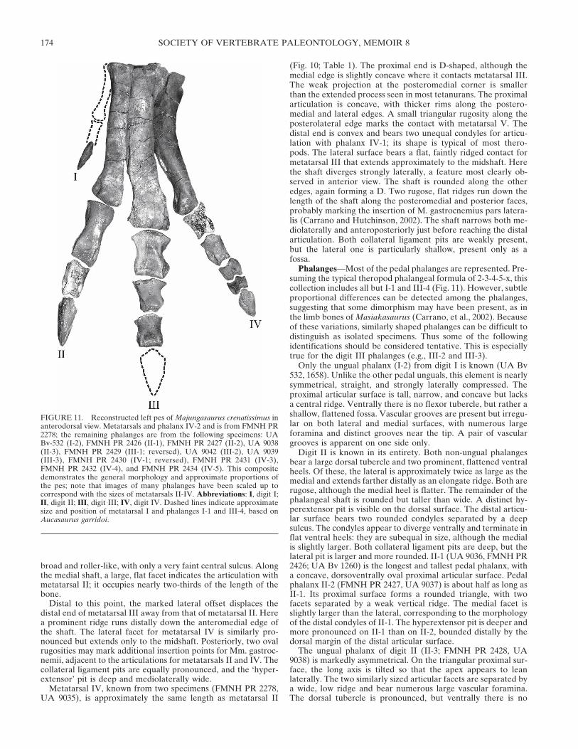
FIGURE 11. Reconstructed left pes of Majungasaurus crenatissimus inanterodorsal view. Metatarsals and phalanx IV-2 and is from FMNH PR2278; the remaining phalanges are from the following specimens: UABv-532 (I-2), FMNH PR 2426 (II-1), FMNH PR 2427 (II-2), UA 9038(II-3), FMNH PR 2429 (III-1; reversed), UA 9042 (III-2), UA 9039(III-3), FMNH PR 2430 (IV-1; reversed), FMNH PR 2431 (IV-3),FMNH PR 2432 (IV-4), and FMNH PR 2434 (IV-5). This compositedemonstrates the general morphology and approximate proportions ofthe pes; note that images of many phalanges have been scaled up tocorrespond with the sizes of metatarsals II-IV. Abbreviations: I, digit I;II, digit II; III, digit III; IV, digit IV. Dashed lines indicate approximatesize and position of metatarsal I and phalanges I-1 and III-4, based onAucasaurus garridoi.
SOCIETY OF VERTEBRATE PALEONTOLOGY, MEMOIR 8174
flexor tubercle. Instead, a deep, longitudinal ventral fossa isflanked by marked lateral and medial ridges. Numerous fo-ramina within this fossa appear to lead to a distally directedchannel, and the fossa may also be confluent with the lateral andmedial vascular grooves. The medial surface is relatively flat,with a distinct ventral groove that wraps onto the ventral surfacenear the proximal end. Its ventral edge forms the larger of thetwo grooves bounding the ventral fossa. The lateral surface ismore rounded, with a distinct triangular arrangement of grooves.The ventral groove is the most distinct and connects with thelarge fossa on the ventral surface. It contains several large fo-ramina, the distalmost leading into a tunnel within the bone andemerging again near the tip.
The first three phalanges of digit III are known in Majungas-aurus. These non-ungual phalanges are remarkably short andwide, with broad, flat articular condyles and correspondinglyshallow articular facets (Fig. 12). Indeed, the proximal articularsurface lacks a vertical ridge entirely, making it difficult to dis-tinguish even III-1 from the remaining phalanges. As with digitII phalanges, the medial condyle tends to be slightly larger thanthe lateral. The ‘hyperextensor’ pit is triangular, wide, and deep,with a marked border where it abuts the distal articular surface.Both collateral pits are deep and rounded, with little differencein size and shape between lateral and medial. The proximal anddistal ends each bear a pair of distinct, flat ventral heels that areseparated by a shallow fossa. The lateral distal heel is abruptlydemarcated from the shaft at its proximal edge, creating a ridgeand furrow that run between the ventral and lateral surfaces. Theproportional breadth of these elements increases distally fromIII-1 to III-3.
All of the phalanges of digit IV are known. They are remark-ably short and blocky, with a very short shaft between theproximal and distal ends. The proximal surface is taller thanwide, with nearly equal dorsal and ventral midline tubercles.The distal articular surface is composed of two prominent,rounded condyles that are separated by a deep sulcus. Becausethe lateral distal condyle is smaller than the medial, the corre-sponding proximal articular facets differ in size accordingly.Whereas the two proximal ventral heels are flat and nearly con-fluent with one another, the distal ventral heels bear dis-tinct proximal edges and are widely separated by the articularsulcus. A hyperextensor pit is present on all phalanges. In con-trast to digit II, the medial collateral pit is markedly deeper thanthe lateral pit on digit IV phalanges. The proximal surface ofIV-1 (FMNH PR 2430, UA 9040) is taller than wide, con-cave, but shallow. The lateral collateral pit of IV-1 is elliptical,whereas those of the other phalanges are rounded. These pha-langes become progressively shorter up to IV-3 and IV-4, whichare little more than closely attached proximal and distal articularsurfaces.
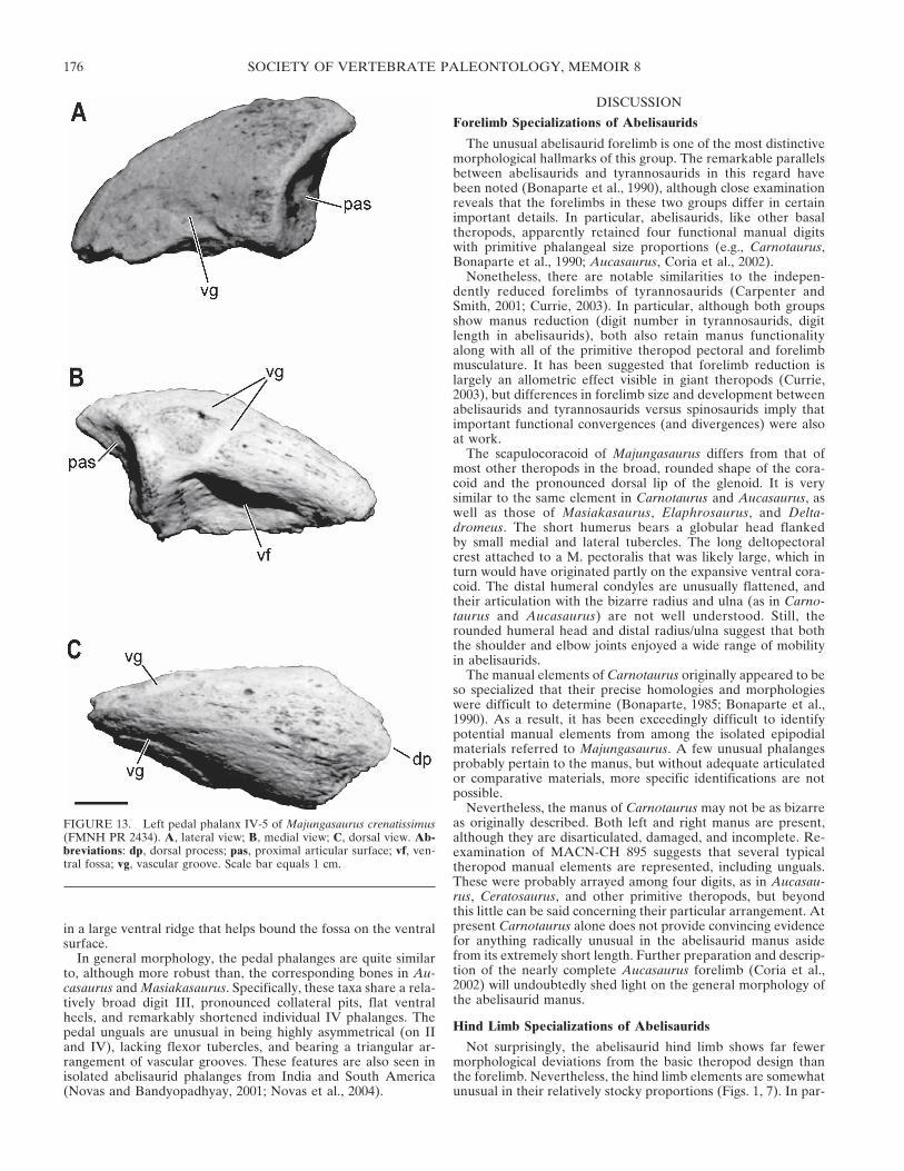
The ungual phalanx of digit IV (IV-5; FMNH PR 2434, UA9043; Fig. 13) is asymmetrical but in a manner opposite to that ofdigit II, as with the phalanges. The proximal end is a roundedtriangle whose two facets are separated by a weak ridge. A ven-tral tubercle is lacking, replaced instead by a deep fossa contain-ing several foramina. The fossa appears to be confluent with theventral vascular grooves on the lateral and medial surfaces. Atriangular arrangement of grooves can be seen on the medialsurface, but on the lateral side only a ventral groove is apparent.The lateral surface is flatter than the medial, terminating below
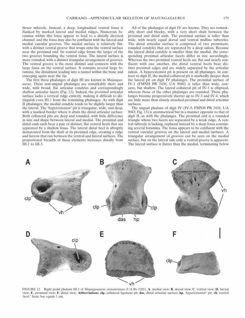
FIGURE 12. Right pedal phalanx III-1 of Majungasaurus crenatissimus (UA Bv-1265). A, medial view; B, dorsal view; C, ventral view; D, lateralview; E, proximal view; F, distal view. Abbreviations: clp, collateral ligament pit; das, distal articular surface; hp, ‘hyperextensor’ pit; vh, ventral‘heel.’ Scale bar equals 1 cm.
CARRANO—APPENDICULAR SKELETON OF MAJUNGASAURUS 175
in a large ventral ridge that helps bound the fossa on the ventralsurface.
In general morphology, the pedal phalanges are quite similarto, although more robust than, the corresponding bones in Au-casaurus and Masiakasaurus. Specifically, these taxa share a rela-tively broad digit III, pronounced collateral pits, flat ventralheels, and remarkably shortened individual IV phalanges. Thepedal unguals are unusual in being highly asymmetrical (on IIand IV), lacking flexor tubercles, and bearing a triangular ar-rangement of vascular grooves. These features are also seen inisolated abelisaurid phalanges from India and South America(Novas and Bandyopadhyay, 2001; Novas et al., 2004).
DISCUSSION
Forelimb Specializations of Abelisaurids
The unusual abelisaurid forelimb is one of the most distinctivemorphological hallmarks of this group. The remarkable parallelsbetween abelisaurids and tyrannosaurids in this regard havebeen noted (Bonaparte et al., 1990), although close examinationreveals that the forelimbs in these two groups differ in certainimportant details. In particular, abelisaurids, like other basaltheropods, apparently retained four functional manual digitswith primitive phalangeal size proportions (e.g., Carnotaurus,Bonaparte et al., 1990; Aucasaurus, Coria et al., 2002).
Nonetheless, there are notable similarities to the indepen-dently reduced forelimbs of tyrannosaurids (Carpenter andSmith, 2001; Currie, 2003). In particular, although both groupsshow manus reduction (digit number in tyrannosaurids, digitlength in abelisaurids), both also retain manus functionalityalong with all of the primitive theropod pectoral and forelimbmusculature. It has been suggested that forelimb reduction islargely an allometric effect visible in giant theropods (Currie,2003), but differences in forelimb size and development betweenabelisaurids and tyrannosaurids versus spinosaurids imply thatimportant functional convergences (and divergences) were alsoat work.
The scapulocoracoid of Majungasaurus differs from that ofmost other theropods in the broad, rounded shape of the cora-coid and the pronounced dorsal lip of the glenoid. It is verysimilar to the same element in Carnotaurus and Aucasaurus, aswell as those of Masiakasaurus, Elaphrosaurus, and Delta-dromeus. The short humerus bears a globular head flankedby small medial and lateral tubercles. The long deltopectoralcrest attached to a M. pectoralis that was likely large, which inturn would have originated partly on the expansive ventral cora-coid. The distal humeral condyles are unusually flattened, andtheir articulation with the bizarre radius and ulna (as in Carno-taurus and Aucasaurus) are not well understood. Still, therounded humeral head and distal radius/ulna suggest that boththe shoulder and elbow joints enjoyed a wide range of mobilityin abelisaurids.
The manual elements of Carnotaurus originally appeared to beso specialized that their precise homologies and morphologieswere difficult to determine (Bonaparte, 1985; Bonaparte et al.,1990). As a result, it has been exceedingly difficult to identifypotential manual elements from among the isolated epipodialmaterials referred to Majungasaurus. A few unusual phalangesprobably pertain to the manus, but without adequate articulatedor comparative materials, more specific identifications are notpossible.
Nevertheless, the manus of Carnotaurus may not be as bizarreas originally described. Both left and right manus are present,although they are disarticulated, damaged, and incomplete. Re-examination of MACN-CH 895 suggests that several typicaltheropod manual elements are represented, including unguals.These were probably arrayed among four digits, as in Aucasau-rus, Ceratosaurus, and other primitive theropods, but beyondthis little can be said concerning their particular arrangement. Atpresent Carnotaurus alone does not provide convincing evidencefor anything radically unusual in the abelisaurid manus asidefrom its extremely short length. Further preparation and descrip-tion of the nearly complete Aucasaurus forelimb (Coria et al.,2002) will undoubtedly shed light on the general morphology ofthe abelisaurid manus.
Hind Limb Specializations of Abelisaurids
Not surprisingly, the abelisaurid hind limb shows far fewermorphological deviations from the basic theropod design thanthe forelimb. Nevertheless, the hind limb elements are somewhatunusual in their relatively stocky proportions (Figs. 1, 7). In par-
FIGURE 13. Left pedal phalanx IV-5 of Majungasaurus crenatissimus(FMNH PR 2434). A, lateral view; B, medial view; C, dorsal view. Ab-breviations: dp, dorsal process; pas, proximal articular surface; vf, ven-tral fossa; vg, vascular groove. Scale bar equals 1 cm.
SOCIETY OF VERTEBRATE PALEONTOLOGY, MEMOIR 8176
ticular, the tibia is short relative to other hind limb elements,and the entire limb appears to be short relative to body length(although the incomplete nature of most abelisaurid skele-tons makes this difficult to determine precisely). It is interest-ing to note that a subset of abelisaurids—notably Majunga-saurus, Lametasaurus, Quilmesaurus, GSI K19/579, and GSIK27/568—have stocky tibiae even compared to other mem-bers of this clade. In contrast, Aucasaurus, Xenotarsosaurus, ISIR91/1, and perhaps Carnotaurus appear to have tibial propor-tions that are closer to those of other theropods (Novas et al.,2004).
Where they can be identified, pelvic and hind limb muscleattachment points appear to be consistent with those seen inother basal theropods (e.g. Hutchinson, 2001). Like coelophy-soids and some tetanurans, abelisaurids lack coelurosaur synapo-morphies such as a marked accessory trochanter at the base ofthe lesser trochanter (insertion of M. puboischiofemoralis inter-nus 2), a well-defined fossa on the anterior pubic peduncle of theilium (origin of M. puboischiofemoralis internus 1), and a fibularfossa restricted to the medial surface only (insertion of M. pop-liteus). No shifts in muscle attachment positions associated withthese morphologies are therefore inferred to have occurred inthese taxa.
However, the abelisaurid hind limb is also derived relative tothose of coelophysoids and more primitive taxa. The inser-tion for M. popliteus, composed largely of the medial fibularfossa, has moved to occupy most of the medial surface of theproximal fibula, unlike in coelophysoids where it originates froma shallow posterior sulcus. The lesser trochanter is more elevatedand accompanied by a distinct bump for insertion of M. iliofemo-ralis, indicating that the deep dorsal muscles were more similarto the avian condition than to those of primitive theropods(Hutchinson, 2001). In addition, the tarsus has achieved thetetanuran condition, with a mediolaterally expanded distaltibia that partially backs the fibula and articulates with the cal-caneum.
The pronounced cnemial crest is associated with a prominentmedial femoral epicondyle in other abelisauroids, and such astructure may also have been present in Majungasaurus. Thismarks part of the origin of Mm. femorotibiales, which contrib-uted to the knee extensor tendon(s) that inserted on the cnemialcrest. The possible elaboration of Mm. femorotibiales, but notother, more proximal knee extensors (such as Mm. iliotibialesand M. ambiens) suggests enhancement of the knee extensionmoment. The extensive lateral fossa on the proximal tibia mayhave housed a large M. tibialis anterior as well.
Consolidation of the tarsus into a single, block-like structurehas been cited as a synapomorphy of Ceratosauria (sensuGauthier, 1986; Rowe, 1989), but recent phylogenetic work (e.g.,Carrano et al., 2002; Rauhut, 2003) suggests that this is con-vergent between coelophysoids and abelisauroids. Further dif-ferences in detail between the fusion patterns in these groupssupport this hypothesis (see below). Regardless, although somedegree of immobility obviously would have been conferred bysuch fusion, it is not clear what effect this might have had ontarsal function because little mobility is inferred between theseelements in theropods where they remain unfused.
The abelisauroid pes is also unusual in bearing markedlyasymmetrical unguals at the ends of relatively broad, shorteneddigits. The digits themselves also appear to be more stronglycurved and bear pronounced, flattened ventral tendon attach-ments. The metatarsals are fairly tightly appressed, not divergingfrom one another until rather close to their distal ends. Theprominence of metatarsal III appears to be a common featureof abelisauroids, although the particular breadth exhibitedby Majungasaurus, for example, may characterize only abelisau-rids.
Phylogenetic Implications
The appendicular morphology of Majungasaurus, particularlythe tarsus, strongly suggests closer affinities with tetanurans thanwith coelophysoids and other more primitive theropods. For ex-ample, the distal tibia has a flat lateral malleolus that backs thefibula, lodging within an elongate tarsal facet that overlaps thedorsal surfaces of both the astragalus and calcaneum. The cal-caneal facet for the tibia faces slightly medially. The tibial distalend is mediolaterally elongate (Novas, 1996), with a centrally-placed buttress marking the articulation with the astragalar as-cending process (Molnar et al., 1996). This process is tall andplate-like rather than triangular and peg-like (Sereno, 1999), andsits superficially on the anterior tibia without being inset. Inthese features the abelisaurid hind limb shows similarities withtetanurans and not with coelophysoids. Likewise, the enlargedfemoral lesser trochanter (accompanied by a distinct M. ilio-femoralis externus insertion) is more derived than that of coe-lophysoids and other primitive theropods, resembling the condi-tion in spinosauroids and other basal tetanurans (Hutchinson,2001).
In other aspects, the appendicular morphology of Majungas-aurus is more similar to those of more primitive theropods, in-cluding coelophysoids. These features include an anteromediallyoriented femoral head (Bonaparte, 1991), a femoral lesser tro-chanter well below the level of the femoral head (Novas, 1991),flattened (rather than elliptical) unguals on pedal digits II-IV(Russell and Dong, 1993), and a relatively broad scapular blade(Gauthier, 1986). It is important to note, however, that theserepresent symplesiomorphies and not synapomorphies of Majun-gasaurus and coelophysoids. Only one appendicular feature—fusion between the astragalus and calcaneum—can be observedthat appears to be synapomorphic (or homoplastic) betweenthese two taxa. Even this characteristic, however, shows somedifferences in detail. In abelisauroids (Majungasaurus, Xenotar-sosaurus, Masiakasaurus), the astragalus and calcaneum arefused to each other, with the ascending process occasionallyfused to the medial fibula and the anterior tibia. By contrast, incoelophysoids (Coelophysis, Syntarsus, Dilophosaurus, Lilien-sternus), the astragalus and calcaneum tend to fuse together butwith little direct involvement of the tibia or fibula.
Thus, the morphology of Majungasaurus is consistent with theresults of previous phylogenetic analyses (Sampson et al., 2001;Carrano et al., 2002; Rauhut, 2003; Wilson et al., 2003) in indi-cating a closer relationship between abelisauroids (and Cerato-saurus) and tetanurans than between abelisauroids and coelo-physoids. Particular similarities in the tarsus suggest that many‘tetanuran’ morphological innovations actually diagnose a moreinclusive clade, and therefore must have appeared considerablyearlier in theropod evolution than had been previously appreci-ated (Carrano et al., 2002; Rauhut, 2003).
CONCLUSIONS
The appendicular morphology of the abelisaurid theropodMajungasaurus crenatissimus is described. Abelisaurid appen-dicular materials have not been well documented, and severalrelatively complete specimens of this Late Cretaceous Malagasytheropod greatly clarify this region of the skeleton in these thero-pods.
The forelimb of Majungasaurus is similar to those of otherabelisaurids and includes a short, highly modified humerus. Nu-merous abelisaurid and abelisauroid synapomorphies are alsofound in the pelvis and hind limb. In addition, the pelvis and hindlimb display a combination of features that strongly suggest aclose affinity between abelisauroids and tetanurans. Function-ally, the abelisaurid forelimb remains obscure and its interpre-tation must await more complete materials. The hind limb ap-
CARRANO—APPENDICULAR SKELETON OF MAJUNGASAURUS 177
pears to show specializations for strong knee extensors, as well asunusual modifications of the metatarsus and pes that may haveadditional locomotor implications.
ACKNOWLEDGMENTS
I thank D. W. Krause, C. A. Forster, and S. D. Sampson forthe invitation to work on the Mahajanga Basin Project, and spe-cifically on the appendicular skeleton of Majungasaurus (thenMajungatholus). I also acknowledge all the members of the 1993-2001 field expeditions for their help in recovering these materi-als, and V. Heisey and M. Getty for skillfully preparing them. F.E. Novas kindly provided a copy of several then-in-press manu-scripts, M. A. Loewen supplied photographs of most of the pha-langes, A. Farke provided the images used in Fig. 6A and B, andR. Ridgley created the image used in Figure 1. Translations ofBonaparte and Novas (1985), Depéret (1896a, b), Deperet andSavornin (1925), Lavocat (1955), Martinez et al. (1986), and No-vas (1991) are available from the Polyglot Paleontologist website(http://ravenel.si.edu/paleo/paleoglot/). This paper benefitedgreatly from the comments of J. A. Wilson, M. C. Lamanna, D. W.Krause, and an anonymous reviewer. This research was sup-ported by grants from the National Science Foundation (DEB-9224396, DEB-9904045, EAR-9418816, EAR-9706302, EAR-0106477, EAR-0116517, EAR-0446488), the Dinosaur Society(1995), and the National Geographic Society (1999, 2001, 2004).
LITERATURE CITED
Bonaparte, J. F. 1985. A horned Cretaceous carnosaur from Patagonia.National Geographic Research 1:149–151.
Bonaparte, J. F. 1991. The Gondwanian theropod families Abelisauridaeand Noasauridae. Historical Biology 5:1–25.
Bonaparte, J. F., and F. E. Novas. 1985. Abelisaurus comahuensis, n. g.,n. sp., Carnosauria del Cretácico Tardio de Patagonia. Ameghiniana21:259–265.
Bonaparte, J. F., F. E. Novas, and R. A. Coria. 1990. Carnotaurus sastreiBonaparte, the horned, lightly built carnosaur from the Middle Cre-taceous of Patagonia. Contributions in Science, Natural History Mu-seum of Los Angeles County 416:1–41.
Britt, B. B., K. C. Cloward, C. A. Miles, and J. H. Madsen, Jr. 1999. Ajuvenile Ceratosaurus (Theropoda, Dinosauria) from Bone CabinQuarry West (Upper Jurassic, Morrison Formation), Wyoming.Journal of Vertebrate Paleontology 19(3, Supplement):33A.
Britt, B. B., D. J. Chure, T. R. Holtz, Jr., C. A. Miles, and K. L. Stadtman.2000. A reanalysis of the phylogenetic affinities of Ceratosaurus(Theropoda, Dinosauria) based on new specimens from Utah, Col-orado, and Wyoming. Journal of Vertebrate Paleontology 20(3,Supplement):32A.
Bryant, H. N., and K. L. Seymour. 1990. Observations and comments onthe reliability of muscle reconstruction in fossil vertebrates. Journalof Morphology 206:109–117.
Carpenter, K., and M. Smith. 2001. Forelimb osteology and biomechanicsof Tyrannosaurus rex; pp. 90–116 in D. H. Tanke and K. Carpenter(eds.), Mesozoic Vertebrate Life: New Research Inspired by thePaleontology of Philip J. Currie. Indiana University Press, Bloom-ington.
Carrano, M. T., and J. R. Hutchinson. 2002. The pelvic and hind limbmusculature of Tyrannosaurus rex (Dinosauria: Theropoda). Jour-nal of Morphology 253:207–228.
Carrano, M. T., S. D. Sampson, and C. A. Forster. 2002. The osteologyof Masiakasaurus knopfleri, a small abelisauroid (Dinosauria:Theropoda) from the Late Cretaceous of Madagascar. Journal ofVertebrate Paleontology 22:510–534.
Carrano, M. T., and S. D. Sampson. 2004. New discoveries of Ma-siakasaurus knopfleri and the morphology of the Noasauridae (Di-nosauria: Theropoda). Journal of Vertebrate Paleontology 24(3,Supplement):44A.
Chakravarti, D. K. 1934. On the systematic position of Lametasaurusindicus. Proceedings of the 21st Indian Science Congress:352.
Chakravarti, D. K. 1935. Is Lametasaurus indicus an armored dinosaur?American Journal of Science, series 5, 30:138–141.
Chatterjee, S. 1978. Indosuchus and Indosaurus, Cretaceous carnosaursfrom India. Journal of Paleontology 52:570–580.
Coria, R. A., L. M. Chiappe, and L. Dingus. 2002. A new close relativeof Carnotaurus sastrei Bonaparte 1985 (Theropoda: Abelisauridae)from the Late Cretaceous of Patagonia. Journal of Vertebrate Pa-leontology 22:460–465.
Currie, P. J. 2003. Allometric growth in tyrannosaurids (Dinosauria:Theropoda) from the Upper Cretaceous of North America andAsia. Canadian Journal of Earth Sciences 40:651–665.
Depéret, C. 1896a. Note sur les dinosauriens sauropodes & théropodesdu Crétacé supérieur de Madagascar. Bulletin de la Sociétégéologique de France, 3e série, 24:176–194.
Depéret, C. 1896b. Sur l’existence de Dinosauriens, Sauropodes etThéropodes, dans le Crétacé supérieur de Madagascar. ComptesRendus Hebdomadaires des Seances de l’Academie des Sciences àParis 122:483–485.
Depéret, C., and J. Savornin. 1928. La faune de Reptiles et de Poissonsalbiens de Timimoun (Sahara algérien). Bulletin de la Sociétégéologique de France, 4e série, 27:257–265.
Dilkes, D. W. 2000. Appendicular myology of the hadrosaurian dinosaurMaiasaura peeblesorum from the Late Cretaceous (Campanian) ofMontana. Transactions of the Royal Society of Edinburgh: EarthSciences, 90:87–125.
Dilkes, D. W. 2001. An ontogenetic perspective on locomotion in theLate Cretaceous dinosaur Maiasaura peeblesorum (Ornithischia:Hadrosauridae). Canadian Journal of Earth Sciences, 38:1205–1227.
Gauthier, J. 1986. Saurischian monophyly and the origin of birds; pp.1–47 in K. Padian (ed.), The Origin of Birds and the Evolution ofFlight. Memoirs of the California Academy of Sciences 8, San Fran-cisco.
Huene, F. v., and C. A. Matley. 1933. The Cretaceous Saurischia andOrnithischia of the Central Provinces of India. Memoirs of the Geo-logical Survey of India: Palaeontologica Indica 21:1–72.
Hutchinson, J. R. 2001. The evolution of femoral osteology and softtissues on the line to extant birds (Neornithes). Zoological Journalof the Linnean Society 131:169–197.
Jasinoski, S. C., A. P. Russell, and P. J. Currie. 2006. An integrativephylogenetic and extrapolatory approach to the reconstruction ofdromaeosaur (Theropoda: Eumaniraptora) shoulder musculature.Zoological Journal of the Linnean Society 146:301–344.
Kellner, A. W. A., and D. d. A. Campos. 2002. On a theropod dinosaur(Abelisauria) from the continental Cretaceous of Brazil. Arquivosdo Museu Nacional, Rio de Janeiro 60:163–170.
Krause, D. W., J. H. Hartman, and N. A. Wells. 1997. Late Cretaceousvertebrates from Madagascar: Implications for biotic change in deeptime; pp. 3–43 in S. D. Goodman and B. D. Patterson (eds.), NaturalChange and Human Impact in Madagascar. Smithsonian InstitutionPress, Washington, D.C.
Krause, D. W., S. D. Sampson, M. T. Carrano, and P. M. O’Connor. 2007.Overview of the history of discovery, taxonomy, phylogeny, andbiogeography of Majungasaurus crenatissimus (Theropoda: Abelis-auridae) from the Late Cretaceous of Madagascar; pp. 1–20 in S. D.Sampson and D. W. Krause (eds.), Majungasaurus crenatissimusfrom the Late Cretaceous of Madagascar. Society of VertebratePaleontology Memoir 8.
Lavocat, R. 1955. Sur une portion de mandibule de théropode provenantdu Crétacé supérieur de Madagascar. Bulletin du Muséum del’Histoire Naturelle, Paris, 2e série, 27:256–259.
Martínez, R., O. Giménez, J. Rodríguez, and G. Bochatey. 1986. Xeno-tarsosaurus bonapartei nov. gen. et sp. (Carnosauria, Abelisauridae),un nuevo Theropoda de la Formación Bajo Barreal, Chubut, Ar-gentina. IV Congreso Argentino de Paleontología y Bioestratigrafía:23–31.
Marsh, O. C. 1881. Principle characters of American Jurassic dinosaurs.Part V. The American Journal of Science and Arts, Series 3, 21:418–423
Marsh, O. C. 1884. The classification and affinities of dinosaurian rep-tiles. Nature 31:68–69.
Matley, C. A. 1921. On the stratigraphy, fossils and geological relation-ships of the Lameta beds of Jubbulpore. Records of the GeologicalSurvey of India 53:142–169.
Matley, C. A. 1924. Note on an armoured dinosaur from the Lameta bedsof Jubbulpore. Records of the Geological Survey of India 55:105–109.
Molnar, R. E. 1990. Problematic Theropoda: “carnosaurs”; pp. 306–317in D. B. Weishampel, P. Dodson, and H. Osmólska (eds.), TheDinosauria. University of California Press, Berkeley.
SOCIETY OF VERTEBRATE PALEONTOLOGY, MEMOIR 8178
Molnar, R. E., A. L. Angriman, and Z. Gasparini. 1996. An AntarcticCretaceous theropod. Memoirs of the Queensland Museum 39:669–674.
Novas, F. E. 1991. Los Tyrannosauridae, gigantescos dinosaurios celu-rosaurios del Cretácico tardio de Laurasia. Ameghiniana 28:401.
Novas, F. E. 1996. Dinosaur monophyly. Journal of Vertebrate Paleon-tology 16:723–741.
Novas, F. E., and S. Bandyopadhyay. 2001. Abelisaurid pedal ungualsfrom the Late Cretaceous of India. Asociación Paleontológica Ar-gentina Publicación Especial, Buenos Aires:145–149.
Novas, F. E., F. L. Agnolin, and S. Bandyopadhyay. 2004. Cretaceoustheropods from India: a review of specimens described by Hueneand Matley (1933). Revista del Museo Argentino de Ciencias Natu-rales, nuevo serie, 6:67–103.
Osmólska, H., E. Roniewicz, and R. Barsbold. 1972. A new dinosaur,Gallimimus bullatus n. gen., n. sp. (Ornithomimidae) from the Up-per Cretaceous of Mongolia. Palaeontologia Polonica 27:103–143.
Ostrom, J. H. 1974. The pectoral girdle and forelimb function of Dei-nonychus (Reptilia: Saurischia): a correction. Postilla 165:1–11.
Owen, R. 1842. Report on British fossil reptiles, part II. Report of theBritish Association for the Advancement of Science 11:60–204.
Rauhut, O. W. M. 2003. The interrelationships and evolution of basaltheropod dinosaurs. Special Papers in Palaeontology 69:1–213.
Rogers, R. R., and J. H. Hartman. 1998. Revised age of the dinosaur-bearing Maevarano Formation (Upper Cretaceous), Mahajanga Ba-sin, Madagascar. Journal of African Earth Sciences 27:160–162.
Rogers, R. R., J. H. Hartman, and D. W. Krause. 2000. Stratigraphicanalysis of Upper Cretaceous rocks in the Mahajanga Basin, north-western Madagascar: implications for ancient and modern faunas.Journal of Geology 108:275–301.
Rogers, R. R., D. W. Krause, K. Curry Rogers, A. H. Rasoamiara-manana, and L. Rahantarisoa. 2007. Paleoenvironment and pa-leoecology of Majungasaurus crenatissimus (Theropoda: Abelisau-ridae) from the Late Cretaceous of Madagascar; pp. 21–31 in S. D.Sampson and D. W. Krause (eds.), Majungasaurus crenatissimus(Theropoda: Abelisauridae) from the Late Cretaceous of Madagas-car. Society of Vertebrate Paleontology Memoir 8.
Romer, A. S. 1923. The pelvic musculature of saurischian dinosaurs.Bulletin of the American Museum of Natural History 48:605–617.
Rowe, T. B. 1989. A new species of the theropod dinosaur Syntarsusfrom the Early Jurassic Kayenta Formation of Arizona. Journal ofVertebrate Paleontology 9:125–136.
Rowe, T. B., and J. A. Gauthier. 1990. Ceratosauria; pp. 151–168 in D. B.Weishampel, P. Dodson, and H. Osmólska (eds.), The Dinosauria.University of California Press, Berkeley.
Russell, D. A., and Z. Dong. 1993. The affinities of a new theropod fromthe Alxa Desert, Inner Mongolia, People’s Republic of China. Ca-nadian Journal of Earth Sciences 30:2107–2127.
Sampson, S. D., and L. M. Witmer. 2007. Craniofacial anatomy of Majun-gasaurus crenatissimus (Theropoda: Abelisauridae) from the LateCretaceous of Madagascar; pp. 32–102 in S. D. Sampson and D. W.Krause (eds.), Majungasaurus crenatissimus (Theropoda: Abelisau-ridae) from the Late Cretaceous of Madagascar. Journal of Verte-brate Paleontology Memoir 8.
Sampson, S. D., M. T. Carrano, and C. A. Forster. 2001. A bizarrepredatory dinosaur from the Late Cretaceous of Madagascar. Na-ture 409:504–506.
Sampson, S. D., D. W. Krause, P. Dodson, and C. A. Forster. 1996. Thepremaxilla of Majungasaurus (Dinosauria: Theropoda), with impli-cations for Gondwanan paleobiogeography. Journal of VertebratePaleontology 16:601–605.
Sampson, S. D., L. M. Witmer, C. A. Forster, D. W. Krause, P. M.O’Connor, P. Dodson, and F. Ravoavy. 1998. Predatory dinosaurremains from Madagascar: implications for the Cretaceous biogeog-raphy of Gondwana. Science 280:1048–1051.
Seeley, H. G.. 1888. On the classification of the fossil animals commonlynamed Dinosauria. Proceedings of the Royal Society of London43:165–171
Sereno, P. C. 1999. The evolution of dinosaurs. Science 284:2137–2147.Snively, E., A. P. Russell, and G. L. Powell. 2004. Evolutionary mor-
phology of the coelurosaurian arctometatarsus: descriptive, morpho-metric and phylogenetic approaches. Zoological Journal of the Lin-nean Society 142:525–553.
Sues, H.-D., and P. Taquet. 1979. A pachycephalosaurid dinosaur fromMadagascar and a Laurasia-Gondwanaland connection in the Cre-taceous. Nature 279:633–635.
Tarsitano, S. F. 1983. Stance and gait in theropod dinosaurs. Acta Pal-aeontologica Polonica 28:251–264.
Walker, A. D. 1964. Triassic reptiles from the Elgin area: Ornithosuchusand the origin of carnosaurs. Philosophical Transactions of theRoyal Society of London B 248:53–134.
Walker, A. D. 1977. Evolution of the pelvis in birds and dinosaurs; pp.319–358 in S. M. Andrews, R. S. Miles, and A. D. Walker (eds.),Problems in Vertebrate Evolution. Linnean Society Symposium Se-ries 4.
Welles, S. P. 1984. Dilophosaurus wetherilli (Dinosauria, Theropoda)osteology and comparisons. Palaeontographica Abteilung A 185:85–180.
Wilson, J. A., P. C. Sereno, S. Srivastava, D. K. Bhatt, A. Khosla, and A.Sahni. 2003. A new abelisaurid (Dinosauria, Theropoda) from theLameta Formation (Cretaceous, Maastrichtian) of India. Contribu-tions from the Museum of Paleontology, University of Michigan31:1–42.
Submitted November 22, 2004; accepted January 5, 2007.
CARRANO—APPENDICULAR SKELETON OF MAJUNGASAURUS 179