tenascin-c aggravates autoimmune myocarditis via dendritic cell activation and th17 cell...
TRANSCRIPT

Tenascin-C Aggravates Autoimmune Myocarditis via Dendritic CellActivation and Th17 Cell DifferentiationTomokoMachino-Ohtsuka, MD; Kazuko Tajiri, MD, PhD; Taizo Kimura, MD, PhD; Satoshi Sakai, MD, PhD; Akira Sato, MD; Toshimichi Yoshida,MD, PhD; Michiaki Hiroe, MD, PhD; Yasuhiro Yasutomi, DVM, PhD; Kazutaka Aonuma, MD, PhD; Kyoko Imanaka-Yoshida, MD, PhD
Background-—Tenascin-C (TN-C), an extracellular matrix glycoprotein, appears at several important steps of cardiac developmentin the embryo, but is sparse in the normal adult heart. TN-C re-expresses under pathological conditions including myocarditis, andis closely associated with tissue injury and inflammation in both experimental and clinical settings. However, thepathophysiological role of TN-C in the development of myocarditis is not clear. We examined how TN-C affects the initiation ofexperimental autoimmune myocarditis, immunologically.
Methods and Results-—A model of experimental autoimmune myocarditis was established in BALB/c mice by immunization withmurine a-myosin heavy chains. We found that TN-C knockout mice were protected from severe myocarditis compared to wild-typemice. TN-C induced synthesis of proinflammatory cytokines, including interleukin (IL)-6, in dendritic cells via activation of a Toll-likereceptor 4, which led to T-helper (Th)17 cell differentiation and exacerbated the myocardial inflammation. In the transferexperiment, dendritic cells loaded with cardiac myosin peptide acquired the functional capacity to induce myocarditis whenstimulated with TN-C; however, TN-C-stimulated dendritic cells generated from Toll-like receptor 4 knockout mice did not inducemyocarditis in recipients.
Conclusions-—Our results demonstrated that TN-C aggravates autoimmune myocarditis by driving the dendritic cell activation andTh17 differentiation via Toll-like receptor 4. The blockade of Toll-like receptor 4-mediated signaling to inhibit the proinflammatoryeffects of TN-C could be a promising therapeutic strategy against autoimmune myocarditis. ( J Am Heart Assoc. 2014;3:e001052doi: 10.1161/JAHA.114.001052)
Key Words: dendritic cell • myocarditis • tenascin-C • Th17 • TLR4
M yocarditis is induced by a variety of agents,including toxins, alcohols, parasites, bacteria, and
viruses.1,2 Postinfectious autoimmune myocarditis oftenresults in idiopathic dilated cardiomyopathy (DCM), whichis sometimes a lethal disorder characterized by leftventricular (LV) enlargement and systolic dysfunction.1,2
Experimental autoimmune myocarditis (EAM) is a mousemodel of such a kind of inflammation-based cardiomyopa-thy.3,4 EAM is known as a CD4+ T-cell-mediated disease,and activation of self-antigen-loaded dendritic cells (DCs) iscritical for the expansion of autoreactive CD4+ T cells.5
Upon encountering immunologic danger, the maturated andactivated DCs act as an important commander regulatingna€ıve CD4+ T cells by means of antigen presentation,costimulatory molecule engagement, and release of acocktail of polarizing cytokines.6 DC activation is mediatedby the recognition of pathogen-associated molecular pat-terns and damage-associated molecular patterns, which aresensed by pattern-recognized receptors such as Toll-likereceptors (TLRs).6
Recently, accumulating evidence has highlighted theproinflammatory role of several extracellular matrix mole-cules including tenascin-C (TN-C), osteopontin, and galec-tin.7–10 Of these, TN-C acts as a damage-associatedmolecular pattern and directly activates inflammatory cellsincluding lymphocytes, macrophages, synovial fibroblasts,and DCs.11–14 TN-C is synthesized by various cell typesin response to inflammatory cytokines and mechanical
From the Cardiovascular Division, Faculty of Medicine, University of Tsukuba,Japan (T.M.-O., K.T., T.K., S.S., A.S., K.A.); Mie University Research Center forMatrix Biology and Department of Pathology and Matrix Biology, Mie UniversityGraduate School of Medicine, Tsu, Japan (T.Y., K.I.-Y.); Department ofCardiology, National Center of Global Health and Medicine, Tokyo, Japan(M.H.); Laboratory of Immunoregulation and Vaccine Research, TsukubaPrimate Research Center, National Institution of Biomedical Innovation,Tsukuba, Japan (Y.Y.).
Correspondence to: Kazutaka Aonuma, MD, PhD, Cardiovascular Division,Faculty of Medicine, University of Tsukuba, 1-1-1 Tennodai, Tsukuba 305-8575, Japan. E-mail: [email protected]
Received July 14, 2014; accepted August 7, 2014.
ª 2014 The Authors. Published on behalf of the American Heart Association,Inc., by Wiley Blackwell. This is an open access article under the terms of theCreative Commons Attribution-NonCommercial License, which permits use,distribution and reproduction in any medium, provided the original work isproperly cited and is not used for commercial purposes.
DOI: 10.1161/JAHA.114.001052 Journal of the American Heart Association 1
ORIGINAL RESEARCH
by KAZUTAKA AONUMA JR. on December 12, 2014http://jaha.ahajournals.org/Downloaded from

stress.12,15,16 TN-C is not normally expressed in the adultheart but is specifically upregulated during clinical conditionsaccompanying inflammation,7,17 such as acute myocardialinfarction,18 hypertensive cardiac fibrosis,19 myocarditis,15,20
and some cases of DCM.21 In particular, our previousanalysis of the myocardium obtained from left ventriculopl-asty specimens of patients with DCM showed that about halfof the patients had significant active inflammation associatedwith TN-C expression.21 Thus, it has been recognized thatTN-C is closely associated with inflammatory status incardiovascular diseases. However, how TN-C acts as aproinflammatory stimulus by mediating the immune systemin myocarditis has not been fully understood.
Here, we show that TN-C leads to TLR4-mediated inflam-matory responses in DCs and exacerbates myocardialinflammation. TN-C knockout (TNKO) mice were protectedfrom severe myocarditis in a model of cardiac myosin-inducedautoimmune myocarditis. Furthermore, we found that TN-Cinduced synthesis of proinflammatory cytokines, includinginterleukin-6 (IL-6), in DCs via activation of TLR4, which led toTh17 cell differentiation. Finally, in the transfer experiment,DCs loaded with cardiac myosin peptide acquired thefunctional capacity to induce myocarditis when stimulatedwith TN-C; however, TN-C-stimulated DCs generated fromTLR4 knockout (TLR4KO) mice did not induce myocarditis inrecipients.
Materials and Methods
MiceTNKO mice, originally generated by Saga et al., were back-crossed with BALB/c mice for more than 12 generations.22
Male TNKO mice and wild-type (WT) littermates (5 to 7 weeksof age) were used in the experiments. Other male BALB/cmice (same age) that were used to obtain bone marrow cellswere purchased from CLEA Japan. TLR4KO mice werepurchased from Oriental Yeast. All animal experiments wereapproved by the Institutional Animal Experiment Committee ofthe University of Tsukuba.
Induction of EAMThe mice were immunized with 100 lg of a-myosin H-chainpeptide (MyHC-a) (MyHC-a614–629 [Ac-RSLKLMATLFSTYA-SADR-OH]; Toray Research Center) emulsified 1:1 in a PBS/complete Freund’s adjuvant (1 mg/mL; H37Ra; Sigma-Aldrich) on days 0 and 7, as described previously.23 Atdifferent time points (0, 2, 5, 8, 11, 14, 17, 21, 25, 29, and33 days) after the first immunization, a total of 44 mice wereanesthetized with pentobarbital (60 mg/kg i.p.) and theirhearts were removed.
Myosin-Specific Bone Marrow-Derived DendriticCell (BMDC)-Induced MyocarditisBMDCs were generated as previously described.24 BMDCswere pulsed overnight with 10 lg/mL MyHC-a peptide andstimulated for another 4 hours with 1 lg/mL lipopolysac-charide (LPS) (Sigma-Aldrich) or 10 lg/mL TN-C and 5 lg/mL anti-CD40L (AbD Serotec).5 Recipient mice received59105 pulsed and activated BMDCs i.p. on days 0, 2, and 4and were killed 10 days after the first injection.
Histopathological and ImmunohistochemicalExaminationThe hearts were fixed in 4% paraformaldehyde in PBS andembedded in paraffin wax. For a histological analysis, 3-lm-thick sections were cut and stained with hematoxylin andeosin. To evaluate the expression of TN-C, we performedimmunohistochemistry as previously described.25 In brief,sections after antigen retrieval were incubated with poly-clonal rabbit anti-TN-C antibodies,15,26 followed by treatmentwith horseradish peroxidase–conjugated goat anti-rabbitantibody (MBL, Nagoya, Japan). The antibody reactions werevisualized using diaminobenzidine chromogen and werecounterstained with hematoxylin. Myocarditis severity wasscored on hematoxylin and eosin–stained sections usinggrades from 0 to 4: 0, no inflammation; 1, >25% of theheart section involved; 2, 25% to 50%; 3, >50% to 75%; and4, >75%, as described previously.23 Two independentresearchers scored the slides separately in a blindedmanner.
Assessment of LV FunctionThe assessment of the LV function was performed in na€ıveor EAM WT and TNKO mice (14 days after immunization).The mice were anesthetized with an intraperitoneal injectionof sodium pentobarbital (50 mg/kg). The LV apex wasexposed via a subdiaphragmatic incision. An apical stab wasmade with a 27-gauge needle containing a fiber pressuresensor (FPI-LS-PT9; FISO Technologies, Inc, Qu"ebec, Can-ada),27 placed to span the long axis of the LV. Pressuremeasurements were obtained at steady state. All signalswere analyzed with a signal conditioner (FPI-LS-10; FISOTechnologies, Inc) and data acquisition system (LabTrax 4,World Precision Instruments, Sarasota, FL) and then storedon disks for off-line analysis using software (LabScribe, iWorxSystems, Inc, Dover, NH). The following indices wereassessed: heart rate, systolic and end-diastolic LV pressures,and maximal and minimum rates of LV pressure development(!dP/dt).
DOI: 10.1161/JAHA.114.001052 Journal of the American Heart Association 2
Proinflammatory Roles of Tenascin-C in Myocarditis Machino-Ohtsuka et al
ORIG
INALRESE
ARCH
by KAZUTAKA AONUMA JR. on December 12, 2014http://jaha.ahajournals.org/Downloaded from

Flow Cytometric Analyses and IntracellularCytokine StainingHeart inflammatory cells were isolated and processed aspreviously described.5,28 For the flow cytometric analysis ofthe surface markers and cytoplasmic cytokines, the cells werestained directly using fluorochrome-conjugated mouse-spe-cific antibodies and analyzed with a FACSCalibur instrument(BD Biosciences). For the analysis of the intracellular cytokineproduction, the cells were stimulated with 50 ng/mL phorbol12-myristate 13-acetate (Sigma-Aldrich), 750 ng/mL ionomy-cin (Sigma-Aldrich), and 10 lg/mL brefeldin A (eBioscience)for 5 hours. The antibodies were purchased from eBioscienceand included CD4, CD45, Forkhead box protein (Foxp)3,interferon (IFN)-c, and IL-17A.
Stimulation of DCsBMDCs (59105) generated as described above werecultured with TN-C (10 lg/mL) for 48 hours. In someexperiments, 0.1 lmol/L TAK242 (Millipore), a selectiveTLR4 signal transduction inhibitor, was added. Culturesupernatants were subjected to measurements of cytokineor chemokine production. For the Western blotting analysis,we used DCs obtained from spleens of na€ıve WT and TNKOmice by anti-CD11c microbeads (Miltenyi Biotec). Toevaluate the TLR4 expression or NF-KB signaling, the DCswere stimulated with TN-C (10 lg/mL) or tumor necrosisfactor-a (20 ng/mL) for 15 minutes and were subjected toanalysis.
In Vitro Th17 InductionWe used a CD4+CD62L+ T-cell isolation kit (Miltenyi Biotec)and anti-CD11c microbeads (Miltenyi Biotec) to isolateCD4+ na€ıve T cells and DCs from spleens, respectively. TheCD4+ na€ıve T cells (19106) were co-cultured with DCs(59104) and stimulated with 10 lg/mL TN-C for 72 hours.In some experiments, DCs were pretreated with TAK242(0.1 lmol/L) or anti-IL-6 antibody (10 lg/mL; R&D Sys-tems). The supernatants were collected and the cytokineswere measured.
Measurements of Cytokines and ChemokinesThe hearts were homogenized in media containing 10% fetalbovine serum. The supernatants were collected after centri-fugation and stored at !80°C. The concentrations ofcytokines and chemokines in the heart homogenates, serum,or culture supernatants were measured by the use ofMillipore multiplex immunoassay panels (Millipore). In somecases, the cytokine levels were confirmed with a Quantikine
ELISA kit (R&D Systems). For measurements of the TN-Cconcentration, the hearts were homogenized in a buffercontaining 150 mmol/L NaCl, 25 mmol/L Tris (pH 7.4),5 mmol/L EDTA, 10 mmol/L sodium pyrophosphate,10 mmol/L NaF, 1 mmol/L Na3NO4, and complete minipro-tease inhibitors (Roche). The proteins were extracted fromthe homogenized samples by adding Triton X-100 (Sigma) toa final concentration of 1%. Suspensions of hearts mixed with1% Triton X were vortexed and incubated on ice for 2 hours.Then, the suspensions were centrifuged at 22 200 g for15 minutes, and the supernatants were collected. Theconcentration of the TN-C was measured by a Tenascin-CLarge (FNIII-B) Assay kit (IBL).
Serum Troponin DeterminationsBlood was collected from mice at the time of sacrifice, and theserum levels of cardiac troponin I were measured with an ELISAkit (Mouse Cardiac Tn-I, Ultra Sensitive; Life Diagnostics).
Western Blot AnalysisThe total lysates from WT and TNKO DCs were immunoblottedand probed with primary antibodies against TLR4, NF-KB, andphospho-NF-KB (Ser536) (Cell Signaling Technology). Horse-radish peroxidase–conjugated secondary antibodies (CellSignaling Technology) were used to identify the binding sitesof the primary antibody.
RNA Extraction and Quantitative Real-TimeReverse Transcription Polymerase Chain ReactionAll of the hearts removed for the reverse transcriptionpolymerase chain reaction were snap frozen and stored at!80°C. For the preparation of the total RNA, the tissue washomogenized using a bead kit (MagNA Lyser Green Beads;Roche Diagnostics) according to the manufacturer’s instruc-tions. The total RNA was extracted using a MagNA PureCompact Instrument (Roche Applied Science) together with aMagNA Pure Compact RNA Isolation Kit (Roche AppliedScience) according to the manufacturer’s instructions. cDNAwas synthesized from 1 lg total RNA with an Omniscript RTkit (Qiagen). A quantitative-reverse transcription polymerasechain reaction analysis was performed with the LightCycler480 system (Roche Applied Science) with a Universal ProbeLibrary (Roche Applied Science). The primers for the mouseTnc were 50-CCCTCTCTCTGTTGAGGTCTTG-30 (sense) and 50-CCCAGCTGACCTCAGTCAC-30 (antisense). The primers for themouse Hprt were 50-TCCTCCTCAGACCGCTTTT-30 (sense) and50-CCTGGTTCATCATCGCTAATC-30 (antisense). Hprt RNA wasused as an internal control.
DOI: 10.1161/JAHA.114.001052 Journal of the American Heart Association 3
Proinflammatory Roles of Tenascin-C in Myocarditis Machino-Ohtsuka et al
ORIG
INALRESE
ARCH
by KAZUTAKA AONUMA JR. on December 12, 2014http://jaha.ahajournals.org/Downloaded from

StatisticsAll data are expressed as means!SEM. The normality wastested with the Shapiro–Wilk test. The TN-C mRNA andprotein levels after the myocarditis induction were com-pared with the baseline levels using an unpaired 2-tailedt test (Figure 1B). The heart-to-body-weight ratios, serumtroponin I concentrations, flow cytometric analyses data,hemodynamic parameters, and cytokines/chemokine levelswere compared between 2 groups by an unpaired 2-tailedt test (Figures 2C through 2H, 3, 4, 5B through 5D, 6, and8C and 8D). A 1-way analysis of variance was used tocompare the levels of the TN-C in multiple groups(Figure 5A). To compare the severity scores of myocarditisbetween 2 groups, the Mann–Whitney U test was used(Figures 2B, 8B, and Table). The Fisher Exact test was usedto compare the prevalence of DC-induced myocarditisbetween the control group and the other 4 groups,
respectively (Table). A value of P<0.05 was considered tobe statistically significant.
Results
Expression of TN-C in the EAM HeartsFirst, we examined the expression of TN-C in WTmice with EAMthat was induced by immunization with cardiac myosin. Around5 to 6 days after the first immunization, small clusters ofinfiltrating inflammatory cells appeared, and TN-C becamedetectable (Figure 1A). The TN-C expression peaked at day 14,and themolecule was localized to the interstitial spaces in areaswhere inflammatory cell infiltration was evident (Figure 1A).The myocardial inflammation and TN-C expression graduallysubsided and disappeared around 25 days after immunization(Figure 1A). A quantitative-reverse transcription polymerase
A B
Figure 1. Tenascin-C (TN-C) expression in cardiac myosin-induced autoimmune myocarditis. BALB/cmice were immunized twice, on days 0 and 7, with 100 lg of cardiac myosin epitope peptide (MyHC-a).A, Representative histology of myocarditis on days 5, 14, and 25, respectively. Hematoxylin and eosinstaining (i) and immunostaining for TN-C (ii). Scale bars=50 lm. B, The expression of TN-C in heartsobtained from immunized mice at the indicated time points. Immunization on days 0 and 7 are indicatedwith red arrows. TN-C mRNA expression was evaluated by quantitative reverse transcription–polymerasechain reaction. The results are reported as the fold change in the gene expression relative to the expressionon day 0. The TN-C protein levels were measured by an ELISA. n=4 per group at each time point. Error barsrepresent the mean!SEM. *P<0.05 vs day 0. CFA indicates complete Freund’s adjuvant.
DOI: 10.1161/JAHA.114.001052 Journal of the American Heart Association 4
Proinflammatory Roles of Tenascin-C in Myocarditis Machino-Ohtsuka et al
ORIG
INALRESE
ARCH
by KAZUTAKA AONUMA JR. on December 12, 2014http://jaha.ahajournals.org/Downloaded from
chain reaction analysis and ELISA showed that TN-C wasexpressed in parallel with the histological findings (Figure 1B).
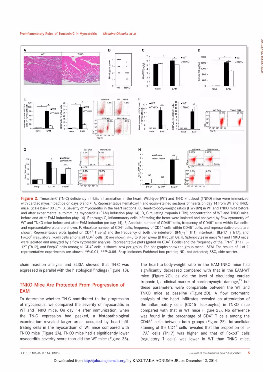
TNKO Mice Are Protected From Progression ofEAMTo determine whether TN-C contributed to the progressionof myocarditis, we compared the severity of myocarditis inWT and TNKO mice. On day 14 after immunization, whenthe TN-C expression had peaked, a histopathologicalexamination revealed larger areas occupied by heart-infil-trating cells in the myocardium of WT mice compared withTNKO mice (Figure 2A). TNKO mice had a significantly lowermyocarditis severity score than did the WT mice (Figure 2B).
The heart-to-body-weight ratio in the EAM-TNKO mice hadsignificantly decreased compared with that in the EAM-WTmice (Figure 2C), as did the level of circulating cardiactroponin I, a clinical marker of cardiomyocyte damage,29 butthese parameters were comparable between the WT andTNKO mice at baseline (Figure 2D). A flow cytometricanalysis of the heart infiltrates revealed an attenuation ofthe inflammatory cells (CD45+ leukocytes) in TNKO micecompared with that in WT mice (Figure 2E). No differencewas found in the percentage of CD4+ T cells among theCD45+ cells between both groups (Figure 2F). Intracellularstaining of the CD4+ cells revealed that the proportion of IL-17A+ cells (Th17) was higher and that of Foxp3+ cells(regulatory T cells) was lower in WT than TNKO mice,
A
E
G H
F
B C D
Figure 2. Tenascin-C (TN-C) deficiency inhibits inflammation in the heart. Wild-type (WT) and TN-C knockout (TNKO) mice were immunizedwith cardiac myosin peptide on days 0 and 7. A, Representative hematoxylin and eosin–stained sections of hearts on day 14 from WT and TNKOmice. Scale bar=100 lm. B, Severity of myocarditis in the heart sections. C, Heart-to-body-weight ratios (HW/BW) in WT and TNKO mice beforeand after experimental autoimmune myocarditis (EAM) induction (day 14). D, Circulating troponin I (TnI) concentration of WT and TNKO micebefore and after EAM induction (day 14). E through G, Inflammatory cells infiltrating the heart were isolated and analyzed by flow cytometry ofWT and TNKO mice before and after EAM induction (on day 14). E, Absolute number of CD45+ cells, frequency of CD45+ cells within live cells,and representative plots are shown. F, Absolute number of CD4+ cells, frequency of CD4+ cells within CD45+ cells, and representative plots areshown. Representative plots (gated on CD4+ T cells) and the frequency of both the interferon (IFN)-c+ (Th1), interleukin (IL)-17+ (Th17), andFoxp3+ (regulatory T-cell) cells among all CD4+ cells (G) are shown. n=5 to 8 per group (B through G). H, Splenocytes in na€ıve WT and TNKO micewere isolated and analyzed by a flow cytometric analysis. Representative plots (gated on CD4+ T cells) and the frequency of the IFN-c+ (Th1), IL-17+ (Th17), and Foxp3+ cells among all CD4+ cells is shown. n=4 per group. The bar graphs show the group mean!SEM. The results of 1 of 2representative experiments are shown. *P<0.01, **P<0.05. Foxp indicates Forkhead box protein; ND, not detected; SSC, side scatter.
DOI: 10.1161/JAHA.114.001052 Journal of the American Heart Association 5
Proinflammatory Roles of Tenascin-C in Myocarditis Machino-Ohtsuka et al
ORIG
INALRESE
ARCH
by KAZUTAKA AONUMA JR. on December 12, 2014http://jaha.ahajournals.org/Downloaded from

whereas the proportion of IFN-c+ cells (Th1) was similarbetween WT and TNKO mice (Figure 2G). The CD4+ T-cellproportions in splenocytes at day 0 did not differ betweenWT and TNKO mice (Figure 2H), indicating that thedeficiency of TN-C did not influence CD4+ T-cell differenti-ation in the non-EAM condition. To further evaluate theeffects of TN-C deficiency on the severity of EAM, weexamined hemodynamic parameters using a pressure sen-sor. In the na€ıve mice, there was no statistical difference inthe heart rate, LV systolic pressure, LV end-diastolicpressure, or !dP/dt between the WT and TNKO mice(Figure 3A through 3E). The LV end-diastolic pressureincreased and !dP/dt decreased more in the WT EAMmice than in the WT na€ıve mice, consistent with severemyocarditis (Figure 3B through 3E). In comparison, TNKOmice showed no significant deterioration in these parame-ters even after the induction of EAM (Figure 3B through 3E).
Taken together, the presence of TN-C in the heart duringthe acute phase of EAM might contribute to the infiltrationof inflammatory cells including Th17 into the heart and tothe progression of myocarditis and LV dysfunction.
TN-C Promotes Proinflammatory Cytokine andChemokine Synthesis in the HeartTo gain new insights into the mechanism of protection againstmyocarditis in TNKO mice, we examined whether a TN-Cdeficiency affected the cytokine and chemokine milieu in theheart. In naive WT and TNKO hearts, the expression ofproinflammatory cytokines and chemokines was quite smallto none (Figure 4). On day 14 following immunization, theheart homogenates from TNKO mice had significantly reducedlevels of the proinflammatory cytokines IL-1a, IL-1b, IL-6, IL-17, tumor necrosis factor-a, and transforming growth factor-b
Table. Prevalence and Severity of MyHC-a-Loaded DC-Induced Myocarditis in WT and TN-C KO Mice
Donors Recipients Activation Prevalence (Day 10) Median Severity Grade at Day 10 (Individual Data)
WT WT PBS 0/7 0
WT WT LPS 7/7* 2 (2, 2, 2, 2, 3, 3, 3)†
WT WT TN-C 7/7* 2 (1, 2, 2, 2, 3, 3, 3)†
TNKO TNKO PBS 0/7 0
TNKO TNKO TN-C 7/7* 2 (1, 1, 2, 2, 2, 3, 3)†
DC indicates dendritic cell; LPS, lipopolysaccharide; MyHC-a, a-myosin H-chain peptide; TN-C, tenascin-C; TNKO, TN-C knockout; WT, wild-type.*P<0.001 (Fisher’s exact test), †P<0.01 (Mann-Whitney U test) vs WT-DCs (with PBS)-induced myocarditis.
A
D E
B C
Figure 3. Effects of tenascin-C (TN-C) deficiency on the hemodynamic parameters in experimentalautoimmune myocarditis (EAM) mice. A, Heart rate; (B) Left ventricular (LV) systolic pressure (LVSP); (C) LVend-diastolic pressure (LVEDP); (D) Maximal rate of the increase in the LV pressure (+dP/dt); and (E), Maximalrate of the decrease in the LV pressure ("dP/dt). Na€ıve or EAM wild-type (WT) and TN-C knockout (TNKO)mice (day 14) were analyzed. n=5 to 7 per group. Bar graphs show the group mean!SEM. *P<0.05.
DOI: 10.1161/JAHA.114.001052 Journal of the American Heart Association 6
Proinflammatory Roles of Tenascin-C in Myocarditis Machino-Ohtsuka et al
ORIG
INALRESE
ARCH
by KAZUTAKA AONUMA JR. on December 12, 2014http://jaha.ahajournals.org/Downloaded from

(Figure 4). Also, the lack of TN-C had significantly reduced thelevels of the following chemokines: macrophage inflammatoryprotein (MIP)-1a (CCL3), MIP-1b (CCL4), MIP-2 (CXCL2),monocyte chemoattractant protein-1 (MCP-1), keratinocytechemoattractant (KC) (CXCL1), and IFN-c-induced protein(IP)-10 (CXCL10) (Figure 4). Thus, protection from autoim-mune myocarditis in TNKO mice is associated with abrogationof proinflammatory molecules and chemokines in the heart.
TN-C Mediates DC Activation and Th17 CellDevelopmentEAM is a CD4+ T-cell-mediated disease,3,4 and DCs are themajor antigen-presenting cells (and key players in the primingof appropriate CD4+ T-cell responses. To assess whether TN-Cis secreted by DCs, we generated BMDCs in vitro by culturingbone marrow cells obtained from WT mice with granulocyte/macrophage colony-stimulating factor. We found that BMDCsactivated with LPS produced only a small amount of TN-C, asdid nonstimulated BMDCs (Figure 5A). Therefore, we culturedWT BMDCs in the presence of TN-C and investigated theeffects of TN-C on BMDCs in proinflammatory cytokine andchemokine production. When BMDCs were stimulated withTN-C, they produced greater amounts of the proinflammatorycytokines IL-1a, IL-1b, IL-6, IL-12p40, and IFN-c than didthose without TN-C (Figure 5B). TN-C stimulation alsopromoted BMDCs to produce the chemokines MCP-1, MIP-1a, MIP-1b, MIP-2, IP-10, regulated upon activation, normalT-cell expressed and secreted (RANTES), and KC and growthfactors G-CSF and granulocyte/macrophage colony-stimulat-ing factor (Figure 5B). These data indicated that TN-C
promotes the production of many types of chemokines andinflammatory cytokines from DCs, which participate in theproliferation, accumulation, and activation of immune cellssuch as monocytes, macrophages, and lymphocytes. More-over, TN-C induced the production of IL-6, a key cytokine inTh17 development,30 by BMDCs in a dose-dependent manner(Figure 5C). On the basis of this significant effect of TN-C onIL-6 secretion by DCs, we next attempted to confirm that TN-C-stimulated BMDCs induce Th17 development. We cocul-tured TN-C-stimulated DCs and na€ıve CD4+ T cells in thepresence of anti-CD3 monoclonal antibody and transforminggrowth factor-b and analyzed IL-17 production in the culturesupernatant. As expected, we found that TN-C-stimulated DCspromoted IL-17 production from CD4+ T cells (Figure 5D).Moreover, we confirmed the neutralization of IL-6 by using aspecific antibody that inhibited the effects of TN-C-stimulatedDCs on the promotion of IL-17 production from CD4+ T cells(Figure 5D). These results indicated that TN-C encouraged thegeneration of Th17 via the secretion of IL-6 from DCs.
TN-C Activates DCs via TLR4Multiple cell-surface receptors are known to bind to the TN-Cmolecule.31 Among them, TLR4 has been shown to be areceptor that transmits important signals to modulateinflammatory response.13 To determine whether TN-C acti-vates DCs via TLR4, we developed BMDCs from TLR4KO mice(TLR4KO-BMDC) and examined whether TN-C induced thecytokine and chemokine secretion. TLR4KO-BMDCs failed toproduce the proinflammatory cytokines IL-1a, IL-1b, IL-6,IL-12p40, and IFN-c even under stimulation with adequate
Figure 4. Tenascin-C (TN-C) deficiency affected the cytokine milieu in the heart. Cytokine and chemokine secretion in homogenized heartsobtained from na€ıve and experimental autoimmune myocarditis (EAM) (on day 14) wild-type (WT) and TN-C knockout (TNKO) mice was assessedby an ELISA. n=4 to 5 per group. The bar graphs show the group mean!SEM. The results of 1 of 2 representative experiments are shown.*P<0.05, **P<0.01. IL indicates interleukin; IP, IFN-c-induced protein; KC, keratinocyte chemoattractant; MCP, monocyte chemoattractantprotein; MIP, macrophage inflammatory protein; ND, not detected; RANTES, regulated on activation, normal T-cell expressed and secreted; TGF,transforming growth factor; TNF, tumor necrosis factor.
DOI: 10.1161/JAHA.114.001052 Journal of the American Heart Association 7
Proinflammatory Roles of Tenascin-C in Myocarditis Machino-Ohtsuka et al
ORIG
INALRESE
ARCH
by KAZUTAKA AONUMA JR. on December 12, 2014http://jaha.ahajournals.org/Downloaded from

amounts of TN-C (Figure 6A). TN-C did not increase thechemokine production, including that of MIP-1a, MIP-1b, MIP-2, RANTES, KC, and IP-10 from TLR4KO-BMDCs (Figure 6A).Furthermore, TLR4 inhibitor TAK242 attenuated TN-C-inducedproduction of IL-1a, IL-1b, IL-6, IL-12p40, IFN-c, MIP-1a,MIP-1b, MIP-2, RANTES, KC, and IP-10 (Figure 6B). Thesedata indicated that TN-C-activated DCs produce proinflam-matory cytokine/chemokine mainly via a TLR4-mediatedsignaling cascade. Next, we cocultured na€ıve CD4+ T cellsand TN-C-stimulated TLR4KO-BMDCs. Despite the presenceof a sufficient amount of TN-C, the IL-17 secretion from CD4+
T cells did not increase (Figure 6C). Furthermore, we
cocultured na€ıve CD4+ T cells and TN-C-stimulated DCs fromWT mice with or without TAK242. Pretreatment with TAK242also resulted in an attenuation of the IL-17 secretion fromCD4+ T cells (Figure 6D). Collectively, DCs activated by theTLR4-mediated signaling mainly contributed to the activationof DCs by TN-C, which promoted Th17 differentiation.
TN-C-Stimulated DCs Gain a Myocarditis-InducingCapacity via TLR4To collect direct evidence that TN-C provides DCs with theability to induce autoimmune myocarditis, we used another
A
C
D
B
Figure 5. Tenascin-C (TN-C) stimulated production of proinflammatory cytokines and chemokines by bone marrow (BM)–derived dendriticcells (DCs) and differentiated na€ıve CD4+ cells into Th17 cells. A, DCs generated from BM (BMDCs) were cultured in the presence or absence oflipopolysaccharide (LPS) 1 lg/mL for 72 hours. TN-C secretions from BMDCs and the TN-C concentration in the medium were measured by anELISA. B, BMDCs were cultured in the presence of 10 lg/mL of TN-C for 48 hours, and the supernatants were subjected to multipleximmunoassay panels for the production of proinflammatory cytokines, chemokines, and growth factors. C, BMDCs were cultured in the presenceof the indicated dose of TN-C for 48 hours. TN-C-dose-dependent IL-6 secretions from BMDCs were measured by an ELISA. D, CD62high na€ıveCD4+ T cells were cultured with DCs, which were obtained from the spleen, in the presence of anti-CD3 mAb (1 lg/mL), TGF-b (2 ng/mL), andTN-C (10 lg/mL) for 72 hours. In some wells, anti-IL-6 Ab (10 lg/mL) was added. The secretion of IL-17 in the supernatants was analyzed byan ELISA. The values are expressed as means!SEM of triplicate culture wells. The results of 1 of 2 representative experiments are shown.*P<0.05, **P<0.01 (compared to no TN-C stimulation). GM-CSF indicates granulocyte/macrophage colony-stimulating factor; IFN, interferon;IL, interleukin; IP, IFN-c-induced protein; KC, keratinocyte chemoattractant; MCP, monocyte chemoattractant protein; MIP, macrophageinflammatory protein; NS, not significant; RANTES, regulated on activation, normal T-cell expressed and secreted; TGF, transforming growthfactor; TNF, tumor necrosis factor.
DOI: 10.1161/JAHA.114.001052 Journal of the American Heart Association 8
Proinflammatory Roles of Tenascin-C in Myocarditis Machino-Ohtsuka et al
ORIG
INALRESE
ARCH
by KAZUTAKA AONUMA JR. on December 12, 2014http://jaha.ahajournals.org/Downloaded from

A
B
C D
Figure 6. Blocking of toll-like receptor (TLR) 4-mediated tenascin-C (TN-C) signaling reduced the IL-6 secretion and Th17 generation. A, Bonemarrow-derived dendritic cells (BMDCs) generated from TLR4 knockout mice were cultured in the presence of 10 lg/mL of TN-C for 48 hours. Thesupernatants were subjected to an ELISA analysis for the production of proinflammatory cytokines and chemokines. B, BMDCs generated fromwild-type mice were cultured in the presence of 10 lg/mL of TN-C for 48 hours. In some wells, TLR4 inhibitor TAK242 (0.1 lmol/L) was added.The supernatants were subjected to an ELISA analysis for the production of proinflammatory cytokines and chemokines. C, CD62high na€ıve CD4+ Tcells were cocultured with BMDCs generated from TLR4 knockout mice in the presence of anti-CD3 mAb (1 lg/mL), TGF-b (2 ng/mL), and fullTN-C (10 lg/mL) for 72 hours. The secretion of IL-17 in the supernatants was analyzed by an ELISA. D, CD62high na€ıve CD4+ T cells wereco-cultured with DCs, which were obtained from the spleen, in the presence of anti-CD3 mAb (1 lg/mL), TGF-b (2 ng/mL), and TN-C (10 lg/mL)for 72 hours. In some wells, TLR4 inhibitor TAK242 (0.1 lmol/L) was added. The secretion of IL-17 in the supernatants was analyzed by an ELISA.The values are expressed as means!SEM of triplicate culture wells. The results of 1 of 2 representative experiments are shown. *P<0.05,**P<0.01. IFN indicates interferon; IL, interleukin; IP, IFN-c-induced protein; KC, keratinocyte chemoattractant; MIP, macrophage inflammatoryprotein; NS, not significant; RANTES, regulated on activation, normal T-cell expressed and secreted; TGF, transforming growth factor.
DOI: 10.1161/JAHA.114.001052 Journal of the American Heart Association 9
Proinflammatory Roles of Tenascin-C in Myocarditis Machino-Ohtsuka et al
ORIG
INALRESE
ARCH
by KAZUTAKA AONUMA JR. on December 12, 2014http://jaha.ahajournals.org/Downloaded from

myocarditis model induced by the transfer of cardiac myosinpeptide-loaded BMDCs. In this model, DC-mediated autoim-mune myocarditis only occurs when DCs are activatedthrough Toll-like receptors,5 and it is a useful model todissect the role of TN-C in the promotion of disease-inducingDCs. Injection of nonstimulated immature DCs did notabsolutely generate EAM, whereas injection of TN-C-stimu-lated WT DCs, and LPS-stimulated DCs induced significantmyocarditis at a high prevalence (Table). In addition, nonac-tivated TNKO-BMDCs could not induce EAM, but exogenousTN-C stimulation of TNKO-BMDCs recovered their ability toinduce EAM (Table).
To determine whether TLR4 expression differed betweenWT and TNKO DCs, we analyzed DCs obtained from the spleenof na€ıve WT and TNKO mice. In the Western blot analysis,TNKO DCs showed a level of TLR4 expression comparable tothat for WT DCs at baseline or in response to TN-C stimulation(Figure 7A). MyD88 and IRAK-1 play crucial roles as adaptormolecules in the signal transduction of TLR4, and theexpression of these proteins leads to the activation of NF-jB.32 To evaluate whether this signaling was altered in theTNKO mice, we examined the difference of NF-jB activation inresponse to TN-C or tumor necrosis factor-a stimulationbetween WT and TNKO DCs. TNKO DCs showed no defect inthe phosphorylation of NF-jBp65, a major component of NF-jB at serine 536, in response to TN-C or tumor necrosisfactor-a stimulation in comparison to WT DCs (Figure 7B).These data confirmed that the protection of TNKO mice frommyocarditis did not depend on their insufficient TLR4- or NF-jB-signaling pathways.
As described above, TLR4-mediated signaling mainlycontributed to the activation of DCs by TN-C. Therefore, todirectly evaluate the role of the TN-C-TLR4 pathway in DCs ondisease induction, we stimulated MyHC-a-loaded-BMDCsgenerated from TLR4KO mice with TN-C and transferredthem into recipient mice. As expected, TN-C-stimulatedBMDCs from TLR4KO mice failed to induce myocarditis into
the recipients (Figure 8A and 8B) with a decreased heart-to-body weight ratio (Figure 8C) and IL-6 production in the heart.(Figure 8D). These results indicated that TN-C activates DCsthrough TLR4 and exacerbates myocarditis.
DiscussionIn the cardiovascular system, TN-C is expressed duringembryonic development and plays important roles with regardto the differentiation of cardiomyocytes and angiogenesis.7,17
TN-C is sparsely detected in normal adults but is upregulatedunder pathological conditions accompanying tissue injury andinflammation.7,17 Because of its specific expression style,TN-C has been used to date as a biomarker33 or a target fornuclear imaging in the diagnosis20 of myocarditis. With thisstudy, we clearly demonstrated the proinflammatory role ofTN-C in the initiation of autoimmune myocarditis. To gain newinsights into the immunological influence of TN-C onautoimmune myocarditis, we used models of EAM inducedin 2 different ways. The first experiment, using MyHC-a/complete Freund’s adjuvant immunization-induced EAM,revealed that the presence of TN-C accelerated myocardialinflammation in vivo. The second experiment, using myosin-specific DC-mediated EAM, provided direct evidence that TN-C is essential for DCs to acquire a sufficient ability to induceautoimmune myocarditis.
DCs are professional antigen-presenting cells and keyplayers in the priming of appropriate antigen-specific T-cellresponses against foreign antigens or sometimes self-tis-sue.6,34 We directly revealed that TN-C aggravates inflamma-tion through interaction with DCs in EAM, and this finding wassimilar to that of a previous experiment using an arthritismodel.12 One of the important functions of DCs duringinflammation is the production of various pro-inflammatorycytokines.6 Actually, we found that TN-C-stimulated DCspromoted more production of proinflammatory cytokines thannonstimulated DCs did (Figure 5B), which was similar toprevious studies that investigated the effects of TN-C or otherextracellular matrix proteins (e.g., osteopontin and galectin-1)on DCs.12,35–39 Our in vivo study also showed that largeramounts of the pro-inflammatory cytokines IL-1a, IL-1b, andIL-6 were contained in the heart homogenates of EAM WTmice than in EAM TNKO mice (Figure 4). As a result, TNKOmice showed less MyHC-a/complete Freund’s adjuvant–induced myocarditis and stable hemodynamic parameterscompared to WT mice (Figures 2 and 3). Taken together, TN-Ccontributed to the progression of EAM through the productionof the cytokines IL-1, IL-6, and IL-12p40, which are criticallynecessary in the pathogenesis of autoimmune heart dis-ease.40–42 As with the cytokines, TN-C also accelerated theproduction of chemokines including MCP-1, MIP-1a, MIP-1b,
A B
Figure 7. Tenascin-C (TN-C) deficiency does not affect Toll-likereceptor (TLR) 4 expression and NF-KB signaling. A, Western blotof TLR4 expression in na€ıve wild-type (WT) and TN-C knockout(TNKO) DCs left untreated or 15 minutes after stimulation withTN-C (10 lg/mL). B, Western blot of phosphorylation of NF-KBp65 at Ser 536 and NF-KB p65 in na€ıve WT and TNKO DCs15 minutes after stimulation with TN-C (10 lg/mL) or TNF-a(20 ng/mL). DCs indicates dendritic cells; TNF, tumor necrosisfactor.
DOI: 10.1161/JAHA.114.001052 Journal of the American Heart Association 10
Proinflammatory Roles of Tenascin-C in Myocarditis Machino-Ohtsuka et al
ORIG
INALRESE
ARCH
by KAZUTAKA AONUMA JR. on December 12, 2014http://jaha.ahajournals.org/Downloaded from

MIP-2, RANTES, KC, and IP-10 by DCs (Figure 5B), and theywere also increased more in the hearts of WT EAM micecompared to TNKO EAM mice (Figure 4). In previous exper-imental myocarditis studies, each of MCP-1,43 MIP-1a,43 MIP-1b,44 MIP-2,45 RANTES,44 KC,46 and IP-1047 has beenidentified as a chemotactic factor effecting the infiltration ofvarious inflammatory cells into inflamed tissue. At the site ofinjury and inflammation, TN-C can provide a scaffold forimmune cell adhesion and migration.10 In fact, our flowcytometric analysis of infiltrating cells into inflamed WT heartsrevealed that peak TN-C expression was associated with anincrease in infiltrating leukocytes at around 14 days after thefirst immunization (Figures 1 and 2E through 2G).
We also revealed that TN-C was important for myosin-loaded DCs to acquire the ability to induce EAM. This DC-mediated myocarditis has been established as a model ofEAM progression into DCM and heart failure even afterresolution of acute inflammatory infiltrates.5 Consistent with aprevious report, our results showed that LPS-stimulated DCsgenerated significant myocarditis, whereas nonactivated DCsdid not (Table).5 We also showed that stimulation of DCs withTN-C instead of LPS successfully induced EAM at a high
prevalence similar to that with LPS stimulation (Table). DC-mediated autoimmunity and heart disease occur only whenDCs are activated through Toll-like receptors.5 Thus, theseresults suggest that TN-C has an ability comparable to that ofLPS for providing DCs with the capacity to induce myocarditisvia TLR4 activation. Furthermore, immature BMDCs fromTNKO mice did not initiate myocarditis in recipient TNKOmice, but the presence of exogenous TN-C provided TNKO-BMDCs with the full ability to induce EAM (Table). Thisindicated that the presence of exogenous TN-C is importantfor acquiring DC activation to induce myocarditis. In clinicalsettings, myocardial tissues obtained from DCM patientssometimes show active myocarditis without evidence of anactive viral invasion into their hearts.48 Taken together, it ispossible that continuous activation of DCs is inducible byextracellular environments regulated in the presence ofinflammation even after the active infection and microbeelements in the inflamed heart are diminished.
We showed that activated BMDCs synthesized TN-C at avery low level (Figure 5A), although other published datademonstrated that DCs obtained from peripheral blood orimmunized draining lymph nodes can produce TN-C.12,49
A
B
(i) (ii)
C D
Figure 8. Transfer of myosin-specific bone marrow–derived dendritic cells (BMDCs) generated from Toll-like receptor 4 knockout (TLR4-KO) mice reduced the myocardial inflammation. BMDCs generated fromwild-type (WT) or TLR4KO mice were pulsed overnight with 10 lg/mL MyHC-a peptide and stimulated foranother 4 hours with 10 lg/mL TN-C and 5 lg/mL anti-CD40L. Recipient mice received 59105 pulsedand activated WT-BMDCs or TLR4KO-BMDCs i.p. on days 0, 2, and 4 and were killed 10 days after the firstinjection. A, Representative histology (hematoxylin and eosin staining) of the myocarditis on day 10 afterthe DC transfer. Left image, WT-BMDCs transferred myocarditis, right image, TLR4KO-BMDCs transferredmyocarditis. Scale bars=100 lm. B, Severity of myocarditis on day 10 after DC transfer. C, Heart-to-body-weight ratios (HW/BW) on day 10 after DC transfer. D, IL-6 secretion in the homogenized hearts 10 daysafter BMDC transfer was assessed by an ELISA. The bar graphs show the group mean!SEM. n=7 per group.*P<0.01. IL indicates interleukin; MyHC, myosin H-chain peptide; ND, not detected; TN-C, tenascin-C.
DOI: 10.1161/JAHA.114.001052 Journal of the American Heart Association 11
Proinflammatory Roles of Tenascin-C in Myocarditis Machino-Ohtsuka et al
ORIG
INALRESE
ARCH
by KAZUTAKA AONUMA JR. on December 12, 2014http://jaha.ahajournals.org/Downloaded from

The differences in the maturation and activation betweenartificially generated BMDCs and in vivo–derived DCs mighthave affected the production of TN-C. In the inflamed heart,interstitial fibroblasts at the site of injury are the major sourceof TN-C.26,50 TN-C molecules secreted by interstitial cells inthe extracellular spaces could modulate immune cell activityin a paracrine fashion.
TN-C influenced the generation of Th17 cells infiltratinginto inflamed heart (Figure 2G). Moreover, na€ıve CD4+ T cellscocultured with TN-C-activated DCs were more differentiatedinto Th17 cells than were those without TN-C stimulation(Figure 5D). Recently, a subset of IL-17-producing Th17 cellshas been described and shown to have a crucial role in theinduction of autoimmune tissue injury.1 Actually, EAM isworsened by the transplantation of Th17 cells, and treatmentby IL-17-blocking antibody or active vaccination against IL-17attenuates the severity of EAM.28,51 We also found that thefrequency of Foxp3+ regulatory T cells was reduced more inthe myocardium of WT mice than TNKO mice (Figure 2G).Foxp3+ regulatory T cells inhibit autoimmunity and protectagainst tissue injury.52 The current consensus is that IL-6induces Th17 differentiation together with transforming
growth factor-b30 and is essential for the initiation of EAMthrough Th17 differentiation.53,54 Conversely, transforminggrowth factor-b-mediated conversion of na€ıve CD4+ cells intoFoxp3+ regulatory T cells is strongly inhibited by IL-6.30 Invitro, TN-C-stimulated DCs produced a high amount of IL-6(Figure 5B and 5C), and then a blockade of IL-6 inhibits theTN-C-mediated Th17 polarization (Figure 5D). Taken together,TN-C influences the generation of pathogenic Th17 cellsthrough its ability to promote IL-6 synthesis from DCs andforms an important link from innate to adaptive immunity.Moreover, IL-1 and granulocyte/macrophage colony-stimulat-ing factor are known as factors promoting the generation andmaintenance of Th17 cells.55,56 Our results indicating that TN-C promoted IL-1 and granulocyte/macrophage colony-stimu-lating factor production by DCs (Figure 5B) might also explainthe Th17 expansion in WT mice. Consistent with our data,there is evidence that TN-C plays a role in promoting T-cellactivation14 and polarization.12,57
Finally, we confirmed that the interaction of TN-C andDCs depended on TLR4-mediated signaling. Originally, TLR4was shown to play a critical role in the recognition of LPSand subsequent signal transduction.32 However, recent
Figure 9. Schematic illustration showing how tenascin-C (TN-C)-stimulated dendritic cells (DCs) inducemyocarditis. TN-C aggravates myocardial inflammation by stimulation of myosin-loaded DCs via Toll-likereceptor (TLR)-4-mediated signaling. DCs stimulated by TN-C produce cytokines and growth factors (IL-6, IL-1, and GM-CSF) that contribute to the generation of cardiac myosin epitope peptide (MyHC)-a-specific Th17cells. Chemokines secreted by TN-C-stimulated DCs help to accumulate inflammatory cells into theinflamed heart. GM-CSF indicates granulocyte/macrophage colony-stimulating factor; IL, interleukin; IP,IFN-c-induced protein; KC, keratinocyte chemoattractant; MHC, major histocompatibility complex; MIP,macrophage inflammatory protein; RANTES, regulated on activation, normal T-cell expressed and secreted;TCR, T-cell receptor.
DOI: 10.1161/JAHA.114.001052 Journal of the American Heart Association 12
Proinflammatory Roles of Tenascin-C in Myocarditis Machino-Ohtsuka et al
ORIG
INALRESE
ARCH
by KAZUTAKA AONUMA JR. on December 12, 2014http://jaha.ahajournals.org/Downloaded from

studies have indicated that TLR4 also plays a critical role ininflammatory responses to endogenous triggers.58 Moreover,it was reported that self-antigen-loaded DCs activated viaTLR4 stimulation by endogenous ligands generated by tissuedamage is sufficient for the initiation of an autoimmuneresponse in genetically susceptible individuals.59 Thus, theTLR4 signaling pathway is an important mediator of autoim-mune reactions that cause inflammation-induced injury in themyocardium. In the present study, the lack of TLR4-mediatedsignaling canceled the effects of TN-C on DCs to producecytokines or chemokines and IL-17 secretion from Th17 cells(Figure 6). TLR4 signaling is composed of 2 distinctpathways. One is a MyD88-dependent pathway that iscritical to the induction of inflammatory cytokines includingIL-1b, IL-6, and IL-12p40.32,60 The other is a Toll/IL-1Rdomain-containing adapter inducing IFN-b (TRIF)-dependentpathway that regulates the enhancement of DC maturationand induction of IP-10.32,60 TAK242, a selective TLR4 signaltransduction inhibitor, interferes with the interactionbetween TLR4 and the adaptor molecules of both path-ways.61 Therefore, TAK242 could reduce IL-1b, IL-6, IL-12p40, and IP-10 secretion from DCs at the same level ofTLR4 deficiency generated by the inhibition of both theMyD88- and TRIF-dependent pathways (Figure 6A and 6B).Regarding the concern that a deficiency of TLR4 and itsdownstream signaling in TNKO mice may have an effect onthe protection from myocarditis, we found no differences inTLR4 expression and NF-KB activation between WT and TNKOmice (Figure 7). Lastly, we confirmed that a TLR4 deficiencycanceled the effect of TN-C in promoting DC-mediatedmyocarditis and attenuated the inflammation in vivo (Fig-ure 8). This result provides direct evidence that blockingthe TLR4-mediated pathway is effective in inhibiting TN-C-mediated myocardial inflammation.
A growing body of evidence suggests that TN-C is highlyexpressed in various inflammatory lesions of the heart, suchas the Coxsackie virus B3–induced viral myocarditis model,another mouse model of myocarditis,62,63 or the angiotensinII–induced hypertensive inflammation/fibrosis heartmodel,19,64 and it may modulate the immune system duringtissue remodeling. Indeed, deletion of TN-C reduces ventric-ular remodeling after myocardial infarction, suggesting thatTN-C may play an important role in the pathology ofremodeling by modulating inflammation.65
In conclusion, TN-C plays crucial roles in Th17 differenti-ation through the induction of IL-6 from DCs as a criticalevent for the initiation of EAM. Our proposed mechanism of aTN-C-mediated progression of myocardial inflammation isillustrated in Figure 9. Our data provide the first evidence thatTN-C activation of DCs via the common receptor, TLR4, iscritical for the expansion of EAM and provides new insight intoa possible therapeutic target for autoimmune myocarditis.
AcknowledgmentsThe authors thank Y. Tsujimura for critical discussions and M. Haraand M. Namikata for providing technical assistance.
Sources of FundingThis work was supported by Health Science Research grantsfrom the Ministry of Health, Labor and Welfare of Japan andthe Ministry of Education, Culture, Sports, Science andTechnology of Japan to Tajiri (No. 25860581), and a researchgrant for intractable diseases from the Ministry of Health,Labor and Welfare of Japan and The Okasan-Kato Foundationto Imanaka-Yoshida.
DisclosuresNone.
References1. Cihakova D, Rose NR. Pathogenesis of myocarditis and dilated cardiomyop-
athy. Adv Immunol. 2008;99:95–114.
2. Kindermann I, Barth C, Mahfoud F, Ukena C, Lenski M, Yilmaz A, Klingel K,Kandolf R, Sechtem U, Cooper LT, Bohm M. Update on myocarditis. J Am CollCardiol. 2012;59:779–792.
3. Eriksson U, Penninger JM. Autoimmune heart failure: new understandings ofpathogenesis. Int J Biochem Cell Biol. 2005;37:27–32.
4. Fairweather D, Kaya Z, Shellam GR, Lawson CM, Rose NR. From infection toautoimmunity. J Autoimmun. 2001;16:175–186.
5. Eriksson U, Ricci R, Hunziker L, Kurrer MO, Oudit GY, Watts TH, Sonderegger I,Bachmaier K, Kopf M, Penninger JM. Dendritic cell-induced autoimmune heartfailure requires cooperation between adaptive and innate immunity. Nat Med.2003;9:1484–1490.
6. Joffre O, Nolte MA, Sp€orri R, Reis e Sousa C. Inflammatory signals in dendriticcell activation and the induction of adaptive immunity. Immunol Rev.2009;227:234–247.
7. Midwood KS, Hussenet T, Langlois B, Orend G. Advances in tenascin-C biology.Cell Mol Life Sci. 2011;68:3175–3199.
8. Lund SA, Giachelli CM, Scatena M. The role of osteopontin in inflammatoryprocesses. J Cell Commun Signal. 2009;3:311–322.
9. Li S, Yu Y, Koehn CD, Zhang Z, Su K. Galectins in the pathogenesis ofrheumatoid arthritis. J Clin Cell Immunol. 2013;4:1000164.
10. Chiquet-Ehrismann R, Orend G, Chiquet M, Tucker RP, Midwood KS. Tenascinsin stem cell niches. Matrix Biol. 2014; doi:10.1016/j.matbio.2014.01.007.[Epub ahead of print].
11. El-Karef A, Yoshida T, Gabazza EC, Nishioka T, Inada H, Sakakura T, Imanaka-Yoshida K. Deficiency of tenascin-C attenuates liver fibrosis in immune-mediated chronic hepatitis in mice. J Pathol. 2007;211:86–94.
12. Kanayama M, Morimoto J, Matsui Y, Ikesue M, Danzaki K, Kurotaki D, Ito K,Yoshida T, Uede T. a9b1 integrin-mediated signaling serves as an intrinsicregulator of pathogenic Th17 cell generation. J Immunol. 2011;187:5851–5864.
13. Midwood K, Sacre S, Piccinini AM, Inglis J, Trebaul A, Chan E, Drexler S, SofatN, Kashiwagi M, Orend G, Brennan F, Foxwell B. Tenascin-C is an endogenousactivator of Toll-like receptor 4 that is essential for maintaining inflammationin arthritic joint disease. Nat Med. 2009;15:774–780.
14. Nakahara H, Gabazza EC, Fujimoto H, Nishii Y, D’Alessandro-Gabazza CN,Bruno NE, Takagi T, Hayashi T, Maruyama J, Maruyama K, Imanaka-Yoshida K,Suzuki K, Yoshida T, Adachi Y, Taguchi O. Deficiency of tenascin C attenuatesallergen-induced bronchial asthma in the mouse. Eur J Immunol. 2006;36:3334–3345.
15. Imanaka-Yoshida K, Hiroe M, Yasutomi Y, Toyozaki T, Tsuchiya T, Noda N, MakiT, Nishikawa T, Sakakura T, Yoshida T. Tenascin-C is a useful marker fordisease activity in myocarditis. J Pathol. 2002;197:388–394.
16. Kanayama M, Kurotaki D, Morimoto J, Asano T, Matsui Y, Nakayama Y, Saito Y,Ito K, Kimura C, Iwasaki N, Suzuki K, Harada T, Li HM, Uehara J, Miyazaki T,
DOI: 10.1161/JAHA.114.001052 Journal of the American Heart Association 13
Proinflammatory Roles of Tenascin-C in Myocarditis Machino-Ohtsuka et al
ORIG
INALRESE
ARCH
by KAZUTAKA AONUMA JR. on December 12, 2014http://jaha.ahajournals.org/Downloaded from
Minami A, Kon S, Uede T. a9 integrin and its ligands constitute critical jointmicroenvironments for development of autoimmune arthritis. J Immunol.2009;182:8015–8025.
17. Imanaka-Yoshida K. Tenascin-C in cardiovascular tissue remodeling: fromdevelopment to inflammation and repair. Circ J. 2012;76:2513–2520.
18. Sato A, Aonuma K, Imanaka-Yoshida K, Yoshida T, Isobe M, Kawase D,Kinoshita N, Yazaki Y, Hiroe M. Serum tenascin-C might be a novel predictor ofleft ventricular remodeling and prognosis after acute myocardial infarction. JAm Coll Cardiol. 2006;47:2319–2325.
19. Nishioka T, Suzuki M, Onishi K, Takakura N, Inada H, Yoshida T, Hiroe M,Imanaka-Yoshida K. Eplerenone attenuates myocardial fibrosis in theangiotensin II-induced hypertensive mouse: involvement of tenascin-Cinduced by aldosterone-mediated inflammation. J Cardiovasc Pharmacol.2007;49:261–268.
20. Sato M, Toyozaki T, Odaka K, Uehara T, Arano Y, Hasegawa H, Yoshida K,Imanaka-Yoshida K, Yoshida T, Hiroe M, Tadokoro H, Irie T, Tanada S, Komuro I.Detection of experimental autoimmunemyocarditis in rats by 111 inmonoclonalantibody specific for tenascin-C. Circulation. 2002;106:1397–1402.
21. Tsukada B, Terasaki F, Shimomura H, Otsuka K, Otsuka K, Katashima T, FujitaS, Imanaka-Yoshida K, Yoshida T, Hiroe M, Kitaura Y. High prevalence ofchronic myocarditis in dilated cardiomyopathy referred for left ventriculopl-asty: expression of tenascin C as a possible marker for inflammation. HumPathol. 2009;40:1015–1022.
22. Saga Y, Yagi T, Ikawa Y, Sakakura T, Aizawa S. Mice develop normally withouttenascin. Genes Dev. 1992;6:1821–1831.
23. Tajiri K, Imanaka-Yoshida K, Matsubara A, Tsujimura Y, Hiroe M, Naka T,Shimojo N, Sakai S, Aonuma K, Yasutomi Y. Suppressor of cytokine signaling 1DNA administration inhibits inflammatory and pathogenic responses inautoimmune myocarditis. J Immunol. 2012;189:2043–2053.
24. Lutz MB, Kukutsch N, Ogilvie AL, Rossner S, Koch F, Romani N, Schuler G.An advanced culture method for generating large quantities of highly puredendritic cells from mouse bone marrow. J Immunol Methods. 1999;223:77–92.
25. Kalembey I, Yoshida T, Iriyama K, Sakakura T. Analysis of tenascin mRNAexpression in the murine mammary gland from embryogenesis to carcino-genesis: an in situ hybridization study. Int J Dev Biol. 1997;41:569–573.
26. Imanaka-Yoshida K, Hiroe M, Nishikawa T, Ishiyama S, Shimojo T, Ohta Y,Sakakura T, Yoshida T. Tenascin-C modulates adhesion of cardiomyocytes toextracellular matrix during tissue remodeling after myocardial infarction. LabInvest. 2001;81:1015–1024.
27. Pinet !E. Fabry-P!erot fiber-optic sensors for physical parameters measurementin challenging conditions. J Sens. 2009;2009:1–9.
28. Valaperti A, Marty RR, Kania G, Germano D, Mauermann N, Dirnhofer S,Leimenstoll B, Blyszczuk P, Dong C, Mueller C, Hunziker L, Eriksson U. Cd11b+
monocytes abrogate Th17 CD4+ T cell-mediated experimental autoimmunemyocarditis. J Immunol. 2008;180:2686–2695.
29. Twerenbold R, Jaffe A, Reichlin T, Reiter M, Mueller C. High-sensitive troponin Tmeasurements: what do we gain and what are the challenges? Eur Heart J.2012;33:579–586.
30. Bettelli E, Carrier Y, Gao W, Korn T, Strom TB, Oukka M, Weiner HL, KuchrooVK. Reciprocal developmental pathways for the generation of pathogeniceffector Th17 and regulatory T cells. Nature. 2006;441:235–238.
31. Orend G. Potential oncogenic action of tenascin-C in tumorigenesis. Int JBiochem Cell Biol. 2005;37:1066–1083.
32. Kawai T, Takeuchi O, Fujita T, Inoue J, Muhlradt PF, Sato S, Hoshino K, Akira S.Lipopolysaccharide stimulates the MyD88-independent pathway and results inactivation of IFN-regulatory factor 3 and the expression of a subset oflipopolysaccharide-inducible genes. J Immunol. 2001;167:5887–5894.
33. Fujimoto N, Onishi K, Sato A, Terasaki F, Tsukada B, Nozato T, Yamada T,Imanaka-Yoshida K, Yoshida T, Ito M, Hiroe M. Incremental prognostic valuesof serum tenascin-C levels with blood B-type natriuretic peptide testing atdischarge in patients with dilated cardiomyopathy and decompensated heartfailure. J Card Fail. 2009;15:898–905.
34. Waldner H. The role of innate immune responses in autoimmune diseasedevelopment. Autoimmun Rev. 2009;8:400–404.
35. Renkl AC, Wussler J, Ahrens T, Thoma K, Kon S, Uede T, Martin SF, SimonJC, Weiss JM. Osteopontin functionally activates dendritic cells and inducestheir differentiation toward a Th1-polarizing phenotype. Blood. 2005;106:946–955.
36. Schulz G, Renkl AC, Seier A, Liaw L, Weiss JM. Regulated osteopontinexpression by dendritic cells decisively affects their migratory capacity. JInvest Dermatol. 2008;128:2541–2544.
37. Fulcher JA, Chang MH, Wang S, Almazan T, Hashimi ST, Eriksson AU, Wen X,Pang M, Baum LG, Singh RR, Lee B. Galectin-1 co-clusters CD43/CD45 on
dendritic cells and induces cell activation and migration through Syk andprotein kinase c signaling. J Biol Chem. 2009;284:26860–26870.
38. Fulcher JA, Hashimi ST, Levroney EL, Pang M, Gurney KB, Baum LG, Lee B.Galectin-1-matured human monocyte-derived dendritic cells have enhancedmigration through extracellular matrix. J Immunol. 2006;177:216–226.
39. Levroney EL, Aguilar HC, Fulcher JA, Kohatsu L, Pace KE, Pang M, Gurney KB,Baum LG, Lee B. Novel innate immune functions for galectin-1: galectin-1inhibits cell fusion by Nipah virus envelope glycoproteins and augmentsdendritic cell secretion of proinflammatory cytokines. J Immunol.2005;175:413–420.
40. Eriksson U, Kurrer MO, Sebald W, Brombacher F, Kopf M. Dual role of the IL-12/IFN-gamma axis in the development of autoimmune myocarditis: inductionby IL-12 and protection by IFN-gamma. J Immunol. 2001;167:5464–5469.
41. Eriksson U, Kurrer MO, Sonderegger I, Iezzi G, Tafuri A, Hunziker L, Suzuki S,Bachmaier K, Bingisser RM, Penninger JM, Kopf M. Activation of dendritic cellsthrough the interleukin 1 receptor 1 is critical for the induction of autoimmunemyocarditis. J Exp Med. 2003;197:323–331.
42. Eriksson U, Kurrer MO, Schmitz N, Marsch SC, Fontana A, Eugster HP, Kopf M.Interleukin-6-deficient mice resist development of autoimmune myocarditisassociated with impaired upregulation of complement C3. Circulation.2003;107:320–325.
43. G€oser S, Ottl R, Brodner A, Dengler TJ, Torzewski J, Egashira K, Rose NR, KatusHA, Kaya Z. Critical role for monocyte chemoattractant protein-1 andmacrophage inflammatory protein-1alpha in induction of experimental auto-immune myocarditis and effective anti-monocyte chemoattractant protein-1gene therapy. Circulation. 2005;112:3400–3407.
44. Leib C, G€oser S, Luthje D, €Ottl R, Tretter T, Lasitschka F, Zittrich S, Pfitzer G,Katus HA, Kaya Z. Role of the cholinergic antiinflammatory pathway in murineautoimmune myocarditis. Circ Res. 2011;109:130–140.
45. Kishimoto C, Kawamata H, Sakai S, Shinohara H, Ochiai H. Enhancedproduction of macrophage inflammatory protein 2 (MIP-2) by in vitro and invivo infections with encephalomyocarditis virus and modulation of myocarditiswith an antibody against MIP-2. J Virol. 2001;75:1294–1300.
46. Ritzman AM, Hughes-Hanks JM, Blaho VA, Wax LE, Mitchell WJ, Brown CR. Thechemokine receptor CXCR2 ligand KC (CXCL1) mediates neutrophil recruit-ment and is critical for development of experimental Lyme arthritis andcarditis. Infect Immun. 2010;78:4593–4600.
47. Yue Y, Gui J, Ai W, Xu W, Xiong S. Direct gene transfer with IP-10 mutantameliorates mouse CVB3-induced myocarditis by blunting Th1 immuneresponses. PLoS One. 2011;6:e18186.
48. Zimmermann O, Kochs M, Zwaka TP, Kaya Z, Lepper PM, Bienek-ZiolkowskiM, Hoher M, Hombach V, Torzewski J. Myocardial biopsy based classificationand treatment in patients with dilated cardiomyopathy. Int J Cardiol.2005;104:92–100.
49. Goh FG, Piccinini AM, Krausgruber T, Udalova IA, Midwood KS. Transcriptionalregulation of the endogenous danger signal tenascin-C: a novel autocrine loopin inflammation. J Immunol. 2010;184:2655–2662.
50. Morimoto S, Imanaka-Yoshida K, Hiramitsu S, Kato S, Ohtsuki M, Uemura A,Kato Y, Nishikawa T, Toyozaki T, Hishida H, Yoshida T, Hiroe M. Diagnosticutility of tenascin-C for evaluation of the activity of human acute myocarditis. JPathol. 2005;205:460–467.
51. Sonderegger I, R€ohn TA, Kurrer MO, Iezzi G, Zou Y, Kastelein RA, BachmannMF, Kopf M. Neutralization of IL-17 by active vaccination inhibits IL-23-dependent autoimmune myocarditis. Eur J Immunol. 2006;36:2849–2856.
52. Sakaguchi S. Naturally arising CD4+ regulatory t cells for immunologic self-tolerance and negative control of immune responses. Annu Rev Immunol.2004;22:531–562.
53. Cruz-Adalia A, Jim!enez-Borreguero LJ, Ram!ırez-Huesca M, Chico-Calero I,Barreiro O, L!opez-Conesa E, Fresno M, S!anchez-Madrid F, Mart!ın P. CD69limits the severity of cardiomyopathy after autoimmune myocarditis. Circula-tion. 2010;122:1396–1404.
54. Yamashita T, Iwakura T, Matsui K, Kawaguchi H, Obana M, Hayama A, MaedaM, Izumi Y, Komuro I, Ohsugi Y, Fujimoto M, Naka T, Kishimoto T, Nakayama H,Fujio Y. IL-6-mediated Th17 differentiation through RORct is essential for theinitiation of experimental autoimmune myocarditis. Cardiovasc Res.2011;91:640–648.
55. Chung Y, Chang SH, Martinez GJ, Yang XO, Nurieva R, Kang HS, Ma L,Watowich SS, Jetten AM, Tian Q, Dong C. Critical regulation of early Th17 celldifferentiation by interleukin-1 signaling. Immunity. 2009;30:576–587.
56. Sonderegger I, Iezzi G, Maier R, Schmitz N, Kurrer M, Kopf M. GM-CSFmediates autoimmunity by enhancing IL-6-dependent Th17 cell developmentand survival. J Exp Med. 2008;205:2281–2294.
57. Ruhmann M, Piccinini AM, Kong PL, Midwood KS. Endogenous activation ofadaptive immunity: tenascin-C drives interleukin-17 synthesis in murinearthritic joint disease. Arthritis Rheum. 2012;64:2179–2190.
DOI: 10.1161/JAHA.114.001052 Journal of the American Heart Association 14
Proinflammatory Roles of Tenascin-C in Myocarditis Machino-Ohtsuka et al
ORIG
INALRESE
ARCH
by KAZUTAKA AONUMA JR. on December 12, 2014http://jaha.ahajournals.org/Downloaded from

58. Liu Y, Yin H, Zhao M, Lu Q. TLR2 and TLR4 in autoimmune diseases: acomprehensive review. Clin Rev Allergy Immunol. 2013; doi: 10.1007/s12016-013-8402-y [Epub ahead of print].
59. Tsan MF, Gao B. Endogenous ligands of Toll-like receptors. J Leukoc Biol.2004;76:514–519.
60. Kaisho T, Takeuchi O, Kawai T, Hoshino K, Akira S. Endotoxin-inducedmaturationof MyD88-deficient dendritic cells. J Immunol. 2001;166:5688–5694.
61. Matsunaga N, Tsuchimori N, Matsumoto T, Ii M. TAK-242 (resatorvid), a small-molecule inhibitor of Toll-like receptor (TLR) 4 signaling, binds selectively toTLR4 and interferes with interactions between TLR4 and its adaptormolecules. Mol Pharmacol. 2011;79:34–41.
62. Ruppert V, Meyer T, Pankuweit S, Jonsdottir T, Maisch B. Activationof STAT1 transcription factor precedes up-regulation of coxsackievirus-
adenovirus receptor during viral myocarditis. Cardiovasc Pathol.2008;17:81–92.
63. Leipner C, Gr€un K, M€uller A, Buchdunger E, Borsi L, Kosmehl H, Berndt A, JanikT, Uecker A, Kiehntopf M, B€ohmer FD. Imatinib mesylate attenuates fibrosis incoxsackievirus B3-induced chronic myocarditis. Cardiovasc Res.2008;79:118–126.
64. Fujita S, Shimojo N, Terasaki F, Otsuka K, Hosotani N, Kohda Y, Tanaka T,Nishioka T, Yoshida T, Hiroe M, Kitaura Y, Ishizaka N, Imanaka-Yoshida K. Atrialnatriuretic peptide exerts protective action against angiotensin II-inducedcardiac remodeling by attenuating inflammation via endothelin-1/endothelinreceptor A cascade. Heart Vessels. 2013;28:646–657.
65. Nishioka T, Onishi K, Shimojo N, Nagano Y, Matsusaka H, Ikeuchi M, Ide T,Tsutsui H, Hiroe M, Yoshida T, Imanaka-Yoshida K. Tenascin-C may aggravateleft ventricular remodeling and function after myocardial infarction in mice. AmJ Physiol. 2010;298:H1072–H1078.
DOI: 10.1161/JAHA.114.001052 Journal of the American Heart Association 15
Proinflammatory Roles of Tenascin-C in Myocarditis Machino-Ohtsuka et al
ORIG
INALRESE
ARCH
by KAZUTAKA AONUMA JR. on December 12, 2014http://jaha.ahajournals.org/Downloaded from

Yoshida, Michiaki Hiroe, Yasuhiro Yasutomi, Kazutaka Aonuma and Kyoko Imanaka-YoshidaTomoko Machino-Ohtsuka, Kazuko Tajiri, Taizo Kimura, Satoshi Sakai, Akira Sato, Toshimichi
DifferentiationC Aggravates Autoimmune Myocarditis via Dendritic Cell Activation and Th17 Cell−Tenascin
Online ISSN: 2047-9980 Dallas, TX 75231
is published by the American Heart Association, 7272 Greenville Avenue,Journal of the American Heart AssociationThe doi: 10.1161/JAHA.114.001052
2014;3:e001052; originally published November 5, 2014;J Am Heart Assoc.
http://jaha.ahajournals.org/content/3/6/e001052World Wide Web at:
The online version of this article, along with updated information and services, is located on the
for more information. http://jaha.ahajournals.orgAccess publication. Visit the Journal at
is an online only OpenJournal of the American Heart AssociationSubscriptions, Permissions, and Reprints: The
by KAZUTAKA AONUMA JR. on December 12, 2014http://jaha.ahajournals.org/Downloaded from