targeting the ang2/tie2 axis inhibits tumor growth and metastasis by impairing angiogenesis and...
TRANSCRIPT

Cancer Cell
Article
Targeting the ANG2/TIE2 Axis Inhibits Tumor Growthand Metastasis by Impairing Angiogenesis andDisabling Rebounds of Proangiogenic Myeloid CellsRoberta Mazzieri,1,2,6 Ferdinando Pucci,1,2,3,6 Davide Moi,1,2 Erika Zonari,1,2 Anna Ranghetti,1,2 Alvise Berti,3
Letterio S. Politi,4 Bernhard Gentner,2,3 Jeffrey L. Brown,5 Luigi Naldini,1,2,3,7,* and Michele De Palma1,2,7,*1Angiogenesis and Tumor Targeting Research Unit2San Raffaele-Telethon Institute for Gene Therapy (HSR-TIGET), Division of Regenerative Medicine, Stem Cells and Gene TherapySan Raffaele Scientific Institute, 20132 Milan, Italy3Vita-Salute San Raffaele University Medical School, 20132 Milan, Italy4Neuroradiology Department, San Raffaele Hospital, 20132 Milan, Italy5AstraZeneca Pharmaceuticals, Waltham, MA 02451, USA6These authors contributed equally to this work7These authors share senior authorship
*Correspondence: [email protected] (M.D.P.), [email protected] (L.N.)
DOI 10.1016/j.ccr.2011.02.005
SUMMARY
Tumor-infiltrating myeloid cells convey proangiogenic programs that counteract the efficacy of antiangio-genic therapy. Here, we show that blocking angiopoietin-2 (ANG2), a TIE2 ligand and angiogenic factorexpressed by activated endothelial cells (ECs), regresses the tumor vasculature and inhibits progressionof late-stage, metastatic MMTV-PyMT mammary carcinomas and RIP1-Tag2 pancreatic insulinomas.ANG2 blockade did not inhibit recruitment of MRC1+ TIE2-expressing macrophages (TEMs) but impededtheir upregulation of Tie2, association with blood vessels, and ability to restore angiogenesis in tumors.Conditional Tie2 gene knockdown in TEMs was sufficient to decrease tumor angiogenesis. Our findingssupport a model wherein the ANG2-TIE2 axis mediates cell-to-cell interactions between TEMs and ECsthat are important for tumor angiogenesis and can be targeted to induce effective antitumor responses.
INTRODUCTION
Among the signaling molecules that regulate the tumor
vasculature are members of the vascular-endothelial growth
factor (VEGF) pathway, some of which represent validated
targets of antiangiogenic therapies (Chung et al., 2010; Kerbel,
2008). However, antiangiogenic treatments targeting the VEGF
pathway rarely induce durable tumor responses, both in mice
and patients with cancer (Bergers and Hanahan, 2008), and
may also favor metastasis in selected tumor models (Ebos
et al., 2009; Paez-Ribes et al., 2009). Recently, tumor resistance
or recurrence after antiangiogenic therapy has been causally
Significance
Recent studies showed that antiangiogenic, cytotoxic, and radderived, proangiogenic cells. The recruited bonemarrow-derivrelapse in certain mouse tumor models. Here, we show that tANG2 monoclonal antibody inhibits tumor angiogenesis, growof tumor-infiltrating TEMs, thus impeding the emergence of evinhibits tumor angiogenesis and growth also in mouse tumoVEGF/VEGFR therapy. Thus, specifically targeting ANG2may pblood vessels while concomitantly disabling rebounds of proa
512 Cancer Cell 19, 512–526, April 12, 2011 ª2011 Elsevier Inc.
linked to the recruitment of bone marrow (BM)-derived myeloid
cells (Shojaei et al., 2007). Damaging the tumor vasculature
indeed enhances tumor hypoxia, which in turn upregulates the
expression of several myeloid cell chemoattractants (e.g.,
stromal cell-derived factor-1, SDF1) that rouse the influx
of myeloid cells to treated tumors (Bergers and Hanahan,
2008; Chan et al., 2009; Du et al., 2008). Once recruited to the
tumors, myeloid cells promote angiogenesis by releasing angio-
genic and tissue-remodeling factors (Coffelt et al., 2010a;
De Palma et al., 2005), and also stimulate tumor cell intravasa-
tion, dissemination, and metastasis (DeNardo et al., 2009; Qian
and Pollard, 2010).
iation treatments enhance tumor infiltration by bonemarrow-ed cells were shown to promote tumor revascularization andargeting the ANG2/TIE2 pathway by a fully humanized anti-th, and metastasis, and disables the proangiogenic activityasive resistance to antiangiogenic therapy. Blocking ANG2r models that were reported to develop resistance to anti-rovide an effective antiangiogenic therapy that targets tumorngiogenic myeloid cells.

Cancer Cell
Disabling Rebounds of Proangiogenic Myeloid Cells
Angiopoietins (ANGs) constitute another important class of
angiogenic molecules (Augustin et al., 2009; Huang et al., 2010;
Saharinen et al., 2010). ANG2 is upregulated by hypoxia and
may trigger angiogenesis via an autocrine loop in endothelial cells
(ECs), which express the ANG2 receptor, TIE2. Experimental
evidence supports the notion that the ANG2-TIE2 axis promotes
angiogenesis in tumors by destabilizing the blood vessels (e.g.,
by decreasing pericyte coverage) and sensitizing ECs to prolifer-
ation signals mediated by other proangiogenic factors, namely
VEGF (Augustin et al., 2009; Saharinen et al., 2010). However,
in the absence of VEGF, ANG2 promotes EC apoptosis and
consequent blood vessel regression (Augustin et al., 2009;
Chae et al., 2010; Holash et al., 1999; Saharinen et al., 2010).
ANG1, another TIE2 ligand, is known to promote vascular matu-
ration by increasing EC-pericyte interactions (Augustin et al.,
2009; Saharinen et al., 2010; Suri et al., 1996). Because the
phenotypes of Angpt1-deficient and Angpt2-overexpressing
mice are similar, ANG2 has long been regarded as an antagonist
for ANG1, although more recent studies have indicated that
ANG2 may function as a context-dependent TIE2 agonist
(Augustin et al., 2009; Saharinen et al., 2010). Genetic or pharma-
cological targeting of ANG2 reduced tumor angiogenesis andde-
layed the growth of subcutaneous tumors to variable extent in
different studies (Brown et al., 2010; Hashizume et al., 2010; Na-
sarre et al., 2009; Oliner et al., 2004); the role of ANG2 in tumor
angiogenesis and growth—and its therapeutic significance as
a molecular target—remains controversial and poorly defined
(Augustin et al., 2009; Saharinen et al., 2010). Furthermore, the
benefits of targeting ANG2 need to be assessed in clinically rele-
vant tumormodels, such as spontaneous andmetastatic tumors.
Expression of the ANG receptor, TIE2, is not restricted to ECs.
TIE2 is weakly expressed by some circulating monocytes and is
significantly upregulated upon their homing to tumors and differ-
entiation into a subset of perivascular macrophages (De Palma
et al., 2003, 2005, 2008). These TIE2-expressing macrophages
(TEMs) have features of M2-polarized tumor-associated macro-
phages (TAMs) (Mantovani and Sica, 2010), promote both devel-
opmental and tumor angiogenesis (Fantin et al., 2010; Pucci
et al., 2009), and are required for the formation of tumor blood
vessels (De Palma et al., 2003, 2005). Because tumor-infiltrating
TEMs promote vascular regrowth following therapy-induced
vascular damage (Kioi et al., 2010; Kozin et al., 2010), targeting
these cells might increase the efficacy of antiangiogenic treat-
ments by counteracting myeloid cell-mediated angiogenesis
and resistance to therapy (Bergers and Hanahan, 2008).
TEMs, but not TIE2�monocytes, respond to ANG2 stimulation
in vitro (Coffelt et al., 2010b; Murdoch et al., 2007; Venneri et al.,
2007), suggesting that the ANG2-TIE2 axis may also regulate
TEM functions in vivo. We then asked whether targeting the
ANG/TIE2 signaling pathway would inhibit tumor angiogenesis
and growth also by interfering with TEM’s proangiogenic activity.
RESULTS
ANG2 Blockade Inhibits Tumor Growth in MammaryTumor ModelsTo specifically neutralize ANG2, we used a fully humanized
monoclonal antibody (clone 3.19.3) that efficiently blocks
ANG2, but not ANG1, binding to TIE2 (Brown et al., 2010).
Biweekly injections of 3.19.3, but not control immunoglobulins
(IgGs) or saline (vehicle) alone, inhibited tumor growth by
�50% in MMTV-PyMT transgenic mice, which spontaneously
develop aggressive and metastatic mammary carcinomas (Fig-
ure 1A; see Figure S1A available online). We observed tumor
inhibition after both early (starting at 7 weeks of age) and late
(starting at 12 weeks of age) treatment schedules, indicating
that ANG2 has a functional role also during late-stage tumorigen-
esis. Of note the 8-week-long early treatment schedule did not
select for resistance to therapy.
In order to obtain synchronized, late-stage tumors, we injected
tumor cells derived from 16-week-old MMTV-PyMT mice ortho-
topically in the third mammary fat pad of syngenic mice. We
then treatedestablished tumors (15dayspost-tumorcell injection)
by either short (2 weeks) or extended (9 weeks) treatment sched-
ules.Both treatmentschedules inhibited tumorgrowth (Figure1B).
Following a short treatment schedule, the tumors remained in-
hibited for another 2weeks but then resumed their growthwithout
showing accelerated growth kinetics. Upon an extended treat-
ment schedule, the tumors remained unceasingly inhibited and
appeared largely necrotic and fibrotic at the end of the experi-
ments (Figure 1C). Overall, ANG2 blockade inhibited the growth
of orthotopic MMTV-PyMT tumors by 70%–80% and extended
mouse survival significantly in several independent experiments.
We observed antitumor activity of 3.19.3 also in subcutaneous
A431 human carcinomas grown in immunodeficient, CD1 athy-
mic mice (Figure S1B). Taken together, these results indicate
that ANG2 blockade inhibits primary tumor growth without elic-
iting detectable resistance to the treatment, even in late-stage
tumors or upon prolonged treatment schedules.
ANG2 Blockade Regresses the Vasculature and InhibitsAngiogenesis in Mammary Tumor ModelsWe then analyzed angiogenesis in spontaneous and orthotopic
MMTV-PyMT carcinomas (Figures 2A and 2B), as well as subcu-
taneously growing A431 human carcinomas (Figure S2A). We
measured vascular parameters by immunofluorescence staining
(IFS) and confocal microscopy of tumor sections (orthotopic
MMTV-PyMT model) and flow cytometry of tumor cell suspen-
sions obtained from multiple tumor biopsies (spontaneous
MMTV-PyMT model).
In orthotopic MMTV-PyMT (Figure 2A) and subcutaneous
A431 (Figure S2A) carcinomas treated according to a short
schedule, 3.19.3 greatly reduced the relative tumor vascular
area (measured by IFS of CD31+ blood vessels). In spontaneous
MMTV-PyMT tumors treated according to a late schedule,
3.19.3 significantly reduced the proportion of viable ECs among
tumor-derived cells (measured by flow cytometry) (Figures 2B;
Figure S2B). Together, these data indicate profound antiangio-
genic activity of 3.19.3 in the tumor models tested.
Although 3.19.3-treated tumors contained vascular-like struc-
tures heavily coated by NG2+ pericytes, the inner EC lining was
often discontinuous or even absent (Figure 2A; Movies S1
and S2). This feature, together with the lower ratio of CD31+/
NG2+ area (Figure 2B), strongly suggested regression of estab-
lished blood vessels in 3.19.3-treated tumors. Consistent with
impaired angiogenesis and vascular regression, 3.19.3 reduced
tumor perfusion and dramatically increased tumor hypoxia (Fig-
ure 2C) and necrosis (Figures 2A and 2C). Of note, large hypoxic
Cancer Cell 19, 512–526, April 12, 2011 ª2011 Elsevier Inc. 513
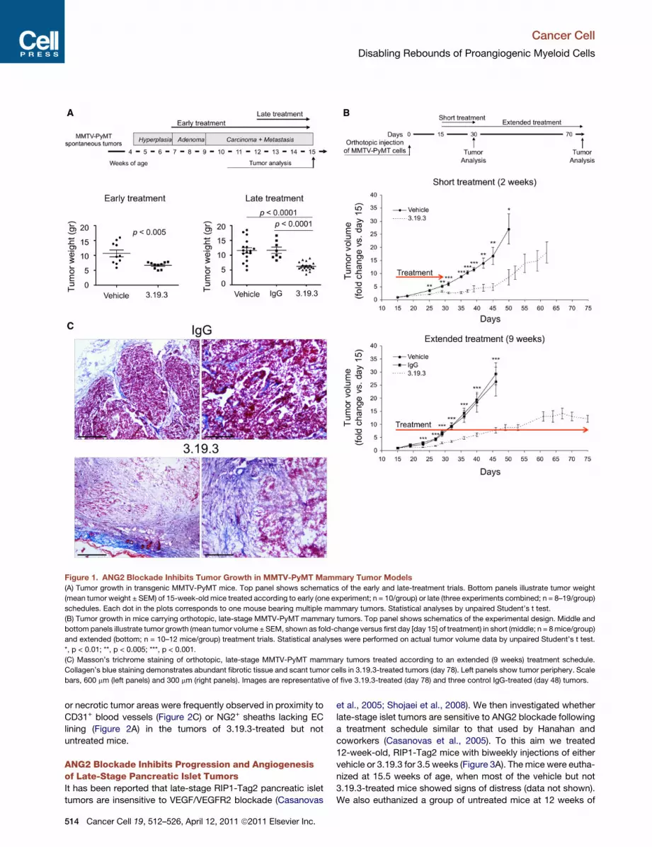
Figure 1. ANG2 Blockade Inhibits Tumor Growth in MMTV-PyMT Mammary Tumor Models
(A) Tumor growth in transgenic MMTV-PyMT mice. Top panel shows schematics of the early and late-treatment trials. Bottom panels illustrate tumor weight
(mean tumor weight ± SEM) of 15-week-old mice treated according to early (one experiment; n = 10/group) or late (three experiments combined; n = 8–19/group)
schedules. Each dot in the plots corresponds to one mouse bearing multiple mammary tumors. Statistical analyses by unpaired Student’s t test.
(B) Tumor growth in mice carrying orthotopic, late-stage MMTV-PyMT mammary tumors. Top panel shows schematics of the experimental design. Middle and
bottom panels illustrate tumor growth (mean tumor volume ± SEM, shown as fold-change versus first day [day 15] of treatment) in short (middle; n = 8mice/group)
and extended (bottom; n = 10–12 mice/group) treatment trials. Statistical analyses were performed on actual tumor volume data by unpaired Student’s t test.
*, p < 0.01; **, p < 0.005; ***, p < 0.001.
(C) Masson’s trichrome staining of orthotopic, late-stage MMTV-PyMT mammary tumors treated according to an extended (9 weeks) treatment schedule.
Collagen’s blue staining demonstrates abundant fibrotic tissue and scant tumor cells in 3.19.3-treated tumors (day 78). Left panels show tumor periphery. Scale
bars, 600 mm (left panels) and 300 mm (right panels). Images are representative of five 3.19.3-treated (day 78) and three control IgG-treated (day 48) tumors.
Cancer Cell
Disabling Rebounds of Proangiogenic Myeloid Cells
or necrotic tumor areas were frequently observed in proximity to
CD31+ blood vessels (Figure 2C) or NG2+ sheaths lacking EC
lining (Figure 2A) in the tumors of 3.19.3-treated but not
untreated mice.
ANG2 Blockade Inhibits Progression and Angiogenesisof Late-Stage Pancreatic Islet TumorsIt has been reported that late-stage RIP1-Tag2 pancreatic islet
tumors are insensitive to VEGF/VEGFR2 blockade (Casanovas
514 Cancer Cell 19, 512–526, April 12, 2011 ª2011 Elsevier Inc.
et al., 2005; Shojaei et al., 2008). We then investigated whether
late-stage islet tumors are sensitive to ANG2 blockade following
a treatment schedule similar to that used by Hanahan and
coworkers (Casanovas et al., 2005). To this aim we treated
12-week-old, RIP1-Tag2 mice with biweekly injections of either
vehicle or 3.19.3 for 3.5 weeks (Figure 3A). The mice were eutha-
nized at 15.5 weeks of age, when most of the vehicle but not
3.19.3-treated mice showed signs of distress (data not shown).
We also euthanized a group of untreated mice at 12 weeks of

Figure 2. ANG2 Blockade Regresses the Vasculature and Inhibits Angiogenesis in MMTV-PyMT Mammary Tumor Models
(A) NG2 (green) and CD31 (red) immunostaining, and TO-PRO-3 (TP3) nuclear staining (blue) of orthotopic, late-stage MMTV-PyMT mammary tumors treated
according to a short (2 weeks) schedule and analyzed immediately after discontinuation of therapy. Top panels show confocal images of representative tumor
sections. N, necrotic tumor areas. Scale bar, 300 mm. Middle panels show images of three-dimensional models obtained by surface rendering of the confocal
Z stacks, after superimposition of multiple confocal planes (section thickness, 16 mm). Scale bar, 150 mm. Bottom panels show superimposition of multiple
confocal Z stacks imaging individual blood vessels. Scale bar, 50 mm. Results are representative of two independent experiments and ten tumors per group
analyzed.
(B) Morphometric (Relative vascular area; Relative pericyte area; EC/pericyte ratio) and flow cytometry (Endothelial cells) analyses of angiogenesis in orthotopic
(morphometric analyses) and spontaneous (flow cytometry) MMTV-PyMT mammary tumors. Each dot in the plots corresponds to one tumor; scatter plots show
mean values ± SEM. Statistical analyses by unpaired Student’s t test.
(C) Top panels illustrate lectin (green), hypoxia (PIMO; red), and CD31 (blue) immunostaining of orthotopic, late-stage MMTV-PyMT mammary tumors treated
according to a short (2 weeks) schedule. N, necrotic tumor areas; H, hypoxic areas; arrows indicate lectin+/CD31+ perfused blood vessels. Scale bar, 150 mm.
Results are representative of two independent experiments and eight tumors per group analyzed. Bottom panel shows quantification of hypoxia in three
representative tumors (mean percentage [%] of PIMO+ area ± SEM). Statistical analyses by unpaired Student’s t test.
Cancer Cell
Disabling Rebounds of Proangiogenic Myeloid Cells
age (t0) in order to obtain pancreatic tissue at a time point coin-
ciding with the initiation of therapy (Figure 3A).
Clone 3.19.3 significantly reduced the mean tumor area
(versus vehicle; Figure 3A) calculated by measuring each of the
islet tumors scored in the largest pancreatic section (28–72
tumors per section; Figure S3). Of note, large tumors exceeding
0.5 mm2 of area were significantly fewer in 3.19.3-treated than
control mice (Figure 3A; Figure S3), indicating that ANG2
blockade effectively inhibited the progression of advanced
tumors in RIP1-Tag2 mice. Furthermore, 3.19.3 did not increase
local invasion by islet tumors, as shown by the similar propor-
tions of noninvasive, partially invasive (IC1), or entirely invasive
(IC2) tumors (Ebos et al., 2009; Paez-Ribes et al., 2009) in
3.19.3-treated and control mice (Figure 3B).
We then studied tumor angiogenesis. Clone 3.19.3 greatly
reduced the relative tumor vascular area, measured by IFS of
Cancer Cell 19, 512–526, April 12, 2011 ª2011 Elsevier Inc. 515

Figure 3. ANG2 Blockade Inhibits Progression and Angiogenesis of Late-Stage, RIP1-Tag2 Pancreatic Islet Tumors
(A) Top panel shows schematics of the late treatment trial. Bottom-left panel illustrates mean tumor area (±SEM; two independent experiments combined;
n = 4–8/group). Bottom-right panel shows proportion (%) of tumors exceeding 0.5 mm2 of area (mean value ± SEM; two independent experiments combined;
n = 4–8/group). Each dot in the plots corresponds to one mouse, of which multiple islet tumors contained in the largest pancreatic section were analyzed.
Statistical analyses by unpaired Student’s t test.
(B) Left panels illustrate hematoxylin and eosin staining (H&E) of pancreatic sections showing examples of noninvasive (left and middle panels) and invasive (right
panel) islet tumors. Scale bars, 300 mm (left panel) and 150 mm (middle and right panels). Right panel illustrates proportion (%; mean value ± SEM) of tumors with
noninvasive, partially invasive (IC1) or entirely invasive (IC2) margins. Each dot in the plots corresponds to one mouse, of which multiple islet tumors contained in
the largest pancreatic section were analyzed.
(C) NG2 (green) and CD31 (red) immunostaining of islet tumors. Scale bar, 150 mm. Results are representative of two independent experiments and three to four
mice per group analyzed.
(D) Morphometric analyses of angiogenesis (Relative vascular area; Relative pericyte area; Endothelial / pericyte ratio; mean values ± SEM) in islet tumors
analyzed at the indicated time points. Each dot in the plots corresponds to one mouse, of which multiple tumors were analyzed. Statistical analyses by unpaired
Student’s t test.
Cancer Cell
Disabling Rebounds of Proangiogenic Myeloid Cells
516 Cancer Cell 19, 512–526, April 12, 2011 ª2011 Elsevier Inc.

Figure 4. ANG2 Blockade Upregulates the
Expression of Proangiogenic Genes in
Mammary Tumors
Gene expression by qPCR in whole-tumor
lysates obtained from spontaneous MMTV-PyMT
tumors treated with 3.19.3 (mean fold change
over reference value [vehicle]). For each mouse
(n = 3–4 mice/group), two to three small tumor
biopsies were obtained and pooled together.
Error bars represent 95% confidence interval
(1.96 3 SEM). Gapdh and Hprt were used as
reference genes. Genes differentially expressed
between 3.19.3- and vehicle-treated tumors are
indicated by the asterisks (*, p < 0.05; **,
p < 0.01; ***, p < 0.001).
Cancer Cell
Disabling Rebounds of Proangiogenic Myeloid Cells
CD31+ blood vessels (Figures 3C and 3D). Of note, the relative
tumor vascular area was similar in t0 (12 weeks of age) and
vehicle-treated (15.5 weeks of age) mice but was reduced by
more than 50% in 3.19.3-treated mice (15.5 weeks of age). As
seen in MMTV-PyMT carcinomas, 3.19.3 enhanced pericyte
coverage of tumor blood vessels (Figures 3C and 3D). These
data indicate profound antiangiogenic activity of ANG2 blockade
in late-stage, pancreatic islet tumors.
ANG2 Blockade Upregulates the Expressionof Proangiogenic Genes in Mammary TumorsWe then analyzed the expression of a panel of angiogenesis-
associated and myeloid cell-growth factor/chemoattractant
genes by qPCR (Figure S4) in tumor lysates obtained from spon-
taneous MMTV-PyMT carcinomas treated according to a late
schedule (Figure 1A). Although none of the investigated genes
showed major changes, the proangiogenic genes Angpt2,
Fgf2, Hgf, Pdgfb, Vegfa, Vegfb, Mmp9, and Sdf1 were all upre-
gulated in 3.19.3-treated versus untreated tumors (Figure 4). Of
note, enhanced expression of Fgf2, Sdf1, and Hgf has been
previously associated with tumor resistance to various antian-
giogenic or radiation treatments (Casanovas et al., 2005; Kozin
et al., 2010; Shojaei et al., 2010).
Whereas Sdf1, Vegfa, and Angpt2 were slightly upregulated,
other myeloid cell-growth factor/chemoattractant genes (e.g.,
Bv8/prokinecitin-1, Csf1/M-CSF, Csf2/GM-CSF, and Csf3/
G-CSF) did not show significant changes in 3.19.3-treated
versus untreated tumors (Figure 4). Interestingly, Tie2was signif-
icantly downregulated, whereas the expression of other EC
receptors (e.g., Vegfr1, Vegfr2, and Cxcr4) was similar in
3.19.3-treated versus untreated tumors (Figure 4). Igf1, which
is an EC antiapoptotic/survival factor highly expressed by
TEMs (Pucci et al., 2009), was strongly upregulated in 3.19.3-
treated versus untreated tumors. In summary these data suggest
that mammary tumors upregulate, albeit marginally, the expres-
sion of several proangiogenic genes following ANG2 blockade;
nevertheless, the treated tumors did not show evidence for
rebound angiogenesis (Figures 2A–2C) or growth resistance
(Figures 1A–1C).
ANG2 Blockade Does Not Inhibit Tumor Infiltration byTEMs but Impedes Their Association with Blood VesselsWe previously showed that TEMs can be distinguished from
TIE2� TAMs by their cell surface marker profile (TEMs: CD11b+/
F4/80+/MRC1high/CD11c�; TIE2� TAMs: CD11b+/F4/80+/
MRC1low/�/CD11c+) (Pucci et al., 2009) and perivascular location
(De Palma et al., 2005). Because TIE2� TAMs express higher
amounts of classic proinflammatory genes than TEMs (Pucci
et al., 2009), here we refer to the former as ‘‘inflammatory TAMs.’’
ANG2 was previously shown to be a TEM chemoattractant
(Coffelt et al., 2010b; Murdoch et al., 2007; Venneri et al., 2007).
We then asked whether ANG2 blockade inhibited TEM recruit-
ment to the tumors.We found thatMMTV-PyMT carcinomas con-
tained substantial numbers of MRC1+ TEMs (Figures 5A and 5B).
Unexpectedly, ANG2 blockade enhanced tumor infiltration by
MRC1+ TEMs, but not inflammatory TAMs or total CD11b+
myeloid cells, both in spontaneous (Figure 5A; Figure S5A) and
orthotopic (Figure 5B) MMTV-PyMT tumors. The enhanced
recruitment of MRC1+ TEMs in 3.19.3-treated tumors may be
fostered by increased tumor hypoxia and/or expression of SDF1
(Figures 2C and 4), which are known TEM-recruiting signals (Kioi
et al., 2010; Kozin et al., 2010). Interestingly, whereas in control
tumors theMRC1+cellsweremostly associatedwithCD31+blood
vessels (a typical TEM feature), in 3.19.3-treated tumors these
cells were more homogeneously spread in the tumor mass and
frequentlydisengaged from thebloodvessels (Figures5Aand5B).
ANG2 blockade did not increase MRC1+ TEM infiltration in
RIP1-Tag2 islet tumors but, similar to findings in MMTV-PyMT
carcinomas, displaced them from the blood vessels (Figure 5C).
This was true also in the scant tumor regions characterized by
a relatively high vascular area (Figure S5B). Together, these
data suggest that ANG2 is not required for TEM recruitment to
the tumors but regulates their interaction with angiogenic blood
vessels.
ANG2 Blockade Impedes Tumor TEM Upregulationof Tie2Because 3.19.3 strongly inhibited tumor angiogenesis, the
finding of increased numbers of MRC1+ TEMs in 3.19.3-treated
Cancer Cell 19, 512–526, April 12, 2011 ª2011 Elsevier Inc. 517

Figure 5. ANG2 Blockade Does Not Inhibit Tumor Infiltration by
TEMs but Impedes Their Association with Blood Vessels
(A) Top panels show MRC1 (red) and CD31 (blue) immunostaining of sponta-
neous, late-stage MMTV-PyMT mammary tumors treated according to a late
schedule and analyzed at 15 weeks of age (see Figure 1A). Scale bar, 150 mm.
Results are representative of three independent experiments and three to five
tumors per group analyzed. Bottom panels illustrate flow cytometry analyses
of myeloid cell infiltrates in tumors treated as above (mean frequency of tumor-
derived cells ± SEM, shown as fold change versus reference [vehicle]). Each
Cancer Cell
Disabling Rebounds of Proangiogenic Myeloid Cells
518 Cancer Cell 19, 512–526, April 12, 2011 ª2011 Elsevier Inc.
MMTV-PyMT carcinomas appeared paradoxical in view of
the proangiogenic activity of these cells. In order to inves-
tigate whether ANG2 blockade altered gene expression in
TEMs, we isolated TEMs (7AAD�/CD11b+/CD31low/MRC1high/
CD11c�), inflammatory TAMs (7AAD�/CD11b+/CD31low/
MRC1low/�/CD11c+), and ECs (7AAD�/CD31high/CD11b�) fromboth 3.19.3-treated and untreated, orthotopic MMTV-PyMT
carcinomas, and analyzed the expression of a panel of genes
of interest (Figure S4) in the sorted cells. In agreement with our
previous gene expression studies performed in the N202
mammary tumor model (Pucci et al., 2009), Tie2, Igf1, Sdf1,
and Mrc1 were all highly upregulated in TEMs versus inflamma-
tory TAMs isolated from untreated MMTV-PyMT tumors (Fig-
ure 6A; Figure S4). Therefore, the higher expression level of
Igf1, Mrc1, and Sdf1 in whole mammary tumor lysates of
3.19.3-treated (versus untreated) mice (Figure 4) is consistent
with their enhanced infiltration by TEMs (Figures 5A and 5B;
Figure S5A).
Interestingly, the expression level of Tie2—but not other
TEM-distinguishing genes—was significantly lower (8- to
10-fold in four independent experiments) in TEMs isolated from
3.19.3-treated than control tumors (Figure 6B; Figure S4; data
not shown). Because Tie2 is strongly upregulated in TEMs locally
in the tumor microenvironment (De Palma et al., 2008), these
data strongly suggested that neutralization of ANG2 had
impeded the upregulation of TIE2 in tumor-infiltrating TEMs.
On the other hand, ANG2 blockade did not change the expres-
sion level of any of the investigated genes in inflammatory
TAMs (Figures 6C; Figure S4). Except for Sdf1, which was upre-
gulated, none of the genes analyzed in ECs (including Tie2)
displayed significant changes following ANG2 blockade (Figures
6D; Figure S4).
To verify that, regardless of their differential expression level of
Tie2, the MRC1+ cells represented bona fide TEMs in both
3.19.3-treated and untreated tumors, we transplanted lethally
irradiated, 6-week-old MMTV-PyMT mice with Tie2-GFP BM
cells (De Palma et al., 2005) and analyzed the tumors at 15weeks
of age after a late treatment schedule (Figure S6). Anti-GFP IFS
of tumor sections specifically marked the MRC1+ cells and
labeled similar proportions of these cells in 3.19.3- and
vehicle-treated mice (Figure S6). Together with the gene expres-
sion data in Figure 6B, these results indicate that the MRC1+
dot in the plots corresponds to onemouse; tumor samples were obtained from
three independent experiments. Statistical analyses by unpaired Student’s t
test.
(B) MRC1 (red) and CD31 (blue) immunostaining of orthotopic, late-stage
MMTV-PyMT mammary tumors treated according to a short schedule (see
Figure 1B) and analyzed 2 weeks after the first treatment. Scale bar, 150 mm.
Results are representative of two independent experiments and ten tumors per
group analyzed.
(C) Top panels show MRC1 (red) and CD31 (green) immunostaining of late-
stage RIP1-Tag2 islet tumors treated according to a late schedule and
analyzed at 15.5 weeks of age (see Figure 3A). Scale bar, 100 mm. Results are
representative of two independent experiments, three to four mice per group
and several tumors analyzed. Bottom panels show morphometric analyses
(mean values ± SEM) of MRC1+ cell infiltration (left) and association with blood
vessels (right) in islet tumors treated as above. Each dot in the plots corre-
sponds to one mouse, of which multiple tumors were analyzed. Statistical
analyses by unpaired Student’s t test.
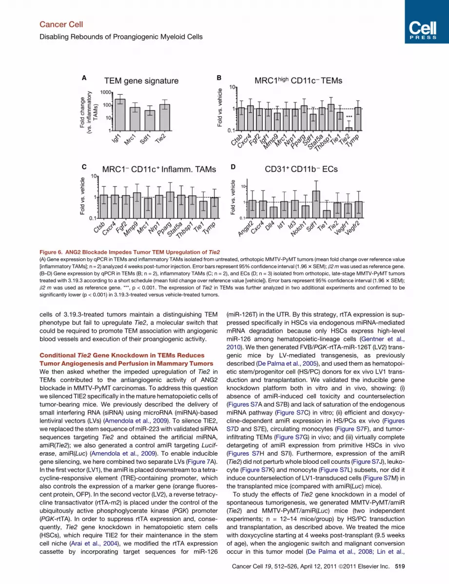
Figure 6. ANG2 Blockade Impedes Tumor TEM Upregulation of Tie2(A) Gene expression by qPCR in TEMs and inflammatory TAMs isolated from untreated, orthotopic MMTV-PyMT tumors (mean fold change over reference value
[inflammatory TAMs]; n = 2) analyzed 4weeks post-tumor injection. Error bars represent 95%confidence interval (1.963SEM); b2mwas used as reference gene.
(B–D) Gene expression by qPCR in TEMs (B; n = 2), inflammatory TAMs (C; n = 2), and ECs (D; n = 3) isolated from orthotopic, late-stage MMTV-PyMT tumors
treated with 3.19.3 according to a short schedule (mean fold change over reference value [vehicle]). Error bars represent 95% confidence interval (1.963 SEM);
b2 m was used as reference gene. ***, p < 0.001. The expression of Tie2 in TEMs was further analyzed in two additional experiments and confirmed to be
significantly lower (p < 0.001) in 3.19.3-treated versus vehicle-treated tumors.
Cancer Cell
Disabling Rebounds of Proangiogenic Myeloid Cells
cells of 3.19.3-treated tumors maintain a distinguishing TEM
phenotype but fail to upregulate Tie2, a molecular switch that
could be required to promote TEM association with angiogenic
blood vessels and execution of their proangiogenic activity.
Conditional Tie2 Gene Knockdown in TEMs ReducesTumor Angiogenesis and Perfusion inMammary TumorsWe then asked whether the impeded upregulation of Tie2 in
TEMs contributed to the antiangiogenic activity of ANG2
blockade in MMTV-PyMT carcinomas. To address this question
we silenced TIE2 specifically in themature hematopoietic cells of
tumor-bearing mice. We previously described the delivery of
small interfering RNA (siRNA) using microRNA (miRNA)-based
lentiviral vectors (LVs) (Amendola et al., 2009). To silence TIE2,
we replaced the stem sequence ofmiR-223with validated siRNA
sequences targeting Tie2 and obtained the artificial miRNA,
amiR(Tie2); we also generated a control amiR targeting Lucif-
erase, amiR(Luc) (Amendola et al., 2009). To enable inducible
gene silencing, we here combined two separate LVs (Figure 7A).
In the first vector (LV1), the amiR is placed downstream to a tetra-
cycline-responsive element (TRE)-containing promoter, which
also controls the expression of a marker gene (orange fluores-
cent protein, OFP). In the second vector (LV2), a reverse tetracy-
cline transactivator (rtTA-m2) is placed under the control of the
ubiquitously active phosphoglycerate kinase (PGK) promoter
(PGK-rtTA). In order to suppress rtTA expression and, conse-
quently, Tie2 gene knockdown in hematopoietic stem cells
(HSCs), which require TIE2 for their maintenance in the stem
cell niche (Arai et al., 2004), we modified the rtTA expression
cassette by incorporating target sequences for miR-126
(miR-126T) in the UTR. By this strategy, rtTA expression is sup-
pressed specifically in HSCs via endogenous miRNA-mediated
mRNA degradation because only HSCs express high-level
miR-126 among hematopoietic-lineage cells (Gentner et al.,
2010). We then generated FVB/PGK-rtTA-miR-126T (LV2) trans-
genic mice by LV-mediated transgenesis, as previously
described (De Palma et al., 2005), and used them as hematopoi-
etic stem/progenitor cell (HS/PC) donors for ex vivo LV1 trans-
duction and transplantation. We validated the inducible gene
knockdown platform both in vitro and in vivo, showing: (i)
absence of amiR-induced cell toxicity and counterselection
(Figures S7A and S7B) and lack of saturation of the endogenous
miRNA pathway (Figure S7C) in vitro; (ii) efficient and doxycy-
cline-dependent amiR expression in HS/PCs ex vivo (Figures
S7D and S7E), circulating monocytes (Figure S7F), and tumor-
infiltrating TEMs (Figure S7G) in vivo; and (iii) virtually complete
detargeting of amiR expression from primitive HSCs in vivo
(Figures S7H and S7I). Furthermore, expression of the amiR
(Tie2) did not perturb whole blood cell counts (Figure S7J), leuko-
cyte (Figure S7K) and monocyte (Figure S7L) subsets, nor did it
induce counterselection of LV1-transduced cells (Figure S7M) in
the transplanted mice (compared with amiR(Luc) mice).
To study the effects of Tie2 gene knockdown in a model of
spontaneous tumorigenesis, we generated MMTV-PyMT/amiR
(Tie2) and MMTV-PyMT/amiR(Luc) mice (two independent
experiments; n = 12–14 mice/group) by HS/PC transduction
and transplantation, as described above. We treated the mice
with doxycycline starting at 4 weeks post-transplant (9.5 weeks
of age), when the angiogenic switch and malignant conversion
occur in this tumor model (De Palma et al., 2008; Lin et al.,
Cancer Cell 19, 512–526, April 12, 2011 ª2011 Elsevier Inc. 519
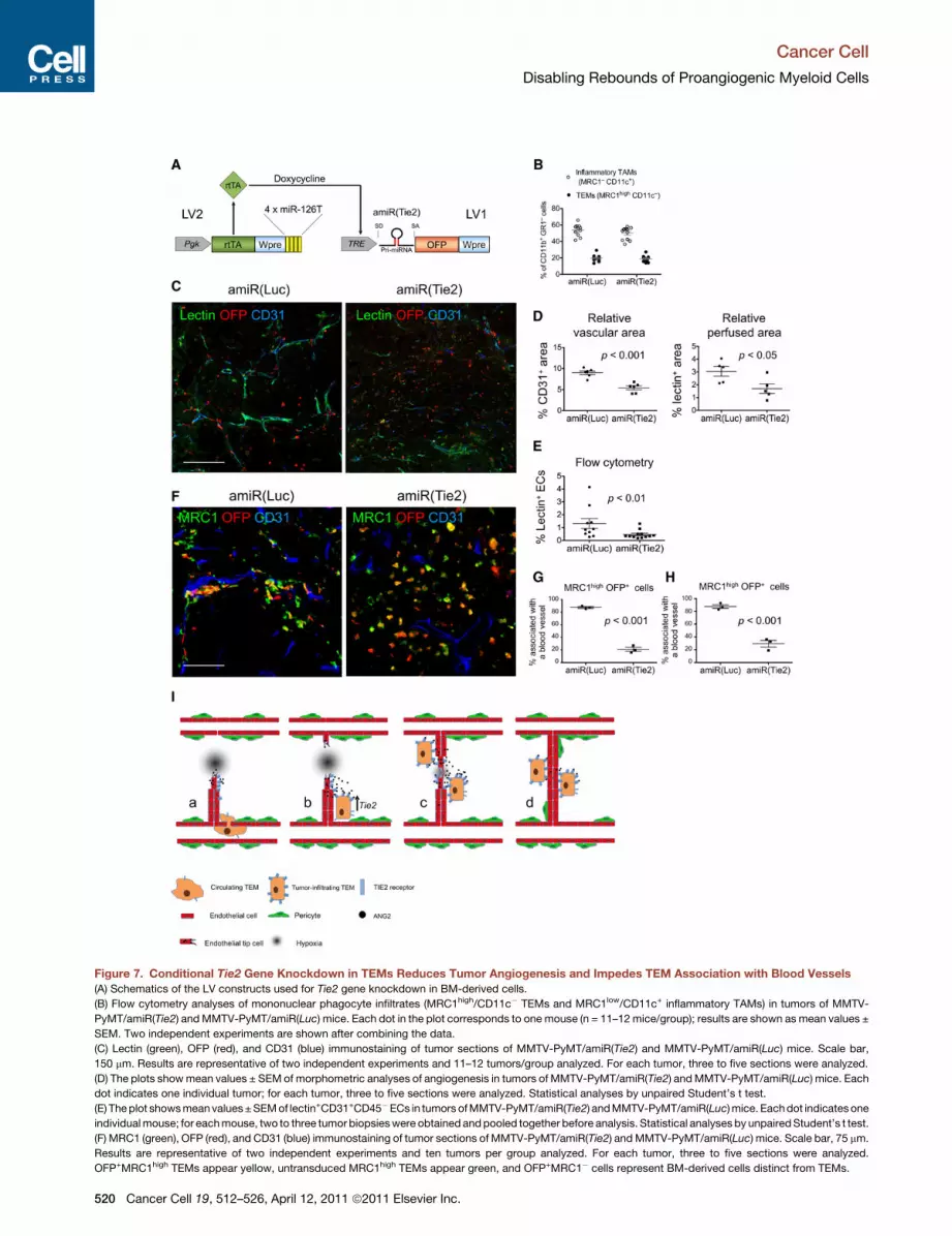
Figure 7. Conditional Tie2 Gene Knockdown in TEMs Reduces Tumor Angiogenesis and Impedes TEM Association with Blood Vessels
(A) Schematics of the LV constructs used for Tie2 gene knockdown in BM-derived cells.
(B) Flow cytometry analyses of mononuclear phagocyte infiltrates (MRC1high/CD11c� TEMs and MRC1low/CD11c+ inflammatory TAMs) in tumors of MMTV-
PyMT/amiR(Tie2) andMMTV-PyMT/amiR(Luc) mice. Each dot in the plot corresponds to one mouse (n = 11–12 mice/group); results are shown as mean values ±
SEM. Two independent experiments are shown after combining the data.
(C) Lectin (green), OFP (red), and CD31 (blue) immunostaining of tumor sections of MMTV-PyMT/amiR(Tie2) and MMTV-PyMT/amiR(Luc) mice. Scale bar,
150 mm. Results are representative of two independent experiments and 11–12 tumors/group analyzed. For each tumor, three to five sections were analyzed.
(D) The plots showmean values ± SEM of morphometric analyses of angiogenesis in tumors of MMTV-PyMT/amiR(Tie2) andMMTV-PyMT/amiR(Luc) mice. Each
dot indicates one individual tumor; for each tumor, three to five sections were analyzed. Statistical analyses by unpaired Student’s t test.
(E) Theplot showsmeanvalues±SEMof lectin+CD31+CD45�ECs in tumors ofMMTV-PyMT/amiR(Tie2) andMMTV-PyMT/amiR(Luc)mice. Eachdot indicatesone
individualmouse; for eachmouse, two to three tumor biopsieswere obtained andpooled together before analysis. Statistical analysesby unpairedStudent’s t test.
(F) MRC1 (green), OFP (red), and CD31 (blue) immunostaining of tumor sections of MMTV-PyMT/amiR(Tie2) andMMTV-PyMT/amiR(Luc) mice. Scale bar, 75 mm.
Results are representative of two independent experiments and ten tumors per group analyzed. For each tumor, three to five sections were analyzed.
OFP+MRC1high TEMs appear yellow, untransduced MRC1high TEMs appear green, and OFP+MRC1� cells represent BM-derived cells distinct from TEMs.
Cancer Cell
Disabling Rebounds of Proangiogenic Myeloid Cells
520 Cancer Cell 19, 512–526, April 12, 2011 ª2011 Elsevier Inc.
Cancer Cell
Disabling Rebounds of Proangiogenic Myeloid Cells
2006). We euthanized the mice at 15 weeks of age and analyzed
their tumors by flow cytometry and IFS of frozen sections.
The frequency of TEMs and inflammatory TAMs among the
tumor-infiltrating CD11b+/Gr1� myelomonocytic cells was
similar in MMTV-PyMT/amiR(Tie2) and MMTV-PyMT/amiR
(Luc) mice (Figure 7B), indicating that Tie2 gene knockdown
did not detectably affect TEM recruitment to the tumors.
However, when we analyzed tumor angiogenesis, we observed
significant differences between MMTV-PyMT/amiR(Tie2) and
MMTV-PyMT/amiR(Luc) mice (Figures 7C and 7D). The CD31+
blood vessels of amiR(Tie2) tumors appeared smaller, fewer,
and less perfused (lectin+) than in the controls (Figure 7C).
Both the CD31+ and lectin+ relative vascular area, measured
by IFS of tumor sections, were significantly lower in amiR(Tie2)
than amiR(Luc) mice (Figure 7D), indicating decreased angio-
genesis and, possibly, increased vessel immaturity or collapse.
Flow cytometric analyses of tumor cell suspensions confirmed
the lower proportion of lectin+/CD31+/CD45� ECs in the tumors
of amiR(Tie2) mice (Figure 7E). We observed decreased angio-
genesis/perfusion also in FVB/amiR(Tie2) mice either challenged
with orthotopic MMTV-PyMT (Figure S7N) or subcutaneous
N202 (Figure S7O) mammary carcinomas. These data indicate
that Tie2 knockdown by RNAi and, consequently, its impeded
upregulation in tumor-infiltrating TEMs reduces tumor angio-
genesis and blood vessel functionality in mammary tumor
models.
Conditional Tie2 Gene Knockdown in TEMs ImpedesTheir Association with Tumor Blood VesselsWenoted that the distribution of OFP+MRC1+ cells differed in the
tumors of amiR(Tie2) and amiR(Luc) mice, both in spontaneous
(Figures 7F and 7G) and orthotopic (Figure 7H) MMTV-PyMT
tumor models. Indeed, there were fewer OFP+MRC1+ TEMs
associated with CD31+ tumor blood vessels in amiR(Tie2) than
amiR(Luc) mice (Figures 7G and 7H), suggesting that Tie2 knock-
down in TEMs had hampered their ability to associate with
angiogenic blood vessels. Together with the ANG2 blockade
data shown above, these findings indicate that TIE2 expression
by TEMs is required for their association with angiogenic blood
vessels, and that disrupting such association (either by inter-
fering with Tie2 expression in TEMs, or by neutralizing the TIE2
ligand, ANG2) limits the formation of intratumoral vascular
networks (Figure 7I).
Whereas Tie2 knockdown in TEMs was sufficient to signifi-
cantly decrease angiogenesis in multiple tumor models (Figures
7C–7E; Figures S7N and 7O), it failed to reproducibly inhibit
tumor growth (data not shown). However, it should be noted
that the frequency of OFP+ TEMs ranged from 50% to 90%
(average: 70%) in the tumors of both amiR(Tie2) and amiR(Luc)
mice (Figure S7P), indicating that Tie2 gene knockdown had
occurred in the majority but not all tumor-infiltrating TEMs.
(G and H) Analysis of OFP+MRC1high TEM/CD31+ blood vessel association in tumo
orthotopic MMTV-PyMT tumors grown in FVB mice). Individual OFP+MRC1high
Histograms show mean values (±SEM) of the percentage of OFP+MRC1high TEM
mice per group and two tumors per mouse; in (H), data were obtained from thre
(I) TEM-EC interactions mediated by ANG2-TIE2 promote vascular morphogene
upregulated upon their extravasation and exposure to ANG2 in the perivascular m
(b) and promote vascular growth (c and d).
ANG2 Blockade Inhibits Spontaneousand Preestablished Mammary Tumor MetastasisWe then asked whether angiogenesis inhibition following ANG2
blockade affected tumor cell dissemination and outgrowth of
pulmonary metastases in MMTV-PyMT transgenic mice. We
analyzed the lungs of 3.19.3-treated and control mice from either
early (one experiment; 8 weeks of treatment; n = 10 mice/group)
or late (two independent experiments; 3 weeks of treatment;
n = 10 mice/group) treatment trials. Both in early and late treat-
ment trials, 3.19.3 effectively inhibited spontaneous metastasis
in MMTV-PyMT mice (Figures 8A and 8B; Figure S8). In order
to discriminate direct versus indirect effects of 3.19.3 on metas-
tasis formation, we used a model in which metastatic growth is
independent of the primary tumor. To this aim we intravenously
injected tumor cells obtained from late-stage MMTV-PyMT
carcinomas into wild-type mice; by this approach, pulmonary
metastases form in the absence of a primary tumor. Starting at
1 day after tumor cell inoculation, mice were treated with
3.19.3 for 25 days or left untreated, and the lungs were analyzed
thereafter. Whereas 3.19.3 did not significantly decrease the
number of metastases in the lung parenchyma, it dramatically
inhibited the progression from a micro- to a macrometastatic
stage, as shown by volumetric analysis of the pulmonary tumor
burden (Figure 8C). These data provide direct evidence that
ANG2 blockade not only inhibits primary tumor growth and its
metastatic dissemination but also directly suppresses the
growth of established metastases.
DISCUSSION
Here, we demonstrate that ANG2 blockade: (i) inhibits angiogen-
esis and induces vascular regression in multiple tumor models,
including tumors that are prone to develop resistance to anti-
VEGF/VEGFR therapy; (ii) inhibits tumor growth in multiple tumor
models, including late-stage spontaneous tumors; (iii) limits the
metastatic dissemination of primary tumors and the outgrowth
of established metastasis; and (iv) impedes, in tumor-infiltrating
TEMs, the transcriptional upregulation of Tie2, which is required
for their association with tumor blood vessels and proangiogenic
activity.
Sustained Antiangiogenic and Antitumor Activityof ANG2 BlockadeWe neutralized ANG2 using an ANG2-specific monoclonal anti-
body (3.19.3) that potently binds ANG2 with at least 500-fold
greater affinity compared with ANG1 (Brown et al., 2010). Clone
3.19.3 markedly reduced the relative tumor vascular area in each
tumor model tested, including spontaneous MMTV-PyMT
mammary and RIP1-Tag2 pancreatic islet tumors. In these tumor
models, ANG2 neutralization increased pericyte coverage of
the remnant blood vessels, similar to previous findings in
rs of amiR(Tie2) and amiR(Luc) mice (G, spontaneousMMTV-PyMT tumors; H,
TEMs were scored as either associated or not with CD31+ blood vessels.
s associated with CD31+ blood vessels. In (G), data were obtained from three
e tumors per group. Statistical analyses by unpaired Student’s t test.
sis in tumors. Circulating TEMs express low-level TIE2 (a), but the receptor is
icroenvironment (b). TEMs adhere to ANG2-expressing sprouting blood vessels
Cancer Cell 19, 512–526, April 12, 2011 ª2011 Elsevier Inc. 521

Figure 8. ANG2 Blockade Inhibits Spontaneous and Preestablished Mammary Tumor Metastasis
(A) Spontaneous metastasis model. Left panels illustrate number of metastatic foci (bars) and total metastatic area (broken line) in individual serial sections (each
bar) obtained from the entire left lung of MMTV-PyMT mice treated with 3.19.3, control IgGs, or vehicle after a late schedule (see Figure 1A), and analyzed at
15 weeks of age. Right panels show H&E of representative whole lung sections.
(B) Spontaneous metastasis model. Number of metastatic foci (mean values ± SEM) per section per mouse in mice treated according to either late (3 weeks; left)
or early (8 weeks, right; see Figure S8) schedules. Each dot represents one mouse. Statistical analyses by Mann-Whitney U test.
(C) Pre-established metastasis model. Top-left panel shows schematics of experimental design. Bottom-left panels illustrate morphometric analyses of
metastasis (mean values ± SEM) in the lungs of mice either treated with 3.19.3 (n = 8) or vehicle alone (n = 9). Each dot in the plots represents one mouse. Results
combine two independent experiments. Statistical analyses by Mann-Whitney U test (left and middle panels) or unpaired Student’s t test (right panel). Right
panels show H&E of representative whole lung sections.
Cancer Cell
Disabling Rebounds of Proangiogenic Myeloid Cells
522 Cancer Cell 19, 512–526, April 12, 2011 ª2011 Elsevier Inc.

Cancer Cell
Disabling Rebounds of Proangiogenic Myeloid Cells
subcutaneous tumors (Hashizume et al., 2010; Nasarre et al.,
2009). Because ANG1 promotes pericyte-EC interactions, it is
possible that ANG2 blockade by 3.19.3 increased ANG1
bioavailability for interaction with TIE2, thus enhancing pericyte
coverage of the blood vessels and suppressing angiogenesis.
In agreement with previous findings (Fiedler et al., 2004), our
gene expression data indicate that ANG2 is highly expressed
in tumor ECs (Figure S4). However, the role of ANG2 in tumor
angiogenesis is still controversial. L1-7N, a previously described
ANG2-specific peptibody (Oliner et al., 2004), did not reduce
vascular density detectably in the tumor models tested (Hashi-
zume et al., 2010). Furthermore, genetic models of Angpt2 defi-
ciency or overexpression failed to unequivocally establish the
importance of ANG2 in tumor angiogenesis (Chae et al., 2010;
Nasarre et al., 2009).
Contrary to some of the earlier data, our findings indicate that
3.19.3 regresses the tumor vasculature and inhibits the growth of
both early and late-stage tumors, pointing to a critical role of
ANG2 during tumor progression. It is possible that some of the
previously reported strategies of ANG2 inhibition might have
overlooked the importance of ANG2 for tumor angiogenesis,
particularly when both ANG2 and ANG1, which have opposite
functions in tumor angiogenesis, were concomitantly targeted,
e.g., by soluble TIE2 delivery or ANG1/ANG2-bispecific anti-
bodies (Huang et al., 2010).
In MMTV-PyMT tumor models, 3.19.3 induced tumor blood
vessel regression, increased tumor hypoxia, fibrosis, and
necrosis, and enhanced the recruitment of MRC1+ TAMs (i.e.,
TEMs) to the tumors. Such tumor responses were previously
found—in the context of other antiangiogenic treatments (e.g.,
anti-VEGF/VEGFR) and/or tumor models—to associate with
the activation of alternate proangiogenic pathways and drug
resistance (Bergers and Hanahan, 2008; Casanovas et al.,
2005; Shojaei et al., 2007). Accordingly, we observed increased
expression of several proangiogenic genes (e.g., Vegfa, Fgf2,
and Sdf1) in 3.19.3-treated mammary tumors. We currently
ignore whether such upregulation of proangiogenic factors in
3.19.3-treated tumors is truly indicative of tumor adaptation to
circumvent ANG2 blockade, rather than representing an epiphe-
nomenon associated with enhanced tumor hypoxia or fibrosis.
In either case we did not find evidence for drug resistance or
rebound angiogenesis in tumors treated according to various
ANG2 blockade schedules, pointing to a requisite role of ANG2
for tumor angiogenesis. Importantly, ANG2 blockade also
inhibited angiogenesis and progression of late-stage, pancreatic
insulinomas in RIP1-Tag2mice, amouse tumormodel previously
shown to develop resistance to anti-VEGF/VEGFR2 therapy
(Casanovas et al., 2005; Shojaei et al., 2008). Although we did
not directly compare ANG2 blockade with VEGF/VEGFR2
blockade, our data suggest that effective ANG2 inhibition may
have the potential to achieve antiangiogenic and antitumor
activity also in tumors that are resistant to anti-VEGF therapy.
Antimetastatic Activity of ANG2 BlockadeAn important finding of this study is that ANG2 blockade mark-
edly inhibited metastasis in two metastasis models. Clone
3.19.3 strongly inhibited spontaneous pulmonary metastases
in MMTV-PyMT mice, both following short and extended
(8 weeks) treatment schedules. These data argue that ANG2
blockade, at variance with certain models of anti-VEGF therapy
(Ebos et al., 2009; Paez-Ribes et al., 2009), has potent antimeta-
static activity and does not select for proinvasive/prometastatic
tumor phenotypes. Because 3.19.3 reduced angiogenesis and
increased pericyte coverage of the remaining blood vessels in
the primary tumors, it is likely that tumor cell intravasation and
dissemination were directly inhibited at the primary tumor site.
Of note the genetic disruption of pericyte coverage elicited
increased metastasis in the Rip1-Tag2 pancreatic islet tumor
model (Xian et al., 2006).
Our data further indicate that 3.19.3 impairs the growth of
micrometastases at the post-seeding step. Indeed, preestab-
lished micrometastases failed to develop into large macrometa-
stases following ANG2 blockade, a phenomenon possibly due to
inhibition of the angiogenic switch that occurs at the metastatic
site concomitant to micro- to macrometastasis transition.
The ANG2-TIE2 Pathway Regulates TEMs’Proangiogenic ActivityTIE2 is expressed at very low level in circulatingmonocytes but is
strongly upregulated (up to 100-fold) in tumor-associated TEMs
(De Palma et al., 2008). Thus, ANG2 may signal both autocrinally
on ECs (Augustin et al., 2009) and iuxtacrinally on perivascular,
TIE2+ macrophages. The latter circumstance is supported by
experimental evidence that ANG2 agonistically enhances the
proangiogenic activity of human blood-derived TIE2+, but not
TIE2� monocytes in vitro (Coffelt et al., 2010b). Because the
MRC1+ TEMs recruited to 3.19.3-treated mammary tumors
expressed much lower Tie2 than those of untreated tumors, it
can be envisioned that EC-derived ANG2 stimulates TIE2
expression on perivascular TEMs and that such feedback may
be essential for the execution of productive angiogenesis.
Although it cannot be excluded that TIE2 upregulation in TEMs
is mediated indirectly by ANG2, ANG2 blockade specifically
modulated the Tie2 mRNA among several angiogenic genes
analyzed, suggesting that this response is intimately linked to
TIE2 signaling. Of note, several growth factors, including
ANGs, can regulate the expression of their receptor tyrosine
kinases at the transcriptional level via autoregulatory feedback
loops (Hashimoto et al., 2004). Intriguingly, we did not observe
transcriptional modulation of Tie2 in ECs following ANG2
blockade; this may suggest that ANG2-mediated modulation of
Tie2 expression involves different TIE2 heterodimers (Seegar
et al., 2010) and/or signaling adaptors (e.g., integrins) in ECs
and TAMs.
To investigate the role of TIE2 in TEMs, we developed a gene
knockdown platform that effectively protects the hematopoietic
compartment from potential toxicity consequent to RNA inter-
ference in HSCs. Indeed, our previous attempts to knock
down Tie2 using constitutive LVs caused obvious hematopoietic
toxicity (data not shown). By using inducible LVs coupled to
detargeting from HSCs (Gentner et al., 2010), we showed that
Tie2 knockdown in BM-derived cells significantly inhibits angio-
genesis and microvascular perfusion in MMTV-PyMT mice,
without affecting hematopoiesis detectably. Remarkably, by
targeting the TIE2 receptor in TEMs, we recapitulated some of
the features of TEM elimination (De Palma et al., 2005), indi-
cating that TIE2 is a pivotal biological effector and therapeutic
target in these cells.
Cancer Cell 19, 512–526, April 12, 2011 ª2011 Elsevier Inc. 523

Cancer Cell
Disabling Rebounds of Proangiogenic Myeloid Cells
TAMs comprise molecularly and functionally distinct subpop-
ulations (Qian and Pollard, 2010). TEMs express lower amounts
of VEGF than classic TAMs (Pucci et al., 2009) and do not reside
in hypoxic, avascular tumor areas (De Palma et al., 2005). Thus, it
is likely that TEMs exert a requisite proangiogenic function by
supporting tumor angiogenesis downstream to VEGF-induced
vascular activation. Because the lower expression of Tie2 in the
TEMs of 3.19.3-treated mammary tumors was not associated
with deregulated expression of a panel of classic pro- and antian-
giogenic genes, one can envision that TIE2 expression by TEMs
regulates blood vessel formation in tumors by noncanonical (e.g.,
VEGF-independent) angiogenic mechanisms. Indeed, our find-
ings of impeded association between TEMs and tumor blood
vessels both after specific Tie2 knockdown in TEMs and extra-
cellular blockade of ANG2 support the concept that the ANG2/
TIE2 axis is crucial to establish cell-to-cell interactions between
TEMsandECs. Such scenario is in agreementwith a recent study
showing that TIE2+ perivascularmacrophages physically interact
with the TIE2+ endothelial tip cells of nascent blood vessels and
are essential to promote vascular anastomosis during embryonic
development (Fantin et al., 2010). Furthermore, TIE2+ hemato-
poietic cells adhere to TIE2+ ECs and stimulate angiogenesis in
para-aortic splanchnopleural mesoderm explant cultures
(Takakura et al., 1998). Both ANG1 and ANG2 induce homomeric
TIE2 complex formation in cell-to-cell endothelial junctions
(Saharinen et al., 2008). Thus, ANG2-mediated TEM-EC interac-
tions may facilitate the navigation of endothelial sprouts through
the dense extracellular matrix, eventually enabling the fusion of
nascent blood vessels in angiogenic tissues (Figure 7I).
Tie2 silencing in TEMs, although consistently inhibited angio-
genesis and tumor blood vessel perfusion by almost 50%, did
not reproducibly inhibit tumor growth in the investigated mouse
tumor models. Similarly, previous reports showed that the
genetic deletion of certain proangiogenic factors in ECs or
myeloid cells may reduce tumor angiogenesis without
decreasing tumor growth rates (Nasarre et al., 2009; Stockmann
et al., 2008). It should be noted that in our Tie2-silencing studies,
from 10% to 50% of the tumor-associated TEMs were TIE2
competent (OFP�) in the different mice. This is expected from
the chimeric composition of hematopoiesis following transplan-
tation of ex vivo-transduced HS/PCs and the nonexhaustive
doxycycline-mediated gene induction. Thus, it cannot be
excluded that fully exhaustive Tie2 targeting in tumor-infiltrating
macrophages would impair angiogenesis and tumor vessel func-
tion to an extent becoming critical for tumor growth.
In conclusion our data indicate that the TIE2 receptor
expressed by perivascular TEMs is a crucial regulator of
ANG2-mediated proangiogenic programs in tumors. Because
tumor-infiltrating myeloid cells are known to convey protumoral
and proangiogenic programs that can counteract the efficacy
of antiangiogenic treatments (Bergers and Hanahan, 2008), the
combined targeting of angiogenic ECs and proangiogenic
TEMs by selective ANG2/TIE2-pathway inhibitors may extend
the reach of antiangiogenic therapy in patients with cancer.
EXPERIMENTAL PROCEDURES
Detailed experimental procedures are available in Supplemental Experimental
Procedures.
524 Cancer Cell 19, 512–526, April 12, 2011 ª2011 Elsevier Inc.
Mice
FVB and CD1 athymic mice were purchased from Charles River Laboratory
(Calco, Milan, Italy). FVB/MMTV-PyMT and C57Bl/6/RIP1-Tag2 mice were ob-
tained from the NCI-Frederick Mouse Repository (MD, USA) and established
as colonies at the San Raffaele animal facility. FVB/Pgk-rtTA-miR-126T trans-
genic mice were generated by LV-mediated transgenesis. FVB/Tie2-GFP
transgenic mice were generated previously (De Palma et al., 2005). All proce-
dures were performed according to protocols approved by the Animal Care
and Use Committee of the Fondazione San Raffaele del Monte Tabor (IACUC
324 and 335) and communicated to the Ministry of Health and local authorities
according to the Italian Law.
ANG2 Blockade by 3.19.3
Tumor-bearing mice were randomized into vehicle (phosphate-buffered
saline), IgG (Endobulin, Baxter, Italy), and treatment (3.19.3; AstraZeneca
Pharmaceuticals, Waltham, MA, USA) groups. Clone 3.19.3 and IgGs were
administered by i.p. injections after a twice-weekly schedule at doses of
10 mg/kg for the indicated periods of time, as described previously (Brown
et al., 2010).
HS/PC Isolation, Transduction, and Transplantation for Tie2
Knockdown in TEMs
BM was obtained from 6- to 12-week–old FVB/PGK-rtTA-miR-126T (LV2)
transgenic mice. Lineage-negative cells enriched in HS/PCs were isolated
using a cell purification kit (StemCell Technologies) and transduced by
concentrated LVs. Briefly, 106 cells/ml were prestimulated for 4–6 hr in
serum-free StemSpan medium (StemCell Technologies) containing a cocktail
of cytokines (IL-3, SCF, TPO, and FLT-3L; all from PeproTech) and transduced
with amiR-expressing LVs (LV1) with a dose equivalent to 108 LV transducing
units/ml, for 12 hr in medium containing cytokines. After transduction, 106 cells
were infused into the tail vein of lethally irradiated, 5.5-week–old, female FVB
or FVB/MMTV-PyMT mice (radiation dose: 1150 cGy split in two doses).
Induction of Tie2 Gene Knockdown by Doxycycline Administration
Starting at 4 weeks after HS/PC transplantation (i.e., 9.5 weeks of age), FVB or
MMTV-PyMTmiceweremoved to doxycycline-containing food (Charles River)
and received i.p. injections of doxycycline (0.5mg/mouse) every third day, until
the end of the experiments (12.5–13.5 weeks of age for FVB and 15 weeks of
age for MMTV-PyMT mice).
Statistical Analysis
In all studies, values are expressed as mean ± standard error of the mean
(SEM) or 95% confidence intervals (1.963 SEM), as indicated. Statistical anal-
yses were performed by unpaired Student’s t test, or Mann-Whitney U test, as
indicated. Differences were considered statistically significant at p < 0.05.
Statistical methods are described in full in the Supplemental Experimental
Procedures.
SUPPLEMENTAL INFORMATION
Supplemental Information includes Supplemental Experimental Procedures,
eight figures, and two movies and can be found with this article online at
doi:10.1016/j.ccr.2011.02.005.
ACKNOWLEDGMENTS
We thank Francesca Sanvito and Martina Rocchi for help with pathology;
Cesare Covino (ALEMBIC) for help with tumor imaging; Lucia Sergi Sergi for
vector production; Giulia Escobar for help with some experiments; and
ChingChing Leow (MedImmune, Gaithersburg, MD, USA) for helpful discus-
sions. This research was supported by grants from the European Research
Council (Starting Grant 243128/TIE2+Monocytes to M.D.P.; Advanced Grant
249845/TARGETINGGENETHERAPY to L.N.), the Associazione Italiana per
la Ricerca sul Cancro (IG-2007 to L.N. and IG-2010 to M.D.P.), AstraZeneca
(to L.N. and M.D.P), Fondazione Guido Berlucchi (to M.D.P.), and the Italian
Ministry of Health ‘‘Challenge in Oncology’’ (to L.N.). E.Z. was supported by
a FIRC fellowship. None of the authors has a financial interest related to this
work.

Cancer Cell
Disabling Rebounds of Proangiogenic Myeloid Cells
Received: July 19, 2010
Revised: December 1, 2010
Accepted: February 14, 2011
Published: April 11, 2011
REFERENCES
Amendola, M., Passerini, L., Pucci, F., Gentner, B., Bacchetta, R., and Naldini,
L. (2009). Regulated and multiple miRNA and siRNA delivery into primary cells
by a lentiviral platform. Mol. Ther. 17, 1039–1052.
Arai, F., Hirao, A., Ohmura, M., Sato, H., Matsuoka, S., Takubo, K., Ito, K., Koh,
G.Y., and Suda, T. (2004). Tie2/angiopoietin-1 signaling regulates hematopoi-
etic stem cell quiescence in the bone marrow niche. Cell 118, 149–161.
Augustin, H.G., Young Koh, G., Thurston, G., and Alitalo, K. (2009). Control of
vascular morphogenesis and homeostasis through the angiopoietin-Tie
system. Nat. Rev. Mol. Cell Biol. 10, 165–177.
Bergers, G., and Hanahan, D. (2008). Modes of resistance to anti-angiogenic
therapy. Nat. Rev. Cancer 8, 592–603.
Brown, J.L., Cao, Z.A., Pinzon-Ortiz, M., Kendrew, J., Reimer, C., Wen, S.,
Zhou, J.Q., Tabrizi, M., Emery, S., McDermott, B., et al. (2010). A humanmono-
clonal anti-ANG2 antibody leads to broad antitumor activity in combination
with VEGF inhibitors and chemotherapy agents in preclinical models. Mol.
Cancer Ther. 9, 145–156.
Casanovas, O., Hicklin, D.J., Bergers, G., and Hanahan, D. (2005). Drug resis-
tance by evasion of antiangiogenic targeting of VEGF signaling in late-stage
pancreatic islet tumors. Cancer Cell 8, 299–309.
Chae, S.S., Kamoun, W.S., Farrar, C.T., Kirkpatrick, N.D., Niemeyer, E., de
Graaf, A.M., Sorensen, A.G., Munn, L.L., Jain, R.K., and Fukumura, D.
(2010). Angiopoietin-2 interferes with anti-VEGFR-2-induced vessel normali-
zation and survival benefit in mice bearing gliomas. Clin. Cancer Res. 16,
3618–3627.
Chan, D.A., Kawahara, T.L., Sutphin, P.D., Chang, H.Y., Chi, J.T., and Giaccia,
A.J. (2009). Tumor vasculature is regulated by PHD2-mediated angiogenesis
and bone marrow-derived cell recruitment. Cancer Cell 15, 527–538.
Chung, A.S., Lee, J., and Ferrara, N. (2010). Targeting the tumour vasculature:
insights from physiological angiogenesis. Nat. Rev. Cancer 10, 505–514.
Coffelt, S.B., Lewis, C.E., Naldini, L., Brown, J.M., Ferrara, N., and De Palma,
M. (2010a). Elusive identities and overlapping phenotypes of proangiogenic
myeloid cells in tumors. Am. J. Pathol. 176, 1564–1576.
Coffelt, S.B., Tal, A.O., Scholz, A., De Palma, M., Patel, S., Urbich, C., Biswas,
S.K., Murdoch, C., Plate, K.H., Reiss, Y., and Lewis, C.E. (2010b).
Angiopoietin-2 regulates gene expression in TIE2-expressing monocytes
and augments their inherent proangiogenic functions. Cancer Res. 70,
5270–5280.
De Palma, M., Venneri, M.A., Roca, C., and Naldini, L. (2003). Targeting
exogenous genes to tumor angiogenesis by transplantation of genetically
modified hematopoietic stem cells. Nat. Med. 9, 789–795.
De Palma, M., Venneri, M.A., Galli, R., Sergi Sergi, L., Politi, L.S., Sampaolesi,
M., and Naldini, L. (2005). Tie2 identifies a hematopoietic lineage of proangio-
genic monocytes required for tumor vessel formation and a mesenchymal
population of pericyte progenitors. Cancer Cell 8, 211–226.
De Palma, M., Mazzieri, R., Politi, L.S., Pucci, F., Zonari, E., Sitia, G.,
Mazzoleni, S., Moi, D., Venneri, M.A., Indraccolo, S., et al. (2008). Tumor-tar-
geted interferon-alpha delivery by Tie2-expressing monocytes inhibits tumor
growth and metastasis. Cancer Cell 14, 299–311.
DeNardo, D.G., Barreto, J.B., Andreu, P., Vasquez, L., Tawfik, D., Kolhatkar,
N., and Coussens, L.M. (2009). CD4(+) T cells regulate pulmonary metastasis
of mammary carcinomas by enhancing protumor properties of macrophages.
Cancer Cell 16, 91–102.
Du, R., Lu, K.V., Petritsch, C., Liu, P., Ganss, R., Passegue, E., Song, H.,
Vandenberg, S., Johnson, R.S., Werb, Z., and Bergers, G. (2008). HIF1alpha
induces the recruitment of bone marrow-derived vascular modulatory cells
to regulate tumor angiogenesis and invasion. Cancer Cell 13, 206–220.
Ebos, J.M., Lee, C.R., Cruz-Munoz, W., Bjarnason, G.A., Christensen, J.G.,
and Kerbel, R.S. (2009). Accelerated metastasis after short-term treatment
with a potent inhibitor of tumor angiogenesis. Cancer Cell 15, 232–239.
Fantin, A., Vieira, J.M., Gestri, G., Denti, L., Schwarz, Q., Prykhozhij, S., Peri,
F., Wilson, S.W., and Ruhrberg, C. (2010). Tissue macrophages act as cellular
chaperones for vascular anastomosis downstream of VEGF-mediated
endothelial tip cell induction. Blood 116, 829–840.
Fiedler, U., Scharpfenecker, M., Koidl, S., Hegen, A., Grunow, V., Schmidt,
J.M., Kriz, W., Thurston, G., and Augustin, H.G. (2004). The Tie-2 ligand
angiopoietin-2 is stored in and rapidly released upon stimulation from
endothelial cell Weibel-Palade bodies. Blood 103, 4150–4156.
Gentner, B., Visigalli, I., Hiramatsu, H., Lechman, E., Ungari, S., Giustacchini,
A., Schira, G., Amendola, M., Quattrini, A., Martino, S., et al. (2010).
Identification of hematopoietic stem cell-specific miRNAs enables gene
therapy of globoid cell leukodystrophy. Sci. Transl. Med. 2, 58ra84.
Hashimoto, T., Wu, Y., Boudreau, N., Li, J., Matsumoto, M., and Young, W.
(2004). Regulation of tie2 expression by angiopoietin–potential feedback
system. Endothelium 11, 207–210.
Hashizume, H., Falcon, B.L., Kuroda, T., Baluk, P., Coxon, A., Yu, D., Bready,
J.V., Oliner, J.D., and McDonald, D.M. (2010). Complementary actions of
inhibitors of angiopoietin-2 and VEGF on tumor angiogenesis and growth.
Cancer Res. 70, 2213–2223.
Holash, J., Maisonpierre, P.C., Compton, D., Boland, P., Alexander, C.R.,
Zagzag, D., Yancopoulos, G.D., and Wiegand, S.J. (1999). Vessel cooption,
regression, and growth in tumors mediated by angiopoietins and VEGF.
Science 284, 1994–1998.
Huang, H., Bhat, A., Woodnutt, G., and Lappe, R. (2010). Targeting the
ANGPT-TIE2 pathway in malignancy. Nat. Rev. Cancer 10, 575–585.
Kerbel, R.S. (2008). Tumor angiogenesis. N. Engl. J. Med. 358, 2039–2049.
Kioi, M., Vogel, H., Schultz, G., Hoffman, R.M., Harsh, G.R., and Brown, J.M.
(2010). Inhibition of vasculogenesis, but not angiogenesis, prevents the recur-
rence of glioblastoma after irradiation in mice. J. Clin. Invest. 120, 694–705.
Kozin, S.V., Kamoun, W.S., Huang, Y., Dawson, M.R., Jain, R.K., and Duda,
D.G. (2010). Recruitment of myeloid but not endothelial precursor cells facili-
tates tumor regrowth after local irradiation. Cancer Res. 70, 5679–5685.
Lin, E.Y., Li, J.F., Gnatovskiy, L., Deng, Y., Zhu, L., Grzesik, D.A., Qian, H., Xue,
X.N., and Pollard, J.W. (2006). Macrophages regulate the angiogenic switch in
a mouse model of breast cancer. Cancer Res. 66, 11238–11246.
Mantovani, A., and Sica, A. (2010). Macrophages, innate immunity and cancer:
balance, tolerance, and diversity. Curr. Opin. Immunol. 22, 231–237.
Murdoch, C., Tazzyman, S., Webster, S., and Lewis, C.E. (2007). Expression of
Tie-2 by humanmonocytes and their responses to angiopoietin-2. J. Immunol.
178, 7405–7411.
Nasarre, P., Thomas, M., Kruse, K., Helfrich, I., Wolter, V., Deppermann, C.,
Schadendorf, D., Thurston, G., Fiedler, U., and Augustin, H.G. (2009). Host-
derived angiopoietin-2 affects early stages of tumor development and vessel
maturation but is dispensable for later stages of tumor growth. Cancer Res.
69, 1324–1333.
Oliner, J., Min, H., Leal, J., Yu, D., Rao, S., You, E., Tang, X., Kim, H., Meyer, S.,
Han, S.J., et al. (2004). Suppression of angiogenesis and tumor growth by
selective inhibition of angiopoietin-2. Cancer Cell 6, 507–516.
Paez-Ribes, M., Allen, E., Hudock, J., Takeda, T., Okuyama, H., Vinals, F.,
Inoue, M., Bergers, G., Hanahan, D., and Casanovas, O. (2009).
Antiangiogenic therapy elicits malignant progression of tumors to increased
local invasion and distant metastasis. Cancer Cell 15, 220–231.
Pucci, F., Venneri, M.A., Biziato, D., Nonis, A., Moi, D., Sica, A., Di Serio, C.,
Naldini, L., and De Palma, M. (2009). A distinguishing gene signature shared
by tumor-infiltrating Tie2-expressing monocytes, blood ‘‘resident’’ mono-
cytes, and embryonic macrophages suggests common functions and devel-
opmental relationships. Blood 114, 901–914.
Qian, B.Z., and Pollard, J.W. (2010). Macrophage diversity enhances tumor
progression and metastasis. Cell 141, 39–51.
Saharinen, P., Bry, M., and Alitalo, K. (2010). How do angiopoietins Tie in with
vascular endothelial growth factors? Curr. Opin. Hematol. 17, 198–205.
Cancer Cell 19, 512–526, April 12, 2011 ª2011 Elsevier Inc. 525
Cancer Cell
Disabling Rebounds of Proangiogenic Myeloid Cells
Saharinen, P., Eklund, L., Miettinen, J., Wirkkala, R., Anisimov, A., Winderlich,
M., Nottebaum, A., Vestweber, D., Deutsch, U., Koh, G.Y., et al. (2008).
Angiopoietins assemble distinct Tie2 signalling complexes in endothelial
cell-cell and cell-matrix contacts. Nat. Cell Biol. 10, 527–537.
Seegar, T.C., Eller, B., Tzvetkova-Robev, D., Kolev, M.V., Henderson, S.C.,
Nikolov, D.B., and Barton, W.A. (2010). Tie1-Tie2 interactions mediate func-
tional differences between angiopoietin ligands. Mol. Cell 37, 643–655.
Shojaei, F., Wu, X., Malik, A.K., Zhong, C., Baldwin, M.E., Schanz, S., Fuh, G.,
Gerber, H.P., and Ferrara, N. (2007). Tumor refractoriness to anti-VEGF treat-
ment is mediated by CD11b+Gr1+ myeloid cells. Nat. Biotechnol. 25,
911–920.
Shojaei, F., Singh, M., Thompson, J.D., and Ferrara, N. (2008). Role of Bv8 in
neutrophil-dependent angiogenesis in a transgenic model of cancer progres-
sion. Proc. Natl. Acad. Sci. USA 105, 2640–2645.
Shojaei, F., Lee, J.H., Simmons, B.H., Wong, A., Esparza, C.O., Plumlee, P.A.,
Feng, J., Stewart, A.E., Hu-Lowe, D.D., and Christensen, J.G. (2010). HGF/
c-Met acts as an alternative angiogenic pathway in sunitinib-resistant tumors.
Cancer Res. 70, 10090–10100.
526 Cancer Cell 19, 512–526, April 12, 2011 ª2011 Elsevier Inc.
Stockmann, C., Doedens, A., Weidemann, A., Zhang, N., Takeda, N.,
Greenberg, J.I., Cheresh, D.A., and Johnson, R.S. (2008). Deletion of vascular
endothelial growth factor in myeloid cells accelerates tumorigenesis. Nature
456, 814–818.
Suri, C., Jones, P.F., Patan, S., Bartunkova, S., Maisonpierre, P.C., Davis, S.,
Sato, T.N., and Yancopoulos, G.D. (1996). Requisite role of angiopoietin-1,
a ligand for the TIE2 receptor, during embryonic angiogenesis. Cell 87,
1171–1180.
Takakura, N., Huang, X.L., Naruse, T., Hamaguchi, I., Dumont, D.J.,
Yancopoulos, G.D., and Suda, T. (1998). Critical role of the TIE2 endothelial
cell receptor in the development of definitive hematopoiesis. Immunity 9,
677–686.
Venneri, M.A., De Palma, M., Ponzoni, M., Pucci, F., Scielzo, C., Zonari, E.,
Mazzieri, R., Doglioni, C., and Naldini, L. (2007). Identification of proangiogenic
TIE2-expressing monocytes (TEMs) in human peripheral blood and cancer.
Blood 109, 5276–5285.
Xian, X., Hakansson, J., Stahlberg, A., Lindblom, P., Betsholtz, C., Gerhardt,
H., and Semb, H. (2006). Pericytes limit tumor cell metastasis. J. Clin. Invest.
116, 642–651.