targeting of glycoprotein i (ge) of varicella-zoster virus to the trans -golgi network by an ayrv...
TRANSCRIPT

JOURNAL OF VIROLOGY, Oct. 1996, p. 6563–6575 Vol. 70, No. 100022-538X/96/$04.0010Copyright q 1996, American Society for Microbiology
Targeting of Glycoprotein I (gE) of Varicella-Zoster Virus to thetrans-Golgi Network by an AYRV Sequence and an Acidic Amino
Acid-Rich Patch in the Cytosolic Domain of the MoleculeZHENGLUN ZHU,1 YUE HAO,1 MICHAEL D. GERSHON,1 RICHARD T. AMBRON,1
AND ANNE A. GERSHON2*
Departments of Anatomy and Cell Biology1 and Pediatrics,2 Columbia UniversityCollege of Physicians and Surgeons, New York, New York
Received 31 January 1996/Accepted 18 June 1996
Previous studies suggested that varicella-zoster virus (VZV) envelope glycoproteins (gps) are selectivelytransported to the trans-Golgi network (TGN) and that the cytosolic domain of gpI (gE) targets it to the TGN.To identify targeting signals in the gpI cytosolic domain, intracellular protein trafficking was studied intransfected cells expressing chimeric proteins in which a full-length or mutated gpI cytosolic domain was fusedto the gpI transmembrane domain and interleukin-2 receptor (tac) ectodomain. Expressed protein wasvisualized with antibodies to tac. A targeting sequence (AYRV) and a second, acidic amino acid-rich region ofthe gpI cytosolic domain (putative signal patch) were each sufficient to cause expressed protein to colocalizewith TGN markers. This targeting was lost when the tyrosine of the AYRV sequence was replaced with glycineor lysine, when arginine was replaced with glutamic acid, or when valine was substituted with lysine. Incontrast, tyrosine could be replaced by phenylalanine and valine could be substituted with leucine. Mutationof alanine to aspartic acid or deletion of alanine abolished TGN targeting. Exposure of transfected cells toantibodies to the tac ectodomain revealed that the TGN targeting of expressed tac-gpI chimeric proteinsoccurred as a result of selective retrieval from the plasmalemma. These data suggest that the AYRV sequenceand a second signaling patch in the cytosolic domain of gpI are responsible for its targeting to the TGN. Theobservations also support the hypothesis that the TGN plays a critical role in the envelopment of VZV.
The intracellular route of maturation and assembly of her-pesviruses has been controversial. While it is clear that nucleo-capsids are assembled in the nucleus and that they acquire anenvelope during budding from the inner nuclear membrane(21), the nature of that envelope is uncertain. One idea, pri-marily supported by observations made on the processing ofherpes simplex virus, is that the envelope derived from theinner nuclear membrane is essentially the final viral envelope(4, 16, 21). Although the sugars of envelope glycoproteins (gps)may be posttranslationally modified during transport throughthe cisternal space, tegument and the core proteins of the viralenvelope are thought to all be present in the particles as theyemerge from the nucleus. A second idea, developed from ob-servations made on pseudorabies (3) and varicella-zoster virus(VZV) (6), is that the envelope acquired from the inner nu-clear membrane is a temporary one that enables envelopedparticles to fuse with the rough endoplasmic reticulum (RER),delivering free nucleocapsids to the cytosol. The nucleocapsidsare then believed to be reenveloped in the trans-Golgi network(TGN) by a process that causes tegument to be trapped be-tween the nucleocapsids and the viral envelope. This secondaryenvelope is thought to be the final one that is found on infec-tious particles. Conceivably, the same mechanism may not beresponsible for the envelopment of all alphaherpesviruses.One VZV gp, gpI, has been investigated as a model to
determine whether a TGN-targeting sequence is present in itsprimary structure (30). These studies have indicated that thecytosolic domain of gpI contains all of the information neededfor targeting that molecule to the TGN. In cells transfectedwith cDNA encoding wild-type gpI (gpIwt), the protein con-
centrates in the TGN; however, when cells are transfected withcDNA encoding a truncated gpI, which lacks cytosolic andtransmembrane domains, the protein does not concentrate inthe TGN and is either secreted or degraded in the RER. Whencells are transfected with a cDNA encoding a chimeric proteinin which the ectodomain of a plasmalemmal marker, the in-terleukin-2 receptor (tac), is fused to the transmembrane andcytosolic domains of gpIwt, the chimeric protein concentratesin the TGN. In contrast, in cells transfected with cDNA en-coding the ectodomain of tac fused only to the transmembranedomain of gpI, the resultant protein concentrates in endo-somes and not the TGN. The full-length tac itself is targeted tothe plasma membrane of transfected cells. Transfection of cellswith cDNA encoding mutant forms of gpI in which one, two, orall three potential glycosylation sites have been deleted doesnot affect the targeting of the resultant proteins. These obser-vations support the idea that the cytosolic domain of at leastone VZV gp contains targeting information that causes theprotein to accumulate in the TGN.This study was carried out to identify the sequence(s) of the
cytosolic domain of gpI that is responsible for its targeting tothe TGN. In addition, since the prototypic TGN protein,TGN38 (1), has been demonstrated to concentrate in the TGNby selective retrieval from the plasma membrane (13, 14, 19),we tested the hypothesis that gpI is similarly routed to theTGN via the plasmalemma. The observation that a chimericprotein, in which the transmembrane domain of gpI was fusedto the ectodomain of tac, is targeted to endosomes is compat-ible with the possibility that the retrieval of gps from theplasma membrane to endosomes is a default pathway; reten-tion in the plasma membrane, as in tac, or concentration in theTGN, as in gpIwt, may require targeting signals in the cytosolicdomains of the proteins. To study potential gpI-targeting se-* Corresponding author.
6563
on January 8, 2016 by guesthttp://jvi.asm
.org/D
ownloaded from

quences, cells were transfected with cDNA encoding chimericproteins in which the ectodomain of tac was fused to thetransmembrane and cytosolic domains of gpI from which spe-cific sequences were deleted or mutated. To investigate thepossibility that the targeting of gpI to the TGN involves selec-tive retrieval from the plasma membrane, cells transfected withcDNA constructs encoding tac-gpI fusion proteins were incu-bated with antibodies to the ectodomain of tac. Since the tacectodomain of the resultant fusion proteins would be exposedto the extracellular medium, retrieval of tac-gpI chimeric pro-teins from the plasma membrane would be expected to causethese antibodies to become concentrated in the TGN. Simi-larly, deletion or mutation of the responsible targeting signal inthe cytosolic domain of gpI would be expected to eliminate theconcentration in the TGN of antibodies to tac. Our observa-tions support the idea that the cytosolic domain of gpI containsa TGN-targeting signal with the sequence AYRV. Additionalportions of the cytosolic domain of gpI may contribute to asecond TGN-targeting patch. Targeting of gpI to the TGNoccurs as a result of selective retrieval from the plasma mem-brane.
MATERIALS AND METHODS
PCR cloning. cDNA encoding gpIwt was cloned from VZV genomic DNA, usingthe amplimers 59-CCCGGTACCGAGGGTCGCCTGTAATAT-39 (VZGP1L5)and 59-CCCTCTAGATGCCCCGGTTCGGTGATCA-39 (VZGP1L3) as previ-ously described (30). Briefly, reaction mixtures were initially incubated for 3 minat 948C and then subjected to 35 cycles of 1 min at 948C, 1 min at 588C, and 3 minat 728C. The PCR-amplified DNA was digested with Asp718 and XbaI and gelpurified, and the resultant fragments were cloned into the multiple cloning sitesof the eucaryotic expression vector pSVK3 (Pharmacia, Piscataway, N.J.). Trans-formed Escherichia coli DH5a clones were selected, and plasmid DNA fromselected subclones was amplified and purified by CsCl gradient centrifugation.All constructs were sequenced to verify their authenticity. Authenticity of clonedcDNA was also verified by in vitro translation, which was carried out with atranscription-translation coupling kit (rabbit reticulocyte lysate system; PromegaBiotec).Preparation of chimeric proteins. We prepared cDNA constructs encoding
chimeric proteins in which selected domains of gpI were fused to the extracel-lular domain of tac. Methods used were essentially those described previously(30). In the first step, two overlapping DNA fragments were amplified by theterminal primers and overlapping internal primers encompassing the desiredmutant. Gel-purified products of this first amplification were used for a secondPCR amplification in which only the terminal primers were used. For prepara-tion of fusion proteins, the terminal 59 tac primer was 59-CCCGGTACCAAGGGTCAGGAAGATGGA-39, which was paired with VZGP1L3. Primers (otherthan the 59 primer) are shown in Tables 1 and 2.Transfection of cells. The targeting of tac-gpI fusion proteins was investigated
in transfected cells. Cos-7 cells were cultured at 378C in Dulbecco’s modifiedEagle’s medium containing heat-inactivated 10% fetal bovine serum, 100 U ofpenicillin per ml, and 100 U of streptomycin per ml in an atmosphere containing
TABLE1.PCRamplimersusedtopreparetac-gpIconstructs(withinternalprimers)
Construct
39primer
Upstreaminternalprimer
Downstreaminternalprimer
R1-7(gpIwt)
59CCCTCTAGATGCCCCGGTTCGGTGATCA39
59GGGGGTAATTTCTGTTGTAAATATGGACGT39
59TTTACAACAGAAATTACCCCCGTAAAC39
R1/R4-6
59CCCTCTAGATGCCCCGGTTCGGTGATCA39
59GTAATACATGGCTTTAACCCTCATTCG39
59GTTAAAGCCATGTATTACGCTGGCCTT39
R1/R5-6
59CCCTCTAGATGCCCCGGTTCGGTGATCA39
59GAAATCGTCGGCTTTAACCCTCATTCG39
59GTTAAAGCCGACGATTTCGAGGACTCG39
R1-2/R4-6
59CCCTCTAGATGCCCCGGTTCGGTGATCA39
59GTAATACATTACCCTATAGGCTTTAAC39
59TATAGGGTAATGTATTACGCTGGCCTT39
R1-2/R5-6
59CCCTCTAGATGCCCCGGTTCGGTGATCA39
59GAAATCGTCTACCCTATAGGCTTTAAC39
59TATAGGGTAGACGATTTCGAGGACTCG39 TABLE 2. PCR amplimers used to prepare tac-gpI constructs
(without internal primers)
Construct 39 primer
R1 ...................59CCCTCTAGAGGCTTTAACCCTCATTCGTTTAGCC39R1-2................59CCCTCTAGATACCCTATAGGCTTTAACC39R1-3................59CCCTCTAGAGCTTTGGTTATACGGGGAC39R1-4................59CCCTCTAGACACTGGAAGGCCAGCGTAA39R1-5................59CCCTCTAGACGTAGATTCCGAGTCCTCG39R1-6................59CCCTCTAGACGCGTTACCAAACTCTTCT39R1dA-R2 .........59CCCTCTAGATACCCTATATTTAACCCTCATTCG39R1A3D-R2 .....59CCCTCTAGATACCCTATAGTCTTTAAC-
CCTCATTCG39R1-R2Y3F......59CCCTCTAGATACCCTAAAGGCTTTAACC39R1-R2Y3G .....59CCCTCTAGATACCCTACCGGCTTTAACC39R1-R2Y3K .....59CCCTCTAGATACCCTTTTGGCTTTAACC39R1-R2R3E .....59CCCTCTAGATACCTCATAGGCTTTAACC39R1-R2V3K .....59CCCTCTAGATTTCCTATAGGCTTTAAC39R1-R2V3L......59CCCTCTAGATAGCCTATAGGCTTTAACC39
6564 ZHU ET AL. J. VIROL.
on January 8, 2016 by guesthttp://jvi.asm
.org/D
ownloaded from
5% CO2. Electroporation for used to transfect cells. For this purpose, 107 cellswere released from dishes by trypsinization (0.5 ml), which was terminated byadding 0.5 ml of cold electroporation buffer (Dulbecco’s modified Eagle’s me-dium, 10% bovine calf serum, 20 mM N-2-hydroxyethylpiperazine-N9-2-ethane-sulfonic acid [HEPES]). The suspended cells (0.8 ml) were transferred into a0.4-cm gap cuvette (Bio-Rad Laboratories, Richmond, Calif.), 40 mg of DNA wasadded, and the preparations were incubated on ice for 5 min. The cells were thenplaced in the chamber of a Gene Pulser electroporation unit (300 V, 960 mF;Bio-Rad). After electroporation, the cells were again incubated on ice for 5 minprior to transfer to a 10-cm-diameter dish containing growth medium. Mediawere changed after 12 h, and cells were incubated for a further period of 48 h.Subcellular markers. Transfected cells were grown on glass coverslips in a
six-well tissue culture dish and fixed for 2 h at room temperature with 2%formaldehyde (freshly prepared from paraformaldehyde). When endosomeswere to be specifically identified, cells were incubated for 60 min prior to fixationwith a2-macroglobulin labeled with the green fluorescent dye fluorescein iso-thiocyanate (FITC). For immunocytochemistry, fixed cells were washed withphosphate-buffered saline (PBS) and permeabilized with 0.1% Triton X-100 inPBS. The cells were then exposed for 2 h at room temperature to mousemonoclonal antibodies to tac (1:100 diluted with PBS–2 mg of bovine serumalbumin [BSA] per ml; Amersham Corp., Arlington Heights, Ill.), rinsed withPBS, and exposed to FITC-labeled goat anti-mouse immunoglobulin G (1:80diluted with PBS–2 mg of BSA per ml; (Kirkegaard & Perry, Gaithersburg, Md.).Coverslips were washed in PBS and mounted in the presence of b-nitrophenol tominimize fading. The lipid N-(ε-7-nitrobenz-2-oxa-1,3-diazo-4-yl-aminocaproyl)-D-erythro-sphingosine (C6-NBD-Cer), which selectively concentrates in the TGN(17), was used as a TGN marker. Immunoreactivity and the fluorescence ofC6-NBD-Cer was visualized by vertical fluorescence microscopy using a narrow-band dichroic mirror-filter combination (exciting filter BP 450-490; dichroicmirror RKP 510, barrier filter BP 525/20).
RESULTSA targeting sequence in its cytosolic domain is sufficient to
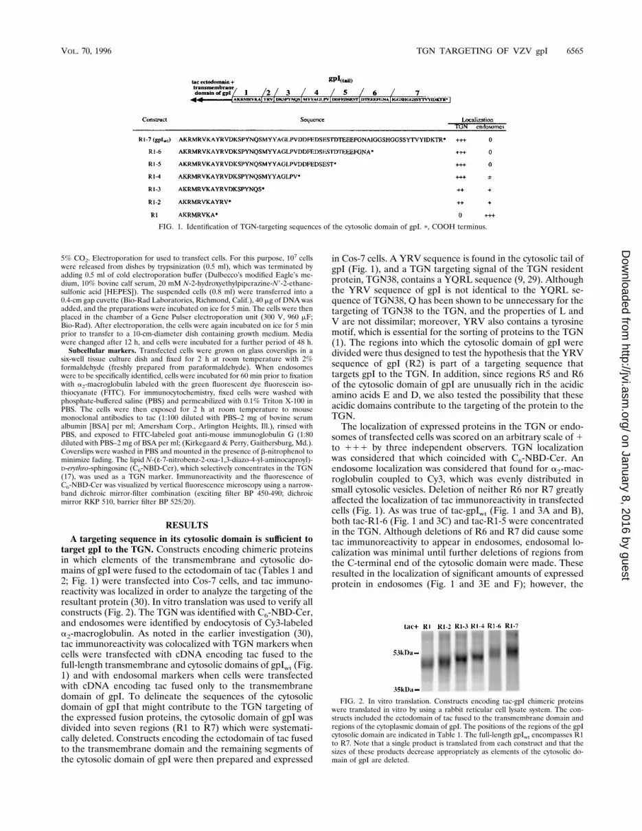
target gpI to the TGN. Constructs encoding chimeric proteinsin which elements of the transmembrane and cytosolic do-mains of gpI were fused to the ectodomain of tac (Tables 1 and2; Fig. 1) were transfected into Cos-7 cells, and tac immuno-reactivity was localized in order to analyze the targeting of theresultant protein (30). In vitro translation was used to verify allconstructs (Fig. 2). The TGN was identified with C6-NBD-Cer,and endosomes were identified by endocytosis of Cy3-labeleda2-macroglobulin. As noted in the earlier investigation (30),tac immunoreactivity was colocalized with TGN markers whencells were transfected with cDNA encoding tac fused to thefull-length transmembrane and cytosolic domains of gpIwt (Fig.1) and with endosomal markers when cells were transfectedwith cDNA encoding tac fused only to the transmembranedomain of gpI. To delineate the sequences of the cytosolicdomain of gpI that might contribute to the TGN targeting ofthe expressed fusion proteins, the cytosolic domain of gpI wasdivided into seven regions (R1 to R7) which were systemati-cally deleted. Constructs encoding the ectodomain of tac fusedto the transmembrane domain and the remaining segments ofthe cytosolic domain of gpI were then prepared and expressed
in Cos-7 cells. A YRV sequence is found in the cytosolic tail ofgpI (Fig. 1), and a TGN targeting signal of the TGN residentprotein, TGN38, contains a YQRL sequence (9, 29). Althoughthe YRV sequence of gpI is not identical to the YQRL se-quence of TGN38, Q has been shown to be unnecessary for thetargeting of TGN38 to the TGN, and the properties of L andV are not dissimilar; moreover, YRV also contains a tyrosinemotif, which is essential for the sorting of proteins to the TGN(1). The regions into which the cytosolic domain of gpI weredivided were thus designed to test the hypothesis that the YRVsequence of gpI (R2) is part of a targeting sequence thattargets gpI to the TGN. In addition, since regions R5 and R6of the cytosolic domain of gpI are unusually rich in the acidicamino acids E and D, we also tested the possibility that theseacidic domains contribute to the targeting of the protein to theTGN.The localization of expressed proteins in the TGN or endo-
somes of transfected cells was scored on an arbitrary scale of1to 111 by three independent observers. TGN localizationwas considered that which coincided with C6-NBD-Cer. Anendosome localization was considered that found for a2-mac-roglobulin coupled to Cy3, which was evenly distributed insmall cytosolic vesicles. Deletion of neither R6 nor R7 greatlyaffected the localization of tac immunoreactivity in transfectedcells (Fig. 1). As was true of tac-gpIwt (Fig. 1 and 3A and B),both tac-R1-6 (Fig. 1 and 3C) and tac-R1-5 were concentratedin the TGN. Although deletions of R6 and R7 did cause sometac immunoreactivity to appear in endosomes, endosomal lo-calization was minimal until further deletions of regions fromthe C-terminal end of the cytosolic domain were made. Theseresulted in the localization of significant amounts of expressedprotein in endosomes (Fig. 1 and 3E and F); however, the
FIG. 1. Identification of TGN-targeting sequences of the cytosolic domain of gpI. p, COOH terminus.
FIG. 2. In vitro translation. Constructs encoding tac-gpI chimeric proteinswere translated in vitro by using a rabbit reticular cell lysate system. The con-structs included the ectodomain of tac fused to the transmembrane domain andregions of the cytoplasmic domain of gpI. The positions of the regions of the gpIcytosolic domain are indicated in Table 1. The full-length gpIwt encompasses R1to R7. Note that a single product is translated from each construct and that thesizes of these products decrease appropriately as elements of the cytosolic do-main of gpI are deleted.
VOL. 70, 1996 TGN TARGETING OF VZV gpI 6565
on January 8, 2016 by guesthttp://jvi.asm
.org/D
ownloaded from

6566 ZHU ET AL. J. VIROL.
on January 8, 2016 by guesthttp://jvi.asm
.org/D
ownloaded from

fusion proteins continued to be concentrated primarily in theTGN until R2 was deleted (Fig. 1 and 3G and H). When R2was deleted from the chimeric constructs, none of the resultantfusion protein concentrated in the TGN; instead, the expressedprotein was located entirely in endosomes, a pattern identicalto that observed in cells transfected with cDNA encoding theectodomain of tac fused only to the transmembrane domain ofgpI. These observations are consistent with the hypothesis thatthe YRV sequence (R2) of the cytosolic domain of gpI isessential for targeting the protein to the TGN. Certainly, theobservation that the addition of only the YRV sequence to R1is sufficient to confer TGN targeting on the resultant chimericprotein suggests that this sequence is a TGN-targeting signal,although it may not be the only one. The presence of the YRVsequence alone, however, did not prevent the appearance ofsome of the expressed protein in endosomes. Endosomes, aswell as the TGN, became labeled when cells were transfectedwith many of the constructs encoding chimeric proteins withdeletions from the C-terminal region of the cytosolic domainof gpI. This observation suggests that sequences other thanYRV may also contribute to gpI targeting, at least to restrictretention in endosomes.A second targeting patch in its cytosolic domain contributes
to the targeting of gpI to the TGN. To determine whetherregions of the cytosolic domain of gpI, in addition to the YRVof R2, contribute to TGN targeting, Cos-7 cells were trans-fected with constructs encoding tac-gpI chimeric proteins inwhich R2 was deleted while other regions of the gpI cytosolicdomain closer to the C terminus were retained. Since the initialstudies (Fig. 1) had indicated that the localization of fusionproteins that contained R1 to R3 (Fig. 3E) was similar to thatof proteins that contained only R1 and R2 (Fig. 3F), thesestudies focused on R4 to R6, which included the acidic regions(R5 and R6) of the molecule. When cells were transfected with
cDNA encoding chimeric proteins that lacked the YRV do-main but contained R4 to R6, the expressed protein concen-trated in the TGN but was also found in endosomes (Table 3;Fig. 4A). The further deletion (individually) of R4 (Table 3;Fig. 4B), R5 (Table 3), or R6 (Table 3; Fig. 4C), may havereduced but did not eliminate targeting to the TGN. Thereseemed to be a reciprocal relationship between targeting ofconstructs with deletions to the TGN and concentration of tacimmunoreactivity in endosomes. Deletion of both R5 and R6greatly increased the concentration of expressed protein inendosomes (Table 3; Fig. 4D). These observations are consis-tent with the idea that R4 to R6 encompass a targeting patchthat contains TGN-targeting information.Combining the targeting sequence with the targeting patch
in its cytosolic domain potentiates the targeting of gpI to theTGN. Expressed proteins containing R2 and those containingR4 to R6 are each targeted to the TGN but also to endosomes.The presence of expressed protein in endosomes thus distin-guishes the trafficking of these chimeric proteins from that ofthe fusion protein containing the full-length cytosolic domainof gpIwt. To test whether the combination of these regionswould be targeted like tac-gpIwt, Cos-7 cells were transfectedwith a construct that encoded a chimeric protein that con-tained R2 and R4 to R6. The corresponding expressed proteinwas essentially confined to the TGN in transfected cells, andalmost no labeling of endosomes was detected (Table 4; Fig.5A). The appearance of tac immunoreactivity in cells trans-fected with the fusion protein containing R2 and R4 to R6could not be distinguished from that of tac-gpIwt (Fig. 5B). Incontrast, expressed protein was found in endosomes when cellswere transfected with constructs encoding proteins that con-tained R2 and only R4 (Table 4; Fig. 5C) or R4 and R5 (Table4; Fig. 5D). These observations suggest that all of the targeting
TABLE 3. Identification of a TGN-targeting patch in the cytosolic domain of gpI
Constructa SequencebLocalization
TGN Endosomes
R1/R4-6 AKRMRVKA___________MYYAGLPVDDFEDSESTDTEEEFGNA 11 1R1/R5-6 AKRMRVKA___________________DDFEDSESTDTEEEFGNA 1 11R1/R4-5 AKRMRVKA___________MYYAGLPVDDFEDSEST 1 11R1/R4 AKRMRVKA___________MYYAGLPV 6 111c
R1-2/R4-6 AKRMRVKAYRV________MYYAGLPVDDFEDSESTDTEEEFGNA 111 0
a See Fig. 1.b COOH termini are on the right.c Also locates strongly in plasma membrane.
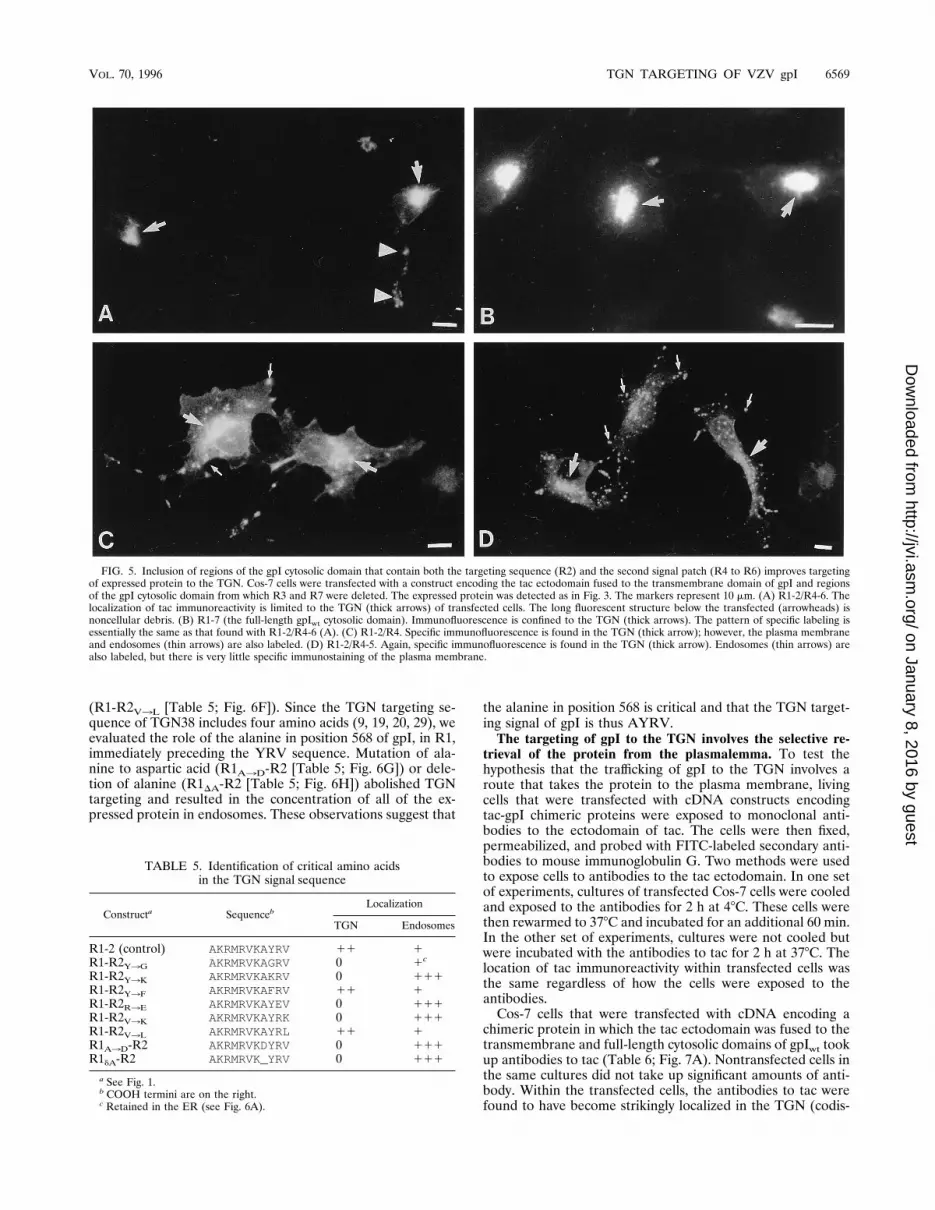
FIG. 3. Progressive deletion of regions of the cytosolic domain of gpI in tac-gpI constructs reveals the existence of a TGN targeting sequence in R2. Cos-7 cells weretransfected with a construct encoding the tac ectodomain fused to the transmembrane domain of gpI and regions of the gpI cytosolic domain. The expressed proteinwas detected with antibodies to tac. The location of the TGN was assessed with C6-NBD-Cer (not illustrated), and the location of endosomes was determined bydemonstrating the uptake of Cy3-labeled a2-macroglobulin (not illustrated). The markers represent 10 mm. (A) Control (construct R1-7, containing the full-length gpIwtcytosolic domain). Note that at low magnification there is little or no background and that specific immunofluorescence (thin arrows) is seen only in the TGN oftransfected cells. Cells that were not transfected (thick arrows) are not fluorescent. (B) Control (R1-7). At higher magnification, the limitation of specific immuno-fluorescence to the TGN (arrow) is more evident, and the absence of immunofluorescence in endosomes, ER, plasma membrane, or other organelles should be noted.(C) R1-6. Localization of specific immunofluorescence is similar to that of the control, primarily in the TGN. Very few small endosomes are evident. (D) R1-4. TheTGN (thick arrow) is still labeled; however, the processes of the cells can also be seen because specific immunofluorescence is present in endosomes (thin arrows), whichare diffusely distributed in the cytoplasm. In addition, the plasma membrane is immunostained, which adds apparent background and outlining to the transfected cells.(E) R1-3. The TGN (thick arrow) is still immunostained, and endosomes (thin arrows) are evident. No immunostaining of the plasma membrane is apparent; however,the expressed protein can be detected by a faint but specific immunofluorescence in the perinuclear cisterna which outlines the nonfluorescent nucleus. (F) R1-2. TheTGN (thick arrow) is brightly immunostained, but there is also some specific immunofluorescence in endosomes (thin arrows) and the perinuclear cisterna. Noimmunostaining of the plasma membrane is apparent; however, the expressed protein can be detected by a faint but specific immunofluorescence in the perinuclearcisterna which outlines the nonfluorescent nucleus. (G and H) R1. There is almost no specific immunofluorescence remaining in the TGN, although the centrosomalregions of transfected cells are thicker than the periphery and show more autofluorescence. As a result, there is a diffuse nonspecific brightness in this region that isdifferent from the more intense and sharply localized specific fluorescence found in immunostained organelles. Expressed protein is found mainly in endosomes;however, faint immunofluorescence can also be detected in the perinuclear cisterna (G). The plasma membrane is not immunostained.
VOL. 70, 1996 TGN TARGETING OF VZV gpI 6567
on January 8, 2016 by guesthttp://jvi.asm
.org/D
ownloaded from

information in the cytosolic domain of gpI is found in R2 andR4 to R6.Mutations of individual amino acids of the targeting se-
quence identify those which are critical for TGN targeting. Ascontrols, Cos-7 cells were transfected with cDNA encodingchimeric proteins in which the ectodomain of tac was fused tothe transmembrane and R1 and R2 of the cytosolic domains ofgpI (Table 5). The targeting of expressed protein was thencompared with that found in cells transfected with tac-R1-2constructs in which individual amino acids of R2 or the lastamino acid of R1 were mutated. Targeting to the TGN wasalmost entirely lost when the tyrosine of the R2 sequence wasreplaced with glycine or lysine (R1-R2Y3G or R1-R2Y3K [Ta-ble 5]). The expressed R1-R2Y3G protein appeared to bemainly retained in the endoplasmic reticulum (Fig. 6A). Intra-
cellular expressed protein in cells transfected with the con-struct R1-R2Y3K was restricted to endosomes (Fig. 6B). Incontrast, tyrosine could be replaced by phenylalanine (R1-R2Y3F [Table 5; Fig. 6C]) without altering the targeting of theexpressed protein. These observations suggest that an aromaticamino acid rather than tyrosine itself is critical in position 569.Targeting to the TGN was also lost when the arginine of R2was replaced with glutamic acid (R1-R2R3E [Table 5; Fig.6D]) or the valine of R2 was replaced with lysine (R1-R2V3K[Table 5; Fig. 6E]). In both cases, intracellular tac immunore-activity was again found only in endosomes. Although TGNtargeting thus was abolished when valine was mutated to abasic amino acid, it could be replaced by another hydrophobicamino acid, leucine, without altering the targeting of expressedprotein to the TGN or increasing localization in endosomes
FIG. 4. Deletion of the region (R2) that includes the TGN-targeting sequence enables a second targeting patch to be detected. Cos-7 cells were transfected witha construct encoding the tac ectodomain fused to the transmembrane domain of gpI and regions of the gpI cytosolic domain from which R2 was deleted. The expressedprotein was detected as for Fig. 3. The markers represent 10 mm. (A) R1/R4-6. Specific immunofluorescence can be detected in the TGN (thick arrow) and inendosomes (thin arrows). In many cells the plasma membrane is also very immunofluorescent, which obscures the visualization of intracellular structures. (B) R1/R5-6.Specific immunofluorescence is evident in the TGN (thick arrow) and in endosomes (thin arrows). There is less immunostaining of the plasma membrane than whenR4 is present. (C) R1/R4-5. The TGN (thick arrow) and endosomes are immunostained. (D) R1/R4. Specific immunofluorescence is primarily located in endosomes(thin arrows); the TGN is not labeled.
TABLE 4. Combination of the putative TGN-targeting signal and patch in the cytosolic domain of gpI
Constructa SequencebLocalization
TGN Endosomes
R1-2/R4-6 AKRMRVKAYRV_______MYYAGLPVDDFEDSESTDTEEEFGNA 111 0R1-2/R4-5 AKRMRVKAYRV________MYYAGLPVDDFEDSEST 11 1R1-2/R4 AKRMRVKAYRV________MYYAGLPV 11 1
a See Fig. 1.b COOH termini are on the right.
6568 ZHU ET AL. J. VIROL.
on January 8, 2016 by guesthttp://jvi.asm
.org/D
ownloaded from
(R1-R2V3L [Table 5; Fig. 6F]). Since the TGN targeting se-quence of TGN38 includes four amino acids (9, 19, 20, 29), weevaluated the role of the alanine in position 568 of gpI, in R1,immediately preceding the YRV sequence. Mutation of ala-nine to aspartic acid (R1A3D-R2 [Table 5; Fig. 6G]) or dele-tion of alanine (R1DA-R2 [Table 5; Fig. 6H]) abolished TGNtargeting and resulted in the concentration of all of the ex-pressed protein in endosomes. These observations suggest that
the alanine in position 568 is critical and that the TGN target-ing signal of gpI is thus AYRV.The targeting of gpI to the TGN involves the selective re-
trieval of the protein from the plasmalemma. To test thehypothesis that the trafficking of gpI to the TGN involves aroute that takes the protein to the plasma membrane, livingcells that were transfected with cDNA constructs encodingtac-gpI chimeric proteins were exposed to monoclonal anti-bodies to the ectodomain of tac. The cells were then fixed,permeabilized, and probed with FITC-labeled secondary anti-bodies to mouse immunoglobulin G. Two methods were usedto expose cells to antibodies to the tac ectodomain. In one setof experiments, cultures of transfected Cos-7 cells were cooledand exposed to the antibodies for 2 h at 48C. These cells werethen rewarmed to 378C and incubated for an additional 60 min.In the other set of experiments, cultures were not cooled butwere incubated with the antibodies to tac for 2 h at 378C. Thelocation of tac immunoreactivity within transfected cells wasthe same regardless of how the cells were exposed to theantibodies.Cos-7 cells that were transfected with cDNA encoding a
chimeric protein in which the tac ectodomain was fused to thetransmembrane and full-length cytosolic domains of gpIwt tookup antibodies to tac (Table 6; Fig. 7A). Nontransfected cells inthe same cultures did not take up significant amounts of anti-body. Within the transfected cells, the antibodies to tac werefound to have become strikingly localized in the TGN (codis-
FIG. 5. Inclusion of regions of the gpI cytosolic domain that contain both the targeting sequence (R2) and the second signal patch (R4 to R6) improves targetingof expressed protein to the TGN. Cos-7 cells were transfected with a construct encoding the tac ectodomain fused to the transmembrane domain of gpI and regionsof the gpI cytosolic domain from which R3 and R7 were deleted. The expressed protein was detected as in Fig. 3. The markers represent 10 mm. (A) R1-2/R4-6. Thelocalization of tac immunoreactivity is limited to the TGN (thick arrows) of transfected cells. The long fluorescent structure below the transfected (arrowheads) isnoncellular debris. (B) R1-7 (the full-length gpIwt cytosolic domain). Immunofluorescence is confined to the TGN (thick arrows). The pattern of specific labeling isessentially the same as that found with R1-2/R4-6 (A). (C) R1-2/R4. Specific immunofluorescence is found in the TGN (thick arrow); however, the plasma membraneand endosomes (thin arrows) are also labeled. (D) R1-2/R4-5. Again, specific immunofluorescence is found in the TGN (thick arrow). Endosomes (thin arrows) arealso labeled, but there is very little specific immunostaining of the plasma membrane.
TABLE 5. Identification of critical amino acidsin the TGN signal sequence
Constructa SequencebLocalization
TGN Endosomes
R1-2 (control) AKRMRVKAYRV 11 1R1-R2Y3G AKRMRVKAGRV 0 1c
R1-R2Y3K AKRMRVKAKRV 0 111R1-R2Y3F AKRMRVKAFRV 11 1R1-R2R3E AKRMRVKAYEV 0 111R1-R2V3K AKRMRVKAYRK 0 111R1-R2V3L AKRMRVKAYRL 11 1R1A3D-R2 AKRMRVKDYRV 0 111R1dA-R2 AKRMRVK_YRV 0 111
a See Fig. 1.b COOH termini are on the right.c Retained in the ER (see Fig. 6A).
VOL. 70, 1996 TGN TARGETING OF VZV gpI 6569
on January 8, 2016 by guesthttp://jvi.asm
.org/D
ownloaded from
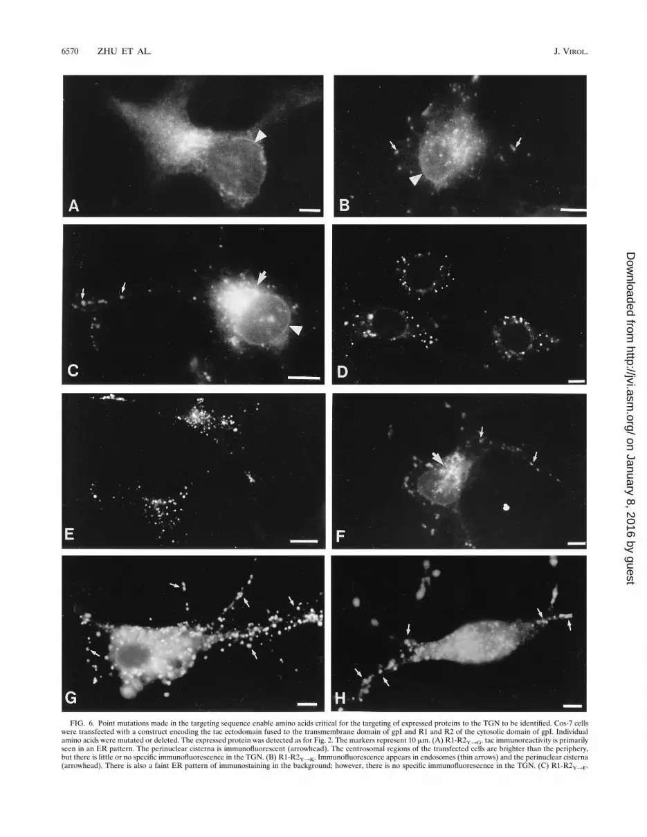
FIG. 6. Point mutations made in the targeting sequence enable amino acids critical for the targeting of expressed proteins to the TGN to be identified. Cos-7 cellswere transfected with a construct encoding the tac ectodomain fused to the transmembrane domain of gpI and R1 and R2 of the cytosolic domain of gpI. Individualamino acids were mutated or deleted. The expressed protein was detected as for Fig. 2. The markers represent 10 mm. (A) R1-R2Y3G. tac immunoreactivity is primarilyseen in an ER pattern. The perinuclear cisterna is immunofluorescent (arrowhead). The centrosomal regions of the transfected cells are brighter than the periphery,but there is little or no specific immunofluorescence in the TGN. (B) R1-R2Y3K. Immunofluorescence appears in endosomes (thin arrows) and the perinuclear cisterna(arrowhead). There is also a faint ER pattern of immunostaining in the background; however, there is no specific immunofluorescence in the TGN. (C) R1-R2Y3F.
6570 ZHU ET AL. J. VIROL.
on January 8, 2016 by guesthttp://jvi.asm
.org/D
ownloaded from
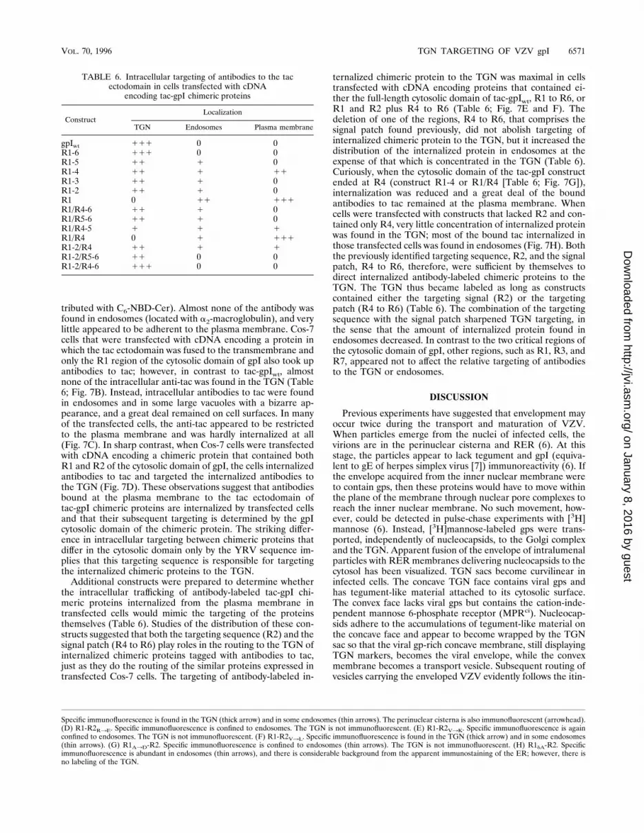
tributed with C6-NBD-Cer). Almost none of the antibody wasfound in endosomes (located with a2-macroglobulin), and verylittle appeared to be adherent to the plasma membrane. Cos-7cells that were transfected with cDNA encoding a protein inwhich the tac ectodomain was fused to the transmembrane andonly the R1 region of the cytosolic domain of gpI also took upantibodies to tac; however, in contrast to tac-gpIwt, almostnone of the intracellular anti-tac was found in the TGN (Table6; Fig. 7B). Instead, intracellular antibodies to tac were foundin endosomes and in some large vacuoles with a bizarre ap-pearance, and a great deal remained on cell surfaces. In manyof the transfected cells, the anti-tac appeared to be restrictedto the plasma membrane and was hardly internalized at all(Fig. 7C). In sharp contrast, when Cos-7 cells were transfectedwith cDNA encoding a chimeric protein that contained bothR1 and R2 of the cytosolic domain of gpI, the cells internalizedantibodies to tac and targeted the internalized antibodies tothe TGN (Fig. 7D). These observations suggest that antibodiesbound at the plasma membrane to the tac ectodomain oftac-gpI chimeric proteins are internalized by transfected cellsand that their subsequent targeting is determined by the gpIcytosolic domain of the chimeric protein. The striking differ-ence in intracellular targeting between chimeric proteins thatdiffer in the cytosolic domain only by the YRV sequence im-plies that this targeting sequence is responsible for targetingthe internalized chimeric proteins to the TGN.Additional constructs were prepared to determine whether
the intracellular trafficking of antibody-labeled tac-gpI chi-meric proteins internalized from the plasma membrane intransfected cells would mimic the targeting of the proteinsthemselves (Table 6). Studies of the distribution of these con-structs suggested that both the targeting sequence (R2) and thesignal patch (R4 to R6) play roles in the routing to the TGN ofinternalized chimeric proteins tagged with antibodies to tac,just as they do the routing of the similar proteins expressed intransfected Cos-7 cells. The targeting of antibody-labeled in-
ternalized chimeric protein to the TGN was maximal in cellstransfected with cDNA encoding proteins that contained ei-ther the full-length cytosolic domain of tac-gpIwt, R1 to R6, orR1 and R2 plus R4 to R6 (Table 6; Fig. 7E and F). Thedeletion of one of the regions, R4 to R6, that comprises thesignal patch found previously, did not abolish targeting ofinternalized chimeric protein to the TGN, but it increased thedistribution of the internalized protein in endosomes at theexpense of that which is concentrated in the TGN (Table 6).Curiously, when the cytosolic domain of the tac-gpI constructended at R4 (construct R1-4 or R1/R4 [Table 6; Fig. 7G]),internalization was reduced and a great deal of the boundantibodies to tac remained at the plasma membrane. Whencells were transfected with constructs that lacked R2 and con-tained only R4, very little concentration of internalized proteinwas found in the TGN; most of the bound tac internalized inthose transfected cells was found in endosomes (Fig. 7H). Boththe previously identified targeting sequence, R2, and the signalpatch, R4 to R6, therefore, were sufficient by themselves todirect internalized antibody-labeled chimeric proteins to theTGN. The TGN thus became labeled as long as constructscontained either the targeting signal (R2) or the targetingpatch (R4 to R6) (Table 6). The combination of the targetingsequence with the signal patch sharpened TGN targeting, inthe sense that the amount of internalized protein found inendosomes decreased. In contrast to the two critical regions ofthe cytosolic domain of gpI, other regions, such as R1, R3, andR7, appeared not to affect the relative targeting of antibodiesto the TGN or endosomes.
DISCUSSION
Previous experiments have suggested that envelopment mayoccur twice during the transport and maturation of VZV.When particles emerge from the nuclei of infected cells, thevirions are in the perinuclear cisterna and RER (6). At thisstage, the particles appear to lack tegument and gpI (equiva-lent to gE of herpes simplex virus [7]) immunoreactivity (6). Ifthe envelope acquired from the inner nuclear membrane wereto contain gps, then these proteins would have to move withinthe plane of the membrane through nuclear pore complexes toreach the inner nuclear membrane. No such movement, how-ever, could be detected in pulse-chase experiments with [3H]mannose (6). Instead, [3H]mannose-labeled gps were trans-ported, independently of nucleocapsids, to the Golgi complexand the TGN. Apparent fusion of the envelope of intralumenalparticles with RER membranes delivering nucleocapsids to thecytosol has been visualized. TGN sacs become curvilinear ininfected cells. The concave TGN face contains viral gps andhas tegument-like material attached to its cytosolic surface.The convex face lacks viral gps but contains the cation-inde-pendent mannose 6-phosphate receptor (MPRci). Nucleocap-sids adhere to the accumulations of tegument-like material onthe concave face and appear to become wrapped by the TGNsac so that the viral gp-rich concave membrane, still displayingTGN markers, becomes the viral envelope, while the convexmembrane becomes a transport vesicle. Subsequent routing ofvesicles carrying the enveloped VZV evidently follows the itin-
TABLE 6. Intracellular targeting of antibodies to the tacectodomain in cells transfected with cDNA
encoding tac-gpI chimeric proteins
ConstructLocalization
TGN Endosomes Plasma membrane
gpIwt 111 0 0R1-6 111 0 0R1-5 11 1 0R1-4 11 1 11R1-3 11 1 0R1-2 11 1 0R1 0 11 111R1/R4-6 11 1 0R1/R5-6 11 1 0R1/R4-5 1 1 1R1/R4 0 1 111R1-2/R4 11 1 1R1-2/R5-6 11 0 0R1-2/R4-6 111 0 0
Specific immunofluorescence is found in the TGN (thick arrow) and in some endosomes (thin arrows). The perinuclear cisterna is also immunofluorescent (arrowhead).(D) R1-R2R3E. Specific immunofluorescence is confined to endosomes. The TGN is not immunofluorescent. (E) R1-R2V3K. Specific immunofluorescence is againconfined to endosomes. The TGN is not immunofluorescent. (F) R1-R2V3L. Specific immunofluorescence is found in the TGN (thick arrow) and in some endosomes(thin arrows). (G) R1A3D-R2. Specific immunofluorescence is confined to endosomes (thin arrows). The TGN is not immunofluorescent. (H) R1dA-R2. Specificimmunofluorescence is abundant in endosomes (thin arrows), and there is considerable background from the apparent immunostaining of the ER; however, there isno labeling of the TGN.
VOL. 70, 1996 TGN TARGETING OF VZV gpI 6571
on January 8, 2016 by guesthttp://jvi.asm
.org/D
ownloaded from

6572 ZHU ET AL. J. VIROL.
on January 8, 2016 by guesthttp://jvi.asm
.org/D
ownloaded from
erary of the MPRci to deliver virions to acidic prelysosomes(late endosomes). Inactivation of virions in these structuresmay account for the cell-associated nature of VZV in vitro.These observations suggest that the targeting of viral gps to theTGN might be critical for the final envelopment of VZV.Since earlier studies indicated that the cytosolic domain of
VZV gpI contains TGN-targeting information (30), the cur-rent investigation was carried out to identify the responsibleregions of this portion of the molecule. The targeting of ex-pressed protein in Cos-7 cells transfected with cDNA encodingthe ectodomain of tac fused to the transmembrane and cyto-solic domains of gpI was studied. To determine the role playedin targeting by specific regions of the cytosolic domain of gpI,segments or individual amino acids of this domain were de-leted or mutated. The selective localization of C6-NBD-Cerserved as a TGN marker (17, 30), and the localization ofa2-macroglobulin, taken up by endocytosis, was used as markerfor endosomes (30).Two regions of the cytosolic domain of gpI were found to be
critical for the targeting of expressed chimeric proteins to theTGN. One was a short stretch of four amino acids with thesequence AYRV, and the other was a much longer sequencenearer the C-terminal end of gpI. Both of these regions of thegpI cytosolic domain contained information which by itself wassufficient to target chimeric proteins expressed in transfectedcells to the TGN. We postulate that the longer acidic sequenceconstitutes a targeting patch, the effectiveness of which de-pends not on a sequence of contiguous amino acids but on thetertiary structure of the molecule. The individual amino acidsof the shorter targeting sequence were each found to be im-portant in signaling. In contrast, relatively large portions of themore distal patch could be deleted without abolishing its TGN-targeting function. While such deletions reduced the propor-tion of the expressed protein that was targeted to the TGN andincreased the relative amount in endosomes, they did not pre-vent some of the expressed protein from concentrating in theTGN. Combining the shorter targeting sequence with thelonger putative signal patch enhanced the targeting of theresultant expressed protein to the TGN and virtually elimi-nated its appearance in endosomes. It is of interest thatTGN38, the prototypic TGN resident protein, also containsboth a targeting sequence of amino acids and another signalpatch, each of which contributes to the concentration of thismolecule in the TGN (18).The role in TGN targeting played by individual amino acids
of the AYRV-targeting sequence was studied by evaluating theeffects of point mutations made in constructs encoding chi-meric proteins that lacked the targeting patch. The targeting ofexpressed proteins in cells transfected with these constructswas thus determined solely by the effectiveness of the targetingsequence and was not subject to a confounding effect of themore distal patch. Although the tyrosine of the AYRV se-
quence could not be replaced with glycine or lysine, it could bereplaced by phenylalanine; therefore, an aromatic amino acid,rather than tyrosine itself, is probably required in this position.Phosphorylation on this tyrosine residue is thus not likely toplay a role in TGN targeting. Like gpI, TGN38, contains afour-amino-acid sequence, YQRL, that is critical for its local-ization in the TGN (9, 19, 20, 29). The tyrosine of the YQRLsequence is thought to be essential; however, its substitutionwith phenylalanine has not been investigated. Since the argi-nine of AYRV could not be replaced with glutamic acid, abasic amino acid may be needed at this residue. Althoughtargeting to the TGN was not disrupted when valine was sub-stituted with leucine, targeting was lost when valine was sub-stituted with lysine, suggesting that a hydrophobic amino acidand not a basic amino acid is critical at this position. Sincealanine could not be deleted (which has the effect of convertingAYRV to KYRV) or substituted with aspartic acid, targeting isevidently lost if an acidic or basic amino precedes the aromaticamino acid in the targeting sequence. Deletion of the entireregion (R3) between the AYRV sequence and the more distalputative signal patch did not alter the targeting of expressedchimeric protein to the TGN, suggesting that valine is the lastamino acid of the AYRV-targeting sequence. Another TGNresident protein, furin, is directed to the TGN by signallingregions in its cytosolic domain (2, 26). Furin also cycles be-tween the plasma membrane and the TGN and concentrates inthe TGN as a result of selective retrieval (24). As is true of gpIand TGN38, two nonoverlapping regions participate in signal-ling (22, 26). One of these is a YKGL motif that is likely to befunctionally similar to the shorter targeting sequence of gpI,and the other is a longer region, the sequence of which is acidic(26) and resembles that found in R4 to R6 of the cytosolicdomain of gpI. In fact, the S residues that have been demon-strated in these regions of furin to be critical for TGN targeting(24) are also present in analogous location in gpI. In additionto TGN38 and furin, the cytosolic domains of other proteins ofinterest contain sequences of amino acids that resemble theAYRV of gpI. One of these is the cation-dependent mannose6-phosphate receptor (MPRcd), which cycles between theplasma membrane and the TGN (11). The cytosolic domain ofthe MPRcd contains both the YQRL that is found in TGN38and an AYRGV sequence that resembles gpI. For it to besimilar to AYRV, one would have to suppose that the addi-tional glycine in the MPRcd is a small linker that does not affectthe signaling function. This supposition has not been tested,and the roles played by YQRL and AYRGV in the intra-cellular targeting of the MPRcd have not been studied. TheAYRGV sequence, however, has been found to be critical forinternalization of the MPRcd (10). The itinerary of the MPRcd,moreover, includes stops at both the plasma membrane andthe TGN (10, 11, 15, 27). Another protein with a similar se-quence in a probable internal cytosolic loop is the putative
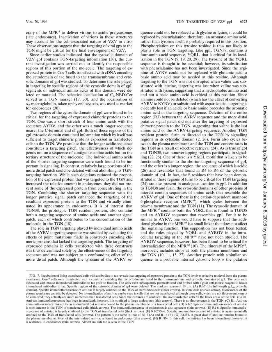
FIG. 7. Incubation of living transfected cells with antibodies to tac reveals that targeting of expressed protein to the TGN involves selective retrieval from the plasmamembrane. Cos-7 cells were transfected with a construct encoding the tac ectodomain fused to the transmembrane and cytosolic domains of gpI. The cells wereincubated with mouse monoclonal antibodies to tac prior to fixation. The cells were subsequently permeabilized and probed with a goat anti-mouse reagent to locateinternalized antibodies to tac. Specific regions of the cytosolic domain of gpI were deleted. The markers represent 10 mm. (A) R1-7 (the full-length gpIwt cytosolicdomain). Specific immunofluorescence of anti-tac is largely confined to the TGN of transfected cells (thick arrows). In some cells (curved arrow), fluorescence of theplasma membrane can also be detected. No internalization of anti-tac can be seen in cells that are not transfected; although these cells, which are not fluorescent, cannotbe visualized, they actually are more numerous than transfected cells. Since the cultures are confluent, the nontransfected cells fill the black areas of the field. (B) R1.Anti-tac immunofluorescence has been internalized; however, it is confined to large endosomes (thin arrows). There is no fluorescence in the TGN. (C) R1. Anti-tacimmunofluorescence has not been internalized but remains bound to the plasma membrane of a transfected cell. (D) R1-2. Specific immunofluorescence of anti-tacis most intense in the TGN of transfected cells (thick arrows). The immunofluorescence of endosomes is also apparent (thin arrows). (E) R1-6. Specific immunoflu-orescence of anti-tac is largely confined to the TGN of transfected cells (thick arrows). (F) R1-2/R4-6. Specific immunofluorescence of anti-tac is again essentiallyconfined to the TGN of transfected cells (arrows). The pattern is the same as that of R1-7 (A) and R1-6 (F). (G) R1/R4. A great deal of anti-tac remains bound tothe plasma membrane. Most of the internalized anti-tac is found in endosomes (thin arrows). (H) R1/R4. In this cell, the anti-tac has been internalized; however, itis restricted to endosomes (thin arrows). Almost no anti-tac is seen in the TGN.
VOL. 70, 1996 TGN TARGETING OF VZV gpI 6573
on January 8, 2016 by guesthttp://jvi.asm
.org/D
ownloaded from

Cu21-transporting ATPase that is defective in Menke’s disease(12, 25). Although this sequence of the Cu21-transportingATPase, LYRV, contains a leucine instead of an alanine, thetwo amino acids are likely to be functionally similar. The in-tracellular distribution of the Cu21-transporting ATPase hasnot yet been reported.Since the targeting of TGN38 to the TGN has been shown to
depend on selective retrieval of the protein from the plasmamembrane, the possibility that a similar mechanism accountsfor the targeting of gpI was investigated. Living cells that hadbeen transfected with tac-gpI constructs were incubated withantibodies to the ectodomain of tac (Fig. 8). To minimize thepossibility that antibodies to tac might enter cells nonspecifi-cally by fluid-phase endocytosis, cells were initially exposed tothe antibodies at 48C. Subsequent warming after the washoutof residual free antibody would enable bound antibodies to beinternalized together with the proteins that they labeled. Inpractice, cooling the cells during exposure to antibodies provedto be unnecessary; transfected cells rapidly internalized anti-bodies to the tac ectodomain, regardless of whether the cellswere exposed to these antibodies at 4 or at 378C. In contrast,nontransfected cells did not take up the antibodies to tac indetectable amounts, even when they were incubated with theantibodies for 2 h at 378C. Since expressed protein remainsentirely at the plasma membrane when cells are transfectedwith the full-length tac (30), the internalization of the chimerictac-gpI proteins suggests that an internalization signal is
present in the cytosolic and/or transmembrane domains of gpI.The observation that antibodies to tac were internalized in alltransfected cells, even in those transfected with constructs thatcontained only the transmembrane and R1 of the gpI cytosolicdomain, suggests that the internalization signal of the gpI mol-ecule is present either in its transmembrane region or in R1 ofthe cytosolic domain. Whether internalized chimeric proteinsonly reached endosomes or selectively concentrated in theTGN, however, was found to depend on signals in other re-gions of the cytosolic domain of gpI. These trafficking signals,determining the routing of protein retrieved from the plasmamembrane, were the same domains of the gpI cytosolic do-main, which were found to direct the targeting of expressedtac-gpI constructs. Again, both the AYRV-targeting sequenceand the larger putative signal patch were found to play impor-tant roles. Either could direct some of the internalized proteinto the TGN, and when both were present in the expressedprotein, essentially all of the retrieved antibody-labeled proteinwas found in the TGN. These observations suggest that selec-tive retrieval from the plasma membrane is the mechanism thatis responsible for the targeting of gpI to the TGN (Fig. 8).The conclusion that selective retrieval from the plasma
membrane is responsible for the targeting of gpI to the TGNimplies that the concentration of gpI in the TGN is likely to bean active process involving membrane recycling, rather than astable process involving the anchoring of gpI in the TGN. Toreach the plasma membrane, gpI would have to be transportedfrom the RER through the cis-Golgi, Golgi stack, TGN, andtransport vesicles. To carry bound antibodies to tac back to theTGN from the plasma membrane, the protein would have tobe retrieved by endocytosis, which would deliver it to endo-somes (Fig. 8). Endosomes are organelles that sort a great dealof membrane traffic (28). For example, both MPRs cycle be-tween the plasma membrane and the TGN via endosomes (23).Endosomes also direct receptors and other membrane proteinsto specialized postendosomal vesicles, including the transcy-totic vesicles of epithelial cells, synaptic vesicles of neurons,and GLUT4-containing vesicles of adipocytes (28). The rout-ing function of the targeting sequence and patch of the cyto-solic domain of gpI, therefore, evidently interacts with a sort-ing mechanism that directs the traffic of membrane fromendosomes. An equilibrium, in which vesicles containing gpIrecycle between the TGN and the plasma membrane, is prob-ably shifted by the presence of the targeting signal and patch inthe cytosolic domain of gpI toward the TGN. Deletion ofeither the shorter signal or the longer patch may shift theequilibrium toward endosomes, while the loss of both signalingregions results in the failure of endosomes to transport gpIback to the TGN.The observation that gpI contains targeting signals that
cause it to concentrate in the TGN is compatible with thehypothesis that the TGN plays a critical role in viral envelop-ment. Neither the shorter targeting sequence nor the longertargeting patch of the gpI cytosolic domain is present in thecytosolic domains of VZV gpII to gpV (5). In fact, cytosolictails of significant length are probably present only in gpI, gpII,gpIV, and gpVI; moreover, there is relatively little sequenceidentity in the putative cytosolic regions of any of the gps,although an AYRYV sequence is present in gpII. It is thus notyet clear what is responsible for routing gps other than gpI tothe TGN. Conceivably, different targeting sequences in theother gps may be responsible. Alternatively, other gps may notbe independently targeted to the TGN, but instead be routedto this organelle because they form a complex with gpI. Theresulting multimeric complex could then be directed to the siteof viral envelopment by the targeting signal in the cytosolic
FIG. 8. Antibodies to the tac ectodomain are internalized and selectivelyconcentrated in the TGN of cells transfected with gpI-tac constructs. The sche-matic diagram (not drawn to scale) shows how selective retrieval of expressedprotein carrying bound antibodies can cause these antibodies to become con-centrated in the TGN of transfected cells. The original forward transport ofexpressed protein to the plasma membrane is presumed to occur through theconstitutive pathway and is not depicted. The antibodies to the tac ectodomainare postulated to bind to the tac ectodomain, which is exposed on the plasmamembrane. The antibodies are then concentrated in the lumen of endosomes,which is topologically equivalent to extracellular space. The endosomes thenpresumably fuse with membranes of the TGN. Antibodies are again intralume-nal, as the directionality of membranes is maintained. Since a change in thesequence of the gpI cytosolic domain of the chimeric proteins disrupts retrievalin the same manner as it disrupts targeting to the TGN, it is likely that targetingof expressed chimeric proteins occurs as a result of selective retrieval from theplasma membrane directed by the cytosolic domain of gpI.
6574 ZHU ET AL. J. VIROL.
on January 8, 2016 by guesthttp://jvi.asm
.org/D
ownloaded from

domain of gpI. If so, then gpI would serve as a navigator gp, arole that is consistent with the fact that gpI is the most abun-dant protein synthesized by cells infected with VZV (8). Al-though the mechanism by which the TGN envelops newlysynthesized virions in VZV-infected cells has not yet beenmade clear, the current observations support the idea that thisorganelle is the site where the process occurs and suggest thatgpI, which contains TGN-targeting signals, may be critical forviral envelopment.
ACKNOWLEDGMENTS
This work was supported by Public Health Service grant AI 127187from the National Institute of Allergy and Infectious Disease.We thank Yonghui Zhang and Wanda Setlik for their assistance.
REFERENCES
1. Bos, K., C. Wraight, and K. K. Stanley. 1993. TGN38 is maintained in thetrans-Golgi network by a tyrosine-containing motif in the cytoplasmic do-main. EMBO J. 12:2219–2228.
2. Bosshart, H., J. Humphrey, E. Deignan, J. Davidson, J. Drazba, L. Yuan, V.Oorschot, P. J. Peters, and J. S. Bonifacino. 1994. The cytoplasmic domainmediates location of furin to the trans-Golgi network en route to the endo-somal/lysosomal system. J. Cell Biol. 126:1157–1172.
3. Card, J. P., L. Rinaman, R. B. Lynn, B.-H. Lee, R. P. Meade, R. R. Miselis,and L. W. Enquist. 1993. Pseudorabies virus infection of the rat centralnervous system: ultrastructural characterization of viral replication, trans-port, and pathogenesis. J. Neurosci. 13:2515–2539.
4. Darlington, R. W., and L. H. Moss. 1968. Herpesvirus envelopment. J. Virol.2:48–55.
5. Davison, A. J., and J. E. Scott. 1986. The complete DNA sequence ofvaricella-zoster virus. J. Gen. Virol. 67:1759–1816.
6. Gershon, A., Z. Zhu, D. L. Sherman, C. A. Gabel, R. T. Ambron, and M. D.Gershon. 1994. Intracellular transport of newly synthesized varicella-zostervirus: final envelopment in the trans-Golgi network. J. Virol. 68:6372–6390.
7. Grose, C. 1991. Glycoproteins of varicella-zoster virus and their herpessimplex virus homologs. Rev. Infect. Dis. 13:S960–S963.
8. Grose, C., and V. Litwin. 1988. Immunology of the varicella-zoster glycopro-teins. J. Infect. Dis. 157:877–881.
9. Humphrey, J. S., P. J. Peters, L. C. Yuan, and J. S. Bonifacino. 1993.Localization of TGN38 to the trans-Golgi network: involvement of a cyto-plasmic tyrosine-containing sequence. J. Cell Biol. 120:1123–1135.
10. Johnson, C. F., W. Chan, and S. Kornfeld. 1990. Cation-dependent mannose6-phosphate receptor contains two internalization signals in its cytoplasmicdomain. Proc. Natl. Acad. Sci. USA 87:10010–10014.
11. Johnson, K. F., and S. Kornfeld. 1992. A his-leu-leu sequence near thecarboxyl terminus of the cytoplasmic domain of the cation-dependent man-nose 6-phosphate receptor is necessary for the lysosomal enzyme sortingfunction. J. Biol. Chem. 267:17110–17115.
12. Kodama, H., T. Abe, M. Takama, I. Takahashi, M. Kodama, and M. Nish-imura. 1993. Histochemical localization of copper in the intestine and kidneyof macular mice: light and electron microscopic study. J. Histochem. Cyto-chem. 41:1529–1535.
13. Ladinsky, M. S., and K. E. Howell. 1993. An electron microscopic study ofTGN38/41 dynamics. J. Cell Sci. Suppl. 17:41–47.
14. Martin, S., B. Reaves, G. Banting, and G. W. Gould. 1994. Analysis of the
co-localization of the insulin-responsive glucose transporter (GLUT4) andthe trans Golgi network marker TGN38 within 3T3-L1 adipocytes. Biochem.J. 300:743–749.
15. Matovcik, L. M., J. Goodhouse, and M. G. Farquhar. 1990. The recyclingitinerary of the 46 kDa mannose 6-phosphate receptor—Golgi to late endo-somes—coincides with that of the 215 kDa M6PR. Eur. J. Cell Biol. 53:203–211.
16. Morgan, C., H. M. Rose, M. Holden, and E. P. Jones. 1959. Electron micro-scopic observations on the development of herpes simplex virus. J. Exp. Med.110:643–656.
17. Pagano, R. E., M. A. Sepanski, and O. C. Martin. 1989. Molecular trappingof a fluorescent ceramide analogue at the Golgi apparatus of fixed cells:interaction with endogenous lipids provides a trans-Golgi marker for bothlight and electron microscopy. J. Cell Biol. 109:2067–2079.
18. Ponnambalam, S., C. Rabouille, J. P. Luzio, T. Nilsson, and G. Warren.1994. The TGN38 glycoprotein contains two non-overlapping signals thatmediate localization to the trans-Golgi network. J. Cell Biol. 125:253–268.
19. Reaves, B., and G. Banting. 1992. Perturbation of the morphology of thetrans-Golgi network following Brefeldin A treatment: redistribution of aTGN-specific integral membrane protein, TGN38. J. Cell Biol. 116:85–94.
20. Reaves, B., A. Wilde, and G. Banting. 1992. Identification, molecular char-acterization and immunolocalization of an isoform of the trans-Golgi-net-work (TGN)-specific integral membrane protein TGN38. Biochem. J. 283:313–316.
21. Roizman, B., and A. E. Sears. 1993. Herpes simplex viruses and their repli-cation, p. 11–68. In B. Roizman, R. J. Whitley, and C. Lopez (ed.), Thehuman herpesviruses. Raven Press, New York.
22. Schafer, W., A. Stroh, S. Berghofer, J. Seiler, N. Vey, M.-L. Kruse, H. F.Kern, H.-D. Klenck, and W. Garten. 1995. Two independent targeting signalsin the cytoplasmic domain determine trans-Golgi network localization andendosomal trafficking of the proprotein convertase furin. EMBO J. 14:2424–2435.
23. Schulze-Garg, C., C. Boker, S. K. Nadinpalli, K. von Figura, and A. Hille-Rehfeld. 1993. Tail-specific antibodies that block return of 46,000 Mr man-nose 6-phosphate receptor to the trans-Golgi network. J. Cell Biol. 122:541–551.
24. Takahashi, S., T. Nakagawa, T. Banno, T. Watanabe, K. Murakami, and K.Nakayama. 1995. Localization of furin to the trans-Golgi network and recy-cling from the cell surface involves serine and tyrosine residues within thecytosolic domain. J. Biol. Chem. 270:28397–28401.
25. Tumer, Z., B. Vural, T. Tonnesen, J. Chelly, A. P. Monaco, and N. Horn.1995. Characterization of the exon structure of the Menkes disease geneusing vectorette PCR. Genomics 26:437–442.
26. Voorhees, P., E. Deignan, E. van Donselaar, J. Humphrey, M. S. Marks, P. J.Peters, and J. S. Bonifacino. 1995. An acidic sequence within the cytoplasmicdomain of furin functions as a determinant of trans-Golgi network localiza-tion and internalization from the cell surface. EMBO J. 14:4961–4975.
27. Watanabe, H., J. H. Grubb, and W. S. Sly. 1990. The overexpressed human46-kDa mannose 6-phosphate receptor mediates endocytosis and sorting ofB-glucuronidase. Proc. Natl. Acad. Sci. USA 87:8036–8040.
28. Whitney, J. A., M. Gomez, D. Sheff, T. E. Kreis, and I. Mellman. 1995.Cytoplasmic coat proteins involved in endosome function. Cell 83:703–713.
29. Wong, S. H., and W. Hong. 1993. The SXYQRL sequence in the cytoplasmicdomain of TGN38 plays a major role in trans-Golgi network localization.J. Biol. Chem. 268:22853–22862.
30. Zhu, Z., M. D. Gershon, Y. Hao, R. T. Ambron, C. A. Gabel, and A. A.Gershon. 1995. Envelopment of varicella-zoster virus (VZV): targeting ofviral glycoproteins to the trans-Golgi network. J. Virol. 69:7951–7959.
VOL. 70, 1996 TGN TARGETING OF VZV gpI 6575
on January 8, 2016 by guesthttp://jvi.asm
.org/D
ownloaded from