t-type ca2+ current activity during oocyte growth and maturation in the ascidian styela plicata
TRANSCRIPT

T-Type Ca2+ Current Activity during Oocyte Growth andMaturation in the Ascidian Styela plicataAlessandra Gallo1, Gian Luigi Russo1,2, Elisabetta Tosti1*
1 Animal Physiology and Evolution Laboratory, Stazione Zoologica Anton Dohrn, Napoli, Italy, 2 Institute of Food Sciences, National Research Council, Avellino, Italy
Abstract
Voltage-dependent calcium currents play a fundamental role during oocyte maturation, mostly L-type calcium currents,whereas T-type calcium currents are involved in sperm physiology and cell growth. In this paper, using anelectrophysiological and pharmacological approach, we demonstrated, for the first time in oocytes, that T-type calciumcurrents are present with functional consequences on the plasma membrane of growing immature oocytes of the ascidianStyela plicata. We classified three subtypes of immature oocytes at the germinal vesicle stage on the basis of their size,morphology and accessory cellular structures. These stages were clearly associated with an increased activity of T-typecalcium currents and hyperpolarization of the plasma membrane. We also observed that T-type calcium currents oscillate inthe post-fertilization embryonic stages, with minimal amplitude of the currents in the zygote and maximal at 8-cell stage. Inaddition, chemical inhibition of T-type calcium currents, obtained by applying specific antagonists, induced a significantreduction in the rate of cleavage and absence of larval formation. We suggest that calcium entry via T-type calcium channelsmay act as a potential pacemaker in regulating cytosolic calcium involved in fertilization and early developmental events.
Citation: Gallo A, Russo GL, Tosti E (2013) T-Type Ca2+ Current Activity during Oocyte Growth and Maturation in the Ascidian Styela plicata. PLoS ONE 8(1):e54604. doi:10.1371/journal.pone.0054604
Editor: Chris D. Wood, Universidad Nacional Autonoma de Mexico, Mexico
Received September 28, 2012; Accepted December 14, 2012; Published January 22, 2013
Copyright: � 2013 Gallo et al. This is an open-access article distributed under the terms of the Creative Commons Attribution License, which permitsunrestricted use, distribution, and reproduction in any medium, provided the original author and source are credited.
Funding: Institutional funds from Stazione Zoologica Anton Dohrn. The funders had no role in study design, data collection and analysis, decision to publish, orpreparation of the manuscript.
Competing Interests: The authors have declared that no competing interests exist.
* E-mail: [email protected]
Introduction
Oocyte maturation represents the last phase of oogenesis and
consists of nuclear and cytoplasmic modifications [1]. Nuclear
maturation is characterized by the meiotic process. In almost all
species studied, the immature oocytes are arrested at first meiotic
prophase (PI) which is characterized by a large nucleus, the
germinal vesicle (GV). The PI arrest persists up to the time of the
hormonal stimulus that resumes meiosis inducing the germinal
vesicle breakdown (GVBD). This leads the oocyte to a second
block occurring at different stages such as metaphase I (MI) in
ascidians, bivalves and gastropods, and metaphase II (MII) in
vertebrates and mammals. Finally, the second meiotic block is
removed by the spermatozoon at fertilization, a cell interaction
process where gametes recognize, bind and fuse to finally generate
a new individual [2–5]. The cytoplasmic maturation is a less clear
process characterized by morphological and functional changes
that are necessary to support fertilization and the following
developmental events [6]. In particular, cytoplasmic maturation is
associated with a considerable increase in the oocyte size
depending on the storage of foodstuffs and informational
macromolecules, such as transcripts and proteins, modifications
of plasma membrane and calcium (Ca2+) signalling [7–12].
Voltage-gated channels are located in the plasma membrane of
many excitable and non-excitable cells, allowing ion currents to
flow through the cell and giving rise to diverse physiological
cellular processes. The role of ion currents in the gametes
physiology has been well described in many animal species
[7,11,13–16]. A pivotal role in gamete physiology is played by
different types of voltage dependent Ca2+ currents; in particular, it
has been shown that the high threshold L-type Ca2+ currents are
either expressed in the immature oocytes and modulate oocyte
growth, cytoplasmic maturation and early embryo development in
a variety of organisms [9,10,17–20]. T-type Ca2+ channels are low
voltage-gated channels that contribute to multiple physiological
functions. By generating low-threshold Ca2+ currents, T-type Ca2+
channels influence action potential in neurons, impulse conduction
in heart cells, myogenic tone in smooth muscle cells and hormone
regulation in endocrine cells. In this respect, the importance of
these channels in the physiopathology of human degenerative
pathologies, such as cardiovascular diseases and cancer is
recognized [21,22].
In the gametes, the T-type Ca2+ currents are involved in the
sperm physiology from spermatogenesis [23] to sperm activation
by mediating the Ca2+ influx during the acrosome reaction process
[24–26]. Limited evidence exists on the role of these currents in
the oocyte. In the ascidian Ciona intestinalis a Ca2+ current, sharing
some features of T-like, has been described in unfertilized oocytes
[18,27] and during spontaneous meiotic maturation in murine
ovarian oocytes [28]. Apart from these few cases, T-type Ca2+
currents have never been reported to play a role during oocyte
growth and maturation.
Ascidians are marine invertebrates commonly present world-
wide representing a well-known experimental model in develop-
mental studies [29,30]. Styela plicata differs from other ascidian
species since it lacks a clear reproductive apparatus; hence, many
of the physiological processes related to its reproduction remain
virtually unknown [31].
PLOS ONE | www.plosone.org 1 January 2013 | Volume 8 | Issue 1 | e54604

In order to characterize oocyte physiology and maturation in
Styela plicata, we identified three different maturation and growth
stages in oocytes collected from the ovary and described, for the
first time, T-type Ca2+ current activity on the plasma membrane
with an apparent functional role in the modulation of subsequent
fertilization and early developmental events.
Materials and Methods
If not otherwise stated, chemicals were purchased from Sigma-
Aldrich (Milan, Italy).
Animals and GametesAscidians Styela plicata were collected from Gulf of Naples, a
location that is not privately-owned nor protected in any way,
according to the authorization of Marina Mercantile (DPR 1639/
68, 09/19/1980 confirmed on 01/10/2000). The field studies did
not involve endangered or protected species. All animal proce-
dures were in compliance with the guidelines of the European
Union (directive 609/86).
After collection, animals were maintained in tanks with running
seawater at 18uC. Before use, they were anesthetized in ice and the
ovary was dissected and oocytes collected with a Pasteur pipette
and transferred to Petri dishes containing artificial sea water
(ASW: 400 mM NaCl; 50 mM MgCl2; 10 mM KCl; 10 mM
CaCl2; 10 mM HEPES, pH 8.2). Spermatozoa were collected
with a fine Pasteur pipette from the spermduct and diluted in ASW
before insemination.
Oocytes with an intact GV were selected according to Jeffery
and Capco [32] and differentiated on the basis of oocyte size,
cytoplasmic pigmentation and accessory cells morphology. The
size of GV oocytes was evaluated, after denudation (see below), by
measuring the diameter on a millimetre grid using an inverted
microscope (Diaphot, Nikon Corporation, Tokyo, Japan). The
surface area was calculated assuming a spherical shape.
Electrophysiology and PharmacologyIn order to obtain nude plasma membrane for electrical
recordings, the chorion and follicle cells of oocytes were removed
by 0.03% (w/v) of Protease E from Streptomyces griseus in ASW. In
Styela plicata, chorion and follicle cells are needed for fertilization;
therefore, oocytes were dechorionated manually using steel
needles after fertilization at the zygote stage, developed 30 min
after the fertilization of fully grown oocytes. The nude oocytes and
zygotes were washed twice in ASW, placed in Petri dishes coated
with 1% (w/v) agar. Zygotes were used for whole-cell clamp
recordings, or left to develop. In this case, at the appropriate stage
(2, 4 and 8-cells embryos) were transferred to the recording
chamber.
All experiments on nude oocytes and embryos were performed
at room temperature (about 20uC) in a bathing solution containing
200 ml ASW. Ion currents were recorded in the whole-cell patch-
clamp technique as previously described [10]. Briefly, patch
pipettes were pulled using a Sutter P-87 (Sutter Instrument,
Novato, CA, USA) with a tip of 1–2 mm in diameter showing a
resistance of 3–5 megaOhms when filled with an intracellular-like
solution (ICS: 200 mM K2SO4; 20 mM NaCl; 200 mM sucrose;
10 mM EGTA; 10 mM HEPES, pH adjusted to 7.5). Following
the formation of a gigaseal, the membrane was ruptured by gentle
suction obtaining the whole-cell voltage-clamp configuration.
Currents and resting potential (RP) were measured using a List
EPC-7 amplifier (HEKA Electronics, Cologne, Germany), filtered
at 3 kHz and digitized with a Digidata 1322A under the control of
pClamp9 software (Axon Instruments, Union City, CA, USA). To
differentiate the high (L-type Ca2+) and low (T-type Ca2+)
threshold currents, different holding voltages were used. Typically,
a holding voltage of 230 mV for the L-type currents and
280 mV for the T-type Ca2+ currents were employed. However,
similar to T-type Ca2+ channels, sodium (Na+) channels activate in
the same negative range of membrane potentials.
Inward currents were elicited by depolarizing voltage steps from
a holding potential of either 230 mV or 280 mV to +20 mV and
+70 respectively in 10 mV increments to generate the voltage-
dependent currents (I/V curves). To further characterize the ion
currents observed at 280 mV, measurements were performed in:
i. Na+-free ASW, prepared by replacing NaCl with 400 mM
choline chloride and using KOH for pH adjustment; ii. Ca2+-free
ASW, obtained substituting CaCl2 with 10 mM MgCl2 and
adding EGTA 10 mM; iii. divalent-free (DW) ASW containing
460 mM NaCl; 10 mM KCl; 5 mM EGTA; 10 mM HEPES,
pH 8.0; iv. 20 mM Ca2+ ASW. Moreover, pharmacological
characterization of currents was performed by pre-incubating,
for 30 minutes, samples in specific inhibitors of T-Type Ca2+
currents (25 mM NiCl2 and 1 mM mibefradil) and Na+ currents
(0.1 mM tetrodotoxin; TTX). Control recordings were performed
in ASW.
Fertilization and Embryo DevelopmentAliquots of fully grown oocytes collected from the same ovary
were fertilized adding about 106 spermatozoa/ml in ASW (control)
and ASW containing 25 mM NiCl2. One hour after fertilization,
the number of 2-cell embryos was counted under the stereomi-
croscope and fertilization rate was calculated. Then embryos were
left to develop to hatched larva in a culture chamber at 18uC.
Two-cell stage control embryos were also incubated in 25 mM
NiCl2 and allowed to develop.
Vitality and morphology of hatched larvae were evaluated at
the inverted microscope. In order to exclude a possible effect of
NiCl2 on the spermatozoa, they were incubated for 30 minutes in
the presence of the same concentration of NiCl2 and added to the
dish containing fully grown oocytes.
Statistical AnalysisThe surface area of the oocytes and embryos was calculated
assuming a spherical shape by using the formula 4pr2. Because
cellular surface area change during growth ion current amplitudes
were normalized and reported per mm2 of surface area to allow for
an independent comparison.
The amplitude of ion currents is reported as mean 6 standard
error (S.E.) for the number of cells (n) in which whole-cell patch-
clamp recordings were performed. Differences between peak
values of electrical currents were analyzed with General Linear
model (GLM) procedure of ANOVA [33]. In the case of values
expressed as percentages, we proceeded to analyse data after
arcsine transformation. Pair-wise comparison of the means were
analyzed by the least significant difference (LSD) test.
Results
Oocytes ClassificationBased on the size, oocyte pigmentation and changes in the
morphology of accessory cells, we selected three subcategories of
GV-containing oocytes as follow:
Stage A (GV-A), corresponding to pre-vitellogenic stage; oocytes
were less or equal to 70 mm in diameter, with a transparent
cytoplasm and a layer of flat follicle cells;
Calcium Currents in Oocyte Growth
PLOS ONE | www.plosone.org 2 January 2013 | Volume 8 | Issue 1 | e54604

Stage B (GV-B), corresponding to vitellogenic stage; oocytes
were 70–140 mm in diameter with a yellow cytoplasm and
surrounded by a layer of columnar follicle cells;
Stage C (GV-C), corresponding to post-vitellogenic stage;
oocytes were higher or equal to 140 mm in diameter, with a
brown cytoplasm and surrounded by a layer of columnar follicle
cells attached to a vitelline coat, with an innermost adherent layer
of test cells (Figure 1).
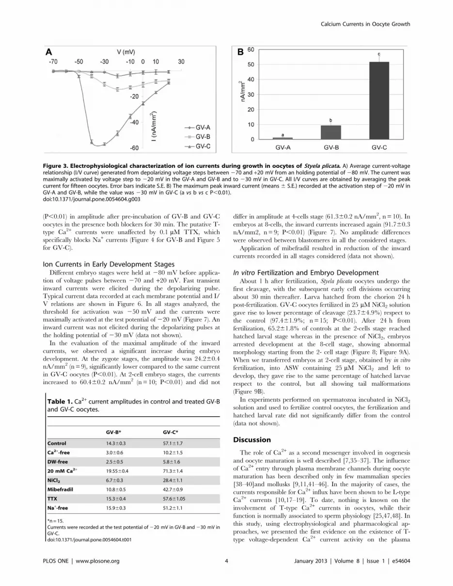
Ion Currents in GV Oocyte StagesThe resting membrane potential of the oocytes was
21162.5 mV in the GV-A stage (n = 20). It increased significantly
(P,0.01) during the oocytes growth as follows: 24862.2 mV at
GV-B stage (n = 25) and 27662.8 mV at GV-C stage (n = 23)
(Figure 2). The oocytes in the three stages were voltage-clamped at
the holding potential of 230 mV. Applying the depolarizing
voltage steps to test potential between 220 and +70 mV, we did
not observe the inward component peaking at test potentials
between 0 and +20 mV, commonly referred to as L-type Ca2+
currents. Depolarizing voltage pulses were then applied to the test
potential between 270 and +20 mV, from a holding potential of
280 mV. Using this voltage-clamp protocol, it was possible to
evoke an inward current in all the three stages. The activation
threshold of the recorded inward current in GV-B and GV-C
oocytes was 250 mV and the currents were maximally activated
by voltage step to 220 mV in the GV-A and GV-B and to
230 mV in GV-C (Figure 3A). Activation and inactivation time
constants at specific test voltage resulted as follows: 7.660.7 and
69.462.4 ms for GV-B at - 20 mV; 4.560.2 and 60.962.6 ms for
GV-C at - 30 mV. The maximum peak inward currents in GV-A
and GV-B at 220 mV were 3.960.2 nA/mm2 and 14.360.1 nA/
mm2, respectively. At 230 mV, the value was of 57.160.6 nA/
mm2 in GV-C (Figure 3B).
The selectivity to cations of recorded currents at GV-B and GV-
C stages was examined since at these voltage steps either T-type
Ca2+ and Na+ currents are activated. Oocytes at GV-B and GV-C
stages, placed in either Ca2+-free ASW or DF-ASW, showed a
significant reduction of the inward component respect to control
(P,0.01). On the other hand, raising the external Ca2+
concentration to 20 mM resulted in a significant (P,0.01) increase
in the amplitude of the inward currents (Table 1). In the absence
of external sodium (Na+-free ASW), the inward currents did not
differ from the control. Therefore, the characteristics and
selectivity of the inward currents in GV-B and GV-C oocytes
were similar to those of T-type Ca2+ currents.
The inward currents in GV-B and GV-C were further
characterized by examining their sensitivity to pharmacological
agents (Table 1). The high sensitivity of T-type Ca2+ currents to be
blocked by Ni2+ was selected as a specific signature of this channel.
In addition, mibefradil was also employed as selective T-type Ca2+
channel blocker at submicromolar concentrations [34]. The
inward currents were found to be sensitive to NiCl2 and mibefradil
at 25 and 1 mM, respectively; in fact, it significantly decreased
Figure 1. Representative images of the three GV stages in oocytes of Styela plicata. Panels A), B) and C) represent pre-vitellogenesis (stageA; GV-A), vitellogenesis (stage B; GV-B) and post-vitellogenesis (stage C; GV-C), respectively. The germinal vesicle is indicated (arrow head) in all stagesreproduced.doi:10.1371/journal.pone.0054604.g001
Figure 2. Membrane resting potential for different oocytestages of Styela plicata. The resting potential (means 6 S.E.) increasedsignificantly through more negative values along the three stages (GV-A, GV-B and GV-C) described in legend of Figure 1 (a vs b vs c P,0.01).doi:10.1371/journal.pone.0054604.g002
Calcium Currents in Oocyte Growth
PLOS ONE | www.plosone.org 3 January 2013 | Volume 8 | Issue 1 | e54604
(P,0.01) in amplitude after pre-incubation of GV-B and GV-C
oocytes in the presence both blockers for 30 min. The putative T-
type Ca2+ currents were unaffected by 0.1 mM TTX, which
specifically blocks Na+ currents (Figure 4 for GV-B and Figure 5
for GV-C).
Ion Currents in Early Development StagesDifferent embryo stages were held at 280 mV before applica-
tion of voltage pulses between 270 and +20 mV. Fast transient
inward currents were elicited during the depolarizing pulse.
Typical current data recorded at each membrane potential and I/
V relations are shown in Figure 6. In all stages analyzed, the
threshold for activation was 250 mV and the currents were
maximally activated at the test potential of 220 mV (Figure 7). An
inward current was not elicited during the depolarizing pulses at
the holding potential of 230 mV (data not shown).
In the evaluation of the maximal amplitude of the inward
currents, we observed a significant increase during embryo
development. At the zygote stages, the amplitude was 24.260.4
nA/mm2 (n = 9), significantly lower compared to the same current
in GV-C oocytes (P,0.01). At 2-cell embryo stages, the currents
increased to 60.460.2 nA/mm2 (n = 10; P,0.01) and did not
differ in amplitude at 4-cells stage (61.360.2 nA/mm2, n = 10). In
embryos at 8-cells, the inward currents increased again (91.760.3
nA/mm2, n = 9; P,0.01) (Figure 7). No amplitude differences
were observed between blastomeres in all the considered stages.
Application of mibefradil resulted in reduction of the inward
currents recorded in all stages considered (data not shown).
In vitro Fertilization and Embryo DevelopmentAbout 1 h after fertilization, Styela plicata oocytes undergo the
first cleavage, with the subsequent early cell divisions occurring
about 30 min thereafter. Larva hatched from the chorion 24 h
post-fertilization. GV-C oocytes fertilized in 25 mM NiCl2 solution
gave rise to lower percentage of cleavage (23.764.9%) respect to
the control (97.461.9%; n = 15; P,0.01). After 24 h from
fertilization, 65.261.8% of controls at the 2-cells stage reached
hatched larval stage whereas in the presence of NiCl2, embryos
arrested development at the 8-cell stage, showing abnormal
morphology starting from the 2- cell stage (Figure 8; Figure 9A).
When we transferred embryos at 2-cell stage, obtained by in vitro
fertilization, into ASW containing 25 mM NiCl2 and left to
develop, they gave rise to the same percentage of hatched larvae
respect to the control, but all showing tail malformations
(Figure 9B).
In experiments performed on spermatozoa incubated in NiCl2solution and used to fertilize control oocytes, the fertilization and
hatched larval rate did not significantly differ from the control
(data not shown).
Discussion
The role of Ca2+ as a second messenger involved in oogenesis
and oocyte maturation is well described [7,35–37]. The influence
of Ca2+ entry through plasma membrane channels during oocyte
maturation has been described only in few mammalian species
[38–40]and mollusks [9,11,41–46]. In the majority of cases, the
currents responsible for Ca2+ influx have been shown to be L-type
Ca2+ currents [10,17–19]. To date, nothing is known on the
involvement of T-type Ca2+ currents in oocytes, while their
function is normally associated to sperm physiology [25,47,48]. In
this study, using electrophysiological and pharmacological ap-
proaches, we presented the first evidence on the existence of T-
type voltage-dependent Ca2+ current activity on the plasma
Figure 3. Electrophysiological characterization of ion currents during growth in oocytes of Styela plicata. A) Average current-voltagerelationship (I/V curve) generated from depolarizing voltage steps between 270 and +20 mV from an holding potential of 280 mV. The current wasmaximally activated by voltage step to 220 mV in the GV-A and GV-B and to 230 mV in GV-C. All I/V curves are obtained by averaging the peakcurrent for fifteen oocytes. Error bars indicate S.E. B) The maximum peak inward current (means 6 S.E.) recorded at the activation step of 220 mV inGV-A and GV-B, while the value was 230 mV in GV-C (a vs b vs c P,0.01).doi:10.1371/journal.pone.0054604.g003
Table 1. Ca2+ current amplitudes in control and treated GV-Band GV-C oocytes.
GV-B* GV-C*
Control 14.360.3 57.161.7
Ca2+-free 3.060.6 10.261.5
DW-free 2.560.5 5.861.6
20 mM Ca2+ 19.5560.4 71.361.4
NiCl2 6.760.3 28.461.1
Mibefradil 10.860.5 42.760.9
TTX 15.360.4 57.661.05
Na+-free 15.960.3 51.261.1
*n = 15.Currents were recorded at the test potential of 220 mV in GV-B and 230 mV inGV-C.doi:10.1371/journal.pone.0054604.t001
Calcium Currents in Oocyte Growth
PLOS ONE | www.plosone.org 4 January 2013 | Volume 8 | Issue 1 | e54604

membrane of GV growing oocytes. We identified three stages of
immature oocytes in Styela plicata on the basis of their size,
morphology and accessory cellular structures (Figure 1) and
characterized the electrical properties of their plasma membranes.
The growth of GV-B and GV-C stages was clearly associated with
an increased T-type Ca2+ current activity and membrane potential
hyperpolarisation. In fact, the smallest oocytes (GV-A) exhibited a
low Ca2+ current activity accompanied by the lowest resting
potential value, which increased significantly during the following
B and C stages.
The increase in Ca2+ current activity indicates that in Styela
plicata oocytes, the initial stages of cytoplasmic maturation and
growth progression may depend on a progressive Ca2+ surge via
the plasma membrane. Intracellular Ca2+ release occurs at
fertilization in all known species [7,49]; in addition, modifications
of Ca2+ stores during oocyte maturation may contribute to
mediate Ca2+ release at fertilization [50,51]. The highest density of
plasma membrane Ca2+ currents in the larger GV-C stage
indicates a plausible role of these currents in filling the internal
Ca2+ stores that in the mature oocytes of ascidian Ciona intestinalis
are responsible for the post-fertilization contraction of the zygote
[52].
The completed maturation process represents a prelude to
fertilization; fully grown oocytes are ready for the signal that
resumes meiosis. In this respect, successful fertilization occurs only
in presence of mature and competent oocytes [53]. In Styela plicata,
fully grown GV oocytes are stored in the ovaries before spawning
[54]. The literature reports many differences in oocyte maturation
and spawning among ascidian species. In Halocynthia roretzi oocytes
mature, just before spawning [55], in Styela canopus oocytes are
spawned with intact GV which breaks down rapidly before
fertilization [56], whereas Styela gibbsii oocytes are spawned after
GVBD occurrence [57]. In Styela plicata, it was reported that
oocytes are spawned with intact GV and that GVBD is triggered
by the fertilization process [32]. In this study, we confirmed this
observation, since the addition of spermatozoa to the GV-C stage
oocytes triggered fertilization and embryo development up to
larval stages, whereas the immature GV-A and B did not fertilize
under the same conditions. Differently than other ascidian species,
such as Ciona intestinalis and Cnemidocarpa irene, incubation of all
immature stages in sea water was not followed by spontaneous
maturation [10,55,57]. Based on these observations, we can
hypothesize that the highest activity of Ca2+ currents recorded in
GV-C stage may be necessary for the oocyte to respond to the
unknown stimulus inducing resumption of the first meiotic block.
The increase of RP also supports this hypothesis. Although the role
of RP is not fully clarified, it has been found to vary during meiotic
progression in some species [39,40] and it appears to be associated
Figure 4. Pharmacological characterization of Ca2+ currents in GV-B oocyte of Styela plicata. A) Representative traces of voltage protocoland current records evoked by stepping membrane potential to voltages between 270 and +20 mV, in 10 mV increments, from a holding potentialof 280 mV in control experiment (left) and after NiCl2 treatment (right). B) Average current-voltage relationship (I/V curve) showing the effects ofmodified ASW and pharmacological agents (NiCl2; TTX; mibefradil) on control currents. All I/V curves were obtained by averaging the peak current fortwenty oocytes. Error bars indicate S.E.doi:10.1371/journal.pone.0054604.g004
Calcium Currents in Oocyte Growth
PLOS ONE | www.plosone.org 5 January 2013 | Volume 8 | Issue 1 | e54604

with a ‘‘stand-by’’ status of the cell. It is possible that as soon as the
plasma membrane receives the signal to grow, the ion exchange
may induce the shift of RP through values closest to the
physiological potential of the mature MI oocyte, supporting the
metabolic activity necessary to prepare the plasma membrane for
GVBD. In agreement with these results, we show here that plasma
membrane potential significantly hyperpolarizes from stage A to
C, reaching the most negative values in stage C, similarly to MI
stage recorded in other ascidian species [24].
These data, along with the mitochondria pattern distribution
recently demonstrated [58], suggest that the GV-C stage in Styela is
considered the mature oocyte competent for fertilization.
The modifications in the plasma membrane potential have
mostly been associated with the physiology of excitable tissues and
related to cell cycle [59]. Several studies have shown that
progression through the cell cycle is dependent upon transient
increases in cytosolic Ca2+, since the inhibition of Ca2+ influx by
the antagonists prevents cell cycle progression [60]. Regulation of
Figure 5. Pharmacological characterization of Ca2+ currents in GV-C oocyte of Styela plicata. A) Representative traces of voltage protocoland current records obtained by applying depolarizing voltage step between 270 mV and +20 mV, with subsequent 10 mV increments, fromholding potentials of 280 mV in control experiment (left) and after NiCl2 treatment (right). B) Average current-voltage relationship (I/V curve)showing the effects of modified ASW and pharmacological agents (NiCl2; TTX; mibefradil) on control currents. All I/V curves are obtained by averagingthe data from twenty oocytes. Error bars indicate S.E.doi:10.1371/journal.pone.0054604.g005
Figure 6. Membrane resting potential during embryo devel-opment in Styela plicata. The RP (means 6 S.E.) shows positive valuesin the zygote. Negative values increased from 2-cell stage up to 8-cellstage (a vs b vs c P,0.01).doi:10.1371/journal.pone.0054604.g006
Calcium Currents in Oocyte Growth
PLOS ONE | www.plosone.org 6 January 2013 | Volume 8 | Issue 1 | e54604

Calcium Currents in Oocyte Growth
PLOS ONE | www.plosone.org 7 January 2013 | Volume 8 | Issue 1 | e54604

some ion channels are dependent upon actin microfilaments [61].
During cleavage, the microfilaments reorganize to form the
cleavage furrow to control changes in cell volume during mitosis.
In Ciona intestinalis, perturbation of ion channels altered actin
filaments organization and mitochondrial migration after contrac-
tion leading to a disturbance in cleavage formation [62] in
agreement with the role of actin filaments in ion channel
regulation [63]. These data are also consistent with the finding
in somatic cells that a Ca2+ entry through T-type Ca2+ channels
may be needed only at specific stages of the cell cycle for the
control of cell growth and proliferation [64].
Functional expression of Ca2+ channels has been described
during development in ascidians [10,27]. In a previous study, we
showed a T-type Ca2+ channel regulation by the cell cycle in the
sea urchin embryo [65]. In the mouse oocytes, T-type Ca2+
currents increase after fertilization and decrease at the beginning
of early development [66]; however, an involvement in the cell
cycle regulation was subsequently shown in the early mouse
embryo where the amplitudes of the T- type Ca2+currents change
in a cell cycle-dependent manner being large in unfertilized
oocytes and decreasing after fertilization throughout the first cell
cycle and increasing again during late telophase [59]. In order to
substantiate a possible role of T- type Ca2+currents in early
development of Styela, we followed the pattern of current activity
from zygote up to the 8- cell stage. The significant decline of either
T-type Ca2+ currents and RP values occurring in the zygote up to
4-cell stage suggests a minor role for these currents in the signalling
events related to the first embryonic mitotic cycle, whereas in
either cases we observed a significant increase at the 8-cell stage
without determining any specific differences among blastomeres.
This finding appears in agreement with the critical role of the 8-
cell stage embryo, where the segregation of cell lines initiates [31]
and is consistent with data reported in Ciona intestinalis for L-type
Ca2+ currents [10]. On the contrary, the lack of spatial distribution
of currents among blastomeres at the 8-cell stage rules out a
possible lineage-specific electrical diversity. The property and
distribution of ion channels in embryos change during develop-
ment [67–69]. In ascidians, it has been described an oscillation of
Ca2+ currents that disappear at MI stage and reappear in the cells
of muscular lineages [70].
Ca2+ influx through T-type Ca2+ channels may also be critical
for cell cycle progression since their inhibition can prevent the
proliferation of a variety of cell types including fibroblasts and
endothelial cells [22]. In this paper, we demonstrated that the
presence of functional T-type Ca2+ currents is critical for either
fertilization and embryo development. In fact GV-C stage oocytes
fertilized in NiCl2 arrest at 8-cell stage which, in ascidians,
represents the fundamental stage for cell lines segregation and
genomic activation [31]. T-type Ca2+ currents play also a pivotal
role in development, since the treatment of 2-cell stage embryos
with NiCl2 does not arrest embryos, but generates hatched larvae
bearing serious morphological abnormalities of the tail, a key
feature for larval metamorphosis in ascidians [31]. These data are
also consistent with the impact of ion channels inhibition on late
development in Ciona intestinalis, that coincides with the time of
passage between maternal to genomic expression [10,62].
Figure 7. Electrophysiological characterization of Ca2+ currents during embryo development in Styela plicata. A) Representative tracesof voltage protocol and current records obtained in response to voltage steps between 270 and +20 mV, in 10 mV increments, from a holdingpotential of 280 mV in the developmental stage reported (left). Images (right) at the light microscope of the stages considered. Arrows indicate thepolar body. B) Average current-voltage relationship (I/V curve) showing that maximal currents generated at the step to 220 mV. All I/V curves areobtained by averaging the data from twenty considered embryo stages. Error bars indicate S.E.doi:10.1371/journal.pone.0054604.g007
Figure 8. Effect of Ca2+ current inhibition on fertilization rate of Styela plicata. Light shading shows percentage of first cleavage (2-cellstage) of GV-C oocyte fertilized in ASW (control) and in NiCl2 (a vs b P,0.01). Dark shading shows percentage of 2-cell stage that reached the larvalstage. Insert reports a normally developed Styela plicata larva 24 h after fertilization. Bar is 240 mm.doi:10.1371/journal.pone.0054604.g008
Calcium Currents in Oocyte Growth
PLOS ONE | www.plosone.org 8 January 2013 | Volume 8 | Issue 1 | e54604

ConclusionsThe present work shows the presence of functionally active T-
type Ca2+ currents in immature growing oocytes of the ascidian
Styela plicata. Several lines of evidence indicate that T-type Ca2+
currents play a role in growth regulation as suggested by their
expression during embryo development and periods of rapid
physiological and pathophysiological growth systems [71]. The
significant increase of T-type Ca2+currents accompanied by the
progressive hyperpolarization of the plasma membrane potential
implies a peculiar role for these currents in regulating cytosolic
Ca2+ during the cytoplasmic maturation and growth of Styela
oocytes.
The absence of a clear GVBD and the fertilization occurring at
the largest immature stage also suggest an important role of these
currents in allowing Ca2+ entry that in turn triggers release of
further intracellular Ca2+ from stores. The variation of T-type
Ca2+ currents during development and the significant reduction of
cleavage rate due to the inhibition of Ca2+ influx indicate that
fertilization and embryo development are modulated by these
currents.
Finally, we documented the difference between physiology of
Styela oocytes and other ascidian species in which the Ca2+ entry at
fertilization and post-fertilization are underlined by different
channels types [10,19]. These latter data further highlight the
evolutionary variability of biological mechanisms that exist among
the ascidian species [72].
Acknowledgments
We thank Drs. L.J. De Felice, M. Costantini and M. Cataldi for useful
comments on the manuscript; Mr. G. Gargiulo and Mr. G. Lanzotti for
computer graphics; Dr. P. Cirino and Mr. A. Macina for providing and
maintaining Styela plicata. We are also indebted with V. Monfrecola for
technical assistance.
Author Contributions
Conceived and designed the experiments: AG GLR ET. Performed the
experiments: AG. Analyzed the data: AG GLR ET. Contributed reagents/
materials/analysis tools: ET. Wrote the paper: AG GLR ET.
Figure 9. Effect of Ca2+ current inhibition on the in vitro fertilization and embryo development in Styela plicata. A) Slightly deformedembryos at 2-cell stage, developed from oocytes fertilized in ASW containing 25 mM NiCl2, arrested at an abnormal 8-cell stage; B) Abnormal hatchedlarvae developed from embryos transferred at 2-cell stage in ASW containing 25 mM NiCl2. Bar is 50 mm in A and 100 mm in B.doi:10.1371/journal.pone.0054604.g009
Calcium Currents in Oocyte Growth
PLOS ONE | www.plosone.org 9 January 2013 | Volume 8 | Issue 1 | e54604
References
1. Eppig JJ (1996) Coordination of nuclear and cytoplasmic oocyte maturation in
eutherian mammals. Reprod Fertil Dev 8: 485–489.
2. Dale B (1983) Fertilization in animals. London: Edward Arnold.3. Yanagimachi R (1994) Mammalian Fertilization. In: J KEN, editor. The
physiology of reproduction. New York: Raven press.
4. Tosti E, Boni R (2011) Oocyte maturation and fertilization: a long history for ashort event. Dubai: Bentham Science Publishers.
5. Russo GL, Wilding M, Marino M, Dale B (1998) Ins and outs of meiosis in
ascidians. Semin Cell Dev Biol 9: 559–567.
6. Dale B, Elder K (1997) In Vitro Fertilization. Cambridge: Cambridge UniversityPress.
7. Boni R, Gualtieri R, Talevi R, Tosti E (2007) Calcium and other ion dynamics
during gamete maturation and fertilization. Theriogenology 68 Suppl 1: S156–164.
8. Cui XS, Kim NH (2007) Maternally derived transcripts: identification and
characterisation during oocyte maturation and early cleavage. Reprod FertilDev 19: 25–34.
9. Cuomo A, Di Cristo C, Paolucci M, Di Cosmo A, Tosti E (2005) Calcium
currents correlate with oocyte maturation during the reproductive cycle inOctopus vulgaris. J Exp Zool A Comp Exp Biol 303: 193–202.
10. Cuomo A, Silvestre F, De Santis R, Tosti E (2006) Ca2+ and Na+ current
patterns during oocyte maturation, fertilization, and early developmental stagesof Ciona intestinalis. Mol Reprod Dev 73: 501–511.
11. Tosti E (2006) Calcium ion currents mediating oocyte maturation events.
Reprod Biol Endocrinol 4: 26.
12. Yamashita M, Mita K, Yoshida N, Kondo T (2000) Molecular mechanisms ofthe initiation of oocyte maturation: general and species-specific aspects. Prog
Cell Cycle Res 4: 115–129.
13. Dale B, Wilding M (2011) Ionic events at fertilization. In: Tosti E, Boni R,editors. Oocyte maturation and fertilization: A long history for a short event.
Dubai: Bentham science publisher. 104–120.
14. Darszon A, Labarca P, Nishigaki T, Espinosa F (1999) Ion channels in spermphysiology. Physiol Rev 79: 481–510.
15. Hagiwara S, Jaffe LA (1979) Electrical properties of egg cell membranes. Annu
Rev Biophys Bioeng 8: 385–416.
16. Tosti E, Boni R (2004) Electrical events during gamete maturation andfertilization in animals and humans. Hum Reprod Update 10: 53–65.
17. Bosma MM, Moody WJ (1990) Macroscopic and single-channel studies of two
Ca2+ channel types in oocytes of the ascidian Ciona intestinalis. J Membr Biol114: 231–243.
18. Dale B, Talevi R, DeFelice LJ (1991) L-type Ca2+ currents in ascidian eggs. Exp
Cell Res 192: 302–306.19. Silvestre F, Cuomo A, Tosti E (2009) Ion current activity and molecules
modulating maturation and growth stages of ascidian (Ciona intestinalis) oocytes.
Mol Reprod Dev 76: 1084–1093.20. Tosti E, Gallo A, Silvestre F (2011) Ion currents involved in oocyte maturation,
fertilization and early developmental stages of the ascidian Ciona intestinalis.
Mol Reprod Dev 78: 854–860.21. Huc S, Monteil A, Bidaud I, Barbara G, Chemin J, et al. (2009) Regulation of T-
type calcium channels: signalling pathways and functional implications. Biochim
Biophys Acta 1793: 947–952.
22. McGivern JG (2006) Pharmacology and drug discovery for T-type calciumchannels. CNS Neurol Disord Drug Targets 5: 587–603.
23. Lee JH, Ahn HJ, Lee SJ, Gye MC, Min CK (2011) Effects of L- and T-type
Ca(2)(+) channel blockers on spermatogenesis and steroidogenesis in theprepubertal mouse testis. J Assist Reprod Genet 28: 23–30.
24. Arnoult C, Cardullo RA, Lemos JR, Florman HM (1996) Activation of mouse
sperm T-type Ca2+ channels by adhesion to the egg zona pellucida. Proc NatlAcad Sci U S A 93: 13004–13009.
25. Darszon A, Lopez-Martinez P, Acevedo JJ, Hernandez-Cruz A, Trevino CL
(2006) T-type Ca2+ channels in sperm function. Cell Calcium 40: 241–252.26. Jagannathan S, Publicover SJ, Barratt CL (2002) Voltage-operated calcium
channels in male germ cells. Reproduction 123: 203–215.
27. Arnoult C, Villaz M (1994) Differential developmental fates of the two calciumcurrents in early embryos of the ascidian Ciona intestinalis. J Membr Biol 137:
127–135.
28. Hotsuliak M, Berdyieva TK, Libert SV (2002) Effects of T-type calcium channelblockers on spontaneous meiotic maturation of mouse ovarian oocytes in vitro.
Fiziol Zh 48: 98–101.
29. Corbo JC, Di Gregorio A, Levine M (2001) The ascidian as a model organism indevelopmental and evolutionary biology. Cell 106: 535–538.
30. Kumano G, Nishida H (2007) Ascidian embryonic development: an emerging
model system for the study of cell fate specification in chordates. Dev Dyn 236:1732–1747.
31. Satoh N (1994) Developmental biology of ascidian. Cambridge: Cambridge
University Press.
32. Jeffery WR, Capco DG (1978) Differential accumulation and localization ofmaternal poly(A)-containing RNA during early development of the ascidian,
Styela. Dev Biol 67: 152–166.
33. SAS. (1988) User’s guide/STAT (Release 6.03 edition). Cary, NC:StatisticalAnalysis System Institute.
34. Lacinova L (2005) Voltage-dependent calcium channels. Gen Physiol Biophys
24 Suppl 1: 1–78.
35. Homa ST (1995) Calcium and meiotic maturation of the mammalian oocyte.
Mol Reprod Dev 40: 122–134.
36. Machaca K (2007) Ca2+ signaling differentiation during oocyte maturation.
J Cell Physiol 213: 331–340.
37. Stricker SA (1999) Comparative biology of calcium signaling during fertilization
and egg activation in animals. Dev Biol 211: 157–176.
38. Lee JH, Yoon SY, Bae IH (2004) Studies on Ca2+-channel distribution in
maturation arrested mouse oocyte. Mol Reprod Dev 69: 174–185.
39. Murnane JM, DeFelice LJ (1993) Electrical maturation of the murine oocyte: an
increase in calcium current coincides with acquisition of meiotic competence.Zygote 1: 49–60.
40. Tosti E, Boni R, Cuomo A (2000) Ca(2+) current activity decreases during
meiotic progression in bovine oocytes. Am J Physiol Cell Physiol 279: C1795–
1800.
41. Colas P, Dube F (1998) Meiotic maturation in mollusc oocytes. Semin Cell DevBiol 9: 539–548.
42. Dube F (1992) Thapsigargin induces meiotic maturation in surf clam oocytes.Biochem Biophys Res Commun 189: 79–84.
43. Guerrier P, Leclerc-David C, Moreau M (1993) Evidence for the involvement ofinternal calcium stores during serotonin-induced meiosis reinitation in oocytes of
the bivalve mollusc Ruditapes philippinarum. Dev Biol 159: 474–484.
44. Leclerc C, Guerrier P, Moreau M (2000) Role of dihydropyridine-sensitive
calcium channels in meiosis and fertilization in the bivalve molluscs Ruditapesphilippinarum and Crassostrea gigas. Biol Cell 92: 285–299.
45. Moreau M, Leclerc C, Guerrier P (1996) Meiosis reinitiation in Ruditapesphilippinarum (Mollusca): involvement of L-calcium channels in the release of
metaphase I block. Zygote 4: 151–157.
46. Tomkowiak M, Guerrier P, Krantic S (1997) Meiosis reinitiation of mussel
oocytes involves L-type voltage-gated calcium channel. J Cell Biochem 64: 152–160.
47. Arnoult C, Villaz M, Florman HM (1998) Pharmacological properties of the T-type Ca2+ current of mouse spermatogenic cells. Mol Pharmacol 53: 1104–
1111.
48. Perez-Reyes E (2003) Molecular physiology of low-voltage-activated t-type
calcium channels. Physiol Rev 83: 117–161.
49. Jaffe LF (1980) Calcium explosions as triggers of development. Ann N Y Acad
Sci 339: 86–101.
50. Boni R, Cuomo A, Tosti E (2002) Developmental potential in bovine oocytes is
related to cumulus-oocyte complex grade, calcium current activity, and calciumstores. Biol Reprod 66: 836–842.
51. Chiba K, Kado RT, Jaffe LA (1990) Development of calcium release
mechanisms during starfish oocyte maturation. Dev Biol 140: 300–306.
52. Brownlee C, Dale B (1990) Temporal and spatial correlation of fertilization
current, calcium waves and cytoplasmic contraction in eggs of Ciona intestinalis.Proc R Soc Lond B Biol Sci 239: 321–328.
53. Menezo Y, Elder K (2011) The enhancers of oocyte competence. In: Tosti E,Boni R, editors. Oocyte maturation and fertilization: A long history for a short
event. Dubai: Bentham science publisher. 64–70.
54. Lambert CC (2011) Signaling pathways in ascidian oocyte maturation: the roles
of cAMP/Epac, intracellular calcium levels, and calmodulin kinase in regulatingGVBD. Mol Reprod Dev 78: 726–733.
55. Sakairi K, Hiroko S (1991) Possible MIS Production by Follicle Cells inSpontaneous Oocyte Maturation of the Ascidian, Halocynthia roretzi. Develop
Growth and Differ 33: 155–162.
56. Conklin EG (1905) The organization and cell lineage of the ascidian egg. J Acad
Natn Sci Philad 13: 1–119.
57. Lambert CC (2005) Signaling pathways in ascidian oocyte maturation: effects of
various inhibitors and activators on germinal vesicle breakdown. Dev GrowthDiffer 47: 265–272.
58. Bezzaouia A, Gallo A, Silvestre F, Tekaya S, Tosti E (2012) Distribution patternand activity of mitochondria during oocyte growth and maturation in the
ascidian Styela plicata. Zygote in press.
59. Day ML, Johnson MH, Cook DI (1998) Cell cycle regulation of a T-type
calcium current in early mouse embryos. Pflugers Arch 436: 834–842.
60. Berridge MJ (1995) Capacitative calcium entry. Biochem J 312 (Pt 1): 1–11.
61. Ullrich N, Sontheimer H (1997) Cell cycle-dependent expression of a glioma-
specific chloride current: proposed link to cytoskeletal changes. Am J Physiol
273: C1290–1297.
62. Tosti E, Romano G, Buttino I, Cuomo A, Ianora A, et al. (2003) Bioactivealdehydes from diatoms block the fertilization current in ascidian oocytes. Mol
Reprod Dev 66: 72–80.
63. Cantiello HF (1997) Role of actin filament organization in cell volume and ion
channel regulation. J Exp Zool 279: 425–435.
64. Capiod T (2011) Cell proliferation, calcium influx and calcium channels.
Biochimie 93: 2075–2079.
65. Yazaki I, Tosti E, Dale B (1995) Cytoskeletal elements link calcium channel
activity and the cell cycle in early sea urchin embryos. Development 121: 1827–1831.
Calcium Currents in Oocyte Growth
PLOS ONE | www.plosone.org 10 January 2013 | Volume 8 | Issue 1 | e54604

66. Yamashita N (1982) Enhancement of ionic currents through voltage-gated
channels in the mouse oocyte after fertilization. J Physiol 329: 263–280.
67. Hirano T, Takahashi K (1984) Comparison of properties of calcium channels
between the differentiated 1-cell embryo and the egg cell of ascidians. J Physiol
347: 327–344.
68. Moody WJ (1998) The development of voltage-gated ion channels and its
relation to activity-dependent development events. Curr Top Dev Biol 39: 159–
185.
69. Tosti E (2010) Dynamic roles of ion currents in early development. Mol Reprod
Dev 77: 856–867.70. Simoncini L, Block ML, Moody WJ (1988) Lineage-specific development of
calcium currents during embryogenesis. Science 242: 1572–1575.
71. Ertel SI, Ertel EA, Clozel JP (1997) T-type Ca2+ channels and pharmacologicalblockade: potential pathophysiological relevance. Cardiovasc Drugs Ther 11:
723–739.72. Lemaire P (2011) Evolutionary crossroads in developmental biology: the
tunicates. Development 138: 2143–2152.
Calcium Currents in Oocyte Growth
PLOS ONE | www.plosone.org 11 January 2013 | Volume 8 | Issue 1 | e54604