t-loops at trypanosome telomeres
TRANSCRIPT

Jorge L.MunÄ oz-JordaÂn1,2,George A.M.Cross1, Titia de Lange2,3 andJack D.Grif®th4
1Laboratory of Molecular Parasitology and 2Laboratory of CellBiology and Genetics, Box 159, The Rockefeller University, 1230York Avenue, New York, NY 10021 and 4Lineberger ComprehensiveCancer Center, University of North Carolina, Chapel Hill, NC 27599,USA
3Corresponding authore-mail: [email protected]
Mammalian telomeres form large duplex loops(t-loops) that may sequester chromosome ends byinvasion of the 3¢ TTAGGG overhang into the duplexTTAGGG repeat array. Here we document t-loops inTrypanosoma brucei, a kinetoplastid protozoan withabundant telomeres due to the presence of many mini-chromosomes. These telomeres contained 10±20 kbduplex TTAGGG repeats and a 3¢ TTAGGG over-hang. Electron microscopy of psoralen/UV cross-linked DNA revealed t-loops in enriched telomericrestriction fragments and at the ends of isolated mini-chromosomes. In mammals, t-loops are large (up to25 kb), often comprising most of the telomere. Despitesimilar telomere lengths, trypanosome t-loops weremuch smaller (~1 kb), indicating that t-loop sizes areregulated. Coating of non-cross-linked minichromo-somes with Escherichia coli single-strand bindingprotein (SSB) often revealed 3¢ overhangs at both telo-meres and several cross-linked minichromosomes hadt-loops at both ends. These results suggest that t-loopsand their prerequisite 3¢ tails can be formed on theproducts of both leading and lagging strand synthesis.We conclude that t-loops are a conserved feature ofeukaryotic telomeres.Keywords: telomere/t-loop/trypanosome
Introduction
The advent of linear chromosomes in eukaryotes wasaccompanied by the acquisition of specialized terminalstructures that preserve chromosome ends. Most eukary-otic telomeres feature a tandem array of short repeats and a3¢ overhang (Wellinger and Sen, 1997). These telomeresare maintained by telomerase, which adds telomericrepeats to the 3¢ end of the chromosome (Greider andBlackburn, 1985; Nugent and Lundblad, 1998).Telomerase-mediated telomere elongation is the predom-inant mechanism by which eukaryotes compensate for thefailure of lagging strand synthesis to complete thereplication of terminal sequences.
Telomeres protect chromosome ends against degrad-ation and end-to-end fusion, and they prevent inappropri-
ate activation of checkpoint pathways that respond tochromosome breaks (Muller, 1938; McClintock, 1941;Sandell and Zakian, 1993; van Steensel et al., 1998;Karlseder et al., 1999). This capping function is mediatedby telomere-associated proteins. The stability ofSaccharomyces cerevisiae telomeres depends on Cdc13p,a single-stranded telomeric DNA binding protein thatprotects telomeres from degradation and prevents acti-vation of the RAD9 checkpoint pathway (Garvik et al.,1995; Lin and Zakian, 1996; Nugent et al., 1996).Similarly, hypotrichous ciliates have short [<20 nucleo-tides (nt)] telomeric overhangs that are bound by a single-strand binding protein, but the in vivo function of thiscomplex has not been established (Gottschling and Cech,1984; Gottschling and Zakian, 1986; Price, 1999).Mammalian telomeres are protected by TRF2, whichbinds along the duplex array of telomeric TTAGGGrepeats (Bilaud et al., 1997; Broccoli et al., 1997;Smogorzewska et al., 2000). Interference with TRF2function results in immediate deprotection of telomeres, asevidenced by loss of the telomeric 3¢ overhang, formationof end-to-end chromosome fusions, activation of an ATM/p53-dependent DNA damage checkpoint, cell cycle arrestand apoptosis (van Steensel et al., 1998; Karlseder et al.,1999).
Electron microscopy (EM) of mammalian telomericDNA has revealed large duplex loops (t-loops) atchromosome ends in vivo (Grif®th et al., 1999). t-loopswere stabilized by cross-linking the DNA in chromatinwith the psoralen 4¢ aminomethyltrioxalen (AMT) and UVlight. Telomeric DNA was enriched by gel-®ltrationchromatography after digestion of the bulk chromosomalDNA with frequently cutting restriction enzymes that donot cleave TTAGGG repeats. t-loops are composed ofduplex TTAGGG repeats and vary with the length of thetelomeric repeat array from 1 to 25 kb. Single-strandedDNA binding protein (SSB) from Escherichia coli boundto the tail±loop junction, indicating that there is a segmentof single-stranded DNA at this site, most likely formed bystrand displacement upon invasion of the 3¢ overhang intothe duplex telomeric repeat tract. t-loops were observedfrequently in a variety of mammalian DNA sources,including HeLa cells, mouse liver, HT1080 cells andprimary peripheral blood leukocytes. A recent reportshowed loops at the ends of ampli®ed micronuclearchromosomes in Oxytricha fallax (Murti and Prescott,1999). These loops were not found in DNA frommacronuclei, in which telomeres are extremely short andhave a protein complex bound to the 3¢ overhang.
Although the mechanism by which t-loops are formedin vivo has not been established, both TRF2 and the relatedtelomeric protein TRF1 have biochemical features sug-gestive of a role in loop formation and/or maintenance.TRF1 can loop and pair telomeric DNA, and TRF2
t-loops at trypanosome telomeres
The EMBO Journal Vol. 20 No. 3 pp. 579±588, 2001
ã European Molecular Biology Organization 579

promotes the formation of t-loops from a model telomeresubstrate (Bianchi et al., 1997, 1999; Grif®th et al., 1998,1999). In addition, proteins involved in DNA recombina-tion and repair have been proposed to contribute to t-loopdynamics. An example is the Rad50±Mre11±Nbs1 re-combinational repair complex that binds to TRF2 andinteracts with mammalian telomeres (de Lange andPetrini, 2000; Zhu et al., 2000).
t-loops could provide an architectural solution to theproblems posed by telomeres by hiding the telomereterminus in the duplex part of the telomere, preventingactivation of DNA damage checkpoints and protectingchromosome ends from inappropriate DNA repair (Grif®thet al., 1999). In addition, insertion of the 3¢ terminus wouldpresumably block telomerase from adding repeats to thechromosome end. Therefore, the rate at which t-loops arerefolded after DNA replication might contribute to theregulation of telomere length.
To determine whether t-loops are conserved, weemployed the advantages of trypanosome genomes. TheTrypanosomatidae are protozoan parasites, of whichTrypanosoma brucei is best known for the phenomenonof antigenic variation, allowing extracellular survival inthe bloodstream of its mammalian host. Each bloodstreamform (BF) cell expresses 10 million copies of a singlespecies of variant surface glycoprotein (VSG) (Cross,1975). The expressed VSG gene (VSG) is invariablypositioned at a telomere (de Lange and Borst, 1982), at theend of a polycistronic transcription unit called an expres-sion site (ES). Only one ES is transcriptionally active atany time, and antigenic variation is achieved either bygene conversion of the transcribed VSG by one of theseveral hundred VSGs dispersed around the genome, or bycoordinated activation/inactivation of different telomericESs (Cross, 1996; Rudenko et al., 1998). ESs are nottranscribed in the non-infectious insect stage (procyclicform, PF) of the life cycle.
Trypanosome telomeres contain long TTAGGG repeatarrays, which grow by 9±12 bp per cell division (Bernardset al., 1983; Blackburn and Challoner, 1984; van der Ploeget al., 1984), presumably through the action of telomerase(Cano et al., 1999). Transcriptionally active telomeresgrow slightly faster and are more susceptible to truncationsthan inactive ones (Pays et al., 1983; Horn and Cross,1997), suggesting complex mechanisms of telomeremaintenance and protection. Position effects within theES (Rudenko et al., 1995; Horn and Cross, 1997),chromatin remodeling and developmentally regulatedrepression effects close to telomeres suggest a role fortelomeres in regulating antigenic variation (Navarro et al.,1999).
Trypanosoma brucei contains 11 large chromosomepairs (Melville et al., 1998, 2000), ranging from 1 to>5 Mbp, which carry the essential genes, and ~100minichromosomes, of 25±150 kb, which are predomin-antly composed of tandem 177 bp repeats (Weiden et al.,1991). Minichromosomes have canonical telomeres atboth ends and some telomeres carry silent VSG genesthat can contribute to the expressed VSG repertoirethrough transposition to ESs in the large chromosomes(van der Ploeg et al., 1984; Weiden et al., 1991).Minichromo-somes represent an abundant source of
telomeres, which were used in this study to address theevolutionary conservation of t-loops.
Results
Trypanosoma brucei telomeres carry ~10±20 kb ofTTAGGG repeats and have a 3 ¢ overhangIt was previously shown that T.brucei telomeres arecomposed of TTAGGG repeats and that most TTAGGGrepeats in the trypanosome genome occupy terminal sites,based on their sensitivity to exonuclease treatment ofintact genomic DNA (Blackburn and Challoner, 1984;van der Ploeg et al., 1984). In order to determine themedian length of the TTAGGG repeat arrays in theT.brucei line used in this study, we applied a techniquepreviously used to measure the length of human telomeres(Saltman et al., 1993). The rate at which the exonucleaseBal31 removes TTAGGG repeat hybridization signal iscompared with the rate at which the enzyme shortensterminal DNA fragments. In T.brucei, this approach isfacilitated by the availability of probes for subtelomericVSG genes and the detailed knowledge of the restrictionmaps of these loci, allowing precise measurements ofBal31 digestion rates on well de®ned terminal restrictionfragments.
Digestion of DNA from BF and PF T.brucei withfrequently cutting enzymes yielded telomeric fragments inthe 10±20 kb range (Figure 1A), suggesting that thetelomeres contain long arrays of TTAGGG repeats. Tomeasure the length of the telomeric repeat array directly,intact PF DNA was treated with Bal31 exonuclease forincreasing times, digested with HinfI±AluI±RsaI andhybridized to a TTAGGG repeat probe (Figure 1B).Quanti®cation of the TTAGGG repeat signal at each timepoint indicated that the exonuclease removed ~2.6% of theTTAGGG repeat signal per minute. The rate at whichBal31 shortened the telomeric fragment that carries VSG221 was determined in parallel (Figure 1B; see Figure 1Cfor restriction map). The same shortening rate wasfound for the telomeric restriction fragment carryingVSG 121 (Figure 1B; see Figure 1C for restrictionmap). As expected, Bal31 did not affect chromosome-internal restriction fragments, such as those carrying non-telomeric copies of VSG 121 (Figure 1B). Comparisonof the two rates (Figure 1D) showed that Bal31removed ~10% of the TTAGGG repeat signal in thetime needed to shorten the telomere by 1.5 kb, implyingthat the average length of the TTAGGG repeat array was~15 kb. This value for the length of the TTAGGG repeatsarray was in agreement with the median length for thetelomeric fragments observed in DNA digested withHinfI and RsaI (Figure 1A), which are expected to removemost of the subtelomeric sequences from the terminalfragments.
We next determined whether T.brucei chromosomeends carry an overhang of the TTAGGG repeat strand.Mammalian chromosomes have up to 200 nt of single-stranded TTAGGG repeats at their 3¢ termini (Makarovet al., 1997; McElligott and Wellinger, 1997; Wright et al.,1997; van Steensel et al., 1998; Huffman et al., 2000) andthis overhang is presumed to be important for theformation of t-loops. The presence of single-strandedTTAGGG repeats can be assessed by annealing labeled
J.L.MunÄ oz-JordaÂn et al.
580

C-strand-speci®c oligonucleotides to genomic DNA.Using this approach, we found that native DNA fromT.brucei contained single-stranded TTAGGG repeats(Figure 1E). The signal was not detected in DNA digestedwith E.coli exonuclease I, which is speci®c for 3¢ single-stranded tails, or in a control hybridization with anoligonucleotide representing the G-rich telomeric strand,consistent with the signal being derived from 3¢ single-stranded TTAGGG tails. Furthermore, the signals werepresent on large fragments (>10 kb) in DNA that wasdigested with AluI±HinfI±RsaI, as would be expected ifthe G-tails are present at the ends of the trypanosometelomeres. The presence of single-stranded telomeric tailswas corroborated by EM analysis of minichromosomescoated with E.coli SSB (see below). These data indicatethat T.brucei telomeres resemble human telomeres in boththe length of the TTAGGG repeat array and the presenceof a 3¢ [TTAGGG]n overhang.
t-loops in T.brucei telomeric DNA from procyclicand bloodstream formsThe telomeric repeat arrays of T.brucei are suf®cientlylong to allow their isolation by differential size fractiona-tion after digestion of genomic DNA with frequentlycutting restriction endonucleases (see Figure 1A). Wepreviously employed this approach to isolate telomericDNA from the human genome, for cloning and for EMvisualization (de Lange et al., 1990; Grif®th et al., 1999).Furthermore, the TTAGGG sequence of trypanosometelomeres, like their human counterparts, lends itself tocross-linking with psoralen (AMT) and UV light, whichcross-links T residues on opposite strands at AT steps,potentially stabilizing t-loops during their isolation.Accordingly, PF or BF T.brucei were permeabilized withdigitonin and treated with AMT and UV light. Followingdeproteinization, the DNA was cleaved with AluI±HinfI±RsaI and size fractionated on a Bio-Gel A-15m
Fig. 1. Analysis of T.brucei telomeric DNA. (A) Southern blotting analysis of telomeric restriction fragments in DNA from PF and BF trypanosomes.The DNAs were digested with RsaI (R), HinfI (H), RsaI±HinfI (RH), MboI (M) or AluI (A). The left panel shows the ethidium bromide; the rightpanel shows a Southern blot probed with a (TTAGGG)27 probe. (B) Bal31 digestion of trypanosome telomeric DNA. Intact genomic DNA from BFtrypanosomes was treated with Bal31 for the indicated times (in minutes) and digested with AluI±HinfI±RsaI (left panel), EcoRI (middle) or XmnI(right panel) and probed as indicated below the panels. The arrows indicate three non-telomeric VSG 121 fragments. (C) Restriction maps of thetelomeric VSG 221 and 121 loci and the chromosome internal 121 genes. E, EcoRI; X, XmnI. (D) Graph of the rate at which Bal31 removed theTTAGGG repeat signal in (B) plotted against the rate at which the exonuclease shortened the VSG 221 telomeric EcoRI fragment in (B). (E) Overhangassay. DNA from BF and PF was digested with AluI±HinfI±RsaI after treatment with (+) or without (±) E.coli exonuclease I. DNA was incubated withradiolabeled single-stranded [TTAGGG]4 or [AATCCC]4 probes as indicated and separated by agarose gel electrophoresis (van Steensel et al., 1998).The gel was dried and exposed on a PhosphorImager (Molecular Dynamics). The signal at the front represents free probe.
t-loops at trypanosome telomeres
581

matrix. Fractions containing large DNA fragments werethen prepared for EM by spreading on a denatured ®lm ofcytochrome c protein, followed by rotary shadowcasting.
EM examination revealed the presence of long DNAmolecules is the early eluting (high molecular weight)fractions of the Bio-Gel column, and many of thesemolecules contained loops at one end. In ®ve experiments,the fraction of long linear DNAs (>10 kb) containing a
loop at one end varied from 8% to as much as 25%(n >100 for each experiment), which is a frequency oft-loops very similar to that observed in mammaliantelomeric restriction fragments. The structure of trypano-some t-loops appeared to be similar to that of t-loops frommammalian cells, containing a single terminal loop ofvariable size and a variably sized unforked tail. Examplesof trypanosome t-loops are shown in Figure 2, where theloops vary in size from 6.3 (A) to 0.63 kb (F). Nodifference was observed in t-loop frequency or structure inDNA from BF and PF trypanosomes.
Trypanosome t-loops are smallMeasurement of loop contour lengths of t-loops inenriched telomeric restriction fragments showed a widevariety of sizes ranging from as small as 0.3 kb to as largeas 8 kb. However, >65% of the loops were quite small(<1.5 kb) and the median length of the 48 t-loops fromtelomeric restriction fragments was ~1.1 kb. Similarly,t-loops at the ends of isolated minichromosomes (seebelow) showed a range in loop sizes from as small as570 bp to as large as 8.4 kb, with a median value of 1.0 kbfor 21 loops analyzed. The combined data on the sizerange of the t-loops in both types of DNA preparations aregiven in Figure 3. Overall, the median size of the loopswas 1.1 kb, and 42 out of 69 t-loops analyzed were verysmall, ranging between 0.5 and 1.0 kb. Furthermore, anumber of trypanosome t-loops measured <500 bp. Larget-loops (>3 kb) were rare in both the enriched telomericrestriction fragments and in isolated minichromosomes.
Fig. 2. Visualization of t-loops from T.brucei DNA photo-cross-linked with AMT and UV light. Trypanosomes were permeabilized with digitonin andtreated with AMT and UV light, followed by endonuclease cleavage of puri®ed DNA and isolation of the telomeric restriction fragments by gel®ltration. DNA fragments were prepared for EM by spreading on a denatured ®lm of cytochrome c protein and rotary shadowcasting withplatinum:paladium. Shown in reverse contrast. t-loops shown in (A±F) measured 6.3, 1.75, 1.5, 1.2, 0.99 and 0.63 kb, respectively. Bar is equivalentto 1 kb.
Fig. 3. Size distribution of trypanosome t-loops. Bar graph depictingthe size distribution of T.brucei t-loops. The data were obtained frommeasurements of 48 t-loops in enriched telomeric restriction fragments(examples shown in Figure 2) and from measurements of 21 t-loops atthe ends of minichromosomes (examples shown in Figure 5).
J.L.MunÄ oz-JordaÂn et al.
582
The level of resolution of the surface spreading methodemployed here is such that circles of <150±200 bp wouldfrequently appear as balls rather than small loops ordonuts. Examination of the minichromosomes by directlyadsorbing the samples to carbon supports and rotaryshadowcasting in the absence of denatured protein alloweda higher resolution inspection of the DNA ends. However,even using this technique, no examples of circles smallerthan those detected by surface spreading were observed(data not shown). Nonetheless, it remains possible thatsome very small loops were present and not scored in theseexperiments.
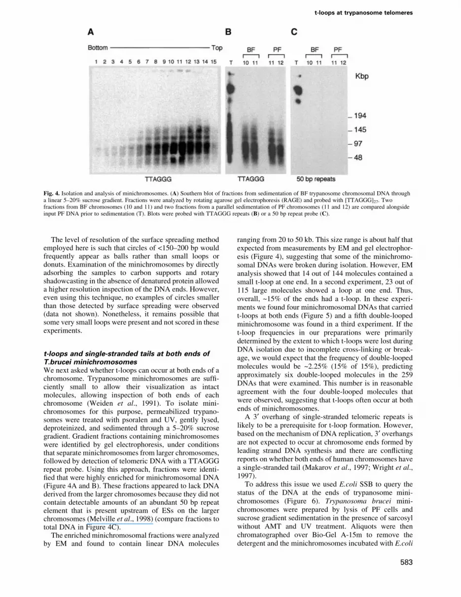
t-loops and single-stranded tails at both ends ofT.brucei minichromosomesWe next asked whether t-loops can occur at both ends of achromosome. Trypanosome minichromosomes are suf®-ciently small to allow their visualization as intactmolecules, allowing inspection of both ends of eachchromosome (Weiden et al., 1991). To isolate mini-chromosomes for this purpose, permeabilized trypano-somes were treated with psoralen and UV, gently lysed,deproteinized, and sedimented through a 5±20% sucrosegradient. Gradient fractions containing minichromosomeswere identi®ed by gel electrophoresis, under conditionsthat separate minichromosomes from larger chromosomes,followed by detection of telomeric DNA with a TTAGGGrepeat probe. Using this approach, fractions were identi-®ed that were highly enriched for minichromosomal DNA(Figure 4A and B). These fractions appeared to lack DNAderived from the larger chromosomes because they did notcontain detectable amounts of an abundant 50 bp repeatelement that is present upstream of ESs on the largerchromosomes (Melville et al., 1998) (compare fractions tototal DNA in Figure 4C).
The enriched minichromosomal fractions were analyzedby EM and found to contain linear DNA molecules
ranging from 20 to 50 kb. This size range is about half thatexpected from measurements by EM and gel electrophor-esis (Figure 4), suggesting that some of the minichromo-somal DNAs were broken during isolation. However, EManalysis showed that 14 out of 144 molecules contained asmall t-loop at one end. In a second experiment, 23 out of115 large molecules showed a loop at one end. Thus,overall, ~15% of the ends had a t-loop. In these experi-ments we found four minichromosomal DNAs that carriedt-loops at both ends (Figure 5) and a ®fth double-loopedminichromosome was found in a third experiment. If thet-loop frequencies in our preparations were primarilydetermined by the extent to which t-loops were lost duringDNA isolation due to incomplete cross-linking or break-age, we would expect that the frequency of double-loopedmolecules would be ~2.25% (15% of 15%), predictingapproximately six double-looped molecules in the 259DNAs that were examined. This number is in reasonableagreement with the four double-looped molecules thatwere observed, suggesting that t-loops often occur at bothends of minichromosomes.
A 3¢ overhang of single-stranded telomeric repeats islikely to be a prerequisite for t-loop formation. However,based on the mechanism of DNA replication, 3¢ overhangsare not expected to occur at chromosome ends formed byleading strand DNA synthesis and there are con¯ictingreports on whether both ends of human chromosomes havea single-stranded tail (Makarov et al., 1997; Wright et al.,1997).
To address this issue we used E.coli SSB to query thestatus of the DNA at the ends of trypanosome mini-chromosomes (Figure 6). Trypanosoma brucei mini-chromosomes were prepared by lysis of PF cells andsucrose gradient sedimentation in the presence of sarcosylwithout AMT and UV treatment. Aliquots were thenchromatographed over Bio-Gel A-15m to remove thedetergent and the minichromosomes incubated with E.coli
Fig. 4. Isolation and analysis of minichromosomes. (A) Southern blot of fractions from sedimentation of BF trypanosome chromosomal DNA througha linear 5±20% sucrose gradient. Fractions were analyzed by rotating agarose gel electrophoresis (RAGE) and probed with [TTAGGG]27. Twofractions from BF chromosomes (10 and 11) and two fractions from a parallel sedimentation of PF chromosomes (11 and 12) are compared alongsideinput PF DNA prior to sedimentation (T). Blots were probed with TTAGGG repeats (B) or a 50 bp repeat probe (C).
t-loops at trypanosome telomeres
583

SSB protein to bind any single-stranded DNA. Singletetramers or octamers of SSB bound along the length or atthe ends of otherwise duplex DNA can be distinguished byEM, and represent the presence of ~75 (single tetramer) or150 nt (octamer) of single-stranded DNA (Chrysogelosand Grif®th, 1982). The minimum length of a single-stranded DNA overhang that will allow binding of an SSBtetramer has not been established. Thus, overhangs lessthan ~75 nt may be missed using this approach. Followingpreparation of the complexes for EM, examination of 138minichromosomes judged to be >50 kb revealed that 70%showed no SSB on either end, 23% showed SSB binding atone end and 7% had SSB at both ends (Figure 6).
To evaluate the length of the overhang, the number ofSSB tetramers bound at an end was counted. For theminichromosomes with SSB bound at just one end or atboth ends, 71 and 70%, respectively, of the ends showed
from one to three tetramers bound, suggestive of over-hangs in the range of 75±225 nt. The remaining 30% of theends showed longer SSB-bound tracts ranging up to ~500nt. Thus, consistent with the annealing data in Figure 1,trypanosome chromosome ends contain substantial re-gions of single-stranded DNA and these overhangs canoccur at both ends of the same chromosome.
Discussion
This report documents the presence of t-loops atchromosome ends in trypanosomes. Although theyhave the same sequence and overall length, trypano-some telomeres had loops that were signi®cantlysmaller than those of mammalian telomeres, indicatingthat t-loop sizes are determined by a speci®c mech-anism. A signi®cant fraction of isolated intact trypano-some minichromosomes had two t-loops and carriedsingle-stranded overhangs at both ends, showing thattelomeres generated by both leading and lagging strandsynthesis can be remodeled into t-loops. Generation ofthe 3¢ overhang for t-loop formation at the endduplicated by leading strand synthesis must involvepost-replicative modi®cation since the replication prod-uct is predicted to be blunt. Our ®ndings, together withthe demonstration of t-loops in mammals and ciliates,indicate that they are a conserved feature of eukaryotictelomeres and their presence at both ends of achromosome is consistent with a requirement fort-loops in the function of all telomeres.
Trypanosome t-loops are relatively smallThe trypanosome telomeres analyzed in this study arecomposed of 10±20 kb of TTAGGG repeats. Humantelomeres have a very similar structure, containing aduplex TTAGGG repeat array in the 5±20 kb range.Despite these similarities, the size distribution of thet-loops observed in these two species was signi®cantlydifferent. Trypanosome telomeres often had small t-loops(median size 1.1 kb), whereas human telomeres very rarelyshowed t-loops in that size range. For instance, HeLa cellswith telomeres in the 20 kb range had t-loops with amedian size of 14 kb and <2% of the t-loops were <1 kb.The smallest human t-loops were observed in cells withtelomeres composed of ~5 kb TTAGGG repeats, but theset-loops were still signi®cantly larger (median 3 kb) thanthose of trypanosomes. Similarly, the loops observed at theends of micronuclear chromosomes of O.fallax were muchlarger [5±10 kb loops (Murti and Prescott, 1999)] thantrypanosome t-loops and more comparable to those ofmammalian cells. Although it is not clear what determinesthe size of the t-loops, the data suggest that there is anactive process involved in establishing or maintainingt-loops of a speci®c size.
Conservation of t-loopsThis study focused on telomeres in trypanosomesbecause of their experimental advantages and becausethey represent a very ancient lineage. Trypanosomesprobably branched off >500 million years ago, longbefore the origin of their metazoan hosts (Stevens andGibson, 1999). The molecular biology of these highlydiverged protozoa is quite distinct from the perceived
Fig. 5. t-loops in T.brucei minichromosomes. Minichromosomesenriched by sucrose gradient sedimentation were prepared for EMas described in Materials and methods. The minichromosome in(A) measures 28.7 kb, and the loops at the left and right ends measure650 and 710 bp, respectively. The minichromosome in (B) is 30.5 kb,with loops of 1310 and 790 bp at the left and right ends, respectively.The molecule in (C) is 21.1 kb (most likely a broken minichromosome)and the loop at the left end is 1470 bp. Shown in reverse contrast.Bar is equivalent to 5 kb.
J.L.MunÄ oz-JordaÂn et al.
584

norm as represented by yeast, plants and mammals.For instance, trypanosomes have a specialized orga-nelle for glycolysis, their mitochondria contain anunusual network of small circular DNAs, mitochondrialRNAs are edited, and nuclear mRNAs are formed bytrans-splicing. Within this context, the conservation oft-loops in trypanosomes stands out as highly signi®cantand predicts that t-loops play an essential role attelomeres in many eukaryotes.
The previous demonstration of looped structures atthe ends of O.fallax micronuclear chromosomes (Murtiand Prescott, 1999) is in agreement with the proposalthat t-loops are highly conserved . Interestingly, thisorganism also provides an example of functional
telomeres that lack t-loops. The macronuclear DNAof Oxytricha and other hypotrichous ciliates is formedby extensive fragmentation and processing of themicronuclear genome, resulting in ampli®ed smallDNA fragments each carrying one gene. These gene-sized molecules are all endowed with short telomeresthat contain <50 bp of duplex telomeric DNA and ashort single-stranded overhang (Price, 1999). Giventheir extreme short size, it was anticipated that thesetelomeres would lack t-loops, (Grif®th et al., 1999) aprediction consistent with the EM analysis. Instead, theends of the macronuclear DNAs may be protected bythe tenaciously bound protein complex (Gottschlingand Zakian, 1986; Horvath et al., 1998; Price, 1999).
Fig. 6. Trypanosoma brucei minichromosomes with single-strand overhangs stained with SSB. Non-cross-linked minichromosomes enriched bysucrose gradient sedimentation were incubated with E.coli SSB and prepared for EM as described in Materials and methods, including adsorption tothin carbon foils, dehydration and rotary shadowcasting with tungsten. (A) A 31 kb minichromosome with single-stranded overhangs at both ends. Theoverhangs on this molecule are longer than most and were selected for greater visibility at low magni®cation. (B±E) Individual minichromosome endswith SSB bound. The size of the particle in (E) corresponds to a single SSB tetramer. Shown in reverse contrast. Bar equals 0.5 mm (A) and0.27 mm (B±E).
t-loops at trypanosome telomeres
585

Collectively, the presence of t-loops in organisms asdiverged as mammals, ciliates and Kinetoplastidaeindicates that this aspect of telomere structure ishighly conserved.
t-loops at yeast telomeres?The ®nding of t-loops in diverged eukaryotes has raisedthe question of whether they occur in budding yeast, wheretelomeres have been characterized extensively. A t-loop-like structure was proposed by Li and Lustig (1996) as anintermediate in the rapid deletions that can occur whenyeast telomeres are excessively long. Telomere folding(without strand invasion) was also proposed byMcEachern and Blackburn (1995) to explain the mechan-ism of telomere length regulation in Kluyveromyces lactis,and Zakian and Ptashne and their colleagues proposed afold-back structure for telomeres in S.cerevisiae based onstudies of transcriptional regulation of subtelomeric genes(de Bruin et al., 2000, 2001). Similarly, based on theability to cross-link the telomeric DNA binding proteinRap1p to subtelomeric Y¢ elements, Grunstein, (1997)proposed that yeast telomeres form a higher order structurein which the telomere is folded back along the sub-telomeric DNA. Technical limitations of the current t-loopassays have hindered direct examination of yeast telomerestructure.
A signi®cant difference between yeast telomeres andthose of trypanosomes and mammals is that yeasttelomeres appear to lack long single-stranded protrusions[except for a short window late in S phase (Wellinger et al.,1993)]. Such telomere tails are presumed to be required forthe strand invasion that creates the t-loop, althoughdifferent scenarios can be envisioned. Furthermore,t-loop formation in mammals has been proposed to dependon the telomeric protein TRF2, and a recent comparison ofthe mammalian and yeast telomeric complexes hassuggested that budding yeast has lost the genes encodingboth TRFs (Li et al., 2000). Interestingly, the major yeasttelomeric DNA binding protein Rap1p has the ability topromote the pairing of single-stranded telomeric DNAwith duplex repeat tracts in vitro (Gilson et al., 1994), anactivity that could be indicative of a role in higher orderremodeling of telomeric DNA .
So far, the protein components of trypanosometelomeres have not been identi®ed. Speci®cally, it willbe of interest to determine whether trypanosomes haveTRF and Rap1p orthologs. Given the ease of genetargeting in trypanosomes and the abundance of theirtelomeres, trypanosomes could become a fruitful systemfor telomere biology once telomeric proteins are in hand.
t-loops and telomeric tails at both ends of eachchromosomeSeveral trypanosome minichromosomes showed t-loops atboth ends. The frequency of double-looped molecules washigh enough to suggest that t-loops are formed at DNAends created by both lagging and leading strand synthesis.The two modes of DNA synthesis are predicted to generatedifferent ends. Lagging strand synthesis generates a3¢ overhang with a length that depends primarily on thesite where primase synthesized the last RNA primer;removal of the RNA primer could contribute an additional8±12 nt to the protrusion. By contrast, leading strand
synthesis should result in a blunt end and formation of a 3¢overhang was therefore suggested to require a nuclease.This dilemma was previously recognized in the context oftests for the presence of 3¢ overhangs at both ends of eachchromosome and there are con¯icting reports on theterminal structure of human chromosome ends (Makarovet al., 1997; Wright et al., 1997). Our data are compatiblewith the view that all chromosome ends carry a 3¢overhang, as an overhang is likely to be required for themaintenance of t-loops. Indeed, EM analysis of mini-chromosomes with bound SSB showed frequent occur-rence of single-stranded DNA (presumably the G-strandoverhang) at both chromosome ends. There are severalmechanisms by which the end created by leading strandsynthesis could acquire a 3¢ overhang. Telomerase couldsynthesize the overhangs, they could be generated by an(unknown) 5¢®3¢ exonuclease, or the newly generatedblunt end could invade the duplex part of the telomere andthe 3¢ end could then be extended by the replicationmachinery. Regardless of the mechanism of their forma-tion, the presence of overhangs and t-loops at both ends oftrypanosome chromosomes further corroborates the ideathat t-loops are required for the protection of all chromo-some ends.
Materials and methods
TrypanosomesMolteno Institute Trypanozoon antigenic type 1.2 (MITat 1.2), clone221a, derived from strain 427 was used. Mice were infected byintraperitoneal injection and observed until the parasitemia reached~5 3 108 trypanosomes/ml. A total of 109 trypanosomes were puri®edthrough DEAE±cellulose as described (Cross, 1975), centrifuged gentlyand suspended in ice-cold trypanosome dilution buffer, TDB (5 mM KCl,80 mM NaCl, 1 mM MgSO4´7H2O, 20 mM Na2HPO4, 2 mMNaH2PO4´2H2O, 20 mM glucose). Procyclic forms of the same strainwere cultured at 27°C in SDM-79 supplemented with fetal bovine serum,to a concentration of 107 trypanosomes/ml. A total of 109 trypanosomeswere collected, washed and resuspended in ice-cold phosphate-buffered saline (PBS) pH 7.3 (137 mM NaCl, 2.7 mM KCl, 4.3 mMNa2HPO4´7H2O, 1.4 mM KH2PO4).
Genomic blottingTo isolate genomic DNA, ~5 3 108 trypanosomes were resuspended inTNE (10 mM Tris pH 7.4, 10 mM EDTA, 100 mM NaCl) and lysedin TNES (10 mM Tris pH 7.4, 100 mM NaCl, 10 mM EDTA, 1% SDS) inthe presence of 100 mg/ml proteinase K. After overnight incubation withproteinase K at 37°C, and phenol/chloroform extractions, DNA wasprecipitated with isopropanol and resuspended in TE (10 mM Tris pH 7.5/1 mM EDTA). RNase A treatment, phenol/chloroform extractions andisopropanol precipitation followed. Bal31 digestions were performed asdescribed elsewhere (de Lange and Borst, 1982). At each time point, thereaction was stopped by increasing the temperature to 65°C for 10 minand DNA was puri®ed using Sephacryl MicroSpin columns (Amersham).For telomere blots, DNA was digested overnight with the restrictionenzymes AluI, HinfI, RsaI, MboI or XmnI, and size fractionated andblotted as described (de Lange and Borst, 1982). Telomeric restrictionfragments were detected using a probe containing TTAGGG repeats andlabeled as previously described (de Lange, 1992). The sequence of the50 bp repeat was obtained from a plasmid provided by P.Borst (pBL-50),and a probe consisting of one repeat (GTGTACTTGCCTGTAC-TAAAAGTATTCTTACAGGGGTTGCAGTATACTGT) was synthe-sized and end labeled. Probes for VSGs 221 and 121 were labeled bystandard methods. Signals were quanti®ed using ImageQuant softwareand PhosphorImager data.
Permeabilization, cross-linking and telomeric DNApreparation for EMBF or PF trypanosomes (5 3 108) washed in TDB or PBS, respectively,were resuspended in resuspension buffer (15 mM Tris±HCl pH 7.4,
J.L.MunÄ oz-JordaÂn et al.
586

15 mM NaCl, 60 mM KCl, 1 mM EDTA, 0.25 mM sucrose) andincubated on ice in the presence of 40 mM digitonin (Sigma) for 5 min.Trypanosomes were collected by spinning at 3000 r.p.m. for 30 s in amicrocentrifuge, and treated for cross-linking and DNA extractionfollowing scaled-down adaptations of previously published protocols.Speci®cally, 50 ml of a solution of AMT (Sigma; 5 mg/ml in H2O) wereadded to 1 ml of permeabilized cells. Trypanosomes were stirred slowlyand exposed to UV light (350 nm) for 30 min. An equal volume of TNESwas added and supplemented with 200 mg of proteinase K. The sampleswere incubated at 55°C for 2 h. After two extractions with phenol/chloroform and precipitation of the DNA with isopropanol, the DNA wasresuspended in TNE and treated with RNase A (20 mg/ml) for 1 h at 37°C.DNA was extracted twice with phenol/chloroform, collected byprecipitation with isopropanol, dissolved and digested withRsaI±HinfI±AluI. The digest was phenol extracted, ethanol precipitatedand resuspended in TE.
Sucrose gradients and pulsed-®eld rotating gelelectrophoresis of minichromosomal DNAThe procedure was adapted from the protocol of Weiden et al. (1991).Brie¯y, digitonin-permeabilized trypanosomes cross-linked with psor-alen/UV as above were resuspended in 100 ml of TNE and immediatelylysed by adding 1 ml of lysis solution [200 mM EDTA, 1% sodium laurylsarcosinate (SLS), 0.5 mg/ml proteinase K] and incubating for 2±3 h atroom temperature. The lysate was loaded onto a 35 ml linear 5±20%sucrose gradient in 100 mM EDTA, 25 mM Tris pH 7.5, 1% SLS.Gradients were centrifuged at 25°C for 16 h at 10 000 r.p.m. in an SW28ultracentrifuge rotor. Fractions of 2 ml were collected from the bottom ofthe gradient. Aliquots of 30 ml from each fraction were mixed with 30 mlof 1.6% low melting point agarose at 65°C and loaded onto a 0.8%agarose gel. Electrophoresis was carried at an angle of 120°, 1±12 s linearramp, and a constant voltage of 180 V for 15 h at 13°C in a rotatingagarose gel electrophoresis (RAGE) apparatus (Stratagene).
Gel chromatography of T.brucei telomeric restrictionfragmentsFollowing restriction of total cross-linked T.brucei DNA, the DNA wasprecipitated with ethanol and resuspended in 10 mM Tris pH 7.5, 1 mMEDTA at a concentration of ~200 mg/ml and applied to a 20 ml column ofBio-Gel A-5m equilibrated in the same buffer. The chromatography wascontrolled with a Pharmacia Gradifract apparatus. The DNA pro®le wasdetermined by absorbance readings at 260 nm and fractions containingthe telomeric restriction fragments were prepared for EM.
Staining of minichromosomes with SSBAliquots of T.brucei minichromosomes in sucrose±sarcosyl werechromatographed over 2 ml columns of Bio-Gel A-5m equilibrated in20 mM HEPES pH 7.5/1 mM EDTA. Escherichia coli SSB was added to1 mg/ml and the sample incubated on ice for 10 min. Glutaraldehyde wasthen added to 0.6% for 5 min at room temperature and the samplechromatographed over a second Bio-Gel column to remove the freeprotein and ®xative. Minichromosomes in the excluded fractions wereprepared for EM by direct adsorption onto glow-charged carbon ®lms inthe presence of spermidine, washed, air dried and rotary shadowcast withtungsten (Grif®th and Christiansen, 1978).
EM methodsTo examine cross-linked T.brucei minichromosomes separated bysucrose sedimentation, the pooled DNA fractions were chromatographedthrough 2 ml columns of Bio-Gel A-5m (Bio-Rad Inc.) equilibrated in TEto remove the sucrose and detergent. Following cross-linking andprocessing, DNA was precipitated with ethanol and dissolved in TE.Cross-linked DNA samples in TE were prepared for EM by spreading ona denatured ®lm of cytochrome c using the droplet variation of the methodof Kleinschmidt as described (Grif®th et al., 1999). The grids were airdried and rotary shadowcast with platinum:paladium (20:1), andexamined in a Philips CM12 instrument at 40 kV. DNA lengths weremeasured by projecting images on EM sheet ®lm onto a Summagraphicsdigitizing tablet attached to a Macintosh computer programmed withsoftware developed in the Grif®th laboratory. Images for publication werescanned using a Nikon LS4500 ®lm scanner, and the contrast adjusted andimages arranged into ®gures using Adobe Photoshop software.
Acknowledgements
We thank Piet Borst for the pBL-50bp clone and Matthew Berriman forpreparing the probe. Agata Smogorzewska and Giulia Celli are thankedfor their assistance with telomere blots and sucrose gradients, andadditional members of the de Lange laboratory are acknowledged fortheir helpful suggestions during the course of this work and theircomments on this manuscript. This work was funded by grants for theNational Institutes of Health AI21729 (G.A.M.C.), 1-F31-AI09893(J.L.M.-J.), GM31819 (J.D.G.), CA70343 (J.D.G.), GM49046 (T.d.L.)and AG16642 (T.d.L.). T.d.L. is supported by an Ellison MedicalFoundation Senior Scholar Award.
References
Bernards,A., Michels,P.A.M., Lincke,C.R. and Borst,P. (1983) Growthof chromosome ends in multiplying trypanosomes. Nature, 303,592±597.
Bianchi,A., Smith,S., Chong,L., Elias,P. and de Lange,T. (1997) TRF1 isa dimer and bends telomeric DNA. EMBO J., 16, 1785±1794.
Bianchi,A., Stansel,R.M., Fairall,L., Grif®th,J.D., Rhodes,D. and deLange,T. (1999) TRF1 binds a bipartite telomeric site with extremespatial ¯exibility. EMBO J., 18, 5735±5744.
Bilaud,T., Brun,C., Ancelin,K., Koering,C.E., Laroche,T. and Gilson,E.(1997) Telomeric localization of TRF2, a novel human teloboxprotein. Nature Genet., 17, 236±239.
Blackburn,E.H. and Challoner,P.B. (1984) Identi®cation of a telomericDNA sequence in Trypanosoma brucei. Cell, 36, 447±457.
Broccoli,D., Smogorzewska,A., Chong,L. and de Lange,T. (1997)Human telomeres contain two distinct Myb-related proteins, TRF1and TRF2. Nature Genet., 17, 231±235.
Cano,M.I.N., Dungan,J.M., Agabian,N. and Blackburn,E.H. (1999)Telomerase in kinetoplastid parasitic protozoa. Proc. Natl Acad. Sci.USA, 96, 3616±3621.
Chrysogelos,S. and Grif®th,J. (1982) E.coli single strand DNA bindingprotein organizes single stranded DNA in nucleosome-like units. Proc.Natl Acad. Sci. USA, 79, 5803±5807.
Cross,G.A.M. (1975) Identi®cation, puri®cation and properties of clone-speci®c glycoprotein antigens constituting the surface coat ofTrypanosoma brucei. Parasitology, 71, 393±417.
Cross,G.A.M. (1996) Antigenic variation in trypanosomes: secretssurface slowly. BioEssays, 18, 283±291.
de Bruin,D., Kantrow,S.M., Liberatore,R.A. and Zakian,V.A. (2000)Telomere folding is required for the stable maintenance of telomereposition effects in yeast. Mol. Cell. Biol., 20, 7991±8000.
de Bruin,D., Zaman,Z. Liberatore,R.A. and Ptashne,M. (2001) Telomerelooping permits gene activation by a downstream UAS in yeast.Nature, 209, 109±113.
de Lange,T. (1992) Human telomeres are attached to the nuclear matrix.EMBO J., 11, 717±724.
de Lange,T. and Borst,P. (1982) Genomic environment of theexpression-linked extra copies of genes for surface antigens ofTrypanosoma brucei resembles the end of a chromosome. Nature,299, 451±453.
de Lange,T. and Petrini,J.H.J. (2000) A new connection at humantelomeres: association of the Mre11 complex with TRF2. Cold SpringHarb. Symp. Quant. Biol., in press.
de Lange,T., Shiue,L., Myers,R.M., Cox,D.R., Naylor,S.L., Killery,A.M.and Varmus,H.E. (1990) Structure and variability of humanchromosome ends. Mol. Cell. Biol., 10, 518±527.
Garvik,B., Carson,M. and Hartwell,L. (1995) Single-stranded DNAarising at telomeres in cdc13 mutants may constitute a speci®c signalfor the RAD9 checkpoint. Mol. Cell. Biol., 15, 6128±6138.
Gilson,E., Muller,T., Sogo,J., Laroche,T. and Gasser,S.M. (1994) RAP1stimulates single- to double-strand association of yeast telomericDNA: implications for telomere±telomere interactions. Nucleic AcidsRes., 22, 5310±5320.
Gottschling,D.E. and Cech,T.R. (1984) Chromatin structure of themolecular ends of Oxytricha macronuclear DNA: phased nucleosomesand a telomeric complex. Cell, 38, 501±510.
Gottschling,D.E. and Zakian,V.A. (1986) Telomere proteins: speci®crecognition and protection of the natural termini of Oxytrichamacronuclear DNA. Cell, 47, 195±205.
Greider,C.W. and Blackburn,E.H. (1985) Identi®cation of a speci®ctelomere terminal transferase activity in Tetrahymena extracts. Cell,43, 405±413.
t-loops at trypanosome telomeres
587

Grif®th,J. and Christiansen,G. (1978) Electron microscopic visualizationof chromatin and other DNA±protein complexes. Annu. Rev. Biophys.Bioeng., 7, 19±35.
Grif®th,J., Bianchi,A. and deLange,T. (1998) TRF1 promotes parallelpairing of telomeric tracts in vitro. J. Mol. Biol., 278, 79±88.
Grif®th,J.D., Comeau,L., Rosen®eld,S., Stansel,R.M., Bianchi,A.,Moss,H. and de Lange,T. (1999) Mammalian telomeres end in alarge duplex loop. Cell, 97, 503±514.
Grunstein,M. (1997) Molecular model for telomeric heterochromatin inyeast. Curr. Opin. Cell Biol., 9, 383±387.
Horn,D. and Cross,G.A.M. (1997) Analysis of Trypanosoma brucei vsgexpression site switching in vitro. Mol. Biochem. Parasitol., 84,189±201.
Horvath,M.P., Schweiker,V.L., Bevilacqua,J.M., Ruggles,J.A. andSchultz,S.C. (1998) Crystal structure of the Oxytricha nova telomereend binding protein complexed with single strand DNA. Cell, 95,963±974.
Huffman,K.E., Levene,S.D., Tesmer,V.M., Shay,J.W. and Wright,W.E.(2000) Telomere shortening is proportional to the size of the G-richtelomeric 3¢-overhang. J. Biol. Chem., 275, 19719±19722.
Karlseder,J., Broccoli,D., Dai,Y., Hardy,S. and de Lange,T. (1999) p53-and ATM-dependent apoptosis induced by telomeres lacking TRF2.Science, 283, 1321±1325.
Li,B. and Lustig,A.J. (1996) A novel mechanism for telomere sizecontrol in Saccharomyces cerevisiae. Genes Dev., 10, 1310±1326.
Li,B., Oestreich,S. and de Lange,T. (2000) Identi®cation of human Rap1:implications for telomere evolution. Cell, 101, 471±483.
Lin,J.J. and Zakian,V.A. (1996) The Saccharomyces CDC13 protein is asingle-strand TG1-3 telomeric DNA-binding protein in vitro thataffects telomere behavior in vivo. Proc. Natl Acad. Sci. USA, 93,13760±13765.
Liu,C. and Lustig,A.J. (1996) Genetic analysis of Rap1p/Sir3pinteractions in telomeric and HML silencing in Saccharomycescerevisiae. Genetics, 143, 81±93.
Makarov,V.L., Hirose,Y. and Langmore,J.P. (1997) Long G tails at bothends of human chromosomes suggest a C strand degradationmechanism for telomere shortening. Cell, 88, 657±666.
McClintock,B. (1941) The stability of broken ends of chromosomes inZea mays. Genetics, 26, 234±282.
McEachern,M.J. and Blackburn,E.H. (1995) Runaway telomereelongation caused by telomerase RNA gene mutations. Nature, 376,403±409.
McElligott,R. and Wellinger,R.J. (1997) The terminal DNA structure ofmammalian chromosomes. EMBO J., 16, 3705±3714.
Melville,S.E., Leech,V., Gerrard,C.S., Tait,A. and Blackwell,J.M. (1998)The molecular karyotype of the megabase chromosomes ofTrypanosoma brucei and the assignment of chromosome markers.Mol. Biochem. Parasitol., 94, 155±173.
Melville,S.E., Leech,V., Navarro,M. and Cross,G.A.M. (2000) Themolecular karyotype of the megabase chromosomes of Trypanosomabrucei stock 427. Mol. Biochem. Parasitol., 111, 261±273.
Muller,H.J. (1938) The remaking of chromosomes. The Collecting Net,Woods Hole, 8, 182±195.
Murti,K.G. and Prescott,D.M. (1999) Telomeres of polytenechromosomes in a ciliated protozoan terminate in duplex DNAloops. Proc. Natl Acad. Sci. USA, 96, 14436±14439.
Navarro,M., Cross,G.A.M. and Wirtz,E. (1999) Trypanosoma bruceivariant surface glycoprotein regulation involves coupled activation/inactivation and chromatin remodeling of expression sites. EMBO J.,18, 2265±2272.
Nugent,C.I. and Lundblad,V. (1998) The telomerase reversetranscriptase: components and regulation. Genes Dev., 12, 1073±1085.
Nugent,C.I., Hughes,T.R., Lue,N.F. and Lundblad,V. (1996) Cdc13p: asingle-strand telomeric DNA-binding protein with a dual role in yeasttelomere maintenance. Science, 274, 249±252.
Pays,E., Laurent,M., Delinte,K., van Meirvenne,N. and Steinert,M.(1983) Differential size variations between transcriptionally active andinactive telomeres of Trypanosoma brucei. Nucleic Acids Res., 11,8137±8147.
Price,C. (1999) Telomeres. Capping off the ends. Nature, 397, 213±214.Rudenko,G., Blundell,P.A., Dirks-Mulder,A., Kieft,R. and Borst,P.
(1995) A ribosomal DNA promoter replacing the promoter of atelomeric VSG gene expression site can be ef®ciently switched on andoff in T. brucei. Cell, 83, 547±553.
Rudenko,G., Cross,M. and Borst,P. (1998) Changing the end: antigenicvariation orchestrated at the telomeres of African trypanosomes.Trends Microbiol., 6, 113±116.
Saltman,D., Morgan,R., Cleary,M.L. and de Lange,T. (1993) Telomericstructure in cells with chromosome end associations. Chromosoma,102, 121±128.
Sandell,L.L. and Zakian,V.A. (1993) Loss of a yeast telomere: arrest,recovery and chromosome loss. Cell, 75, 729±739.
Smogorzewska,A., van Steensel,B., Bianchi,A., Oelmann,S.,Schaefer,M.R., Schnapp,G. and de Lange,T. (2000) Control ofhuman telomere length by TRF1 and TRF2. Mol. Cell. Biol., 20,1659±1668.
Stevens,J.R. and Gibson,W. (1999) The molecular evolution oftrypanosomes. Parasitol. Today, 15, 432±437.
van der Ploeg,L.H.T., Liu,A.Y.C. and Borst,P. (1984) Structure of thegrowing telomeres of trypanosomes. Cell, 36, 459±468.
van Steensel,B., Smogorzewska,A. and de Lange,T. (1998) TRF2protects human telomeres from end-to-end fusions. Cell, 92, 401±413.
Weiden,M., Osheim,Y.N., Beyer,A.L. and Van der Ploeg,L.H. (1991)Chromosome structure: DNA nucleotide sequence elements of asubset of the minichromosomes of the protozoan Trypanosoma brucei.Mol. Cell. Biol., 11, 3823±3834.
Wellinger,R.J. and Sen,D. (1997) The DNA structures at the ends ofeukaryotic chromosomes. Eur. J. Cancer, 33, 735±749.
Wellinger,R.J., Wolf,A.J. and Zakian,V.A. (1993) Saccharomycestelomeres acquire single-strand TG1-3 tails late in S phase. Cell, 72,51±60.
Wright,W.E., Tesmer,V.M., Huffman,K.E., Levene,S.D. and Shay,J.W.(1997) Normal human chromosomes have long G-rich telomericoverhangs at one end. Genes Dev., 11, 2801±2809.
Zhu,X.D., Kuster,B., Mann,M., Petrini,J.H. and Lange,T. (2000) Cell-cycle-regulated association of RAD50/MRE11/NBS1 with TRF2 andhuman telomeres. Nature Genet., 25, 347±352.
Received September 20, 2000; revised December 5, 2000;accepted December 6, 2000
J.L.MunÄ oz-JordaÂn et al.
588