systemic postganglionic adrenergic studies do not distinguish parkinson's disease from multiple...
TRANSCRIPT
Systemic Postganglionic Adrenergic Studies Do Not DistinguishParkinson's Disease From Multiple System Atrophy
Axel Lipp1,2, Paola Sandroni1, and Phillip A. Low1,*
1Department of Neurology, Mayo Clinic, Rochester, MN
2Department of Neurology, Charité - University Medicine Berlin, Germany
AbstractBackground—Multiple system atrophy (MSA) affects the preganglionic adrenergic neuron andParkinson's disease (PD) involves the postganglionic counterpart. Widespread postganglionicdenervation should result in denervation supersensitivity and a failure of the axon to releasenorepinephrine (NE). We examined if pharmacological dissection of the adrenergic neuron candistinguish between MSA and PD.
Method—We measured blood pressure, heart rate, and plasma NE responses to direct(phenylephrine) and indirect (tyramine) acting adrenergic agonists in 15 patients with probable MSA,16 patients with idiopathic PD, and 16 age- and gender-matched controls.
Results—Baroreflex sensitivity was impaired in MSA and intact in PD. Pressor responses tophenylephrine (direct acting) were higher in MSA (p < 0.01) and PD patients (p = 0.04) than controls.Blood pressure responses to tyramine (indirect acting) were increased in MSA only (p < 0.01).Tyramine increased plasma catecholamine levels in all groups with no significant differencesbetween groups.
Conclusion—There is denervation supersensitivity in PD patients that is, however, insufficient toshift the dose-response curve to the left. The excessive pressor responses to both tyramine andphenylephrine in MSA are due to baroreflex failure. We conclude that this diagnostic approach lackssufficient sensitivity to differentiate PD and MSA.
KeywordsMSA; PD; baroreflex; tyramine; phenylephrine; autonomic; denervation supersensitivity
IntroductionAutonomic denervation occurs in both Parkinson's disease (PD) and multiple system atrophy(MSA). There is severe and generalized preganglionic adrenergic involvement in MSA [1]. Incontrast the adrenergic lesion in PD is postganglionic [2,3]. In some organs, such as the heart,an evaluation of postganglionic adrenergic denervation will reliably distinguish PD from MSA[4-6]. Using directly acting adrenergic labels such as MIBG [4-6], hydroxyephedrine [7] orfluorodopa [8] uptake is uniformly reduced in PD and normal in MSA. It is of interest that
*To whom correspondence should be addressed: Mayo Foundation, Autonomic Reflex Laboratory, Department of Neurology, 200 FirstStreet SW, Rochester, Minnesota 55905, Phone: 507-284-3375, Fax: 507-284-3133, Email: E-mail: [email protected]'s Disclaimer: This is a PDF file of an unedited manuscript that has been accepted for publication. As a service to our customerswe are providing this early version of the manuscript. The manuscript will undergo copyediting, typesetting, and review of the resultingproof before it is published in its final citable form. Please note that during the production process errors may be discovered which couldaffect the content, and all legal disclaimers that apply to the journal pertain.
NIH Public AccessAuthor ManuscriptJ Neurol Sci. Author manuscript; available in PMC 2010 June 15.
Published in final edited form as:J Neurol Sci. 2009 June 15; 281(1-2): 15–19. doi:10.1016/j.jns.2009.03.006.
NIH
-PA Author Manuscript
NIH
-PA Author Manuscript
NIH
-PA Author Manuscript
laboratory denervation occurs in the context of little clinical involvement. It is not knownwhether similar pharmacologic evidence of systemic adrenergic denervation occurs and if PDcan be differentiated from MSA pharmacologically. We tested the hypothesis that systemicadrenergic pharmacologic dissection will similarly distinguish PD from MSA. It is well-knownthat a disorder with diffuse and severe postganglionic adrenergic involvement, such as pureautonomic failure (PAF), diabetic autonomic [9] or autoimmune autonomic neuropathy [10]will have denervation supersensitivity and a larger pressor response to directly actingadrenergic agonists, whereas a preganglionic disorder (MSA) will not [11]. Postganglionicdenervation will result in receptor upregulation so that there will be a pressor response tosubthreshold or low dose phenylephrine. The dose-response curve for a postganglionic disorderis shifted to the left and steeper, whereas the preganglionic disorder (MSA) is not shifted tothe left but is steeper (due to baroreflex failure) [11,12]. We undertook pharmacologicdissection studies using a directly acting agonist, phenyephrine; and an indirectly acting agent(tyramine), which releases norepinephrine from intact postganglionic axons.
MethodsSubjects
The study cohort comprised fifteen patients with multiple system atrophy of the Parkinsoniantype (MSA-P) according to the consensus criteria [1], sixteen patients with idiopathicParkinson's disease, and sixteen age- and sex-matched, predominantly sedentary controlsubjects. The study was approved by the Institutional Review Board of the Mayo Clinic, andall subjects gave written informed consent before participation. Participants refrained fromexercise, alcohol, and caffeine for 24 hours and from nicotine at least four hours. All vasoactivemedication, including levodopa and fludrocortisone, was discontinued at least five half-livesbefore the study. Patients with concomitant conditions that potentially involve the CNS oraffect autonomic testing (particularly diabetes, hypertension and coronary or congestive heartdiseases) were excluded from the study. Prior to the study, all participants underwent clinicalautonomic testing in order to assess cardiovagal (heart rate response to deep breathing),adrenergic (blood pressure responses to passive head up tilt and to Valsalva maneuver), andsudomotor (quantitative sudomotor axon reflex test - QSART, thermoregulatory sweat test -TST) function.
MeasurementsHeart rate was measured by a three-lead electrocardiogram. Arterial blood pressure wasmeasured on a beat-to-beat basis by finger photoplethysmography (Finometer model 1, FMS)and regularly verified by manual sphygomanometry on the contralateral arm.
ProtocolSubjects were placed in a supine position and indwelling catheters were inserted into the cubitalveins bilaterally for administration of vasoactive medication and to obtain blood samples. Aftera 20-minute period of quiet rest, heart rate, blood pressure, and respiration (chest expansion)were continuously recorded for a further 10-minute baseline period. To test for hemodynamichypersensitivity to vasoactive agents, increasing dosages of tyramine (10, 20, and 30 mcg/kg)and phenylephrine (25, 50, 100, and 200 mcg) were administered at five minute intervals, butnot before blood pressure returned to baseline. Trials were separated by a fifteen minute restperiod and a subsequent ten minute baseline recording. Escalation of tyramine andphenylephrine boluses was stopped when blood pressure increased more than 30 mmHg orbaseline systolic blood pressure exceeded 180 mmHg. Blood samples for plasmacatecholamine levels were taken at baseline and 60 seconds after administration of tyramineboluses, respectively.
Lipp et al. Page 2
J Neurol Sci. Author manuscript; available in PMC 2010 June 15.
NIH
-PA Author Manuscript
NIH
-PA Author Manuscript
NIH
-PA Author Manuscript
Data Acquisition and Data AnalysisData were digitized with 14 bit resolution and a sample frequency of 500 Hz using WinDAQdata acquisition software (WinDAQ; Dataq Instruments, Akron, OH). R-R intervals, systolicand diastolic blood pressure, and respiration were analyzed offline with signal processingsoftware (CODAS, Dataq Instruments, Akron, OH). Hemodynamic effects of vasoactive drugswere assessed as changes in systolic blood pressure and heart rate over baseline. To controlfor Finometer offset and recording artifacts, we also measured integrated blood pressureresponses (area under the curve, AUCSBP) over a 20 and 40 second period in response tophenylephrine and tyramine, respectively. Baroreflex sensitivity was assessed using therelationship between R-R interval and systolic blood pressure during phenylephrine bolusesonly, since the sequential administration of depressor and pressor agents (modified Oxfordtechnique) would have confounded our primary analysis (pressor effect of phenylephrine/tyramine). To index the adrenergic component of the baroreflex we assessed blood pressurerecovery time (BP-recovery time), defined as the time taken for systolic blood pressure to returnto baseline (± 2 SD) after phenylephrine boluses.
All data are expressed as mean ± standard error of the mean. Phenylephrine induced changesin systolic blood pressure were compared between patients and controls by multiple-comparison analyses (unpaired t-test). Analysis of variance (one-way ANOVA) would not beappropriate in this regard, since differences between patients and controls but not withinpatients were considered (quod vide O'Brien, 1983) [13].
ResultsClinical characteristics
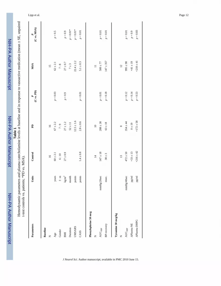
Clinical characteristics of patients and control subjects are summarized in Table 1. All patientsexhibited parkinsonism, which was not or only minimally responsive to levodopa treatment inthe case of MSA. Autonomic impairment (combined cardiovagal, adrenergic, sudomotorfailure), scored using the Composite Autonomic Severity Scale (CASS) [14], was significantlyhigher in MSA, matching the criteria for probable MSA-P [1]. PD patients were slightly olderthan both MSA and control subjects, who in turn did not differ with respect to age. The severityof parkinsonism in MSA and PD patients was matched, although the Unified MSA RatingScale (UMSARS) [15] was significantly higher in MSA than PD patients (Table 1); this reflectsthe fact that UMSARS measures also deficits such as oculomotor and cerebellar impairmentin addition to extrapyramidal dysfunction.
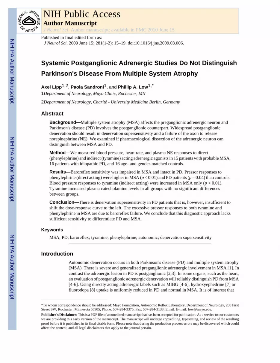
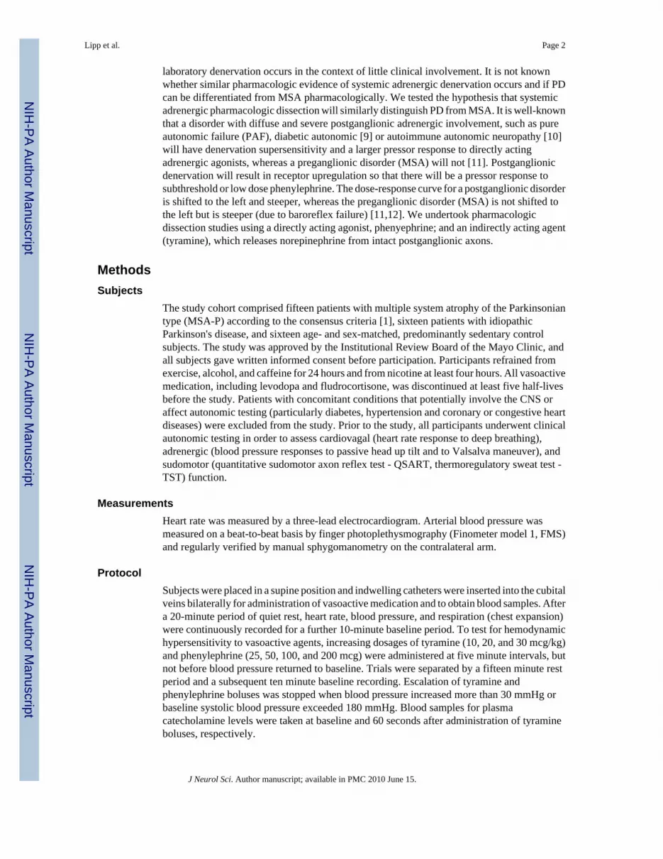
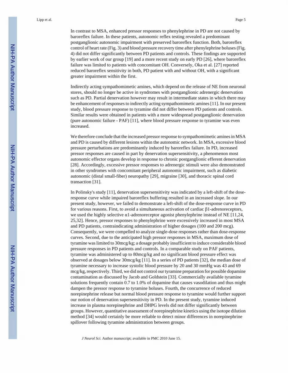
Hemodynamic effects of vasoactive medicationPhenylephrine induced changes in systolic blood pressure were higher in patients than controlsubjects (Table 1, Fig. 1). In 8/16 PD and 10/15 MSA patients, however, administration ofhigher dosages of phenylephrine (100 and 200 mcg) was contraindicated due to resting supinehypertension and excessive blood pressure responses to lower doses. Thus, group-wisecomparison was performed at a dose of 50 mcg phenylephrine. Blood pressure responses tophenylephrine were significantly higher in MSA (33.0 ± 4.6 mmHg) and PD (20.2 ± 1.8 mmHg)patients when compared to healthy controls (15.0 ± 1.4 mmHg; control vs. PD, p = 0.04; controlvs. MSA, p < 0.01). The augmented pressor effect of phenylephrine in MSA and PD patientsis even more pronounced when data are expressed as AUCSBP (control: 147 ± 18 mmHg/20sec;MSA: 500 ± 77 mmHg/20sec; p < 0.01; PD: 288 ± 38 mmHg/20sec; p < 0.01). Tyramineincreased systolic blood pressure in a dose-dependent fashion (Table 1, Fig. 2). Blood pressureresponse to 30 mcg/kg tyramine was not significantly different in PD patients and controls(PD: AUCSBP = 354 ± 44 mmHg/40sec, control: AUCSBP = 472 ± 50 mmHg/40sec, p = 0.12).In MSA, however, changes in arterial blood pressure exceeded those observed in controlssignificantly (AUCSBP = 996 ± 98 mmHg/40sec, p < 0.01).
Lipp et al. Page 3
J Neurol Sci. Author manuscript; available in PMC 2010 June 15.
NIH
-PA Author Manuscript
NIH
-PA Author Manuscript
NIH
-PA Author Manuscript
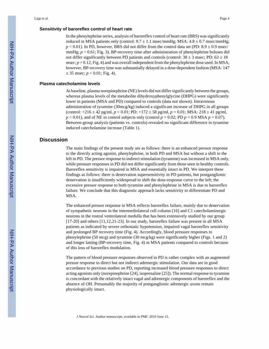
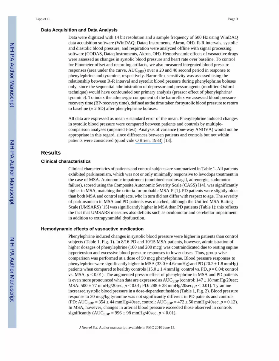
Sensitivity of baroreflex control of heart rateIn the phenylephrine series, analysis of baroreflex control of heart rate (BRS) was significantlyreduced in MSA patients only (control: 9.7 ± 1.1 msec/mmHg; MSA: 4.8 ± 0.7 msec/mmHg;p < 0.01). In PD, however, BRS did not differ from the control data set (PD: 8.9 ± 0.9 msec/mmHg; p < 0.61; Fig. 3). BP-recovery time after administration of phenylephrine boluses didnot differ significantly between PD patients and controls (control: 38 ± 3 msec; PD: 63 ± 18msec; p = 0.12, Fig. 4) and was overall independent from the phenylephrine dose used. In MSA,however, BP-recovery time was substantially delayed in a dose-dependent fashion (MSA: 147± 35 msec; p < 0.01; Fig. 4).
Plasma catecholamine levelsAt baseline, plasma norepinephrine (NE) levels did not differ significantly between the groups,whereas plasma levels of the metabolite dihydroxyphenylglycine (DHPG) were significantlylower in patients (MSA and PD) compared to controls (data not shown). Intravenousadministration of tyramine (30mcg/kg) induced a significant increase of DHPG in all groups(control: +216 ± 42 pg/ml, p < 0.01; PD: +172 ± 58 pg/ml, p = 0.01; MSA: 218 ± 41 pg/ml,p < 0.01), and of NE in control subjects only (control p = 0.02; PD p = 0.9 MSA p = 0.07).Between-group analysis (patients vs. controls) revealed no significant difference in tyramineinduced catecholamine increase (Table 1).
DiscussionThe main findings of the present study are as follows: there is an enhanced pressor responseto the directly acting agonist, phenylephrine, in both PD and MSA but without a shift to theleft in PD. The pressor response to indirect stimulation (tyramine) was increased in MSA only,while pressure responses in PD did not differ significantly from those seen in healthy controls.Baroreflex sensitivity is impaired in MSA and essentially intact in PD. We interpret thesefindings as follows: there is denervation supersensitivity in PD patients, but postganglionicdenervation is insufficiently widespread to shift the dose-response curve to the left; theexcessive pressor response to both tyramine and phenylephrine in MSA is due to baroreflexfailure. We conclude that this diagnostic approach lacks sensitivity to differentiate PD andMSA.
The enhanced pressor response in MSA reflects baroreflex failure, mainly due to denervationof sympathetic neurons in the intermediolateral cell column [16] and C1 catecholaminergicneurons in the rostral ventrolateral medulla that has been extensively studied by our group[17-20] and others [11,12,21-23]. In our study, baroreflex failure was present in all MSApatients as indicated by severe orthostatic hypotension, impaired vagal baroreflex sensitivityand prolonged BP recovery time (Fig. 4). Accordingly, blood pressure responses tophenylephrine (50 mcg) and tyramine (30 mcg/kg) were significantly higher (Figs. 1 and 2)and longer lasting (BP-recovery time, Fig. 4) in MSA patients compared to controls becauseof this loss of baroreflex modulation.
The pattern of blood pressure responses observed in PD is rather complex with an augmentedpressor response to direct but not indirect adrenergic stimulation. Our data are in goodaccordance to previous studies on PD, reporting increased blood pressure responses to directacting agonists only (norepinephrine [24], isoprenaline [25]). The normal response to tyramineis concordant with the relatively intact vagal and adrenergic components of baroreflex and theabsence of OH. Presumably the majority of postganglionic adrenergic axons remainphysiologically intact.
Lipp et al. Page 4
J Neurol Sci. Author manuscript; available in PMC 2010 June 15.
NIH
-PA Author Manuscript
NIH
-PA Author Manuscript
NIH
-PA Author Manuscript
In contrast to MSA, enhanced pressor responses to phenylephrine in PD are not caused bybaroreflex failure. In these patients, autonomic reflex testing revealed a predominantpostganglionic autonomic impairment with preserved baroreflex function. Both, baroreflexcontrol of heart rate (Fig. 3) and blood pressure recovery time after phenylephrine boluses (Fig.4) did not differ significantly between PD patients and controls. These findings are supportedby earlier work of our group [19] and a more recent study on early PD [26], where baroreflexfailure was limited to patients with concomitant OH. Conversely, Oka et al. [27] reportedreduced baroreflex sensitivity in both, PD patient with and without OH, with a significantgreater impairment within the first.
Indirectly acting sympathomimetic amines, which depend on the release of NE from neuronalstores, should no longer be active in syndromes with postganglionic adrenergic denervationsuch as PD. Partial denervation however may result in intermediate states in which there maybe enhancement of responses to indirectly acting sympathomimetic amines [11]. In our presentstudy, blood pressure response to tyramine did not differ between PD patients and controls.Similar results were obtained in patients with a more widespread postganglionic denervation(pure autonomic failure - PAF) [11], where blood pressure response to tyramine was evenincreased.
We therefore conclude that the increased pressor response to sympathomimetic amines in MSAand PD is caused by different lesions within the autonomic network. In MSA, excessive bloodpressure perturbations are predominantly induced by baroreflex failure. In PD, increasedpressor responses are caused in part by denervation supersensitivity, a phenomenon mostautonomic effector organs develop in response to chronic postganglionic efferent denervation[28]. Accordingly, excessive pressor responses to adrenergic stimuli were also demonstratedin other syndromes with concomitant peripheral autonomic impairment, such as diabeticautonomic (distal small-fiber) neuropathy [29], migraine [30], and thoracic spinal cordtransaction [31].
In Polinsky's study [11], denervation supersensitivity was indicated by a left-shift of the dose-response curve while impaired baroreflex buffering resulted in an increased slope. In ourpresent study, however, we failed to demonstrate a left-shift of the dose-response curve in PDfor various reasons. First, to avoid a simultaneous activation of cardiac β1-adrenoreceptors,we used the highly selective α1-adrenoreceptor agonist phenylephrine instead of NE [11,24,25,32]. Hence, pressor responses to phenylephrine were excessively increased in most MSAand PD patients, contraindicating administration of higher dosages (100 and 200 mcg).Consequently, we were compelled to analyze single-dose responses rather than dose-responsecurves. Second, due to the anticipated high pressor responses in MSA, maximum dose oftyramine was limited to 30mcg/kg; a dosage probably insufficient to induce considerable bloodpressure responses in PD patients and controls. In a comparable study on PAF patients,tyramine was administered up to 80mcg/kg and no significant blood pressure effect wasobserved at dosages below 30mcg/kg [11]. In a series of PD patients [32], the median dose oftyramine necessary to increase systolic blood pressure by 20 and 30 mmHg was 43 and 69mcg/kg, respectively. Third, we did not control our tyramine preparation for possible dopaminecontamination as discussed by Jacob and Goldstein [33]. Commercially available tyraminesolutions frequently contain 0.7 to 1.0% of dopamine that causes vasodilation and thus mightdampen the pressor response to tyramine boluses. Fourth, the concurrence of reducednorepinephrine release but normal blood pressure response to tyramine would further supportour notion of denervation supersensitivity in PD. In the present study, tyramine inducedincrease in plasma norepinephrine and DHPG levels did not differ significantly betweengroups. However, quantitative assessment of norepinephrine kinetics using the isotope dilutionmethod [34] would certainly be more reliable to detect minor differences in norepinephrinespillover following tyramine administration between groups.
Lipp et al. Page 5
J Neurol Sci. Author manuscript; available in PMC 2010 June 15.
NIH
-PA Author Manuscript
NIH
-PA Author Manuscript
NIH
-PA Author Manuscript
In conclusion, reduced neurotransmission in postganglionic autonomic fibers of PD patientsappeared to be counteracted in part by an increased postsynaptic sensitivity. This denervationsupersensitivity is not suitable for an early differentiation of Parkinson etiologies (PD vs.MSA). However, PD patients might be at risk of excessive blood pressure alterations inresponse to commonly used dosages of vasoactive substances.
AcknowledgementsThis work was supported in part by National Institutes of Health (NS 32352, NS 44233, NS 43364, and Mayo CTSA(UL1 RR24150), and Mayo Funds.
References1. Gilman S, Low PA, Quinn N, et al. Consensus statement on the diagnosis of multiple system atrophy.
J Neurol Sci 1999;163(1):94–98. [PubMed: 10223419]2. Braak H, Del TK, Rub U, de Vos RA, Jansen Steur EN, Braak E. Staging of brain pathology related
to sporadic Parkinson's disease. Neurobiol Aging 2003;24(2):197–211. [PubMed: 12498954]3. Braak H, Bohl JR, Muller CM, Rub U, de Vos RA, Del TK. Stanley Fahn Lecture 2005: The staging
procedure for the inclusion body pathology associated with sporadic Parkinson's disease reconsidered.Mov Disord 2006;21(12):2042–2051. [PubMed: 17078043]
4. Druschky A, Hilz MJ, Platsch G, et al. Differentiation of Parkinson's disease and multiple systematrophy in early disease stages by means of I-123-MIBG-SPECT. J Neurol Sci 2000;175(1):3–12.[PubMed: 10785250]
5. Orimo S, Ozawa E, Nakade S, Sugimoto T, Mizusawa H. (123)I-metaiodobenzylguanidine myocardialscintigraphy in Parkinson's disease. J Neurol Neurosurg Psychiatry 1999;67(2):189–194. [PubMed:10406987]
6. Braune S, Reinhardt M, Schnitzer R, Riedel A, Lucking CH. Cardiac uptake of [123I]MIBG separatesParkinson's disease from multiple system atrophy. Neurology 1999;53(5):1020–1025. [PubMed:10496261]
7. Raffel DM, Koeppe RA, Little R, et al. PET measurement of cardiac and nigrostriatal denervation inParkinsonian syndromes. J Nucl Med 2006;47(11):1769–1777. [PubMed: 17079809]
8. Goldstein DS, Holmes C, Li ST, Bruce S, Metman LV, Cannon RO III. Cardiac sympatheticdenervation in Parkinson disease. Ann Intern Med 2000;133(5):338–347. [PubMed: 10979878]
9. Low PA, Suarez GA. Diabetic neuropathies. Baillieres Clin Neurol 1995;4(3):401–425. [PubMed:8599718]
10. Low PA, Dyck PJ, Lambert EH, et al. Acute panautonomic neuropathy. Ann Neurol 1983;13(4):412–417. [PubMed: 6301359]
11. Polinsky RJ, Kopin IJ, Ebert MH, Weise V. Pharmacologic distinction of different orthostatichypotension syndromes. Neurology 1981;31(1):1–7. [PubMed: 7192816]
12. Jordan J, Tank J, Shannon JR, et al. Baroreflex buffering and susceptibility to vasoactive drugs.Circulation 2002;105(12):1459–1464. [PubMed: 11914255]
13. O'Brien PC. The appropriateness of analysis of variance and multiple-comparison procedures.Biometrics 1983;39(3):787–794. [PubMed: 6652209]
14. Low PA. Composite autonomic scoring scale for laboratory quantification of generalized autonomicfailure. Mayo Clin Proc 1993;68(8):748–752. [PubMed: 8392653]
15. Wenning GK, Tison F, Seppi K, et al. Development and validation of the Unified Multiple SystemAtrophy Rating Scale (UMSARS). Mov Disord 2004;19(12):1391–1402. [PubMed: 15452868]
16. Oppenheimer DR. Lateral horn cells in progressive autonomic failure. J Neurol Sci 1980;46(3):393–404. [PubMed: 6247458]
17. Benarroch EE, Smithson IL, Low PA, Parisi JE. Depletion of catecholaminergic neurons of the rostralventrolateral medulla in multiple systems atrophy with autonomic failure. Ann Neurol 1998;43(2):156–163. [PubMed: 9485056]
18. Benarroch EE. New findings on the neuropathology of multiple system atrophy. Auton Neurosci2002;96(1):59–62. [PubMed: 11911503]
Lipp et al. Page 6
J Neurol Sci. Author manuscript; available in PMC 2010 June 15.
NIH
-PA Author Manuscript
NIH
-PA Author Manuscript
NIH
-PA Author Manuscript
19. Sandroni P, Ahlskog JE, Fealey RD, Low PA. Autonomic involvement in extrapyramidal andcerebellar disorders. Clin Auton Res 1991;1(2):147–155. [PubMed: 1822762]
20. Lipp A, Tank J, Franke G, Arnold G, Luft FC, Jordan J. Osmosensitive mechanisms contribute to thewater drinking-induced pressor response in humans. Neurology 2005;65(6):905–907. [PubMed:16186532]
21. Jordan J, Shannon JR, Biaggioni I, Norman R, Black BK, Robertson D. Contrasting actions of pressoragents in severe autonomic failure. Am J Med 1998;105(2):116–124. [PubMed: 9727818]
22. Sharabi Y, Eldadah B, Li ST, et al. Neuropharmacologic distinction of neurogenic orthostatichypotension syndromes. Clin Neuropharmacol 2006;29(3):97–105. [PubMed: 16772807]
23. Young TM, Asahina M, Watson L, Mathias CJ. Hemodynamic effects of clonidine in two contrastingmodels of autonomic failure: multiple system atrophy and pure autonomic failure. Mov Disord2006;21(5):609–615. [PubMed: 16404729]
24. Wilcox CS, Aminoff MJ. Blood pressure responses to noradrenaline and dopamine infusions inParkinson's disease and the Shy-Drager syndrome. Br J Clin Pharmacol 1976;3(2):207–214.[PubMed: 788743]
25. Tamura N, Shimazu K, Yamamoto T, Maeda A, Hamaguchi K. Cardiovascular alpha-, beta 1- andbeta 2-adrenoceptor functions in neurogenic orthostatic hypotension. Rinsho Shinkeigaku 1995;35(10):1120–1124. [PubMed: 8821496]
26. Barbic F, Perego F, Canesi M, et al. Early abnormalities of vascular and cardiac autonomic controlin Parkinson's disease without orthostatic hypotension. Hypertension 2007;49(1):120–126.[PubMed: 17101845]
27. Oka H, Yoshioka M, Onouchi K, et al. Characteristics of orthostatic hypotension in Parkinson'sdisease. Brain 2007;130(Pt 9):2425–2432. [PubMed: 17673498]
28. Benarroch, EE. Basic Neuroscience with Clinical Applications. Vol. 1. Philadelphia: ButterworthHeinemann; 2005.
29. Scobie IN, Rogers PT, Brown PM, Godfrey H, Sonksen PH. Supersensitivity to both tyramine andnoradrenaline in diabetic autonomic neuropathy. J Neurol Neurosurg Psychiatry 1987;50(3):275–278. [PubMed: 3559608]
30. Boccuni M, Alessandri M, Fusco BM, Cangi F. The pressor hyperresponsiveness to phenylephrineunmasks sympathetic hypofunction in migraine. Cephalalgia 1989;9(4):239–245. [PubMed:2611881]
31. Senard JM, Arias A, Berlan M, Tran MA, Rascol A, Montastruc JL. Pharmacological evidence ofalpha 1- and alpha 2-adrenergic supersensitivity in orthostatic hypotension due to spinal cord injury:a case report. Eur J Clin Pharmacol 1991;41(6):593–596. [PubMed: 1726151]
32. Reid JL, Calne DB, George CF, Vakil SD. Circulatory effects of intravenous tyramine andnoradrenalin in Parkinsonism. J Clin Pharmacol New Drugs 1972;12(11):465–471. [PubMed:4484661]
33. Goldstein DS, Holmes C, Jacob G, et al. Vasodilation during systemic tyramine administrationresponse. Circulation 2004;109(3):E17–E18. [PubMed: 14744961]
34. Esler M, Jennings G, Lambert G, Meredith I, Horne M, Eisenhofer G. Overflow of catecholamineneurotransmitters to the circulation: source, fate, and functions. Physiol Rev 1990;70(4):963–985.[PubMed: 1977182]
Lipp et al. Page 7
J Neurol Sci. Author manuscript; available in PMC 2010 June 15.
NIH
-PA Author Manuscript
NIH
-PA Author Manuscript
NIH
-PA Author Manuscript
Figure 1.Dose-depended changes in systolic blood pressure (mean ± SEM) in response to intravenousadministration of phenylephrine (25 to 200mcg) expressed as area under the curve (20 sec.window). Administration of 100 and 200mcg phenylephrine was contraindicated in severalMSA (10/15, filled circle) and PD (7/15, open circle) patients due to resting supinehypertension. Since corresponding results are biased by sample selection, group-wise analysiswas limited to a dose of 50 mcg phenylephrine (shaded area). Controls (asterisk).
Lipp et al. Page 8
J Neurol Sci. Author manuscript; available in PMC 2010 June 15.
NIH
-PA Author Manuscript
NIH
-PA Author Manuscript
NIH
-PA Author Manuscript

Figure 2.Dose-depended changes in systolic blood pressure (mean ± SEM) in response to intravenousadministration of tyramine (10, 20, and 30 mcg/kg). Analysis was limited to dosage of 30 mcg/kg of tyramine (shaded area). Controls (asterisk), PD (open circle), MSA (filled circle).
Lipp et al. Page 9
J Neurol Sci. Author manuscript; available in PMC 2010 June 15.
NIH
-PA Author Manuscript
NIH
-PA Author Manuscript
NIH
-PA Author Manuscript
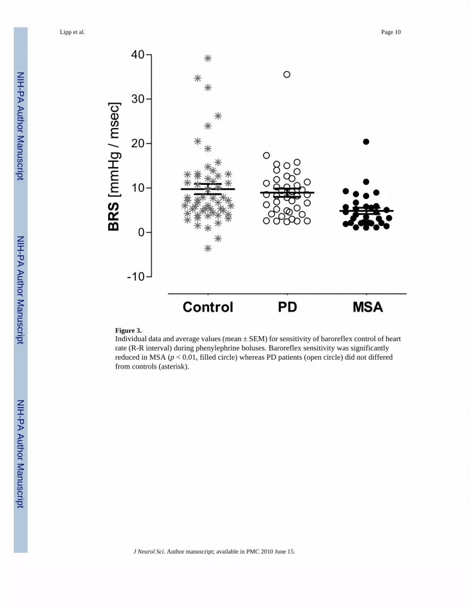
Figure 3.Individual data and average values (mean ± SEM) for sensitivity of baroreflex control of heartrate (R-R interval) during phenylephrine boluses. Baroreflex sensitivity was significantlyreduced in MSA (p < 0.01, filled circle) whereas PD patients (open circle) did not differedfrom controls (asterisk).
Lipp et al. Page 10
J Neurol Sci. Author manuscript; available in PMC 2010 June 15.
NIH
-PA Author Manuscript
NIH
-PA Author Manuscript
NIH
-PA Author Manuscript
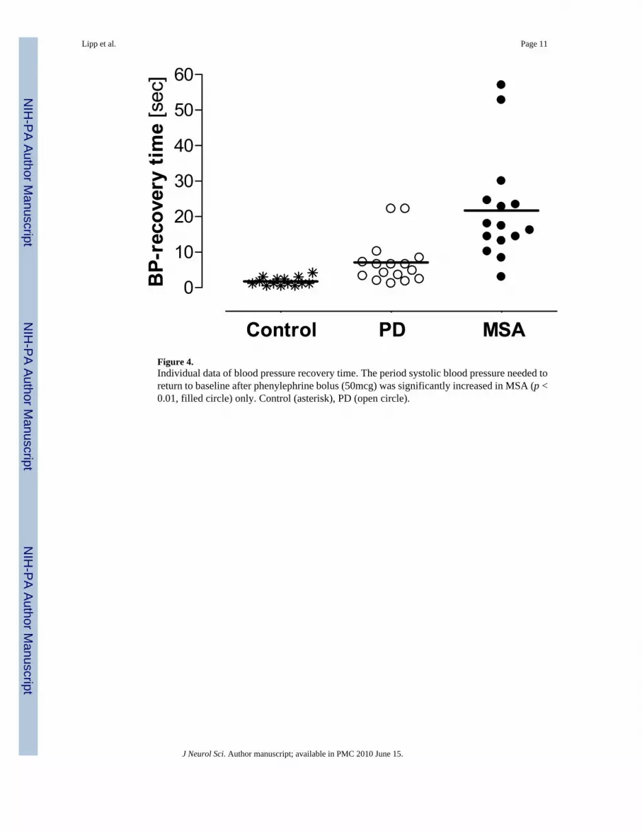
Figure 4.Individual data of blood pressure recovery time. The period systolic blood pressure needed toreturn to baseline after phenylephrine bolus (50mcg) was significantly increased in MSA (p <0.01, filled circle) only. Control (asterisk), PD (open circle).
Lipp et al. Page 11
J Neurol Sci. Author manuscript; available in PMC 2010 June 15.
NIH
-PA Author Manuscript
NIH
-PA Author Manuscript
NIH
-PA Author Manuscript
NIH
-PA Author Manuscript
NIH
-PA Author Manuscript
NIH
-PA Author Manuscript
Lipp et al. Page 12Ta
ble
1H
emod
ynam
ic p
aram
eter
s and
pla
sma
cate
chol
amin
e le
vels
at b
asel
ine
and
in re
spon
se to
vas
oact
ive
med
icat
ion
(mea
n ±
SE, u
npai
red
t-tes
t con
trols
vs.
patie
nts;
*PD
vs.
MSA
).
Para
met
ers
Uni
tsC
ontr
olPD
P(C
vs.
PD)
MSA
P(C
vs.
MSA
)
Bas
elin
e
N16
1615
Age
year
s60
± 2
.167
± 2
.2p
= 0.
0362
± 2
.1p
= 0.
5
Gen
der
f : m
6 : 1
07
: 97
: 8
BM
Ikg
/m2
27 ±
0.9
27 ±
1.2
p =
0.9
27 ±
0.7
p =
0.9
Dur
atio
nm
onth
52 ±
11
7 ±
3p
< 0.
01*
UM
SAR
Spo
ints
12.5
± 1
.622
.4 ±
2.2
p <
0.01
*
CA
SSpo
ints
1.4
± 0.
82.
8 ±
0.6
p <
0.01
5.1
± 0.
3p
< 0.
01
Phen
ylep
hrin
e 50
mcg
N14
1011
AU
CSB
Pm
mH
g/20
sec
147
± 18
288
± 38
p <
0.01
500
± 77
p <
0.01
BP-
reco
very
mse
c.38
± 3
63 ±
18
p =
0.18
147
± 35
7p
< 0.
01
Tyr
amin
e 30
mcg
/kg
N13
812
AU
CSB
Pm
mH
g/40
sec
472
± 50
354
± 44
p =
0.12
995
± 98
p <
0.01
ΔPla
sma
NE
pg/m
l+3
3 ±
130
± 19
p =
0.14
+41
± 19
p =
0.9
ΔPla
sma
DH
PGpg
/ml
+216
± 4
2+1
72 ±
58
p =
0.53
+218
± 4
1p
= 0.
83
J Neurol Sci. Author manuscript; available in PMC 2010 June 15.