synthesis of lamivudine stearate and antiviral activity of stearic acid-g-chitosan oligosaccharide...
TRANSCRIPT

This article appeared in a journal published by Elsevier. The attachedcopy is furnished to the author for internal non-commercial researchand education use, including for instruction at the authors institution
and sharing with colleagues.
Other uses, including reproduction and distribution, or selling orlicensing copies, or posting to personal, institutional or third party
websites are prohibited.
In most cases authors are permitted to post their version of thearticle (e.g. in Word or Tex form) to their personal website orinstitutional repository. Authors requiring further information
regarding Elsevier’s archiving and manuscript policies areencouraged to visit:
http://www.elsevier.com/copyright
Author's personal copy
European Journal of Pharmaceutical Sciences 41 (2010) 498–507
Contents lists available at ScienceDirect
European Journal of Pharmaceutical Sciences
journa l homepage: www.e lsev ier .com/ locate /e jps
Synthesis of Lamivudine stearate and antiviral activity of stearic acid-g-chitosanoligosaccharide polymeric micelles delivery system
Qian Lia,b, Yong-Zhong Dub, Hong Yuanb, Xing-Guo Zhangc, Jing Miaoc, Fu-De Cuia,∗, Fu-Qiang Hub,∗∗
a College of Pharmaceutical Sciences, Shenyang Pharmaceutical University, Shenyang 110016, PR Chinab College of Pharmaceutical Sciences, Zhejiang University, Yuhangtang 388, Hangzhou 310058, PR Chinac Department of Pharmacology, First Affiliated Hospital, Zhejiang University School of Medicine, Hangzhou 310003, PR China
a r t i c l e i n f o
Article history:Received 11 May 2010Received in revised form 9 August 2010Accepted 13 August 2010Available online 20 August 2010
Keywords:LamivudineEsterificationChitosan oligosaccharideStearic acidPolymeric micellesAnti-HBV activity
a b s t r a c t
To increase lipophilicity of water-soluble antiviral drug, the prodrug of Lamivudine (LA), Lamivudinestearate (LAS) was synthesized via ester linkage between LA and stearic acid. After the esterification,the octanol–water partition coefficient (log P) of LA increased from −0.95 to 1.82. Stearic acid-g-chitosanoligosaccharide (CSO-SA) micelles have demonstrated fast internalization and accumulation ability totumor cells. Herein, the CSO-SA with 3.79% amino substitution degree (SD) was prepared for loading LAS.The critical micelle concentration (CMC) of CSO-SA was about 0.032 mg/ml. The micelles with 1 mg/mlCSO-SA concentration had 460.8 nm average diameters with a narrow size distribution and 29.7 mVsurface zeta potential. After LAS was incorporated, the micellar size decreased and the zeta potentialincreased. The LAS loaded CSO-SA micelles (CSO-SA/LAS) possessed high entrapment efficiency and drugloading. LA release from CSO-SA/LAS showed a pH-dependent behavior. The release rate of LA fromCSO-SA/LAS increased significantly as the pH of release medium reduced from 7.4 to 6.2. CSO-SA/LASpresented a low cytotoxicity and high cellular uptake percentage of LAS against HBV transfected tumorcells (HepG2.2.15). In vitro anti-HBV activities of CSO-SA/LAS presented more conspicuous inhibitoryeffects on antigen expression and DNA replication compared with LA and LAS.
© 2010 Elsevier B.V. All rights reserved.
1. Introduction
Despite a high rate of viral clearance by immunocompetentadults and the availability of efficient vaccines, chronic infectionwith hepatitis B virus (HBV) poses a huge health burden on theglobal community (Hoofnagle, 1990). The coexistence of repeatedcycles of HBV replication and immune lysis of infected hepatocytesis associated with fibrosis, cirrhosis, and hepatocellular carcinoma(Raney et al., 2003).
Current strategies for treating hepatitis B have focused on clear-ing active HBV infection through suppression of viral replication.Antiviral agents, particularly nucleoside/nucleotide analogues, arequite effective inhibitors of HBV replication. Lamivudine (LA)mainly targets nucleus of host cells and inhibits reverse transcrip-tase to terminate HBV replication (Asselah et al., 2005). Developedas the first therapeutical agent for oral use in the therapy of HBVinfection, LA obtains significant depressant effect via short-termtreatment. However, relapse is easily triggered after drug with-
∗ Corresponding author. Tel.: +86 24 23986353; fax: +86 24 23986353.∗∗ Corresponding author. Tel.: +86 571 88208439; fax: +86 571 88208439.
E-mail addresses: [email protected] (F.-D. Cui), [email protected] (F.-Q. Hu).
drawal and drug resistance is most likely to be generated afterlong-term medication (Craxi et al., 2006). To date both in vitro andin vivo experiments have demonstrated that the viral inhibitoryextent plays a pivotal role in evading resistant occurrence, whichcan also be avoided potentially by depressing the viral load tounmeasured level (Shaw et al., 2006).
There are some restrictions of antiviral chemotherapeutics inapplications, such as non-specific distribution in vivo, enzymicmetabolism, difficulties in transporting through biological mem-brane and getting into the molecular targets abundantly (Langer,1998), which results in low therapeutic effects, large doses,adverse reactions and drug tolerance. Accordingly, through target-ing pharmaceutics engineering, delivering the exogenous agentsto molecular targets can considerably enhance the efficacy of drugaction and reduce side effects towards normal organs. Previousresearch has indicated that the molecular action sites of anti-HBVchemotherapeutics are mostly at subcellular organelles of hostcells besides that of HBV absorption inhibitors (Zoulim and Perrillo,2008). Thus the vehicle technology targeting subcellular organelleshas become a chief link of achieving molecular targeted therapynowadays (Vladimir and Torchilin, 2008). So far, much researchhas been focused on the intracellular delivery through particu-late delivery systems and achieved perinuclear accumulation of
0928-0987/$ – see front matter © 2010 Elsevier B.V. All rights reserved.doi:10.1016/j.ejps.2010.08.004

Author's personal copy
Q. Li et al. / European Journal of Pharmaceutical Sciences 41 (2010) 498–507 499
nanoparticles after internalization in cells (Chawla and Amiji, 2002;Desai et al., 1997; Lai et al., 2007). However, the fast release rateand perinuclear accumulation of antiviral agent are not conduciveto in vivo transport to target the cells without drug leaking and drugaccumulation at molecular action site (Cavalli et al., 2009).
In previous research, the amphipathic cationic graft copolymerswere synthesized by coupling reaction between the amino groupsof chitosan oligosaccharide (CSO) and carboxyl group of stearicacid (SA). They were able to self-assemble into micelles in aque-ous medium and encapsulate chemotherapeutical drugs (You etal., 2007a,b, 2008b) or biomacromolecule (Hu et al., 2006a,c). More-over, these micelles possessed rapid cellular uptake ability. It hasbeen reported that stearic acid grafted chitosan oligosaccharidepolymeric micelles possess subcellular organelles target abilities(You et al., 2008a, 2007a).
In most cases, hydrophobic drugs are incorporated into thehydrophobic core of micelles by hydrophobic interaction and otheradditional interactions such as metal–ligand coordination bond-ing (Yokoyama et al., 1996; Nishiyama et al., 2001), electrostaticinteraction (Kabanov et al., 1996). Thus undoubtedly, the tenu-ous hydrophobicity of LA should be adjusted. In this paper, theprodrug of LA, Lamivudine stearate (LAS) was synthesized via theesterification between stearic acid and LA. Stearate-grafted chi-tosan oligosaccharides (CSO-SA) with low substitution degree (SD)was synthesized as an intracellular delivery carrier for LAS, andthe properties of formed micelles were investigated. Based on thephysiochemical properties of LAS and LAS loaded micelles, the invitro antiviral activities of LAS loaded CSO-SA micelles against HBVtransfected human hepatoblastoma cells (HepG2.2.15) were inves-tigated compared with those of LA and LAS.
2. Materials and methods
2.1. Materials
Lamivudine (LA) was from Hangzhou Coben Pharmaceuti-cal Co., Ltd.; 1,3-dicyclohexylcarbodi-achtungtrenungimide (DCC)and 4-dimethylamino-pyridine (DMAP) were provided by Shang-hai Medpep Co., Ltd.; chitosan oligosaccharide (CSO) with about18.0 kDa weight average molecular weight was obtained by enzy-matic degradation of 95% deacetylated chitosan (Mw = 45.0 kDa),which was supplied by Yuhuan Marine Biochemistry Co., Ltd.(Zhejiang, China) (You et al., 2007a; Ye et al., 2008). Stearic acid(SA, 99%), 1-ethyl-3-(3-dimethylaminopropyl) carbodiimide (EDC),and 2,4,6-trinitrobenzene sulfonic acid (TNBS) were purchasedfrom Sigma (St. Louis, MO, USA); 3-[4,5-dimethylthiazol-2-yl]-2,5-diphenyltetrazolium bromide (MTT) was purchased from Sigma(St. Louis, MO, USA); Trypsin, Dulbecco’s modified Eagle’s medium(DMEM) and G418 were purchased from GIBCO BRL (Gaithersberg,MD, USA). Fetal bovine serum (FBS) was purchased from SijiqingBiologic (Hangzhou, China). All other solvents were of analytical orchromatographic grade.
2.2. Synthesis of Lamivudine stearate (LAS) (Xue et al., 2003)
LA, SA, DCC and DMAP were dried under reduced pressurewith slight heating for 20 min. 1.29 g (5.6 mmol) LA, 2.778 g(13.44 mmol) DCC and 142 mg (1.12 mmol) DMAP were dissolvedin 50 ml dry dichloromethane, and 3.18 g (11.2 mmol) SA wasdissolved in 50 ml dry dichloromethane, respectively. After the SAsolution was dropped into another dichloromethane solution, thereaction was carried out at 30 ◦C under the protection of nitrogenwith 400 rpm agitation for 4 days (EMS-8C, Shanghai LongtuoInstrument and Equipment Co., Ltd., China). The dichloromethanein final reaction mixture was removed by atmospheric distillation.
Then the crude compound was checked by thin-layer chromatog-raphy (TLC) method and purified by column chromatographymethod over silica gel using methanol/dichloromethane (1:20,v/v) as the eluent.
2.3. Characteristics of Lamivudine stearate
The octanol–water partition coefficients of LA and LAS weredetermined by shake-flask method (Lien and Ren, 2002). Waterphase and n-octyl alcohol phase were mutually saturated by shak-ing treatment in a thermostatic water bath at 25 ◦C for 24 h, andthen stood for phase separation. The LA and LAS were then dis-solved into n-octyl alcohol-saturated water and water-saturatedn-octyl alcohol, respectively. 2.5 ml LA or LAS solution was mixedwith same volume of water-saturated n-octyl alcohol or n-octylalcohol-saturated water, respectively. After 2 h intensive shakingat room temperature to reach the partitioning equilibrium, thewater phase was separated and centrifuged at 2000 rpm for 10 min.The concentration of the solute in water phase was determined byhigh-performance liquid chromatography (HPLC) method.
The stability of LAS at different pH was determined, 0.03 mg/mlLAS PBS solutions with different pH (5.0, 6.2, 7.4 and 8.0) wereprepared by adding 1 ml of 0.3 mg/ml LAS solution (in DMSO) to9 ml preheated 0.01 M phosphate-buffered saline (PBS), and keptat 37 ◦C in a water bath for 96 h. HPLC method was used to ana-lyze the LAS content at intervals. Furthermore, the stability ofLAS in PBS (pH 7.4, 0.01 M) solution containing 10% (w/w) fetalbovine serum (FBS) was investigated. LAS solution was preparedby adding 1 ml of 0.3 mg/ml LAS solution (in DMSO) to 9 ml pre-heated PBS (pH 7.4, 0.01 M) solution containing 10% (w/w) fetalbovine serum (FBS), and kept at 37 ◦C in a water bath for 48 h.At predefined time intervals, 0.1 ml LAS solution was taken, anddiluted by 0.9 ml methanol followed by vortex (1 min) and cen-trifugation (10 000 rpm for 10 min, 3K30, SIGMA LabrorzentrifugenGmbH, Germany). The concentration of LAS in the supernatant wasdetermined by HPLC method.
Melting points were determined in one end open capillarytubes on a melting point apparatus (Buchi B-540, Switzerland).Infrared (IR) and proton nuclear magnetic resonance (1H NMR)spectra were recorded for the compounds on IR spectrometer (vec-tor22, Bruker, Germany) and NMR spectrometer (AC-80, BrukerBiospin, Germany), respectively. LA, LAS and SA were dissolvedin dimethylsulfoxide-d6 with 1 wt%. Electrospray ionization massspectrometry (ESI-MS) was performed with a MS spectrometer(Esquire-LC-00075). LA and LAS were dissolved in methanol matrix.Mass spectra were then recorded in the m/z 1000–50 range.
2.4. Synthesis of stearic acid grafted chitosan oligosaccharide
Chitosan oligosaccharide (CSO) was obtained by enzymaticdegradation of 95% deacetylated chitosan (Mw = 450.0 kDa) accord-ing to a previous study (Hu et al., 2006b). The chemical conjugateof CSO-SA was then synthesized by coupling carboxyl group ofSA and amine group of CSO in the presence of 1-ethyl-3-(3-dimethylaminopropyl) carbodiimide (EDC) (Hu et al., 2008). Briefly,500 mg CSO was dissolved in 20 ml deionized water (DI water), and200 mg SA was dissolved in 20 ml hot ethanol, respectively. Theywere then mixed at 80 ◦C under stirring. After 930 mg EDC wasadded into the mixture, the coupling reaction was carried out for 5 hat 80 ◦C under stirring with 250 rpm. The final reaction mixture wasdialyzed against 10% (v/v) ethanol aqueous solution using a dialysismembrane (MWCO: 3.5 kDa, Spectrum Laboratories, Laguna Hills,CA) for 48 h with frequent exchange of fresh 10% (v/v) ethanol aque-ous solution to remove by-products. Finally, the dialyzed productwas lyophilized (LABCONCO, FreeZone 2.5 Plus, USA).
Author's personal copy
500 Q. Li et al. / European Journal of Pharmaceutical Sciences 41 (2010) 498–507
1H NMR spectrum of obtained CSO-SA was acquired on a NMRspectrometer (AC-80, Bruker Biospin, Germany) in D2O with 1 wt%.
2.5. Characteristics of stearic acid grafted chitosanoligosaccharide
The substitution degree (SD) of CSO-SA, defined as the num-ber of SA groups per 100 amino groups of CSO, was determinedby the TNBS method (Bernkop-Schnurch and Krajicek, 1998). 2 mlof 4% NaHCO3 and 2 ml of 0.1% TNBS solution were added into2 ml of CSO-SA solution with 125 �g/ml CSO-SA, and the mix-ture was incubated for 2 h at 37 ◦C. 2 ml of 2 N HCl solution wasthen added into the mixture to neutralize the remaining NaHCO3.The final reaction mixture was examined by UV spectroscopy (TU-1800PC, Beijing Purkinje General Instrument Co., Ltd., China) withthe absorbance wavelength set at 344 nm. The SD of CSO-SA wascalculated from a calibration curve, which was obtained from theCSO solution.
The critical micelle concentration (CMC) of synthesized CSO-SA micelles was determined by fluorescence measurement usingpyrene as a probe (Lee et al., 1998). The fluorescence emission spec-tra of pyrene in different concentrations (1 × 10⁻³ to 0.5 mg/ml)of CSO-SA micelles were measured using fluorometer (F-2500,HITACHI Co., Japan), with the excitation wavelength set at 334 nm,the excitation slit at 10 nm and emission slit at 2.5 nm. From thepyrene emission spectra, the intensity ratio of the first peak (I1,374 nm) to the third peak (I3, 384 nm) was determined for thecalculation of CMC.
2.6. Preparation of LAS loaded CSO-SA micelles
LAS loaded CSO-SA micelles (CSO-SA/LAS) were prepared bydialysis method. CSO-SA (5 mg) was dissolved in 2.5 ml water. Acertain amount of LAS was dissolved in dimethyl sulfoxide (DMSO)and then added into CSO-SA solution. The mixed solution wasprobe-type ultrasonicated for 30 times (active every 2 s for a 3 sduration) in ice bath (400 W, JY92-II, Scientz Biotechnology Co.,Ltd., China), followed by dialysis (MWCO: 7 kDa, Spectrum Labora-tories, Laguna Hills, CA) against pure water (Milli-Q, Millipore, USA)overnight at room temperature with frequent exchange of freshlydistilled water. After dialysis, the micelle solution was diluted to5 ml and lyophilized.
2.7. Particle size and zeta potential of CSO-SA micelles and LASloaded CSO-SA micelles
The hydrodynamic diameter of CSO-SA micelles and LAS loadedCSO-SA micelles (CSO-SA/LAS) with CSO-SA concentration of100 �g/ml in DI water were measured by dynamic light scattering(DLS) using a Zetasizer (3000 HS, Malvern Instruments Ltd., UK).The zeta potential of the micelles with the same concentration wasdetected in DI water solution by the Zetasizer.
2.8. AFM investigation
The surface morphologies of CSO-SA micelles and CSO-SA/LASin DI water were observed by an atomic force microscopy (SPA3800N, SEIKO, Japan). Explorer atomic force microscope was atapping mode, using high resonant frequency (F0 = 129 kHz), pyra-midal cantilevers with silicon probes having force constants of20 N/m. Scan speed was set at 2 Hz.
Herein, the drug loading of LAS loaded micelles was16.77 ± 0.34%, and the concentration of CSO-SA was 100 �g/ml.
2.9. Content determination of LA and LAS
The concentration of LA and LAS was determined by HPLCmethod (1100 series, Agilent, USA). The mobile phase was amixture of methanol and water. The column was a DiamonsilC18 (250 mm × 4.6 mm) with 5 �m particles. The flow rate was0.75 ml/min, the detection wavelength was 270 nm, and the col-umn temperature was 25 ◦C. 20 �l sample was injected into thecolumn. The program of mobile phase was conducted as follows:the ratio of the volume of methanol to water varied from 70:30to 95:5 after 4.5 min, and then returned to original ratio (70:30)at 25.5 min. The line relationships of calibration curves for LA andLAS were y = 60.899x + 3.4718 and y = 25.896x + 2.1848, where theconcentration range of LA was 2.5–25 �g/ml and LAS 1.6–40 �g/ml(R2 = 0.9993 and 0.9997, y was the peak area and x was the concen-tration), the limit of detection was 0.01 �g/ml.
2.10. Measurement of entrapment efficiency and drug loading
The CSO-SA/LAS solution (0.4 ml) was placed in ultrafilter tubewith molecular weight cutoff of 10 kDa (MicroconYM-10, Milli-pore Co., Bedford, USA) and centrifuged at 10 000 rpm for 15 min(3K30, SIGMA Labrorzentrifugen GmbH, Germany). The LAS con-tent in filtrate (Cf) was measured. 0.9 ml methanol was added into0.1 ml CSO-SA/LAS solution, and the mixture was ultrasonicatedfor 10 min to extract all the LAS. The mixture was centrifugedat 10 000 rpm for 10 min (3K30, SIGMA Labrorzentrifugen GmbH,Germany) and the LAS content (Ct) in the supernatant was alsodetermined. The concentration of LAS was determined by HPLCmethod. The drug entrapment efficiency (EE, %) and drug loading(DL, %) were then calculated by the following equations:
EE (%) = (Ct − Cf )Ct
× 100 (1)
DL (%) = (Ct − Cf )(Ct − Cf + Cm)
× 100 (2)
where Cm represented the concentration of CSO-SA in 0.1 ml drugloaded micelles solution.
2.11. Stability of LAS in CSO-SA micelles
A series of CSO-SA/LAS at different pH (5.0, 6.2, 7.4 and 8.0) wereprepared by dissolving the freeze-dried CSO-SA/LAS in differentPBS (pH 5.0, 6.2, 7.4 and 8.0) and keeping the obtained CSO-SA/LASsolution at 37 ◦C in water bath for 96 h. The concentrations of LASand CSO-SA micelles were 30 �g/ml and 100 �g/ml, respectively.Furthermore, the stability of LAS encapsulated in CSO-SA micellesin PBS (pH 7.4, 0.01 M) solution containing 10% FBS was investi-gated by dissolving the freeze-dried CSO-SA/LAS in PBS solutioncontaining 10% FBS, and keeping the obtained CSO-SA/LAS solu-tion at 37 ◦C in water bath for 48 h. At different time intervals,0.1 ml sample solution was diluted by 0.9 ml methanol, and themixture was ultrasonicated for 10 min to extract LAS. The mixturewas centrifuged at 10 000 rpm for 10 min, and the LAS content inthe supernatant was determined by HPLC method.
2.12. In vitro LA release from micelles
In vitro cumulative release of LA from micelles was investigatedby using 0.01 M phosphate-buffered saline containing 0.5% sodiumdodecyl sulfate (pH 6.2 or pH 7.4) as the dissolution medium. Thefreeze-dried CSO-SA/LAS were weighed, dissolved in 1 ml of dis-tilled water, and introduced into dialysis membrane bag (MWCO:
Author's personal copy
Q. Li et al. / European Journal of Pharmaceutical Sciences 41 (2010) 498–507 501
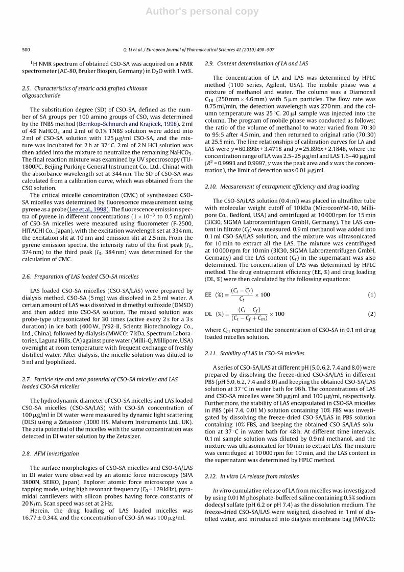
Fig. 1. Synthetic route of Lamivudine stearate.
7 kDa, Spectrum Laboratories, Laguna Hills, CA). The release exper-iment was initiated by placing the end-sealed dialysis bag in 26 mldissolution medium, and shaken at 65 rpm at 37 ◦C. At predefinedtime points, the medium was replaced completely, and the LAconcentrations in dissolution medium were determined by HPLCmethod. All release tests were performed thrice.
2.13. In vitro cytotoxicity assay
HBV transfected human hepatoblastoma cells (HepG2.2.15)were kindly provided by the State Key Lab for Diagnosis and Treat-ment of Infectious Diseases, Zhejiang University, China. This cellline was cultured in the air containing 5% (v/v) CO2 at 37 ◦C inDMEM supplemented with 10% (w/w) fetal bovine serum and500 �g/ml G418.
Cytotoxicity of LA, LAS, CSO-SA micelles and CSO-SA/LAS againstHepG2.2.15 cell line was evaluated by MTT assay (Zhang et al.,2007). Briefly, 1.0 × 10⁵ cells were seeded in 24-well plates andincubated for 24 h to allow the cells to attach. The cells wereexposed to serial concentrations of LA, LAS, CSO-SA micelles andCSO-SA/LAS at 37 ◦C for 72 h. After the incubation, 60 �l of MTTsolution was added and incubated for further 4 h, and then themedium was replaced with 400 �l of DMSO. The absorbance wasread on a Sunrise absorbance microplate reader (Bio-Rad, Model680, USA) at dual wavelength of 570 nm. The data reported repre-sented the means of triple measurement.
2.14. Quantification of cellular uptake
1.0 × 10⁵ HepG2.2.15 cells were seeded in 24-well plates andgrown for 24 h to allow the cells to attach. The cells were exposedto LA, LAS and CSO-SA/LAS diluted in culture medium (the concen-trations of LA, LAS and CSO-SA were 40 nmol/ml, 40 nmol/ml and100 �g/ml) at 37 ◦C. At predetermined time, the cells were washedthree times with ice-cold phosphate buffers (pH 7.4) to termi-nate the uptake and remove the free LA, LAS or CSO-SA/LAS whichwere adsorbed on the cell membrane. The cells were collectedby treatment with 50 �l trypsin (2.5 mg/ml) for 5 min and dilutedwith 450 �l methanol, followed by water-bath sonication underairproof and ice bath conditions. Subsequently, the supernatantwas obtained using hypothermal centrifugation at 22 000 × g for10 min. The concentration of LA and LAS in supernatant was deter-mined by HPLC method.
2.15. Virological assessment
1.0 × 10⁵ HepG2.2.15 cells were seeded in 24-well plates andincubated for 24 h to allow the cells to attach. The LA, LAS, CSO-SAmicelles or CSO-SA/LAS were added with the given concentrationsand incubated for 3 days, 6 days or 9 days. The culture mediafrom the wells were collected for the virological assessment. HBsAgand HBeAg productions were determined by commercial enzymeimmunoassay kits (AXSYM System, Abbott, Wiesbaden, Germany).
HBV DNA was quantified by a commercial real-time polymerasechain reaction (PCR) kit (PG Biotech, Shenzhen, China). All theexperiments were performed in triplicate.
3. Results
3.1. Synthesis of Lamivudine stearate (LAS)
LAS was synthesized via the reaction of carboxyl group of SA andhydroxyl group of LA in the presence of DCC and DMAP. The syn-thetic route of Lamivudine stearate is shown in Fig. 1. The purity ofthe synthesized compounds was checked by TLC and the objectivecomponent I of this study was identified by spectral data as LAS.
LAS yield: 34.2%; m.p.: 115–117 ◦C; IR (KBr): 3352, 3100, 2917,2849, 1743, 1626, 1486, 1366, 1286, 1165, 1097 cm−1; 1H NMR(dimethylsulfoxide-d6) ı (ppm): 7.70 (d, J = 7 Hz, 1H), 7.26 (d,J = 33 Hz, 2H), 6.23 (t, J = 5.5 Hz, 1H), 5.75 (d, J = 7.5 Hz, 1H), 5.36(dd, J1 = 3.5 Hz, J2 = 5 Hz, 1H), 4.33–4.42 (m, 2H), 3.44 (dd, J1 = 5.5 Hz,J2 = 12 Hz, 1H), 3.10 (dd, J1 = 5.5 Hz, J2 = 11.5 Hz, 1H), 2.34 (t, J = 7 Hz,2H), 1.53 (t, J1 = 7 Hz, 2H), 1.23 (s, 26H), 1.13 (s, 2H), 0.86 (t, J = 6.5 Hz,3H); ESI-MS: m/z = 496.31 [M+1]+.
LA m.p.: 160–162 ◦C; 1H NMR (dimethylsulfoxide-d6) ı (ppm):7.80 (d, J = 7.5 Hz, 1H), 7.22 (d, J = 35 Hz, 2H), 6.21 (t, J = 5.2 Hz, 1H),5.74 (d, J = 7.5 Hz, 1H), 5.29 (t, J = 5.9 Hz, 1H), 5.17 (d, J = 4.6 Hz, 1H),3.71–3.77 (m, 2H), 3.41 (dd, J1 = 5.5 Hz, J2 = 11.7 Hz, 1H), 3.05 (dd,J1 = 5.0 Hz, J2 = 11.7 Hz, 1H); ESI-MS: m/z = 230.30 [M+1]+, 459.03[2M+1]+.
SA m.p.: 68–70 ◦C; 1H NMR (dimethylsulfoxide-d6) ı (ppm):11.91 (s, 1H), 2.18 (t, J = 7.5 Hz, 2H), 1.48 (t, J = 7.5 Hz, 2H), 1.24(s, 28H), 0.86 (t, J = 7 Hz, 3H); ESI-MS: m/z = 283.47 [M−1]+, 567.13[2M−1]+.
The IR spectrum of LAS (Fig. 2) showed amino group peak at3352 cm−1, C O bond of easter peak at 1743 cm−1, and C–O bondof easter peak at 1097 cm−1. In the 1H NMR spectra (Fig. 3), the
Fig. 2. IR spectrum of LAS.
Author's personal copy
502 Q. Li et al. / European Journal of Pharmaceutical Sciences 41 (2010) 498–507
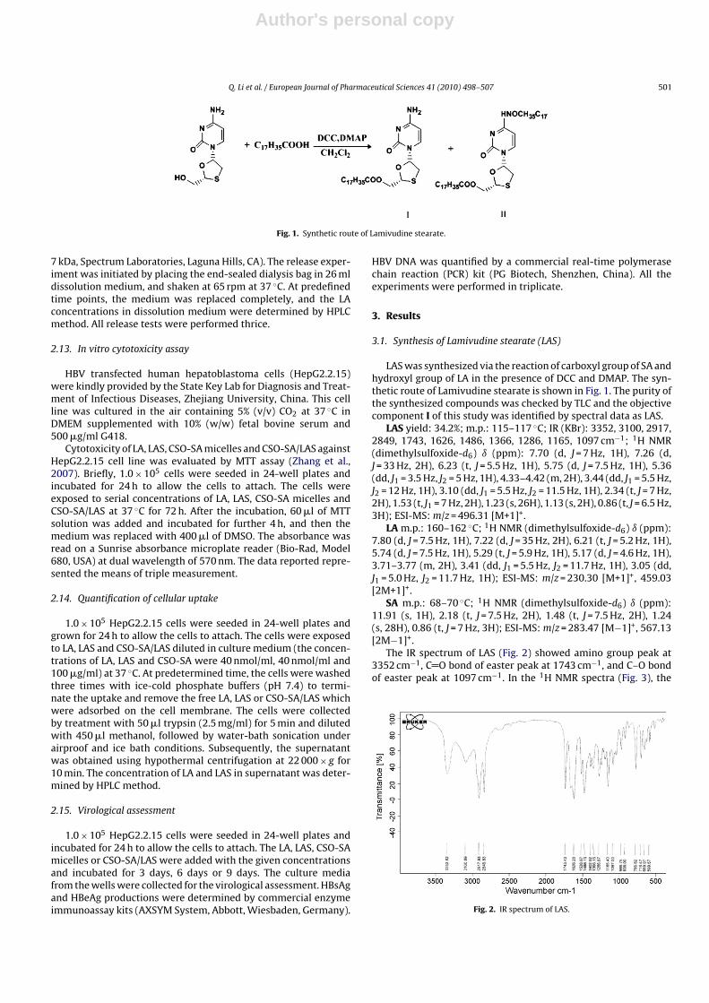
Fig. 3. 1H NMR spectra of LAS (A); LA (B); SA (C); CSO-SA (D). The important peakswere pointed out.
signals of the respective protons of LAS, LA and SA were verifiedon the basis of their chemical shifts, multiplicities and couplingconstants. The spectrum of LAS (Fig. 3A) presented a doublet peakat ı7.26 ppm corresponding to amino group and a multiplet peakat ı4.33–4.42 ppm for the protons of 6′-CH2. The spectrum of LA(Fig. 3B) showed a triplet peak at ı5.17 ppm (J = 4.5 Hz, 1H) forhydroxy group and a multiplet peak at ı3.71–3.77 ppm (2H) forthe protons of 6′-CH2. The spectrum of SA (Fig. 3C) showed a sin-glet at ı11.91 ppm (1H) attributed to the proton of carboxyl group.It was found that the peaks of carboxyl group and hydroxy groupdisappeared in the spectrum of LAS, and the peaks for the protonsof 6′-CH2 in Fig. 3A shifted to low magnetic field in comparisonwith those for the protons of the same group in Fig. 3B. Moreover,LAS was further confirmed with mass spectrometry and exhibitedan identical mass value corresponding to the mass values of LA andSA. In summary, LAS was the prospective compound which wassynthesized via the esterification between the carboxyl group ofSA and the hydroxyl group of LA.
The octanol–water partition coefficients (log P) of LA and LASwere determined by shake-flask method. The shake-flask methodwas used commonly in the measurement of partition coeffi-cients. It was found that the log P of LAS and LA were 1.82 ± 0.10and −0.95 ± 0.03, respectively. These results indicated that thehydrophobicity of LAS was significantly enhanced by esterificationin comparison with that of LA.
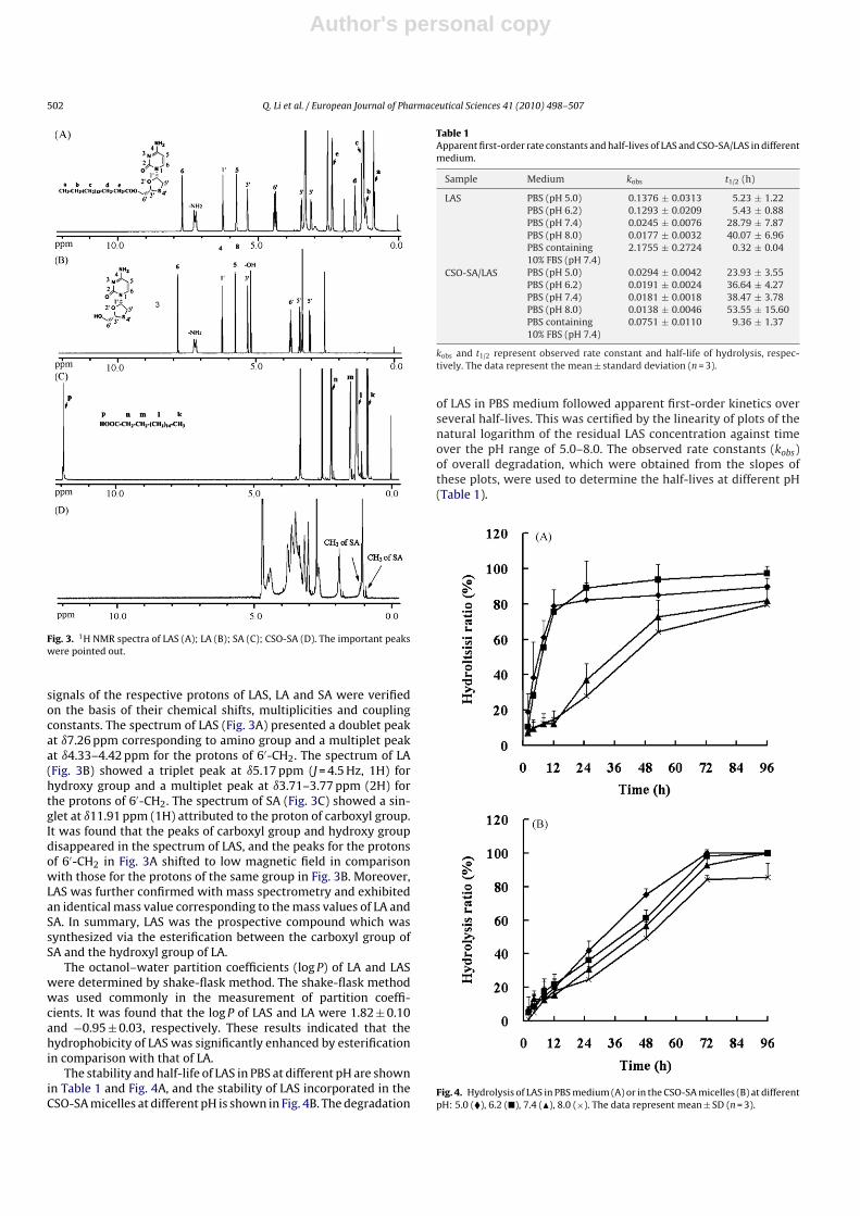
The stability and half-life of LAS in PBS at different pH are shownin Table 1 and Fig. 4A, and the stability of LAS incorporated in theCSO-SA micelles at different pH is shown in Fig. 4B. The degradation
Table 1Apparent first-order rate constants and half-lives of LAS and CSO-SA/LAS in differentmedium.
Sample Medium kobs t1/2 (h)
LAS PBS (pH 5.0) 0.1376 ± 0.0313 5.23 ± 1.22PBS (pH 6.2) 0.1293 ± 0.0209 5.43 ± 0.88PBS (pH 7.4) 0.0245 ± 0.0076 28.79 ± 7.87PBS (pH 8.0) 0.0177 ± 0.0032 40.07 ± 6.96PBS containing10% FBS (pH 7.4)
2.1755 ± 0.2724 0.32 ± 0.04
CSO-SA/LAS PBS (pH 5.0) 0.0294 ± 0.0042 23.93 ± 3.55PBS (pH 6.2) 0.0191 ± 0.0024 36.64 ± 4.27PBS (pH 7.4) 0.0181 ± 0.0018 38.47 ± 3.78PBS (pH 8.0) 0.0138 ± 0.0046 53.55 ± 15.60PBS containing10% FBS (pH 7.4)
0.0751 ± 0.0110 9.36 ± 1.37
kobs and t1/2 represent observed rate constant and half-life of hydrolysis, respec-tively. The data represent the mean ± standard deviation (n = 3).
of LAS in PBS medium followed apparent first-order kinetics overseveral half-lives. This was certified by the linearity of plots of thenatural logarithm of the residual LAS concentration against timeover the pH range of 5.0–8.0. The observed rate constants (kobs)of overall degradation, which were obtained from the slopes ofthese plots, were used to determine the half-lives at different pH(Table 1).
Fig. 4. Hydrolysis of LAS in PBS medium (A) or in the CSO-SA micelles (B) at differentpH: 5.0 (�), 6.2 (�), 7.4 (�), 8.0 (×). The data represent mean ± SD (n = 3).
Author's personal copy
Q. Li et al. / European Journal of Pharmaceutical Sciences 41 (2010) 498–507 503
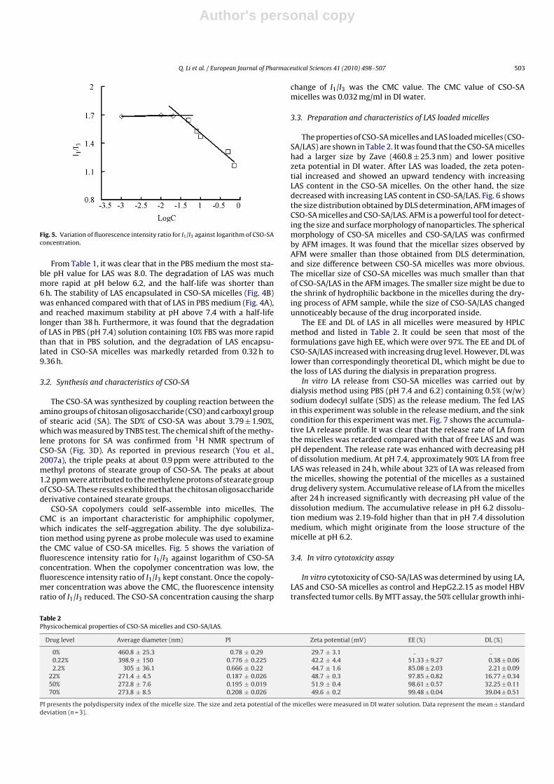
Fig. 5. Variation of fluorescence intensity ratio for I1/I3 against logarithm of CSO-SAconcentration.
From Table 1, it was clear that in the PBS medium the most sta-ble pH value for LAS was 8.0. The degradation of LAS was muchmore rapid at pH below 6.2, and the half-life was shorter than6 h. The stability of LAS encapsulated in CSO-SA micelles (Fig. 4B)was enhanced compared with that of LAS in PBS medium (Fig. 4A),and reached maximum stability at pH above 7.4 with a half-lifelonger than 38 h. Furthermore, it was found that the degradationof LAS in PBS (pH 7.4) solution containing 10% FBS was more rapidthan that in PBS solution, and the degradation of LAS encapsu-lated in CSO-SA micelles was markedly retarded from 0.32 h to9.36 h.
3.2. Synthesis and characteristics of CSO-SA
The CSO-SA was synthesized by coupling reaction between theamino groups of chitosan oligosaccharide (CSO) and carboxyl groupof stearic acid (SA). The SD% of CSO-SA was about 3.79 ± 1.90%,which was measured by TNBS test. The chemical shift of the methy-lene protons for SA was confirmed from 1H NMR spectrum ofCSO-SA (Fig. 3D). As reported in previous research (You et al.,2007a), the triple peaks at about 0.9 ppm were attributed to themethyl protons of stearate group of CSO-SA. The peaks at about1.2 ppm were attributed to the methylene protons of stearate groupof CSO-SA. These results exhibited that the chitosan oligosaccharidederivative contained stearate groups.
CSO-SA copolymers could self-assemble into micelles. TheCMC is an important characteristic for amphiphilic copolymer,which indicates the self-aggregation ability. The dye solubiliza-tion method using pyrene as probe molecule was used to examinethe CMC value of CSO-SA micelles. Fig. 5 shows the variation offluorescence intensity ratio for I1/I3 against logarithm of CSO-SAconcentration. When the copolymer concentration was low, thefluorescence intensity ratio of I1/I3 kept constant. Once the copoly-mer concentration was above the CMC, the fluorescence intensityratio of I1/I3 reduced. The CSO-SA concentration causing the sharp
change of I1/I3 was the CMC value. The CMC value of CSO-SAmicelles was 0.032 mg/ml in DI water.
3.3. Preparation and characteristics of LAS loaded micelles
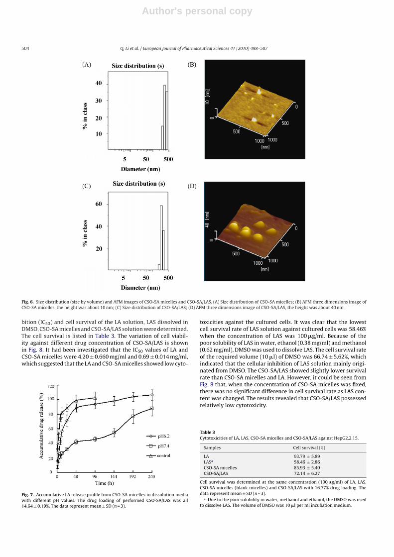
The properties of CSO-SA micelles and LAS loaded micelles (CSO-SA/LAS) are shown in Table 2. It was found that the CSO-SA micelleshad a larger size by Zave (460.8 ± 25.3 nm) and lower positivezeta potential in DI water. After LAS was loaded, the zeta poten-tial increased and showed an upward tendency with increasingLAS content in the CSO-SA micelles. On the other hand, the sizedecreased with increasing LAS content in CSO-SA/LAS. Fig. 6 showsthe size distribution obtained by DLS determination, AFM images ofCSO-SA micelles and CSO-SA/LAS. AFM is a powerful tool for detect-ing the size and surface morphology of nanoparticles. The sphericalmorphology of CSO-SA micelles and CSO-SA/LAS was confirmedby AFM images. It was found that the micellar sizes observed byAFM were smaller than those obtained from DLS determination,and size difference between CSO-SA micelles was more obvious.The micellar size of CSO-SA micelles was much smaller than thatof CSO-SA/LAS in the AFM images. The smaller size might be due tothe shrink of hydrophilic backbone in the micelles during the dry-ing process of AFM sample, while the size of CSO-SA/LAS changedunnoticeably because of the drug incorporated inside.
The EE and DL of LAS in all micelles were measured by HPLCmethod and listed in Table 2. It could be seen that most of theformulations gave high EE, which were over 97%. The EE and DL ofCSO-SA/LAS increased with increasing drug level. However, DL waslower than correspondingly theoretical DL, which might be due tothe loss of LAS during the dialysis in preparation progress.
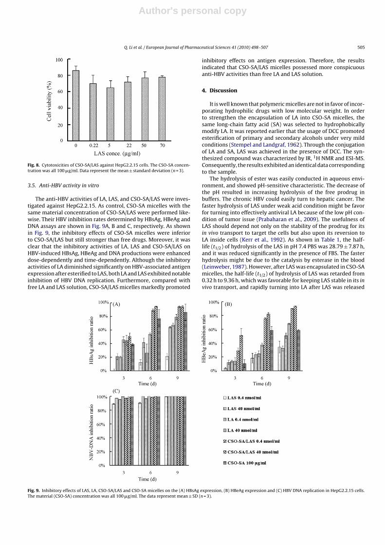
In vitro LA release from CSO-SA micelles was carried out bydialysis method using PBS (pH 7.4 and 6.2) containing 0.5% (w/w)sodium dodecyl sulfate (SDS) as the release medium. The fed LASin this experiment was soluble in the release medium, and the sinkcondition for this experiment was met. Fig. 7 shows the accumula-tive LA release profile. It was clear that the release rate of LA fromthe micelles was retarded compared with that of free LAS and waspH dependent. The release rate was enhanced with decreasing pHof dissolution medium. At pH 7.4, approximately 90% LA from freeLAS was released in 24 h, while about 32% of LA was released fromthe micelles, showing the potential of the micelles as a sustaineddrug delivery system. Accumulative release of LA from the micellesafter 24 h increased significantly with decreasing pH value of thedissolution medium. The accumulative release in pH 6.2 dissolu-tion medium was 2.19-fold higher than that in pH 7.4 dissolutionmedium, which might originate from the loose structure of themicelle at pH 6.2.
3.4. In vitro cytotoxicity assay
In vitro cytotoxicity of CSO-SA/LAS was determined by using LA,LAS and CSO-SA micelles as control and HepG2.2.15 as model HBVtransfected tumor cells. By MTT assay, the 50% cellular growth inhi-
Table 2Physicochemical properties of CSO-SA micelles and CSO-SA/LAS.
Drug level Average diameter (nm) PI Zeta potential (mV) EE (%) DL (%)
0% 460.8 ± 25.3 0.78 ± 0.29 29.7 ± 3.10.22% 398.9 ± 150 0.776 ± 0.225 42.2 ± 4.4 51.33 ± 9.27 0.38 ± 0.062.2% 305 ± 36.1 0.666 ± 0.22 44.7 ± 1.6 85.08 ± 2.03 2.21 ± 0.09
22% 271.4 ± 4.5 0.187 ± 0.026 48.7 ± 0.3 97.85 ± 0.82 16.77 ± 0.3450% 272.8 ± 7.6 0.195 ± 0.019 51.9 ± 0.4 98.61 ± 0.57 32.25 ± 0.1170% 273.8 ± 8.5 0.208 ± 0.026 49.6 ± 0.2 99.48 ± 0.04 39.04 ± 0.51
PI presents the polydispersity index of the micelle size. The size and zeta potential of the micelles were measured in DI water solution. Data represent the mean ± standarddeviation (n = 3).
Author's personal copy
504 Q. Li et al. / European Journal of Pharmaceutical Sciences 41 (2010) 498–507
Fig. 6. Size distribution (size by volume) and AFM images of CSO-SA micelles and CSO-SA/LAS. (A) Size distribution of CSO-SA micelles; (B) AFM three dimensions image ofCSO-SA micelles, the height was about 10 nm; (C) Size distribution of CSO-SA/LAS; (D) AFM three dimensions image of CSO-SA/LAS, the height was about 40 nm.
bition (IC50) and cell survival of the LA solution, LAS dissolved inDMSO, CSO-SA micelles and CSO-SA/LAS solution were determined.The cell survival is listed in Table 3. The variation of cell viabil-ity against different drug concentration of CSO-SA/LAS is shownin Fig. 8. It had been investigated that the IC50 values of LA andCSO-SA micelles were 4.20 ± 0.660 mg/ml and 0.69 ± 0.014 mg/ml,which suggested that the LA and CSO-SA micelles showed low cyto-
Fig. 7. Accumulative LA release profile from CSO-SA micelles in dissolution mediawith different pH values. The drug loading of performed CSO-SA/LAS was all14.64 ± 0.19%. The data represent mean ± SD (n = 3).
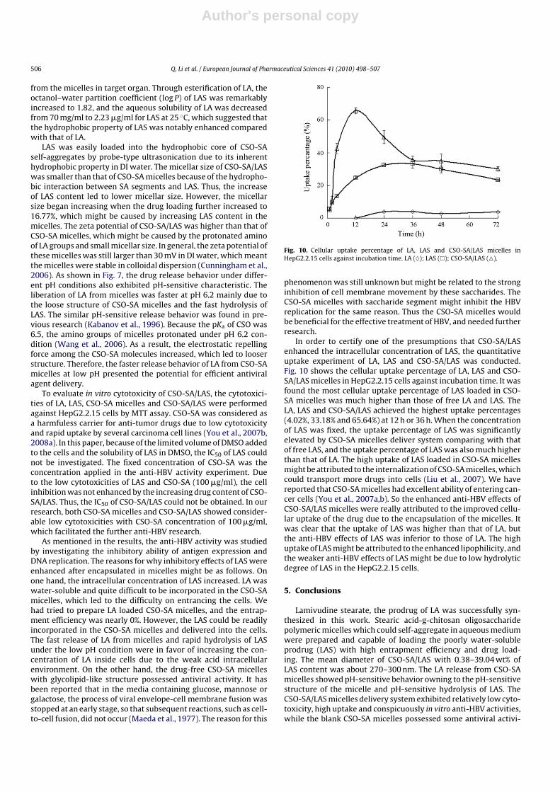
toxicities against the cultured cells. It was clear that the lowestcell survival rate of LAS solution against cultured cells was 58.46%when the concentration of LAS was 100 �g/ml. Because of thepoor solubility of LAS in water, ethanol (0.38 mg/ml) and methanol(0.62 mg/ml), DMSO was used to dissolve LAS. The cell survival rateof the required volume (10 �l) of DMSO was 66.74 ± 5.62%, whichindicated that the cellular inhibition of LAS solution mainly origi-nated from DMSO. The CSO-SA/LAS showed slightly lower survivalrate than CSO-SA micelles and LA. However, it could be seen fromFig. 8 that, when the concentration of CSO-SA micelles was fixed,there was no significant difference in cell survival rate as LAS con-tent was changed. The results revealed that CSO-SA/LAS possessedrelatively low cytotoxicity.
Table 3Cytotoxicities of LA, LAS, CSO-SA micelles and CSO-SA/LAS against HepG2.2.15.
Samples Cell survival (%)
LA 93.79 ± 5.89LASa 58.46 ± 2.86CSO-SA micelles 85.93 ± 5.40CSO-SA/LAS 72.14 ± 6.27
Cell survival was determined at the same concentration (100 �g/ml) of LA, LAS,CSO-SA micelles (blank micelles) and CSO-SA/LAS with 16.77% drug loading. Thedata represent mean ± SD (n = 3).
a Due to the poor solubility in water, methanol and ethanol, the DMSO was usedto dissolve LAS. The volume of DMSO was 10 �l per ml incubation medium.
Author's personal copy
Q. Li et al. / European Journal of Pharmaceutical Sciences 41 (2010) 498–507 505
Fig. 8. Cytotoxicities of CSO-SA/LAS against HepG2.2.15 cells. The CSO-SA concen-tration was all 100 �g/ml. Data represent the mean ± standard deviation (n = 3).
3.5. Anti-HBV activity in vitro
The anti-HBV activities of LA, LAS, and CSO-SA/LAS were inves-tigated against HepG2.2.15. As control, CSO-SA micelles with thesame material concentration of CSO-SA/LAS were performed like-wise. Their HBV inhibition rates determined by HBsAg, HBeAg andDNA assays are shown in Fig. 9A, B and C, respectively. As shownin Fig. 9, the inhibitory effects of CSO-SA micelles were inferiorto CSO-SA/LAS but still stronger than free drugs. Moreover, it wasclear that the inhibitory activities of LA, LAS and CSO-SA/LAS onHBV-induced HBsAg, HBeAg and DNA productions were enhanceddose-dependently and time-dependently. Although the inhibitoryactivities of LA diminished significantly on HBV-associated antigenexpression after esterified to LAS, both LA and LAS exhibited notableinhibition of HBV DNA replication. Furthermore, compared withfree LA and LAS solution, CSO-SA/LAS micelles markedly promoted
inhibitory effects on antigen expression. Therefore, the resultsindicated that CSO-SA/LAS micelles possessed more conspicuousanti-HBV activities than free LA and LAS solution.
4. Discussion
It is well known that polymeric micelles are not in favor of incor-porating hydrophilic drugs with low molecular weight. In orderto strengthen the encapsulation of LA into CSO-SA micelles, thesame long-chain fatty acid (SA) was selected to hydrophobicallymodify LA. It was reported earlier that the usage of DCC promotedesterification of primary and secondary alcohols under very mildconditions (Stempel and Landgraf, 1962). Through the conjugationof LA and SA, LAS was achieved in the presence of DCC. The syn-thesized compound was characterized by IR, 1H NMR and ESI-MS.Consequently, the results exhibited an identical data correspondingto the sample.
The hydrolysis of ester was easily conducted in aqueous envi-ronment, and showed pH-sensitive characteristic. The decrease ofthe pH resulted in increasing hydrolysis of the free prodrug inbuffers. The chronic HBV could easily turn to hepatic cancer. Thefaster hydrolysis of LAS under weak acid condition might be favorfor turning into effectively antiviral LA because of the low pH con-dition of tumor issue (Prabaharan et al., 2009). The usefulness ofLAS should depend not only on the stability of the prodrug for itsin vivo transport to target the cells but also upon its reversion toLA inside cells (Kerr et al., 1992). As shown in Table 1, the half-life (t1/2) of hydrolysis of the LAS in pH 7.4 PBS was 28.79 ± 7.87 h,and it was reduced significantly in the presence of FBS. The fasterhydrolysis might be due to the catalysis by esterase in the blood(Leinweber, 1987). However, after LAS was encapsulated in CSO-SAmicelles, the half-life (t1/2) of hydrolysis of LAS was retarded from0.32 h to 9.36 h, which was favorable for keeping LAS stable in its invivo transport, and rapidly turning into LA after LAS was released
Fig. 9. Inhibitory effects of LAS, LA, CSO-SA/LAS and CSO-SA micelles on the (A) HBsAg expression, (B) HBeAg expression and (C) HBV DNA replication in HepG2.2.15 cells.The material (CSO-SA) concentration was all 100 �g/ml. The data represent mean ± SD (n = 3).
Author's personal copy
506 Q. Li et al. / European Journal of Pharmaceutical Sciences 41 (2010) 498–507
from the micelles in target organ. Through esterification of LA, theoctanol–water partition coefficient (log P) of LAS was remarkablyincreased to 1.82, and the aqueous solubility of LA was decreasedfrom 70 mg/ml to 2.23 �g/ml for LAS at 25 ◦C, which suggested thatthe hydrophobic property of LAS was notably enhanced comparedwith that of LA.
LAS was easily loaded into the hydrophobic core of CSO-SAself-aggregates by probe-type ultrasonication due to its inherenthydrophobic property in DI water. The micellar size of CSO-SA/LASwas smaller than that of CSO-SA micelles because of the hydropho-bic interaction between SA segments and LAS. Thus, the increaseof LAS content led to lower micellar size. However, the micellarsize began increasing when the drug loading further increased to16.77%, which might be caused by increasing LAS content in themicelles. The zeta potential of CSO-SA/LAS was higher than that ofCSO-SA micelles, which might be caused by the protonated aminoof LA groups and small micellar size. In general, the zeta potential ofthese micelles was still larger than 30 mV in DI water, which meantthe micelles were stable in colloidal dispersion (Cunningham et al.,2006). As shown in Fig. 7, the drug release behavior under differ-ent pH conditions also exhibited pH-sensitive characteristic. Theliberation of LA from micelles was faster at pH 6.2 mainly due tothe loose structure of CSO-SA micelles and the fast hydrolysis ofLAS. The similar pH-sensitive release behavior was found in pre-vious research (Kabanov et al., 1996). Because the pKa of CSO was6.5, the amino groups of micelles protonated under pH 6.2 con-dition (Wang et al., 2006). As a result, the electrostatic repellingforce among the CSO-SA molecules increased, which led to looserstructure. Therefore, the faster release behavior of LA from CSO-SAmicelles at low pH presented the potential for efficient antiviralagent delivery.
To evaluate in vitro cytotoxicity of CSO-SA/LAS, the cytotoxici-ties of LA, LAS, CSO-SA micelles and CSO-SA/LAS were performedagainst HepG2.2.15 cells by MTT assay. CSO-SA was considered asa harmfuless carrier for anti-tumor drugs due to low cytotoxicityand rapid uptake by several carcinoma cell lines (You et al., 2007b,2008a). In this paper, because of the limited volume of DMSO addedto the cells and the solubility of LAS in DMSO, the IC50 of LAS couldnot be investigated. The fixed concentration of CSO-SA was theconcentration applied in the anti-HBV activity experiment. Dueto the low cytotoxicities of LAS and CSO-SA (100 �g/ml), the cellinhibition was not enhanced by the increasing drug content of CSO-SA/LAS. Thus, the IC50 of CSO-SA/LAS could not be obtained. In ourresearch, both CSO-SA micelles and CSO-SA/LAS showed consider-able low cytotoxicities with CSO-SA concentration of 100 �g/ml,which facilitated the further anti-HBV research.
As mentioned in the results, the anti-HBV activity was studiedby investigating the inhibitory ability of antigen expression andDNA replication. The reasons for why inhibitory effects of LAS wereenhanced after encapsulated in micelles might be as follows. Onone hand, the intracellular concentration of LAS increased. LA waswater-soluble and quite difficult to be incorporated in the CSO-SAmicelles, which led to the difficulty on entrancing the cells. Wehad tried to prepare LA loaded CSO-SA micelles, and the entrap-ment efficiency was nearly 0%. However, the LAS could be readilyincorporated in the CSO-SA micelles and delivered into the cells.The fast release of LA from micelles and rapid hydrolysis of LASunder the low pH condition were in favor of increasing the con-centration of LA inside cells due to the weak acid intracellularenvironment. On the other hand, the drug-free CSO-SA micelleswith glycolipid-like structure possessed antiviral activity. It hasbeen reported that in the media containing glucose, mannose orgalactose, the process of viral envelope-cell membrane fusion wasstopped at an early stage, so that subsequent reactions, such as cell-to-cell fusion, did not occur (Maeda et al., 1977). The reason for this
Fig. 10. Cellular uptake percentage of LA, LAS and CSO-SA/LAS micelles inHepG2.2.15 cells against incubation time. LA (♦); LAS (�); CSO-SA/LAS (�).
phenomenon was still unknown but might be related to the stronginhibition of cell membrane movement by these saccharides. TheCSO-SA micelles with saccharide segment might inhibit the HBVreplication for the same reason. Thus the CSO-SA micelles wouldbe beneficial for the effective treatment of HBV, and needed furtherresearch.
In order to certify one of the presumptions that CSO-SA/LASenhanced the intracellular concentration of LAS, the quantitativeuptake experiment of LA, LAS and CSO-SA/LAS was conducted.Fig. 10 shows the cellular uptake percentage of LA, LAS and CSO-SA/LAS micelles in HepG2.2.15 cells against incubation time. It wasfound the most cellular uptake percentage of LAS loaded in CSO-SA micelles was much higher than those of free LA and LAS. TheLA, LAS and CSO-SA/LAS achieved the highest uptake percentages(4.02%, 33.18% and 65.64%) at 12 h or 36 h. When the concentrationof LAS was fixed, the uptake percentage of LAS was significantlyelevated by CSO-SA micelles deliver system comparing with thatof free LAS, and the uptake percentage of LAS was also much higherthan that of LA. The high uptake of LAS loaded in CSO-SA micellesmight be attributed to the internalization of CSO-SA micelles, whichcould transport more drugs into cells (Liu et al., 2007). We havereported that CSO-SA micelles had excellent ability of entering can-cer cells (You et al., 2007a,b). So the enhanced anti-HBV effects ofCSO-SA/LAS micelles were really attributed to the improved cellu-lar uptake of the drug due to the encapsulation of the micelles. Itwas clear that the uptake of LAS was higher than that of LA, butthe anti-HBV effects of LAS was inferior to those of LA. The highuptake of LAS might be attributed to the enhanced lipophilicity, andthe weaker anti-HBV effects of LAS might be due to low hydrolyticdegree of LAS in the HepG2.2.15 cells.
5. Conclusions
Lamivudine stearate, the prodrug of LA was successfully syn-thesized in this work. Stearic acid-g-chitosan oligosaccharidepolymeric micelles which could self-aggregate in aqueous mediumwere prepared and capable of loading the poorly water-solubleprodrug (LAS) with high entrapment efficiency and drug load-ing. The mean diameter of CSO-SA/LAS with 0.38–39.04 wt% ofLAS content was about 270–300 nm. The LA release from CSO-SAmicelles showed pH-sensitive behavior owning to the pH-sensitivestructure of the micelle and pH-sensitive hydrolysis of LAS. TheCSO-SA/LAS micelles delivery system exhibited relatively low cyto-toxicity, high uptake and conspicuously in vitro anti-HBV activities,while the blank CSO-SA micelles possessed some antiviral activi-

Author's personal copy
Q. Li et al. / European Journal of Pharmaceutical Sciences 41 (2010) 498–507 507
ties as well. Overall, the present micelles are promising carrier foreffective therapy of anti-HBV drugs.
Acknowledgments
We are grateful for financial support of National Basic ResearchProgram of China (973 Program) under Contract 2009CB930300,National HighTech Research and Development Program (863) ofChina (2007AA03Z318).
References
Asselah, T., Ripault, M.P., Castelnau, C., 2005. The current status of antiviral therapyof chronic hepatitis B. J. Clin. Virol. 34, S115–S124.
Bernkop-Schnurch, A., Krajicek, M.E., 1998. Mucoadhesive polymers as platforms forperoral peptide delivery and absorption: synthesis and evaluation of differentchitosan–EDTA conjugates. J. Control. Release 50, 215–223.
Cavalli, R., Donalisio, M., Civra, A., Ferruti, P., Ranucci, E., Trotta, F., Lembo, D., 2009.Enhanced antiviral activity of Acyclovir loaded into �-cyclodextrin-poly (4-acryloylmorpholine) conjugate nanoparticles. J. Control. Release 137, 116–122.
Chawla, J.S., Amiji, M.M., 2002. Biodegradable poly (epsilon-caprolactone) nanopar-ticles for tumor-targeted delivery of tamoxifen. Int. J. Pharm. 249, 127–138.
Craxi, A., Antonucci, G., Camma, C., 2006. Treatment options in HBV. J. Hepatol. 44,S77–S83.
Cunningham, D., Littleford, R.E., Smith, W.E., Lundahl, P.J., Khan, I., McComb, D.W.,2006. Practical control of SERRS enhancement. Faraday Discuss. 132, 135–145.
Desai, M.P., Labhasetawar, V., Walter, E., Levy, R.J., Amidon, G.L., 1997. The mecha-nism of uptake of biodegradable microparticles in Caco-2 cells is size dependent.Pharm. Res. 14, 1568–1573.
Hoofnagle, J.H., 1990. Alpha-interferon therapy of chronic hepatitis B. Current statusand recommendations. J. Hepatol. 11, S100–S107.
Hu, F.Q., Li, Y.H., Yuan, H., Zeng, S., 2006a. Novel self-aggregates of chitosanoligosaccharide grafted steric acid: preparation, characterization and proteinassociation. Pharmazie 61, 194–198.
Hu, F.Q., Ren, G.F., Yuan, H., Du, Y.Z., Zeng, S., 2006b. Shell cross-linked stearic acidgrafted chitosan oligosaccharide self-aggregated micelles for controlled releaseof paclitaxel. Colloids Surf. B 50, 97–103.
Hu, F.Q., Zhao, M.D., Yuan, H., You, J., Du, Y.Z., Zeng, S., 2006c. A novel chitosanoligosaccharide–stearic acid micelles for gene delivery: properties and in vitrotransfection studies. Int. J. Pharm. 315, 158–166.
Hu, F.Q., Wu, X.L., Du, Y.Z., You, J., Yuan, H., 2008. Cellular uptake and cytotoxi-city of shell crosslinked stearic acid grafted chitosan oligosaccharide micellesencapsulating doxorubicin. Eur. J. Pharm. Biopharm. 69, 117–125.
Kabanov, A.V., Bronich, T.K., Kabanov, V.A., 1996. Soluble stoichiometric complexesfrom poly (N-ethyl-4-vinylpyridinium) cations and poly (ethylene oxide) block-polymethacrylate anions. Macromolecules 29, 67–97.
Kerr, S.G., Kalman, T.I., Med, J., 1992. Highly water-soluble lipophilic prodrugs of theanti-HIV nudeoside analog 2′ ,3′-dideoxycytidine and its 3′-fluoro derivative. J.Med. Chem. 35, 1996–2001.
Lai, S.K., Hida, K., Man, S.T., Chen, C., Machamer, C., Schroer, T.A., Hanes, J., 2007.Privileged delivery of polymer nanoparticles to the perinuclear region of livecells via a nonclathrin, non-degradative pathway. Biomaterials 28, 2876–2884.
Langer, R., 1998. Drug delivery and targeting. Nature 392, 5–10.
Lee, K.Y., Kwon, I.C., Kim, Y.H., Jo, W.H., Jeong, S.Y., 1998. Preparation of chitosanself-aggregates as a gene delivery system. J. Control. Release 51, 213–220.
Leinweber, F.J., 1987. Possible physiological roles of carboxylic ester hydrolases.Drug Metab. Rev. 18, 379–439.
Lien, E.J., Ren, S.J., 2002. Partition coefficients. Encyclopedia Pharm. Technol.,2012–2019.
Liu, S.Q., Wiradharma, N., Gao, S.J., Tong, Y.W., Yang, Y.Y., 2007. Biofunctionalmicelles self-assembled from a folate-conjugated block copolymer for targetedintracellular delivery of anticancer drugs. Biomaterials 28, 1423–1433.
Maeda, Y., Kim, J., Koseki, I., Mekada, E., Shiokawa, Y., Okada, Y., 1977. Modificationof cell membranes with viral envelopes during fusion of cells with HVJ (sendaivirus). III. Effects of mono- and di-saccharides on cell fusion and membranemovement of fused cells. Exp. Cell Res. 108, 95–106.
Nishiyama, N., Kato, Y., Sugiyama, Y., Kataoka, K., 2001. Cisplatin-loadedpolymer–metal complex micelle with time-modulated decaying property as anovel drug delivery system. Pharm. Res. 18, 1035–1041.
Prabaharan, M., Grailer, J.J., Pilla, S., Steeber, D.A., Gong, S., 2009. Folate-conjugatedamphiphilic hyperbranched block copolymers based on Boltorn-H40, poly-(l-lactide) and poly(ethylene glycol) for tumor-targeted drug delivery.Biomaterials 30, 3009–3019.
Raney, A.K., Hamatake, R.K., Hong, Z., 2003. Agents in clinical development for thetreatment of chronic hepatitis B. Expert Opin. Investig. Drugs 12, 1281–1295.
Shaw, T., Bartholomeusz, A., Locarnini, S., 2006. HBV drug resistance: mechanisms,detection and interpretation. J. Hepatol. 44, 593–606.
Stempel, A., Landgraf, F.W., 1962. Quinazolines and 1,4-benzodiazopines-9,2-carbobenzoxyglycylamidobenzophenones and their conversion to 1,4-benzodiazopines. J. Org. Chem. 27, 4675–4677.
Vladimir, P., Torchilin, 2008. Tat peptide-mediated intracellular delivery of pharma-ceutical nanocarriers. Adv. Drug Deliv. Rev. 60, 548–558.
Wang, Q.Z., Chen, X.G., Liu, N., Wang, S.X., Liu, C.S., Meng, X.H., Liu, C.G., 2006. Pro-tonation constants of chitosan with different molecular weight and degree ofdeacetylation. Carbohydr. Polym. 65, 194–201.
Xue, K.C., Gu, Y., Zhang, S.Q., Jiang, Y.P., Wu, D.C., 2003. Preparation of lamivudylpalmitate solid lipid nanoparticles. J. Fourth Mil. Med. Univ. 10, 890–892.
Ye, Y.Q., Yang, F.L., Hu, F.Q., Du, Y.Z., Yuan, H., Yu, H.Y., 2008. Core-modified chitosanbased polymeric micelles for controlled release of doxorubicin. Int. J. Pharm.352, 294–301.
Yokoyama, M., Okano, T., Sakurai, Y., Suwa, S., Kataoka, K., 1996. Introduction ofcisplatin into polymeric micelle. J. Control. Release 39, 351–356.
You, J., Hu, F.Q., Du, Y.Z., Yuan, H., 2007a. Polymeric micelles with glycolipid-likestructure and multiple hydrophobic domains for mediating-target delivery ofpaclitaxel. Biomacromolecules 8, 2450–2456.
You, J., Hu, F.Q., Du, Y.Z., Yuan, H., Ye, B.F., 2007b. High cytotoxicity and resistant-cell reversal of novel paclitaxel loaded micelles by enhancing molecular-targetdelivery of the drug. Nanotechnology 18, 495101.
You, J., Hu, F.Q., Du, Y.Z., Yuan, H., 2008a. Improved cytotoxicity of doxorubicin byenhancing its nuclear delivery mediated via nanosized micelles. Nanotechnol-ogy 19, 255103.
You, J., Li, X., Cui, F.D., Du, Y.Z., Yuan, H., Hu, F.Q., 2008b. Folate conjugated polymermicelles for active-targeting to cancer cells: preparation, in vitro evaluation oftargeting ability and cell cytotoxicity. Nanotechnology 19, 045102.
Zhang, Y.D., Hu, Z.Y., Ye, M.Y., Pan, Y.F., Chen, J.J., Luo, Y.L., Zhang, Y.Q., He, L.X., Wang,J.W., 2007. Effect of poly (ethylene glycol)-blockpolylactide micelles on hep-atic cells of mouse: low cytotoxicity, but efflux of the micelles by ATP-bindingcassette transporters. Eur. J. Pharm. Biopharm. 66, 268–280.
Zoulim, F., Perrillo, R., 2008. Hepatitis B: reflections on the current approach toantiviral therapy. J. Hepatol. 48, 2–19.