synthesis of cholesterol-conjugated magnetic nanoparticles for purification of human paraoxonase 1
TRANSCRIPT

Synthesis of Cholesterol-Conjugated MagneticNanoparticles for Purification of Human Paraoxonase 1
Zahoor Qadir Samra & Sadaf Shabir & Zainab Rehmat &Mariam Zaman & Aqsa Nazir & Nadia Dar &
Muhammad Amin Athar
Received: 6 July 2009 /Accepted: 26 October 2009 /Published online: 10 November 2009# Humana Press 2009
Abstract Human serum paraoxonase 1 (PON1) is known as an antioxidant and is alsoinvolved in the detoxification of many compounds. In this study, a novel purificationstrategy was employed to purify the PON1 by using cholesterol-conjugated magneticnanoparticles. Magnetic nanoparticles were synthesized and conjugated with cholesterolthrough diazotized p-aminohippuric acid. In Fourier transform infrared spectrum ofcholesterol-p-aminohippuric acid-Fe3O4 nanoparticles, the appearance of peaks at 3,358.3,1,645 cm−1, and at 2,334.9 cm−1 confirmed the conjugation. The molecular weight ofpurified PON1 was nearly 45 kDa on sodium dodecyl sulfate (SDS)–polyacrylamide gelelectrophoresis (PAGE), and isoelectric point was 5.3. The specific activity was438 U mg−1 protein, and the purification fold was 515 with 73% yield. The Km valueswere 1.3 and 0.74 mM with paraoxon and phenyl acetate, respectively. Western blot of 2D-PAGE confirmed the homogeneity and stability of the enzyme. Mg+2, Mn+2, glycerol,(NH4)2SO4, PEG 6000, Triton X-100, and phenylmethylsulfonyl fluoride did not show anyeffect on activity. Pb+2, Co+2, Zn2+, ethanol, β-mercaptoethanol, and acetone reduced theactivity while Ni2+, Cd2+, Cu2+, iodoacetic acid, SDS, dimethylformamide, DMSOinhibited the activity. In vitro enzyme activity was slightly reduced by acetyl salicylicand acetaminophen and reduced 50% with amino glycosides and ampicillin antibiotics atconcentrations of 0.6 and 30 mg ml−1, respectively. This is the first report for the synthesisof cholesterol-conjugated magnetic nanoparticles for simple purification of PON1 enzyme.
Keywords Paraoxonase 1 . Nanoparticles . FTIR . Antibiotics . 2D-PAGE .Western blot
Appl Biochem Biotechnol (2010) 162:671–686DOI 10.1007/s12010-009-8840-4
Z. Q. Samra (*) : S. Shabir : Z. Rehmat :M. Zaman : A. Nazir :M. A. AtharInstitute of Biochemistry and Biotechnology, Quaid-i-Azam Campus, University of the Punjab,Lahore 54590, Pakistane-mail: [email protected]
N. DarJinnah Degree College for Women, Mozang Road, Lahore 54590, Pakistan

Introduction
Paraoxonase 1 (PON1; aryldialkylphosphatase, EC 3.1.8.1) is the glycosylated esteraseenzyme synthesized in liver and released into serum after association with high-densitylipoprotein (HDL) [1, 2]. PON1 name is derived from its property to hydrolyze paraoxonand to detoxify the broad range of substrates such as aryl esters, carbamates, cycliccarbonates, lactones, commonly used insecticides, and chemical warfare organophosphatecompounds [3–8]. Paraoxon inhibits acetyl cholinesterase and is metabolically generated invivo from parathion (insecticide) by mitochondrial oxidation [9]. PON1 is conserved inmammals, but its activity is not detected in fish, birds, and arthropods. In mammals, PON1is a member of a multigene family and located on human chromosome 7 [10, 11].
PON1 is known as a bioscavenger that protects against cardiovascular diseases byinhibiting the formation of oxidized high-density lipoproteins and low-density lipoproteins[12]. The pharmacological modulation of PON1 expression may be a useful approach forprotection of cardiovascular diseases and detoxification of organophosphate compounds.Due to the active role of PON1 in lipid metabolism, cardiovascular disease, andatherosclerosis, it has also gained attention to understand its role in the metabolism ofpharmaceutical drugs [13]. Serum PON1 level depends upon a number of physiological andpathological conditions such as renal disease, diabetes mellitus, HDL deficiencies, livercirrhosis, high-fat diets, and nutritional and environmental factors [5, 14].
With the advent of nanotechnology, the affinity purification of proteins, enzymes, andimmunoglobulins through ligands or biomolecules immobilized onto magnetic nano-particles has become an important tool since last decade [15, 16]. The affinity purificationonto ligand-conjugated magnetic nanoparticles has the following advantages (a) enhancedstability, (b) easy separation from mixture, (c) possible modulation of the catalyticproperties, and (d) prevention of microbial growth [17]. Due to the specific size and shapeof magnetic nanoparticles, these have applications as drug delivery [18, 19], tissue repair[20], cell engineering [21], and for diagnosis [22, 23]. The surface of the magneticnanoparticles can be modified chemically, and the binding can be done by carbodiimideactivation method due to the simplicity and efficiency of the reagent [24, 25]. Cumbersomechromatographic procedures about the purification of paraoxonase 1 are available [13, 26–29]. By utilizing these procedures, a moderate level of enzyme activity is lost. There is aneed to develop the strategy for easy purification of paraoxonase 1 from any source.
Due to affinity of human paraoxonase 1 with cholesterol (CHO), we describe a simpleand novel method for purification of human paraoxonase 1 by using cholesterol-conjugatedmagnetic nanoparticles. The effect of some analgesics and antibiotics is also studied on theactivity of purified PON1 due to physiological importance of these medicines.
Materials and Methods
Analytical-grade reagents such as iron(II) chloride tetrahydrate, iron(III) chloridehexahydrate, paraoxon, phenyl acetate, cholesterol, p-aminohippuric acid, acrylamide,bisacrylamide, sodium dodecyl sulfate (SDS), phenylmethylsulfonyl fluoride (PMSF),Triton X-100, ethanol, dimethylformamide (DMF), dimethyl sulfoxide (DMSO), β-mercaptoethanol, dithiothreitol, iodoacetate, ethylenediaminetetraacetic acid (EDTA),ammonia solution, acetyl salicylic acid, acetaminophen, sodium ampicillin, gentamicinsulfate, kanamycin sulfate, streptomycin sulfate, alkaline-phosphatase-conjugated rabbitantimouse immunoglobulin G (IgG), and other chemicals required for routine analysis were
672 Appl Biochem Biotechnol (2010) 162:671–686

purchased from Sigma-Aldrich. Bradford reagent and nitrocellulose membrane werepurchased from Bio-Rad. Protein markers were purchased from Fermentas Inc. SephadexG-75 was purchased from Pharmacia. Healthy human plasma (after screening for hepatitisB and C virus) was used in this study. Deionized water produced by Milli-Q was usedthroughout the experiments.
Synthesis of Magnetic Nanoparticles and Determination of Amino Group
Magnetic nanoparticles (Fe3O4) were synthesized by mixing iron(II) and iron(III) ions in1:2 ratios under alkaline hydrothermal conditions [30]. The synthesized magneticnanoparticles were analyzed by Fourier transform infrared (FTIR) spectroscopy. Thenumber of amino groups (–NH2) attached to the Fe3O4 was determined spectrophoto-metrically using molar absorption coefficient 6,623 M−1cm −1 [31].
Attachment of p-Aminohippuric Acid with Fe3O4
The p-aminohippuric acid (PAHA) was conjugated with Fe3O4 by the modification ofmethod [32]. PAHA (200 mg) was dissolved in 120 ml distilled water and mixed with200 mg of Fe3O4. The mixture was sonicated for 10 min at 4 °C on ice, and pH 8.0 wasadjusted with 1.0 M NaOH. The mixture was warmed for 15 min at 37 °C. After cooling,200 mg of 1-ethyl-3-(3-dimethyl-aminopropyl)-carbodiimide-HCL (ECD) was added. ThepH 6.4 was adjusted with 2.0 M HCl and stirred for 6 h in dark at 25 °C. ECD (100 mg)was again added to the mixture and kept at 25 °C for another 15 h. The PAHA-Fe3O4-conjugated particles were separated by applying external magnetic force and washed with1.0 l deionized water and finally with 2.0 ml acetone. The particles were air-dried andprocessed for FTIR analysis.
Coupling of Cholesterol with PAHA-Conjugated Fe3O4
The CHO was coupled with PAHA-conjugated Fe3O4 by the modifications in method [33].PAHA-conjugated Fe3O4 (200 mg) was suspended in 30 ml of deionized water, and pH wasadjusted to 1.5 with 1.0 M HCl. The solution was cooled to 0 °C for 30 min, and 0.5 ml ofNaNO2 solution (100 mg/ml distilled water) was slowly added with constant shaking. After5 min, the excess nitrite was destroyed by adding 0.5 ml of ammonium sulfamate solution(60 mg ml−1 distilled water). The cholesterol solution (8 mg per 10 ml of 50% (v/v) acetonein 0.1 M borate buffer, pH 9.0) was added dropwise with constant stirring at 0 °C. After 1 h,conjugated CHO-PAHA-Fe3O4 was removed and washed with water to remove theunconjugated cholesterol. The particles were dried at 37 °C and processed for FTIR analysis.
Fourier Transform Infrared Spectroscopy
Fe3O4, PAHA, PAHA-Fe3O4, CHO, and CHO-PAHA-Fe3O4 were homogenized separatelyin Nujol, and the FTIR spectrum was taken on FTIR spectrophotometer (Perkin-Elmer) at25 °C.
Protein Determination and Enzyme Assays
During purification of PON1 enzyme, the protein concentration was determined byBradford reagent assay using bovine serum albumin as standard [34]. Paraoxonase and aryl
Appl Biochem Biotechnol (2010) 162:671–686 673

esterase activities were determined using diethyl-p-nitrophenyl phosphate (paraoxon) andphenyl acetate as substrate, respectively [23]. The final substrate concentration of paraoxonand phenyl acetate was 2 mM at pH 8.0. The activity of PON1 enzyme was determined byusing the molar extinction coefficients of paraoxon and phenyl acetate as 18,290 and1,310 M−1cm−1 at 412 and 270 nm, respectively. One unit of enzyme activity was definedas the amount of enzyme required to hydrolyze 1.0 μM of substrate per minute at 37 °C.All assays were conducted in duplicate.
Purification of PON1 Enzyme
All purification steps were carried out at 4 °C unless otherwise stated. Before purification,total protein concentration and PON1 activity of human plasma were measured.
Triton X-100 Treatment and Ammonium Sulfate Precipitation
Human plasma (10 ml) was mixed with Triton X-100 (1% v/v) and CaCl2 solution (finalconcentration 10 mM) and gently stirred for 16 h. The mixture was centrifuged for 20 minat 6,000 rpm. The supernatant was separated and mixed with ammonium sulfate to 60%saturation and then to 70% saturation. The solution was stirred slowly for 5 h. Theprecipitates were collected at 12,000 rpm for 25 min. The precipitates were dissolved in5.0 ml of PON buffer (20 mM Tris–HCl pH 8.0, 1.0 mM CaCl2, and 5.0 μM EDTA) anddialyzed against 2.0 l of the same buffer.
Affinity Cholesterol-Conjugated Magnetic Nanoparticles
The dialyzed solution was mixed with 0.2 g of cholesterol-conjugated Fe3O4 nanoparticlesand gently shaken for 30 min. The particles were separated and washed twice with 20 ml ofPON buffer. The washed particles were mixed again with 5.0 ml of PON buffer containing0.2% deoxycholate for 30 min. The magnetic nanoparticles were separated again. Thesupernatant was dialyzed against 1 l of PON buffer and concentrated to 1.5 ml onmicrocentrifuge filter unit (10,000-Da cut).
Gel Permeation Chromatography
Concentrated fractions from affinity cholesterol-conjugated magnetic nanoparticles wereapplied on Sephadex G-75 column (1.5×30 cm) pre-equilibrated with PON buffer. A 2.0-mlfraction was collected at a flow rate of 0.5 ml min−1, and fractions containing the PON1activity were pooled, concentrated, and stored at −20 °C until further use. The proteinconcentration was determined by Bradford reagent.
Production of Anti-PON1Antibodies
Purified human paraoxonase 1 (1.0 mg ml−1l) was mixed with denaturing buffer (50 mMTris–HCl, pH 7.4, 0.01% SDS, 0.005 M β-mercaptoethanol) in 1:1 ratio and kept at 60 °Cfor 30 min to denature the enzyme to avoid the self-tolerance of mammalian antigen. MaleBalb/C mice, 6–7 weeks old, were subcutaneously injected with denatured PON1 (80–100 µg per injection) after mixing with Freund’s complete adjuvants in 1:1 ratio at 2-weekintervals with a total of five injections. After checking the anti-PON1 antibody titre in miceblood by indirect enzyme-linked immunosorbent assay, whole blood was isolated by
674 Appl Biochem Biotechnol (2010) 162:671–686

cardiac puncture, and serum was isolated. Preimmune serum was obtained from mice tailbefore immunization and used as control.
Characterization of PON1 Enzyme
SDS–Polyacrylamide Gel Electrophoresis and Western Blot
Protein samples obtained during purification steps were analyzed on slab 10% SDS–polyacrylamide gel electrophoresis (PAGE) as described [35]. The gel was stained withCoomassie Brilliant Blue R-250 and destained in solution containing methanol, acetic acid,and water in 40:10:50 ratio, respectively. The proteins samples were electrophoreticallytransferred onto nitrocellulose membrane [36]. The nitrocellulose membrane was treatedwith blocking buffer (3% BSA in Tris-buffered saline–Tween 20) and incubated withprimary antibodies (mouse anti-PON1 antibodies) followed by incubation with secondaryantibody (alkaline-phosphatase-conjugated rabbit antimouse IgG). After washing in Tris-buffered saline–Tween 20, color reaction was developed using nitro blue tetrazolium and 5-bromo-4-chloro-3-indolyl phosphate as substrates. Preimmune serum and secondaryantibody were used as control.
Effects of pH and Temperature
The effect of pH on purified human serum paraoxonase 1 was measured at 37 °C over thepH range 5–11 in 50-mM buffer (sodium acetate buffer, pH 5–6; Tris–HCL buffer pH 7–9;glycine–NaOH buffer pH 9–11) using 2.0-mM paraoxon concentration. All assays wereconducted in duplicate.
The effect of temperature on paraoxonase 1 was determined by incubating the enzyme atdifferent temperatures (10, 20, 30, 40, 50, and 60 °C) for 30 min. The enzyme activity wasmeasured under standard assay conditions which were considered as 100%. All assays wereconducted in duplicate.
Effects of Metal Ions, Detergents, Solvents, and Reducing Agents
The effects of metal ions (MgCl2, MnCl2, PbCl2, CoCl2, CuCl2, CdCl2, ZnCl2, andNiCl2,) were studied by adding the metal ion separately in a final concentration of0.5 to 3.0 mM. The effects of other reagents such as, PMSF, iodoacetate, SDS,β-mercaptoethanol, DMF, DMSO, ethanol, acetone, glycerol, ammonium sulfate, andpolyethylene glycol (PEG) 6000 were also determined separately by adding compounds infinal concentration of 2% to 7%. The purified PON1 enzyme was preincubated separatelywith above reagents dissolved in PON buffer for 15 min at 37 °C, and the activitywas measured under standard assay conditions. The activity of the enzyme without theaddition of above reagents was considered as 100%. All assays were conducted induplicate.
Effect of Stabilizers on PON1 Activity
The effect of stabilizers was checked as described [24] with little modification. Purifiedparaoxonase 1 (200 U ml−1) was taken in 5 ml of PON buffer pH 8.0 containing 20%concentration of (NH4)2SO4, PEG 6000, and glycerol separately and incubated for 30 daysat 4 °C. Enzyme solution (0.05 ml) was taken on each day, and the enzyme activity was
Appl Biochem Biotechnol (2010) 162:671–686 675

checked under standard assay conditions for 30 days. Control enzyme assays wereconducted without stabilizers. All assays were carried out in duplicates.
Effect of Medicines on PON1 Activity
The inhibitory or noninhibitory effects of different final concentration of some medicinessuch as acetyl salicylic acid (2, 4, 6, 8, 10 mg ml−1), acetaminophen (2, 4, 6, 8,10 mg ml−1), sodium ampicillin (10, 20, 30, 40, 50 mg ml−1), gentamicin sulfate (2, 4, 6,8,10 mg ml−1), kanamycin sulfate (2, 4, 6, 8,10 mg ml−1), and streptomycin sulfate (2, 4, 6,8,10 mg ml−1) was determined (in vitro). Purified paraoxonase 1 (50 U ml−1) was mixedwith the above medicines separately for 15 min, and the enzyme activity was checked understandard assay conditions. Paraoxonase 1 enzyme activity without medicines wasconsidered as 100%.
2D-PAGE, Western Blot, and Kinetic Study
The purity of the enzyme was checked by 2D-PAGE. The isoelectric point of PON1enzyme was determined in isoelectric focusing (IEF) gel in tube (1.0×50 mm) according tothe O’Farrell method, and the isoelectric focused gel was also placed on SDS–polyacrylamide gel (10%) for second dimension [37, 38]. The protein separated in 2D-PAGE was transferred onto nitrocellulose membrane and processed further for immuno-chemical reaction as described above. The kinetic parameters of PON1 enzyme werestudied under standard assay conditions using different concentrations of substrate (0.5 to3.0 mM of paraoxon and phenyl acetate). All assays were carried out in duplicates. TheMichaelis–Menton constant (Km) was determined by Lineweaver–Burk plot method.
Results
FTIR Analysis
The binding of cholesterol with magnetic nanoparticles was achieved through diazotizationreaction of p-aminohippuric acid. The conjugation was confirmed by FTIR spectroscopy.The FTIR spectra of Fe3O4, PAHA, PAHA-Fe3O4, CHO, and CHO-PAHA-Fe3O4 areshown in Fig. 1a–e, respectively.
In FTIR spectrum of Fe3O4, the bend and stretch peaks of –NH2 group present on Fe3O4
were observed at 1,637 and at 3,300 cm−1, respectively (Fig. 1a (a and b, respectively)). InFTIR spectrum of PAHA, the bend peaks of –NH2, –NH, and –COOH groups wereobserved at 3,342, 3,415, and 3,215 cm−1, respectively (Fig. 1b (a, b, and c)). The stretchpeaks of –amino group, aromatic ring, and –C=O group were observed at 1,631.4, 1,599.3,and 1,900 cm−1, respectively (Fig. 1b (d, e, and f)).
In FTIR spectrum of PAHA-Fe3O4, the stretch peaks of –NH2, –NH, and –C=O groups ofPAHAwere observed at 3,336.9, 3,405, and 1,571 cm−1, respectively (Fig. 1c (a, b, and c)).
Fig. 1 FTIR spectra for conjugation of cholesterol with magnetic nanoparticles through diazotized p-aminohippuric acid. The details about the peaks are explained in text section. a FTIR spectrum of magneticnanoparticles, b FTIR spectrum of p-aminohippuric acid, c FTIR spectrum of conjugation of magneticnanoparticles with p-aminohippuric acid, d FTIR spectrum of cholesterol, e FTIR spectrum of conjugation ofcholesterol through diazotized p-aminohippuric-acid-conjugated magnetic nanoparticles
b
676 Appl Biochem Biotechnol (2010) 162:671–686

Appl Biochem Biotechnol (2010) 162:671–686 677

The bend peak of –COOH group appeared at 3,215 cm−1 in PAHA spectrum (Fig. 1b (b))which disappeared in spectrum of PAHA-Fe3O4 and new peak of N=C– has appeared at2,353.3 cm−1 (Fig. 1c, (d)). The appearance of new peak indicated the successfulconjugation of –COOH group of PAHA with –NH2 group of Fe3O4 through carbodiimidereagent. The bend peak of aromatic ring and –C=C– of PAHA was observed at 1,900 and1,630.9 cm−1, respectively (Fig. 1c (b and f)).
In FTIR spectrum of CHO, the broad stretch peak of phenol group was observed at3,375.6 cm−1, and stretch peak of –C=C– was observed at 1,630.9 cm−1 (Fig. 1d (a and b)).In CHO-PAHA-Fe3O4 spectrum, the appearance of bend peaks of phenol group at3,358.3 cm−1 (Fig. 1e (a)) and slightly shifted peak of aromatic ring at 1,645 cm−1
(Fig. 1e (b)) confirmed the binding of CHO with PAHA-Fe3O4. The additional stretch peaksat 2,354.9 cm−1 in pure CHO spectrum (Fig. 1d (c)) and CHO-PAHA-Fe3O4 spectrum(Fig. 1e (c)) advocated the binding confirmation. The stretch peak at 2,334.9 cm−1 (Fig. 1e
Fig. 1 (continued)
678 Appl Biochem Biotechnol (2010) 162:671–686

(d)) confirmed the diazotization (–N=N–) bonding. The characteristic peaks of Nujol wereobserved in all spectra at 2,928, 1,461.8, and at 1,376 cm−1. The proposed structure of CHO-PAHA-Fe3O4 is shown (Fig. 2).
Purification
Human serum paraoxonase 1 was purified to homogeneity from human plasma by usingaffinity cholesterol-conjugated magnetic nanoparticles and Sephadex-G75 chromato-graphic techniques. Different concentrations of sodium deoxycholate in PON buffer(0.1%, 0.2%, 0.3%, 0.4%, 0.5%) were used to elute the PON1 enzyme from CHO-PAHA-Fe3O4 particles. The complete elution of adsorbed enzyme on CHO-PAHA-Fe3O4
was observed with 0.2% deoxycholate in PON buffer. The dialyzed and concentratedPON1 fractions were loaded on to Sephadex-G75 column and eluted in PON1 buffer. Thefractions indicating the enzyme activity were pooled and concentrated (Fig. 3). Thepurified enzyme was analyzed on SDS–PAGE and only single band of nearly 45 kDawas observed which clearly demonstrated the homogeneity of the purified enzyme(Fig. 4a). The purification of enzyme was 516-fold with 73% yield. The specific activitywas 438 U mg−1. The purification steps and activity of human serum paraoxonase 1are shown (Table 1). In Western blot analysis, a single protein band was observedwhich advocated the purity and integrity of the enzyme stored at −20 °C for 1 month(Fig. 4b).
Biochemical Properties of Purified PON1 Enzyme
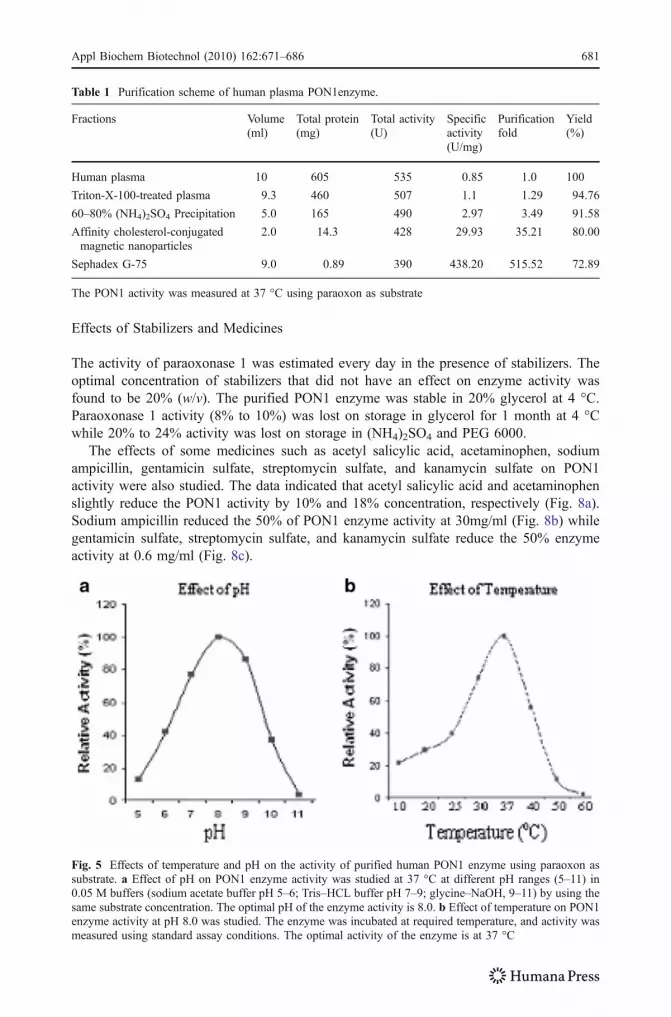
The enzyme is functionally active at pH 8.0 with paraoxon and phenyl acetate. The enzymeactivity was reduced below pH 7.0 and at alkaline pH (Fig. 5a). The optimal activity ofparaoxonase 1 was observed at 37 °C and completely lost at 50 °C (Fig. 5b). The enzymewas stable for 8 to 10 h at 37 °C in PON buffer.
Various metal ions and organic compounds were tested to investigate the inhibitory orstimulatory effect on PON1 enzyme activity; Mg+2 and Mn+2 did not show any inhibitoryor stimulatory effect whereas Pb+2, Co+2, and Zn2+ reduced the enzyme activity while Ni2+,Cd2+, and Cu2+ inhibited the enzyme activity. Glycerol, PEG 6000, ammonium sulfate, andPMSF did not inhibit the enzyme activity while β-mercaptoethanol, ethanol, and acetone
Fig. 2 Proposed structure of conjugation of cholesterol with diazotized p-aminohippuric acid magneticnanoparticles
Appl Biochem Biotechnol (2010) 162:671–686 679

moderately reduced the enzyme activity. Iodoacetic acid, SDS, DMF, and DMSOcompletely destroyed the enzyme activity (Table 2).
The isoelectric point of purified PON1 was observed at 5.3, and the PON1 enzymeshowed a single spot in Western blot of 2D-PAGE which corresponds to purified PON1enzyme. It indicated that the enzyme was purified to homogeneity (Fig. 6). The Km valuesof PON1 enzyme were found as 1.3 and 0.74 mM for paraoxon and phenyl acetate,respectively, by Lineweaver–Burk plot (Fig. 7).
Fig. 3 A chromatographic profile of purification of PON1 enzyme on Sephadex G-75 resin. The fractionscollected from affinity cholesterol-conjugated magnetic nanoparticles were applied on Sephadex G-75column (1.5×30 cm) pre-equilibrated with PON buffer. A 2-ml peak fraction was collected and assayed forPON1 enzyme activity. The fractions showed that the enzyme activity (shadowed area) was pooled andcharacterized
Fig. 4 SDS–PAGE (10%) and Western blot analysis of purified PON1 enzyme. a (lane 1) standardmolecular weight protein marker, (lane2) Triton-X-100-treated human serum, (lane 3) 60–80% ammonium-sulfate-precipitated protein fraction, (lane 4), partially purified PON1 enzyme from affinity cholesterol-conjugated magnetic nanoparticles, (lane 5), purified PON1 enzyme from Sephadex G-75 resin. Afterelectrophoresis, gel was stained with Coomassie brilliant blue R-250. A single protein band of nearly 45 kDaof PON1 enzyme is detected. b (Lane 1) a single immunoreactive protein band of nearly 45 kDa indicatedthe purity and stability of the enzyme (arrow)
680 Appl Biochem Biotechnol (2010) 162:671–686
Effects of Stabilizers and Medicines
The activity of paraoxonase 1 was estimated every day in the presence of stabilizers. Theoptimal concentration of stabilizers that did not have an effect on enzyme activity wasfound to be 20% (w/v). The purified PON1 enzyme was stable in 20% glycerol at 4 °C.Paraoxonase 1 activity (8% to 10%) was lost on storage in glycerol for 1 month at 4 °Cwhile 20% to 24% activity was lost on storage in (NH4)2SO4 and PEG 6000.
The effects of some medicines such as acetyl salicylic acid, acetaminophen, sodiumampicillin, gentamicin sulfate, streptomycin sulfate, and kanamycin sulfate on PON1activity were also studied. The data indicated that acetyl salicylic acid and acetaminophenslightly reduce the PON1 activity by 10% and 18% concentration, respectively (Fig. 8a).Sodium ampicillin reduced the 50% of PON1 enzyme activity at 30mg/ml (Fig. 8b) whilegentamicin sulfate, streptomycin sulfate, and kanamycin sulfate reduce the 50% enzymeactivity at 0.6 mg/ml (Fig. 8c).
Fig. 5 Effects of temperature and pH on the activity of purified human PON1 enzyme using paraoxon assubstrate. a Effect of pH on PON1 enzyme activity was studied at 37 °C at different pH ranges (5–11) in0.05 M buffers (sodium acetate buffer pH 5–6; Tris–HCL buffer pH 7–9; glycine–NaOH, 9–11) by using thesame substrate concentration. The optimal pH of the enzyme activity is 8.0. b Effect of temperature on PON1enzyme activity at pH 8.0 was studied. The enzyme was incubated at required temperature, and activity wasmeasured using standard assay conditions. The optimal activity of the enzyme is at 37 °C
Table 1 Purification scheme of human plasma PON1enzyme.
Fractions Volume(ml)
Total protein(mg)
Total activity(U)
Specificactivity(U/mg)
Purificationfold
Yield(%)
Human plasma 10 605 535 0.85 1.0 100
Triton-X-100-treated plasma 9.3 460 507 1.1 1.29 94.76
60–80% (NH4)2SO4 Precipitation 5.0 165 490 2.97 3.49 91.58
Affinity cholesterol-conjugatedmagnetic nanoparticles
2.0 14.3 428 29.93 35.21 80.00
Sephadex G-75 9.0 0.89 390 438.20 515.52 72.89
The PON1 activity was measured at 37 °C using paraoxon as substrate
Appl Biochem Biotechnol (2010) 162:671–686 681

Discussion
A new methodology was developed for the affinity purification of human serum paraoxonase1. Paraoxonase 1 is an important enzyme to hydrolyze the toxic organophosphorus, nervegasses, insecticides, and pharmaceutical drugs [9, 39]. Human serum paraoxonase 1 waspurified in three sequential steps: ammonium sulfate precipitation, affinity separation by
Fig. 6 2D-PAGE of purified human PON1 enzyme and Western blot analysis of 2D-PAGE. a Firstdimension IEF was placed onto 10% SDS–PAGE for second dimension. A 45-kDa protein spot (arrowhead)corresponds to purified PON1 enzyme and indicates the purity of the protein. (lane1) Standard molecularweight protein marker, (lane 2) purified PON1 enzyme. b The purified PON1 enzyme separated on 2D-PAGE was transferred onto nitrocellulose membrane and proceeded for immunochemical reaction. Animmunoreactive protein spot of 45 kDa (arrow) was highlighted that corresponds to purified PON1 enzyme.(lane1) Standard molecular weight protein marker, (lane 2) an immunoreactive PON1 protein
Table 2 Effect of metal ions and organic compounds on human PON1 enzyme activity.
Metal ions (1.0 mM) Residual activity (%) Organic compounds (5%) Residual activity (%)
Control 100 Glycerol 100
Mn2+ 100 Ammonium sulfate 100
Mg2+ 100 PEG 6000 100
Co2+ 70 Triton X-100 100
Pb2+ 65 PMSF 100
Zn2+ 30 Ethanol 57
Cd2+ 0 β-mercaptoethanol 48
Cu2+ 0 Acetone 18
Ni2+ 0 SDS 0
DMF 0
DMSO 0
Iodoacetate 0
The purified enzyme was incubated with the reagents separately and then enzyme activity was measured at37 °C paraoxon as substrate. (All assays were conducted in duplicate)
PMSF phenylmethylsulfonyl fluoride, SDS sodium dodecyl sulfate, DMF dimethyl formamide, DMSOdimethyl sulfoxide
682 Appl Biochem Biotechnol (2010) 162:671–686

cholesterol-conjugated magnetic nanoparticles, and gel permeation chromatography. Para-oxonase 1 was detached from HDL by treating the serum with nonionic detergent TritonX-100. The maximum enzyme activity was observed at 60–70% ammonium sulfatesaturation. A 0.2% deoxycholate solution was found suitable for the complete elution of
Fig. 8 Effects of medicines on purified PON1 enzyme activity (in vitro). a Acetylsalicylic acid (circles) andacetaminophen (triangles) slightly reduced the enzyme activity. b Sodium ampicillin (squares) reduced the50% enzyme activity at 30 mg/ml. c Gentamicin sulfate (squares), kanamycin sulfate (x marks), andstreptomycin sulfate (triangles) reduced the 50% enzyme activity at 0.6 mg/ml
Fig. 7 Lineweaver–Burk plot forhuman serum paraoxonase 1.Paraoxon (filled circles) andphenyl acetate (empty circles)are used as substrates understandard assay conditions
Appl Biochem Biotechnol (2010) 162:671–686 683

PON1 from cholesterol-conjugated magnetic nanoparticles. The single spot of PON1 wasobtained by Western blot of 2D-PAGE that confirmed the homogeneity of purified enzyme.The optimum pH of the paraoxonase 1 activity was found to be 8.0 which is similar to theprevious reports [27]. A Lineweaver–Burk plot was made by using different concentrationsof paraoxon and phenyl acetate (0.5 to 5 mM) at 37 °C, pH 8.0. The Km values were 1.3 and0.67 mM with paraoxon and phenyl acetate substrates, respectively, which is in closeagreement with the reported values [13, 27].
Paraoxonase 1 activity was also studied in the presence of some metal ions andorganic compounds due to their biological importance. Co2+ is used in diagnosis whileZn2+ is used in therapy as well as in the agricultural field. Cd2+, Cu2+, Ni2+, and Pb2+ areused in metal alloy and are environmentally hazardous metals. The Triton X-100 and SDSare nonionic detergents, and PMSF is used as protease inhibitor. β-Mercaptoethanol andiodoacetate are used as reducing and oxidizing agents. Ammonium sulfate, ethanol, andacetone are used as protein precipitant. The optimal concentration of Mn2+ and Mg2+ ionsthat did not have any effect on paraoxonase 1 activity was 1.00 mM while 5%concentration of organic reagents was optimal to study the enzyme activity. It wasobserved that the enzyme activity was not inhibited by metal ions (Mn2+ and Mg2+),glycerol, ammonium sulfate, PEG 6000, Triton X-100, and PMSF while Co2+, Pb2+, Zn2+,ethanol, β-mercaptoethanol, and acetone moderately inhibit (70% to 30%) the enzymeactivity. Cd2+, Cu2+, Ni2+, SDS, DMF, DMSO, and iodoacetate totally inhibited the enzymeactivity (Table 2).
Paraoxonase 1 not only protects from the oxidation of lipids conjugated with HDL andLDL but can also hydrolyze many pharmaceutical compounds. It is also reported thatsome diuretic and hypocholesterolemic drugs, such as spironolactone, mevastatin,lovastatin, and simvastatin, pravastatin, and prulifloxacin inhibit the PON1 activity[40–42]. So there is need to study the effect of some pharmaceutical medicines onparaoxonase 1 activity to understand its role during therapy. The analgesic (acetylsalicylicacid and acetaminophen) and amino glycoside antibiotics (gentamicin sulfate, kanamycinsulfate, and streptomycin sulfate) are commonly used in fever and during infections. Inorder to study the effect of these medicines, purified human serum paraoxonase 1 wasincubated (in vitro) with medicines, and the enzyme activity was noted. It was observedthat acetylsalicylic acid and acetaminophen slightly reduced the PON1 enzyme activitywhile ampicillin and amino glycoside antibiotics effectively reduced the PON1 enzymeactivity at low concentration.
PON1 is polymorphic in nature as R and Q allozyme and is substrate dependent. Theparaoxon and fenitroxon are hydrolyzed by the R allozyme while phenyl acetate is hydrolyzedby both allozymes. The other compounds such as diazoxon and the nerve gasses (soman andsarin) are hydrolyzed by Q allozyme [3]. The effect of macronutrients on PON1 enzyme levelin human diet has not been studied, but in rodents it is reported that monounsaturated fattyacid increased the PON1 enzyme activity than saturated or highly polyunsaturated fatty acid[43]. An atherogenic diet and reused or degraded cooking oil decrease PON1 enzyme levelactivity in rabbit, mice, and human while polyphenols of tea and fruit juice increase thePON1 enzyme activity in both mice and humans [44, 45].
The purification strategy described here is simple, easy, less expensive, and less timeconsuming. A high yield of purified PON1 with optimal activity was obtained from smallvolume as compared to reported cumbersome methodology for purification of PON1. Thestudy of effect of pharmaceutical medicines on PON1 enzyme activity may be helpful inantibiotic therapy. This is the first report about the new purification strategy which may beequally important like other purification procedures for PON1 enzyme.
684 Appl Biochem Biotechnol (2010) 162:671–686

References
1. Mackness, M. I., Arrol, S., Mackness, B., & Durrington, P. N. (1997). Lancet, 349, 851–852.2. Mochizuki, H., Scherer, S. T., Nickle, D. J., Majer, M., Huizenga, J. J., Tsui, L. C., et al. (1998). Gene,
213, 149–157.3. Davies, H. G., Richter, R., Keifer, J. M., Broomfield, C. A., Sowalla, J., & Furlong, C. E. (1996). Nature
Genetics, 14, 334–336.4. Billecke, S., Draganov, D., Counsell, R., Stetson, P., Watson, C., Hsu, C., et al. (2000). Drug Metabolism
& Disposition, 28(11), 1335–1342.5. Durringhton, P. N., Mackness, B., & Mackness, M. J. (2001). Arteriosclerosis Thrombosis Vascular
Biology, 21, 473–480.6. Sorgob, M. A., & Vilanova, E. (2002). Toxicology Letter, 128, 215–228.7. Cherry, N., Mackness, M., Durrington, P., Povey, A., Dippnall, M., Smith, T., et al. (2002). Lancet, 359,
763–764.8. Mackness, B. P., Durrington, A., Povey, S., Thomson, M., Dippnall, M., Mackness, T., et al. (2003).
Pharmacogenetics, 13, 81–88.9. La Du, B. N. (1992). Pharmacogenetics of drug metabolism. New York: Pergamon Press.10. Sorenson, R. C., Primo-Parma, S. I., Teiber, J., & La Du, B. N. (1996). Genomics, 33, 498–509.11. Draganov, D. I., Stetson, P. L., Watson, C. E., Billecke, S. S., & La Du, B. N. (2000). Journal of
Biological Chemistry, 43, 33435–33442.12. Aviram, M., Billecke, S., Sorenson, R., Bisgaier, C., Newton, R., Rosenblat, M., et al. (1998).
Arteriosclerosis Thrombosis Vascular Biology, 10, 1617–1624.13. Sinan, S., Kockar, F., Gencer, N., Yildirim, H., & Arslan, O. (2006). Biochemistry, 71, 46–50.14. Costa, L. G., Cole, T. B., Jarvik, G. P., & Furlong, C. E. (2003). Annual Review of Medicine, 54, 371–
392.15. Tischer, W., & Wedekind, F. (1999). Topics in Current Chemistry, 200, 95–125.16. Jia, H., Guangyu, Z., & Wang, P. (2003). Biotechnology Bioengineering, 84, 407–413.17. Bornscheuer, U. T. (2003). Angewandte Chemie International, 42, 3336–3337.18. Schutt, W., Gruttner, C., Hafeli, U., Zborowski, M., Teller, J., Putzar, H., et al. (1997). Hybridoma, 16,
109–117.19. Rudge, S. R., Kurtz, T. L., Vessely, C. R., Catterall, L. G., & Williamson, D. L. (2000). Biomaterials, 21,
1411–1420.20. Josephson, L., Perez, J. M., & Weissleder, R. (2001). Angewandte Chemie International, 40, 3204–3206.21. Katz, E., Sheeney-Haj-Ichia, L., Buckmann, A. F., & Willner, I. (2002). Angewandte Chemie
International, 41, 1343–1346.22. Gan, K. N., Smolen, A., Eckerson, H. W., & La Du, B. N. (1991). Drug Metabolism & Disposition, 19,
100–106.23. Gupta, P. K., & Hung, C. T. (1989). Life Sciences, 44, 175–186.24. Sahoo, K. S., & Labhasetwar, V. (2003). Drug Discovery Today, 8, 1112–1120.25. Huang, S. H., Liao, M. H., & Chen, D. H. (2003). Biotechnology Progress, 19, 1095–1100.26. Takeshi, K., Tomoichiro, O., Mitsuaki, I., Tohru, E., Takayuki, F., Eiji, S., et al. (2000). Journal of Lipid
Research, 41, 1358–1363.27. Golmanesh, L., Mehrani, H., & Tabei, M. (2008). Journal of Biochemistry Biophysics Methods, 70,
1037–1042.28. Furlong, C. E., Costa, L. G., Hasett, C., Richter, R. J., Sundstorm, J. A., Alder, D. A., et al. (1993).
Chemical Biology Interaction, 87, 35–48.29. Durrington, P. N., Mackness, B., & Mackness, M. I. (2002). Arteriosclerosis Thrombosis Vascular
Biology, 22, 1248–1250.30. Samra, Z. Q., & Athar, M. A. (2009). Biotechnology Bioprocess Engineering (in press).31. Liao, M. H., & Chen, D. H. (2002). Journal of Materials Chemistry, 12, 3654–3659.32. Weiler, E. W. (1980). Planta, 148, 262–272.33. Moesta, P., Hahn, M. G., & Grisebach, H. (1983). Planta Physiology, 73, 233–237.34. Bradford, M. M. (1976). Analytical Biochemistry, 72, 248–251.35. Laemmli, U. K. (1970). Nature, 227, 680–685.36. Towbin, H. K., Staehelin, T., & Gordon, J. (1979). Proceeding of National Academy of Science, 76,
4350–4359.37. Harlow, E. D., & Lane, D. (1988). Antibodies, a laboratory manual. USA: CSH.38. Walker, J. (1994). Methods in molecular biology: Basic protein and peptide protocol (Vol. 32). Totowa:
Humana.39. Tougou, K., Nakamura, S., & Wantanabe, S. (1998). Drug Metabolism & Disposition, 26, 355–359.
Appl Biochem Biotechnol (2010) 162:671–686 685

40. Malin, R., Laaksonen, R., & Knuuti, J. (2001). Pharmacogenetics, 11, 625–633.41. Tomas, M., Senti, M., Gracia-Faria, F., Vila, J., Torrents, A., Covas, M., et al. (2000). Arteriosclerosis
Thrombosis Vascular Biology, 20, 2113–2119.42. Leviev, I., & James, R. (2000). Atherosclerosis, 151, 41–48.43. Kudchodkar, B. J., Lacko, A. G., Dory, L., & Fungwe, T. V. (2000). Journal of Nutrition, 30, 2427–
2433.44. Shih, D. M., Gu, L., Xia, Y. R., Navab, M., Li, W. F., Hama, S., et al. (1998). Nature, 394, 284–287.45. Aviram, M., Rosenblat, M., Billecke, S., Erogul, J., Sorenson, R., Bisgaier, C. L., et al. (1999). Free
Radical Biology & Medicine, 26(7–8), 892–904.
686 Appl Biochem Biotechnol (2010) 162:671–686