studies on some endoparasites of camels in the southeastern area of egypt
TRANSCRIPT
SCVMJ, XIII (1) 2008 81
STUDIES ON SOME ENDOPARASITES OF CAMELS
IN THE SOUTHEASTERN AREA OF EGYPT
**Mona A. Mahmoud ; *Amin, M.M.;*Youssef, R.R.;**El-Kattan,
A.;**Azza S. A. Goda and**T.R. Abou El-Naga.
*Department of Internal Medicine and Infectious Diseases Fac. Of Vet. Med.
Cairo Univ.
**Animal Health Department, Desert Research Center.
ABSTRACT In the present study, a total of 261 camels (185 local and76 imported)
from Halaieb, Shalateen and Abu-Ramad, were subjected to parasitological
examination for determination of the prevalence of gastrointestinal nematodes
and blood parasites. 41.08% of local and 51.3% of imported camels were found
infested with nematodes. Trichostrongylus spp. was the prevalent one in both
local (20%) and imported camels (36.84%). The other recovered nematodes
were Nematodiru, Trichuris and Strongyloid spp.The blood parasites,
Trypanosoma evansi and Theileria spp. were recorded in 79.4 and 75% of local
and imported camels respectively. Mixed infection of blood parasites and
gastrointestinal nematodes were also found. Spring showed the highest infection
rate of both blood parasites and gastrointestinal nematodes in the study area. In
conclusion, Trypanosoma evansi, Theileria species and gastrointestinal
nematodes constitute large problems affecting camel health and productivity
especially during rainy seasons (Winter and Spring). In brief, the data obtained
proved that imported camels play an important role in the epizootology of
parasitic infection of camels.
INTRODUCTION
Camels play an important
socio-economic role within the pasto-
ral and agricultural system in the dry
and semidry zones of Asia and Africa.
The survival of millions of human
being is dependent on the camel in
such areas. For meat, milk and hair
production and still an important mean
of drought and transportation for large
sectors of pastoral societies (El–
Sawalhy et al., 1996). In 1998, the
total number of camels in Egypt was
135 thousands camels representing
1.1, 0.9 and 0.7% from the total
number of camels in Arabian cou-
ntries, Africa and in the world respect-
82 Mona A. Mahmoud et al.,
ively (Anon, 2000). More than 42
thousands of camels inhabit El-
Shalateen area and the total number of
imported camels from Sudan to Egypt
through El–Shalateen Veterinary qua-
rantine was 91.299 camels in 2001
(Mahran, 2004). Camels in Egypt
annually produce about 20.8, 2.3, 0.62
and 0.09 thousand tons of consumable
milk, meat, hides and fibers respec-
tively (Wardeh, 1992). Due to the few
number of camels raised in Egypt, it is
essential to import camels from other
countries as Sudan, Somalia, Djibouti
and Kenya, thus the occurrence of
exotic diseases at any time could be
expected and also the free movements
of camels throughout the borders leads
to the transmission and spreading of
diseases (Abd El-Aziz, 1996). The area
of the present study (Halaieb, Shalat-
een and Abo-Ramad triangle, Red Sea
Governorate, Egypt) representing the
South Eastern border of Egypt and
considered the major point of entry of
the imported camel from Sudan and
other African countries into Egypt.
The main objective of this
work is to investigate some of endo-
parasites affecting local and imported
camels in the study area.
MATERIAL & METHODS
Study Area:
This region forms a triangular
area reach about 18000 km2, its base is
about 300 km, begins at Halaieb (Lat-
eral line 23º North, Long 36º 45ˉ) and
end at Shalateen (Lateral line 23º 20ˉ,
Long 36º 10ˉ) (El-Shaer, 1999).
Animals:
A total number of 261
dromedary camels (185 local Rashidi
and 76 imported from Sudan) during
the period from August 2003 till
September 2004, were randomly invo-
lved in this study. They were repre-
sented various ages (3–8 years) of
both sexes.
Samples:
1- Faecal samples:
A total of 261 faecal samples
were examined using concentration
floatation technique according to (So-
ulsby, 1982) for oviscopical exami-
nation of gastrointestinal nematodes.
2- Blood samples:
Two blood samples for each
camel were collected from the jugular
vein of 261 camels; the first blood
sample was evacuated in 5ml EDTA
containing vaccutainer for blood sm-
ears, the second used for serum collec-
tion to be used latterly in serological
examination. Detection of blood para-
sites using stained smears technique
according to (Coles, 1986).
3- Clinical examination:
Camels were thoroughly exa-
mined according to the methods desc-
ribed by (Higgins, 1986).
4- Serological examination:
Indirect Enzyme Linked Immuno
Sorbent Assay (ELISA):
SCVMJ, XIII (1) 2008 83
Antigens: Crude antigen of Trypanosoma
evansi was supplied by the Molecular
Biology Unit, Faculty of Vet. Medi-
cine, Cairo University, Egypt.
(ELISA) for determination of antib-
odies of Trypanosoma evansi in camel
sera was done according to (Zwey-
garth et al., 1986 and El–Sawalhy
and Ebeid, 1994).
RESULTS
Infected camels with blood and
gastrointestinal parasites showed signs
of illness including, loss of body we-
ight, edema, decreased animal pro-
duction. The overall prevalence rates
for gastrointestinal nematodes was
41.08and 51.3% for local and impo-
rted camels respectively, while the
seasonal prevalence rates were 40.9,
52.2, 34.6 and 50% in Summer, Autu-
mn, Winter and Spring respect-
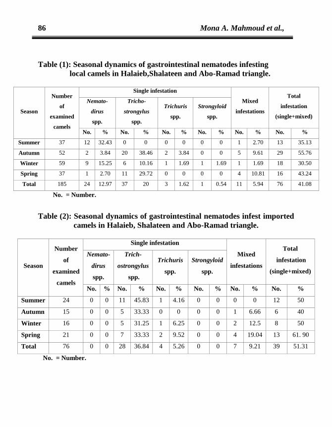
ively,(Tables 1,2 and 3).
In local camels, (Table 1), the
most prevalent spp. was Trichostr-
ongylus (20%) followed by Nemat-
odirus (12.9%) then Trichuris (1.62%)
and Strongyloid spp. (0.54%). In
imported camels, Trichostrongylus
spp. was recorded in 36.84% of exa-
mined camels followed by Trichuris
spp. (5.26%) (Table 2). Mixed infest-
ations were recorded in 5.9%, and
9.2% of local and imported camels
respectively. The highest rate of mixed
infestation was found in Spring
(13.7%) followed by Autumn (8.9%).
Trichostrongylus spp. was found in all
mixed infestation with other species.
(Table 3).
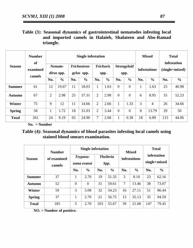
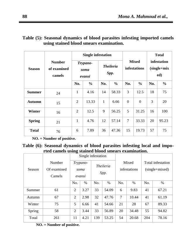
The prevalence of blood para-
sites (T. evansi and Theleria spp.) was
presented in Tables (4 and 5). The
overall infection rate of local camels
was 79.4%. The highest prevalence
94.5% was recorded in spring. In
imported camels, the overall preva-
lence was 75% with highest in winter
(100%). Mixed infection with gastroi-
ntestinal nematodes and T. evansi was
recorded in 7.5 and 14.4% of local and
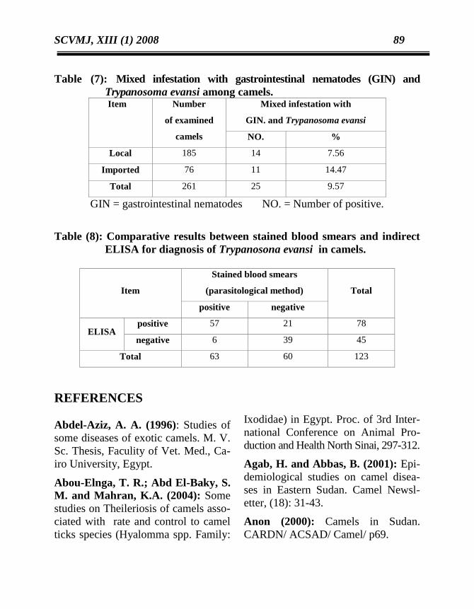
imported camels respectively (Table 7).
Table (8) showed that, 63.1% of tested
camels' sera showed antibodies against
T. evansi while the rate of infection was
51.9% using stained blood smears.
DISCUSSION
The conditions under which
camels are usually kept are not con-
ducive to parasite transmission but su-
rprisingly large numbers of helminthes
are known to occur in the camel (El-
Behari, 1985). Internal parasites cons-
titute an important disease problem
that affect the health and productivity
of camel and is often more sound in
areas and in certain seasons of the year
(Majid et al., 1997). It was necessary,
in our study, to investigate the most
prevalent parasites infesting camels.
84 Mona A. Mahmoud et al.,
The clinical signs (signs of
illness including, loss of body weight,
edema, decreased animal production)
appeared on examined animals were
like that recorded by Mourad and El-
Sherif, (2002). The overall prevalence
rate of gastrointestinal nematodes infe-
station was 41.1 and 51.31% in local
and imported camels respectively, a
relatively similar rate was obtained by
Awad, (1996) who recorded 52.0% in
Egypt and (Anwar and Khan 1998)
recorded 42% in Pakistan, while
higher and lower rates were reported
by Sharrif et al., (1998) 84% in
Jordan, El-Salahy et al., (2000) 60%
in Egypt and Kumar et al., (2001)
37.8% in India. Mourad and El-
Sherif, (2002) in Egypt found that,
Trichostrongylus spp. was the most
prevalent one recorded in 20 and
36.84% of local and imported camels
respectively. These findings were in
agreement with Awad, (1996) and
Magzoub et al., (1997).This might be
a result of adaptation and higher resis-
tance of Trichostrongylus larvae to the
hot dry climatic and other enviro-
nmental conditions (El-Salahy et al.,
2000).
Mixed infestation was reported
with a prevalence of 5.9 and 9.2% in
local and imported camels respect-
ively. Mixed infestation with 2 or 3
parasites in the same camel was
common (El-Molla et al., 1981;
Magzoub et al., 1997 and Sharrif et
al., 1998). Regarding the seasonal variat-
ions, it is well known that the preval-
ence of gastrointestinal helminthes
may vary widely from region to other
as well as during the various seasons
within the same region (El-Behari,
1985). Similar finding was obtained in
this study. The rate of infestation was
peaked during autumn (52.2%)
followed by spring (50%). while the
lowest one was observed during
Winter (34.6%). Similar results had
been recorded by Awad (1996) who
reported an infection rate 67% in
autumn and 51.1% in spring. In fact,
helminthes infestation is precipitated
by the sever stress brought on by the
long migratory trip in which the ca-
mels are subjected to in the early rainy
season (early weeks of autumn).
During this time, camels are also
weak and have poor nutritional status
besides unsuitable climatic condition
in autumn which favor the mainte-
nance and development of larval
stages, while Winter is not drastically
affect the survival of different stages
of parasites (Awad, 1996, and Agab
and Abbas, 2001).
In the present study, the total
prevalence of blood parasites using
stained blood smear examination was
9.4% in local camels and 75% in
imported camels. The prevalence rate
of Trypanosoma evansi was 23.7 and
SCVMJ, XIII (1) 2008 85
27.6% in local and imported camels
respectively, while Theileria spp. was
76.7 and 67.1% of local and imported
camels respectively.
Mixed infestation with the both
blood parasites was recorded in 21.08,
and 19.73% of local and imported
camels respectively. The variations of
rates of Trypanosoma evansi which
were recorded in Egypt, either higher
rates Mahran, 2004 (11.5%), and
Baraka et al. 2005 (26.6%), or lower
rates of same spp. Fayed et al., 1984
(3.7%) Laila et al., 2001 (4.1%). In
our opinion, these variations may be
attributed to nature of the study area,
climatic condition and animal influen-
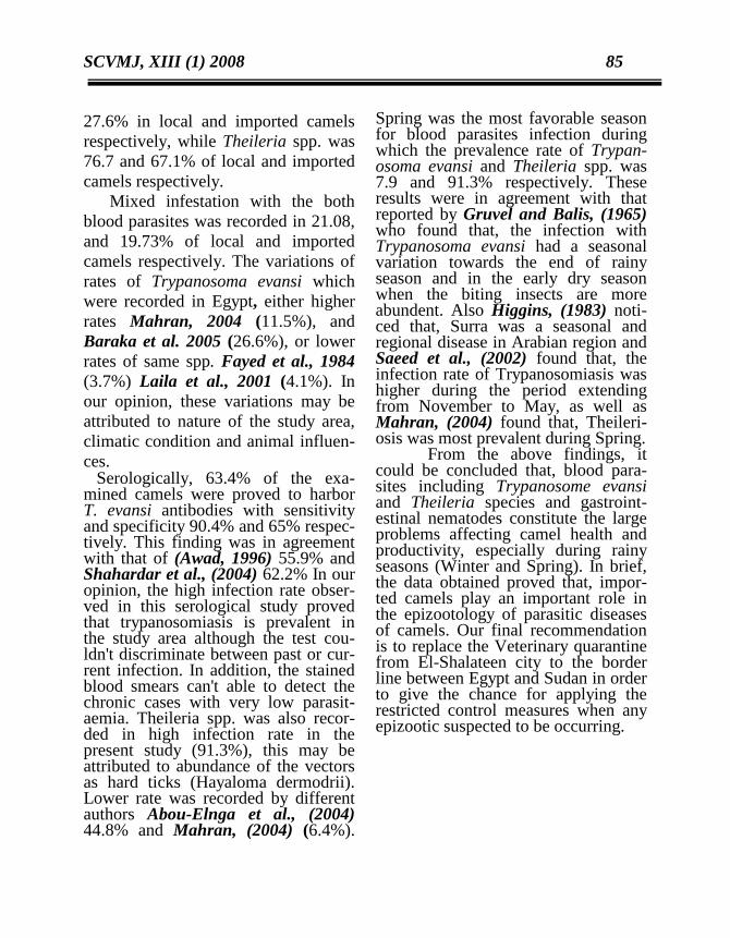
ces. Serologically, 63.4% of the exa-mined camels were proved to harbor T. evansi antibodies with sensitivity and specificity 90.4% and 65% respec-tively. This finding was in agreement with that of (Awad, 1996) 55.9% and Shahardar et al., (2004) 62.2% In our opinion, the high infection rate obser-ved in this serological study proved that trypanosomiasis is prevalent in the study area although the test cou-ldn't discriminate between past or cur-rent infection. In addition, the stained blood smears can't able to detect the chronic cases with very low parasit-aemia. Theileria spp. was also recor-ded in high infection rate in the present study (91.3%), this may be attributed to abundance of the vectors as hard ticks (Hayaloma dermodrii). Lower rate was recorded by different authors Abou-Elnga et al., (2004) 44.8% and Mahran, (2004) (6.4%).
Spring was the most favorable season for blood parasites infection during which the prevalence rate of Trypan-osoma evansi and Theileria spp. was 7.9 and 91.3% respectively. These results were in agreement with that reported by Gruvel and Balis, (1965) who found that, the infection with Trypanosoma evansi had a seasonal variation towards the end of rainy season and in the early dry season when the biting insects are more abundent. Also Higgins, (1983) noti-ced that, Surra was a seasonal and regional disease in Arabian region and Saeed et al., (2002) found that, the infection rate of Trypanosomiasis was higher during the period extending from November to May, as well as Mahran, (2004) found that, Theileri-osis was most prevalent during Spring.
From the above findings, it could be concluded that, blood para-sites including Trypanosome evansi and Theileria species and gastroint-estinal nematodes constitute the large problems affecting camel health and productivity, especially during rainy seasons (Winter and Spring). In brief, the data obtained proved that, impor-ted camels play an important role in the epizootology of parasitic diseases of camels. Our final recommendation is to replace the Veterinary quarantine from El-Shalateen city to the border line between Egypt and Sudan in order to give the chance for applying the restricted control measures when any epizootic suspected to be occurring.
86 Mona A. Mahmoud et al.,
Table (1): Seasonal dynamics of gastrointestinal nematodes infesting
local camels in Halaieb,Shalateen and Abo-Ramad triangle.
Season
Number
of
examined
camels
Single infestation
Mixed
infestations
Total
infestation
(single+mixed)
Nemato-
dirus
spp.
Tricho-
strongylus
spp.
Trichuris
spp.
Strongyloid
spp.
No. % No. % No. % No. % No. % No. %
Summer 37 12 32.43 0 0 0 0 0 0 1 2.70 13 35.13
Autumn 52 2 3.84 20 38.46 2 3.84 0 0 5 9.61 29 55.76
Winter 59 9 15.25 6 10.16 1 1.69 1 1.69 1 1.69 18 30.50
Spring 37 1 2.70 11 29.72 0 0 0 0 4 10.81 16 43.24
Total 185 24 12.97 37 20 3 1.62 1 0.54 11 5.94 76 41.08
No. = Number.
Table (2): Seasonal dynamics of gastrointestinal nematodes infest imported
camels in Halaieb, Shalateen and Abo-Ramad triangle.
Season
Number
of
examined
camels
Single infestation
Mixed
infestations
Total
infestation
(single+mixed)
Nemato-
dirus
spp.
Trich-
ostrongylus
spp.
Trichuris
spp.
Strongyloid
spp.
No. % No. % No. % No. % No. % No. %
Summer 24 0 0 11 45.83 1 4.16 0 0 0 0 12 50
Autumn 15 0 0 5 33.33 0 0 0 0 1 6.66 6 40
Winter 16 0 0 5 31.25 1 6.25 0 0 2 12.5 8 50
Spring 21 0 0 7 33.33 2 9.52 0 0 4 19.04 13 61. 90
Total 76 0 0 28 36.84 4 5.26 0 0 7 9.21 39 51.31
No. = Number.
SCVMJ, XIII (1) 2008 87
Table (3): Seasonal dynamics of gastrointestinal nematodes infesting local
and imported camels in Halaieb, Shalateen and Abo-Ramad
triangle.
Season
Number
of
examined
camels
Single infestation Mixed
infestations
Total
infestation
(single+mixed) Nemato-
dirus spp.
Trichostron-
gylus spp.
Trichuris
spp.
Strongyloid
spp.
No. % No. % No. % No. % No. % No. %
Summer 61 12 19.67 11 18.03 1 1.63 0 0 1 1.63 25 40.98
Autumn 67 2 2.98 25 37.31 2 2.98 0 0 6 8.95 35 52.23
Winter 75 9 12 11 14.66 2 2.66 1 1.33 3 4 26 34.66
Spring 58 1 1.72 18 31.03 2 3.44 0 0 8 13.79 29 50
Total 261 24 9.19 65 24.90 7 2.68 1 0.38 18 6.89 115 44.06
No. = Number
Table (4): Seasonal dynamics of blood parasites infesting local camels using
stained blood smears examination.
NO. = Number of positive.
Season Season
Number
of examined
camels
Single infestation Mixed
infestations
Total
infestation
single+mixed
Trypano-
soma evansi
Theileria
Spp.
No. % No. % No. % No. %
Summer 37 1 2.70 19 51.35 3 8.10 23 62.16
Autumn 52 0 0 31 59.61 7 13.46 38 73.07
Winter 59 3 5.08 32 54.23 16 27.11 51 86.44
Spring 37 1 2.70 21 56.75 13 35.13 35 94.59
Total 185 5 2.70 103 55.67 39 21.08 147 79.45
88 Mona A. Mahmoud et al.,
Table (5): Seasonal dynamics of blood parasites infesting imported camels
using stained blood smears examination.
Se Season Season
Number
of examined
camels
Single infestation
Mixed
infestations
Total
infestation
(single+mix
ed)
Trypano-
soma
evansi
Theileria
Spp.
No. % No. % No. % No. %
Summer 24 1 4.16 14 58.33 3 12.5 18 75
Autumn 15 2 13.33 1 6.66 0 0 3 20
Winter 16 2 12.5 9 56.25 5 31.25 16 100
Spring 21 1 4.76 12 57.14 7 33.33 20 95.23
Total 76 6 7.89 36 47.36 15 19.73 57 75
NO. = Number of positive.
Table (6): Seasonal dynamics of blood parasites infesting local and impo-
rted camels using stained blood smears examination.
Season
Number
Of examined
Camels
Single infestation
Mixed
infestations
Total infestation
(single+mixed)
Trypano-
soma
evansi
Theileria
Spp.
No. % No. % No. % No. %
Summer 61 2 3.27 33 54.09 6 9.83 41 67.21
Autumn 67 2 2.98 32 47.76 7 10.44 41 61.19
Winter 75 5 6.66 41 54.66 21 28 67 89.33
Spring 58 2 3.44 33 56.89 20 34.48 55 94.82
Total 261 11 4.21 139 53.25 54 20.68 204 78.16
NO. = Number of positive.
SCVMJ, XIII (1) 2008 89
Table (7): Mixed infestation with gastrointestinal nematodes (GIN) and
Trypanosoma evansi among camels. Item Number
of examined
camels
Mixed infestation with
GIN. and Trypanosoma evansi
NO. %
Local 185 14 7.56
Imported 76 11 14.47
Total 261 25 9.57
GIN = gastrointestinal nematodes NO. = Number of positive.
Table (8): Comparative results between stained blood smears and indirect
ELISA for diagnosis of Trypanosona evansi in camels.
Item
Stained blood smears
(parasitological method)
Total
positive negative
ELISA positive 57 21 78
negative 6 39 45
Total 63 60 123
REFERENCES
Abdel-Aziz, A. A. (1996): Studies of
some diseases of exotic camels. M. V.
Sc. Thesis, Faculity of Vet. Med., Ca-
iro University, Egypt.
Abou-Elnga, T. R.; Abd El-Baky, S.
M. and Mahran, K.A. (2004): Some
studies on Theileriosis of camels asso-
ciated with rate and control to camel
ticks species (Hyalomma spp. Family:
Ixodidae) in Egypt. Proc. of 3rd Inter-
national Conference on Animal Pro-
duction and Health North Sinai, 297-312.
Agab, H. and Abbas, B. (2001): Epi-
demiological studies on camel disea-
ses in Eastern Sudan. Camel Newsl-
etter, (18): 31-43.
Anon (2000): Camels in Sudan.
CARDN/ ACSAD/ Camel/ p69.
90 Mona A. Mahmoud et al.,
Sharrif, L. A. Al-Ani, F. K.; andAl-
Rawashdeh, O. F.; (1998): Camels
diseases in Jordon. Proceedings of The
Third Annual Meeting for Animal
Production Under Arid Conditions,
United Arab Emirates University, 2:
77-92.
Anwar, A. H. and Khan, M. N.
(1998): Parasitic founa of camel in
Pakistan. Proceedings of The Third
Annual Meeting for Animal Pro-
duction Under Arid conditions, United
Arab Emirates University, 2: 69-76.
Awad, H. (1996): Studies on parasitic
infection in camels. Ph. D. Thesis,
Faculty of Vet. Med. Cairo University,
Egypt.
Baraka, T. A.; Abdou, T. A.; and
Abou-Elnga, R. (2005): Clinical and
laboratory studies of rumen perfor-
mance and blood hemato-biochemical
status in Trypanosome infected cam-
els. 4th
Int. Sci. Conf. April 2005, 1-
14.
Coles, E. H. (1986) : Veterinary Clin-
ical pathology, 4th Ed. Sounders Comp.
Philadelphia, London, Toronto. Sch-
midt.
El-Behari, S. (1985): Helminthiasis of
camels. British Vet. J., 141 : 315-326 .
El-Molla, A. and Fahmy, F. (1981): Trials with some anthelmintics against
gastrointestinal nematodes in camels.
Zagazig Vet. Journal, IV : 24-30.
El-Salahy, M.; Monib, M. and Ar-
afa, M. I. (2000): Prasitological stud-
ies of some gastrointestinal parasites
of camels in Assiut governorate with
special reference to zoonotic nema-
todes. Assiut Vet. Med. J., 43 (87) :
280-294.
El-Sawalhy, A. A. and Ebeid, M. A.
(1994): Evaluation of different meth-
ods of diagnosis for Camel Trypano-
somiasis Beni-Suef. Vet. Med. Res. IV
(I/I) : 70-79.
El-Sawalhy, A. A.; Montaser, A. M.
and Rizk, L. G. (1996) :Proceedings
of the 4th
sci. cong. Vet. Med. J. Giza,
44 (2) : 323-329.
El-Shaer, H. (1999): Characterization
of camel breed in Shlateen- Halaieb
triangle in Southern Egypt. ( ARDN/
ACSAD/ Camel / P 67).
Fayed, A. A.; Arab, R. M.; and Abo-
zeid, A. A. (1984): Studies on some
diagnostic aids and chemotherapeutic
druges of camels trypanosomiasis.
Vet. Med. J., 32 (3) : 219-227.
Gruvel, J. and Balis, J. (1965): Tryp-
anosoma evansi infection in camels in
Chad Republic. Principle vectors Re-
vue. Eler. Med. Vet. Pays. Top. 18:
435-449.
Higgins, A. J. (1983): Observation on
the diseases of the Arabian camel
(Camelus dromedarius) and their
SCVMJ, XIII (1) 2008 91
control.A review. Vet. Bull., 53 (12):
1089-1097.
Higgins, A. J. (1986): The camel in
health and disease. Baillere, Tindall,
London, Philadelphia Toronto.
Kumar, R.; Sena, D. S.; Mal, G. and
Sahani, M. S. (2001): Epizootiol-
ogical, Haemato-biochemical and the-
rapeutic studies on gastrointestinal
nematodosis in Indian dromedary ca-
mel. International Conference of Repr-
oduction and Production of Camelids,
Al-Ain,United Arab Emirates, p.15.
Laila, S. Ahmed; Allawy, T. A. and
Mohamed, A. A. M. (2001) : Some
epidemiological aspects of camel try-
panosomiasis in Upper Egypt. Inter-
national Conference on Reproduction
and Production of Camelids, Al-Ain,
United Arab Emirates, p. 14.
Magzoub, M.; Omer, O. H.; and
Haroun, E. M. (1997): Gastrointe-
stinal parasites of dromedary camels
in Gassim region, Saudi Arabia. Ind.
Vet. J., 74: 373-376.
Mahran, O. M. (2004): Some studies
on blood parasites in camels (Camelus
dromedarius) at Shalateen city, Red
Sea Governorate. Assuit Vet. Med. J.,
50 (102) : 172-184.
Majid, A. A.; Yagoub, I. A. and
Ibtisam, A. G. (1997): Internal para-
sites of camels in central Sudan State.
ACSAD/ CARDN. The annual tech-
nical report, p. 99.
Mourad, S, D. and EL-Sherif, A. M.
(2002): Epizootiological study on the
relation between Imported camels fr-
om Sudan and some parasitic diseases
in Egypt. 10th
Sci. Cong. 2002 Fac.
Vet. Med. Assuit Univ. Egypt. 321-
331.
Saeed, E. M.; El-Amin, E. A. and
El-Bashir, M. O. (2002) : Investig-
ation of cameline Trypano-somiasis in
Mid-Eastern Sudan using Ag ELISA
conjunction with parasitological exa-
mination. Deutscher Tropentag, Witz-
enhausen. Proc. of Challenges to Org-
anic Farming and Sustainable Land
Use in the Tropics and Subtropics.
Shahardar, R. A.; Mishra, A. K.
and Rao, J. R. (2004): Detection of
antibodies against Trypano-soma eva-
nsi in dromedary camels by ELISA
using solubilized antigens. Ind. J. of
Animal Science, 74 (1): 3-6.
Sharrif, L.; Al-Rawashdeh, O. M.;
Al-Qudah, K. M. and Al-Ani, F. K.
(1998):Prevalence of gastrointestinal
helminthes, hydatid cysts and nasal
myiasis in camels in Jordan. Procee-
ding of The Third Annual Meeting for
Animal Production Under Arid Cond-
itions, United Arab Emirates Univer-
sity, 2 : 108-114.
92 Mona A. Mahmoud et al.,
Soulsby, E. J. L. (1982): Helminth,
arthropods and protozoa of domesti-
cated animals. 7th
Ed. The English
language book society. Balliere, Tin-
dall and Cassel. London.
Wardeh, M.F. (1992): The impor-
tance of the dromedary camel in the
Arab countries. ACSAD, Camel New-
sletter, (9): 15-19.
Zweygarth, E.; Sabawa, C. and Rot-
tcher, D. (1986): An enzyme-lin-ked
immunosorbent assay for the detection
of antibodies to T.bruci and T. evansi
in Camelus dromdarus using
peroxidase-conjugated protein A. Tro-
pical Med. and Para., (37): 105-106.
الملخص العربى
فىى مطقةىة المثلىال حاليى ( جمل مستورد 66جمل محلي و 281)جمال 162اجريت هذة الدراسة على
فوجىد أن , اجراء فحوص لقفيليات الدم وقفيليىات امماىاءعلى هىذة الجمىالوقد تم , والشالتين وأبو رماد
وتىم %. 12,15و الجمىال المسىتوردة% 82,18طسبة االصابة بالقفيليات الداخليىة فىى الجمىال المحليىة
التوصل الى أن جطس التريكوسىتروطجيلس كىان مىن أكثىر القفيليىات اصىابة فىى كىال مىن الجمىال المحليىة
يليهىىىا االصىىىابة بكىىىال مىىىن الطيمىىىاتوديرس وجىىىطس , %56,88مىىىال المسىىىتوردة بطسىىىبة والج% 11بطسىىىبة
وأوضىحت الدراسىة الىى أن االصىابة بتريباطوسىوما ايفىاطلاي والثيليريىا .تركيوريس ثم جطس ستروطجيلويد
كما وجدت اصابات ملدوجة . فى الجمال المستوردة% 61فى الجمال المحلية و% 8,,6كاطت بطسبة
واثبتت الدراسة أن اعلى مادل لالصابة كان فى موسم الربيى وذلى . الدم وقفيليات االمااء من قفيليات
والخالصىىة أن كىىال مىىن التريباطوسىىوما ايفىىاطلاي والثيليريىىا وقفيليىىات االماىىاء تسىىب . فىىى مطقةىىة البحىىال
قىد اثبتىت و. مشاكل صحية واطتاجية كثيرة للجمال فى هذة المطاقق وخاصة فىى موسىمى الشىتاء والربيى
. الدراسة أن الجمال المستوردة تلا دورا هاما فى طةل الادوي للجمال المحلية