sterically stabilized liposomes as a platform for salinomycin metal coordination compounds:...
TRANSCRIPT

215
Sterically stabilized liposomes as a platform for salinomycin metal coordination compounds: physicochemical characterization
and in vitro evaluation D. Momekova1*, G. Momekov2, J. Ivanova3, I. Pantcheva4, E. Drakalska1, N. Stoyanov5, 6, M. Guenova5, 6,
A. Michova5-7, K. Balashev8, S. Arpadjan4, M. Mitewa4, S. Rangelov9, N. Lambov1
1Department of Pharmaceutical Technology and Biopharmaceutics, 2Lab. of Experimental Chemotherapy, Department of Pharmacology, Pharmacotherapy and Toxicology, Faculty of Pharmacy, Medical University-Sofia, 2 Dunav Str., 1000 Sofia, Bulgaria
3Department of Chemistry, Biochemistry, Physiology and Pathophysiology, Faculty of Medicine, 4Department of Analytical Chemistry, Faculty of Chemistry and Pharmacy, 8Department of Physical Chemistry, Faculty of Chemistry and Pharmacy,
University of Sofia “St. Kliment Ohridski”, Sofia, Bulgaria5Laboratory of Hematopathology and Immunology, 6Center of Excellence, Translational Research in Hematology,
National Specialized Hospital for Active Treatment of Hematological Diseases, Sofia, Bulgaria7Department of Immunology, National Center of Infectious and Parasitic Diseases, Sofia, Bulgaria
9Institute of Polymers, Bulgarian Academy of Sciences, Sofia, Bulgaria*Correspondence: [email protected]
Sterically stabilized DPPC:CHOL:DSPE-PEG-2000 liposomal formulations of the lipophilic complexes of salinomycin with Na(I), K(I), Mn(II), Co(II), and Ni(II) ions were prepared by film-hydration method at different drug-to-DPPC molar ratios. For the K(I) and Na(I) complexes, optimal loading was established at a drug-to-DPPC molar ratio of 0.5:1, whereas for the Me(II) complexes, it was encountered at 0.1:1. DLS revealed uniform LUV populations (130-160 nm) with monomodal size distribution, further corroborated by AFM. Free and entrapped salinomycinates exhibited cytotoxicity in three human tumor cell lines, whereby the liposomal agents were superior vs. free complexes. DNA-fragmentation and flow cytometric assays showed that the cytotoxicity of free and liposomal salinomycinates is mediated by the induction of apoptosis and G1 arrest. The ability of the carriers to retain the bio-activity of the entrapped cargo gives us reason to conclude that the presented DPPC:CHOL:DSPE-PEG-2000 liposomes are suitable platforms for the salinomycin complexes, needing further evaluation and optimization.
Key words: Salinomycin – Metal complexes – Sterically stabilized liposomes – Cytotoxicity – Apoptosis.
J. DRUG DEL. SCI. TECH., 23 (3) 215-223 2013
Salinomycin is a natural antibiotic produced by Streptomyces albus [1-3]. It is a representative monocarboxylic polyether ionophore, comprising a distinctive tricyclic spiroketal ring system in its structure (Figure 1) [2]. As a class, the polyether antibiotics (also denoted as carboxylic ionophores) share the ability to facilitate bilateral ion flux across cellular membranes, altering the normal concentration gradients and conversely interfering with natural ion transport systems in both prokaryotic and eukaryotic cells [1, 4-8]. Salinomycin in particular is an ionophore with a preference for K+ and has a broad spectrum of antimicrobial activity with noteworthy effects against Gram-positive bacteria [9-11] and protozoa [12-14]. This drug is widely used for the chemoprophylaxis and treatment of coccidiosis in poultry and ruminants [12, 15], and as a growth promoter additive in cattle, sheep and swine breeding [16-18]. Recently there has been tremendous research into novel anticancer agents capable of bypassing or modulating chemotherapy-resistance mechanisms in cancer cells [19-21] and especially able to selectively target the generally non-responsive population of cancer stem cells [22-24]. Cancer stem cells (CSCs) comprise a minority cell population that lacks differentiation markers and have a high capacity for cell proliferation but, unless stimulated, may divide infrequently [23, 25]. It is well established that the destruction of differentiated tumor cells by cytoreductive treatment without the simultaneous killing of tumor stem cells will not lead to permanent tumor eradication; thus, CSCs have become an increasingly important target of cancer therapy [23, 24]. Salinomycin has been shown recently to selectively eradicate breast cancer CSCs [22, 26, 27], a finding which was eventually corroborated in other malignancies such as: gastric cancer [28], prostate cancer [29, 30], colon cancer [31], lung adenocarcinoma [32], leukemias [33],
and uterine sarcoma [34]. Mechanistic investigations have shown that salinomycin activates unconventional cell death signaling pathways in human cancer cells, including cell lines with a low responsive-ness to chemotherapy and apoptotic stimuli [27, 35, 36]. Moreover, salinomycin has been well documented to modulate the multi-drug resistance phenotype in tumor cells overexpressing P-glycoprotein or other ATP-binding cassette xenobiotic transporters [37-39]. On these grounds, salinomycin could be considered a promising anticancer lead compound that bypasses multiple mechanisms of apoptosis resistance in human cancer cells and is especially active against non-responsive tumor stem cell populations [7, 27]. The promising pharmacological properties of this agent have fuelled drug design programs aimed at developing more active or better-tolerated analogues [10, 13, 40]. Due to the presence of oxygen atoms in various groups (carboxylic, ether, hydroxyl), polyether antibiotics represent potential ligands able to bind metal cations, and much research interest has been focused upon synthesis of their coordination compounds in
Figure 1 - Schematic diagram of the adsorption process of a single liposome: (A) before contact with the mica, and (B) after adsorption.

216
an attempt to enhance biological activity. The polyether antibiotics are best known as monovalent ionophores for their affinity to bind alkali and Ag+ ions [41, 42] but more recently, salinomycin and monensin have been shown to form complexes with metal(II) ions (e.g. Zn2+, Cu2+, Co2+, Ni2+ , etc.) [40, 43-46]. These exert enhanced cytotoxic activity against tumor cell lines, and/or superior antimicrobial effects, as compared to the free ligands [40, 43, 45-47]. Despite the enormous scope for evaluation of salinomycin and its derivatives as anticancer agents, they suffer from inherent safety and pharmaceutical peculiarities which have been large caveats for advancing the field beyond the non-clinical level. The ionophoric activity of salinomycin at cellular and mitochondrial membranes is responsible for prominent adverse and toxic effects such as inotropic and sympathomimetic activities, myocardial lesions and rhabdomy-olysis [48], neuronal cell death [49] and adrenal medulla lesions [50]. An exemplary human exposure of estimated 1 mg/kg of the antibiotic resulted in significant intoxication with cardiovascular effects and rhabdomyolysis [48]. Moreover, salinomycin and its salts are excep-tionally lipophilic, and practically insoluble in water. Salinomycin has a large molecular mass and is a P-gp substrate [38] which conditions an unfavorable biopharmaceutical and pharmacokinetic profile. In an attempt to both modify the structure of the lead compound and to develop a suitable drug carrier, we hereby present the for-mulation of a series of salinomycin metal complexes in nano-sized vehicles based on sterically stabilized liposomes. The choice of the drug delivery system is based on the favorable biocompatibility and safety of sterically stabilized liposomes, their ability to beneficially modify the pharmacokinetic properties and the organ toxicity of cy-totoxic agents, and to ascertain tumor-targeted delivery via enhanced permeation and retention (EPR) effect [51-53]. Moreover, an additional beneficial feature of liposomal formulations is the solubilization they afford in case of water insoluble cytotoxic agents [54, 55] such as the polyether antibiotics. Liposomal formulations of the structurally related ionophore monensin have been elaborated and validated as a suitable drug delivery system for this compound [56-59]. However, to the best of our knowledge, this is the first report on the design and characterization of liposomal carriers of metal complexes of salino-mycin.
I. MATERIALS AND METHODS1. Chemicals and reagents The commercially available pharmaceutical grade salinomycin so-dium (C42H69O11Na; SalNa) was provided by Biovet Ltd., Bulgaria, and was used without further purification. Solvents (MeCN, MeOH, DMSO, hexane, THF) and metal salts (KNO3, Co(NO3)2.6H2O, MnCl2.4H2O and Ni(NO3)2.6H2O) and the tetrazolium salt 3-(4,5-dimethylthiazol-2-yl)-2,5-diphenyl tetrazolium bromide (MTT) were purchased from Merck (Germany). Dipalmitoyl phosphatidyl choline (DPPC), cholesterol (CHOL), phosphate buffer (pH 7.4), chloroform, RPMI 1640, fetal calf serum (FCS) were purchased from Sigma Chemical Co. (United States). Distearoyl phosphatidyl ethanolamine-PEG2000 (DSPE-PEG2000) was a generous gift from Lipoid GmbH (Germany). Formic acid, 2-propanol, L-glutamine and ethidium bromide were purchased from AppliChem GmbH, Darmstadt, Germany.
2. Synthesis of salinomycin complexes with potassium(I), manganese(II), cobalt(II) and nickel(II) ions2.1. Synthesis of K(I) salinomycinate The antibiotic (sodium salinomycin, 0.25 mmol, 193 mg) was dissolved in 20 mL mixed solvent (MeOH:MeCN = 10:1). KNO3 (0.5 mmol, 51 mg) dissolved in 2 mL water was added to the solu-tion of the ligand. The mixture was stirred for 15 min. After 10 days, a white solid phase was isolated. The precipitate was washed with water and dried at room temperature over P4O10. The complex was
characterized using various spectroscopic methods such as IR, NMR, FAB-MS techniques (unpublished results). The elemental analysis of the complex of salinomycin with ions of K(I) revealed that the new compound possessed a composition of SalK. Yield 158 mg, 80 %. Anal. Calcd. for C42H69O11K (MW = 789): H, 8.81; C, 63.93; K, 4.95. Found: H, 8.80; C, 63.80; K, 4.90 %.
2.2. Synthesis of Mn(II,) Co(II), and Ni(II)) disalinomycinates MnCl2.4H2O (0.5 mmol, 198 mg), dissolved in 2 mL water, was added to a solution of sodium salinomycin (0.25 mmol, 193 mg in MeOH:MeCN = 10:1) and the procedure described above was ap-plied. The coordination mode of salinomycin was evaluated by IR, EPR and FAB-MS spectroscopies (author’s personal communication). The complex obtained is of composition [Mn(Sal)2(H2O)2]. Yield 139 mg, 70 %. Anal. Calcd. for C84H142O24Mn (MW = 1591): H, 9.00; C, 63.42; Mn, 3.45. Found: H, 8.50; C, 63.88; Mn, 3.45 %. Synthesis and structure elucidation of the complexes of salinomycin with ions of Co(II) and Ni(II) have been previously described [40]. The metal complexes of salinomycin are not soluble in water and possess particular solubility in MeOH, MeCN, EtOH, CHCl3, DMSO and hexane. Elemental analysis (C, H) of the compounds was performed with a VarioEL V5.18.0 Elemental Analyzer. For the AAS determination of metal content, the samples were dissolved with conc. HNО3. AAS was conducted with a Perkin Elmer 1100 B using a stock standard solution (Merck, 1000 µg/mL); the working reference solutions were prepared after suitable dilution.
3. Preparation of liposomes DPPC, CHOL (2:1 molar ratio; 10 μmol total lipid/L), 5 mol% DSPE-PEG2000 and the salinomycin complexes in a chosen drug-to-DPPC molar ratio were placed into 50 mL round-bottomed flasks and dissolved in chloroform. Thereafter, the solvent was evaporated in vacuo using a rotary vacuum evaporator (Buchi, Germany). Phosphate buffered saline (PBS) (pH 7.4) was added to the dry lipid/salinomycin derivative film and the resulting dispersions were subjected to eleven freeze-thaw cycles. Thereafter, the dispersions were extruded five times through polycarbonate filters of pore size 200 nm and eight times through polycarbonate filters of pore size 100 nm using a T001 thermobarrel extruder (Lipex Biomembranes Ltd., Canada) at 45 °C. The unentrapped compounds were removed by gel filtration through a Sephadex G50 column (Pharmacia, Sweden), equilibrated with PBS (pH 7.4). The phospholipid content of the final formulations was determined by phosphate analysis, as previously described [60].
4. Determination of drug entrapment efficiency The liposomes were analysed by atomic absorption spectrometry for the determination of drug loading efficiency. The amount of Na, K, Co, Mn and Ni was determined by a Perkin Elmer AAAnalyst 400 flame atomic absorption spectrometer. The instrumental parameters were optimized to obtain maximum signal-to-noise ratio for standard analyte solutions aspirated into the flame.
5. Size and size distribution determination by dynamic light scattering (DLS) The size and the size distribution of the liposomes were determined using a NanoZS zetasizer apparatus (Malvern, United Kingdom). The light source was a 4 mW He-Ne laser operating with polarized light of wavelength of 632.8 nm. DLS measures the time correlation of the intensity of the scattered light, which is related to the time correlation of the field. The time correlation function decays exponentially with a decay rate Γ, which depends on the size of the particles. Polydisperse samples were characterized by a distribution of decay rates. The light scattering data were analyzed using the Contin method. The intensity correlation function was measured three times for each sample at a fixed angle of 173° (backscatter detection). The
Sterically stabilized liposomes as a platform for salinomycin metal coordination compounds:physicochemical characterization and in vitro evaluation
D. Momekova, G. Momekov, J. Ivanova, I. Pantcheva, E. Drakalska, N. Stoyanov, M. Guenova,A. Michova, K. Balashev, S. Arpadjan, M. Mitewa, S. Rangelov, N. Lambov
J. DRUG DEL. SCI. TECH., 23 (3) 215-223 2013
217
hydrodynamic diameter at θ = 173°, d173, was calculated using the Stokes-Einstein equation:
D = kT/(3πηD)
where kT is the thermal energy factor, D is the diffusion coefficient and η is the temperature-dependent viscosity of water. The measure-ments were made at 25 °C in phosphate buffered saline (pH 7.4) and run in triplicate.
6. ζ-potential measurement The ζ-potential was determined using a NanoZS zetasizer appara-tus (Malvern Instruments Ltd., United Kingdom). The measurements were performed at 25 °C in a PBS of pH 7.4.
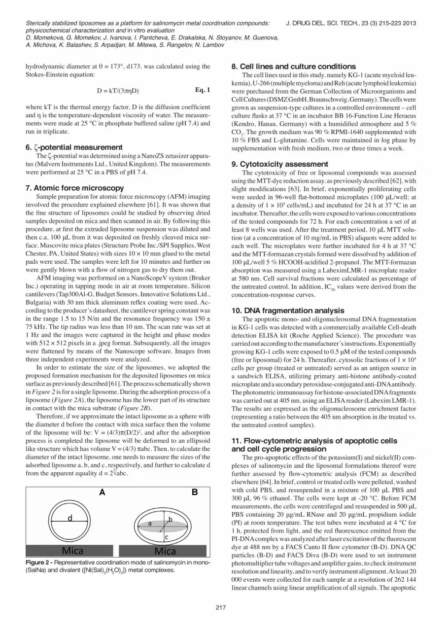
7. Atomic force microscopy Sample preparation for atomic force microscopy (AFM) imaging involved the procedure explained elsewhere [61]. It was shown that the fine structure of liposomes could be studied by observing dried samples deposited on mica and then scanned in air. By following this procedure, at first the extruded liposome suspension was diluted and then c.a. 100 μL from it was deposited on freshly cleaved mica sur-face. Muscovite mica plates (Structure Probe Inc./SPI Supplies, West Chester, PA, United States) with sizes 10 × 10 mm glued to the metal pads were used. The samples were left for 10 minutes and further on were gently blown with a flow of nitrogen gas to dry them out. AFM imaging was performed on a NanoScopeV system (Bruker Inc.) operating in tapping mode in air at room temperature. Silicon cantilevers (Tap300Al-G, Budget Sensors, Innovative Solutions Ltd., Bulgaria) with 30 nm thick aluminum reflex coating were used. Ac-cording to the producer’s datasheet, the cantilever spring constant was in the range 1.5 to 15 N/m and the resonance frequency was 150 ± 75 kHz. The tip radius was less than 10 nm. The scan rate was set at 1 Hz and the images were captured in the height and phase modes with 512 × 512 pixels in a .jpeg format. Subsequently, all the images were flattened by means of the Nanoscope software. Images from three independent experiments were analyzed. In order to estimate the size of the liposomes, we adopted the proposed formation mechanism for the deposited liposomes on mica surface as previously described [61]. The process schematically shown in Figure 2 is for a single liposome. During the adsorption process of a liposome (Figure 2A), the liposome has the lower part of its structure in contact with the mica substrate (Figure 2B). Therefore, if we approximate the intact liposome as a sphere with the diameter d before the contact with mica surface then the volume of the liposome will be: V = (4/3)p(D/2)2, and after the adsorption process is completed the liposome will be deformed to an ellipsoid like structure which has volume V = (4/3) pabc. Then, to calculate the diameter of the intact liposome, one needs to measure the sizes of the adsorbed liposome a, b, and c, respectively, and further to calculate d from the apparent equality d = 2√abc.
8. Cell lines and culture conditions The cell lines used in this study, namely KG-1 (acute myeloid leu-kemia), U-266 (multiple myeloma) and Reh (acute lymphoid leukemia) were purchased from the German Collection of Microorganisms and Cell Cultures (DSMZ GmbH, Braunschweig, Germany). The cells were grown as suspension-type cultures in a controlled environment – cell culture flasks at 37 °C in an incubator BB 16-Function Line Heraeus (Kendro, Hanau, Germany) with a humidified atmosphere and 5 % CO2. The growth medium was 90 % RPMI-1640 supplemented with 10 % FBS and L-glutamine. Cells were maintained in log phase by supplementation with fresh medium, two or three times a week.
9. Cytotoxicity assessment The cytotoxicity of free or liposomal compounds was assessed using the MTT-dye reduction assay, as previously described [62], with slight modifications [63]. In brief, exponentially proliferating cells were seeded in 96-well flat-bottomed microplates (100 μL/well; at a density of 1 × 10⁵ cells/mL) and incubated for 24 h at 37 °C in an incubator. Thereafter, the cells were exposed to various concentrations of the tested compounds for 72 h. For each concentration a set of at least 8 wells was used. After the treatment period, 10 μL MTT solu-tion (at a concentration of 10 mg/mL in PBS) aliquots were added to each well. The microplates were further incubated for 4 h at 37 °C and the MTT-formazan crystals formed were dissolved by addition of 100 μL/well 5 % HCOOH-acidified 2-propanol. The MTT-formazan absorption was measured using a LabeximLMR-1 microplate reader at 580 nm. Cell survival fractions were calculated as percentage of the untreated control. In addition, IC50 values were derived from the concentration-response curves.
10. DNA fragmentation analysis The apoptotic mono- and oligonucleosomal DNA fragmentation in KG-1 cells was detected with a commercially available Cell-death detection ELISA kit (Roche Applied Science). The procedure was carried out according to the manufacturer’s instructions. Exponentially growing KG-1 cells were exposed to 0.5 μM of the tested compounds (free or liposomal) for 24 h. Thereafter, cytosolic fractions of 1 × 10⁴ cells per group (treated or untreated) served as an antigen source in a sandwich ELISA, utilizing primary anti-histone antibody-coated microplate and a secondary peroxidase-conjugated anti-DNA antibody. The photometric immunoassay for histone-associated DNA fragments was carried out at 405 nm, using an ELISA reader (Labexim LMR-1). The results are expressed as the oligonucleosome enrichment factor (representing a ratio between the 405 nm absorption in the treated vs. the untreated control samples). 11. Flow-cytometric analysis of apoptotic cells and cell cycle progression The pro-apoptotic effects of the potassium(I) and nickel(II) com-plexes of salinomycin and the liposomal formulations thereof were further assessed by flow-cytometric analysis (FCM) as described elsewhere [64]. In brief, control or treated cells were pelleted, washed with cold PBS, and resuspended in a mixture of 100 μL PBS and 300 μL 96 % ethanol. The cells were kept at -20 °C. Before FCM measurements, the cells were centrifuged and resuspended in 500 μL PBS containing 20 μg/mL RNase and 20 μg/mL propidium iodide (PI) at room temperature. The test tubes were incubated at 4 °C for 1 h, protected from light, and the red fluorescence emitted from the PI-DNA complex was analyzed after laser excitation of the fluorescent dye at 488 nm by a FACS Canto II flow cytometer (B-D). DNA QC particles (B-D) and FACS Diva (B-D) were used to set instrument photomultiplier tube voltages and amplifier gains, to check instrument resolution and linearity, and to verify instrument alignment. At least 20 000 events were collected for each sample at a resolution of 262 144 linear channels using linear amplification of all signals. The apoptotic
Eq. 1
3
Figure 2 - Representative coordination mode of salinomycin in mono- (SalNa) and divalent ([Ni(Sal)2(H2O)2]) metal complexes.
Sterically stabilized liposomes as a platform for salinomycin metal coordination compounds:physicochemical characterization and in vitro evaluationD. Momekova, G. Momekov, J. Ivanova, I. Pantcheva, E. Drakalska, N. Stoyanov, M. Guenova,A. Michova, K. Balashev, S. Arpadjan, M. Mitewa, S. Rangelov, N. Lambov
J. DRUG DEL. SCI. TECH., 23 (3) 215-223 2013

218
(cells with fractional DNA content; sub-G0 cells), G1, G2 and S-phase cellular populations were defined on histograms and expressed as percentages by means of ModFit LT version 3.0 software.
12. Data processing and statistic The cytotoxicity assays were carried out in eight separate experi-ments, whereas the apoptosis induction evaluation was conducted in quadruplicate. The MTT data were normalized as a percentage of the untreated control (set as 100 %) and fitted to sigmoidal concentration-response curves, and the corresponding IC50 values were calculated using non-linear regression analysis (GraphPad Prizm software pack-age). Statistical processing exploited Student’s t-test with p ≤ 0.05 set as the significance level.
II. RESULTS 1. Structural elucidation of salinomycin metal complexes The interaction of the commercially available sodium salinomycin with K(I) ions results in the formation of complex compound of a composition SalK. The data obtained are indicative that the potas-sium complex is isostructural to the commercially available SalNa (Figure 1) [65]. Sodium salinomycin interacts with divalent metal ions Mn(II), Co(II), and Ni(II) to form homometallic mononuclear species of a composition [M(Sal)2(H2O)2] where M = Mn, Co, and Ni. Based on the data from the spectroscopic studies as well as on similarities in the coordination mode of monensin and salinomycin, an octahedral structure of divalent metal complexes of salinomycin ([M(Sal)2(H2O)2].nH2O, n = 0, 2) was proposed (Figure 1) [40, 45, 66].
2. Preparation and characterization of the liposomal formulations of salinomycin coordination compounds Sterically stabilized DPPC:CHOL:DSPE-PEG-2000 liposomal formulations of the commercially available sodium salinomycin and the other complexes were prepared by the lipid film hydration method, followed by eleven freeze/thawing and a total of thirteen extrusion cycles. Different formulations were prepared using varying drug-to-DPPC molar ratios, namely 0.1:1, 0.25:1, 0.5:1 and 1:1. Thereafter, the liposomes where characterized in terms of loading efficiency, size and size distribution patterns, ζ-potential and morphology. Consider-
ing the method of preparation; i.e. the inclusion of the drugs into the primary lipid film, and due to their high lipophilicity and membrane-affinity, salinomycinates are thought to be incorporated within the DPPC:CHOL:DSPE-PEG-2000 bilayer of liposomes. The drug entrapment efficiency for different formulations of the mono- and divalent metal salinomycinates was determined by AAS. In the case of the sodium and potassium complexes of salinomycin, the optimal loading was established at a drug-to-DPPC molar ratio of 0.5:1 (Table I). The further increase of the salinomycinate content in the lipid film, however, was consistent with membrane destabili-zation, higher phospholipid loss during the preparation process and, conversely, with lower entrapment efficiency. The liposomal inclusion of the divalent metal complexes, which invariably contain two salinomycinate ligands and hence have substan-tially higher molecular weight as compared to the sodium and potassium derivatives, was associated with a far more pronounced deterioration in membrane integrity and phospholipid loss and, conversely, the optimal entrapment efficiency was encountered at the lowest molar ratio of 0.1:1 (Table I). These findings could be also ascribed to the well-established propensity of bivalent ions to interact with phosphate groups of phospholipids and deteriorate membrane integrity [67, 68]. These findings were paralleled by the DLS data. The preparation method allowed the formation of well-defined nano-sized unilamellar vesicles with diameters ranging roughly from 130 to 160 nm (Table II and Figure 3). The empty liposomes as well as the formulations with optimal drug-loading efficiencies were characterized with monomodal size distribution patterns and polydispersity indices (PDI) ranging approximately from 0.06 to 0.1. Of note, the size distribution param-eters remained generally unaltered following 2-month storage of the liposomal dispersions at 4 °C (Table II). In a dissimilar fashion the vesicles prepared with higher than the optimal initial drug-to-DPPC ratios in the lipid film where distinguished by prominent heterogeneity and PDI higher than ca. 0.8 (data not shown).
Table I - Entrapment efficiency (%) of the metal complexes in DPPC:CHOL:DSPE-PEG2000 liposomes.
Complex Complex:DPPC molar ratio0.1:1 0.25:1 0.5:1 1:1
SalNaSalK
[Mn(Sal)2(H2O)2][Co(Sal)2(H2O)2][Ni(Sal)2(H2O)2]
58 %61 %60 %58 %64 %
60 %66 %43 %28 %38 %
74 %76 %ND*ND*ND*
40 %55 %ND*ND*ND*
*Not determined – lipid film deteriorates.
Table II - Vesicle size of the prepared liposomal formulations, as determined by DLS and AFM.Liposomal formulation
(complex:DPPC molar ratio)DLS-data AFM-diameter (nm)
Immediately after preparation After 2 months storage (4 °C)Diameter (nm) PDI* Diameter (nm) PDI*
Non-loaded vesiclesLipo-SalNa (0.5:1)Lipo-SalK (0.5:1)Lipo-[Mn(Sal)2(H2O)2](0.1:1)Lipo-[Co(Sal)2(H2O)2] (0.1:1)Lipo-[Ni(Sal)2(H2O)2] (0.1:1)
133.1 ± 2.01159.9 ± 1.45151.3 ± 1.57139.0 ± 2.26157.9 ± 1.77133.8 ± 0.78
0.068 ± 0.0090.124 ± 0.0180.062 ± 0.0230.064 ± 0.0040.070 ± 0.0050.073 ± 0.001
138.2 ± 3.21 156.9 ± 2.68156.4 ± 0.35135.6 ± 0.35155.8 ± 1.98129.4 ± 0.78
0.081 ± 0.0130.105 ± 0.0300.120 ± 0.0030.094 ± 0.0010.090 ± 0.0440.075 ± 0.019
142.2 ± 11.5 182.1 ± 33.5159.2 ± 21.5167.4 ± 18.8176.2 ± 18.2139.3 ± 22.9
*Polydispersity indices.
Figure 3 - Representative monomodal DLS size distribution data (by intensity) for liposomal [Co(Sal)2(H2O)2] (complex-to-DPPC 0.1:1).
Sterically stabilized liposomes as a platform for salinomycin metal coordination compounds:physicochemical characterization and in vitro evaluation
D. Momekova, G. Momekov, J. Ivanova, I. Pantcheva, E. Drakalska, N. Stoyanov, M. Guenova,A. Michova, K. Balashev, S. Arpadjan, M. Mitewa, S. Rangelov, N. Lambov
J. DRUG DEL. SCI. TECH., 23 (3) 215-223 2013

219
The AFM images displayed concave, well-defined objects, ran-domly attached to the mica surface (Figure 4). The size-determination data from this method are summarized in Table II and well corroborate the DLS findings. These findings were paralleled by the phosphorus assay data which showed a less than 25 % decrease in the phospholipid content during the preparation process in the case of the optimal formulations, whereas at the higher drug-to-lipid ratios, the phospholipid loss exceeded 40 % (data not shown). The ζ-potential of the empty and loaded liposomes was determined as well. Whereas the non-loaded liposomes had a ζ-potential of -12 mV, the incorporation of the metal complexes invariably imparted a net positive surface charge on the prepared vesicles, as follows: SalNa (+ 1.14 mV), SalK (+ 2.15 mV), [Mn(Sal)2(H2O)2] (+ 1.84 mV), [Co(Sal)2(H2O)2] (+ 0.4 mV), and [Ni(Sal)2(H2O)2] (+ 4.04 mV).
3. Cytotoxicity The cytotoxic effects of free and liposomal metal salinomycinates, after continuous 72-h exposure was determined by the MTT-dye re-duction assay in a panel of human cell lines originating from distinct hematological malignancies, namely acute myeloid leukemia (KG-1), acute lymphoid leukemia (Reh), and multiple myeloma (U-266). The coordination metal compounds and their liposomal formulations exhibited strong, concentration-dependent cytotoxic effects, caus-ing 50 % inhibition of cellular viability at nanomolar concentrations (Table III). In general, the divalent metal species were more active as compared to the sodium and potassium salinomycinates, and could be ranged in order of decreasing potency as follows: [Mn(Sal)2(H2O)2] >
[Co(Sal)2(H2O)2] > [Ni(Sal)2(H2O)2]. Most importantly, the liposomal encapsulation of the metal complexes was not detrimental to their cytotoxic effects – liposomal formulation proved to exert comparable cytotoxicity to that of free complexes and in some occasions they even proved to outclass the non-encapsulated agents. The non-loaded liposomes were devoid of cytotoxic activity over a wide range of concentrations (corresponding to 5-200 μM DPPC), corroborating our previous investigations and indicating the excellent bio- and cytocompatibility of the DPPC:CHOL:DSPE-PEG-2000 carriers [69].
4. DNA-fragmentation analysis To determine whether free and liposomal salinomycin complexes induce apoptotic cell death in KG-1 cells, the levels of mono-and oligonucleosomal fragmentation of genomic DNA (a key hallmark feature of apoptosis) were detected by a commercially available ELISA-kit. A statistically significant increase in the cytosolic levels of histone-associated DNA fragments was observed in response to metal salinomycinate exposure (0.5 μM, 24 h treatment) in KG-1 cells, which was more profound following treatment with liposomal complexes (Figure 5). These findings indicate that the inhibitory effects
Figure 4 - Liposomes [Co(Sal)2(H2O)2] (complex-to-DPPC 0.1:1) depos-ited on mica support. (A) 2D image with the section analysis performed across one liposome (the inset). (B) 3D image of the surface. From the exemplary AFM image of [Co(Sal)2(H2O)2] (complex-to-DPPC 0.1:1), one can estimate D as follows: d = 2 √abc = 2 √254 x 260 x 10.3 = 175.9 nm, where a = 254 nm, b = 260 nm and c = 10.5 nm are the sizes measured from the sections made across the liposome, as described in the “Materials and methods” section.
33
Table III - Cytotoxic activity of free or liposomal salinomycin coordination compounds in a panel of human leukemic cell lines as determined by the MTT-dye reduction assay after 72 h treatment.
Treatment series IC50 (µM)KG-Ia U-266b Rehc
SalNaLipo-SalNaSalKLipo-SalK[Mn(Sal)2(H2O)2]Lipo-[Mn(Sal)2(H2O)2][Co(Sal)2(H2O)2]Lipo-[Co(Sal)2(H2O)2][Ni(Sal)2(H2O)2]Lipo-[Ni(Sal)2(H2O)2]
0.379 ± 0.0220.301 ± 0.029*0.356 ± 0.0330.242 ± 0.017*0.181 ± 0.0110.215 ± 0.0200.195 ± 0.0110.154 ± 0.013*0.344 ± 0.0280.278 ± 0.019*
0.453 ± 0.0390.495 ± 0.0270.480 ± 0.0410.453 ± 0.021*0.198 ± 0.017 0.135 ± 0.010*0.291 ± 0.0110.243 ± 0.020*0.394 ± 0.0370.340 ± 0.029*
0.435 ± 0.0370.421 ± 0.023*0.451 ± 0.0320.383 ± 0.029*0.168 ± 0.0090.102 ± 0.011*0.189 ± 0.0140.179 ± 0.019*0.258 ± 0.0240.230 ± 0.012*
aAcute myeloid leukemia. bMultiple myeloma. cAcute lymphoid leukemia. *Statistically significant vs. the non-entrapped compound at p ≤ 0.05 (t-test).
Figure 5 - Apoptotic DNA fragmentation in KG-1 cells exposed to either free or liposomal metal salinomycinates for 24 h. The data are presented as enrichment factors, proportional to the levels of cellular mono-and oligonucleosomal DNA fragments, as determined by a commercially available Cell death detection ELISA kit. *Denotes statistically significant differences vs. the untreated control, whereas # indicates statistically significant differences vs. the non-entrapped drugs (p ≤ 0.05 set as significance level; t-test).
Sterically stabilized liposomes as a platform for salinomycin metal coordination compounds:physicochemical characterization and in vitro evaluationD. Momekova, G. Momekov, J. Ivanova, I. Pantcheva, E. Drakalska, N. Stoyanov, M. Guenova,A. Michova, K. Balashev, S. Arpadjan, M. Mitewa, S. Rangelov, N. Lambov
J. DRUG DEL. SCI. TECH., 23 (3) 215-223 2013

220
of the salinomycinates on the growth of leukemic cells are at least partly mediated by the induction of programmed cell death through apoptosis. The more pronounced DNA-fragmentation after exposure to the liposomal formulations indicates the superior pro-apoptotic activity thereof in corroboration to the cytotoxicity data.
5. Flow cytometric analysis Further evidence for the involvement of apoptotic mechanisms was gathered using flow-cytometric analysis (FCM) of cells after treat-ment with either free or liposomal SalK and [Ni(Sal)2(H2O)2]. These particular complexes were chosen as representative mono- and divalent metal salinomycinates on the basis of their superior drug loading ef-ficiency in liposomes. The 24-h exposure of KG-1 cells to either free or liposomal salinomycinates (at 0.5 or 1.0 μM) was associated with a strong, concentration-dependent increase in the fraction of apoptotic (sub-G0) cells as compared to the untreated control (Figure 6). The juxtaposition of the data for free or liposomal complexes showed that at the higher concentration, the evaluated pro-apoptotic activity was equal for the free vs. encapsulated nickel(II) complex and superior in the case of the liposomal potassium salinomycinate. The induction of apoptosis was concomitant with a profound, concentration-dependent modulation of the cell cycle progression of the viable, non-apoptotic cells. Treatment with either liposomal or free potassium and nickel salinomycinates led to significant G1 arrest with a marked decrease of the fraction of cells in S-phase and practical disappearance of the G2 population (Figure 6).
III. DISCUSSION This paper is the first account on elaborating liposomal coordination compounds of salinomycin. While in general the cytotoxic agents that have been formulated in liposomes are water soluble, and conversely encapsulated within the inner aqueous compartment of vesicles, liposo-mal entrapment has proven to be a feasible drug delivery approach for lipophilic agents as well [70-72], including some metal coordination compounds [54, 55, 73, 74], which are loaded within the lipid bilayer.The chosen method for preparation (lipid film hydration with succes-sive freeze-thawing and extrusion cycles) affords the formation of liposomes of uniform size and monomodal size distribution, which are crucial prerequisites for the optimal biopharmaceutical and phar-macokinetic performance of colloidal drug delivery systems. The sodium and potassium complexes could be incorporated into vesicular membranes without detrimental effects on their integrity at higher levels as compared to their metal(II) containing counterparts, presum-ably due to the significantly higher molecular mass of the latter. The membrane saturation limits that could be determined based on these observations are 0.5:1 (drug-to-DPPC) for SalNa and SalK and 0.1:1 (drug-to-DPPC) for the other salinomycinates studied. The cytotoxicity of metal complexes and their liposomal formu-lations was tested in a panel of human tumor cell lines after a 72-h exposure. All novel compounds proved to be superior as compared to sodium salinomycin. These findings are in agreement to the pre-liminary pharmacological evaluation of the cobalt(II) and nickel(II) complexes, which have been described as potent cytotoxic agents superior to both SalNa and salinomycinic acid (SalH) [40]. The newly synthesized manganese(II) complex, however, proved to be the most active of the series tested. The data well corroborate the observation that the introduction of metal centers augments the activity of polyether antibiotics, as evidenced by the pharmacological evaluation of various metal complexes of monensin and salinomycin [40, 43, 45, 46]. In general, the superior activity of the divalent metal complexes of salinomycin could be ascribed to the importance of the metal centers as pharmacophores, in line with recent accounts on the potent anticancer cytotoxicity of manganese [75, 76], cobalt [77-81] and nickel [82-87] coordination compounds. The most remarkable finding of the preliminary pharmacological
testing, however, is the similar or even superior (in some instances) cytotoxicity of liposomal salinomycinates as compared to the free agents. The prepared liposomal carriers are distinguished by a posi-tive net surface charge which gives them profound pre-binding af-finity towards the negatively charged human cells, so the observed increased cytotoxicity of liposomal complexes could be at least partly due to the ζ-potential of the vesicles. Moreover, the incorporation of salinomycinates within the liposomal membrane would hamper their interactions with serum proteins abundant in the cell culture medium containing 10 % fetal bovine serum. The established superior activity of liposomal salinomycinates are in concordance to the well-established increase of the biological activity of the structurally related ionophore monensin in multi-drug resistant cancer cells or in vivo tumor models, when applied as sterically stabilized or plain liposomes [57, 88]. In order to elucidate the mechanistic aspects of the observed cyto-toxicity, we examined the ability of tested compounds, either free or liposomal, to recruit the apoptotic cell death signaling pathways using two independent read-out systems – an ELISA kit for apoptotic DNA fragmentation and flow-cytometric analysis. The data clearly indicate that the metal coordination compounds induce apoptosis in malignant cells, whereby in general, the divalent metal complexes proved to be superior in this respect. Moreover, the FCM analysis showed that free and liposomally entrapped potassium and nickel salinomycinates proved to evoke prominent cell cycle arrest in KG-1 cells. The most important finding was that in all treatment groups, the G2 population,
Figure 6 - Effects of free or liposomal potassium or nickel salinomy-cinates on the apoptotic rate and cell cycle progression in KG-1 cells. Exponentially growing cells were cultured in RPMI 1640 supplemented with 10 % FBS, and treated with either free or liposomal agents, at 0.5 and 1.0 μM. After 24 h exposure, cells were collected, washed with PBS, digested with RNase and then cellular DNA was stained with propidium iodide as described in the materials and methods section. Thereafter cell cycle distribution was examined by flow cytometric analysis. The percentage of cell cycle distribution data for each treatment group shown is the mean of three independent experiments (see inset). *Denotes statistically significant differences vs. the untreated control, whereas # indicates statistically significant differences vs. the non-entrapped drugs (p ≤ 0.05 set as significance level; t-test).
Sterically stabilized liposomes as a platform for salinomycin metal coordination compounds:physicochemical characterization and in vitro evaluation
D. Momekova, G. Momekov, J. Ivanova, I. Pantcheva, E. Drakalska, N. Stoyanov, M. Guenova,A. Michova, K. Balashev, S. Arpadjan, M. Mitewa, S. Rangelov, N. Lambov
J. DRUG DEL. SCI. TECH., 23 (3) 215-223 2013

221
which accounted for approximately 20 % of the control KG-1 cells, was significantly decreased up to total disappearance. A similarly significant but less prominent decrease in the S-phase population was encountered as well, whereas the predominant non-apoptotic cells were the G1 population. These findings were evident in all treatment groups, even in cases whereby the proportion of apoptotic cells was not high. The most pronounced G1 arrest was encountered after treat-ment with the liposomal formulation of the nickel salinomycinates, whereby both S and G2 populations virtually disappeared. These findings give further justification for the role of apoptotic cell signaling for the cytotoxicity mode of action of salinomycin, well appreciated in previous investigations of the drug [7, 26, 33, 35, 36]. Taking into consideration the fact that deregulation of apoptotic signaling cascades are frequently encountered in malignant cells, the ability of a given compound to induce apoptosis is considered a hall-mark prerequisite for potent and selective antineoplastic activity [21, 22]. More importantly, the liposomal formulations were characterized with similar or even superior pro-apoptotic activity vs. the free salino-mycinates, in agreement with the MTT-bioassay data, which further validates sterically stabilized liposomes as a versatile drug delivery platform for salinomycin complexes as potential anti-cancer drugs. Taken together, the findings from the presented study and espe-cially the established ability of the carriers to retain the biological activity of the entrapped cargo give us reason to conclude that the DPPC:CHOL:DSPE-PEG2000 sterically stabilized liposomes are a suitable vehicle for the cytotoxic complexes of salinomycin, needing further, more thorough evaluation and optimization.
REFERENCES
1. Westley J.W. - Polyether antibiotics: versatile carboxylic acid ionophores produced by Streptomyces. - Adv. Appl. Microbiol., 22, 177-223, 1977.
2. Mitani M., Yamanishi T., Miyazaki Y. - Salinomycin: a new mono-valent cation ionophore. - Biochem. Biophys. Res. Commun., 66, 1231-1236, 1975.
3. Mitani M., Yamanishi T., Miyazaki Y.,Otake N. - Salinomycin effects on mitochondrial ion translocation and respiration. - Antimicrob. Agents Chemother., 9, 655-660, 1976.
4. Schafer H., Clauss W.,Hornicke H. - Cationophore properties of the new polyether antibiotic Salinomycin investigated in distal rabbit colon in vivo and in vitro. - Comp. Biochem. Physiol., 79, 387-392, 1984.
5. Dembitsky V. M. - Astonishing diversity of natural surfactants. 2. Polyether glycosidic ionophores and macrocyclic glycosides. - Lipids, 40, 219-248, 2005.
6. Miyazaki Y., Shibuya M., Sugawara H., Kawaguchi O.,Hirsoe C. - Salinomycin, a new polyether antibiotic. - J. Antibiot. (Tokyo), 27, 814-821, 1974.
7. Naujokat C., Fuchs D.,Opelz G. - Salinomycin in cancer: a new mission for an old agent. - Mol. Med. Report, 3, 555-559, 2010.
8. Taylor R.W., Pfeiffer D.R. - Cation complexation and transport by carboxylic ionophores. - In:Polyether Antibiotics: Naturally Occuring Acid Ionophores, J.W. Westley (Ed.), Marcel Dekker, New York, 1982, p. 103-184.
9. Butaye P., Devriese L.A.,Haesebrouck F. - Antimicrobial growth promoters used in animal feed: effects of less well known an-tibiotics on gram-positive bacteria. - Clin. Microbiol. Rev., 16, 175-188, 2003.
10. Huczynski A., Janczak J., Stefanska J., Antoszczak M.,Brzezinski B. - Synthesis and antimicrobial activity of amide derivatives of polyether antibiotic-salinomycin. - Bioorg. Med. Chem. Lett., 22, 4697-4702, 2012.
11. Laukova A., Baran M.,Kalacnjuk G.I. - The effect of salinomycin and lasalocid on laboratory cultures of Enterococcus faecium and Staphylococcus gallinarum strains. - Folia Microbiol. (Praha), 40, 271-273, 1995.
12. Danforth H.D., Ruff M.D., Reid W.M.,Miller R.L. - Anticoccidial
activity of salinomycin in battery raised broiler chickens. - Poult. Sci., 56, 926-932, 1977.
13. Hammann P., Raether W.,Vertesy L. - Anticoccidial activity of salinomycin derivatives. - J. Antibiot. (Tokyo), 46, 523-525, 1993.
14. Migaki T.T., Chappel L.R., Babcock W.E. - Anticoccidial efficacy of a new polyether antibiotic, salinomycin, in comparison to monensin and lasalocid in battery trials. - Poult. Sci., 58, 1192-1196, 1979.
15. Lindsay D.S., Blagburn B.L., Sundermann C.A., Ernest J.A. - Chemoprophylaxis of cryptosporidiosis in chickens, using halofuginone, salinomycin, lasalocid, or monensin. - Am. J. Vet. Res., 48, 354-355, 1987.
16. Corah L.R. - Polyether ionophores--effect on rumen function in feedlot cattle. - Vet. Clin. North. Am. Food Anim. Pract., 7, 127-132, 1991.
17. Merchen N.R., Berger L.L. - Effect of salinomycin level on nutrient digestibility and ruminal characteristics of sheep and feedlot performance of cattle. - J. Anim. Sci., 60, 1338-1346, 1985.
18. De Schrijver R., Moreels A., Fremaut D. - Supplementing salinomycin to diets for growing-finishing pigs. - Dtsch. Tierarztl. Wochenschr., 97, 520-523, 1990.
19. Chen K.G., Sikic B.I. - Molecular pathways: regulation and therapeutic implications of multidrug resistance. - Clin. Cancer Res., 18, 1863-1869, 2010.
20. Choi C.H. - ABC transporters as multidrug resistance mecha-nisms and the development of chemosensitizers for their reversal. - Cancer Cell Int., 5, 30, 2005.
21. Chabner B.A., Amrein P.C., Druker B.J., Michaelson D., Mitsia-des C.S., Goss P.E., Ryan D.P., Ramachandra S., Richardson P.G., Supko J.G., Wilson W.H. - Chemotherapy of neoplastic disease. - In:Goodman & Gilman's The Pharmacological Basis of Therapeutics, L.L. Brunton, J.S. Lazo and K.L. Parker Eds., McGraw Hill, New York, 2006, p. 1315-1403.
22. Beug H. - Breast cancer stem cells: eradication by differentiation therapy? - Cell, 138, 623-625, 2009.
23. Martin-Belmonte F., Perez-Moreno M. - Epithelial cell polarity, stem cells and cancer. - Nat. Rev. Cancer, 12, 23-38, 2011.
24. Nguyen L.V., Vanner R., Dirks P., Eaves C.J. - Cancer stem cells: an evolving concept. - Nat. Rev. Cancer, 12, 133-143, 2012.
25. Seton-Rogers S. - Cancer stem cells. VEGF promotes stemness. - Nat. Rev. Cancer, 11, 831, 2011.
26. Gupta P.B., Onder T.T., Jiang G., Tao K., Kuperwasser C., Weinberg R.A., Lander E.S. - Identification of selective inhibi-tors of cancer stem cells by high-throughput screening. - Cell, 138, 645-659, 2009.
27. Huczynski A. - Salinomycin: a new cancer drug candidate. - Chem. Biol. Drug Des., 79, 235-238, 2012.
28. Zhi Q.M., Chen X.H., Ji J., Zhang J.N., Li J.F., Cai Q., Liu B.Y., Gu Q.L., Zhu Z.G., Yu Y.Y. - Salinomycin can effectively kill ALDH(high) stem-like cells on gastric cancer. - Biomed. Pharmacother., 65, 509-515, 2011.
29. Ketola K., Hilvo M., Hyotylainen T., Vuoristo A., Ruskeepaa A.L., Oresic M., Kallioniemi O., Iljin K. - Salinomycin inhibits prostate cancer growth and migration via induction of oxidative stress. - Br. J. Cancer, 106, 99-106, 2012.
30. Kim K.Y., Yu S.N., Lee S.Y., Chun S.S., Choi Y.L., Park Y.M., Song C.S., Chatterjee B., Ahn S.C. - Salinomycin-induced apoptosis of human prostate cancer cells due to accumulated reactive oxygen species and mitochondrial membrane depolarization. - Biochem. Biophys. Res. Commun., 413, 80-86, 2011.
31. Dong T.T., Zhou H.M., Wang L.L., Feng B., Lv B., Zheng M.H. - Salinomycin selectively targets 'CD133+' cell subpopulations and decreases malignant traits in colorectal cancer lines. - Ann. Surg. Oncol., 18, 1797-1804, 2011.
32. Wang Y. - Effects of salinomycin on cancer stem cell in human lung adenocarcinoma A549 cells. - Med. Chem., 7, 106-111, 2011.
33. Fuchs D., Daniel V., Sadeghi M., Opelz G., Naujokat C. - Salino-mycin overcomes ABC transporter-mediated multidrug and apoptosis resistance in human leukemia stem cell-like KG-1a
Sterically stabilized liposomes as a platform for salinomycin metal coordination compounds:physicochemical characterization and in vitro evaluationD. Momekova, G. Momekov, J. Ivanova, I. Pantcheva, E. Drakalska, N. Stoyanov, M. Guenova,A. Michova, K. Balashev, S. Arpadjan, M. Mitewa, S. Rangelov, N. Lambov
J. DRUG DEL. SCI. TECH., 23 (3) 215-223 2013

222
cells. - Biochem. Biophys. Res. Commun., 394, 1098-1104, 2010.
34. Kim J.H., Chae M., Kim W.K., Kim Y.J., Kang H.S., Kim H.S., Yoon S. - Salinomycin sensitizes cancer cells to the effects of doxorubicin and etoposide treatment by increasing DNA damage and reducing p21 protein. - Br. J. Pharmacol., 162, 773-784, 2011.
35. Fuchs D., Heinold A., Opelz G., Daniel V., Naujokat C. - Salino-mycin induces apoptosis and overcomes apoptosis resistance in human cancer cells. - Biochem. Biophys. Res. Commun., 390, 743-749, 2009.
36. Kim J.H., Yoo H.I., Kang H.S., Ro J., Yoon S. - Salinomycin sensitizes antimitotic drugs-treated cancer cells by increasing apoptosis via the prevention of G2 arrest. - Biochem. Biophys. Res. Commun., 418, 98-103, 2012.
37. Kim W.K., Kim J.H., Yoon K., Kim S., Ro J., Kang H.S., Yoon S. - Salinomycin, a p-glycoprotein inhibitor, sensitizes radiation-treated cancer cells by increasing DNA damage and inducing G2 arrest. - Invest. New Drugs, 30, 1311-1318, 2012.
38. Lagas J.S., Sparidans R.W., van Waterschoot R.A., Wagenaar E., Beijnen J.H., Schinkel A.H. - P-glycoprotein limits oral avail-ability, brain penetration, and toxicity of an anionic drug, the antibiotic salinomycin. - Antimicrob. Agents Chemother., 52, 1034-1039, 2008.
39. Riccioni R., Dupuis M.L., Bernabei M., Petrucci E., Pasquini L., Mariani G., Cianfriglia M., Testa U. - The cancer stem cell selective inhibitor salinomycin is a p-glycoprotein inhibitor. - Blood Cells Mol. Dis., 45, 86-92, 2010.
40. Ivanova J., Pantcheva I.N., Zhorova R., Momekov G., Simova S., Stoyanova R., Zhecheva E., Ivanova S., Mitewa M. - Syn-thesis, spectral properties, antibacterial and antitumor activity of salinomycin complexes with Co(II), Ni(II), Cu(II) and Zn(II) transition metal ions. - J. Chem. Chem. Eng., 6, 551-562, 2012.
41. Cox B.G., Vantruong N., Rzeszotarska J., Schneider H. - Rates and equilibria of alkali-metal and silver ion complex-formation with monensin in ethanol. - J. Am. Chem. Soc., 106, 5965-5969, 1984.
42. Lutz W.K., Wipf H.K., Simon W. - Alkali-cation specificity and carrier qualities of the antibiotics nigericin and monensin. - Helv. Chim. Acta, 53, 1741-1746, 1970.
43. Mitewa M., Pantcheva I., Alexandrova R. - Antitumor activity of the polyether ionophorous antibiotic monensin and its metal(II) complexes. - In: Recent Researches in Modern Medicine, O. Braissant, H. Wakamatsu, I. Kuo-Kang, K. Allegaert, Y. Lenbury and A. Wachholtz Eds., WSEAS Press, Cambridge, 2011, p. 439-444.
44. Pantcheva I.N., Dorkov P., Atanasov V.N., Mitewa M., Shivachev B.L., Nikolova R.P., Mayer-Figge H., Sheldrick W.S. - Crystal structure and properties of the copper(II) complex of sodium monensin A. - J. Inorg. Biochem., 103, 1419-1424, 2009.
45. Pantcheva I.N., Mitewa M.I., Sheldrick W.S., Oppel I.M., Zhorova R., Dorkov P. - First divalent metal complexes of the polyether ionophore Monensin A: X-ray structures of [Co(Mon)2(H2O)2] and [Mn(Mon)2(H2O)2] and their bactericidal properties. - Curr. Drug. Discov. Technol., 5, 154-161, 2008.
46. Pantcheva I.N., Zhorova R., Mitewa M., Simova S., Mayer-Figge H., Sheldrick W.S. - First solid state alkaline-earth complexes of monensic acid A (MonH): crystal structure of [M(Mon)2(H2O)2] (M = Mg, Ca), spectral properties and cytotoxicity against aerobic Gram-positive bacteria. - Biometals, 23, 59-70, 2009.
47. Alexandrova R., Zhivkova T., Pantcheva I.N., Mitewa M. - Cytotoxic and antiproliferative activities of monensic acid and its metal(II) complexes against drug sensitive and multidrug resistant human tumor cell lines. - Int. J. Biol. Biom. Eng., 5, 93-101, 2011.
48. Story P., Doube A. - A case of human poisoning by salinomycin, an agricultural antibiotic. - N. Z. Med. J., 117, U799, 2004.
49. Boehmerle W., Endres M. - Salinomycin induces calpain and cytochrome c-mediated neuronal cell death. - Cell Death Dis., 2, e168, 2011.
50. Chen-Pan C., Pan I.J., Yamamoto Y., Sakogawa T., Yamada J., Hayashi Y. - Prompt recovery of damaged adrenal medullae induced by salinomycin. - Toxicol. Pathol., 27, 563-572, 1999.
51. Lasic D.D., Vallner J.J., Working P.K. - Sterically stabilized liposomes in cancer therapy and gene delivery. - Curr. Opin. Mol. Ther., 1, 177-185, 1999.
52. Woodle M.C. - Controlling liposome blood clearance by surface-grafted polymers. - Adv. Drug Deliv. Rev., 32, 139-152, 1998.
53. Torchilin V.P. - Recent advances with liposomes as pharmaceuti-cal carriers. - Nat. Rev. Drug Discov., 4, 145-160, 2005.
54. Boldyrev I.A., Gaenko G.P., Moiseeva E.V., Deligeorgiev T., Kaloianova S., Lesev N., Vasilev A., Molotkovskii Iu G. - 1,10-phenantroline europium complexes: their inclusion in liposomes and cytotoxicity. - Bioorg. Khim., 37, 408-413, 2011.
55. Kaluderovic G.N., Dietrich A., Kommera H., Kuntsche J., Mader K., Mueller T., Paschke R. - Liposomes as vehicles for water insoluble platinum-based potential drug: 2-(4-(Tetrahydro-2H-pyran-2-yloxy)-undecyl)-propane-1,3-diamminedichloroplati-num( II). - Eur. J. Med. Chem., 54, 567-572, 2012.
56. Ferdous A.J., Stembridge N.Y., Singh M. - Role of monensin PLGA polymer nanoparticles and liposomes as potentiator of ricin A immunotoxins in vitro. - J. Control. Release, 50, 71-78, 1998.
57. Griffin T., Rybak M.E., Recht L., Singh M., Salimi A., Raso V. - Potentiation of antitumor immunotoxins by liposomal monensin. - J. Natl. Cancer Inst., 85, 292-298, 1993.
58. Shaik M.S., Chatterjee A., Singh M. - Effects of monensin liposomes on the cytotoxicity, apoptosis and expression of multidrug resistance genes in doxorubicin-resistant human breast tumour (MCF-7/dox) cell-line. - J. Pharm. Pharmacol., 56, 899-907, 2004.
59. Singh M., Ferdous A.J., Kanikkannan N., Faulkner G. - Stealth monensin immunoliposomes as potentiator of immunotoxins in vitro. - Eur. J. Pharm. Biopharm., 52, 13-20, 2001.
60. Rouser G., Fleischer S., Yamamoto A. - Two dimensional thin-layer chromatographic separation of polar lipids and determina-tion of phospholipids by phosphorus analysis of spots. - Lipids, 5, 494-496, 1970.
61. Teschke O., de Souza E. F. - Liposome structure imaging by atomic force microscopy: verification of improved liposome stability during adsorption of multiple aggregated vesicles. - Langmuir, 18, 6513-6520, 2002.
62. Mosmann T. - Rapid colorimetric assay for cellular growth and survival: application to proliferation and cytotoxicity assays. - J. Immunol. Methods, 65, 55-63, 1983.
63. Konstantinov S.M., Eibl H., Berger M.R. - BCR-ABL influences the antileukaemic efficacy of alkylphosphocholines. - Br. J. Haematol., 107, 365-380, 1999.
64. Watson J., Erta E. - Flow cytometry. - In: Animal Cell Culture, R. Freshley Ed., IRL Press, Oxford, 1992, p. 165-121.
65. Paulus E.F., Kurz M., Matter H., Vértesy L. - Solid-state and solution structure of the salinomycin-sodium complex: stabi-lization of different conformers for an ionophore in different environments. - J. Am. Chem. Soc., 120, 8209-8221, 1998.
66. Pantcheva I.N., Ivanova J., Zhorova R., Mitewa M., Simova S., Mayer-Figge H., Sheldrick W.S. - Nickel(II) and zinc(II) dimon-ensinates: crystal structure, spectral properties and bactericidal activity. - Inorg. Chim. Acta, 363, 1879-1886, 2010.
67. Sokolov I.V., Lishko U.K. - Fusion of planar bilayer phospholipid membranes with liposomes. - Biokhimiia, 44, 317-323, 1979.
68. Antonov V.F., Shevchenko E. V. - Lipid pores and stability of cell membranes. - Vestn. Ross. Akad. Med. Nauk, 48-55, 1995.
69. Momekova D., Momekov G., Rangelov S., Lambov N. - In vitro biocompatibility study of free and liposomaly-grafted copolymers bearing short blocks of aliphatic lipid-mimetic units – cytotoxicity and hemolytic activity. - J. Drug Deliv. Sci. Technol., 17, 393-397, 2007.
70. Bode C., Trojan L., Weiss C., Kraenzlin B., Michaelis U., Teifel M., Alken P., Michel M.S. - Paclitaxel encapsulated in cationic liposomes: a new option for neovascular targeting for the treat-ment of prostate cancer. - Oncol. Rep., 22, 321-326, 2009.
71. Fetterly G.J., Straubinger R.M. - Pharmacokinetics of paclitaxel-containing liposomes in rats. - AAPS Pharm. Sci., 5, E32, 2003.
72. Holvoet C., Vander Heyden Y., Lories G., Plaizier-Vercammen J. - Preparation and evaluation of paclitaxel-containing liposomes. - Pharmazie, 62, 126-132, 2007.
Sterically stabilized liposomes as a platform for salinomycin metal coordination compounds:physicochemical characterization and in vitro evaluation
D. Momekova, G. Momekov, J. Ivanova, I. Pantcheva, E. Drakalska, N. Stoyanov, M. Guenova,A. Michova, K. Balashev, S. Arpadjan, M. Mitewa, S. Rangelov, N. Lambov
J. DRUG DEL. SCI. TECH., 23 (3) 215-223 2013

223
73. Kutsenko O.K., Trusova V.M., Gorbenko G.P., Deligeorgiev T., Vasilev A., Kaloianova S., Lesev N. - Fluorescence study of lipid bilayer interactions of Eu(III) coordination complexes. - J. Fluoresc., 21, 1689-1695, 2011.
74. Stanimirov S., Vasilev A., Haupt E., Petkov I., Deligeorgiev T. - Synthesis and spectral properties of novel fluorescent poly(oxyethylene phosphate) tris(beta-diketonate) europium (III) complexes. - J. Fluoresc., 19, 85-95, 2009.
75. Qiu-Yun C., Dong-Fang Z., Juan H., Wen-Jie G., Jing G. - Synthesis, anticancer activities, interaction with DNA and mitochondria of manganese complexes. - J. Inorg. Biochem., 104, 1141-1147, 2010.
76. Zhou D.F., Chen Q.Y., Qi Y., Fu H.J., Li Z., Zhao K.D., Gao J. - Anticancer activity, attenuation on the absorption of calcium in mitochondria, and catalase activity for manganese complexes of N-substituted di(picolyl)amine. - Inorg. Chem., 50, 6929-6937, 2011.
77. Hou M.H., Lu W.J., Huang C.Y., Fan R.J., Yuann J.M. - Effects of polyamines on the DNA-reactive properties of dimeric mith-ramycin complexed with cobalt(II): implications for anticancer therapy. - Biochemistry, 48, 4691-4698, 2009.
78. Nagababu P., Kumar D.A., Reddy K.L., Kumar K.A., Mustafa M.B., Shilpa M., Satyanarayana S. - DNA Binding and pho-tocleavage studies of cobalt(III) ethylenediamine pyridine complexes: [Co(en)2(py)2]3+ and [Co(en)2(mepy)2]3+. - Met. Based Drugs, 2008, 275084, 2008.
79. Roth T., Eckert C., Fiebig H.H., Jung M. - Comparative action of cobalt carbonyl complexes on cancer cells using human tumor xenografts. - Anticancer Res., 22, 2281-2284, 2002.
80. Woo Y., Koh D., Lim Y. - Complete assignments of NMR data of salens and their cobalt (III) complexes. - Magn. Reson. Chem., 47, 184-189, 2009.
81. Zhou C., Du X., Li H. - Studies of interactions among cobalt(III) polypyridyl complexes, 6-mercaptopurine and DNA. - Bioelec-trochemistry, 70, 446-451, 2007.
82. Afrasiabi Z., Sinn E., Lin W., Ma Y., Campana C., Padhye S. - Nickel (II) complexes of naphthaquinone thiosemicarbazone and semicarbazone: synthesis, structure, spectroscopy, and biological activity. - J. Inorg. Biochem., 99, 1526-1531, 2005.
83. Chen Z., Wang X., Zhu Y., Li Y., Guo Z. - Selective guanosine binding and cytotoxicity of a benzimidazole derived dinickel complex. - J. Inorg. Biochem., 101, 1894-1902, 2007.
84. Frezza M., Hindo S.S., Tomco D., Allard M.M., Cui Q.C., Heeg M.J., Chen D., Dou Q.P., Verani C.N. - Comparative activities of nickel(II) and zinc(II) complexes of asymmetric [NN'O] ligands as 26S proteasome inhibitors. - Inorg. Chem., 48, 5928-5937, 2009.
85. Kamalakannan P., Venkappayya D. - Synthesis and characteri-zation of cobalt and nickel chelates of 5-dimethylaminomethyl-2-thiouracil and their evaluation as antimicrobial and anticancer agents. - J. Inorg. Biochem., 90, 22-37, 2002.
86. Matkar S.S., Wrischnik L.A., Jones P.R., Hellmann-Blumberg U. - Two closely related nickel complexes have different effects on DNA damage and cell viability. - Biochem. Biophys. Res. Commun., 343, 754-761, 2006.
87. Ye N., Park G., Przyborowska A.M., Sloan P.E., Clifford T., Bauer C.B., Broker G.A., Rogers R.D., Ma R., Torti S.V., Brechbiel M.W., Planalp R.P. - Nickel(II), copper(II) and zinc(II) binding properties and cytotoxicity of tripodal, hexadentate tris(ethylenediamine)--analogue chelators. - Dalton Trans., 1304-1311, 2004.
88. Sudhan Shaik M., Kanikkannan N., Singh M. - Conjugation of anti-My9 antibody to stealth monensin liposomes and the effect of conjugated liposomes on the cytotoxicity of immunotoxin. - J. Control. Release, 76, 285-295, 2001.
MANUSCRIPT
Received 9 October 2012, accepted for publication 17 December 2012.
Sterically stabilized liposomes as a platform for salinomycin metal coordination compounds:physicochemical characterization and in vitro evaluationD. Momekova, G. Momekov, J. Ivanova, I. Pantcheva, E. Drakalska, N. Stoyanov, M. Guenova,A. Michova, K. Balashev, S. Arpadjan, M. Mitewa, S. Rangelov, N. Lambov
J. DRUG DEL. SCI. TECH., 23 (3) 215-223 2013
