stability and structure of xanthosine-metal ion complexes in aqueous solution, together with...
TRANSCRIPT
1480 Inorg. Chem. 1989,
spectra can be attributed to transitions involving these two elec- tronic levels. The value of the separation of the vibrational structure of 1200-1 300 cm-I also supports this assignment, since the C-0 stretching vibration mode occurs at ca. 1250 cm-l. We suggest that the presence of this band at ca. 10000-13 000 cm-' is diagnostic of the semiquinone character of a coordinated di- oxolene ligand. Although this criterion cannot be exploited in all of the cases, due to the possible presence of other more intense bands, it is extremely useful and simple to apply. A scan of the literature showed for instance that in M4(DTBSQ)* complexes (M = Co, Ni, Mn),I4 where the semiquinone nature of the ligands was established by X-ray crystal structure, bands are present at ca 13 000 cm-I thus confirming our assignment.
The determination of the origin of the pattern of bands oc- curring in the UV region is not straightforward. On the basis of the relative intensities and the values of the dissymmetry factors, we assign the bands at highest energy to the same transitions occurring in the catecholate derivatives, Le. 3bl - 4bl and 3bl - 3a2. The band with a vibronic progression observed in the near-UV region is tentatively assigned to the 2a2 - 3bl transition, which has a - a* character, on the basis of its absence in the spectra of catecholate complexes and of its intensity.
Finally, the broad band in the visible region of the CD spectrum is tentatively assigned to the n - P* electric dipole forbidden and magnetic dipole allowed 7b2 -+ 3bl transition.
We assign to charge-transfer transitions the bands observed at 21 200 and 19 200 cm-I for the nickel(I1)-DTBSQ and -TCSQ derivatives, respectively. These bands show a red shift on passing from the DTBSQ to the TCSQ derivative, consistent with a MLCT character of the electronic excitation. It appears rea-
28, 1480-1489
sonable to assign this transition to the spin-allowed t2g - a* quartet transition, taking into account its intensity and dissym- metry factor. This assignment requires a difference of optical electronegativity between the nickel(I1) and the semiquinone ligand of about 1 .27 Other assignments involving doublet terms appear less probable, taking into account the energy difference between the ground state and the first doublet excited states.
The proposed assignment for both catecholates and semi- quinonates agrees well with the observed chemical properties of these systems. Oxidation of divalent metal-catecholate adducts can occur either on the metal, yielding metal(II1) catecholates, or on the ligand, yielding metal(I1) semiquinonates. We have foundI9 that for CTH complexes the former process occurs with chromium-, manganese-, iron-, and cobalt-catecholate adducts, in agreement with the results of previous investigations on other chemically related Nickel(I1) represents the crossover and together with copper(I1) and zinc(I1) forms stable semi- quinonate adducts.
Acknowledgment. The financial support of the Ministry of Public Education and of the CNR is gratefully acknowledged.
Registry No. SS-CTH, 53187-81-8; C X - N ~ ( S S - C T H ) ( C I ~ ~ ) ~ , 80531- 86-8; Zn(SS-CTH)(PF,),, 1 19565-86-5; Ni(SS-CTH)(DTBSQ)CIO,, 119565-88-7; Ni(SS-CTH)(TCCat), 119480-47-6; Ni(SS-CTH)- (TCSQ)ClO, 119480-49-8; Zn(SS-CTH)(DTBSQ)PF,, 119480-51-2; Zn(SS-CTH)(TCCat) , 1 19480-52-3; Ni(SS-CTH)(DTBSQ)PF,, 11961 5-74-6; 3,5-di-ferr-butyl-l,2-dihydroxybenzene, 1020-3 1-1.
(27) Lever, A. B. P. Inorganic Electronic Spectroscopy, 2nd ed.; Elsevier: Amsterdam, 1984.
Contribution from the Institute of Inorganic Chemistry, University of Basel, Spitalstrasse 5 1, CH-4056 Basel, Switzerland
Stability add Structure of Xanthosine-Metal Ion Complexes in Aqueous Solution, Together with Intramolecular Adenosine-Metal Ion Equilibria Yoshiaki Kinjo,' Roger Tribolet, Nicolas A. CorfC, and Helmut Sigel* Received August 30, 1988
The stability constants of the 1:l complexes formed between MgZt, ea2+, SrZt, Bazt, Mn2+, Co2+, Ni2', Cu2+, Zn2+, Cd2', C~(2,2'-bipyridyl)~', or Cu(l,10-phenanthroline)2' and xanthosine (Xao) were determined by potentiometric pH titration in aqueous solution ( I = 0.1 (NaNO,); 25 "C). Xanthosine is deprotonated at N-1 with pKHxao = 5.47, and in accord herewith M(Xao-H)' complexes are formed. The anion (Xao-H)- displays a dichotomy between metal ion binding at N-1 and binding at N-7. The ratios for the metal ion distribution between these two sites are estimated: Cu2', like the proton, strongly favors binding at N-I; Ni2+ also binds about 70% at this site; Mn2+ and Cd2+ prefer N-7 binding by about 75%; Co2' and Zn2+ are more equally distributed between the two sites. In connection with these structural evaluations previous conclusions regarding metal ion binding in M(adenosine)2t complexes are reconsidered; it is suggested that the amino group next to N-l in adenosine gives rise to steric hindrance in the case of N-1 metal ion coordination. When this and some other effects are taken into account, it is concluded that Ni2', Cu2', and Zn2+ coordinate to adenosine preferably via the N-7 site. From the relationships obtained for N-1 and N-7 coordination between the log K stability constants and the ligand pK, values, other stability constants may be estimated, provided the ligand pK, is known. This procedure is applied to calculate with pKHH(Xao) = 0.74 and the indicated relationships the stability constants of the complexes formed between Mn2', Co2+, Ni2+, Cu2+, Zn2+, or Cd2' and neutral xanthosine; the resulting constants are compared with some experimentally obtained estimations. Metal ion coordination occurs in these M(Xao)2t complexes at the N-7 site. Finally it is emphasized that the indicated relationships may be used to judge the validity of published stability constants for nucleoside-metal ion complexes; this is an important aspect, as here many pitfalls are buried.
Enzymes utilizing nucleotides as substrates are in general also metal ion dependent (e.g. ref 2-4). This explains why great efforts are being made to understand and to quantify the interactions between metal ions and nucleotides in the solid state (e.g. ref 5 and 6) and in solution (e.g. ref 7-1 1).
~ ~ ~~
( I ) Work done at the University of Basel during a leave from the University of the Ryukyus, Okinawa, Japan.
(2) (a) Eichhorn, G. L. Met. Ions Bioi. Syst. 1980, I O , 1-21. (b) Wu, F. Y.-H.; Wu, C.-W. Met. Ions Bioi. Syst. 1983, 15, 157-192.
(3) Kalbitzer, H. R. Mer. Ions Biol. Sysr. 1987, 22, 81-103. (4) Mildvan, A. S. Magnesium 1987, 6, 28-33.
Our own efforts are presently concentrated on the stability and structure of the complexes between nucleoside monophosphates and the divalent alkaline-earth or 3d transition-metal ion^.'^.'^
(5) Gellert, R. W.; Bau, R. Met. Ions Bioi. Syst. 1979, 8, 1-55. (6) Swaminathan, V.; Sundaralingam, M. CRC Crir. Reu. Biochem. 1979,
6, 245-336. (7) Martin, R. B.; Miriam, Y . H. Met. Ions Bioi. Syst. 1979, 8, 57-124. (8) Diebler, H. J . Mol. Cafal. 1984, 23, 209-217. (9) Sigel, H. Chimia 1987, 41, 11-26.
(IO) Sigel, H. Eur. J . Biochem. 1987, 165, 65-72. (11) Martin, R:B. Met. Ions Bioi. Syst . 1988, 23, 315-330. (12) Massoud, S. S.; Sigel, H. Inorg. Chem 1988, 27, 1447-1453.
0020-1669/89/1328-1480$01.50/0 0 1989 American Chemical Society
Solution Structures of Nucleoside-Metal Complexes
0 -
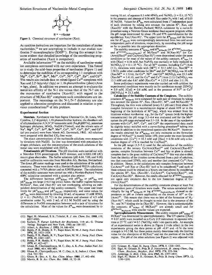
R = -ribose Figure 1. Chemical structure of xanthosine (Xao).
As xanthine derivatives are important for the metabolism of purine n~cleot ides , '~ we are attempting to include in our studies xan- thosine 5'-monophosphate (XMP2-). During this attempt it be- came evident that detailed knowledge of the coordinating prop- erties of xanthosine (Xao) is compulsory.
Available information15-21 on the stability of xanthosine-metal ion complexes contrasted with our own experience. This forced us to study the acid-base properties of xanthosine (Figure 1) and to determine the stabilities of its corresponding 1:l complexes with Mg2+, Ca2+, Sr2+, Ba2+, Mn2+, Co2+, Ni2+, Cu2', Zn2', and Cd2+. The results also include data on mixed-ligand Cu2+ complexes with Xao and 2,2'-bipyridyl (bpy) or 1,lO-phenanthroline (phen) (Arm = bpy, phen). In addition we present an attempt to evaluate the metal ion affinity of the N-1 site versus that of the N-7 site in the monoanion of xanthosine (Xao-H)-; with regard to the structure of M(Xao-H)' complexes such considerations are im- portant. The reasonings on the N- l /N-7 dichotomy are also applied to adenosine complexes and discussed in relation to pre- vious considerationsz2 of this problem.
Experimental Section Materials. Xanthosine was from Sigma Chemical Co., St. Louis, MO.
Cytidine, 2,2'-bipyridyl, 1 ,IO-phenanthroline hydrate, the disodium salt of ethylenediamine-N,N,N',N'-tetraacetic acid (Na2H2EDTA), potassium hydrogen phthalate, H N 0 3 , NaOH (Titrisol), and the nitrate salts of Na', Mg2+, Ca2', Sr2', Ba2+, Mn2+, Co2', Ni2', Cu2+, Zn2', and Cd2' (all pro analysi) were from Merek AG, Darmstadt, FRG. All solutions were prepared with distilled C02-free water.
The exact concentrations of the nucleoside solutions were measured by titrations with NaOH. Its titer was determined with potassium hy- drogen phthalate, and the concentrations of the stock solutions of the metal ions were established with EDTA.
Potentiometric pH Titrations. The pH titrations were carried out with a Metrohm E536 potentiograph, E655 dosimat, and 6.0202.100 (JC) macro glass electrodes. The buffer solutions (pH 4.64, 7.00, and 9.00) used for calibration were also from Metrohm AG, Herisau, Switzerland. The direct pH meter readings were used in the calculations of the acidity constants. These were done with a Hewlett-Packard 9825A calculator, connected with a 7470A plotter and an 82905B printer. The calculations of the stability constants were carried out with a Hewlett-Packard Vectra 60PC calculator connected with a quietjet plus printer.
The differences between pK"(xao) and pKHxao or pKHxao and pKH(Xa+H) are large (>4.5 log units); hence, the buffer regions between H(Xao)', Xao, and (Xao-H)- are not overlapping, allowing an inde- pendent determination of the acidity constants. The upper and lower limits for pKHH(xao) and PKH(X,,~), respectively, and some values for pKHxno were determined by titrating 20 mL of aqueous 15 mM HNO, (I = 0.1 (NaNO,); 25 "C) in the presence and absence of 4.5 mM xanthosine under N2 with 2 mL of 0.2 M NaOH and by using the differences in NaOH consumption between such a pair of titrations for the calculations. Most of the values for KHxao were determined by ti-
(13) Sigel, H.; Massoud, S. s.; Tribolet, R. J. Am. Chem. SOC. 1988, 110, 6857-6865.
(14) Karlson, P. Kurzes Lehrbuch der Biochemie, 11th ed.; G. Thieme Verlag: Stuttgart and New York, 1980; pp 104-107.
(1 5 ) Albert, A. Biochem. J. 1953, 54, 646-654. (16) Reddy, P. R.; Reddy, K. V.; Taqui Khan, M:M. J. Inorg. Nucl. Chem.
(17) Reddy, P. R.; Reddy, K. V.; Taqui Khan, M. M. J. Inorg. Nucl. Chem. 1976, 38, 1923-1924.
1978.40. 1265-1267. (18) Reddy, P. R.; Reddy, K. V.; Taqui Khan, M. M. J. Inorg. Nucl. Chem.
1979, 41, 423-427. (19) Ghose, R.; Chattopadhyaya, M. C.; Dey, A. K. Proc. Indian Natl. Sci.
Acad. 1980, 46A, 486-493. (20) Ghose, R.; Chattopadhyaya, M. C.; Dey, A. K. Indian J. Chem. 1980,
(21) Ghose, R.; Dey, A. K. Rev. Chim. Miner. 1980, 17, 492-496. (22) Martin, R. B. Acc. Chem. Res. 1985, 18, 32-38.
19A, 783-785.
Inorganic Chemistry, Vol. 28, No. 8, 1989 1481
trating 50 mL of aqueous 0.3 mM H N 0 3 and N a N 0 , (I = 0.1; 25 "C) in the presence and absence of 0.56 mM Xao under N2 with 1 mL of 0.05 M NaOH. Values for pxa0 were calculated from 17 independent pairs of such titrations by taking into account the species H+, Xao, and (Xao-H)- with the Hewlett-Packard 9825A calculator by a curve-fit procedure using a Newton-Gauss nonlinear-least-squares program within the pH range determined by about 3% and 97% neutralization for the equilibrium Xao/(Xao-H)-. The upper limit for p H ( x a o ) and the lower one for were correspondingly determined from two and seven independent pairs of titrations, respectively, by extending the pH range as far as possible into the appropriate direction.
The stability constants flM(xao) and KMM(xalFH) (or Kc"(h:a(h)(xao) and KCu(Arm)Cu(Arm)(Xa,H)) of M(Xao)2+ and M(Xao-H) (or Cu- (Arm)(Xao)zt and Cu(Arm)(Xao-H)') were determined under the same conditions as for most of the values of the acidity constant, KHxao, i.e. with [Xao] = 0.56 mM, but N a N 0 3 was partially or fully replaced by M(N03)2 (I = 0.1; 25 "C). For all M2+ systems, including Cu2+/Arm ( l : l ) , titrations were made with [M2'] = 33.3 mM (Le., Xao:M2' = 159.5); under other conditions for Mn2+ and Zn2' M(N03)2 was 30 mM (Xao:M2' = 1:53.6), for Co2', Ni2', and CdZt M(NO3)2 was 23.3 mM (Xao:M2' = 1:41.6), and for Cu2' and Cu2'/Arm (1:l) CU(NO,)~ was 13.3 mM and also 6.67 mM (Xao:Cu2' = 1:23.8 and also 1:11.9).
Some preliminary experiments were also carried out with cytidine; the experimental conditions were exactly as for xanthosine except [HN03] = 0.9 mM, [Cyd] = 0.6 mM, and in the presence of Ni2' or Cu2' [M(NO,),] = 33.3 mM.
Calculation of the Stability Constants of the Complexes. The stability constants KMM(XapH) were computed for each pair of titrations by taking into account the species H', Xao, (Xao-H)-, M2', and M ( X ~ O - H ) ' . ~ ~ Throughout, the data were collected (every 0.1 pH unit) from about 5% complex formation to a neutralization degree of about 85% or to the beginning of the hydrolysis of M2'(aq), which was evident from the titrations without xanthosine: e.g., for the Cu2' system (with the highest concentration) the pH range 3.5-4.4 was evaluated and for the Mn2' system the pH range evaluated was 5.1-5.8. In the case of the xanthosine systems with Co2', Ni2+, Cu2', and Cd2+ the experimental data could be slightly better satisfied with a curve-fitting procedure24 taking into account in addition to the mentioned species also M(Xao)2+. However, the results obtained for KMM(Xao) are only estimates as the formation degree of M(Xao)2' is mostly below 10%; even under the most favorable conditions with [Cu2+] = 33.3 mM and [Xao] = 0.56 mM only a for- mation degree of 15% is reached for Cu(Xao)2t.
In the pH range (3.5-5.1) used for the calculation of the stability constants of the ternary Cu(Arm)(Xao)2' and Cu(Arm)(Xao-H)' species, complex formation between Cu2' and bpy or phen is already complete due to the great stability of C u ( A r ~ n ) ~ ' ; ~ ~ this was also evident from the identity of the titration curves obtained from a pair of solutions, one that contained HNO, only and another that contained Cu2'/Arm in addition. Hence, in the calculations only complex formation between Cu(Arm)2' and Xao or (Xao-H)- had to be considered,26 and each of these systems could be treated as a binary one (see above) by considering the species H', Xao, (Xao-H)-, Cu(Arm)2t, Cu(Arm)(Xao)2t, and Cu(Arm)(Xao-H)'. However, the results obtained for Kc"(h)cII(h)(xao) are again only estimates due to the low formation degree of Cu- (Arm)(Xao)2'.
For the determinations of the stability constants always at least five independent pairs of titrations were made. The values calculated indi- vidually for log K'M(XapH) or log @u(A"")Cu(Am)(Xa~H) showed no de- pendence on pH or (where applicable) on the excess of M2' or Cu- (Arm)2t. This means that there was no hint of the formation of M2- (Xao-H),', which could be thought to exist due to the presence of the N-1 and N-7 binding site in (Xao-H)-. However, this is understandable: the constants KMM(Xao) of M(Xao)2t are already small and for K M ( X a o - H ) ~ 2 ( ~ a p ~ ) even smaller values are expected.
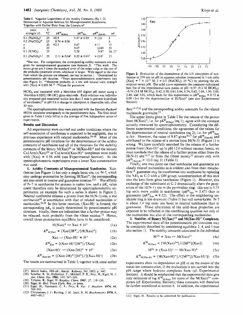
Spectrophotometric Measurements. The acidity constant pF?',(,,) of H(Xao)' was determined by spectrophotometry. The UV spectra ([Xao] = 0.07 mM) were recorded on a Cary 219 instrument in aqueous solu- tions at 25 "C and I = 0.5 (NaC10,) with 1-cm quartz cells. An example of an experimental series is shown in Figure 2 (vide infra); in the two experiments giving the data points a t pH -0.97 and -0.76 the ionic strength is >0.5 M, but these points mainly determine only the limiting value for the absorption of H(Xao)' and not the position of the buffer region. The pH of the other solutions (see Figure 2) was adjusted with
(23) Griesser, R.; Sigel, H. Inorg. Chem. 1970, 9, 1238-1243. (24) Sigel, H.; Griesser, R.; Prijs, B. 2. Naturforsch., B: Anorg. Chem., Org.
Chem., Biochem., Biophys., Biol. 1972, 278, 353-364. (25) Anderegg, G. Helv. Chim. Acta 1963, 46, 2397-2410. (26) Sigel, H.; Huber, P. R.; Griesser, R; Prijs, B. Inorg. Chem. 1973, 12,
1198-1200.
1482 Inorganic Chemistry, Vol. 28, No. 8, 1989 Kinjo et al.
Table I. Negative Logarithms of the Acidity Constants (Eq 1-3) Determined in Aqueous Solution for Monoprotonated Xanthosine, Together with Earlier Data from the Literature"
ionic T, strength ( r ) "C pKHH(Xao) pKHxaa PK"[X&~H) ref
0.1 (NaNO# 25 <0.9 5.47 f 0.03 >12.0 b 0.5 (NaCIO,)d 25 0.74 f 0.06 b e 20 <2.5 5.67 > I 1 1 5 0 25 5.67 12.00 32 0.1 (KNOp) 25 5.58 9.87 16, 17 0.1 (NaN03)c,f 25 2.11 & 0.04' 9.22 f 0.01f > 1 2 . 6 33
' S e e text. For comparison, the corresponding acidity constants are also given for monoprotonated guanosine (see footnote f). *This work. The errors given are 3 rimes the standard error of the mean value or the sum of the probable systematic errors, whichever is larger. With regard to the sites from which the protons are released, see text in section 1. cDetermined by potentiometric pH titration. From spectrophotometric experiments (see also Figure 2). 'Measured probably at a low and natural ionic strength with [Xao] = 0.002 M.I5 /Values for guanosine.
HCIO, and measured with a Metrohm 605 digital pH meter using a Metrohm 6.0202.100 (JC) glass electrode. Each solution was individu- ally prepared and measured within less than 5 min to prevent hydrolysis of xantho~ine;~' at pH 0.6 a change in absorption is observable only after 30 min.
The spectrophotometric data were analyzed with the Hewlett-Packard 9825A calculator analogously to the potentiometric data. The final result given in Table I (vide infra) is the average of five independent series of experiments. Results and Discussion
All experiments were carried out under conditions where the self-association of xanthosine is expected to be negligible, due to previous experience with related nucleoside^.^^-^^ Most of the potentiometric pH titrations for the determination of the acidity constants of xanthosine and all of the titrations for the stability constants of the binary M(Xao)2+ or M(Xao-H)+ and the ternary Cu(Arm)(Xao)2+ or Cu(Arm)(Xao-H)+ complexes were made with [Xao] = 0.56 mM (see Experimental Section). In the spectrophotometric experiments even a lower Xao concentration was used.
1. Acidity Constants of Monoprotonated Xanthosine. Xan- thosine (see Figure 1) has only a single basic site, i.e. N-7, which may undergo protonation by forming H(Xao)+; the corresponding site also exists in inosine and guanosine m o i e t i e ~ . ~ ~ - ~ ' The affinity of N-7 in xanthosine for protons is rather low, and a pKa value could therefore only be determined by spectrophotometric ex- periments; an example for such a series is shown in Figure 2. Neutral xanthosine releases a proton from the H(N-1) site, a site a t t r i b ~ t i o n ~ ~ in accordance with that of related nucleosides or nucleotides.22s2s In this latter reaction, (Xao-H)- is formed; the corresponding pKa is easily determined by potentiometric pH titrations. Finally, there are indications that a further proton may be released, most probably from the ribose residue.32 Hence, overall three protonation equilibria have to be considered:
H(Xao)+ Xao + H+ ( la )
Xao F= (Xao-H)- + H+ (2a)
KHXao = [(Xao-H)-] [H+]/[Xao] (2b)
(Xao-H)- (xa0-2H)~- + H+ (3a) KH(Xao.H) = [(Xao-2H)'-] [H+] /[(Xao-H)-] (3b)
The results are summarized in Table I, together with some earlier
(27) Merck Index, 10th ed.; Merck: Rahway, NJ, 1983; p 1445. (28) Scheller, K. H.; Hofstetter, F.; Mitchell, P. R.; Prijs, B.; Sigel, H. J .
Am. Chem. Soc. 1981, 103, 247-260. (29) Tribolet, R.; Sigel, H. Biophys. Chem. 1987, 27, 119-130. (30) Sigel, H. Eiol. Trace Elem. Res., in press. (31 ) Sigel, H.; Naumann, C. F.; Prijs, B. Eur. J . Biochem. 1974, 46,
(32) Christensen, J. J.; Rytting,, J. H.; Izatt, R. M. Biochemistry 1970, 9, 589-593.
4907-491 3.
0 6 2 1 1 0604 Xao A
I 1
-10 -05 00 0 5 1 0 15 20 2 5 30 PH
Figure 2. Evaluation of the dependence of the UV absorption of xan- thosine at 239 nm on pH in aqueous solution (measured in 1-cm cells; [Xao] = 7 X M; I = 0.5 (NaC10,); 25 "C) by plotting the ab- sorption versus pH. The solid curve represents the computer-calculated best fits of the experimental data points at pH -0.97 (9.3 M HCIO,),
2.49, and 3.01, which leads for this experiment to pK"(xa,,) = 0.72 * 0.09 (30) for the deprotonation of H(Xao)+ (see also Experimental Section).
datalS-17,32 and the corresponding acidity constants for the related nucleoside guanosine.33
The upper limits given in Table I for the release of the proton from H(Xao)+, i.e. for p@H(Xao) (eq l ) , agree with the constant actually measured by spectrophotometry. Considering the dif- ferent experimental conditions, the agreement of the values for the deprotonation of neutral xanthosine (eq 2), Le. for pKHxao, is fair. However, the value of 9.87 givenI6J7 for pKH(xao.~) and attributed to the release of a proton from H(N-3) (Figure 1) is wrong. We have carefully searched for the release of a further proton from (Xao-H)- up to pH 12.0 without success; hence, we must conclude that the release of a further proton either from the H(N-3) ~ i t e ' ~ ~ " or from the ribose moiety32 occurs only with pKH(Xao.H) > 12.0 (eq 3) (Table I ) .
Finally, one may point out that xanthosine and guanosine are closely related nucleosides as is evident also from their metabo- lism:I4 guanosine may be transformed into xanthosine by replacing the NH2 at C-2 with a O H group; tautomerization of this enol into the keto form gives xanthosine (Figure 1 ) . However, this exchange of the substituent at C-2 dramatically alters the prop- erties of the H(N-I) site in the pyrimidine ring: this site is 3.75 log units more acidic in xanthosine (pKHxao = 5.47) than in guanosine (pKHGUo = 9.22). The effect in the neighboring im- idazole ring is less dramatic (Table I) but still remarkable: N-7 is about 1.4 log units less basic in neutral xanthosine than in guanosine. These alterations of the acid-base properties are expected to be reflected in the coordinating qualities not only of the nucleosides but also of the corresponding nucleotides.
2. Stability of Binary M(Xao)'+ and M(Xao-H)+ Complexes. The experimental data of the potentiometric pH titrations may be completely described by considering equilibria 2,4, and 5 (see also section 1) . The stability constants calculated in the individual
-0.76 (5.8 M HCIO,),0.42,0.50, 0.61 0.64,0.74, 0.82, 1.04, 1.50, 2.00,
M2+ + Xao M(Xao)2+ (4a)
M2+ + (Xao-H)- M(Xao-H)+ (Sa)
KMM(Xao-H) = [M(Xao-H)+I/([M2+l [(xao-H)-l) (5b)
experiments show no dependence on pH or on the excess of the metal ion concentration, if the evaluation is not carried into the pH range where hydroxo complexes form (cf. Experimental Section). It should be emphasized that the experimental data give only estimates of log KMM(xaol for some of the M(Xao)'+ com- plexes (cf. Experimental Section); these constants will therefore be further considered in section 8.. In addition, the experimental
(33) Sigel, H. Results to be submitted for publication.
Solution Structures of Nucleoside-Metal Complexes Inorganic Chemistry, Vol. 28, No. 8, 1989 1483
Table 11. Logarithms of the Stability Constants of Binary M(Xao-H)+ (Eq 5 ) and Ternary Cu(Arm)(Xao-H)+ Complexes (Eq 7) , Together with the Estimates Obtained for Some of the Corresponding M(Xao)2+ (Eq 4) and Cu(Arm)(Xao)2+ Species (Eq 6), As Determined by Potentiometric pH Titrations in Water at 25 O C and I = 0.1 (NaNOp)"
M2+ log KMM(Xaof log KMM(Xac-H) A log KC"
Mg2+ Ca2+ Sr2+ Ba2+ Mn2+ co2+ Ni2+ CUI+ Zn2+ Cd2+ Cu(bPY)2+ Cu(phen)*+
<0.6c <0.6c <0.6' <0.6c
0.84 f 0.05 1.65 f 0.05 2.09 f 0.05 2.58 f 0.03 1.32 f 0.02 1.96 f 0.05
0.5 f 0.2 0.7 f 0.2 0.8 f 0.3
0.70 f 0.12 0.64 f 0.18 2.49 f 0.03 -0.09 * 0.04 0.7 f 0.3 2.48 f 0.03 -0.10 f 0.04
"The resulting values for A log Kcu (eq 8 and 9) are also listed. The error limits given are 3 times the standard error of the mean value or the sum of the probable systematic errors, whichever is larger. The values of the error limits for A log Kc, were calculated according to the error propagation method of Gauss. *See also Table VII. CDetailed evaluations of the experimental data indicated that for all four alka- line-earth ions log KMM(XapH) N 0.2 f 0.2.
data also provide no evidence for the formation of M2(Xao-H),+ complexes; the stability of such species is expected to be very low (see Experimental Section).
The stability constants determined for M(Xao-H)+ complexes (eq 5) and the estimates for some M(Xao)2+ species (eq 4; see also section 8) show the usual trends (Table 11): Complex stability with the alkaline-earth ions is low. For the divalent 3d metal ions a stability sequence corresponding to the Irving-Williams series34 is observed; the relatively large stability differences between the M(Xao-H)+ complexes with Cu2+ and Mn2+ (1.7 log units) or Cu2+ and Zn2+ (1.3 log units) are character is ti^,^ of nitrogen donor ligands (see also section 5).
Unfortunately, the agreement between the present results and previous data is poor: the constants given in ref 15 for the M- (Xao-H)+ complexes of Co2+, Ni2+, Cu2+, and Zn2+ are about 0.8-1.2 log units too large. This discrepancy cannot be solely attributed to different experimental conditions (20 "C and I = natural?); instead it appears, on the basis of the experimental part in ref 15, that the hydrolysis of the M2+(aq) ions was neglected. The data of ref 16 and 17 have been determined under conditions (25 "C; I = 0.1 M (KN03)) similar to ours, but the agreement is even worse: the logarithms of the stability constants given for the M(Xao-H)+ complexes of Mg2+, Ca2+, MnZ+, Co2+, and Zn2+ are all between 2.22 and 2.48; the corresponding values for the Ni2+ and Cu2+ complexes are 2.88 and 2.91, respectively. Hence, these previous constants are between about 0.3 and 2.2 log units too large, if compared with the results in Table 11. The situation with the constants given in ref 19-21 is similar: for Co2+, Ni2+, and Cu2+ log KMM(Xao.H) varies between 2.88 and 3.42 (25 "C; I = 0.1 M (KNO,)); i.e., these constants are about 0.8-1.2 log units too large (as are those of ref 15). Most probably hydrolysis of M2+(aq) was neglected also in the experiments of ref 19-21. All these previous values in ref 15-17 and 19-21 have to be rejected. That the data of ref 16 and 17 are erroneous is evident already from the fact that the stability constant given for Ca- (Xao-H)+ (log K = 2.37) is larger than the one for Zn(Xao-H)+ (log K = 2.26) or for Co(Xao-H)+ (log K = 2.23); for a ligand like xanthosine such properties would be in contrast to all expe- r i e n ~ e . ~ ~ . , ~ This contrast does however not exist; the stability constants listed in Table I1 agree with experience.
3. Stability of Ternary Cu(Arm)(Xao)2+ and Cu(Arrn)(Xao- H)+ Complexes. The stability of the mixed-ligand complexes of
(34) Irving. H.; Williams, R. J. P. Nature 1948, 162, 746-747; J. Chem. SOC.
( 3 5 ) Sigel, H.; McCormick, D. B. Acc. Chem. Res. 1970, 3, 201-208. 1953, 3192-3210.
xanthosine, Cu2+, and an aromatic amine (Arm), Le. 2,2'-bipyridyl (bpy) or 1,lO-phenanthroline (phen), was also determined. The potentiometric pH data can be completely satisfied in a way analogous to that given in section 2 for the binary systems; this means that now equilibria 2, 6, and 7 have to be considered in the calculations. The corresponding constants are listed in Table
Cu(Arm)2+ + Xao * Cu(Arm)(Xao)2+
[ Cu(Arm) ( X ~ O ) ~ ' ]
[Xao] -
p(ATm)Cu(Arm)(Xao) -
Cu(Arm)2f + (Xao-H)- * Cu(Arm)(Xao-H)+
[Cu(Arm)(Xao-H)+]
[ C ~ ( A r m ) ~ + l [(Xao-H)-] p(Arm)Cu(Arm)(Xao-H) =
11; the values for Cu(Arm)(Xao)*+ are again only estimates (section 2). For Cu(bpy)(Xao-H)+ log *(bpy)h(bpy)(~ao-~) = 2.36 has previously been determined,19 in fair agreement with the present result.
The relative stability of ternary complexes toward their binary parent complexes is best quantified by considering equilibrium 8a.36,37 The corresponding constant defined by eq 8b is calculated
(8a) Cu(Arm)2+ + Cu(Xao-H)+ Cu(Arm)(Xao-H)+ + Cu2+
[ Cu(Arm) (Xao-H)+] [ Cu2+]
[ C ~ ( A r r n ) ~ + ] [ Cu(Xao-H)+] (8b) 1 O A 1% Kcu =
with eq 9. Equilibrium 8a is e ~ p e c t e d , ~ - , ~ to be on its left side
A log KC, = log *(Arrn)Cu(Arrn)(Xao-H) - log KCUCu(Xao-H)
= log Fu(Xao-H)Cu(Xao-H)(Arrn) - log KCUCu(Arrn)
(9)
with negative values for A log Kcu due to the general rule that KMMCL) > KM(L)M(L)2. Indeed, statistical consideration^^^^^^ for the coordination of a bidentate ligand followed by a monodentate (or bidentate) ligand to the tetragonal or Jahn-Teller-distorted oc- tahedral coordination sphere of Cuz+ give A log Kcu,statist = -0.5 (or -0.9).
The constants listed in Table I1 for A log K,-, are slightly negative but are somewhat larger than the statistically expected value. This result appears to be in agreement with the coordination of a negatively charged nitrogen site to Cu(Arm)2+, as will be further discussed in section 7. It should be pointed out that no positiue values for A log Kcu are obtained; such an observation is usually made if (i) the ternary complex is formed with heter- oaromatic N bases and 0-donor ligands23v36-38 and/or (ii) intra- molecular aromatic-ring stacks are formed between suitable ligand parts within the mixed-ligand ~ o m p l e ~ . ~ ~ , ~ ~ , ~ ~ ~ ~
On the basis of previous experience with mixed-ligand com- p l e ~ e s ~ ~ , ~ ~ , ~ ~ ~ ~ ~ negative A log KM values (eq 8 and 9) are also expected for other M2+/Arm/(Xao-H)- systems. This means that the stability of other ternary M(Arm)(Xao-H)+ complexes may be estimated by subtracting 0.2 log unit from the stability constants of the binary complexes in Table 11; the resulting estimate is expected to be correct within f 0 . 2 log unit. However, there is a limit to this procedure: 2,2'-bipyridyl and 1 ,IO-phenanthroline
Sigel, H. Angew. Chem. 1975, 87, 391-400; Angew. Chem., Int. Ed. Engl. 1975, 14, 394-402. Sigel, H. Coordination Chemistry-20; Banerjea, D., Ed.; Pergamon Press: Oxford and New York, 1980; pp 27-45. Sigel, H.; Fischer, B. E.; Prijs, B. J. Am. Chem. SOC. 1977, 99,
Malini-Balakrishnan, R.; Scheller, K. H.; Haring, U. K.; Tribolet, R.; Sigel, H. Inorg. Chem. 1985, 24, 2067-2076. Fischer, B. E.; Sigel, H. J . Am. Chem. SOC. 1980, 102, 2998-3008. Sigel, H.; Malini-Balakrishnan, R.; Haring, U. K. J . Am. Chem. SOC.
Fischer, B. E.; Sigel, H. Inorg. Chem. 1979, 18, 425-428.
4489-4496.
1985, 107, 513775148,
1484 Inorganic Chemistry, Vol. 28, No. 8. 1989
form stacking adducts with purine derivative^,^,^^,^^ and therefore formation of (Arm)(Xao) and (Arm)(Xao-H)- stacks has to be considered. The stability constant for these adducts is expectedgs4 to be on the order of 10; Le., log K = 1. Hence, in the ternary systems with Mg2+, Ca2+, Sr2+, BaZ+, and Mnz+ (and possibly even Zn2+) the situation is governed by the stability of the mentioned aromatic-ring stacking adducts and not by the metal ions, though these may still coordinate to bpy or phen, forming an unbridged ternary stacking adduct. Finally, in the bridged ternary complexes, where the metal ion coordinates to both ligands as in Cu- (Arm)(Xao-H)+, no intramolecular stacks can be formed for steric reasons.
With these reasonings in mind, the stability constants deter- mined earlier18*20 for the ternary M(bpy)(Xao-H)' and M- (phen)(Xao-H)+ complexes must be rejected as being too large:18,20 with the exception of the Ni2+ systems, the A log KM values for all bpy or phen systems with Mg2+, CaZ+, MnZ+, Co2+, Zn2+, and Cu2+ are claimed to be positive;18 for Ca2+/bpy/(Xao-H)- even A log Kca = +1.04 is given. In this case, the error most probably occurred in the calculation procedure.
4. Sites of Metal Ion Coordination to Deprotonated Xanthosine. Purine nucleosides show a dichotomy between N-1 and N-7 for metal ion binding.z2 This is tentatively expressed for M(Xao-H)+ complexes in the intramolecular equilibrium loa. Release of the
Kinjo et al.
complexes with a sole N-1 or N-7 coordination are defined in eq 13 and 14, respectively. Combination of eq 12-14 gives eq 15,
KN.1 = [M(N-1/Xao-H)+]/[M2+] [(Xao-H)-] (13)
KN.7 = [M(N-7/Xao-H)+]/[Mz+][(Xao-H)-] (14)
and together with eq 11 also eq 16 and 17 are obtained. Kexp = KN-l -k KN-7 (15)
R --ribose
proton from the H(N-1) site of neutral xanthosine leads to a negatively charged nitrogen, which should be an excellent binding site for many metal ions: The corresponding isomer, shown on the left side in equilibrium loa, is designated as M(N-l/Xao-H)+. On the right side in equilibrium 10a N-7 binding is indicated, a coordination type well-known for many purine deri~atives,~-"J~ and this species is now designated as M(N-7/Xao-H)+, but its correct structure is difficult to assess. There is obviously the possibility of dislocating the negative charge in (Xao-H)- from N-1 toward 0-6; this could favor chelate formation involving N-7 and 0-6. However, so far no examples for such a direct chelation of 6-oxopurine derivatives are k n o ~ n ; ~ . ~ ~ there are indications for an indirect chelation, i.e. with a water between 0 - 6 and the N-7-bound metal ion.7 This view is also supported by X-ray studies of solid complexe~.~ This structural ambiguity is indicated by the thin dotted line between 0 - 6 and Mz+ in equilibrium loa; evidently a similar water bridge could also be formed between 0 - 6 and the N-1-bound metal ion. With these uncertainties in mind, which will be discussed somewhat further in section 6, the intramolecular equilibrium 1 Oa is rewritten in a simplified way:
M(N-1 /Xao-H)+ M(N-7/Xao-H)+ ( lob)
This equilibrium is quantified by the dimensionless equilibrium constant Kl:
KI = [M(N-7/Xao-H)+]/[M(N-l/Xao-H)+] (11)
Hence, equilibrium 5a may be rewritten M2+ + (Xao-H)- + M(N-1 /Xao-H)+ * M(N-7/Xao-H)+
The experimentally accessible stability constant Kexp (cf. also eq 5b) is then given by eq 12b. The stability constants for the
Kexp = K M ~ ( x a o - ~ ) =
(1 2a)
[M(N-1 /Xao-H)'] + [M(N-7/Xao-H)+]
[ M2+] [ (Xao-H)-] (12b)
(43) Tribolet, R.; Malini-Balakrishnan, R.; Sigel, H. J . Chem. SOC., Dalton Trans. 1985, 2291-2303.
(44) Scheller, K. H.; Sigel, H. J . Am. Chem. SOC. 1983, 105, 3005-3014.
(17) Kexp KN-7 & = - - I = KN-I Kexp - KN-7
Evidently values for either KN.1 or KNS7 have to be obtained to find an answer with regard to the position of equilibrium 10. As the acidity constant for the release of the proton from N-1 (eq 2) in xanthosine is known (Table I; section l ) , an attempt is made in sections 6 and 7 to estimate values for KN.1 (eq 13) and to use these for further evaluations; however, this evaluation is restricted to the 3d metal ions, as well as Zn2+ and Cd2+, because the stability of the M(Xao-H)+ complexes with the alkaline-earth ions is very low (Table 11).
5. Correlations between Complex Stability and Ligand Basicity: Construction of Base Line Plots Quantifying the Metal Ion Af f i ty of N-1 and N-7. The existence of a linear relationship between log KMML and P P H L is well-known for many series of structurally related ligands (see ref 45 and references therein). This advance is due to Martin,22 who showed that logarithms of the stability constants for Ni2+, Cu2+, and Znz+ binding at pyridine or purine N- 1 type nitrogens and imidazole or purine N-7 type nitrogens display a linear relationship with pK, for the three metal ions and the two types of nitrogen ligating sites. On this basis the dichotomy of metal ion binding to N-1 versus that to N-7 in several purine nucleosides including adenosine was resolved for Ni*+, Cuz+, Zn2+,46 and Pd(diethylenetriamine)2+.2z-47 An uncertainty oc- curring with adenosine is that the experimentally determined stability constants are smaller than the values estimated from the log K versus pKa plots."
We believe that this apparent discrepancy is to a large part due to steric hindrance: The data for 2-methylp~r id ine~~ and other ortho-substituted pyridines49 do not fit the mentioned correlations, in contrast to the case for meta- and para-substituted pyridines and pyridine itself.22 CH3 and N H z groups are comparable in size and therefore we consider the given N-1 reference lines11*22-46 as not representative of the N-1 complexation tendency of adenosine, which carries an amino group next to N-1; Le., the available reference lines provide estimates of log K for N-1 adenosine complexation that are too large. This view asks for the construction of base lines taking into account steric hindrance and also for a reevaluation of the metal ion distribution data2zv46 between N-1 and N-7 of adenosine; of course, this distribution also strongly depends on the validity of the pKa value estimated for N-7 of neutral adenosine46 (for further comments see section 9).
These reasonings on steric hindrance are confirmed by equi- librium constants determined now for cytidine systems (see Ex- perimental section: I = 0.1 (NaN03); 25 "c) : p&(Qdd, = 4.14
0.6. The first two values agree excellently with literature data>0,51 Cytidine carries, like adenosine, an amino group next to its
f 0.02 (3(T), log ~ c u ( c y d ) = 1.56 f 0.06, and log K N,(Cyd <
(45) Martin, R. B.; Sigel, H. Comments Inorg. Chem. 1988, 6, 285-314. (46) Kim, S-H.; Martin, R. B. Inorg. Chim. Acta 1984, 91, 19-24. (47) Kim, S.-H.; Martin, R. B. Inorg. Chim. Acta 1984, 91, 11-18. (48) Kahmann, K.; Sigel, H.; Erlenmeyer, H. Helu. Chim. Acta 1964, 47,
1754-17fi3 . - . - -
(49) Sigel, H.; Wynberg, H.; van Bergen, T. J.; Kahmann, K. Helu. Chim. Acta 1972, 55, 610-613.
(50) (a) Fiskin, A. M.; Beer, M. Biochemistry 1965, 4, 1289-1294. (b) Martin, R. B. Fed. Proc., Fed. Am. SOC. Exp. Eiol. 1961, 20, (Suppl. lo), 54-59.
( 5 1 ) Smith, R. M.; Martell, A. E. Critical Stabilily Constants; Plenum Press: New York and London, 1975; Vol. 2.
Solution Structures of Nucleoside-Metal Complexes Inorganic Chemistry, Vol. 28, No. 8, 1989 1485
Table 111. Equilibrium Data Used for the Construction of log K versus pKa Plots for N-7 or Imidazole-like and N-1 or Pyridine-like Binding Sites: Negative Logarithms of the Acidity Constants, pKa, of the Ligands in Their Acidic Form and the Logarithms of the Stability Constants, log K, of the Corresponding Metal Ion Complexes
acidic log K no. ligand ligand form pK. M2' complex Mn2' Co2+ Ni2' Cu2' Zn2+ Cd2+
N-7 or Imidazole-like Systems 1 adenosine" H ~ ( A ~ o ) ~ + -1.56 M(HAdo),' 0.16 -0.89 2 1-methylinosineb H(CH,Ino)' 1.4 M ( C H , I ~ O ) ~ ' 1.0 1.4 0.3 3 inosineb H(Ino)+ 1.4 M(Ino)2+ 0.8 1.1 1.3 0.3lC 0.86d 4 guanosineb H(Guo)' 2.33 M(Guo)~' 1.0 1.4 1.9 0.80b*C 1.1 7e 5 imidazole/ H(Im)+ 7.04 M(Im)2t 1.25 2.40 3.03 4.21 2.51 2.71 6 1-methylimidazole" H(CH,Im)' 7.39 M(CH31m)2t 2.778 3.44 4.61 2.98
N- 1 or Pyridine-like Systems 7 pyridineh H(PY)+ 5.26 M ( P ~ ) ~ + 0.14,'0.24' 1.25 1.87 2.49 1.00 1.32'[ 8 4-(2-thienyl)pyridinem H(th-py)' 5.59 M(th-py)2' 1.91 2.57 1.10 9 4-methylpyridine" H(CH,py)+ 6.18 M(CH3py)" 1.58' 2.11 2.88 1.40 1.57p
I O 7-methylinosine" (CHJno)' 6.57 M ( C H , I ~ O - H ) ~ + 3.04 1.43 1 1 inosineb Ino 8.7 M(Ino-H)2t 2.1 2.8 2.4 12 ammonia' NH,' 9.38 M(NH3)2t 1.27 2.08 2.74 4.18 2.41 2.67
D 2 0 at I = 0.1-3 (NaNO,); 27 0C.28 'In D20 at I = 0.1-4 (NaNO,); 27 0C.28 / I = 0.1 (NaNO,); 25 OC." S I = 1.0 (NaC10,); 21 "C: Kim, S.-H.; Martin, R. B. Unpublished results, 1982. Martin, R. B. Personal communication, March 1988. "I = 0.1 (NaNO,); 25 0C.55 ' I = 0.5; 25 0C.56 J I = 0.5; 25 0C,57 k I = 0.1-1; 20-30 "C. Average of 14 values listed in ref 51 and 56-59 (see footnote I ) . 'A view on the constants given in the literature shows that the influence of ionic strength and temperature is small. '"I = 0.1 (NaCIO,); 25 0C?8v49 The correct structure of the ligand is given in ref 49. " I = 0.1 (NaCIO,); 25 0C.48 ' I = 0.5-1.0; 25 OC; average from ref 51 and 56 (see footnote I ) . P I = 0.1-1.0; 25-30 OC; average from ref 51, 56, 57, and 59 (see footnote I ) . 9 I = 0.1 (NaNOd; 25 "C for pK. and Mn(NHd2' values.54 The other values are literature averages ( I = 0-2; 20-30 "C) taken from ref 54 (see footnote I ) .
" I = 1.0 (NaC10,); 21 0C.46 *I = 1.0 (NaC104); 25 0C.53 'In D 2 0 at I = 0.1-5 (NaNO,); 27 0C.28
pyridine-like nitrogen, and the given equilibrium d a t a for this nucleoside do not fit on the previous base line plots for Cuz+46 and Ni2+;I1 Le., the present stability constants a r e smaller than the previous ones.46 In accordance with the present results are constants obtained earlier for 2-aminopyridine and these d a t a do also not fit on the mentioned base line. Consequently, we have not included in t h e construction of the base line plots described below any cytidine or other 2-aminopyridine-like data . However, in agreement with previous c o n c l u s i ~ n s , ~ ~ ~ ~ ~ we could not observe s ter ic hindrance by a (smaller) keto oxygen in a position next to N-1 as occurs, for example, in inosine; it could be t h a t this or tho oxygen is undergoing hydrogen bonding with a coordinated water molecule and that this diminishes or cancels its s ter ic influence. This type of hydrogen bonding would also explain why steric hindrance in complexes of 2-aminopyridine is more pronounced (see also section 9) than in those of cytidine; the latter ligand has not only an amino group but also a keto oxygen in a position next to its coordinating pyridine-like N-3. Hence, for cytidine complexes one may expect tha t par t of the steric effect of the 4-amino group is compensated for by hydrogen bonding of a metal ion coordinated water molecule to the 2-keto oxygen. Certainly, a final decision on this aspect requires more experimental da ta .
T h e procedure of Martin11~zz~46 for the construction of base line plots for t h e metal ion affinity of N-1 and N-7 of purine nu- cleotides is now extended to Mn2+, eo2+, and CdZ+, and tha t for the complexation of Niz+, Cuz+, and ZnZ+ is repeated by taking into account the restrictions described above. T h e equilibrium constants used for the log K versus pK, plots a r e summarized in Table 111; as far as possible, we have selected constantsZ8~46~51~53-59
Sun, M. S.; Brewer, D. G. Can. J . Chem. 1967, 45, 2729-2739. Lonnberg, H.; Vihanto, P. Inorg. Chim. Acta 1981, 56, 157-161. Saha, N.; Sigel, H. J . Am. Chem. SOC. 1982, 104, 4100-4105. Banerjea, D.: Kaden, T. A,; Sigel, H. Inorg. Chem. 1981, 20,
Perrin, D. D. Stability Constants of Metal-Ion Complexes; IUPAC Chemical Data Series 22; Pergamon Press: Oxford and New York, 1979; Part B. Martell, A. E.; Smith, R. M. Critical Sfability Constants; Plenum Press: New York and London, 1982; Vol. 5, 1st suppl. SillCn, L. G.; Martell, A. E. Stability Constants of Metal-Ion Com- plexes; Special Publication 17; The Chemical Society: London, 1964. SillCn, L. G.; Martell, A. E. Stability Constants of Metal-Ion Com- plexes; Special Publication 25; The Chemical Society: London, 197 1;
2586-2590.
Suppl. 1 .
Table IV. Correlations between M2' Complex Stability with Imidazole-like (N-7) or Pyridine-like (N- 1) Ligands and Ligand Binding Site Basicity"
M2+ m b R SDb Regression Lines for Imidazole-like or N-7 Type Ligands
Mn2+ 0.30' -0.86 0.07d Co2' 0.315 f 0.026 0.312 f 0.135 0.993 0.057 Ni2' 0.377 f 0.020 0.519 f 0.095 0.996 0.046 Cu2' 0.499 f 0.019 0.766 f 0.084 0.997 0.053 Zn2' 0.416 f 0.016 -0.246 f 0.069 0.997 0.045 Cd2' 0.328 f 0.020' 0.404 f O.07Oe 1.000 0.05'
Regression Lines for Pyridine-like or N-1 Type Ligands Mn2' 0.262 f 0.021 -1.189 f 0.145 0.997 0.029 Co2' 0.204 f 0.031 0.244 f 0.236 0.977 0.045 Ni2' 0.235 f 0.025 0.633 f 0.179 0.984 0.036 Cu2+ 0.415 f 0.010 0.296 f 0.065 0.999 0.013 Zn2' 0.367 f 0.025 -0.923 f 0.178 0.991 0.034 Cd2+ 0.332 f 0.013 -0.450 f 0.094 0.999 0.015
"Slopes ( m ) and intercepts (b) are given for the straight base line plots of log K versus pK, as calculated by the least-squares procedure from the experimental equilibrium constants of the systems given in Table 111, together with the corresponding correlation coefficients (R). The column at the far right lists the standard deviations (SD) resulting from the differences between the experimental (Table 111) and calcu- lated (from the straight-line equations) log K values of the individual systems listed in Table 111 ( I = 0.1-1 (see text and Table 111); 25 "C). Straight-line equation: y = mx + b. x may represent the pK, value of any N-7 or N-1 type ligand. The errors given with m and b correspond to one standard deviation ( l a ) . bThese SD values times 2 or 3 are considered as reasonable error limits for any stability constant calcu- lation in the pKa range of the employed experimental data (see Table I11 or Figure 3). 'This value is an estimate based on the other slopes in the table; together with the imidazole/Mn2+ data of Table 111 the N-7 type base line is thus defined. dEstimated error limit. CDue to the perfect fit of the experimental data (three points only; see Table 111) on the calculated straight line, the calculated errors are very small, i.e. for m f0.001, for b f0.004, and for SD fO.OOO; therefore, the estimates given above are considered as more realistic error limits.
that were measured a t (or close to) I = 0.1 and 25 "C. This latter a im is relatively well achieved for the N - 1 or pyridine-like da ta , and these a re mainly important for the evaluations in sections 6 and 7 .
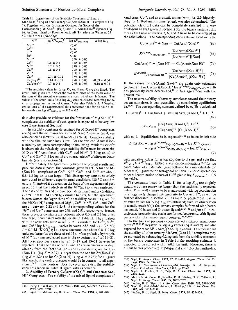
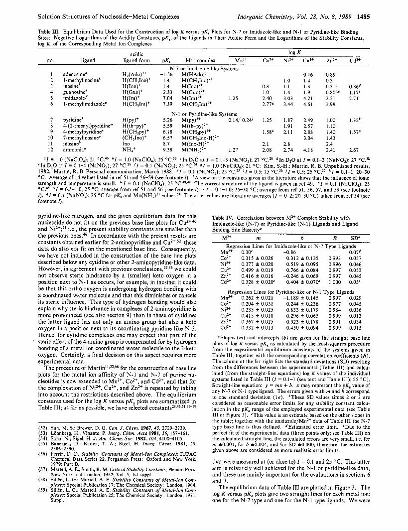
The equilibrium da ta of Table 111 are plotted in Figure 3. The log K versus pK, plots give two straight lines for each metal ion: one for the N-7 type and one for the N-1 type ligands. W e were
1486 Inorganic Chemistry, Vol. 28, No. 8, 1989 Kinjo et al.
Table V. Logarithms of the Stability Constants for Metal Ion Coordination at N-1 or N-7 of N-I-Deprotonated Xanthosine, Together with the Intrinsic [N-l] / [N-7] Binding RatiosG
[M(N- 1 /Xao-H)+1/ M2+ or H+ log Ksxp (eq 12b)b log KN.I (eq 13)' log Kp4.7 (eq 14)d log K N . ~ - log Kp4.7 [M(N-i /Xao-H)+P
Mn2+ 0.84 f 0.05 0.24 f 0.09 0.71 f 0.07 -0.47 f 0.12 0.34 f 0.09 co2+ 1.65 f 0.05 1.36 f 0.14 1.34 f 0.18 0.02 f 0.23 1.0 f 0.6 NiZ+ 2.09 f 0.05 1.92 f 0.11 1.60 f 0.28 0.32 f 0.30 2.1 f 1.4 cu2+ 2.58 f 0.03 2.57 f 0.04 <2f >0.6f >4f Zn2+ 1.32 f 0.02 1.08 f 0.10 0.95 f 0.14 0.13 f 0.17 1.3 f 0.5 Cd2+ 1.96 f 0.05 1.37 f 0.05 1.83 f 0.07 -0.46 f 0.09 H + 5.47 f 0.038 5.47 f 0.038
0.35 f 0.07 - 300' 3.0 f 0.5h 2.5 f 0.5
"The errors given in columns 2 and 3 are 3 times the standard errors; those in the other columns were calculated according to the error propagation method of Gauss. bValues of log KMM(Xao.~) from Table 11. CCalculated with p P x a Q = 5.47 and the N-1 base line equations of Table IV; the errors are 3 times the S D values given in Table IV. "Calculated via eq 15. 'See text in section 6 and eq 18. fcalculated with the upper limit of 2.61 for log Kcrp and the lower h i t of 2.53 for log KN.~. Direct calculation with the above values gives log KN.7 = 0.94 f 2.16, log KN.1 - log KN.7 = 1.63 f 2.16, and [M(N-l/Xao-H)+]/[M(N-7/Xao-H)+] = 43 f 212. gValue for pKHxao from Table I. hEstimation; see text in section 6. 'Ratio for [H(N-1) of (Xao)O]/[H(N-7) of H(Xao-H)"]; see text in section 6.
rather surprised to observe that the data for the ammonia systems of Co2+, Ni2+, Cu2+, and Zn2+ fit within the error limits on the base lines for the pyridine-like or N-1 type ligands. This could be a result of mere chance, but it allowed us to construct corre- sponding base lines also for Mn2+ and Cd2+ (Figure 3). Similarly, regarding the use of the imidazole data (Table 111) for the N-7 base lines, one might argue that a statistical correction should be introduced to account for the possibility that H(Im)+ has two acidic protons, the release of one giving the coordinating neutral imidazole ligand. However, again the imidazole data fit well without such a correction on the N-7 base lines for Co2+, Ni2+, Cu2+, and Zn2+; therefore, we used the unaltered data also in constructing the base lines for Mn2+ and Cd2+ (Figure 3). The results of the least-squares calculations for the base lines are summarized in Table IV; the slopes of the base lines quantifying N-7 and N-1 type coordination for Ni2+, Cu2+, and Zn2+ are in excellent agreement with the slopes published by Kim and Martin.46
6. Metal Ion and Proton Affinity of N-1 and N-7 in Depro- tonated Xanthosine. The equilibrium constants of the xanthosine systems (Tables I and 11) are also inserted into Figure 3 (solid diamond points). The point for Cu(Xao-H)+ fits exactly on the reference N-l line; the point for Ni(Xao-H)+ is also close to the N-1 line, whereas the point for Mn(Xao-H)+ is significantly above; the data points for the other three xanthosine metal ion systems are in each case resting between the two individual N-1 and N-7 base lines. This appears to indicate, for example, that for Cu2+ N-1 binding is dominating and that Mn2+ coordinates to a sig- nificant extent at N-7.
A more quantitative evaluation is possible by applying eq 15: the values for Kexp are known (Table 11), and values for KN-1 can now be calculated with pKHX, = 5.47 (Table I) and the base line equations given in Table IV. The only unknown in eq 15 is KN.7, and consequently this stability constant quantifying N-7 coor- dination to N- 1 deprotonated xanthosine can now also be calcu- lated. Knowledge of the stability constants KN.1 and KN-7 allows further calculation of the intrinsic IN- I ] / [N-71 binding ratios; from eq 13 and 14 follows
[M(N-1 /Xao-H)+] KN.1
[M(N-7/Xao-H)+] K N - ~
Hence, this ratio is best derived from the difference log KN.1 - log KN.7. The results of these calculations are summarized in Table V.
There is one further aspect: knowledge of the metal-binding tendency of N-7 in (Xao-H)-, Le. of log KN-7 (Table V), now allows together with the N-7 type base line equations of Table IV for each metal ion calculation of a pKaINS7 value, the acidity constant of N- 1-deprotonated but N-7-protonated zwitterionic xanthosine, H(Xao-H)*. Obviously, no value can be obtained from the CuZ+ system (see Table V), and the Mn2+ system was not used due to the uncertainty of the corresponding N-7 base line (Table IV). The results for PKHH(%~H) (=PK*,~.~) values from the N-7 affinity of the other metal ions (in parentheses) are 3.26 f 0.54 (Co2+),
(18) = -
Y!
- 2 - 1 0 1 2 3 4 5 6 7 8 9 1 0 0 " " " ' ' 8 ' . ' " ' " 1 " " " " I '
PKa Figure 3. Relationship between log K and pKa for the 1:l complexes of Cd2+, Cu2+, Zn2+, Mn2+, Ni2+, and Co2+ (from top to bottom) with imidazole-like or N-7 type ligands (0, broken lines) and pyridine-like or N-1 type ligands (0, full lines). The least-squares lines are drawn through the data sets listed in Table 111; the inserted numbers correspond to the ligand numbers in Table 111. The resulting equations for the base lines (reference lines) are summarized in Table IV. The points due to the complexes formed between M2+ and (Xao-H)- are inserted for com- parison ( 6 ) ; the corresponding data are listed in Tables I and 11. The two arrows placed on each pK, axis correspond to the acidity constants of xanthosine; Le., pKHH(Xao) = 0.74 and pKHxao = 5.47 (Table I) .
2.87 & 0.38 (Niz+), 2.88 & 0.23 (Zn2+), and 4.35 A 0.35 (Cd*+); the given error limits correspond to one standard deviation (1 u). Clearly, the scatter of the data is large, which is not surprising considering the errors in log KN.7 and the relatively small slopes of the N-7 base lines, but the values overlap within 3a and an estimate for pKHH(xaeH) may still be obtained. Calculation of the arithmetic mean using the number of data points on the N-7 base lines as weighting factors gives pKHH(Xao.H) = 3.2 & 0.4 (3a); the mean without the Cd2+ value gives pKHH(Xao.H) = 3.0 0.1 (3a). We consider as the best estimate with a conservative error limit pKHH(Xa*H) = 3.0 f 0.5. Combination of this value with pKHxao
Solution Structures of Nucleoside-Metal Complexes
Table VI. Comparison of the Metal Ion Affinity of N-1 and N-7 in N- 1 -Deprotonated Xanthosine, (Xao-H)-: Percentages for the Two Isomeric Complexes in Equilibrium I O , Together with the Corresponding Intramolecular Equilibrium Constant KI (Eq 11)"
log A Kl M2+ (eq 20)* (eq 11, 17, 19)
Mn2+ 0.60 * 0.10 2.98 f 0.94 Co2+ 0.29 f 0.15 0.95 f 0.67 Ni2+ 0.17 f 0.12 0.48 f 0.41 Cu2+ 0.01 f 0.05 -0 (<0.25)d Zn2+ 0.24 f 0.10 0.74 f 0.41 Cd2+ 0.59 f 0.07 2.89 f 0.63
90 M(N-7/ Xao-H)+
75 f 6 49 f 18 32 f 19
4 2 f 14 74 f 4
(eq 21)
-0 (<20)d
9O M(N-I/ Xao-H)+C
25 51 68
58 26
- 100 (>80)
'The error limits correspond to 3 times the standard errors, calcu- lated via the error propagation method of Gauss. bCalculated with eq 20 from the values listed in Table V. CThese percentages follow from the difference from 100% with the values in column 4; the error limits are the same as in column 4. dThe complete calculation based on log A gives K , = 0.02 f 0.12 and % Cu(N-7/Xao-H)+ = 2 f 11 . The limits given in parentheses are calculated with log A < 0.1.
= 5.47 f 0.03 (Table I) allows calculation of the intrinsic [N- l ] / [N-7] binding ratio for the proton, Le. of [H(N-1) of (Xao)O]/[H(N-7) of H(Xao-H)']; this ratio is given in the bottom line of Table V.
The acidity constant pKHH(Xao.H) = 3.0 f 0.5 estimated for H(Xao-H)' is reasonable, as is seen from the following com- parisons. Deprotonation of N-7 in H(Xao)+ occurs with pKHH(xao) = 0.74 f 0.06 (Table I ) ; hence, the negative charge at N-1 in H(XaeH)* leads to an interaction difference with the N-7 position of ApK, = 2.3 & 0.5. This value agrees excellently with corre- sponding N- 1 /N-7 interaction differenceP for guanosine and inosine; Le., ApK, = 2.0 and 2.2, respectively.
The results in Table V show that especially the proton but also Cu2+ favor N- 1 binding to the deprotonated (Xao-H)- species; N-7 binding is only dominating for the complexes with Mnz+ and Cd2+. However, even here the experimentally measured stability constants (log Kexp) contain a noticeable contribution from N-1 coordination (log KN-I).60
7. Position of the Intramolecular Equilibrium between N-1 and N-7 Binding in M(Xao-H)+ Complexes. A closely related approach to the [N-l]/[N-7] ratio considered in eq 18 and the far right-hand column in Table V is based on the intramolecular equilibrium 10. Comparison of eq 11 and 18 makes the interrelation clear. The constants of Table V allow calculation of KI (eq 17) via eq 19 and 20. Most instructive is the percentagewise quantification
KI = 10lOgA - 1
log A = log Kexp - log KN.1 (19)
(20)
of the isomeric complexes occurring in equilibrium 10; Le., by eq 21.
% [M(N-7/Xao-H)+] = 100K1/(l + K,) (21)
The corresponding results are listed in Table VI.6o Cu(Xao- H)+ occurs largely as the N-1 isomer, while for the M(Xao-H)+
(60) One should mention that the evaluations in sections 6 (Table V ) and 7 (Table VI) might also be made with pKHH(Xa*H) = 3.0 f 0.5 and the N-7 base lines of Table IV. For comparison these results are given below for [M(N-7/Xao-H)+] (%) (eq 10); use of @'H(x**H) = 2.5, 3.0, and 3.5 gave the following (in this order) for the M(Xao-H)+ systems: Mn2+ ( 1 1 f 6/16 f 8/22 f I l ) , Co2+ (28 f 12/41 f 17/58 f 24), Ni2+ (23 f 8/36 f 12/56 i 19), Cu2+ (27 f IO/@ f 18/85 f 32), Zn2+ (30 i 10/48 i 16/78 i 25), Cd2+ (18 i 5/27 * 8/39 i 12). Comparison of these results with those in the fourth column of Table VI shows that for most metal ions the agreement is excellent. The only real exceptions are the Mn2+ and Cd2+ systems: calculation with P K ~ ~ ( ~ , , , . ~ ) and the N-7 reference lines gives a lower amount of the N-7-coordinated isomer. We are not certain about the origin of this discrepancy, but most probably the reference lines are not well enough defined; for the Mn2+ N-7 reference line this seems quite obvious (see Tables 111 and IV). In any case, we believe that the evaluations given in sections 6 and 7 via p P x a o = 5.47 i 0.03 and the N-l reference lines are more reliable, because for these data the error limits are significantly smaller (see also Table IV and text in section 5 ) .
Inorganic Chemistry, Vol. 28, No. 8, 1989 1487
complexes of MnZ+, Coz+, Ni2+, ZnZ+, and Cd2+ a real distribution of the metal ion between the N-1 and N-7 sites is observed. The Irving-Williams sequence like order of the percentages for the isomers is interesting, indicating a systematic alteration of the relative affinities of the N-1 and N-7 sites toward the divalent 3d metal ions.
As the binary Cu(Xao-H)+ complex forms mainly via N-1 coordination, the same property may be surmised for the two ternary complexes Cu(bpy)(Xao-H)+ and Cu(phen)(Xao-H)'. This interpretation agrees with A log Kcu = -0.1 for both cases (cf. Table 11). As the negative charge (Le., the electron density) is probably somewhat less well localized on N-1 in (XaO-H)- as is the case with 0 donors such as phenolates or carboxylates, the observed A log Kcu values agree with general experience: e.g., for coordination of the neutral 4-methylpyridine to Cu(bpy)2+ one obtains A log Kcu = -0.661 and for the corresponding coordination of acetate A log Kcu = 0.039 (see also ref 36-38, 54, and 5 5 ) .
8. Stability of Mz+ Complexes with Neutral Xanthosine and Influence of pH on the N-1 versus N-7 Site Distribution. Neutral xanthosine (see Figure 1) offers N-7 as a binding site to metal ions. The stability of these complexes is defined by eq 4 in section 2. The low proton affinity of the N-7 site also suggests a low metal ion affinity, and indeed (see section 2) only estimates for log Phl(xao) of some systems could be obtained (Table 11). However, the known acidity constant of N-7-protonated xanthosine, pK"(xao) = 0.74 (Table I), together with the base line equations listed in Table IV for N-7 metal ion binding allows calculation of the corresponding stability constants. The measured (exp) and calculated (calc) stability constants agree satisfactorily; the "best" values for log KMM(Xao) are listed in the fourth column of Table VII. These stability constants for the M(Xao)Z+ complexes follow the Irving-Williams sequence.
The existence of the complexes M(Xao)z+ and M(Xao-H)+ (eq 4 and 5) means that also equilibrium 22 must exist. In fact, the
M(Xao)z+ M(Xao-H)+ + H + (22a)
KHM(Xao) = [H'] [M(Xao-H)'] / [ M ( X ~ O ) ~ + ] (22b)
corresponding acidity constant may be calculated with eq 23.
PKHM(Xao) = PKHXao -k log KMM(Xao) - log KMM.I(Xao-H) (23)
Comparison of these pKHM(Xao) values in Table VI1 with pKHxao = 5.47 (Table I) shows as expected a significant acidification of the H+(N-1) site by the bound metal ions.
As the metal ion is coordinated in these M(Xao)Z+ complexes via N-7, it will change its binding position, at least in some cases, from N-7 to N-1 upon deprotonation of the N-1 site by giving the M(Xao-H)+ species (discussed in sections 6 and 7). An equation derived by MartinZZ defines the crossover pH ( =pH,) for migration of a metal ion from N-7 to N-1 with increasing pH; when this equation is applied to the present situation, one obtains
pHc = PKHXao + log KMM(Xao) - log (KN-l - KN-7) (24)
This equation defines the crossover pH for the situation where the amount of N-7-bound metal ion, which occurs in the M(Xao)z+ and M(N-7/Xao-H)+ species, equals that of the N-1-bound metal ion, which occurs only in M(N-l/Xao-H)+; in other words, where R = 1 for the ratio given in eq 25.
[ M ( X ~ O ) ~ + ] + [M(N-7/Xao-H)+]
[M(N-1 /Xao-H)+] R = (25)
I t is evident t h a t for MnZ+ a n d Cd2+ no pH, value can be calculated, because N-7 binding is also dominating in the M- (Xao-H)+ complexes (see Table VI); Le., a value of R = 1 is never reached. The results calculated for the crossover pH of the other metal ion systems with eq 24 and the equilibrium constants listed in Tables I, V, and VI1 (log KMM(Xao) values) are given in the column at the right in Table VII. The pH, values are below
(61) Sigel, H. Chimia 1967, 21, 489-500.
1488 Inorganic Chemistry, Vol. 28, No. 8, 1989 Kinjo et al.
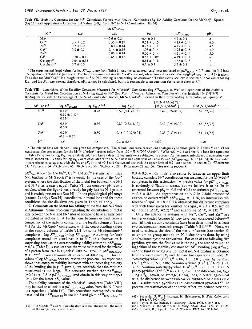
Table VII. Stability Constants for the M2+ Complexes Formed with Neutral Xanthosine (Eq 4),# Acidity Constants for the M(Xao)2t Species (Eq 22), and Approximate Crossover pH Values (pH,) from N-7 to N- l Coordination (Eq 24)
Mn2+ N.
co2+ 0.5 f 0.2 Ni2+ 0.7 f 0.2 cu2+ 0.8 f 0.3 Zn2+ Cd2+ 0.70 f 0.12 C O P Y )2+ 0.64 f 0.18 Cu(phen)2t 0.7 f 0.3
-0.6 f 0.2 -0.4 f 0.4 0.55 f 0.17 0.80 f 0.14 1.14 f 0.16 0.06 f 0.14 0.65 f 0.15
0.53 f 0.13 0.77 f 0.1 1 1.06 f 0.14 0.06 f 0.14 0.68 f 0.09 0.64 f 0.18 0.7 f 0.3
4.2 f 0.4 4.35 f 0.14 1.15 f 0.12 3.95 f 0.15 4.21 f 0.14 4.19 f 0.11 3.62 f 0.18 3.7 f 0.3
b 6.0 4.6
-4.0 5.0 b C
c
"The experimental (exp) values for log KMMtXao) are from Table 11, and the calculated (calc) ones are based on pKHH(,,) = 0.74 and the N-7 base line equations of Table IV (see text). The fourth column contains the "best" constant; where two values exist, the weighted mean with f 3 u is given. The value for Mn(Xao)2+ is a rough estimate. 'As N-7 binding is dominating, no crossover pH value exists: see text in section 8. 'No values for log KN.I and log KN.7 are known; therefore, pH, cannot be calculated, but it is reasonable to assume that the value is close to 3.7.
Table VIII. Logarithms of the Stability Constants Measured for M(Ado)2+ Complexes (log KMM(Ado)), as Well as Logarithms of the Stability Constants for Metal Ion Coordination at N - l (log KN.I) or N-7 (log KN.7) of Neutral Adenosine, Together with the Intrinsic [N-l]/[N-7] Binding Ratios and the Percentage of the N-7-Coordinated Isomer, M ( N - ~ / A ~ o ) ~ + , Formed in the Corresponding Intramolecular Equilibria"
[ M( N- 1 / M2+ or HC log KM~(Ado) log KN.lortho * log Kp4.7' [ M ( N - ~ / A ~ o ) ~ + ] % M ( N - ~ / A ~ O ) ~ + '
Ni2+ -0.1 7d 0.28 0.59 (0.41/0.78) 0.49 (0.74/0.32) 67 (57/76) 0.30 f 0.13' 0.321
cu2+ 0.84d 0.709
0.59 0.87 (0.62/1.12) 0.52 (0.93/0.30) 66 (52/77)
Zn2+ -0.2gd -0.80 -0.16 (-0.37/0.05) 0.23 (0.37/0.14) 81 (73/88)
H+ 3.6' 3.6 0.2 f 0.5' -2500 -0.04
-0.3 f 0.2h
OThe related data for H(Ado)+ are given for comparison. The calculations were carried out analogously to those given in Tables V and VI for xanthosine: the percentages for the M(N-1/Ado)2+ species follow from 100 - % M(N-7/Ado)*+. 'With pKa = 3.6 and the N - l base line equations in Table IV values for log KN., were calculated: from these values 1.2 log units were subtracted to account for the amino substituent in adenosine (see text in section 9). 'Values for log KN.7 were calculated with the N-7 base line equations of Table IV and p@H(N.~/~&,) = 0.2 (f0.5); the first value in parentheses is calculated with the lower pKa limit of -0.3 and the second one with the upper limit of 0.7 (see also text in section 9). dReference 63. 'Reference 64. /Reference 65. ZReference 50. *Reference 28. 'References 22 and 66. 'See text in section 9.
pKHxao = 5.47 for the Ni2+, Cu2+, and Zn2+ systems, as in these N-1 binding in M(Xao-H)+ is favored. In the case of the Coz+ system, where the distribution of the metal ion between the N-1 and N-7 sites is nearly equal (Table VI), the crossover pH is only reached when the ligand has already largely lost its N-1 proton and is mainly present as (Xao-H)-. In the physiological pH range (around 7) only (Xao-H)- coordinates to metal ions and for these conditions the site distributions given in Table VI apply.
9. Comments on the Metal Ion Affinity of the N-1 and N-7 Sites in Adenosine. Some problems regarding the distribution of metal ions between the N-1 and N-7 sites of adenosine have already been indicated in section 5. A further one becomes evident from a comparison of the stability constants in the fourth column of Table VI1 for the M(Xao)Z+ complexes, with the corresponding values in the second column of Table VI11 for some M(adenosine)2+
complexes metal ion coordination to N-7, this observation is surprising because the corresponding acidity constant, pKHH(Xao) = 0.74 (Table I), is smaller than the value estimated for the release of a proton from N-7 in H(Ado)' with N-1 free, i.e. Pf&(N.7/Ado) = I . 1 .46,62 Even allowance of an error of f 0 . 2 log unit for t h e values of log p,(Ad,,) does not resolve the problem. As experience shows that complex stability increases with an increasing basicity of the binding site (e.g., Figure 3), we conclude that the estimate mentioned is too large. We conclude further that pKHH(Xao) (=0.74) = 0.8 2 p K H ~ ( ~ . 7 , ~ d o ) and obtain in this way an upper limit for the latter pK, value.
The stability constants of the M(Ado)2+ complexes (Table VIII) may be used to calculate a PKHH(N.7/Ado) value from the N-7 base line equations (Table IV). This procedure corresponds to that described for pKH,(xa,,) in section 6 and gives pKHH(N.71Ado) =
Complexes: log K M ~ ( x a o ) > log K'M(Ado). Assuming for both
(62) If in M(Ado)2+ also N-1 coordination is taken into account, the result of the comparison is even worse.
0.0 f 0.5, which might also rather be taken as an upper limit because complete N-7 coordination was assumed for the M(Ado)2+ complexes in this estimation. A precise value for PKHH(N.7/Ado) is evidently difficult to assess, but we believe it to be (in its extremes) between pKa = -0.5 and +0.8 and estimate p@H(N.7/Ado) = 0.2 f 0.5. As deprotonation at N-7 in 2-fold protonated adenosine occurs PKHH2(Ado), = -1.56, an interaction dif- ference of ApK, = 1.8 f 0.5 is obtained; this difference compares well with those given for xanthosine (ApK, = 2.3 f 0.5; section 6), inosine (ApK, =2.2),46 and guanosine (ApK, = 2.0).46
Only the adenosine systems with NiZ+, Cuz+, and Zn2+ are further evaluated because (i) they have been considered before2z*46 and (ii) their stability constants have been determined by at least two independent research groups (Table VIII).63"6 Next, we need to estimate the size of the steric influence (see section 5) of an amino group next to an N-1 site; this is done by using 2-substituted pyridine derivatives. For each of the following five pyridine systems the first value is the pK,, the second value the logarithm of the stability constant for Mz+ binding (log KMML), and the third value log KN., for metal ion N-1 binding as obtained from the mentioned pK, and the base line equations of Table IV: 2-methylpyridine ( C U ~ + ) , ~ ~ 6.06, 1.3, 2.8 1; 2-methylpyridine (Ni2+),48 6.06, 51 , 2.06; 2-aminopyridine ( C U ~ ' ) , ~ ~ 6.96, 1.7 1, 3.18; 2-amino-3-methylpyridine (CU~'),~' 7.23, 1.91, 3.30; 2- phenylpyridine ( C U ~ + ) , ~ ~ 4.74, 0.7, 2.26. The difference log KN-I - log KMML equals, on average, 1.5 log units, in perfect agreement with the difference between two earlier published base lines valid for 3,4-substituted pyridines and 2-substituted pyridines.49 To prevent overestimation of the steric effect, we deduce now only
(63) Schneider, P. W.; Brintzinger, H.; Erlenmeyer, H. Hefu. Chim. Acfa
(64) Taylor, R. S . ; Diebler, H. Bioinorg. Chem. 1976, 6, 247-264. (65) Mariam, Y. H.; Martin, R. B. Inorg. Chim. Acfa 1979, 35, 23-28. (66) Tribolet, R.; Sigel, H. Eur. J . Biochem. 1987, 163, 353-363.
1964, 47, 992-1002.
Solution Structures of Nucleoside-Metal Complexes
1.2 log units from calculated log KN-1 values to obtain log KN-loltho constants.
By applying the results summarized in the last two paragraphs, we obtain the estimates given in the third column of Table VIII, which characterize the stability of N- 1 type M(Ado)2+ complexes. Column 4 lists the stabilities for N-7 type complexation by using p K H ~ ( ~ . 7 / ~ + , ) .= 0.2; the first value in parentheses refers to the lower pK, limit of -0.3 and the second one to the upper limit of 0.7. Now the ratios of N-1 over N-7 binding can be calculated (fifth column), as well as the percentages for metal ion coordi- nation at N-7 in the M(Ado)2+ complexes (last column); the lower and upper limits are given in parentheses. The corresponding data for the interaction between the proton and adenosine are listed at the bottom of the table. It may be noted that now the measured stability constants (considering f0.2 log unit as the error range) are in much better accord with the calculated values for log KN.1 and log KN.7 than p r e v i o u ~ l y ; " ~ ~ ~ * ~ ~ this also supports the present evaluation..
Though rather tentative with regard to the absolute size of the values, the data in Table VI11 show that the metal ions coordinate preferably at the N-7 site of adenosine and nor a t the N-1 site as previously c o n c l ~ d e d . ~ ~ * ~ ~ Moreover, it should be noted that assumption of a larger P K H ~ ( ~ . 7 / ~ d o ) value would further favor N-7 coordination; the same is true if a larger steric hindrance (i.e., > 1.2 log units) due to the amino group next to the N- 1 site is assumed. However, in agreement with the conclusions of Martin,22 it is evident that the intramolecular equilibrium between N-7- and N- 1 -bonded isomers exists and that proton binding occurs ov- erwhelmingly at N-1 of neutral a d e n o ~ i n e . ~ ~ , ~ ~ More final con- clusions can only be made after experiments have been carried out (for all metal ions) that allow construction of base lines for o-amino N-1 type ligands. Caveat and Conclusions
The reevaluation of the metal ion binding tendency of N-1 versus that of N-7 in adenosine and the conclusion that the N-7 site is favored for Ni2+, Cu2+, and Zn2+ binding is important: now, the formation of macrochelates in complexes of adenine nucleo- t i d e ~ ~ * ' ~ ~ ' ~ involving N-7 is expected. A more detailed reevaluation of the adenosine metal ion systems is clearly needed, but this should be based on more comprehensive experimental data. It is especially desirable to define the influence of ortho substituents on the N-1 reference lines more exactly; it appears that there are three such N-1 base lines. (i) One base line corresponds to unsubstituted N-1 or pyridine-like ligands; in fact, these reference lines are given in Table IV and they include also ligands with an o-carbonyl oxygen (Table 111). The steric influence of this group is small (Le., smaller than that of an amino Indeed, the data for inosine and 7-methylinosine (Table 111) fit well on the N-1 reference lines (Figure 3); hence, if there is any steric influence, it is offset by an oxo-metal ion interaction possibly via a water molecule (see section 5 ) . (ii) There are certainly independent reference lines for N- 1 type ligands with an o-amino group, as for 2-aminopyridine or adenosine; these base lines are expected (see section 9) to be about 1-1.5 log units, depending on the metal
(67) Reily, M. D.; Hambley, T. W.; Marzilli, L. G . J . Am. Chem. Soc. 1988, 110, 2999-3007.
Inorganic Chemistry, Vol. 28, No. 8, 1989 1489
ion, below the reference lines of Table IV. (iii) It appears highly likely that in N-1 or pyridine-like ligands with an o-amino and an o-carbonyl group the steric effect of the amino group is partially offset by a "positive" oxo-metal ion interaction. A ligand of this type is cytidine (see also section 5), and there are indications that metal ions not only coordinate to this ligand via the pyridine-like N but also interact simultaneously with the carbonyl o ~ y g e n . ~ . ~ Consequently, these base lines should be situated between those of points i and ii; indeed, use of the cytidine results of section 5 give for Cu2+ and Ni2+ reference lines that are about 0.45 and (at least) 1 log unit, respectively, below those of Table IV.
The results obtained for xanthosine (sections 6-8) show that for xanthosine 5'-monophosphate (XMP) an overlap between N- 1 deprotonation and the release of a proton from the phosphate group must be expected. In any case, in the neutral pH range XMP will exist mainly as a 3-fold negatively charged species; hence, formation of M(X-H-MP)- complexes will occur. Moreover, in the physiological pH range M(X-H.MP)- species will have a high probability for macrochelate formation.
There is one further aspect of general importance: the equations of the base lines quantifying the pyridine-like (or N-1) and im- idazole-like (or N-7) coordination tendency of metal ions allows us to judge the reliability of published stability constants for nucleoside complexes. For example, stability constants publisheda for the M(inosine-H)+ complexes of Mn2+, Co2+, Ni2+, Cuz+, and Zn2+ are clearly too large if judged on the basis of the reference lines of Figure 3 or the equations listed in Table IV; in contrast, the corresponding constants for Co2+, Ni2+, and Zn2+ in ref 53 are in accordance with the expectations. Similarly, the stability constants published in ref 69 for M(guanosine)2+ complexes have to be rejected, while those of ref 53 can be recommended. In addition, stability constants provided for M(uridine-H)+ (cf. ref 70) and M(cytidine)2+ (cf. ref 71) are also not reliable (with regard to the latter complexes see also section 5). It is important for researchers who apply stability constants of nucleoside-metal ion complexes, e.g. to evaluate the metal ion affinity of certain sites in DNA or RNA, to be aware of such pitfalls; otherwise, com- pletely misleading conclusions may be reached. In such situations the base line equations of Table IV, together with the comments given above, may be used as a first guide in judging the validity of published stability data.
Acknowledgment. We thank Rita Baumbusch and Dr. Salah S. Massoud for carrying out some of the initial pH titrations used for the calculation of the acidity constants of xanthosine. A research grant from the Swiss National Science Foundation (H.S.) and support from the Japanese Ministry of Education and the University of the Ryukyus (Y.K.) are gratefully acknowledged.
Reddy, P. R.; Reddy, K. V.; Taqui Khan, M. M. Indian J . Chem. 1983,
Khan, B. T.; Raju, M.; Zakeeruddin, S. M. J . Chem. SOC., Dalton Tram. 1988, 67-7 1. Khan, B. T.; Raju, R. M.; Zakeeruddin, S. M. J . Coord. Chem. 1987,
(a) Khan, B. T.; Raju, R. M. Indian J . Chem. 1981, ZOA, 860. (b) Reddy, P. R.; Rao, V. B. M. Polyhedron 1985, 4, 1603-1609. (c) Reddy, P. R.; Sudhakar, K. Proc. Indian Acad. Sci., Chem. Sci. 1987,
ZZA, 999-1000.
16, 237-244.
98, 289-295.