sinonasal tract angiosarcoma: a clinicopathologic and immunophenotypic study of 10 cases with a...
TRANSCRIPT

ORIGINAL RESEARCH
Sinonasal Tract Angiosarcoma: A Clinicopathologicand Immunophenotypic Study of 10 Cases with aReview of the Literature
Brenda L. Nelson Æ Lester D. R. Thompson
Received: 22 April 2007 / Accepted: 20 July 2007 / Published online: 25 October 2007
� Humana Press Inc. 2007
Abstract Background Primary sinonasal tract angio-
sarcoma are rare tumors that are frequently misclassified,
resulting in inappropriate clinical management. There are
only a few reported cases in the English literature. Mate-
rials and Methods Ten patients with sinonasal tract
angiosarcoma were retrospectively retrieved from the
Otorhinolaryngic Registry of the Armed Forces Institute of
Pathology. Results Six males and four females, aged 13
to 81 years (mean, 46.7 years), presented with epistaxis
and bloody discharge. Females were on average younger
than their male counterparts (37.8 vs. 52.7 years, respec-
tively). The tumors involved the nasal cavity alone (n = 8)
or the maxillary sinus (n = 2), with a mean size of 4.3 cm;
the average size was different between the genders: males:
2.8 cm; females: 6.4 cm. Histologically, all tumors had
anastomosing vascular channels lined by remarkably
atypical endothelial cells protruding into the lumen, neol-
umen formation, frequent atypical mitotic figures, necrosis,
and hemorrhage. All cases tested (n = 6) demonstrated
immunoreactivity with antibodies to Factor VIII-RA,
CD34, CD31, and smooth muscle actin, while non-reactive
with keratin and S-100 protein. The principle differential
diagnosis includes granulation tissue, lobular capillary
hemangioma (pyogenic granuloma), and Kaposi’s sarcoma.
All patients had surgery followed by post-operative radia-
tion (n = 4 patients). Follow-up was available in all
patients: Six patients died with disease (mean,
28.8 months); two patients had died without evidence of
disease (mean, 267 months); and two are alive with no
evidence of disease at last follow-up (mean, 254 months).
Conclusions Sinonasal tract angiosarcoma is a rare
tumor, frequently presenting in middle-aged patients as a
large mass usually involving the nasal cavity with char-
acteristic histomorphologic and immunophenotypic
features. Sinonasal tract angiosarcoma will often have a
poor prognosis making appropriate separation from other
conditions important.
Keywords Angiosarcoma � Sinonasal tract �Nasal cavity � Vascular � Hemangioma � Sarcoma �Immunohistochemistry � Prognosis � Survival �Differential diagnosis
Introduction
Angiosarcomas are high-grade, malignant vascular tumors
that make up only about 2% of all sarcomas [1, 2]. While
angiosarcomas may occur in any region of the body, well
over half occur in the head and neck, usually involving the
skin and superficial soft tissues, particularly the scalp [1,
3–7]. Despite this fact, angiosarcoma accounts for less than
0.1% of all sinonasal tract malignancies [3, 8–13]. Primary
sinonasal tract angiosarcomas are exceedingly uncommon
and only a few cases have been reported in the English
literature [9, 14–34]. The rarity of these tumors may result
in the misclassification and subsequent inappropriate
management. Further, many synonyms have been applied
to angiosarcomas (epithelioid hemangioendothelioma;
B. L. Nelson
Department of Anatomic Pathology, Naval Medical Center,
San Diego, CA, USA
L. D. R. Thompson (&)
Department of Pathology, Woodland Hills Medical Center,
Southern California Kaiser Permanente Group,
5601 De Soto Avenue, Woodland Hills, CA 91365, USA
e-mail: [email protected]
Head and Neck Pathol (2007) 1:1–12
DOI 10.1007/s12105-007-0017-2

malignant hemangioendothelioma; malignant angioendo-
thelioma; lymphangiosarcoma; hemangiosarcoma;
hemangioblastoma), but the use of these terms in the
sinonasal tract is discouraged, especially since hemangio-
endothelioma represents a unique entity. This report
focuses on the clinical presentation, histologic features,
immunohistochemical profiles, and therapeutic approaches
of sinonasal angiosarcomas in relation to patient prognosis
and outcome.
Material and Methods
Ten cases of angiosarcoma involving the involving the
nasal cavity (n = 8) or paranasal sinuses (sphenoid, max-
illary, ethmoid, and frontal sinuses; n = 2) were retrieved
from the files of the Otorhinolaryngic-Head & Neck Tumor
Registry of the Armed Forces Institute of Pathology
(AFIP), Washington, DC, between 1970 and 1995. These
tumors were chosen from a review of 20,156 (0.05%)
benign or malignant primary sinonasal tract tumors seen in
consultation during this time. All cases were obtained from
civilian sources, including university medical centers.
Materials within the AFIP files were supplemented by a
review of the patient demographics (gender, age, and race),
symptoms at presentation (epistaxis, nasal obstruction,
nasal discharge), including duration (Table 1). Follow-up
information was obtained by direct written and oral com-
munication with the referring pathologist, patient’s
physicians, tumor registries, and patients or patient’s
family members. Follow-up data was available for all ten
patients and included information regarding exact tumor
site, specific treatment modalities used, the presence or
absence of recurrent or metastatic disease, and the current
status of the disease and patient. It is important to add that
we conducted this research from a tertiary pathology
review center, conducting a retrospective review of these
patients and we did not treat the patients. As we did not
prosect the specimen, we had to rely on the contributing
pathologist for an accurate assessment of the margins of
resection. Submitted diagnoses included juvenile naso-
pharyngeal angiofibroma, hemangioma, hemangiosarcoma,
malignant vascular tumor, malignant hemangiopericytoma,
and hemangioendothelioma. This clinical investigation was
conducted in accordance and compliance with all statutes,
directives, and guidelines of the Code of Federal Regula-
tions, Title 45, Part 46, and the Department of Defense
Directive 3216.2 relating to human subjects in research.
Hematoxylin and eosin-stained slides from all cases
were reviewed to confirm that the established histopatho-
logic criteria for the diagnosis of angiosarcoma were met.
A number of macroscopic and histologic observations were
recorded for each of the tumors as follows: tumor location
(Fig. 1); tumor size (greatest dimension in centimeters);
extravasated blood (absent or present [Fig. 2]); respiratory
epithelium (present of absent); anastomosing vascular
channels (Fig. 3); pleomorphism (moderate or severe
[Fig. 3]); tumor cell spindling; neolumen formation
(Figs. 2, 4, 5); mitotic figures (number of mitotic figures
Table 1 Clinical characteristics
Clinical characteristics Number
Gender
Females 4
Males 6
Age (in years)
Range 13–81
Mean 46.7
Women (mean) 37.8
Men (mean) 52.7
Symptoms
Duration (range, in months) 2–24
Duration (mean, in months) 10.7
Epistaxis 6
Obstructive symptoms 3
Nasal discharge 1
Anatomic site
Nasal cavity alone 8
Maxillary sinus alone 2
Size (cm)
Range 1.8–8
Mean 4.3
Female (mean) 6.4
Male (mean) 2.8
Maxillary sinus 8.0
Nasal cavity 2.9
Fig. 1 A vascular neoplasm abuts the nasal cartilage in this
angiosarcoma of the nasal cavity
2 Head and Neck Pathol (2007) 1:1–12
per 10 high power fields [magnification at 40· with a 10·objective lens using Olympus BX40 microscope]); atypical
mitotic figures (present or absent, and defined by abnormal
chromosome spread, tripolar or quadripolar forms, circular
forms, or indescribably bizarre [Fig. 5]); necrosis (absent
or present); and the presence of other microscopic patho-
logic findings.
Immunophenotypic analysis was performed in all cases
with suitable material by a standardized EnvisionTM
method employing 4 lm-thick, formalin fixed, paraffin
embedded sections. Table 2 documents the pertinent,
commercially available immunohistochemical antibody
panel used. The analysis was performed on a single rep-
resentative block for each tumor. When required,
proteolytic antigen retrieval was performed by predigestion
for 3 min with 0.05% Protease VIII (Sigma Chemical Co.,
St. Louis, MO) in a 0.1 M phosphate buffer, pH of 7.8, at
37�C. Heat induced epitope retrieval was performed, as
required, by using formalin-fixed, paraffin-embedded tissue
treated with a buffered citric acid solution pH 6.0 (Citra,
Dako Corporation, Carpinteria, CA) and heated for 20 min
in a steamer. Standard positive controls were used
throughout, with serum used as the negative control. The
antibody reactions were described as either positive or
negative. The Ki-67 antibody reaction was recorded as the
fraction of positive cells, separating them into four groups:
\10%, 11–50%, 51–90%, and [90%.
A review of the English literature based on a MEDLINE
search from 1966 to 2007 was performed and all cases
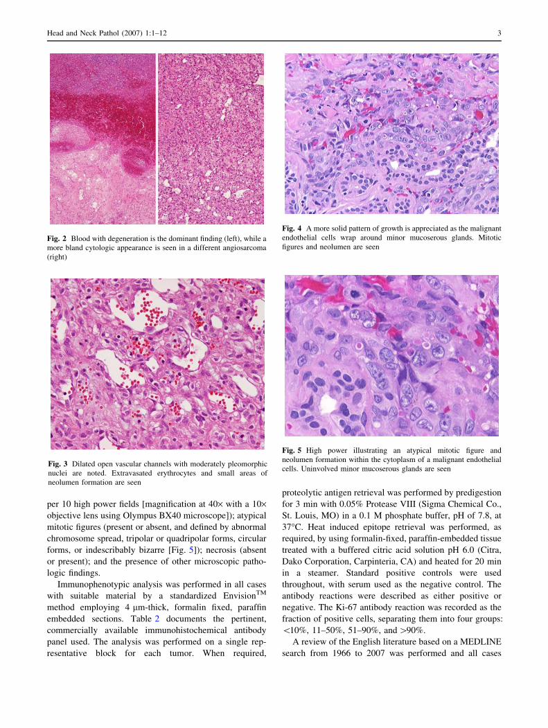
Fig. 2 Blood with degeneration is the dominant finding (left), while a
more bland cytologic appearance is seen in a different angiosarcoma
(right)
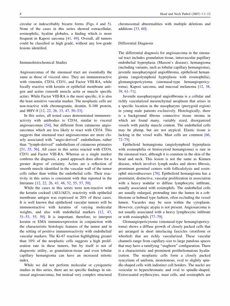
Fig. 3 Dilated open vascular channels with moderately pleomorphic
nuclei are noted. Extravasated erythrocytes and small areas of
neolumen formation are seen
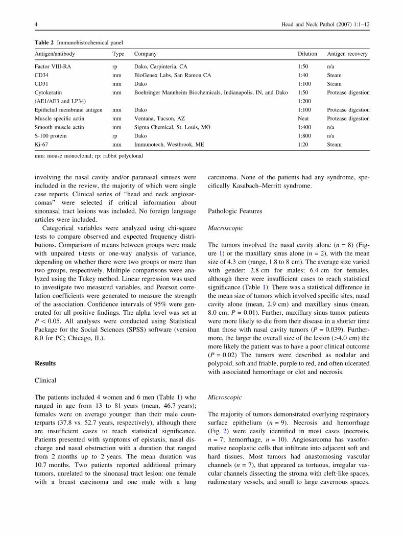
Fig. 4 A more solid pattern of growth is appreciated as the malignant
endothelial cells wrap around minor mucoserous glands. Mitotic
figures and neolumen are seen
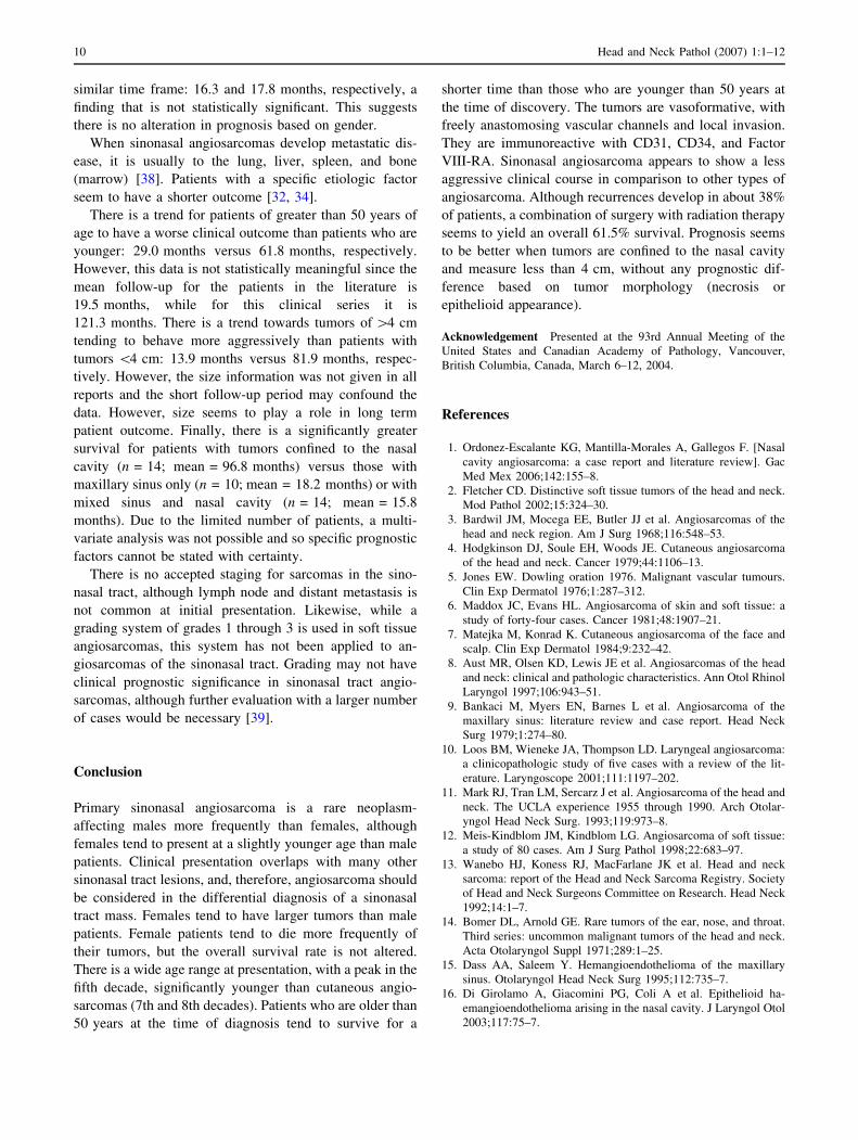
Fig. 5 High power illustrating an atypical mitotic figure and
neolumen formation within the cytoplasm of a malignant endothelial
cells. Uninvolved minor mucoserous glands are seen
Head and Neck Pathol (2007) 1:1–12 3
involving the nasal cavity and/or paranasal sinuses were
included in the review, the majority of which were single
case reports. Clinical series of ‘‘head and neck angiosar-
comas’’ were selected if critical information about
sinonasal tract lesions was included. No foreign language
articles were included.
Categorical variables were analyzed using chi-square
tests to compare observed and expected frequency distri-
butions. Comparison of means between groups were made
with unpaired t-tests or one-way analysis of variance,
depending on whether there were two groups or more than
two groups, respectively. Multiple comparisons were ana-
lyzed using the Tukey method. Linear regression was used
to investigate two measured variables, and Pearson corre-
lation coefficients were generated to measure the strength
of the association. Confidence intervals of 95% were gen-
erated for all positive findings. The alpha level was set at
P \ 0.05. All analyses were conducted using Statistical
Package for the Social Sciences (SPSS) software (version
8.0 for PC; Chicago, IL).
Results
Clinical
The patients included 4 women and 6 men (Table 1) who
ranged in age from 13 to 81 years (mean, 46.7 years);
females were on average younger than their male coun-
terparts (37.8 vs. 52.7 years, respectively), although there
are insufficient cases to reach statistical significance.
Patients presented with symptoms of epistaxis, nasal dis-
charge and nasal obstruction with a duration that ranged
from 2 months up to 2 years. The mean duration was
10.7 months. Two patients reported additional primary
tumors, unrelated to the sinonasal tract lesion: one female
with a breast carcinoma and one male with a lung
carcinoma. None of the patients had any syndrome, spe-
cifically Kasabach–Merritt syndrome.
Pathologic Features
Macroscopic
The tumors involved the nasal cavity alone (n = 8) (Fig-
ure 1) or the maxillary sinus alone (n = 2), with the mean
size of 4.3 cm (range, 1.8 to 8 cm). The average size varied
with gender: 2.8 cm for males; 6.4 cm for females,
although there were insufficient cases to reach statistical
significance (Table 1). There was a statistical difference in
the mean size of tumors which involved specific sites, nasal
cavity alone (mean, 2.9 cm) and maxillary sinus (mean,
8.0 cm; P = 0.01). Further, maxillary sinus tumor patients
were more likely to die from their disease in a shorter time
than those with nasal cavity tumors (P = 0.039). Further-
more, the larger the overall size of the lesion ([4.0 cm) the
more likely the patient was to have a poor clinical outcome
(P = 0.02) The tumors were described as nodular and
polypoid, soft and friable, purple to red, and often ulcerated
with associated hemorrhage or clot and necrosis.
Microscopic
The majority of tumors demonstrated overlying respiratory
surface epithelium (n = 9). Necrosis and hemorrhage
(Fig. 2) were easily identified in most cases (necrosis,
n = 7; hemorrhage, n = 10). Angiosarcoma has vasofor-
mative neoplastic cells that infiltrate into adjacent soft and
hard tissues. Most tumors had anastomosing vascular
channels (n = 7), that appeared as tortuous, irregular vas-
cular channels dissecting the stroma with cleft-like spaces,
rudimentary vessels, and small to large cavernous spaces.
Table 2 Immunohistochemical panel
Antigen/antibody Type Company Dilution Antigen recovery
Factor VIII-RA rp Dako, Carpinteria, CA 1:50 n/a
CD34 mm BioGenex Labs, San Ramon CA 1:40 Steam
CD31 mm Dako 1:100 Steam
Cytokeratin
(AE1/AE3 and LP34)
mm Boehringer Mannheim Biochemicals, Indianapolis, IN, and Dako 1:50
1:200
Protease digestion
Epithelial membrane antigen mm Dako 1:100 Protease digestion
Muscle specific actin mm Ventana, Tucson, AZ Neat Protease digestion
Smooth muscle actin mm Sigma Chemical, St. Louis, MO 1:400 n/a
S-100 protein rp Dako 1:800 n/a
Ki-67 mm Immunotech, Westbrook, ME 1:20 Steam
mm: mouse monoclonal; rp: rabbit polyclonal
4 Head and Neck Pathol (2007) 1:1–12

These spaces are filled with erythrocytes and lined by plump,
enlarged, atypical, spindled (n = 7) or epithelioid endothe-
lial cells which protruded into the vascular spaces in multiple
layers or papillae (Fig. 3). Intracytoplasmic vacuoles, or
neolumen, were identified (n = 8) and contained erythro-
cytes (Figs. 3, 4, and 5). This feature was more characteristic
of the epithelioid growth pattern. The endothelial cells
demonstrated pleomorphic nuclei (severe, n = 3; moderate,
n = 7) with coarse and heavy nuclear chromatin deposition,
irregular nuclear contours, and prominent nucleoli (Fig. 3).
Mitotic figures (Fig. 4) were seen in all of the cases and were
easily identified. Additionally, atypical mitotic figures were
present in most of the tumors (n = 8) (Fig. 5). No extracel-
lular eosinophilic hyaline globules were identified.
Inflammatory cells were present to a variably degree in all
cases, although without a dominant cell type identified.
Immunohistochemical Results
All tested cases (n = 6) demonstrated immunoreactivity
with antibodies to Factor VIII-RA, CD34 (Fig. 6), CD31,
and smooth muscle actin, while non-reactive with keratin
and S-100 protein (Table 3). Epithelial membrane antigen
(n = 1) and muscle specific actin (n = 2) showed varied
immunoreactivity. Ki-67 was reactive, at [10% in all the
tested cases (Fig. 6).
Treatment and Follow-up
All patients were treated with surgery alone or with surgery
followed by post-operative radiation (n = 4) and chemo-
therapy (n = 2). Follow-up was available in all ten patients
(mean follow-up, 121 months; Table 4). Six patients died
with disease (mean, 28.4 months), 2 were alive with no
evidence of disease (mean, 254 months), and 2 were dead
with no evidence of their disease (mean, 267 months) and
died as the result of other primary malignancies.
The four female patients (mean follow-up, 78 months; 3
dead with disease, mean 29 months) tended to have a
poorer outcome than the six male patients (mean follow-up
151 months, 3 dead with disease, mean 29 months), but
this may be related to the size of the tumors. A male and a
female patient each died with no evidence of the primary
angiosarcoma, but succumbed to other primary malignan-
cies (mean follow-up, 267 months). Radiation and
radiation with chemotherapy did not alter the patient out-
come by a statistically identifiable amount. Age did not
reach statistical significant as a predictor of patient out-
come. Size of greater than or equal to 4 cm correlated with
poor outcome (P = 0.02). Finally, tumors involving the
maxillary sinuses tended to do worse than those which
involved the nasal cavity alone (P = 0.039).
Discussion
Despite the fact that the majority of angiosarcomas affect
the skin and soft tissues of the head and neck, angiosar-
comas within the sinonasal tract account for \0.1% of all
malignancies in this region [1, 3, 3–13]. This study reports
Table 3 Pathology findings
Microscopic characteristic Number of cases
Respiratory epithelium present 9
Anastomosing vascular channels 7
Tumor cell spindling 7
Mitotic activity 1–18
Atypical mitotic figures present 8
Pleomorphism
Moderate 7
Severe 3
Neolumen formation 8
Necrosis present 7
Extravasated blood present 10
Immunohistochemical results
Factor VIII-RA 100%
CD34 100%
CD31 100%
Keratin 0%
Epithelial membrane antigen 20%
Muscle specific actin 33%
Smooth muscle actin 100%
S-100 protein 0%
Ki-67 ([10%) 100%
Fig. 6 CD34 strongly stained the neoplastic cells’ cytoplasm and was
distributed in a haphazard fashion (left). Ki-67 strongly and diffuse
reacted with the nuclei in many cases (right)
Head and Neck Pathol (2007) 1:1–12 5

the largest single series of sinonasal tract angiosarcomas
reported in the English language to date, with the vast
majority of studies reporting only a single case [9, 14–34].
Angiosarcoma has been reported to develop in nearly all
anatomic sites, but when this high-grade vascular neoplasm
occurs in the sinonasal tract, a number of differential
diagnostic considerations are raised, along with a different
outcome than primary angiosarcomas in other anatomic
sites.
Retrospective analysis of any disease is a difficult
undertaking in modern medicine, and even more so when
the entity is rare. Terminology has evolved over the past
few decades, rendering the many names used in the past for
angiosarcoma surfeit. Clinical presentation, gender differ-
ences, anatomic site of distribution, size, histologic and
immunohistochemical features, and patient outcome has
not been well characterized by the many single case
reports. The information in this study is combined with that
gleaned from the literature (Table 5) in an attempt to more
fully elucidate the nature of this uncommon tumor and
perhaps contribute to more meaningful clinical
management.
Clinical Information
In our series, sinonasal angiosarcomas were more common
in men than women (male: female, 3:2), a finding sup-
ported by the literature (male: female, 2.2) (Table 5). It is
interesting that this is similar to angiosarcoma of the skin,
in which there is also a distinct male predilection [6, 35].
There is a wide age range at presentation (8–82 years),
with a mean of 46.8 years, substantially younger than the
8th decade mean age at presentation for soft tissue and skin
angiosarcomas [6, 8, 11, 36–39]. While women tended to
be younger than men at presentation (42.6 versus
48.8 years), this difference was not statistically significant.
The patients tended to have symptoms for an average of
Table 4 Patient outcome
All
patients
A, NED D, NED D, D
All patients with
follow-up (years)
10 (10.1) 2 (21.2) 2 (22.3) 6 (2.4)
Follow-up range
(years)
0.1–32.7 9.6–32.8 18.7–25.8 0.1–6.3
Gender
Males (years) 6 (12.6) 2 (21.2) 1 (25.8) 3 (2.4)
Females (years) 4 (6.5) n/a 1 (18.7) 3 (2.4)
Age
\40 years 5 (12.1) 1 (32.8) 1 (18.7) 3 (3.1)
‡40 years 5 (8.1) 1 (9.6) 1 (25.8) 3 (1.7)
Size*
\4.0 cm 4 (13.3) 2 (21.2) n/a 2 (5.4)
‡4.0 cm 3 (0.4) n/a n/a 3 (0.4)
Anatomic site
Nasal cavity
alone
8 (12.5) 2 (21.2) 2 (22.3) 4 (3.4)
Maxillary sinus
alone
2 (0.4) n/a n/a 2 (0.4)
Treatment received
Surgery alone 6 (11.9) 1 (32.8) 1 (25.8) 4 (3.3)
Surgery with
radiation
2 (5.0) 1 (9.6) n/a 1 (0.4)
Surgery with
radiation/chemotherapy
2 (9.7) n/a 1 (18.7) 1 (0.7)
A, NED: Alive, No Evidence of Disease; D, NED: Dead, No Evidence
of Disease; D, D: Dead of Disease, * Size was not reported in all
cases; n/a: not applicable
Table 5 Review of literature combined with current cases [9, 14–34]
Characteristics Number (39)
Gender
Females 12
Males 26
Age (in years)
Range 8–82
Mean 46.8
Women (mean) 42.6
Men (mean) 48.8
Symptoms*
Duration (range, in months) 0.3–96
Duration (mean, in months) 9.8
Epistaxis 20
Obstructive symptoms 18
Nasal discharge 5
Anatomic site
Nasal cavity alone 14
Paranasal sinus alone 10
Combination of sinuses & nasal cavity 14
Size (cm)*
Range 0.7–8
Mean 3.9
Female (mean) 6.0
Male (mean) 2.9
Paranasal sinus alone 6.8
Nasal cavity 2.2
Combination of sinuses & nasal cavity 4.4
Patient survival (mean, months)
A, NED 21 (47)
A, D 1 (2.0)
D, NED 3 (187)
D, D 14 (18.2)
* Not reported for all cases; A, NED: Alive, No Evidence of Disease;
A, D: Alive, with disease; D, NED: Dead, No Evidence of Disease; D,
D: Dead of Disease
6 Head and Neck Pathol (2007) 1:1–12

9.8 months, with epistaxis and obstruction identified most
frequently (20 and 18, respectively). Other symptoms
included nasal discharge, expanding or enlarging mass,
sinusitis, epiphora, pain (headache, otalgia, tooth-ache),
diplopia, ptosis and headaches. Needless to say, none of
these symptoms is specific for this tumor, although the high
rate of epistaxis is probably related to the vascular nature of
the neoplasm. In fact, we posit that the overall better
clinical prognosis for sinonasal tract angiosarcomas when
compared to their skin, soft tissue or visceral counterparts,
may be due to the earlier stage at diagnosis because of
epistaxis as a presenting symptom. This results in an earlier
detection of the tumor, and possibly a better outcome than
angiosarcomas in other anatomic sites [1, 9, 10, 12, 40–44].
Within the sinonasal tract, a single anatomic site is affected
more commonly than multiple sites (nasal cavity alone =
14; single sinus = 14; multiple areas = 10). Any of the
paranasal sinuses can be involved (maxillary, ethmoid,
sphenoid, cavernous sinus), but the maxillary sinus seems
to be involved more frequently than the others. It is curious
that when multiple sites are involved by tumor, the mean
size of tumor (4.4 cm) is less than if a single paranasal
sinus is involved (mean, 6.8 cm). This discrepancy is
accounted for by the overall lack of size data from the
single case reports. Overall, the tumor size within the nasal
cavity alone is less than the paranasal sinuses alone or if
there is a combination of nasal cavity and paranasal sinus
(2.2 cm vs. 6.8 cm vs. 4.4 cm, respectively). It is our
impression from this data, that perhaps a lesion within the
confines of the nasal cavity is more likely to be evaluated
earlier than a lesion which affects the sinuses [16, 17, 26,
45]. Tumors in female patients tend to be larger (mean,
6.0 cm) than tumors in male patients (mean, 2.9 cm),
although there are insufficient cases to reach statistical
significance.
While no patients in our clinical series had any docu-
mented environmental exposure as a possible etiologic
factor, three patients reported in the literature had prior
radiation exposure [25, 34, 46], one patient reported
working in a coal mine for decades [9], and one patient
reported exposure to vinyl chloride [32]. Therefore, it is
possible that rare cases may have an environmental
etiology.
Radiographic Studies
Angiosarcoma is an aggressive infiltrative tumor that will
often invade adjacent soft tissues, cartilage and bone
(Fig. 1). Sinonasal tract angiosarcoma may be radiolucent
or radio-opaque. A soft tissue density, it may be associated
with bone erosion or occasionally have well-delimited
borders. Because of its ability to erode bone, computed
tomography (CT) may allow for an accurate determination
of the extent of the mass, showing enhancement with
contrast. Magnetic resonance imaging (MRI) shows the
tumor to be bright on T2-weighted images. Angiography is
an excellent modality to identify the extent of the tumor
and show the feeder vessel(s) if they are present, while also
allowing for pre-surgical angiographic embolization, if
desired [19, 22, 29, 30, 34].
Pathology
Macroscopically, the tumors are nodular and polypoid,
although with increased size, they tend to infiltrate the
surrounding tissues. The tumors are soft and friable, purple
to red, and often associated with hemorrhage, clot and
necrosis.
Respiratory epithelium was present and intact in 9 of 10
cases in this series, a histologic finding similar to
descriptions of intact epidermis overlying skin primary
angiosarcomas [47, 48]. Ulceration, therefore, is not a
common finding, except in lesions that are large or have
involved more than one anatomic site [15, 22–24, 30, 32,
35]. As may be expected, all our cases and the majority of
those in the literature demonstrated histologic evidence of
blood or extravasated erythrocytes. The vasoformative
pattern seems to be quite universal, with tortuous, irregular,
freely anastomosing vascular channels dissecting through
stroma and creating cleft-like spaces, rudimentary vessels,
capillary-sized vessels and large cavernous spaces. These
vascular spaces and channels were filled with erythrocytes
and lined by plump, enlarged, atypical endothelial cells
which protruded into the vascular spaces in multiple layers
or papillae. An epithelioid appearance can be seen focally
in many cases, but it is usually not the dominant pattern.
Tumor cell spindling is also present, and may sometimes
expand the differential diagnosis. Intracytoplasmic neolu-
men were identified in the majority of cases (8 of 10 cases),
but this feature is not always histologically demonstrated or
described in the case reports. Neolumina seem to be more
easily identified in areas that are epithelioid in appearance.
The presence of erythrocytes within these spaces certainly
confirms the vasoformative nature of this tumor (Figs. 3
and 4).
The neoplastic cells usually show profound nuclear
pleomorphism, with the nuclei showing enlargement with
coarse and heavy nuclear chromatin distribution. The
nuclear contours are frequently irregular or ‘‘moth-eaten.’’
Prominent, irregular nucleoli are seen (Fig. 3). Mitotic
figures were easily identified in all cases, ranging from 1 to
18 figures per 10 high power fields. Atypical forms, also
readily identifiable in most cases, consisted of abnormal
chromosome spread, tripolar or quadripolar forms, and
Head and Neck Pathol (2007) 1:1–12 7
circular or indescribably bizarre forms (Figs. 4 and 5).
None of the cases in this series showed extracellular,
eosinophilic, hyaline globules, a finding which is more
frequent in Kaposi sarcoma [41, 49]. Overall, all tumors
could be classified as high grade, without any low-grade
lesions identified.
Immunohistochemical Studies
Angiosarcomas of the sinonasal tract are essentially the
same as those of visceral sites. They are immunoreactive
with vimentin, CD34, CD31, and Factor VIII-RA, while
focally reactive with keratin or epithelial membrane anti-
gen and actins (smooth muscle actin or muscle specific
actin). While Factor VIII-RA is the most specific, it is also
the least sensitive vascular marker. The neoplastic cells are
non-reactive with chromogranin, desmin, S-100 protein,
and HHV-8 [12, 22, 26, 33, 47, 50–53].
In this series, all tested cases demonstrated immunore-
activity with antibodies to CD34, similar to visceral
angiosarcomas [54], but different from cutaneous angio-
sarcomas which are less likely to react with CD34. This
suggests that sinonasal tract angiosarcomas are more clo-
sely associated with ‘‘angio-derived’’ endothelium, rather
than ‘‘lymph-derived’’ endothelium of cutaneous primaries
[51, 55, 56]. All cases in this series reacted with CD34,
CD31 and Factor VIII-RA. Whereas as a single marker
confirms the diagnosis, a panel approach does allow for a
greater degree of certainty. Actins are a reflection of
smooth muscle identified in the vascular wall of the tumor
cells rather than within the endothelial cells. Their reac-
tivity in this series is consistent with that reported in the
literature [12, 22, 26, 43, 48, 52, 55, 57, 58].
While the cases in this series were non-reactive with
the keratin cocktail (AE1/AE3), reactivity with epithelial
membrane antigen was expressed in 20% of these cases.
It is well known that epithelioid vascular tumors will be
immunoreactive with keratins of varying molecular
weights, and also with endothelial markers [12, 43,
51–53, 55, 59]. It is important, therefore, to interpret
keratin or EMA immunoexpression in conjunction with
the characteristic histologic features of the tumor and in
the setting of positive immunoreactivity with endothelial/
vascular markers. The Ki-67 reaction highlighting greater
than 10% of the neoplastic cells suggests a high prolif-
eration rate in these tumors, but by itself is not of
diagnostic utility, as granulation-tissue and even lobular
capillary hemangioma can have an increased mitotic
index.
While we did not perform molecular or cytogenetic
studies in this series, there are no specific findings in sin-
onasal angiosarcomas, but instead very complex structural
chromosomal abnormalities with multiple deletions and
additions [33, 60].
Differential Diagnosis
The differential diagnosis for angiosarcoma in the sinona-
sal tract includes granulation tissue, intravascular papillary
endothelial hyperplasia (Masson’s disease), hemangioma
(including variants, such as lobular capillary hemangioma),
juvenile nasopharyngeal angiofibroma, epithelioid heman-
gioma (angiolymphoid hyperplasia with eosinophilia),
glomangiopericytoma (sinonasal-type hemangiopericy-
toma), Kaposi sarcoma, and mucosal melanoma [12, 38,
39, 61–71].
Juvenile nasopharyngeal angiofibroma is a cellular and
richly vascularized mesenchymal neoplasm that arises in
a specific location in the nasopharynx (pterygoid region)
in young male patients exclusively. Histologically, there
is a background fibrous connective tissue stroma in
which are found many, variably sized, disorganized
vessels with patchy muscle content. The endothelial cells
may be plump, but are not atypical. Elastic tissue is
lacking in the vessel walls. Mast cells are common [66,
72–75].
Epithelioid hemangioma (angiolymphoid hyperplasia
with eosinophilia or histiocytoid hemangioma) is rare in
the sinonasal tract, although it is common in the skin of the
head and neck. This lesion is not the same as Kimura
disease, which involves lymph nodes and shows fibrosis,
prominent germinal centers with folliculolysis and eosin-
ophil microabscesses [76]. Epithelioid hemangioma has a
prominent, distinctive, vascular proliferation in association
with a heavy nodular to diffuse lymphocytic infiltrate,
usually associated with eosinophils. The endothelial cells
are usually enlarged, protruding into the lumen in a cob-
blestone or hobnail type fashion, often occluding the vessel
lumen. Vacuoles may be seen within the cytoplasm.
However, cytologic atypia is not present. Angiosarcoma is
not usually associated with a heavy lymphocytic infiltrate
or with eosinophils [77–79].
Glomangiopericytoma (sinonasal-type hemangiopericy-
toma) shows a diffuse growth of closely packed cells that
are arranged in short interlacing fascicles (storiform or
whorled) that are richly vascularized. These vascular
channels range from capillary-size to large patulous spaces
that may have a ramifying ‘‘staghorn’’ configuration. There
is a characteristic and prominent peritheliomatous hyalin-
ization. The neoplastic cells form a closely packed
syncytium of uniform, monotonous, oval to slightly spin-
dle-shaped cells with indistinct cell borders. The nuclei are
vesicular to hyperchromatic and oval to spindle-shaped.
Extravasated erythrocytes, mast cells, and eosinophils are
8 Head and Neck Pathol (2007) 1:1–12

almost always present. The tumor cells are immunoreactive
with actins but not with the vascular markers CD34, CD31,
or Factor VIII-RA [61, 65].
Mucosal malignant melanoma, when it is non-pig-
mented, may present with a peritheliomatous palisade of
neoplastic cells. However, a freely anastomosing vascular
pattern is not identified in melanomas of the sinonasal tract
[62, 80].
Kaposi sarcoma primary in the sinonasal tract is rare,
with only isolated clinical reports [81–83]. The histology is
identical to other anatomic sites, although the plaque-tumor
stage with its sieve-like vasoformative pattern, slightly
atypical spindled tumor cells and with eosinophilic, glassy-
hyaline intra- and extra-cellular globules (PAS positive) is
most common. HHV-8 is usually positive, helping to
confirm the diagnosis.
The term pyogenic granuloma is a misnomer, since it is
not related to an infection and does not have granuloma
formation histologically. The equivalent better term is
lobular capillary hemangioma (LCH). The lesion is clini-
cally polypoid. The low power view often shows surface
epithelial ulceration and fibrinoid necrosis. LCH exhibits a
distinct lobular architecture with a mixture of thin and
thick blood vessels comprising the center the lesion
(Fig. 7). The lobules are quite cellular and composed of
small, closely packed capillaries with slit-like or indistinct
lumina. The endothelial cells can be quite plump, but the
nuclei are bland. Mitotic activity is usually brisk. The
center and superficial portions of LCH show well-formed
capillaries or large angulated vessels with branching
lumina. These vessels may have thick walls resembling
small arteries or venules. The stroma ranges from
edematous to fibrotic. A variable inflammatory infiltrate
composed of small lymphocytes, plasma cells, mast cells,
and neutrophils is also present. The low power organiza-
tion, lobular architecture, and lack of cytologic atypia helps
to separate it from angiosarcoma [17, 63, 77, 84, 85]. The
distinct separation of LCH from granulation tissue can at
times be challenging. However, granulation tissue seems to
have a perpendicular arrangement of the capillaries to the
surface and is rich with inflammatory cells and histiocytes.
Mitotic figures will be present, but are not atypical. There
are no freely anastomosing vascular channels [67].
Recanalization of thrombosed vessels within the sinonasal
tract may develop (Masson’s vegetant endothelial hyper-
plasia or intravascular papillary endothelial hyperplasia).
The vessel wall is usually easy to identify, and there is no
atypia of the endothelial cells as they line the papillary
projections within the organizing spaces [4, 68, 86, 87].
Other entities may occasionally enter the differential
diagnosis (angiomatous polyp [88–90], angiomyolipoma
[91], arteriovenous malformation [92, 93], vascular leio-
myoma [63, 94]), but these lesions do not have cytologic
atypia or the complex arborizing architectural arrangement
seen in angiosarcoma.
Treatment and Prognosis
Surgical resection with radiation and/or chemotherapy is
the treatment of choice, although the majority of patients
in this clinical series were managed by surgery alone.
Recurrences developed in only two of our patients but are
more common in the reported cases (38.4% in total).
Radiation alone was used in approximately 41%, with
radiation and chemotherapy in an additional 18%. In spite
of the recurrence rate, there is an approximately 61%
survival overall survival (Table 5), distinctly unique from
the uniformly fatal prognosis within two years for skin
and soft tissue angiosarcomas [3, 9, 14, 17, 19–21, 24,
25, 27–34, 95–97]. While many patients survive, the
reported cases in the literature only follow the patient for
the first 6–12 months, stating they are disease free at that
time. In this clinical series, 60% of patient died with
disease, an average of 2.4 years after initial surgery.
Therefore, with longer follow-up, the death rate from
disease may in fact be higher than presently reported in
the literature.
While 69% of male patients are either alive or have died
but without evidence of disease, only 42% of female
patients fit in the same category. While this difference is
noticeable, the actual overall follow-up time is similar:
64.8 and 67 months, respectively. Similarly, 58% of
female patients died with evidence of disease, while only
31% of male patients did, but they both died within a
Fig. 7 The most difficult differential diagnosis is with lobular
capillary hemangioma (pyogenic granuloma). The lobular configura-
tion, granulation tissue and lack of atypical mitotic figures and no
‘‘neolumen’’ help to make the separation
Head and Neck Pathol (2007) 1:1–12 9
similar time frame: 16.3 and 17.8 months, respectively, a
finding that is not statistically significant. This suggests
there is no alteration in prognosis based on gender.
When sinonasal angiosarcomas develop metastatic dis-
ease, it is usually to the lung, liver, spleen, and bone
(marrow) [38]. Patients with a specific etiologic factor
seem to have a shorter outcome [32, 34].
There is a trend for patients of greater than 50 years of
age to have a worse clinical outcome than patients who are
younger: 29.0 months versus 61.8 months, respectively.
However, this data is not statistically meaningful since the
mean follow-up for the patients in the literature is
19.5 months, while for this clinical series it is
121.3 months. There is a trend towards tumors of [4 cm
tending to behave more aggressively than patients with
tumors \4 cm: 13.9 months versus 81.9 months, respec-
tively. However, the size information was not given in all
reports and the short follow-up period may confound the
data. However, size seems to play a role in long term
patient outcome. Finally, there is a significantly greater
survival for patients with tumors confined to the nasal
cavity (n = 14; mean = 96.8 months) versus those with
maxillary sinus only (n = 10; mean = 18.2 months) or with
mixed sinus and nasal cavity (n = 14; mean = 15.8
months). Due to the limited number of patients, a multi-
variate analysis was not possible and so specific prognostic
factors cannot be stated with certainty.
There is no accepted staging for sarcomas in the sino-
nasal tract, although lymph node and distant metastasis is
not common at initial presentation. Likewise, while a
grading system of grades 1 through 3 is used in soft tissue
angiosarcomas, this system has not been applied to an-
giosarcomas of the sinonasal tract. Grading may not have
clinical prognostic significance in sinonasal tract angio-
sarcomas, although further evaluation with a larger number
of cases would be necessary [39].
Conclusion
Primary sinonasal angiosarcoma is a rare neoplasm-
affecting males more frequently than females, although
females tend to present at a slightly younger age than male
patients. Clinical presentation overlaps with many other
sinonasal tract lesions, and, therefore, angiosarcoma should
be considered in the differential diagnosis of a sinonasal
tract mass. Females tend to have larger tumors than male
patients. Female patients tend to die more frequently of
their tumors, but the overall survival rate is not altered.
There is a wide age range at presentation, with a peak in the
fifth decade, significantly younger than cutaneous angio-
sarcomas (7th and 8th decades). Patients who are older than
50 years at the time of diagnosis tend to survive for a
shorter time than those who are younger than 50 years at
the time of discovery. The tumors are vasoformative, with
freely anastomosing vascular channels and local invasion.
They are immunoreactive with CD31, CD34, and Factor
VIII-RA. Sinonasal angiosarcoma appears to show a less
aggressive clinical course in comparison to other types of
angiosarcoma. Although recurrences develop in about 38%
of patients, a combination of surgery with radiation therapy
seems to yield an overall 61.5% survival. Prognosis seems
to be better when tumors are confined to the nasal cavity
and measure less than 4 cm, without any prognostic dif-
ference based on tumor morphology (necrosis or
epithelioid appearance).
Acknowledgement Presented at the 93rd Annual Meeting of the
United States and Canadian Academy of Pathology, Vancouver,
British Columbia, Canada, March 6–12, 2004.
References
1. Ordonez-Escalante KG, Mantilla-Morales A, Gallegos F. [Nasal
cavity angiosarcoma: a case report and literature review]. Gac
Med Mex 2006;142:155–8.
2. Fletcher CD. Distinctive soft tissue tumors of the head and neck.
Mod Pathol 2002;15:324–30.
3. Bardwil JM, Mocega EE, Butler JJ et al. Angiosarcomas of the
head and neck region. Am J Surg 1968;116:548–53.
4. Hodgkinson DJ, Soule EH, Woods JE. Cutaneous angiosarcoma
of the head and neck. Cancer 1979;44:1106–13.
5. Jones EW. Dowling oration 1976. Malignant vascular tumours.
Clin Exp Dermatol 1976;1:287–312.
6. Maddox JC, Evans HL. Angiosarcoma of skin and soft tissue: a
study of forty-four cases. Cancer 1981;48:1907–21.
7. Matejka M, Konrad K. Cutaneous angiosarcoma of the face and
scalp. Clin Exp Dermatol 1984;9:232–42.
8. Aust MR, Olsen KD, Lewis JE et al. Angiosarcomas of the head
and neck: clinical and pathologic characteristics. Ann Otol Rhinol
Laryngol 1997;106:943–51.
9. Bankaci M, Myers EN, Barnes L et al. Angiosarcoma of the
maxillary sinus: literature review and case report. Head Neck
Surg 1979;1:274–80.
10. Loos BM, Wieneke JA, Thompson LD. Laryngeal angiosarcoma:
a clinicopathologic study of five cases with a review of the lit-
erature. Laryngoscope 2001;111:1197–202.
11. Mark RJ, Tran LM, Sercarz J et al. Angiosarcoma of the head and
neck. The UCLA experience 1955 through 1990. Arch Otolar-
yngol Head Neck Surg. 1993;119:973–8.
12. Meis-Kindblom JM, Kindblom LG. Angiosarcoma of soft tissue:
a study of 80 cases. Am J Surg Pathol 1998;22:683–97.
13. Wanebo HJ, Koness RJ, MacFarlane JK et al. Head and neck
sarcoma: report of the Head and Neck Sarcoma Registry. Society
of Head and Neck Surgeons Committee on Research. Head Neck
1992;14:1–7.
14. Bomer DL, Arnold GE. Rare tumors of the ear, nose, and throat.
Third series: uncommon malignant tumors of the head and neck.
Acta Otolaryngol Suppl 1971;289:1–25.
15. Dass AA, Saleem Y. Hemangioendothelioma of the maxillary
sinus. Otolaryngol Head Neck Surg 1995;112:735–7.
16. Di Girolamo A, Giacomini PG, Coli A et al. Epithelioid ha-
emangioendothelioma arising in the nasal cavity. J Laryngol Otol
2003;117:75–7.
10 Head and Neck Pathol (2007) 1:1–12
17. Fu YS, Perzin KH. Non-epithelial tumors of the nasal cavity,
paranasal sinuses, and nasopharynx: a clinicopathologic study. I.
General features and vascular tumors. Cancer 1974;33:1275–88.
18. Fukushima K, DeJima K, Koike S et al. A case of angiosarcoma
of the nasal cavity successfully treated with recombinant inter-
leukin-2. Otolaryngol Head Neck Surg 2006;134:886–7.
19. Kimura Y, Tanaka S, Furukawa M. Angiosarcoma of the nasal
cavity. J Laryngol Otol 1992;106:368–9.
20. Kurien M, Nair S, Thomas S. Angiosarcoma of the nasal cavity
and maxillary antrum. J Laryngol Otol 1989;103:874–6.
21. Lanigan DT, Hey JH, Lee L. Angiosarcoma of the maxilla and
maxillary sinus: report of a case and review of the literature. J
Oral Maxillofac Surg 1989;47:747–53.
22. Lopes M, Duffau H, Fleuridas G. Primary spheno-orbital angio-
sarcoma: case report and review of the literature. Neurosurgery
1999;44:405–7.
23. Maezaka A, Konishi T. Case of angiosarcoma of the maxillary
sinus. Otorhinolaryngol Tokyo 1963;35:943–5.
24. McClatchey KD, Batsakis JG, Rice DH et al. Angiosarcoma of
the maxillary sinus: report of case. J Oral Surg 1976;34:1019–21.
25. Narula AA, Vallis MP, el-Silimy OE et al. Radiation induced
angiosarcomas of the nasopharynx. Eur J Surg Oncol
1986;12:147–52.
26. Oliveira P, Correia R, Castro E et al. Primary columellar angio-
sarcoma: a case report. Ear Nose Throat J 2005;84:45–6, 51.
27. Sharma BG, Nawalkha PL. Angiosarcoma of the maxillary
antrum: report of a case with brief review of literature. J Laryngol
Otol 1979;93:181–6.
28. Sobol SM, Matthieu DE, Agee JH. Angiosarcoma of the maxil-
lary sinus. Ear Nose Throat J 1990;69:813–8.
29. Solomons NB, Stearns MP. Haemangiosarcoma of the maxillary
antrum. J Laryngol Otol 1990;104:831–4.
30. Triantafillidou K, Lazaridis N, Zaramboukas T. Epithelioid
angiosarcoma of the maxillary sinus and the maxilla: a case
report and review of the literature. Oral Surg Oral Med Oral
Pathol Oral Radiol Endod 2002;94:333–7.
31. Velegrakis GA, Panayiotides JG, Skoulakis CE et al. Angiosar-
coma of the maxillary sinus. J Laryngol Otol 2000;114:381–4.
32. Williamson IG, Ramsden RT. Angiosarcoma of maxillary
antrum–association with vinyl chloride exposure. J Laryngol Otol
1988;102:464–7.
33. Wong KF, So CC, Wong N et al. Sinonasal angiosarcoma with
marrow involvement at presentation mimicking malignant lym-
phoma: cytogenetic analysis using multiple techniques. Cancer
Genet Cytogenet 2001;129:64–8.
34. Zakrzewska JM. Angiosarcoma of the maxilla–a case report and
review of the literature including angiosarcoma of maxillary
sinus. Br J Oral Maxillofac Surg 1986;24:286–92.
35. Holden CA, Spittle MF, Jones EW. Angiosarcoma of the face and
scalp, prognosis and treatment. Cancer 1987;59:1046–57.
36. Haustein UF. Angiosarcoma of the face and scalp. Int J Dermatol
1991;30:851–6.
37. Girard C, Johnson WC, Graham JH. Cutaneous angiosarcoma.
Cancer 1970;26:868–83.
38. Panje WR, Moran WJ, Bostwick DG et al. Angiosarcoma of the
head and neck: review of 11 cases. Laryngoscope 1986;96:1381–
4.
39. Weiss SW, Lasota J, Miettinen MM. Angiosarcoma of soft tissue.
In: Fletcher CDM, Unni K, Mertens F, editors. Pathology and
genetics of tumours of soft tissue and bone, World Health
Organization Classification of Tumours, Kleihues P, Sobin LH,
series editors. Lyon, France: IARC Press, 2002. p. 175–7.
40. Fanburg-Smith JC, Furlong MA, Childers EL. Oral and salivary
gland angiosarcoma: a clinicopathologic study of 29 cases. Mod
Pathol 2003;16:263–71.
41. Kempson RL, Fletcher CDM, Evans HL, Hendrickson MR,
Sibley RK. Tumors of soft tissue. 3rd ed. Washington, DC:
American Registry of Pathology, 1998.
42. Mark RJ, Poen JC, Tran LM et al. Angiosarcoma. A report of 67
patients and a review of the literature. Cancer 1996;77:2400–6.
43. Schammel DP, Tavassoli FA. Uterine angiosarcomas: a mor-
phologic and immunohistochemical study of four cases. Am J
Surg Pathol 1998;22:246–50.
44. Fedok FG, Levin RJ, Maloney ME et al. Angiosarcoma: current
review. Am J Otolaryngol 1999;20:223–31.
45. Nishiwaki Y, Tada Y, Nakatani C et al. A case of angiosarcoma
of the nose. J Dermatol 2002;29:593–8.
46. Namyslowski G, Scierski W, Turecka L et al. A very rare case of
low-grade angiosarcoma of the nose and paranasal sinuses. Ot-
olaryngol Pol 2005;59:105–8.
47. Mentzel T, Kutzner H, Wollina U. Cutaneous angiosarcoma of
the face: clinicopathologic and immunohistochemical study of a
case resembling rosacea clinically. J Am Acad Dermatol
1998;38:837–40.
48. Gallardo MA, Bosch RJ, Vidal L et al. Angiosarcoma arising on
rhinophyma. Eur J Dermatol 2000;10:555–8.
49. McCarthy WD, Pack GT. Malignant blood vessel tumors. A
report of 56 cases of angiosarcoma and Kaposi’s sarcoma. Surg
Gynecol Obstet 1950;91:465–82.
50. Lasota J, Miettinen M. Absence of Kaposi’s sarcoma-associated
virus (human herpesvirus-8) sequences in angiosarcoma. Virch-
ows Arch 1999;434:51–6.
51. Mentzel T, Beham A, Calonje E et al. Epithelioid hemangioen-
dothelioma of skin and soft tissues: clinicopathologic and
immunohistochemical study of 30 cases. Am J Surg Pathol
1997;21:363–74.
52. Al-Abbadi MA, Almasri NM, Al-Quran S et al. Cytokeratin and
epithelial membrane antigen expression in angiosarcomas: an
immunohistochemical study of 33 cases. Arch Pathol Lab Med
2007;131:288–292.
53. Miettinen M, Fetsch JF. Distribution of keratins in normal
endothelial cells and a spectrum of vascular tumors: implications
in tumor diagnosis. Hum Pathol 2000;31:1062–7.
54. Ramani P, Bradley NJ, Fletcher CD. QBEND/10, a new mono-
clonal antibody to endothelium: assessment of its diagnostic
utility in paraffin sections. Histopathology 1990;17:237–42.
55. Orchard GE, Zelger B, Jones EW et al. An immunocytochemical
assessment of 19 cases of cutaneous angiosarcoma. Histopa-
thology 1996;28:235–40.
56. Sauter B, Foedinger D, Sterniczky B et al. Immunoelectron
microscopic characterization of human dermal lymphatic micro-
vascular endothelial cells. Differential expression of CD31,
CD34, and type IV collagen with lymphatic endothelial cells vs
blood capillary endothelial cells in normal human skin, lym-
phangioma, and hemangioma in situ. J Histochem Cytochem
1998;46:165–76.
57. Tabata M, Sugihara K, Matsui R et al. Angiosarcoma of the
tongue: report of a case with immunohistochemical findings. J
Oral Pathol Med 1999;28:92–5.
58. Traweek ST, Kandalaft PL, Mehta P et al. The human hemato-
poietic progenitor cell antigen (CD34) in vascular neoplasia. Am
J Clin Pathol 1991;96:25–31.
59. Wenig BM, Abbondanzo SL, Heffess CS. Epithelioid angiosar-
coma of the adrenal glands. A clinicopathologic study of nine
cases with a discussion of the implications of finding ‘‘epithelial-
specific’’ markers. Am J Surg Pathol 1994;18:62–73.
60. Mandahl N, Jin YS, Heim S et al. Trisomy 5 and loss of the Y
chromosome as the sole cytogenetic anomalies in a cavernous
hemangioma/angiosarcoma. Genes Chromosom Cancer
1990;1:315–6.
Head and Neck Pathol (2007) 1:1–12 11
61. Thompson LD, Miettinen M, Wenig BM. Sinonasal-type
hemangiopericytoma: a clinicopathologic and immunophenotypic
analysis of 104 cases showing perivascular myoid differentiation.
Am J Surg Pathol 2003;27:737–49.
62. Thompson LDR, Wieneke JA, Miettinen M. Sinonasal tract
melanomas: A clinicopathologic study of 115 cases with a pro-
posed staging system. Am J Surg Pathol 2003;27:594–611.
63. Thompson LDR, Fanburg-Smith JC. Tumors of the Nasal Cavity
and Paranasal sinuses: Benign soft tissue tumours. In: Barnes EL,
Eveson JW, Reichart P, Sidransky D, editors. Pathology and
genetics of tumours of the head and neck. World Health Orga-
nization Classification of Tumours., Kleihues P, Sobin LH, series
editors. Lyon, France: IARC Press, 2005. p. 47–51.
64. Thompson LDR, Fanburg-Smith JC. Tumors of the Nasal Cavity
and Paranasal sinuses: Malignant soft tissue tumours. In: Barnes
EL, Eveson JW, Reichart P, Sidransky D, editors. Pathology and
genetics of head and neck tumours. World Health Organization
Classification of Tumours. Kleihues P, Sobin LH, series editors.
Lyon, France: IARC Press, 2005. p. 35–42.
65. Thompson LDR, Fanburg-Smith JC, Wenig BM. Tumors of the
Nasal Cavity and Paranasal sinuses: borderline and low malignant
potential tumours of soft tissues. In: Barnes EL, Eveson JW,
Reichart P, Sidransky D, editors. Pathology and genetics of head
and neck tumours. World Health Organization Classification of
Tumours. Kleihues P, Sobin LH, series editors. Lyon, France:
IARC Press, 2005. p. 43–5.
66. Thompson LDR, Fanburg-Smith JC. Tumours of the Nasophar-
ynx: Nasopharyngeal angiofibroma. In: Barnes EL, Eveson JW,
Reichart P, Sidransky D, editors. Pathology and genetics of head
and neck tumours. World Health Organization Classification of
Tumours. Kleihues P, Sobin LH, series editors. Lyon, France:
IARC Press, 2005. p. 102–3.
67. Alameda F, Fontane J, Corominas JM et al. Reactive vascular
lesion of nasal septum simulating angiosarcoma in a cocaine
abuser. Hum Pathol 2000;31:239–41.
68. Kuo T, Sayers CP, Rosai J. Masson’s ‘‘vegetant intravascular
hemangioendothelioma:’’ a lesion often mistaken for angiosar-
coma: study of seventeen cases located in the skin and soft
tissues. Cancer 1976;38:1227–36.
69. Wenig BL, Sciubba JJ, Cohen A et al. Nasal septal hemangioma.
Otolaryngol Head Neck Surg 1985;93:436–41.
70. Wenig BM, Heffner DK. Contact ulcers of the larynx. A reac-
quaintance with the pathology of an often underdiagnosed entity.
Arch Pathol Lab Med 1990;114:825–8.
71. Stern Y, Braslavsky D, Segal K et al. Intravascular papillary
endothelial hyperplasia in the maxillary sinus. A benign lesion
that may be mistaken for angiosarcoma. Arch Otolaryngol Head
Neck Surg 1991;117:1182–4.
72. Bremer JW, Bryan Neal H, De Sando LW et al. Angiofibroma:
treatment trends in 150 patients during 40 years. Laryngoscope
1986;96:222–31.
73. Harrison DF. The natural history, pathogenesis, and treatment of
juvenile angiofibroma. Personal experience with 44 patients.
Arch Otolaryngol Head Neck Surg 1987;113:936–42.
74. Jacobsson M, Petruson B, Svendsen P et al. Juvenile nasopha-
ryngeal angiofibroma. A report of eighteen cases. Acta
Otolaryngol 1988;105:132–9.
75. Neel HB, Whicker JH, Devine KD et al. Juvenile angiofibroma.
Review of 120 cases. Am J Surg 1973;126:547–56.
76. Chen H, Thompson LD, Aguilera NS et al. Kimura disease: a
clinicopathologic study of 21 cases. Am J Surg Pathol
2004;28:505–13.
77. Kapadia SB, Heffner DK. Pitfalls in the histopathologic diagnosis
of pyogenic granuloma. Eur Arch Otorhinolaryngol
1992;249:195–200.
78. Olsen TG, Helwig EB. Angiolymphoid hyperplasia with eosin-
ophilia. A clinicopathologic study of 116 patients. J Am Acad
Dermatol 1985;12:781–96.
79. Fetsch JF, Weiss SW. Observations concerning the pathogenesis
of epithelioid hemangioma (angiolymphoid hyperplasia). Mod
Pathol 1991;4:449–55.
80. Wenig BM, Dulguerow P, Kapadia SB, Prasad ML, Fanburg-
Smith JC, Thompson LDR. Tumors of the nasal cavity and par-
anasal sinuses: neuroectodermal tumours. In: Barnes EL, Eveson
JW, Reichart P, Sidransky D, editors. Pathology and genetics of
tumours of the head and neck, World Health Organization
Classification of Tumours, Kleihues P, Sobin LH, series editors.
Lyon, France: IARC Press, 2005. p. 65–75.
81. Wyatt ME, Finlayson CJ, Moore-Gillon V. Kaposi’s sarcoma
masquerading as pyogenic granuloma of the nasal mucosa. J
Laryngol Otol 1998;112:280–2.
82. Zhang R, Huang X, Tan C. Kaposi’s sarcoma associated with
HIV in otorhinolaryngology: report of 21 cases. Lin Chuang Er
Bi Yan Hou Ke Za Zhi 1997;11:64–5.
83. Fliss DM, Parikh J, Freeman JL. AIDS-related Kaposi’s sarcoma
of the sphenoid sinus. J Otolaryngol 1992;21:235–7.
84. Kapella M, Panosetti E, Rombaux P et al. Lobular capillary
haemangioma of the nasal cavity: observation of three specific
cases. Acta Otorhinolaryngol Belg 2001;55:241–6.
85. Mills SE, Cooper PH, Fechner RE. Lobular capillary hemangi-
oma: the underlying lesion of pyogenic granuloma. A study of 73
cases from the oral and nasal mucous membranes. Am J Surg
Pathol 1980;4:470–9.
86. Lancaster JL, Alderson DJ, Sherman IW et al. Papillary endo-
thelial hyperplasia (Masson’s tumour) of the maxillary sinus. J
Laryngol Otol 1998;112:500–2.
87. Torne ER, Umbert MP. Masson’s pseudoangiosarcoma of the
tongue: report of two cases. J Cutan Pathol 1985;12:66–71.
88. Batsakis JG, Sneige N. Choanal and angiomatous polyps of the
sinonasal tract. Ann Otol Rhinol Laryngol 1992;101:623–5.
89. Batsakis JG, Rice DH. The pathology of head and neck tumors:
vasoformative tumours. 9A. Head Neck Surg 1981;3:231–9.
90. Batsakis JG, Rice DH. The pathology of head and neck tumors:
vasoformative tumours. 9B. Head Neck Surg 1981;3:326–39.
91. Banerjee SS, Eyden B, Trenholm PW et al. Monotypic angio-
myolipoma of the nasal cavity: a heretofore undescribed
occurrence [In Process Citation]. Int J Surg Pathol 2001;9:309–
15.
92. Cansiz H, Yener M, Kalekoglu N et al. Arteriovenous malfor-
mation of the maxillary sinus and mandible: a case report. Ear
Nose Throat J 2003;82:608–10, 612, 614.
93. Rossi S, Fletcher CD. Angiosarcoma arising in hemangioma/
vascular malformation: report of four cases and review of the
literature. Am J Surg Pathol 2002;26:1319–1329.
94. Hanna GS, Akosa AB, Ali MH. Vascular leiomyoma of the
inferior turbinate-report of a case and review of the literature. J
Laryngol Otol 1988;102:1159–60.
95. Goldenberg AS. The symptoms of an angiosarcoma mimicking
pulpal pain. J Endodontics 1983;9:65–70.
96. Goudaert M, Leduc M, Vandenbussche F et al. Angiosarcome du
maxillaire superier. Rev Stomato-Odont 1968;23:159–72.
97. Nishizawa S, Matsumoto K, Funasaka S et al. Malignant fibrous
histiocytoma of the nasal septum. A report of an unusual lesion.
ORL J Otorhinolaryngol Relat Spec 1982;44:335–9.
12 Head and Neck Pathol (2007) 1:1–12