simultaneous correction of secondary unilateral cleft lip nasal
TRANSCRIPT
Simultaneous correction of secondary unilateralcleft lip nasal deformity using autologous iliaccartilage during alveolar cleft repairYijun Li
Children’s Hospital of Chongqing Medical UniversityLishu Liao
Children’s Hospital of Chongqing Medical UniversityXiaorong Wang
Children’s Hospital of Chongqing Medical UniversityShengyu Tang
Children’s Hospital of Chongqing Medical UniversityYuxiang Zhong
Children’s Hospital of Chongqing Medical UniversityLi Liang
Children’s Hospital of Chongqing Medical UniversityLi Xiang
Children’s Hospital of Chongqing Medical UniversityWanshan Li ( [email protected] )
Children’s Hospital of Chongqing Medical University
Article
Keywords: autologous iliac cartilage, secondary nasal deformity, unilateral cleft lip, simultaneouscorrection
Posted Date: May 17th, 2022
DOI: https://doi.org/10.21203/rs.3.rs-1664734/v1
License: This work is licensed under a Creative Commons Attribution 4.0 International License. Read Full License
Simultaneous correction of secondary unilateral cleft lip nasal
deformity using autologous iliac cartilage during alveolar cleft
repair
Yijun Li1234, MDS, Lishu Liao1234, MDS, Xiaorong Wang1234, MDS, Shengyu Tang1234, MDS,
Yuxiang Zhong1234, MDS, Li Liang1234, MDS, Li Xiang1234, MDS, Wanshan Li1234★, DDS
1Oral Department, Children’s Hospital of Chongqing Medical University, No. 136,
Zhongshan 2nd Road, Yuzhong District, Chongqing 400014, P.R. China. 2National Clinical
Research Center for Child Health and Disorders. 3Ministry of Education Key Laboratory of
Child Development and Disorders. 4Chongqing Key Laboratory of Pediatrics.
Wanshan Li
E-Mail: [email protected];
Tel: +8613608308336.
Fax: +8602363632433
ABSTRACT
Objective: To evaluate the postoperative outcomes of the correction of secondary nasal
deformity associated with unilateral cleft lip with autologous iliac cartilage graft, we try to find
a new source of cartilage graft for nasal deformity.
Methods: The operation to graft autologous iliac cancellous bone to treat alveolar cleft
and cartilage to repair nasal deformity was performed simultaneously in 17 patients with
unilateral cleft lip and palate with complex nasal deformities from 2020 to 2021. Patient
satisfaction, two-dimensional linear quantitative and three-dimensional radiographic results
after the surgery were used to evaluate the effect of the surgery.
Results: Satisfactory effectiveness was achieved in all 17 patients after the operation.
Seventeen patients were followed up for 6 to 24 months and presented symmetrical nostrils,
higher nasal tip and natural nasal shape. Only two patients exhibited red nasal tips and abscess
formation, which were cured by mupirocin ointment. Moreover, there was no endochondral
ossification in the 17 patients after the operation.
Conclusions: Correcting the secondary nasal deformity associated with unilateral cleft lip
with autologous iliac cartilage graft is feasible. It provides a new source of cartilage to avoid
adding a new operative approach for cartilage obtaining and reduces the financial burden on
patients' families. As for the positive results of this treatment, it is worth being widely used in
clinical practice.
Key Words: autologous iliac cartilage, secondary nasal deformity, unilateral cleft lip,
simultaneous correction
INTRODUCTION
Even after surgery, the cleft lip and palate complicated with the alveolar cleft may be
complicated with secondary nasal deformity (1). The Team Approach (TEAM) in the cleft lip
and palate is the most effective method to cure severe cleft lip and palate but needs enough to
withstand several surgeries, injuries, and expenses. (2) According to statistics, most parts of the
patient with cleft lip and palate cured in our hospital come from rural areas of southwest China
(Yunnan, Sichuan, Chongqing and Guizhou), where the economic, transportation, and
healthcare conditions are disadvantageous. TEAM in the cleft lip and palate heavily bears on
the patients and their families. Hence, we tried to find a method to repair multiple deformities
in a single-stage surgery to minimize the number of surgical interventions and costs.
In our hospital, alveolar grafting with autologous iliac bone, a classic surgical method, is
usually used to repair the alveolar cleft. By accident, we found in one clinical practice of iliac
bone harvesting that the iliac cartilage in young adolescents is abundant and more accessible to
sculpture. Hence, we proposed whether the correction of nasal deformity with autologous iliac
cartilage is feasible. However, autologous cartilage iliac is rarely mentioned as an autograft
material. Because the iliac cartilage gradually ossifies with age.
Some autologous cartilages, such as costal, conchal, and nasal septal cartilage, are used as
autograft material(3-8). Past conchal cartilage transplantation may damage auricle-supporting
structures and lead to secondary donor area deformities. Furthermore, conchal and septal
cartilage cannot provide enough volumes and be hard to maintain a long-lasting shape. Though
costal cartilage can provide larger volumes and maintain a long-lasting shape, it may occur
along with postoperative complications such as pneumothorax and infection. Besides, same as
the conchal cartilage and septal cartilage graft, costal cartilage graft needs to increase incisions,
surgical injuries, and costs. Therefore, we proposed the opinion that iliac cartilage can be used
as a source of graft in secondary rhinoplasty owing to its significant volumes and its strength
and ability to sculpt maintain a long-lasting shape.
Since February 2020, adolescent patients with complex secondary nasal deformity have
been treated with this method in our institute. This method has been applied to provide patients
with complete, simultaneous correction of nasal deformities and alveolar cleft. Positive clinical
outcomes have been achieved and are reported as follows.
METHODS
Patient selection and study design
Forty-eight patients with complex secondary nasal deformity and alveolar cleft were
treated at our hospital from June 2020 to June 2021. Only 17 patients met the inclusion and
exclusion criteria (Fig 1): 6 females and 11 males whose ages ranged from 6.75 to 12.75 years
with a mean age of 9.931 years (Table 1). The study was approved ethically by the Ethics
Committee of our hospital (No.2019-36). All patients’ parents were informed of the aim of the
study, and they signed a formal consent before participating.
Inclusion criteria were as follows: (1) patients with alveolar cleft presented complex nasal
deformity who underwent an initial operation for a unilateral cleft lip and palate between
approximately three months after birth and one year of age, and (2) the institutional review
board-approved parent- or guardian-signed informed consent. Exclusion criteria were (1)
previous treatment with filling materials, such as other autologous cartilage or a prosthesis;(2)
the presence of other craniofacial anomalies; or a family history of scar diathesis and (3) cases
with incomplete clinical data and missing follow-up.
Surgical Procedure
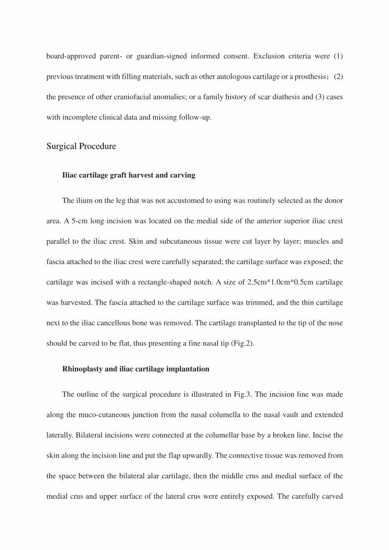
Iliac cartilage graft harvest and carving
The ilium on the leg that was not accustomed to using was routinely selected as the donor
area. A 5-cm long incision was located on the medial side of the anterior superior iliac crest
parallel to the iliac crest. Skin and subcutaneous tissue were cut layer by layer; muscles and
fascia attached to the iliac crest were carefully separated; the cartilage surface was exposed; the
cartilage was incised with a rectangle-shaped notch. A size of 2.5cm*1.0cm*0.5cm cartilage
was harvested. The fascia attached to the cartilage surface was trimmed, and the thin cartilage
next to the iliac cancellous bone was removed. The cartilage transplanted to the tip of the nose
should be carved to be flat, thus presenting a fine nasal tip (Fig.2).
Rhinoplasty and iliac cartilage implantation
The outline of the surgical procedure is illustrated in Fig.3. The incision line was made
along the muco-cutaneous junction from the nasal columella to the nasal vault and extended
laterally. Bilateral incisions were connected at the columellar base by a broken line. Incise the
skin along the incision line and put the flap upwardly. The connective tissue was removed from
the space between the bilateral alar cartilage, then the middle crus and medial surface of the
medial crus and upper surface of the lateral crus were entirely exposed. The carefully carved
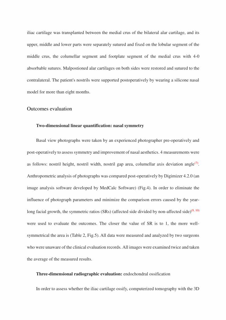
iliac cartilage was transplanted between the medial crus of the bilateral alar cartilage, and its
upper, middle and lower parts were separately sutured and fixed on the lobular segment of the
middle crus, the columellar segment and footplate segment of the medial crus with 4-0
absorbable sutures. Malpostioned alar cartilages on both sides were restored and sutured to the
contralateral. The patient's nostrils were supported postoperatively by wearing a silicone nasal
model for more than eight months.
Outcomes evaluation
Two-dimensional linear quantification: nasal symmetry
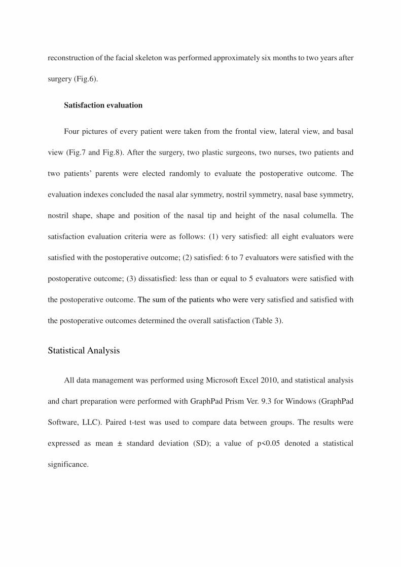
Basal view photographs were taken by an experienced photographer pre-operatively and
post-operatively to assess symmetry and improvement of nasal aesthetics. 4 measurements were
as follows: nostril height, nostril width, nostril gap area, columellar axis deviation angle(7).
Anthropometric analysis of photographs was compared post-operatively by Digimizer 4.2.0 (an
image analysis software developed by MedCalc Software) (Fig.4). In order to eliminate the
influence of photograph parameters and minimize the comparison errors caused by the year-
long facial growth, the symmetric ratios (SRs) (affected side divided by non-affected side)(9, 10)
were used to evaluate the outcomes. The closer the value of SR is to 1, the more well-
symmetrical the area is (Table 2, Fig.5). All data were measured and analyzed by two surgeons
who were unaware of the clinical evaluation records. All images were examined twice and taken
the average of the measured results.
Three-dimensional radiographic evaluation: endochondral ossification
In order to assess whether the iliac cartilage ossify, computerized tomography with the 3D
reconstruction of the facial skeleton was performed approximately six months to two years after
surgery (Fig.6).
Satisfaction evaluation
Four pictures of every patient were taken from the frontal view, lateral view, and basal
view (Fig.7 and Fig.8). After the surgery, two plastic surgeons, two nurses, two patients and
two patients’ parents were elected randomly to evaluate the postoperative outcome. The
evaluation indexes concluded the nasal alar symmetry, nostril symmetry, nasal base symmetry,
nostril shape, shape and position of the nasal tip and height of the nasal columella. The
satisfaction evaluation criteria were as follows: (1) very satisfied: all eight evaluators were
satisfied with the postoperative outcome; (2) satisfied: 6 to 7 evaluators were satisfied with the
postoperative outcome; (3) dissatisfied: less than or equal to 5 evaluators were satisfied with
the postoperative outcome. The sum of the patients who were very satisfied and satisfied with
the postoperative outcomes determined the overall satisfaction (Table 3).
Statistical Analysis
All data management was performed using Microsoft Excel 2010, and statistical analysis
and chart preparation were performed with GraphPad Prism Ver. 9.3 for Windows (GraphPad
Software, LLC). Paired t-test was used to compare data between groups. The results were
expressed as mean ± standard deviation (SD); a value of p<0.05 denoted a statistical
significance.
RESULTS
Postoperatively, only two patients (11.8%) presented with red nasal tips and abscess
formation and were cured by mupirocin ointment and complete aseptic drainage. One patient
(5.9%) had numbness on the medial side of the donor site, getting better without any special
treatment. The appearance of the nose of all 17 patients was significantly improved and kept
stable during 6-month to 2-years follow-up. The detailed data are shown in Table 2. The iliac
cartilage donor site had no deformity and iliac crest bone fracture. There was no restriction on
both lower limb movements, such as claudication.
The SRs of nostril height, nasal width, nostril gap area and columellar axis deviation angle
of the 17 patients were compared by two-dimensional linear quantification before and after the
surgery (Table 2, Fig.5). The ratio of nostril height, width and gap area between the affected
side and non-affected side showed significant improvement after than before surgery (P < 0.05),
the columellar axis deviation was significantly being towards the midline after than before the
surgery (P < 0.05). The computerized tomography with 3D reconstruction comparison showed
no endochondral ossification in the 17 patients after the operation (Fig.6). The patient
satisfaction evaluation presented more than 94% satisfaction for all evaluation indexes after the
surgery (Table 3).
DISCUSSION
Even after an excellent primary rhinoplasty, some distortions can often persist. Secondary
cleft lip nasal deformity mainly results in residual deformity, iatrogenic deformities and growth-
related(1). Patients with alveolar cleft usually present complex nasal deformity owing to the lack
of maxilla continuity. Therefore, the alveolar cleft repair is necessary to supply support to the
nostril base of the affected side in secondary rhinoplasty of the cleft lip patients with the alveolar
cleft. All 17 patients in the present study had an alveolar cleft repair and simultaneous correction
of secondary nasal deformity.
At present, cleft lip nasal deformity is usually repaired using transplantation, such as
autologous (3-8)and allogeneic transplantation. (11,12) Compared with allogeneic transplantation,
autologous transplantation was used more widely, such as costal cartilage, conchal cartilage or
septal cartilage. Every autologous transplantation has its advantages and disadvantages. For
example, costal cartilage is abundant and solid and easy to carve, while the surgery to harvest
costal cartilage causes extreme trauma and potentially severe postoperative complications, such
as pneumothorax and infection. Besides, conchal cartilage and septal cartilage have convenient
access, and the surgical wound is hidden; but there is little cartilage in the nasal septum and
auricular, and they are hard to carve, lacking high strength and good elasticity. Alveolar grafting
with iliac bone is a classic and crucial surgical procedure to repair secondary cleft lip deformity.
In the surgery of alveolar grafting, the authors found that the iliac cartilage in young adolescents
is thicker, stronger and more accessible to sculpture. D.Tao De-tao et al.(13) had an experimental
study on iliac crest cartilage and costal cartilage graft in alae nasi soft tissue and on maxilla
surface. The results showed that the iliac crest cartilage and costal cartilage grafted on the
maxilla surface had become mature osseous tissues. However, the cartilages grafted on the alae
nasi soft tissue had no histological change. It demonstrated that the cartilages graft has no
relationship with the donor area but is closely related to the recipient. This experiment provides
a reliable basis for using iliac cartilage as a graft material. Moreover, because of the histological
similarity between the iliac cartilage and the costal cartilage, we guess the iliac cartilage might
be a suitable implant to correct the nasal deformity.
In this study, secondary nasal deformity of the unilateral cleft lip was repaired
simultaneously with correction of alveolar cleft using autologous iliac cartilage as
transplantation. Compared with the accesses of other transplantation, when the iliac bone is
harvested to graft onto the alveolar deformity lesion, the iliac cartilage can also be extracted
from the same incision. Therefore, because we harvest cartilage from the donor site with the
iliac crest, it reduces incisions, injuries, operation time, and costs, thereby reducing the risk of
deformity and the occurrence of complications of harvesting cartilage from another donor site
and the financial burden on patients' families. Usually, there is much cartilage in the ilium of
immature adolescents, and its high strength and good elasticity make it easier to carve(14).
Therefore, the technique is available and particularly suitable for pediatric patients. The absence
of other surgical involvements such as conchal cartilage or costal cartilage is an advantage of
our method. During the operation, the harvested rectangle-shaped iliac cartilage segment was
trimmed, carved, and transplanted to reconstruct the height of the columellar and restore the
shape of the nasal tip. 6 to 2 years’ follow-up indicated significant improvement of the nasal
appearance of the 17 patients. Furthermore, patient satisfaction evaluation presented more than
94% satisfaction for all evaluation indexes after the surgery. The height of the nostril on the
affected side was significantly higher after than before surgery (P < 0.05), and the width, gap
area and the columellar axis deviation angle were significantly shorter after than before surgery
(P < 0.05).

While, iliac cartilage used as autograft material was rarely mentioned because the iliac
cartilage gradually ossifies with age(14, 15). Some scholars think that secondary reconstruction
of nasal deformities based on alveolar graft and orthodontic treatment is the best stage for
correcting nasal deformities(16). However, iliac cartilage mostly ossifies at this stage which is
hard to find. While in our institute, orthodontic treatment and reconstruction of dental arch
morphology are usually performed before alveolar repair. Then, simultaneous correction of
secondary nasal deformity of the unilateral cleft lip using autologous iliac cartilage during
alveolar cleft repair was performed, minimizing the postoperative nasal morphology changes
to the greatest extent, thus reducing the probability of reoperation of nasal deformity(1, 17). Thus,
we accidentally found a cartilage iliac in clinical practice that did not ossify and used it as an
autograft material. In this study, we used three methods to evaluate the effect of the operation.
In addition to the satisfaction survey and two-dimensional linear quantitative evaluation, a
three-Dimensional radiographic evaluation was introduced to evaluate whether the
endochondral ossification exists(18, 19). The study demonstrated that iliac cartilage did not show
apparent absorption and ossification in the follow-up period.
However, some limitations existed in this clinical study. The most prolonged follow-up
period is only two years, and longer positive results did not be followed. We will continue to
follow up to get more positive results. Besides, the mechanism of iliac cartilage ossification did
not verify. Therefore, we removed the fascia on the surface of the iliac cartilage in order to
avoid ossifying. The mechanism of iliac cartilage ossification remains to be studied. Moreover,
we will try to perform the related animal testing next step.
CONCLUSION
The positive results have certified that when repairing the alveolar cleft, iliac cartilage can
be used as a new graft source to repair the nasal deformity simultaneously. It provides a new
source of cartilage to avoid adding a new operative approach for cartilage obtaining and reduces
the financial burden on patients' families. As for the positive results of this treatment, it can be
widely applied in clinical practice.
Data availability The datasets generated during and analysed during the current study are
available from the corresponding author on reasonable request.
References
1. Allori AC, Mulliken JB. Evidence-Based Medicine: Secondary Correction of Cleft Lip
Nasal Deformity. Plast Reconstr Surg. 2017; 140(1):166e-176e.
2. Bing S, Yuchuan F, Ningbei Y, et al. [Application of team approach and key techniques
of cleft lip and palate]. Hua Xi Kou Qiang Yi Xue Za Zhi. 2017; 35(1):8-17.
3. Cheon YW, Park BY. Long-term evaluation of elongating columella using conchal
composite graft in bilateral secondary cleft lip and nose deformity. Plast Reconstr Surg.
2010; 126(2):543-553.
4. Wei J, Herrler T, Xu H, Li Q, Dai C. Double Composite Tissue Z-plasty Technique for
Anatomical Restoration of Severe Nasal Deformity in Secondary Unilateral Cleft Lip.
Ann Plast Surg. 2017; 79(4):359-364.
5. Jackson O, Wingate N, Lee A, Kaye AE. The conchal butterfly graft in secondary
reconstruction of the bilateral cleft lip nasal deformity. Int J Pediatr Otorhinolaryngol.
2020; 129:109737.
6. Zhang L, Wei S, Qi L, et al. Simultaneous Correction of Complex Secondary
Deformities of Cleft Lip Using Autogenous Costal Cartilage Combined With Rib Grafts.
J Craniofac Surg. 2020; 31(2):497-500.
7. Talaat WM, Ghoneim MM, El-Shikh YM, Elkashty SI, Ismail M, Keshk T.
Anthropometric Analysis of Secondary Cleft Lip Rhinoplasty Using Costal Cartilage
Graft. J Craniofac Surg. 2019. 30(8): 2464-2468.
8. Vass G, Mohos G, Bere Z, et al. Secondary correction of nasal deformities in cleft lip
and palate patients: surgical technique and outcome evaluation. Head Face Med. 2016;
12(1):34.
9. Li L, Liao L, Zhong Y, Li Y, Xiang L, Li W. A modified Mohler technique for patients
with unilateral cleft lip based on geometric principles--A primary report. J
Craniomaxillofac Surg. 2015; 43(5):663-70.
10. Li L, Liao L, Zhong Y, Li Y, Xiang L, Li W. Variation trends of the postoperative
outcomes for unilateral cleft lip patients by modified Mohler and Tennison-Randall
cheiloplasties. J Craniomaxillofac Surg. 2016; 44(11):1786-1795.
11. Kim J, Kim J, Uhm KI, et al. Secondary Cleft Nasal Deformity Correction Using
Bioabsorbable Mesh. J Craniofac Surg. 2016; 27(5):1143-6.
12. Lin PY, Gibson AP, Teichgraeber JF, Greives MR. Resorbable Plates in Secondary Cleft
Nasal Reconstruction. Cleft Palate Craniofac J. 2018; 55(2):226-230.
13. TAO De-tao, TAO Zhen-jiang, LIU Lai-kui, JIANG Hong-bing. Experimental study on
iliac crest cartilage and costal cartilage graft in alae nasi and on maxilla surface.
Stomatology. 2006; 26(2):143-145.
14. Grissom LE, Harty MP, Guo GW, Kecskemethy HH. Maturation of pelvic ossification
centers on computed tomography in normal children. Pediatr Radiol. 2018;
48(13):1902-1914.
15. Fabricant PD, Hirsch BP, Holmes I, et al. A radiographic study of the ossification of the
posterior wall of the acetabulum: implications for the diagnosis of pediatric and
adolescent hip disorders. J Bone Joint Surg Am. 2013; 95(3):230-6.
16. Broadbent TR, Woolf RM. Cleft lip nasal deformity. Ann Plast Surg. 1984; 12(3):216-
34.
17. Mundra LS, Lowe KM, Khechoyan DY. Alveolar Bone Graft Timing in Patients With
Cleft Lip & Palate. J Craniofac Surg. 2022; 33(1):206-210.
18. Chaisooktaksin N, Chimruang J, Worasakwutiphong S, Tansalarak R. Three-
dimensional Changes of Maxillary Alveolar Morphology After Using Modified
Nasoalveolar Molding in Patients with Complete Unilateral Cleft lip and Palate. Cleft
Palate Craniofac J. 2022:10556656221086816.
19. Stoop CC, Janssen NG, Ten Harkel TC, Rosenberg A. A Novel and Practical Protocol
for Three-Dimensional Assessment of Alveolar Cleft Grafting Procedures. Cleft Palate
Craniofac J. 2022:10556656221074210.
Author contributions
Yijun Li wrote the manuscript. Lishu Liao and Wanshan Li performed the operations. Yijun Li,
Shengyu Tang, Xiaorong Wang, Li Xiang, Li Liang and Yuxiang Zhong collected the data.
Yijun Li and Xiaorong Wang analysed the data. Wanshan Li reviewed and edited the manuscript.
Competing interests
The authors declare that no competing interests
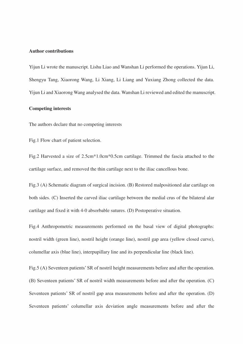
Fig.1 Flow chart of patient selection.
Fig.2 Harvested a size of 2.5cm*1.0cm*0.5cm cartilage. Trimmed the fascia attached to the
cartilage surface, and removed the thin cartilage next to the iliac cancellous bone.
Fig.3 (A) Schematic diagram of surgical incision. (B) Restored malpositioned alar cartilage on
both sides. (C) Inserted the carved iliac cartilage between the medial crus of the bilateral alar
cartilage and fixed it with 4-0 absorbable sutures. (D) Postoperative situation.
Fig.4 Anthropometric measurements performed on the basal view of digital photographs:
nostril width (green line), nostril height (orange line), nostril gap area (yellow closed curve),
columellar axis (blue line), interpupillary line and its perpendicular line (black line).
Fig.5 (A) Seventeen patients’ SR of nostril height measurements before and after the operation.
(B) Seventeen patients’ SR of nostril width measurements before and after the operation. (C)
Seventeen patients’ SR of nostril gap area measurements before and after the operation. (D)
Seventeen patients’ columellar axis deviation angle measurements before and after the
operation.
Fig.6 (A) pre-operative situation (B) 1year after surgery: there was no endochondral ossification.
Fig.7 Patient 1:1 frontal view, 2 lateral views, and 1 basal view photos were taken before and
after the operation. 2 plastic surgeons, 2 nurses, 2 patients and 2 patients' parents determined
the postoperative satisfaction by comparing the effects before and after the operation.
Fig.8 Patient 2:1 frontal view, 2 lateral views, and 1 basal view photos were taken before and
after the operation. 2 plastic surgeons, 2 nurses, 2 patients and 2 patients' parents determined
the postoperative satisfaction by comparing the effects before and after the operation.
Tables
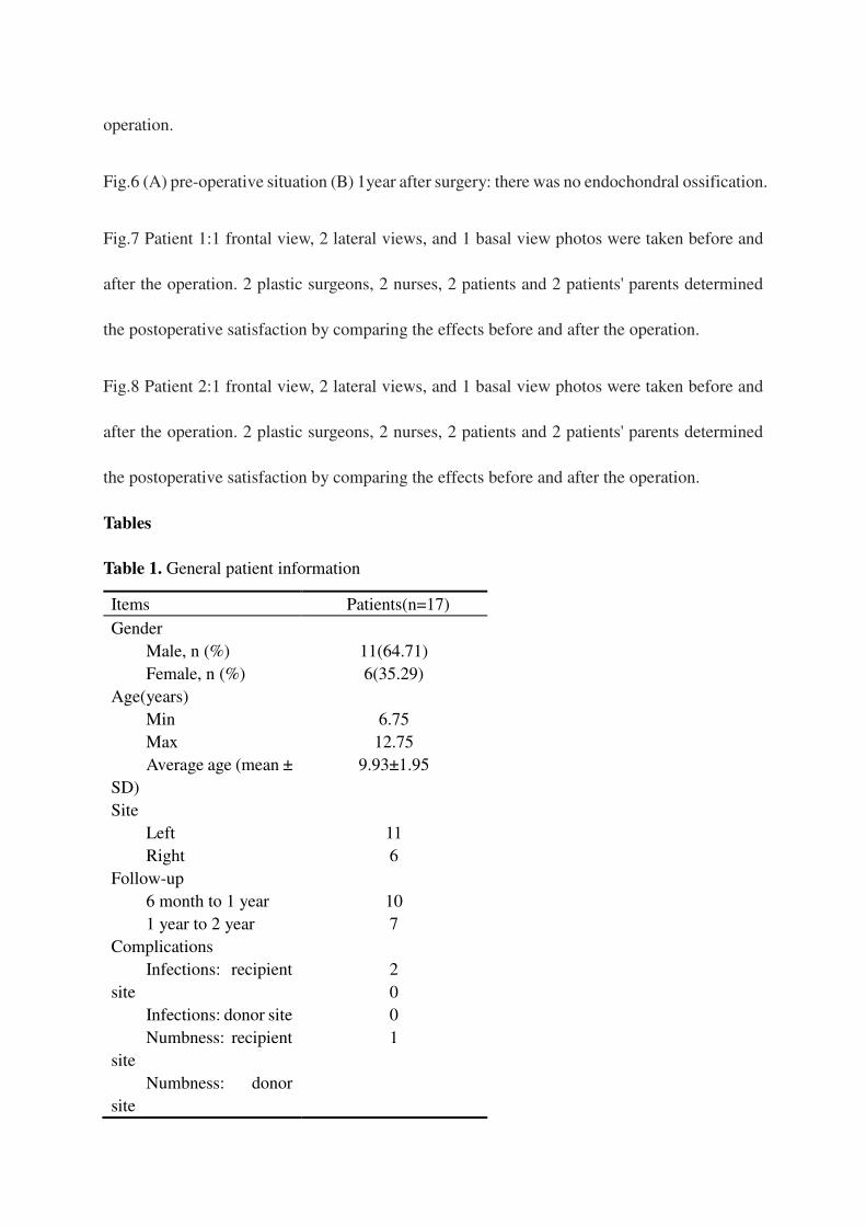
Table 1. General patient information
Items Patients(n=17)
Gender
Male, n (%)
Female, n (%)
11(64.71)
6(35.29)
Age(years)
Min
Max
Average age (mean ±
SD)
6.75
12.75
9.93±1.95
Site
Left
Right
11
6
Follow-up
6 month to 1 year
1 year to 2 year
10
7
Complications
Infections: recipient
site
Infections: donor site
Numbness: recipient
site
Numbness: donor
site
2
0
0
1
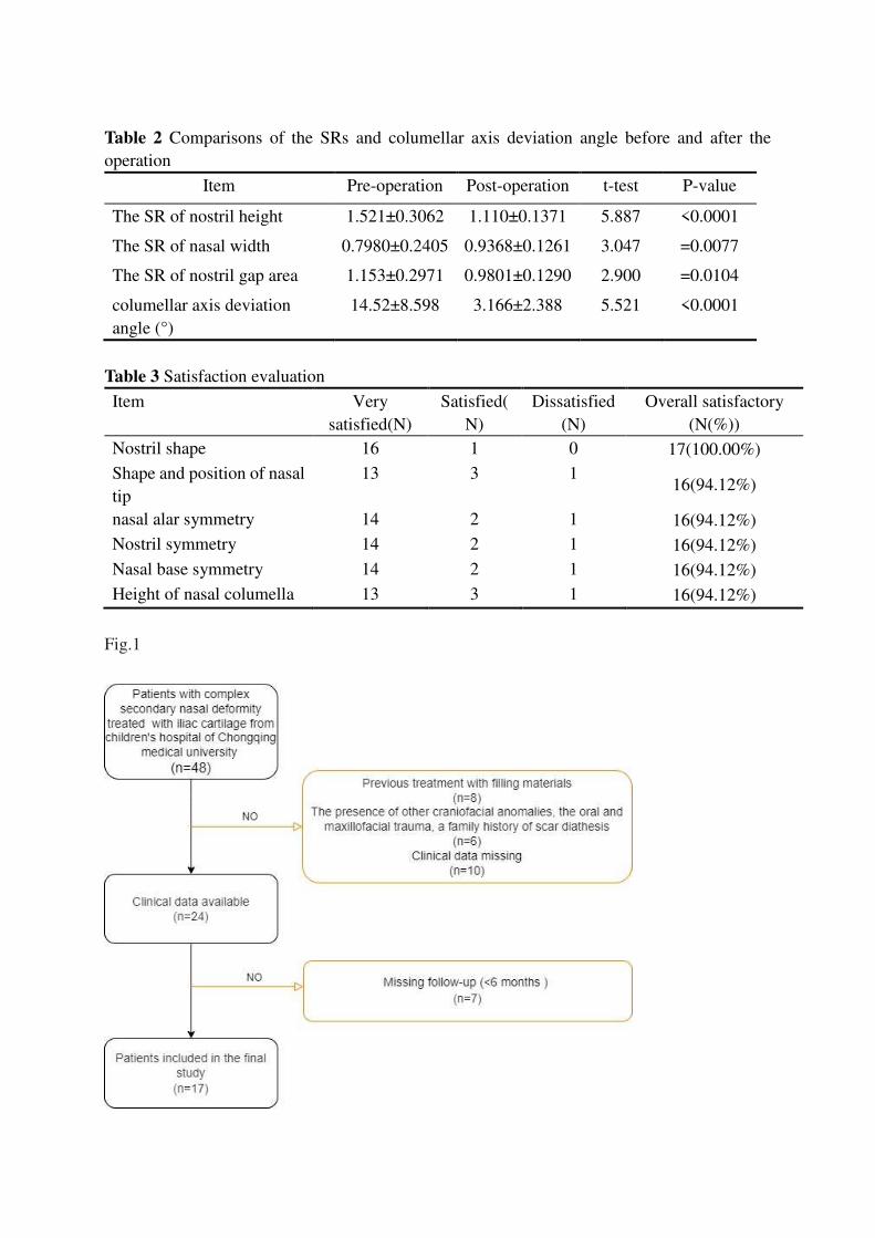
Table 2 Comparisons of the SRs and columellar axis deviation angle before and after the
operation
Item Pre-operation Post-operation t-test P-value
The SR of nostril height 1.521±0.3062 1.110±0.1371 5.887 <0.0001
The SR of nasal width 0.7980±0.2405 0.9368±0.1261 3.047 =0.0077
The SR of nostril gap area 1.153±0.2971 0.9801±0.1290 2.900 =0.0104
columellar axis deviation
angle (°)
14.52±8.598 3.166±2.388 5.521 <0.0001
Table 3 Satisfaction evaluation
Item Very
satisfied(N)
Satisfied(
N)
Dissatisfied
(N)
Overall satisfactory
(N(%))
Nostril shape 16 1 0 17(100.00%)
Shape and position of nasal
tip
13 3 1 16(94.12%)
nasal alar symmetry 14 2 1 16(94.12%)
Nostril symmetry 14 2 1 16(94.12%)
Nasal base symmetry 14 2 1 16(94.12%)
Height of nasal columella 13 3 1 16(94.12%)
Fig.1
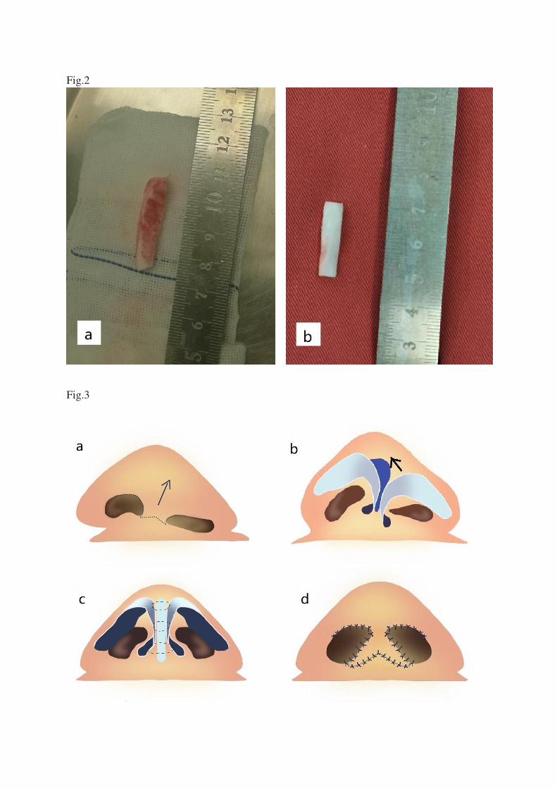
Fig.2
Fig.3
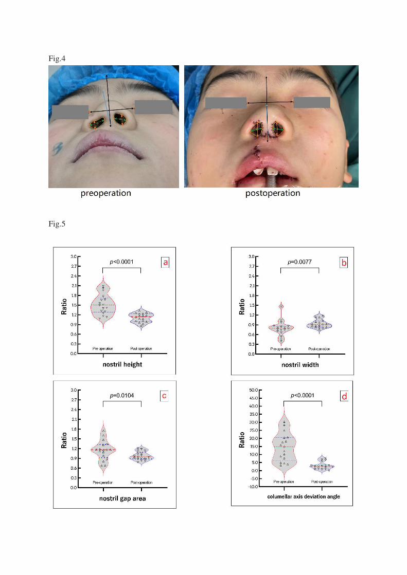
Fig.4
Fig.5
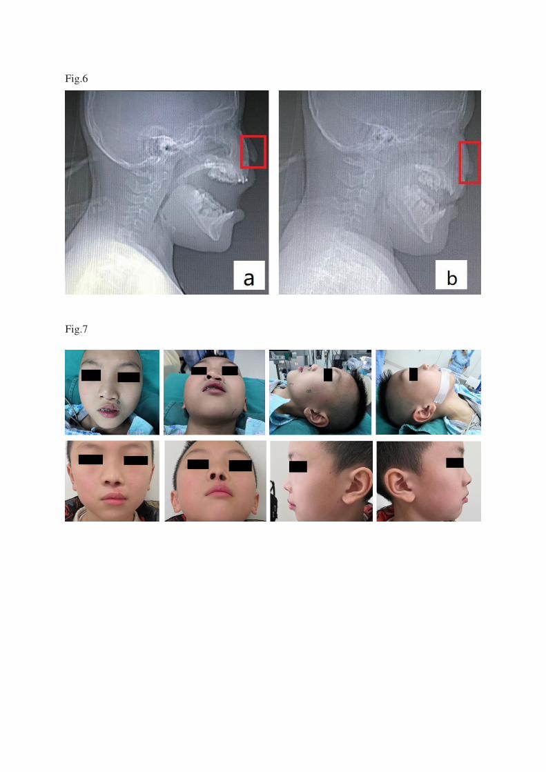
Fig.6
Fig.7
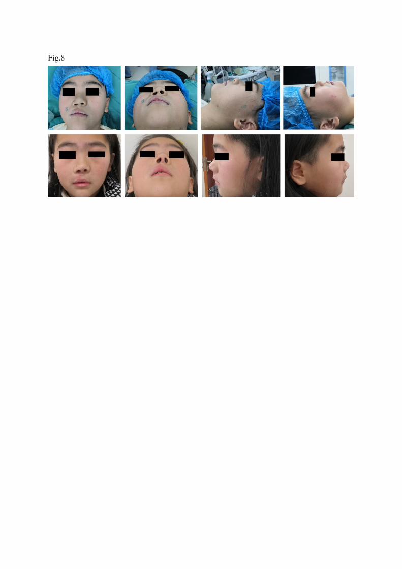
Fig.8