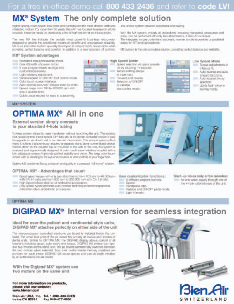
sepdec2007.pdf - lvi global
TRANSCRIPT
s i m p l i c i t y
e s t h e t i c s
ve
rs
at
ilit
yz i r c o n i a
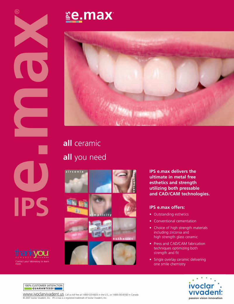
all ceramic
all you need
IPS e.max delivers theultimate in metal freeesthetics and strengthutilizing both pressableand CAD/CAM technologies.
IPS e.max offers:
• Outstanding esthetics
• Conventional cementation
• Choice of high strength materials including zirconia and high strength glass ceramic
• Press and CAD/CAM fabrication techniques optimizing both strength and fit
• Single overlay ceramic delivering one smile chemistry
www.ivoclarvivadent.us Call us toll free at 1-800-533-6825 in the U.S., or 1-800-263-8182 in Canada© 2007 Ivoclar Vivadent, Inc. IPS e.max is a registered trademark of Ivoclar Vivadent, Inc.
100% CUSTOMER SATISFACTION G U A R A N T E E D !
thankyouIVOCLAR VIVADENT
R E W A R D S • • • • • • •
TM
Contact your laboratory to learnmore.
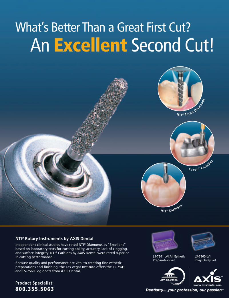
What’s Better Than a Great First Cut?
An Excellent Second Cut!
NTI® Turbo Dia
mon
ds
Razor™ Carbid
esNTI® Carbides
Product Specialist:800.355.5063 Dentistry... your profession, our passionSM
NTI® Rotary Instruments by AXIS DentalIndependent clinical studies have rated NTI® Diamonds as “Excellent” based on laboratory tests for cutting ability, accuracy, lack of clogging, and surface integrity. NTI® Carbides by AXIS Dental were rated superior in cutting performance.
Because quality and performance are vital to creating fine esthetic preparations and finishing, the Las Vegas Institute offers the LS-7541 and LS-7560 Logic Sets from AXIS Dental.
LS-7560 LVIInlay-Onlay Set
LS-7541 LVI All Esthetic Preparation Set
4 LVI VISIONS • SEPTEMBER • OCTOBER • NOVEMBER • DECEMBER 2007
6 Your Obligations as a Physician of the Mouthwilliam g. dickerson, dds, lvim
10 Interview withd. gary wolford, dds
16 Utilizing Neuromuscular Principles in Implant Dentistryjames h. clarke, jr., dds, fagd
24 Create Your Visionalan j. singleton, dds
36 The Weathers’ Report Your Patients Benefit When You Raise Your Fees
arthur “kit” weathers, jr., dds
42 Functional Classification of Craniomandibular Disordersd. gary wolford, dds
52 Uncovering the Cervical-Occlusal Link: A New Approach to Myo-Trode Placementjohn pawlowicz, iii dmd
60 Ask Heidiheidi dickerson, dds, lvim
62 Controlling Overhead – Part IIlori kemmet, dds, lvim
70 Product Reviewsmichael a. miyasaki, dds, lvim
76 Guidelines to Diagnosing Functional Wearw. scott wagner, dmd
The Las Vegas Institute for Advanced Dental Studies (LVI) publishes LVI Visions. Please send any comments or suggestions to 9501 Hillwood Dr., Las Vegas, NV 89134.Telephone (888) 584-3237 or (702) 341-7978. Web address • www.lviglobal.com.
Copyright ©2007 – Las Vegas Institute for Advanced Dental Studies.All rights reserved. No part of this publication may be reproduced in any form without written permission from the
Las Vegas Institute for Advanced Dental Studies. LVI does not verify any claims or other information appearing in any of the advertisements contained in this publication.
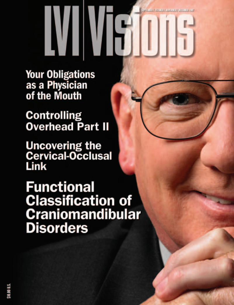
Cover Photo:D. Gary Wolford, DDS
LVI VISIONS • SEPTEMBER • OCTOBER • NOVEMBER • DECEMBER 2007 5
6 LVI VISIONS • SEPTEMBER • OCTOBER • NOVEMBER • DECEMBER 2007
E D I T O R I A LB Y W I L L I A M G . D I C K E R S O N , D D S , LV I M
Your Obligations as a Physician
of the Mouth
LVI VISIONS • SEPTEMBER • OCTOBER • NOVEMBER • DECEMBER 2007 7
The conclusion of the article
was that the dentist that di-
agnosed the least was the
best dentist and the one that diag-
nosed the most was the worst. In ac-
tuality, the dentists that diagnosed
the least were uneducated or felt
guilty about diagnosing what needed
to be done. For those of us who went
on the internet to see the authors x-
rays, we can verify that his oral
health was in a horrible condition.
And the truth was that the dentist
who diagnosed the most was the best
Several years ago, Reader’s Digest ran an article titled
“Is Your Dentist Ripping You Off”. The author visited 28
different offices to be “diagnosed”. There was such a discrepancy in the
diagnosis, ranging from no treatment recommended to several
thousand dollars in diagnosed treatment.
and most comprehensive dentist of-
fering complete restorative dentistry.
Those that diagnosed nothing were
the worst. They were unable to diag-
nose what they could not see.
The same concept is being applied
today when it comes to comprehen-
sive “mouth doctor” dentistry. For
those that have not been trained in
this area, they can not diagnose what
they can not see. The less they know,
the more everything seems normal.
It is like a physician that is unable to
diagnose a disease because he or she
has never been trained to recognize
the obvious signs and symptoms that
one who has been trained, would
easily recognize.
So let’s talk about your duties as a
dentist when a patient enters your
practice. It is your obligation to diag-
nose the condition of the mouth even
if you are not trained to treat it. It
would be like a surgeon who does not
like doing brain surgery or does not
know how, NOT diagnosing a brain
lesion because he does not want to or
does not know how to treat it.
8 LVI VISIONS • SEPTEMBER • OCTOBER • NOVEMBER • DECEMBER 2007
However, many dentists do this un-
consciously. The problem is that
most dentists do not diagnose many
pathological conditions because
they do not recognize the signs and
symptoms.
Although it is your obligation to
diagnose the pathologic condition
of the mouth, it is the patient’s right
to say no to treatment. Too many
dentists get upset and take it per-
sonal when a patient decides not to
have the treatment done. Just as it is
the right of a cancer patient to reject
cancer therapy, not accepting treat-
ment is the patient’s right and no
one should be indignant if that is
their decision. Just make sure you
document that the patient was in-
formed of the problem and that the
patient refused the treatment.
Much of the population is not in
their mandibular physiologic comfort-
able position. Why that is was dis-
cussed in the previous issue of this
magazine. But if a patient is NOT in
their physiologic position, it is the
dentist’s obligation to let them know.
However, just because someone is not
in the right physiologic mandibular
position, does that mean they need to
be treated? Does everyone who is not
in their physiologic position need to
have a full mouth reconstruction? The
answer is of course, NO. Not every-
one who is not in their physiologic
mandibular position needs restorative
treatment nor do they need any treat-
ment if they are asymptomatic. The
bottom line is; every dentist should
diagnose their patient’s mouth as if it
were their own. The ethical question
to ask is, what would you do if it were
YOUR mouth?
When a patient is evaluated, the
treating dentist needs to determine
two things about the patient’s habit-
ual biting position.
1. Are they in their comfortable
position or not?
2. Do they have related symp-
toms?
Someone who is not in their com-
fortable mandibular position may
not have any symptoms. What are
some related symptoms? Headache,
migraines, neck pain, shoulder pain,
facial pain, back pain, ringing of the
ears, vertigo and many others. Some
signs of occlusion disharmony are
worn teeth, abfractions, exostosis,
fractured teeth, malocclusion, etc.
So if they do not have any symptoms
and are not in their comfortable po-
sition, do they need to be treated?
The answer is; only if they need
restorative treatment anyway due to
decay or restorative breakdown. If
they do not have symptoms, they do
not need to be treated. However, it is
still the obligation of the treating
dentist to inform the patient that they
are predisposed to developing prob-
lems so if in fact they do start to de-
velop problems they will seek the
help of a qualified dentist.
Even if someone has symptoms
and is not in their comfortable
mandibular position, they may
CHOOSE not to be moved into that
position. They may decide that the
treatment is worse than the symp-
toms or do not want to make treat-
ment of their disorder a priority.
That is the prerogative and the right
of the patient. It is their right to
have treatment or not to have treat-
ment and live with the symptoms.
Just as a cancer victim can choose
not to have treatment, so can your
patients choose not to have dental
treatment. However, if these pa-
tients decide to be restored in their
current pathologic position, I would
advise the treating dentist not to
guarantee the longevity of the
restorations. Whatever damage they
have done to their teeth because
they are “trying” to get comfortable,
The bottom line is; every dentist should
diagnose their patient’s mouth as if
it were their own.
LVI VISIONS • SEPTEMBER • OCTOBER • NOVEMBER • DECEMBER 2007 9
determine if the symptoms ARE
bite related. By means of orthotic
mandibular repositioning appli-
ance, the diagnosing dentist can de-
termine if the symptoms are bite re-
lated and if they can eliminate those
symptoms. Once the correct com-
fortable position is found and
symptoms are eliminated, the den-
tist then determines which treat-
ment option to permanently place
the patient in that position.
Is the correct position close
enough to their existing position that
it can be accomplished by means of
a coronoplasty? Sometimes as little
as 0.5 mm off of muscular harmony
can make a huge difference and the
patient may have painful symptoms.
A comprehensive bite adjustment
may alleviate those symptoms.
There have been cases of mine and
other LVI faculty where this was
achieved, even the elimination of a
lifetime of migraines.
If the bite is so far off that a corono-
plasty cannot correct the problem,
then orthodontics and/or restorative
becomes the treatment option. Ortho
is ALWAYS an option in neuromuscu-
lar complete comprehensive den-
tistry. Although not many adults
will choose orthodontics because
of the time involvement, it should
always be discussed. They may
need restorative treatment anyway
due to unaesthetic teeth or broken
down dentition. Ortho may make
them straight and correct the posi-
tion, but the teeth will end up
straight and unaesthetic.
In conclusion, it is the obligation
of every dentist out there to diag-
nose their patient’s mouth as if it
were their own and to learn as much
as possible about dentistry so they
can offer complete comprehensive
treatment or at least diagnose it. Re-
member, a dental degree is just a li-
cense to learn more about dentistry.
The question is; are you really
learning more? Are you still doing
only what you were taught in dental
school? Have you not evolved to a
different level of patient care? Are
you truly a physician of the mouth?
If not, then you owe it to your pa-
tients to become one, so you can
offer them the best possible treat-
ment and care they can receive.
The ethical question to ask is, what wouldyou do if it were YOUR mouth?
they will continue to do so after
being restored. The beauty is that
the diagnosing dentists can by
means of an orthotic mandibular
repositioning appliance determine
if the symptoms are bite related and
if they can eliminate those symp-
toms before ever touching a tooth.
So what are the treatment choices?
1. No Treatment
2. Existing CO treatment
3. Aesthetic Treatment
4. Comprehensive Restorative Treatment
Comprehensive Restorative Treatment
Comprehensive Restorative Treat-
ment can be several options. Corono-
plasty (equilibration), orthodontics,
posterior restorations, full arch, full
mouth or a combination of these are
the options. Depending on the situa-
tion, any of those treatments are a vi-
able way to correct the pathologic
situation of the bite.
The first step before beginning
any comprehensive treatment is to
LVI VISIONS • SEPTEMBER • OCTOBER • NOVEMBER • DECEMBER 2007 11
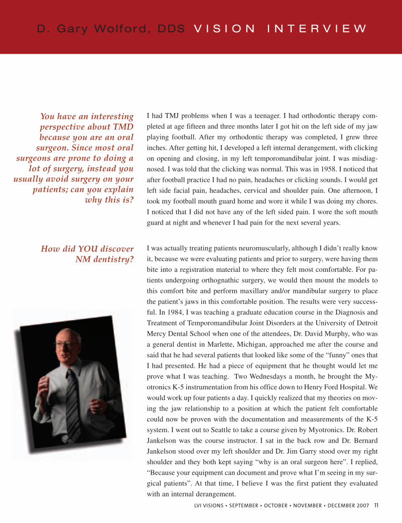
I had TMJ problems when I was a teenager. I had orthodontic therapy com-
pleted at age fifteen and three months later I got hit on the left side of my jaw
playing football. After my orthodontic therapy was completed, I grew three
inches. After getting hit, I developed a left internal derangement, with clicking
on opening and closing, in my left temporomandibular joint. I was misdiag-
nosed. I was told that the clicking was normal. This was in 1958. I noticed that
after football practice I had no pain, headaches or clicking sounds. I would get
left side facial pain, headaches, cervical and shoulder pain. One afternoon, I
took my football mouth guard home and wore it while I was doing my chores.
I noticed that I did not have any of the left sided pain. I wore the soft mouth
guard at night and whenever I had pain for the next several years.
You have an interesting perspective about TMD because you are an oral
surgeon. Since most oral surgeons are prone to doing a
lot of surgery, instead youusually avoid surgery on your
patients; can you explain why this is?
I was actually treating patients neuromuscularly, although I didn’t really know
it, because we were evaluating patients and prior to surgery, were having them
bite into a registration material to where they felt most comfortable. For pa-
tients undergoing orthognathic surgery, we would then mount the models to
this comfort bite and perform maxillary and/or mandibular surgery to place
the patient’s jaws in this comfortable position. The results were very success-
ful. In 1984, I was teaching a graduate education course in the Diagnosis and
Treatment of Temporomandibular Joint Disorders at the University of Detroit
Mercy Dental School when one of the attendees, Dr. David Murphy, who was
a general dentist in Marlette, Michigan, approached me after the course and
said that he had several patients that looked like some of the “funny” ones that
I had presented. He had a piece of equipment that he thought would let me
prove what I was teaching. Two Wednesdays a month, he brought the My-
otronics K-5 instrumentation from his office down to Henry Ford Hospital. We
would work up four patients a day. I quickly realized that my theories on mov-
ing the jaw relationship to a position at which the patient felt comfortable
could now be proven with the documentation and measurements of the K-5
system. I went out to Seattle to take a course given by Myotronics. Dr. Robert
Jankelson was the course instructor. I sat in the back row and Dr. Bernard
Jankelson stood over my left shoulder and Dr. Jim Garry stood over my right
shoulder and they both kept saying “why is an oral surgeon here”. I replied,
“Because your equipment can document and prove what I’m seeing in my sur-
gical patients”. At that time, I believe I was the first patient they evaluated
with an internal derangement.
How did YOU discover NM dentistry?
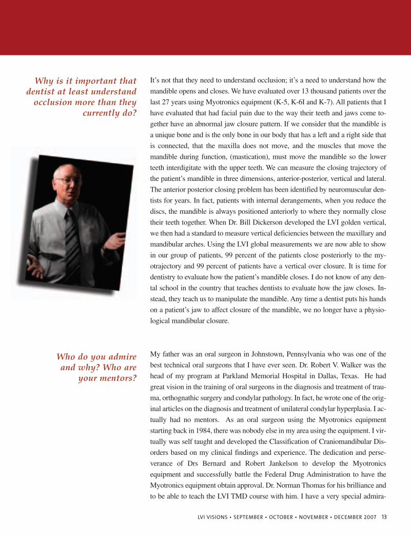
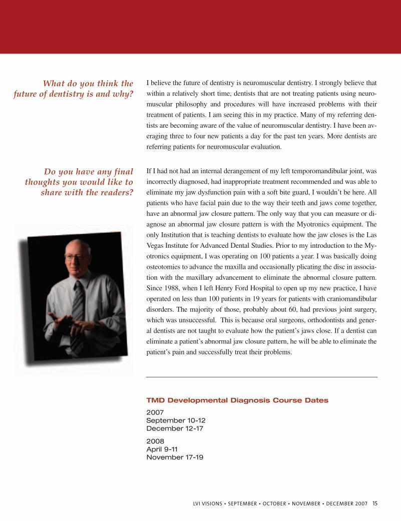
D. Gary Wol ford, DDS V I S I O N I N T E R V I E W
12 LVI VISIONS • SEPTEMBER • OCTOBER • NOVEMBER • DECEMBER 2007
I started teaching the principles of neuromuscular dentistry in earnest after I
came back from the Seattle, Myotronics meeting. Now I could prove that we
could eliminate the patient’s pain by constructing an orthotic to the myotra-
jectory. My therapy successes using conservative therapy were immediate.
Since I no longer had to treat patients surgically, but could treat them with an
orthotic and have orthodontic and reconstructive prosthetic work done on pa-
tients to eliminate their pain, was very rewarding. The patients did not have
the complications of sensory loss that is involved with orthognathic surgery.
You have had a long and successful career that has not
only taught dentists how toincorporate TMD Diagnosis
and Treatment Planning intotheir offices, but how to do
it using the principles of muscular harmony. Can you
explain how you fell into teaching this aspect
of dentistry?
I was very honored to be asked to join the LVI faculty. I met Dr. Dickerson at a
North American Neuromuscular Study Club meeting. He attended as a guest in
Seattle, Washington the day before a Myotronics meeting. I presented my Classi-
fication of Craniomandibular Disorders at that meeting and Dr. Dickerson asked
me to join the faculty and become one of the instructors in the TMD course.
Can you explain to everyonewhy you decided to join the
distinguished LVI faculty?
The greatest aspect of my life, besides my wife Claudia and three children
Lynn, Donald and Daniel, is the fact that they have tolerated my passion for
neuromuscular dentistry. Secondly, was realizing that the neuromuscular den-
tal philosophy can eliminate many patients’ pain and suffering and allow them
to return to a normal life situation. Lastly, being a faculty member at LVI,
where I have the opportunity to teach many dentists how to diagnose and treat
patients with craniomandibular disorders.
You’ve had such a fulfillinglife, what would you consider the greatest
aspect of your life?
I most value becoming a Diplomate at the American Board of Oral and Max-
illofacial Surgery, obtaining a Fellowship in ICCMO and becoming a lecturer
at the Las Vegas Institute for Advanced Dental Studies.
Which professional accomplishment(s)
do you value most?
Any dentist who becomes involved with neuromuscular dentistry very quick-
ly realizes how much pain relief can provide to patients. The large number of
dentists attending LVI courses emphasizes that neuromuscular dentistry is
successful.
Many dentists are afraid toincorporate TMD into their
practices, mainly I think because they don’t
understand it. Do youthink it is something most
dentists will find rewarding?
D. Gary Wol ford, DDS V I S I O N I N T E R V I E W
LVI VISIONS • SEPTEMBER • OCTOBER • NOVEMBER • DECEMBER 2007 13
It’s not that they need to understand occlusion; it’s a need to understand how the
mandible opens and closes. We have evaluated over 13 thousand patients over the
last 27 years using Myotronics equipment (K-5, K-6I and K-7). All patients that I
have evaluated that had facial pain due to the way their teeth and jaws come to-
gether have an abnormal jaw closure pattern. If we consider that the mandible is
a unique bone and is the only bone in our body that has a left and a right side that
is connected, that the maxilla does not move, and the muscles that move the
mandible during function, (mastication), must move the mandible so the lower
teeth interdigitate with the upper teeth. We can measure the closing trajectory of
the patient’s mandible in three dimensions, anterior-posterior, vertical and lateral.
The anterior posterior closing problem has been identified by neuromuscular den-
tists for years. In fact, patients with internal derangements, when you reduce the
discs, the mandible is always positioned anteriorly to where they normally close
their teeth together. When Dr. Bill Dickerson developed the LVI golden vertical,
we then had a standard to measure vertical deficiencies between the maxillary and
mandibular arches. Using the LVI global measurements we are now able to show
in our group of patients, 99 percent of the patients close posteriorly to the my-
otrajectory and 99 percent of patients have a vertical over closure. It is time for
dentistry to evaluate how the patient’s mandible closes. I do not know of any den-
tal school in the country that teaches dentists to evaluate how the jaw closes. In-
stead, they teach us to manipulate the mandible. Any time a dentist puts his hands
on a patient’s jaw to affect closure of the mandible, we no longer have a physio-
logical mandibular closure.
Why is it important thatdentist at least understand
occlusion more than theycurrently do?
My father was an oral surgeon in Johnstown, Pennsylvania who was one of the
best technical oral surgeons that I have ever seen. Dr. Robert V. Walker was the
head of my program at Parkland Memorial Hospital in Dallas, Texas. He had
great vision in the training of oral surgeons in the diagnosis and treatment of trau-
ma, orthognathic surgery and condylar pathology. In fact, he wrote one of the orig-
inal articles on the diagnosis and treatment of unilateral condylar hyperplasia. I ac-
tually had no mentors. As an oral surgeon using the Myotronics equipment
starting back in 1984, there was nobody else in my area using the equipment. I vir-
tually was self taught and developed the Classification of Craniomandibular Dis-
orders based on my clinical findings and experience. The dedication and perse-
verance of Drs Bernard and Robert Jankelson to develop the Myotronics
equipment and successfully battle the Federal Drug Administration to have the
Myotronics equipment obtain approval. Dr. Norman Thomas for his brilliance and
to be able to teach the LVI TMD course with him. I have a very special admira-
Who do you admire and why? Who are
your mentors?
14 LVI VISIONS • SEPTEMBER • OCTOBER • NOVEMBER • DECEMBER 2007
Dentists do not want to know what they are doing wrong. Dentists do not un-
derstand mandibular closure pathways. This is a problem because no dental
school teaches a dentist how to evaluate how the jaw closes. Every patient who
has pain when their teeth and jaws close together has an abnormal closure pat-
tern. The concern is why they don’t want to understand. Ninety-nine percent
of my patients have a regular general dentist and seventy-five percent have had
orthodontic therapy. It’s about time dental schools start to teach dentists how
to evaluate how the patients jaw closes.
Why do you believe it isthat some are afraid to find
out about the truth of NM dentistry?
The failure of dentistry to evaluate how the mandible closes. The emphasis on
the use of MRI’s to evaluate disc position, which is only 20 percent accurate.
The failure of dentistry to recognize the success of neuromuscular dentistry.
What is the biggest problemin dentistry today? What
bothers you the most?
I have never claimed to be very smart. I have always claimed to be very practical,
observant and an excellent diagnostician. We evaluate patients with a history, ex-
tensive clinical examination, radiographic evaluation including a PA and lateral
ceph, panorex with the patient biting their teeth together, axially-corrected tomo-
grams and electromyographic, computerized mandibular scanning and elec-
trosonography. We construct the orthotic to the neuromuscular position and we are
able to eliminate the patient’s pain. Any dentist utilizing the clinical Classification
of Craniomandibular Disorders, the appropriate clinical evaluation and construct-
ing an orthotic to the patient’s myotrajectory can successfully complete Phase I of
treating neuromuscular dentistry.
How can an average dentist,who is nowhere near assmart as you, apply the
principles that you teach intheir practices?
Come to LVI. Complete the core courses. Take the TMD course so you can fit
all the pieces together and treat your patients to the neuromuscular position.If you could give a piece of
advice to all the dentists outthere what would it be?
D. Gary Wol ford, DDS V I S I O N I N T E R V I E W
tion for the courage and dedication to neuromuscular dentistry that Dr. Bill Dick-
erson has provided at LVI. My mother, who has been legally blind since she was
19 years old, has always been very supportive and never complains about her in-
ability to see.
LVI VISIONS • SEPTEMBER • OCTOBER • NOVEMBER • DECEMBER 2007 15
I believe the future of dentistry is neuromuscular dentistry. I strongly believe that
within a relatively short time, dentists that are not treating patients using neuro-
muscular philosophy and procedures will have increased problems with their
treatment of patients. I am seeing this in my practice. Many of my referring den-
tists are becoming aware of the value of neuromuscular dentistry. I have been av-
eraging three to four new patients a day for the past ten years. More dentists are
referring patients for neuromuscular evaluation.
What do you think the future of dentistry is and why?
If I had not had an internal derangement of my left temporomandibular joint, was
incorrectly diagnosed, had inappropriate treatment recommended and was able to
eliminate my jaw dysfunction pain with a soft bite guard, I wouldn’t be here. All
patients who have facial pain due to the way their teeth and jaws come together,
have an abnormal jaw closure pattern. The only way that you can measure or di-
agnose an abnormal jaw closure pattern is with the Myotronics equipment. The
only Institution that is teaching dentists to evaluate how the jaw closes is the Las
Vegas Institute for Advanced Dental Studies. Prior to my introduction to the My-
otronics equipment, I was operating on 100 patients a year. I was basically doing
osteotomies to advance the maxilla and occasionally plicating the disc in associa-
tion with the maxillary advancement to eliminate the abnormal closure pattern.
Since 1988, when I left Henry Ford Hospital to open up my new practice, I have
operated on less than 100 patients in 19 years for patients with craniomandibular
disorders. The majority of those, probably about 60, had previous joint surgery,
which was unsuccessful. This is because oral surgeons, orthodontists and gener-
al dentists are not taught to evaluate how the patient’s jaws close. If a dentist can
eliminate a patient’s abnormal jaw closure pattern, he will be able to eliminate the
patient’s pain and successfully treat their problems.
Do you have any finalthoughts you would like to
share with the readers?
TMD Developmental Diagnosis Course Dates
2007September 10-12December 12-17
2008April 9-11November 17-19
C A S E R E P O R T :
UTILIZING NEUROMUSCULAR
PRINCIPLES IN
IMPLANT DENTISTRYJAMES H. CLARKE, JR., D.D.S., F.A.G.D.
GRADUATE, LVIDIPLOMATE, AMERICAN BOARD OF ORAL IMPLANTOLOGY/ IMPLANT DENTISTRY
LVI VISIONS • SEPTEMBER • OCTOBER • NOVEMBER • DECEMBER 2007 17
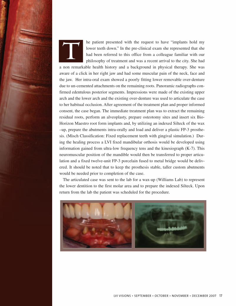
he patient presented with the request to have “implants hold my
lower teeth down.” In the pre-clinical exam she represented that she
had been referred to this office from a colleague familiar with our
philosophy of treatment and was a recent arrival to the city. She had
a non remarkable health history and a background in physical therapy. She was
aware of a click in her right jaw and had some muscular pain of the neck, face and
the jaw. Her intra-oral exam showed a poorly fitting lower removable over-denture
due to un-cemented attachments on the remaining roots. Panoramic radiographs con-
firmed edentulous posterior segments. Impressions were made of the existing upper
arch and the lower arch and the existing over-denture was used to articulate the case
to her habitual occlusion. After agreement of the treatment plan and proper informed
consent, the case began. The immediate treatment plan was to extract the remaining
residual roots, perform an alveoplasty, prepare osteotomy sites and insert six Bio-
Horizon Maestro root form implants and, by utilizing an indexed Silteck of the wax
–up, prepare the abutments intra-orally and load and deliver a plastic FP-3 prosthe-
sis. (Misch Classification: Fixed replacement teeth with gingival simulation.) Dur-
ing the healing process a LVI fixed mandibular orthosis would be developed using
information gained from ultra-low frequency tens and the kinesiograph (K-7). This
neuromuscular position of the mandible would then be transferred to proper articu-
lation and a fixed twelve-unit FP-3 porcelain fused to metal bridge would be deliv-
ered. It should be noted that to keep the prosthesis stable, taller custom abutments
would be needed prior to completion of the case.
The articulated case was sent to the lab for a wax-up (Williams Lab) to represent
the lower dentition to the first molar area and to prepare the indexed Silteck. Upon
return from the lab the patient was scheduled for the procedure.
T
18 LVI VISIONS • SEPTEMBER • OCTOBER • NOVEMBER • DECEMBER 2007
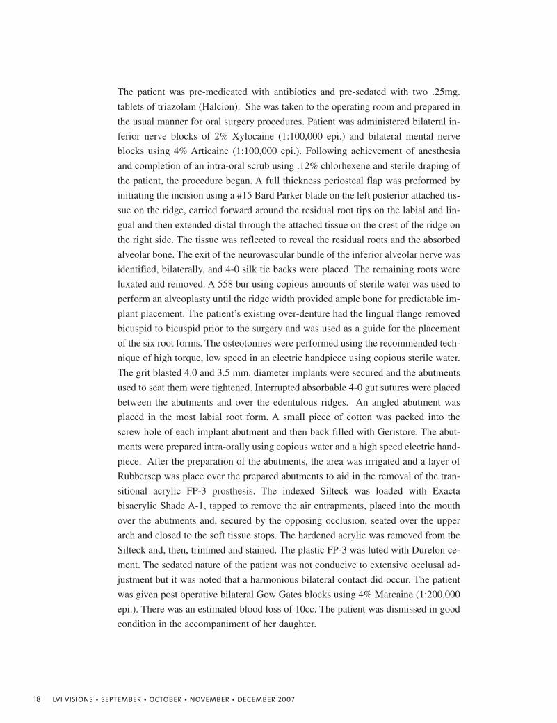
The patient was pre-medicated with antibiotics and pre-sedated with two .25mg.
tablets of triazolam (Halcion). She was taken to the operating room and prepared in
the usual manner for oral surgery procedures. Patient was administered bilateral in-
ferior nerve blocks of 2% Xylocaine (1:100,000 epi.) and bilateral mental nerve
blocks using 4% Articaine (1:100,000 epi.). Following achievement of anesthesia
and completion of an intra-oral scrub using .12% chlorhexene and sterile draping of
the patient, the procedure began. A full thickness periosteal flap was preformed by
initiating the incision using a #15 Bard Parker blade on the left posterior attached tis-
sue on the ridge, carried forward around the residual root tips on the labial and lin-
gual and then extended distal through the attached tissue on the crest of the ridge on
the right side. The tissue was reflected to reveal the residual roots and the absorbed
alveolar bone. The exit of the neurovascular bundle of the inferior alveolar nerve was
identified, bilaterally, and 4-0 silk tie backs were placed. The remaining roots were
luxated and removed. A 558 bur using copious amounts of sterile water was used to
perform an alveoplasty until the ridge width provided ample bone for predictable im-
plant placement. The patient’s existing over-denture had the lingual flange removed
bicuspid to bicuspid prior to the surgery and was used as a guide for the placement
of the six root forms. The osteotomies were performed using the recommended tech-
nique of high torque, low speed in an electric handpiece using copious sterile water.
The grit blasted 4.0 and 3.5 mm. diameter implants were secured and the abutments
used to seat them were tightened. Interrupted absorbable 4-0 gut sutures were placed
between the abutments and over the edentulous ridges. An angled abutment was
placed in the most labial root form. A small piece of cotton was packed into the
screw hole of each implant abutment and then back filled with Geristore. The abut-
ments were prepared intra-orally using copious water and a high speed electric hand-
piece. After the preparation of the abutments, the area was irrigated and a layer of
Rubbersep was place over the prepared abutments to aid in the removal of the tran-
sitional acrylic FP-3 prosthesis. The indexed Silteck was loaded with Exacta
bisacrylic Shade A-1, tapped to remove the air entrapments, placed into the mouth
over the abutments and, secured by the opposing occlusion, seated over the upper
arch and closed to the soft tissue stops. The hardened acrylic was removed from the
Silteck and, then, trimmed and stained. The plastic FP-3 was luted with Durelon ce-
ment. The sedated nature of the patient was not conducive to extensive occlusal ad-
justment but it was noted that a harmonious bilateral contact did occur. The patient
was given post operative bilateral Gow Gates blocks using 4% Marcaine (1:200,000
epi.). There was an estimated blood loss of 10cc. The patient was dismissed in good
condition in the accompaniment of her daughter.
LVI VISIONS • SEPTEMBER • OCTOBER • NOVEMBER • DECEMBER 2007 19
Before Orthotic After Case Finishing
20 LVI VISIONS • SEPTEMBER • OCTOBER • NOVEMBER • DECEMBER 2007
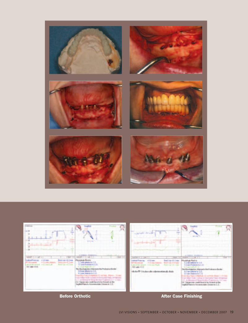
During the healing process, the patient underwent a tens and K-7 evaluation where-
by a more physiological mandibular position was determined. A new wax up was
performed using the transitional FP-3 as a base for support and then later another
fixed orthotic was made chair side. The patient wore this for a period of time until
the muscles were relaxed and the joint noise dissipated.
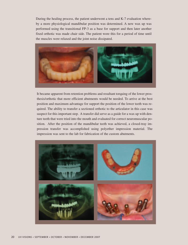
It became apparent from retention problems and resultant torquing of the lower pros-
thesis/orthotic that more efficient abutments would be needed. To arrive at the best
position and maximum advantage for support the position of the lower teeth was re-
quired. The ability to transfer a sectioned orthotic to the articulator in this case was
suspect for this important step. A transfer did serve as a guide for a wax up with den-
ture teeth that were tried into the mouth and evaluated for correct neuromuscular po-
sition. After the position of the mandibular teeth was achieved, a closed-tray im-
pression transfer was accomplished using polyether impression material. The
impression was sent to the lab for fabrication of the custom abutments.
LVI VISIONS • SEPTEMBER • OCTOBER • NOVEMBER • DECEMBER 2007 21
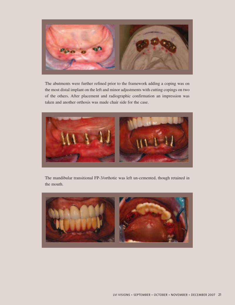
The abutments were further refined prior to the framework adding a coping was on
the most distal implant on the left and minor adjustments with cutting copings on two
of the others. After placement and radiographic confirmation an impression was
taken and another orthosis was made chair side for the case.
The mandibular transitional FP-3/orthotic was left un-cemented, though retained in
the mouth.
22 LVI VISIONS • SEPTEMBER • OCTOBER • NOVEMBER • DECEMBER 2007
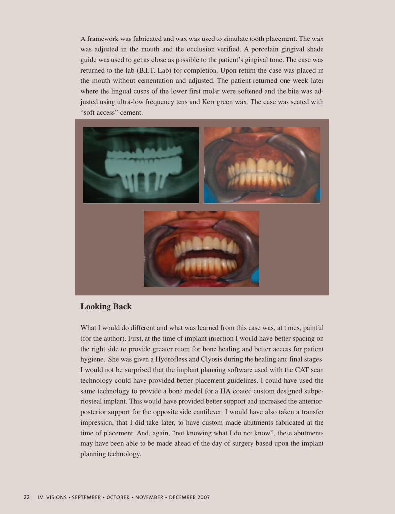
A framework was fabricated and wax was used to simulate tooth placement. The wax
was adjusted in the mouth and the occlusion verified. A porcelain gingival shade
guide was used to get as close as possible to the patient’s gingival tone. The case was
returned to the lab (B.I.T. Lab) for completion. Upon return the case was placed in
the mouth without cementation and adjusted. The patient returned one week later
where the lingual cusps of the lower first molar were softened and the bite was ad-
justed using ultra-low frequency tens and Kerr green wax. The case was seated with
“soft access” cement.
Looking Back
What I would do different and what was learned from this case was, at times, painful
(for the author). First, at the time of implant insertion I would have better spacing on
the right side to provide greater room for bone healing and better access for patient
hygiene. She was given a Hydrofloss and Clyosis during the healing and final stages.
I would not be surprised that the implant planning software used with the CAT scan
technology could have provided better placement guidelines. I could have used the
same technology to provide a bone model for a HA coated custom designed subpe-
riosteal implant. This would have provided better support and increased the anterior-
posterior support for the opposite side cantilever. I would have also taken a transfer
impression, that I did take later, to have custom made abutments fabricated at the
time of placement. And, again, “not knowing what I do not know”, these abutments
may have been able to be made ahead of the day of surgery based upon the implant
planning technology.
LVI VISIONS • SEPTEMBER • OCTOBER • NOVEMBER • DECEMBER 2007 23
The transfer of the bite from the orthotic was more difficult because the indirect
transfer necessitated by the lack of draw and poor height of the hand prepped abut-
ments. The orthotic broke in the distal sections and this would delay the healing of
the muscles and the bite and resulted in delayed results and increased chair time and
real time. The effective time and lab cost involved made the case more of a “learn-
ing experience” than a profitable one
These are the types of cases I seek to treat and I learn something each time. My biggest
“get” is even though I have been placing and restoring implants for over twenty-three
years I am behind by not utilizing the technology afforded by CAT scan technology.
Therefore, I will involve myself in the implant programs of LVI and Dr. Leo Malin.
James H. Clarke, Jr., D.D.S., F.A.G.D.Diplomate, American Board of Oral Implantology/ Implant Dentistry Fellow, American Academy of Implant Dentistry
Dr. James H. Clarke, Jr. graduated from Baylor Dental School in 1974 and then
began his private practice in Houston. His focus from the beginning of his career
was to distinguish himself as a consumate professional through thorough commu-
nication with his patients and the ability to deliver these results through constant
education on a higher level. His belief is that this journey is never ending. His stud-
ies and achievements thus far have enabled him to earn the distinctions he has,
beginning with a Fellowship in the Academy of General Dentistry. His extensive
studies of implant dentistry and the application of that science to his patients'
needs earned him a Fellowship from the Misch Implant Institute, a Fellowship in the
American Academy of Implant Dentistry, a Diplomate in the International Congress
of Oral Implantology and a Mastership from the American Academy of Implant
Prosthodontics. With a Diplomate Board Certification from the American Board of
Oral Implantology/Implant Dentistry and with Graduate status from the prestigious
Las Vegas Institute for Advanced Dental Studies his use of neuromuscular princi-
ples in treatment planning forms a core of excellence. He continues to study at LVI
to seek a greater understanding of the physiology of applied neuromuscular treat-
ment to deliver results of excellence in oral rehabilitation.
LVI- Advanced Anterior, Carp, Occ I, II, III, Scan, K-7, Full MouthNeuromuscular Dentures, Anatomy & Physiology, TMD, Implant I
17222 Red Oak Drive Suite 100 Houston, Texas 77090-2614 Ph: 281-440-1044 Fx: 281-440-9011
www.lvihoustondentist.com
Alan J. Singleton, DDS
LVI VISIONS • SEPTEMBER • OCTOBER • NOVEMBER • DECEMBER 2007 25
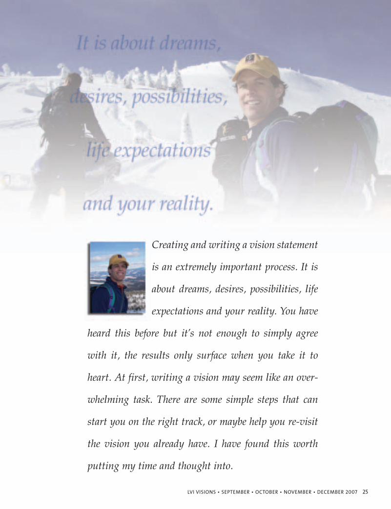
Creating and writing a vision statement
is an extremely important process. It is
about dreams, desires, possibilities, life
expectations and your reality. You have
heard this before but it’s not enough to simply agree
with it, the results only surface when you take it to
heart. At first, writing a vision may seem like an over-
whelming task. There are some simple steps that can
start you on the right track, or maybe help you re-visit
the vision you already have. I have found this worth
putting my time and thought into.
26 LVI VISIONS • SEPTEMBER • OCTOBER • NOVEMBER • DECEMBER 2007
It really does start to all
happen when you em-
brace this, and do it for
yourself. It is not a one
shot deal. Make it a part
of your life. So where do
you start? I have myself struggled
with this in the past. I have found a
few things out over the years that
have made it easier. Don’t be afraid
to just get started and write some
things down. The vision that you cre-
ate today can be different from the
vision you create tomorrow. I have
resisted creating a vision for fear that
it was not perfect and I may change
my mind. I have come to realize that
one of the beauties of creating my
own vision is that I can always create
a new one. You can create your reali-
ty. By thinking, dreaming and writ-
ing your own vision you start to take
control of your reality. Failing to cre-
ate your own vision allows someone
else to create your reality for you
(ask me how I know). You should al-
ways be looking at your vision to
make sure it is what you want, know-
ing that you can adjust and re-create
anytime you want. In fact, the
process of re-thinking and re-creat-
ing may allow you to get closer and
closer to that ideal vision that is you.
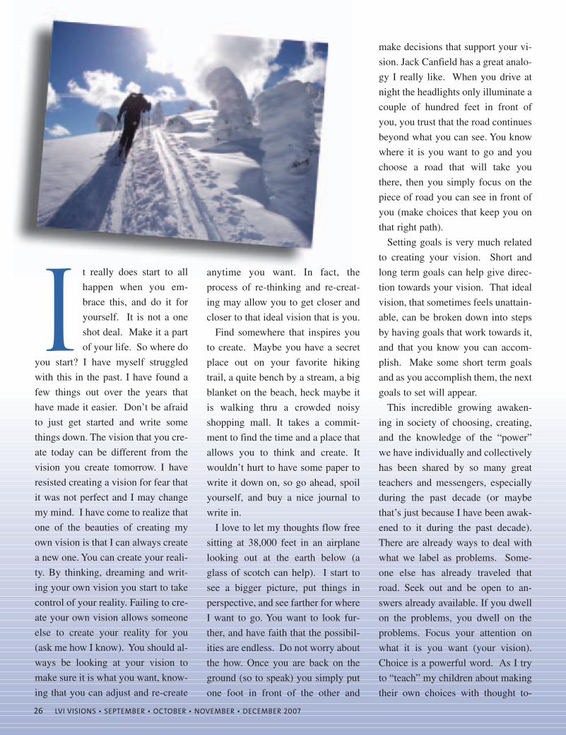
Find somewhere that inspires you
to create. Maybe you have a secret
place out on your favorite hiking
trail, a quite bench by a stream, a big
blanket on the beach, heck maybe it
is walking thru a crowded noisy
shopping mall. It takes a commit-
ment to find the time and a place that
allows you to think and create. It
wouldn’t hurt to have some paper to
write it down on, so go ahead, spoil
yourself, and buy a nice journal to
write in.
I love to let my thoughts flow free
sitting at 38,000 feet in an airplane
looking out at the earth below (a
glass of scotch can help). I start to
see a bigger picture, put things in
perspective, and see farther for where
I want to go. You want to look fur-
ther, and have faith that the possibil-
ities are endless. Do not worry about
the how. Once you are back on the
ground (so to speak) you simply put
one foot in front of the other and
make decisions that support your vi-
sion. Jack Canfield has a great analo-
gy I really like. When you drive at
night the headlights only illuminate a
couple of hundred feet in front of
you, you trust that the road continues
beyond what you can see. You know
where it is you want to go and you
choose a road that will take you
there, then you simply focus on the
piece of road you can see in front of
you (make choices that keep you on
that right path).
Setting goals is very much related
to creating your vision. Short and
long term goals can help give direc-
tion towards your vision. That ideal
vision, that sometimes feels unattain-
able, can be broken down into steps
by having goals that work towards it,
and that you know you can accom-
plish. Make some short term goals
and as you accomplish them, the next
goals to set will appear.
This incredible growing awaken-
ing in society of choosing, creating,
and the knowledge of the “power”
we have individually and collectively
has been shared by so many great
teachers and messengers, especially
during the past decade (or maybe
that’s just because I have been awak-
ened to it during the past decade).
There are already ways to deal with
what we label as problems. Some-
one else has already traveled that
road. Seek out and be open to an-
swers already available. If you dwell
on the problems, you dwell on the
problems. Focus your attention on
what it is you want (your vision).
Choice is a powerful word. As I try
to “teach” my children about making
their own choices with thought to-
LVI VISIONS • SEPTEMBER • OCTOBER • NOVEMBER • DECEMBER 2007 27
wards the outcomes and conse-
quences I realize how important it is
for me to be conscious of choices. In
all things; relationships, people, the
environment, work, stress, play, be-
havior, giving, receiving, etc. From
our environment and surroundings
we are constantly receiving stimuli.
With this stimuli, combined with our
wants, desires, paradigms and vi-
sions, we are always making choices.
When we fail to be conscious of our
choices we wonder why things turn
out the way they do. Make an effort
to be conscious of your choices.
Be sure to look at the whole picture
when creating your vision. By that I
mean, look at your whole life. Den-
tistry is a large part of my life; it does
not however define me. When we
meet someone new and are asked the
question “What do you do?” we tend
to define ourselves by our “job”. All
the people I have met (especially thru
LVI) are much more than that. I
heard someone respond to the ques-
tion “How are you?”, by saying
“Smart and handsome.” Can you an-
swer the question “What do you do?”
in a way that encompasses more than
your job. Look at who you are emo-
tionally, physically, and spiritually in
relation to your health, family, rela-
tionships, work and hobbies.
What do you want to be? What do
you want to do? What do you want
to have? These are 3 separate parts of
the whole picture. For example I
want to be healthy, blissful, patient
and respected. Things I want to do
(be specific); backcountry skiing,
travel for pleasure, play with my
kids, cosmetic and restorative den-
tistry (your job fits in this category).
Examples of things I want to have;
beautiful, warm family home, a
sweet ride (Mercedes-Benz SLR
McLaren works for me), financial
freedom (in excess), freedom to
make choices, and a balance of time
between friends, family, personal re-
lationships and private time. The
have comes last, first find out what
you want to be. Write your vision
around all the parts of your life so
you create the balance that is right for
you. If it is 90% dentistry and that is
where your passion and emotion are,
than that is great. It can also be 10%
dentistry. It is not right or wrong as
long as it is your truth. When you are
doing dentistry, you want to have the
passion and energy to give 100% to
it. The same applies to the other areas
of your life. Find the balance that al-
lows you to do that.
Consider a quote I first heard
adapted into an inspirational turning
point in the movie Coach Carter but
originally given by Nelson Mandela
at his inaugural speech in 1994:
Our deepest fear is not that we are
inadequate.
Our deepest fear is that we are
powerful beyond measure.
It is our light, not our darkness,
that most frightens us.
We ask ourselves, who am I to be
brilliant, gorgeous, talented, fabu-
lous?
Actually, who are you not to be?
You are a child of God; your play-
ing small doesn't serve the world.
There is nothing enlightened about
shrinking so that other people won't
feel insecure around you.
We were born to make manifest the
glory of God that is within us.
It's not just in some of us; it's in
everyone.
And as we let our light shine, we
unconsciously give other people per-
mission to do the same.
As we are liberated from our own
fear, our presence automatically lib-
erates others.
I still have many questions, and as
I write these words I know more an-
swers will come for me. As you write
your vision more answers will come
to you. I feel there is a balance that
will feel “right”. My right is not the
same as everyone else’s right, and
that is what makes it right. If you are
always worrying and thinking about
what another person is thinking, or
how they may react, you are allowing
someone else to create your reality.
If you want to truly create for your-
self, you have to have the attitude
and tell yourself that it does not mat-
ter. It has nothing to do with self cen-
tered greed. As Nelson Mandela so
eloquently states, our actions to cre-
ate, and live, our own true vision
gives others around us the freedom
and opportunity to create their own
vision, based on their ideal truth. A
vision at the expense of another
never comes without a catch. It is the
act of unconditional giving that re-
sults in receiving back more than you
gave. Your vision will manifest when
it allows everyone’s free will.
I learned the golden rule in early
grade-school; “Do unto others as you
would have others do unto you.”
Indulge me in a philosophical
thought; we are all more alike than
we sometimes may believe. On some
large scale we are all one and the
same; respect yourself and those
around you enough to create your
own vision, and although we are all
one, we have different wants and
therefore different visions. Since we
want different things, there is more
than enough to go around. Stop wor-
rying about your engramed para-
digms of what you “should do and
should be”. Write your vision about
what you want! When you actually
write the words, use words like “I
have” and “I am”. If you say “I want”
then you will always be “wanting”.
Feel the emotions of already having.
Make it in the present.
Take some time and list the kinds
of dentistry you like to do, the types
of people you like to work with, and
the environment you like to work in.
Then write a vision that encompass-
es all of this. In my opinion there are
different types of niche practices.
For me the definition of a niche prac-
tice is one that is true to your vision.
It can therefore be different for dif-
ferent dentists depending on what
they want. If you love doing full den-
tures than your niche practice is a
schedule full of edentulous patients.
If it is to do full mouth reconstruction
only, than that is your niche. It can
also be anywhere in between. The
first step is creating the vision that
you want. Don’t worry how you will
make it happen. Just create your
ideal vision. The tools you need to
make it happen can be found at LVI.
When the student is ready the teacher
will appear. When you know what it
is you want, then you will be ready
for the tools to help take you there.
Being focused and directed toward
your vision is different from trying to
force it to happen. It will not help to
force the issue; just always make
decisions that support your vision. It
may happen overnight, it may take
longer. Be grateful for what you have
30 LVI VISIONS • SEPTEMBER • OCTOBER • NOVEMBER • DECEMBER 2007
now in the present, and then more of
that will come to you and propel you
towards your vision.
This is an incredible honor to write
an article about visions in a maga-
zine entitled Visions. Each time I
visit my vision, I am able to be more
focused towards what I want. The
process of writing this article, and
sharing it, has allowed me to do just
that. Thank you for allowing me this
opportunity. I hope you are inspired
to create your own.
Make a commitment to start
writing something down. Look at it a
few times a day. Is it what you want?
Add to it, take away from it, share it
with your friends and be grateful for
how far you have already come. You
may be amazed at how it becomes
easier and easier. I give myself total
creative freedom and therefore am
not obliged to what I have written.
Remember you can always change it.
This gives you the freedom to start,
have fun with it and remember to al-
ways be true to yourself.
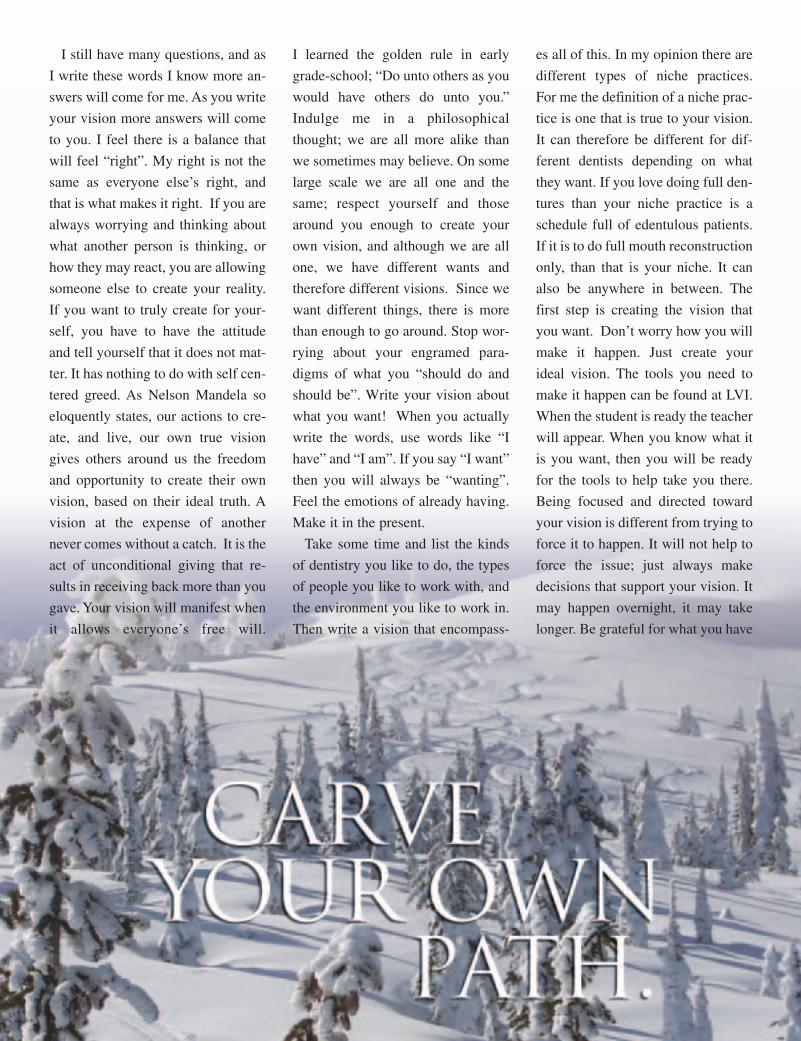
Carve your own path.
Dr. Alan Singleton is a graduate of theUniversity of Alberta, earning his B.Sc.in 1990 and his D.D.S. from theirSchool of Dentistry in 1992. He is agraduate of LVI Global. Dr. Singletonlives in Penticton, BC, Canada. He pro-vides advanced dental care in both thesmall town of Osoyoos, BC, and thecity of Penticton, BC. His general den-tal practice has a focus on Neuromus-cular Restorative Dentistry and Ortho-dontics. His restorative practiceprovides services which include amal-gam replacement with non metal alter-natives to full mouth reconstruction. DrSingleton has provided comprehensiveOrthodontic care for over 14 years,treating both adolescents and adults.Dr. Singleton is an involved member ofnumerous professional organizations.Dr Singleton is proud to be a clinical in-structor at LVI.
32 LVI VISIONS • SEPTEMBER • OCTOBER • NOVEMBER • DECEMBER 2007
“I am a CE junkie and I must have said ten times that this IACA meeting was probablythe best single meeting I have ever attended in my 30 plus years of dentistry. My wifeis not into attending meetings like this, and she even liked it, and we are signed up fornext years meeting in Orlando, including our team members.”
Roger Roubal, DDSOmaha, Nebraska
THIS WAS NOT YOUR TYPICAL DENTAL MEETING!Read what attendees are saying about the 2007 conference.
“Since being back from the IACA, my team and I are so excited about what we learnedthere, being around incredible minds, sharing ideas, meeting old friends and makingnew ones, the atmosphere was just unbelievable. I've been to a lot of dental meetingsADA, AACD, Greater NY, AGD, and State meetings. The IACA is by far the best meeting that anyone can go to.”
Chong Lee, DDSMcLean, Virginia
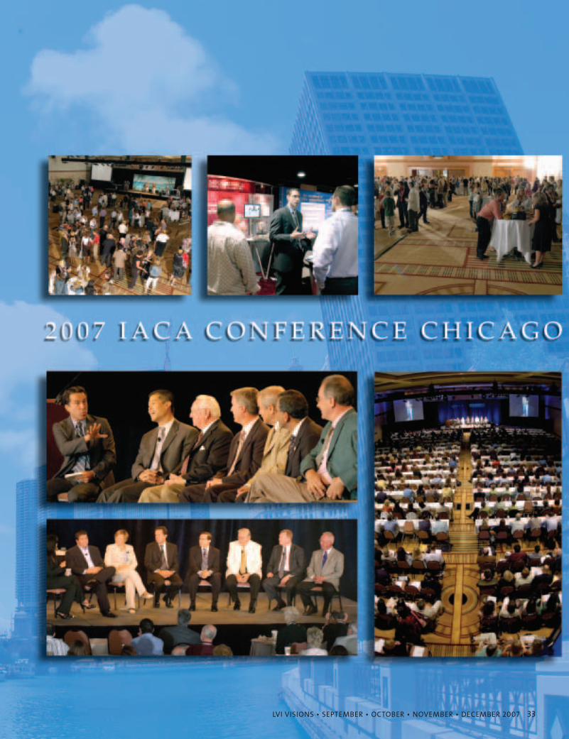
LVI VISIONS • SEPTEMBER • OCTOBER • NOVEMBER • DECEMBER 2007 33

34 LVI VISIONS • SEPTEMBER • OCTOBER • NOVEMBER • DECEMBER 2007
“I've just arrived home from my first IACA with my entire team, it’s after 3:00AM in the morning and I am wired and excited to get back to the office on Tues-day. It felt like the Super Bowl of dentistry with Bob Jankelson, Norm Thomas,Bill Dickerson, Jay Gerber, Anne-Maree Cole, Heidi Dickerson, Mike Miyasaki,Ashley Johnson, Sherry Blair, Brad Durham and on and on and on…”
Drew Markham, DDSHuntsville, Ontario
“What impressed me the most was seeing all of those giants in dentistryright in there with the rest of us in the meeting rooms, learning that onemore thing that will help them grow.”
David Miles, DMDAiken, South Carolina
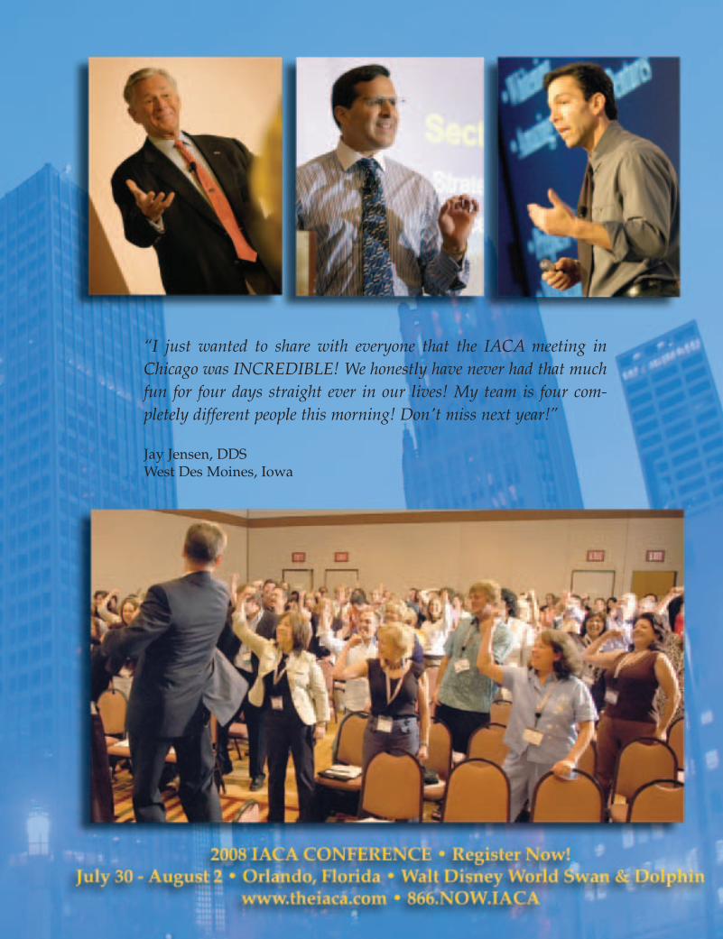
“I just wanted to share with everyone that the IACA meeting inChicago was INCREDIBLE! We honestly have never had that muchfun for four days straight ever in our lives! My team is four com-pletely different people this morning! Don't miss next year!”
Jay Jensen, DDSWest Des Moines, Iowa
36 LVI VISIONS • SEPTEMBER • OCTOBER • NOVEMBER • DECEMBER 2007
Quality, Service or Low Price –Choose Two
It’s impossible to provide the high-
est quality, the best service and the
lowest price without killing your
business. You can have two out of the
three, but the laws of economics pre-
clude maintaining all three at the
same time. Beware of companies that
promise the highest quality, the low-
est price and the best service. Invari-
able, at least one of the three must be
compromised. No one can be all
things for all people.
Patients like knowing their dentist is successful
Your patients might joke about
making the next three payments on
your Mercedes, but they really don’t
want their dentist driving a Ford
Pinto. Human nature is such that
most people want the best quality
and service available, and they also
realize that quality costs more.
The Ritz-Carlton chain of luxu-
ry hotels is a great example of a
company dedicated to high quali-
ty and five-star service; while
definitely NOT trying to cater to
travelers searching for the best
deal. We can learn a lot from
Ritz-Carlton.
WalMart and McDonalds provide
low price and decent service, but
they realize they cannot also have
the highest quality, and shoppers
understand the difference. For eco-
nomic reasons, many low income
dental patients are forced to shop
for the cheapest price, but are those
the patients you really want to at-
tract? Before you answer, consider
the following:
Surprisingly enough, raising your fees might be the best thing that ever
happens to your practice AND to your patients. In fact, if your patients knew all
the facts, many would actually WANT you to raise your fees.
I firmly believe that most of your loyal patients would prefer to pay a little more
rather than suffer reduced quality and/or service. Or worse yet, watch their
favorite dentist be forced into early retirement. Selecting and maintaining your
ideal fee structure is one of the most important facets of a healthy practice.
Arthur "Kit" Weathers, Jr. DDS
Maintaining the Standard of Care
If a general dentist performs a proce-
dure normally done by a specialist, the
GP is held to the same standard of care
as the specialist. That means, if you do a
root canal, you must use the rubber
dam, locate and clean all canals (includ-
ing that pesky MB2 in upper molars),
completely obturate the entire root canal
system, etc., or you can be held liable.
If you obtain the necessary training
and skills to perform endodontic ther-
apy equal to that done by the en-
dodontist, why would you then charge
50 percent less for the same service?
Surveys by Dental Economics, The
McGill Advisory, Dentistry Today and
others routinely report that GPs are
charging approximately 50 percent
less than what endodontists charge for
the exact same procedure. For exam-
ple, in 2006, the average general prac-
titioner’s fee for an anterior root canal
was approximately $500 while en-
dodontists averaged around $750 for
treating the same tooth.
If you cannot perform to the same
level as the endodontist, you should
not do the root canal, but if you can
do it as well, perhaps it’s time to ad-
just your fees. It’s also time to clear-
ly identify what type of practice you
would like to have.
WalMart or Ritz-Carlton?
I believe that most patients want
the best dental care available, but
some have to settle for the lowest
price. It’s important that you decide
which market you want to attract
When most people hear the name
Ritz-Carlton, words like “tops in their
field,” “prestigious,” “highest quality,”
“the finest,” and “in a class of their
own,” come to mind. Wouldn’t you
like for your patients to make similar
statements about your practice?
How Do You Decide What to Charge?
If you want to market your practice
based primarily on having the lowest
fees, the decision is easy. Simply call
all the dentists in your area, check
their fees and make yours lower.
Word will get around and you will
soon be covered up with patients. You
will then need to learn how to cut cor-
ners, eliminate service and work as
fast as possible. While you’re at it,
you might as well sign up for every
HMO and PPO you can find.
If, however, you do not want to
compete on price, I have a few sug-
gestions. Whatever you do, don’t
price yourself in the middle of the
pack or you will commit financial
suicide. This common and often mis-
understood practice virtually guaran-
tees a mediocre practice at best.
Remember that increasing your fees
by only 10 percent adds 28 percent
to your bottom line if your
overhead is 65 percent.
38 LVI VISIONS • SEPTEMBER • OCTOBER • NOVEMBER • DECEMBER 2007
People who want to avoid average
or below average treatment will usu-
ally look for the dentist with the
highest fees. Although charging high
fees cannot guarantee high quality,
the public usually assumes that to be
the case. They can’t tell if your preps
are good, your margins are tight or if
you filled that mesio-buccal canal all
the way to the terminus, so they as-
sume your fees must be high because
you provide the best treatment.
If you have the latest and greatest,
high-tech equipment, that’s another
indication that you must be at the top
of your profession. How else could
you have paid for all that fancy stuff?
One of the best ways to adjust your
fees is to hire a professional to com-
pare your fees with other doctors in
your zip code. This way you can
quickly and systematically maximize
your fees. Dr. Charles Blair has re-
viewed hundreds of practices, and he
computes over 100 different statistics
for each participating doctor’s dental
practice and provides industry
benchmarks for comparison purpos-
es. Go to www.drcharlesblair.com
and check out Dr. Blair’s Revenue
Enhancement Program and new In-
surance Coding Manual.
Dr. Blair does not recommend rais-
ing all of your fees by the same per-
centage across the board. He warns
doctors to be careful of “sore-thumb”
fees such as new patient exams,
cleanings, x-rays and recall exams,
and by using over 100 statistical pa-
rameters to analyze each doctor’s
practice, Dr. Blair has determined
that the average practice is losing
over $100,000 in profitability annu-
ally to management and coding er-
rors that are fairly easy to correct. If
you take time to analyze your prac-
tice, it will be one of the best invest-
ments you will ever make.
“In the eyes of the patient, no fee is
a bargain,” says Blair, “and every prac-
tice loses a few patients every year
over fees, or at least that’s what they
say! You can cut your fees next year
and lose a few patients; you can keep
the same fees next year and lose a few
patients; or rebalance and raise your
fees next year and lose a few patients.”
Even small increases add up
Remember that increasing your
fees by only 10 percent adds 28 per-
cent to your bottom line if your over-
head is 65 percent. Raising fees by 20
percent adds 57 percent, and it’s all
profit. Incidentally, your fees should
never end in a zero. If your current
fee is $450, for example, consider
raising it to something like $497.
Also, I suggest you never discount
your fees for two reasons. First, you
are telling the patient, “Yes, I can do
your case for less money because
I’ve grossly inflated my original
quote.” Secondly and most impor-
tantly, by giving a discount you run
the risk of lowering the value of your
work in the future.
Let’s say you discount your single
tooth implant fee from $3997 to
$2997 as long as the patient “promis-
es not to tell anyone about the dis-
count.” Three of her closest friends
ask who did her work, and she glad-
ly gives them your name. When her
friends ask how much she paid for
the implant and crown, she says
$2997, and as promised, she “doesn’t
tell them about the discount.”
When the three friends show up at
your office and you quote them
$3997 for an implant and crown, they
gasp, “You charged my friend $1,000
less.” So now you give the three
friends the $1,000 discount, but
ONLY if they “absolutely promise
not to mention the discount” to any
of their friends…
You get the idea. Between the lady
and her three friends, you have es-
sentially given away a free implant
and crown. If you hadn’t offered the
discount, you could have treated one
less patient and collected the same
amount of money, and by treating
four patients instead of three you had
to work 25% harder. Not to mention
the fact that you had to pay the lab
fee for an extra crown, buy an extra
implant, pay your assistant, discount
future “friends” and on and on…
Leave the discounts to Southwest
Airlines and automobile salesmen.
“Good is the enemy of great.”
In his best selling book, Good to
Great, Jim Collins says, “The vast
majority of companies never become
great, precisely because the vast ma-
jority become quite good -- and that
is their main problem.”
This is a great time to raise your
fees and upgrade your practice. The
baby boomers are retiring and they
want to look and feel younger. Best
of all, most of them have finally
saved enough money to pay for the
things they want. Ask Harley-
LVI VISIONS • SEPTEMBER • OCTOBER • NOVEMBER • DECEMBER 2007 39
Davidson how many boomers are fi-
nally getting that bike they’ve al-
ways wanted.
For most people, looking younger
includes making their teeth whiter
and more beautiful. White teeth, with
nice rounded mamelons, definitely
help people look younger, and that’s
exactly what the boomers want. The
one common denominator of every
“Extreme Makeover” show is im-
proving the patient’s smile.
Isn’t it time to change your good
your practice into one that’s great.
Most dentists settle into the good cat-
egory, and complacency holds them
there. Incidentally, if you really want
to take your practice to the next level,
you MUST involve your team.
Why involve team members in the business?
Educate your team about the busi-
ness of dentistry if you want to maxi-
mize your profitability. Most doctors
are uncomfortable discussing fees and
business concepts with their team, but
unless your team understands the
goals of the practice they cannot help
you achieve them. Unwittingly, they
may even be sabotaging your efforts.
The team sees large amounts of
money coming into the practice, and
unless they understand overhead ex-
penses, the cost of acquiring and
maintaining necessary technology,
and keeping up with advances in
continuing education (which allows
your practice to provide high quality
service), they cannot help motivate
your patients to want the best dental
care possible.
TEAM = Together EveryoneAchieves More
Therefore, if you want to provide
quality dental treatment, meet or ex-
ceed the standard of care, and mini-
mize stress in your practice, your team
must to be striving for the same goals.
Summary
It’s been said, “If you don’t
charge what you’re worth, you be-
come worth what you charge.”
That’s true, but I suggest you take
the opposite approach. Do NOT
charge what you are worth.
“Charge MORE than you’re worth,
and you will still become worth
what you charge.”
Finally, the most important thing of
all is to enjoy what you do. Albert
Schweitzer said it best, “Success is not
the key to happiness. Happiness is the
key to success. If you love what you
are doing, you will be successful.”
Remember also that nothing is
ever perfect. Anyone who says that
it takes sunshine to bring happiness
has never danced in the rain.
“Don’t be afraid
to give up
the good
to go for
the great.”
Theodore Roosevelt
40 LVI VISIONS • SEPTEMBER • OCTOBER • NOVEMBER • DECEMBER 2007
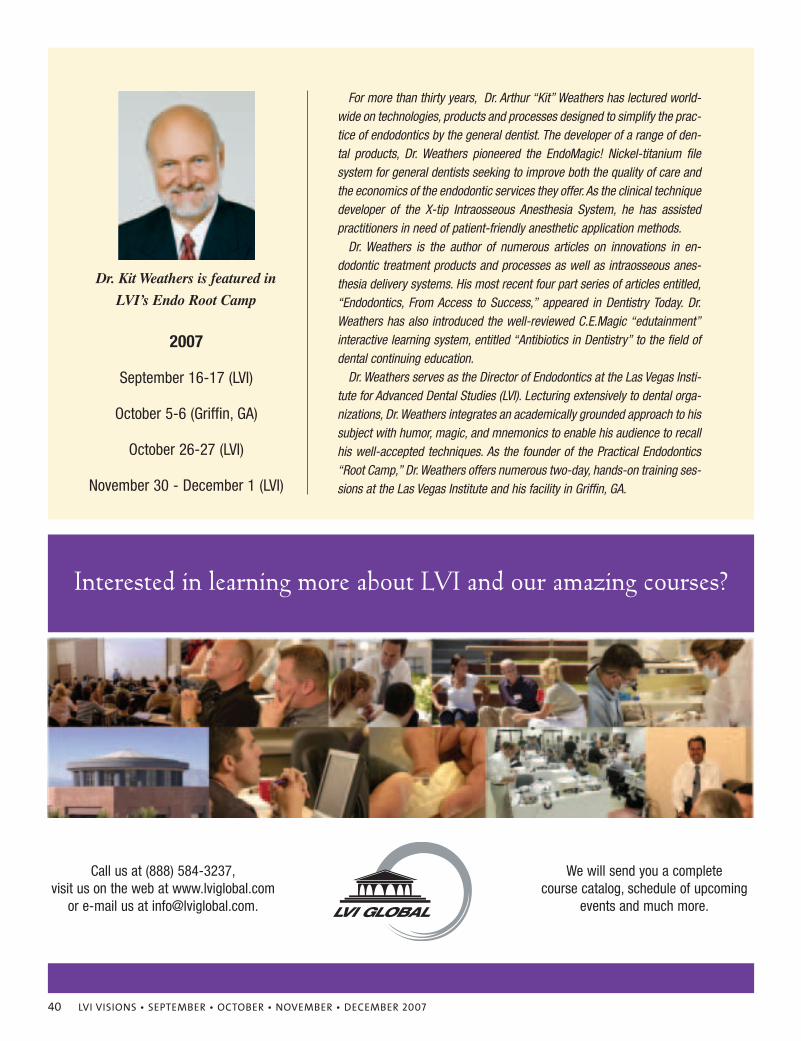
Dr. Kit Weathers is featured in
LVI’s Endo Root Camp
2007
September 16-17 (LVI)
October 5-6 (Griffin, GA)
October 26-27 (LVI)
November 30 - December 1 (LVI)
For more than thirty years, Dr. Arthur “Kit” Weathers has lectured world-wide on technologies, products and processes designed to simplify the prac-tice of endodontics by the general dentist. The developer of a range of den-tal products, Dr. Weathers pioneered the EndoMagic! Nickel-titanium filesystem for general dentists seeking to improve both the quality of care andthe economics of the endodontic services they offer. As the clinical techniquedeveloper of the X-tip Intraosseous Anesthesia System, he has assistedpractitioners in need of patient-friendly anesthetic application methods.
Dr. Weathers is the author of numerous articles on innovations in en-dodontic treatment products and processes as well as intraosseous anes-thesia delivery systems. His most recent four part series of articles entitled,“Endodontics, From Access to Success,” appeared in Dentistry Today. Dr.Weathers has also introduced the well-reviewed C.E.Magic “edutainment”interactive learning system, entitled “Antibiotics in Dentistry” to the field ofdental continuing education.
Dr. Weathers serves as the Director of Endodontics at the Las Vegas Insti-tute for Advanced Dental Studies (LVI). Lecturing extensively to dental orga-nizations, Dr. Weathers integrates an academically grounded approach to hissubject with humor, magic, and mnemonics to enable his audience to recallhis well-accepted techniques. As the founder of the Practical Endodontics“Root Camp,” Dr. Weathers offers numerous two-day, hands-on training ses-sions at the Las Vegas Institute and his facility in Griffin, GA.
Interested in learning more about LVI and our amazing courses?
We will send you a complete course catalog, schedule of upcoming
events and much more.
Call us at (888) 584-3237,visit us on the web at www.lviglobal.com
or e-mail us at [email protected].
42 LVI VISIONS • SEPTEMBER • OCTOBER • NOVEMBER • DECEMBER 2007
LVI VISIONS • SEPTEMBER • OCTOBER • NOVEMBER • DECEMBER 2007 43
Historically, temporomandibular joint disorders (TMD) involve
specific clinical findings. Costen1, in 1934, described TMJ syn-
drome to include (1) preauricular pain, (2) tenderness to palpa-
tion of the closing (elevator) muscles of the mandible, (3) de-
creased opening and (4) joint noise. Joint noise during the period of 1970
through 1990 was specifically defined as a reciprocal clicking caused by dis-
placement of the articular disc anteriorly to the condylar head on closure and
reduction to normal position on opening. Greene and Laskin2, in 1968, intro-
duced the term myofascial pain disorders (MPD) to include patients that had
preauricular pain, decreased opening and pain to palpation over the elevator
muscles of the mandible. Patients with pure MPD do not have any joint noise.
Farrar and McCarty3 defined internal derangements as a result of a tear of the
discal ligament with reciprocal clicking in the temporomandibular joint.
Dr. Bernard Jankelson developed the Myotronics equipment that measures
mandibular movement and electromyographic evaluation of the muscles of
mastication in 1966. This equipment provides the documentation as to how the
mandible functions and to that point in space where the closing muscles want
to close the mandible. Dr. William Dickerson changed the world of dentistry by
founding the Las Vegas Institute for Advanced Dental Studies and developing
a neuromuscular protocol for treating patients.
Over the last fifteen to twenty years, temporomandibular joint disorders have
become a catch-all term for general dentists, physicians and lay people. The
following classification of craniomandibular disorders includes the majority of
factors that can affect mandibular movement and function. The classification is
the result of 40 years of clinical evaluation and treatment of patients with cran-
iomandibular dysfunction.
The Functional Classification of Craniomandibular Disorders is divided into
eight categories, which are indicated by Roman numerals. Each of the subdivi-
sions under the Roman numerals is listed in order of lesser to more severe prob-
lems. I is myofascial pain dysfunction (MPD). II is internal derangements,
which are caused by a tear of the discal ligaments. III is intracapsular pain, not
related to internal derangements. IV is chronic hypomobility (decreased open-
ing). V is mandibular hypermobility. VI is Growth disorders – skeletal abnor-
malities. VII is Traumatic injuries – fractures – hemarthrosis. VIII is abnormal
jaw closure.
This is a functional classification in which each of the eight categories are the re-
sult of an injury or dysfunction of a specific craniomandibular anatomical entity.
I. Masticatory Muscle Disorders (muscle pain)
1.Protective muscle splinting
2. Muscle spasm activity
3.Muscle inflammation (myositis)
Normal incisal opening is 50 mm (the distance between the incisal edge of
the maxillary incisors to the incisal edge of the mandibular incisors on wide
opening). Normal lateral excursions are 10 mm. Any mandibular opening
greater than 50 mm and/or lateral or protrusive movement greater than 10 mm
is diagnostic of hypermobility (subluxation) of the mandibular condyle. Pa-
tients with protective muscle splinting have an incisal opening which can be in-
creased to 50 mm with gentle passive opening of the patients lower jaw with
finger pressure. Muscle spasm is defined when the incisal distance cannot be
increased to 50 mm with finger pressure by the practitioner. Trigger points can
be palpated in the masseter, medial pterygoid or temporalis muscles. Muscle
inflammation (myositis) usually involves pericoronitis of the mandibular third
molars or as a result of multiple inferior alveolar nerve injections. The incisal
opening is less than 30 mm and cannot be increased by finger manipulation.
Lateral excursions are usually normal (10 mm.) unless the lateral pterygoid
muscles are involved. Myositis may also be caused by a buccally erupting max-
illary third molar. Masticatory muscle disorders are the most common cause of
facial pain and the reason patients seek treatment. Patients with protective mus-
cle splinting and muscle spasms all close posterior to myotrajectory.
II. Disc Interference Disorders (internal derangements)
1. Early derangement
2. Mid derangement
3. Late derangement
4. Dislocated disc (Closed Lock)
Patients with an internal derangement have a reciprocal click on opening
and closing. The opening click is always at a greater incisal opening than the
closing click. The click is produced following a tear of the discal ligament
which attaches the articular disc to the condylar head. The lateral and medial
discal ligaments are most commonly torn. The posterior ligament is never
torn. Based on my clinical evaluation of over 14,000 patients, an early de-
rangement is defined as an opening click which occurs within 0-20 mm of
44 LVI VISIONS • SEPTEMBER • OCTOBER • NOVEMBER • DECEMBER 2007

Websites that Generate Business!
website design
search engine positioning
opening, mid derangement between 20-35 mm of opening and late derange-
ment at a greater incisal opening than 35 mm. A dislocated disc occurs in a
patient with a history of reciprocal clicks who does not have a click on open-
ing and closing and the opening is usually less than 35 mm. An important
clinical evaluation to determine the difference between muscle inflammation
or myositis and a dislocated disc is to have the patient attempt to move the jaw
from one side to the other. Normal lateral excursions are 10 mm. Patients with
myositis will have a marked, limited decreased opening, but their lateral ex-
cursions will be normal. In patients with a dislocated disc (closed lock), the
patient will not be able to move the mandible more than 3-5 mm toward the
contra-lateral side. They will have normal excursions toward the affected side,
if the internal derangement is unilateral.
III. Inflammatory Disorders of the Joint (joint pain)
1. Synovitis and capsulitis
2. Retro-discitis
3. Inflammatory arthritis
A. Degenerative arthritis
B. Traumatic arthritis
Synovitis and capsulitis refer to an inflammatory intracapsular clinical situ-
ation in which the patient has pain on opening and palpation with the teeth sep-
arated and with wide opening. Retro-discitis occurs when the patient has pain
and discomfort on closing. Inflammatory changes in the joint include degen-
erative and traumatic arthritis. These will have radiographic evidence of loss of
condylar contour and sclerosis involving the anterior aspect of glenoid fossa
and condylar head. Clinically, patients with degenerative arthritis will exhibit
crepitus on opening and closing.
IV. Chronic Mandibular Hypomobilities (decreased movement)
1. Contracture of elevator muscles
2. Capsular fibrosis
3. Ankylosis
A. Fibrous
B. Osseous
4. Coronoid hyperplasia
46 LVI VISIONS • SEPTEMBER • OCTOBER • NOVEMBER • DECEMBER 2007
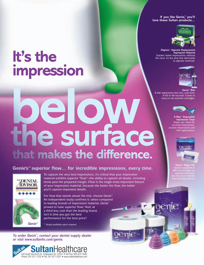
Silgimix™ Alginate Replacement Impression Material
Quicker, better impressions—without the mess. It’s the dust-free alternative
to alginate materials.
Genie™ BiteA bite registration that sets, rock hard,
in 60 or 90 seconds. Comes in easy-to-use automix cartridges.
3-Way™ Disposable Impression Trays
Single use—ideal for accurate impressions,
counter impressions and bite registrations.
Genie™ Magic MixGenie is available in
380 ml cartridges forautomix machines.
Get 5% more materialthan foil bag systems.
If you like Genie,™ you’ll love these Sultan products…
To capture the very best impressions, it’s critical that your impression
material exhibits superior “flow”—the ability to capture all details, including
those past the prepared margin. Flow is the single most important feature
of your impression material, because the better the flow, the better
you’ll capture important details.
For flow that stands above the rest, choose Genie™.
An independent study confirms it: when compared
to leading brands of impression material, Genie™
proved to have superior flow.*And, at
a third less cost than the leading brand,
isn’t it time you got the best
performance for the best price?
* Study available upon request.
To order Genie™, contact your dental supply dealer or visit www.sultanhc.com/genie.
Genie’s™ superior flow… for incredible impressions, every time.
Genie™
330 South Van Brunt St., Englewood, NJ 07631 • Toll Free: 800-637-8582Phone: 201-871-1232 • Fax: 201-871-0321 • www.sultanhealthcare.com
SultanHealthcare
These patients have decreased mandibular opening and may have decreased
lateral excursions. The patients are painless unless an attempt is made to in-
crease the opening. This may be the result of connective tissue disease (scle-
roderma, lupus), following trauma (capsular fibrosis, fibrous and osseous), in-
traoral surgery (scar tissue) and skeletal abnormalities (coronoid hyperplasia).
I emphasize that these conditions are painless but have decreased opening and
occasionally, decreased lateral excursions. In unilateral cases, decreased later-
al excursions will occur towards the contralateral side. Hypomobilities are very
often misdiagnosed as a closed lock.
V. Mandibular Hypermobilities (excessive movement)
1. Subluxation
2. Spontaneous dislocation of condyle
A. Disc condyle function normal
B. Associated with internal derangement
3. Chronic (recurrent) dislocation of condyle
A. Disc condyle function normal
B. Associated with internal derangement
Hypermobility is most commonly misdiagnosed as “TMJ”. This is mandibu-
lar subluxation. The patient has a partial tear of the capsular ligament fibers that
limit anterior condylar translation. Subluxation occurs when the mandibular
condyle moves past the most dependent portion of the articular eminence. This
can be diagnosed with fluoroscopy and tomograms of the temporomandibular
joint. Subluxation is commonly misdiagnosed or not diagnosed with transcra-
nial radiographs. Spontaneous dislocation occurs when the condyle moves past
the midpoint of the articular eminence, and the patient cannot return the
mandible to maximum intercuspation without manipulation of the condyle. Dis-
location of the condyle can occur with or without internal derangements. If sub-
luxation occurs with an associated internal derangement, you have two clicks on
opening and two clicks on closing. This is very difficult to treat conservatively.
Patients with a subluxation should have a bite block placed between their teeth
when dental work, including prophylaxis is performed. Subluxation occurs
when the incisal opening is greater than 50 mm, and lateral excursions and
mandibular protrusion are greater than 10 mm. Subluxation can also occur with
incisal openings less than 50 mm. This can be documented with axially correct-
ed tomograms and computerized mandibular scanning.
48 LVI VISIONS • SEPTEMBER • OCTOBER • NOVEMBER • DECEMBER 2007
VI. Growth Disorders of the Jaws (skeletal malrelationships)
1.Aberration of development
A. Vertical maxillary deficiency (vertical hypoplasia)
B. Vertical maxillary excess (vertical hyperplasia)
C. Maxillary hypoplasia (Anterior – posterior)
D. Maxillary hyperplasia (Anterior – Posterior)
E. Mandibular AP hypoplasia (Class II)
F. Mandibular AP hyperplasia (Class II)
G. Apertognathia
i. Posterior
ii. Anterior
iii. Lateral
H. Asymmetry
i. Maxilla
ii. Mandible
2.Class I, II, III
A. In centric occlusion
B. In myotrajectory
3.Acquired change in joint structure
A. Adaptive changes
B. Proliferative changes
C. Degenerative changes
4.Unilateral condylar hyperplasia
Vertical maxillary deficiency is defined clinically as a patient who does not
show maxillary incisors with the teeth at rest or smiling. These patients normal-
ly have a mandibular plane angle that is less than 25 degrees. Vertical maxillary
excess is defined with patients who show maxillary mucosa when smiling.
These patients generally have a high mandibular plane angle (greater than 30 de-
grees). Apertognathia should be defined as anterior, posterior or lateral. Aper-
tognathia must be evaluated to rule out whether it is skeletal or functional.
Posterior apertognathia usually occurs after iatrogenic treatment of jaw dys-
function problems or with decreased posterior maxillary growth. Lateral aper-
tognathia may be due to abnormal mandibular growth or tongue habit. Anterior
apertognathia is usually due to increased posterior maxillary growth or anterior
tongue thrust. Mandibular asymmetry will produce abnormal opening and clos-
ing. Class I, II and III relationships are less important in evaluating the patients
LVI VISIONS • SEPTEMBER • OCTOBER • NOVEMBER • DECEMBER 2007 49
with craniomandibular disorders, however most patients will have a Class III
molar relationship with a myotrajectory bite. Acquired change in joint structure
must be evaluated. Unilateral condylar hyperplasia is a one-sided growth of the
condyle producing a mandibular asymmetry and is more common in women.
VII. Traumatic Injuries (fractures of the jaws and/or teeth)
1. Fracture
A. Open
B. Closed
C. Non-displaced
D. Displaced
2. Hemarthrosis
Traumatic injuries include fractures of the mandible and maxilla. These are
usually treated separately in an attempt to return the patient to former function
prior to the fracture. In my practice over the last 40 years, patients who sustain
mandibular and/or midface fractures that are appropriately treated rarely de-
velop craniomandibular disorders. Hemarthrosis is the acute occlusal emer-
gency that occurs when a patient sustains trauma to the mandible that produces
bleeding inside the joint. These injuries should be treated on an emergency
basis with impressions being made and a soft bite guard being constructed to
fit over the lower teeth. This will aid in decreasing continued trauma to the in-
tracapsular structures as a result of swallowing function.
VIII. Abnormal Jaw Closure
1. Posterior
2. Lateral
3. Vertical
All patients that have myofascial pain (I), internal derangements (II) and in-
flammatory disorders (III) close posterior to myotrajectory and/or have in-
creased vertical overclosure.
Obviously, the clinician must be aware of other clinical entities including
malignancies, neurological diseases, infections, and systemic diseases.
Bibliography:
1. Costen, JB: Syndrome of sinus and ear symptoms dependent upon disturbed function of the temporomandibular joint. Ann Otol Rhinol Laryngol 1934;.43:1-5.
2. Laskin, DM: Etiology of the pain-dysfunction syndrome. J Am Dent Assoc 1969;79:149-153.
3. Farrar, WB, McCarty WL: A Clinical Outline of Temporomandibular Joint Diagnosis and Treatment, 7th Ed. Montgomery, Ala.: Normandie Publications; 1982.
4. Wolford, DG: Evaluation of Patients With Facial Pain and/or Craniomandibular Disorders. Neuromuscular Dentistry – The Next Millennium Anthology Volume;
ICCMO 1999; 109-117
50 LVI VISIONS • SEPTEMBER • OCTOBER • NOVEMBER • DECEMBER 2007
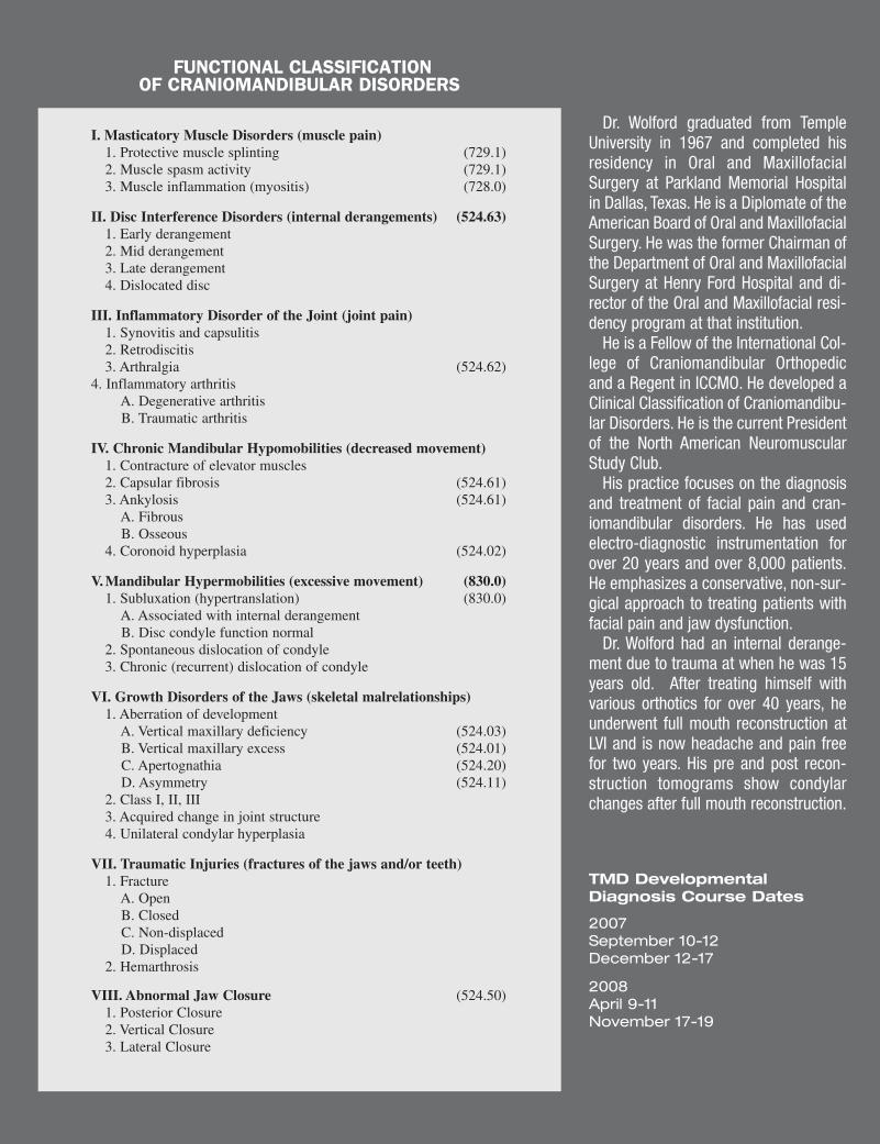
I. Masticatory Muscle Disorders (muscle pain)1. Protective muscle splinting (729.1)2. Muscle spasm activity (729.1)3. Muscle inflammation (myositis) (728.0)
II. Disc Interference Disorders (internal derangements) (524.63)1. Early derangement2. Mid derangement3. Late derangement4. Dislocated disc
III. Inflammatory Disorder of the Joint (joint pain)1. Synovitis and capsulitis2. Retrodiscitis3. Arthralgia (524.62)
4. Inflammatory arthritisA. Degenerative arthritisB. Traumatic arthritis
IV. Chronic Mandibular Hypomobilities (decreased movement)1. Contracture of elevator muscles2. Capsular fibrosis (524.61)3. Ankylosis (524.61)
A. FibrousB. Osseous
4. Coronoid hyperplasia (524.02)
V. Mandibular Hypermobilities (excessive movement) (830.0)1. Subluxation (hypertranslation) (830.0)
A. Associated with internal derangementB. Disc condyle function normal
2. Spontaneous dislocation of condyle3. Chronic (recurrent) dislocation of condyle
VI. Growth Disorders of the Jaws (skeletal malrelationships)1. Aberration of development
A. Vertical maxillary deficiency (524.03)B. Vertical maxillary excess (524.01)C. Apertognathia (524.20)D. Asymmetry (524.11)
2. Class I, II, III3. Acquired change in joint structure4. Unilateral condylar hyperplasia
VII. Traumatic Injuries (fractures of the jaws and/or teeth)1. Fracture
A. OpenB. ClosedC. Non-displacedD. Displaced
2. Hemarthrosis
VIII. Abnormal Jaw Closure (524.50)1. Posterior Closure2. Vertical Closure3. Lateral Closure
FUNCTIONAL CLASSIFICATION OF CRANIOMANDIBULAR DISORDERS
Dr. Wolford graduated from TempleUniversity in 1967 and completed hisresidency in Oral and MaxillofacialSurgery at Parkland Memorial Hospitalin Dallas, Texas. He is a Diplomate of theAmerican Board of Oral and MaxillofacialSurgery. He was the former Chairman ofthe Department of Oral and MaxillofacialSurgery at Henry Ford Hospital and di-rector of the Oral and Maxillofacial resi-dency program at that institution.
He is a Fellow of the International Col-lege of Craniomandibular Orthopedicand a Regent in ICCMO. He developed aClinical Classification of Craniomandibu-lar Disorders. He is the current Presidentof the North American NeuromuscularStudy Club.
His practice focuses on the diagnosisand treatment of facial pain and cran-iomandibular disorders. He has usedelectro-diagnostic instrumentation forover 20 years and over 8,000 patients.He emphasizes a conservative, non-sur-gical approach to treating patients withfacial pain and jaw dysfunction.
Dr. Wolford had an internal derange-ment due to trauma at when he was 15years old. After treating himself withvarious orthotics for over 40 years, heunderwent full mouth reconstruction atLVI and is now headache and pain freefor two years. His pre and post recon-struction tomograms show condylarchanges after full mouth reconstruction.
TMD Developmental Diagnosis Course Dates
2007September 10-12December 12-17
2008April 9-11November 17-19
52 LVI VISIONS • SEPTEMBER • OCTOBER • NOVEMBER • DECEMBER 2007
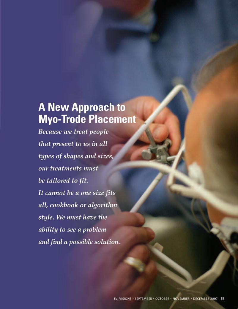
LVI VISIONS • SEPTEMBER • OCTOBER • NOVEMBER • DECEMBER 2007 53
Because we treat people
that present to us in all
types of shapes and sizes,
our treatments must
be tailored to fit.
It cannot be a one size fits
all, cookbook or algorithm
style. We must have the
ability to see a problem
and find a possible solution.
A New Approach toMyo-Trode Placement
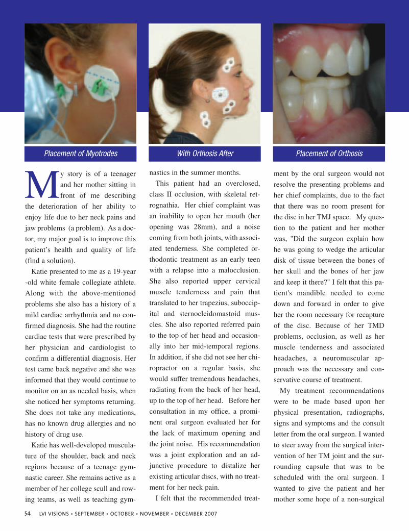
54 LVI VISIONS • SEPTEMBER • OCTOBER • NOVEMBER • DECEMBER 2007
My story is of a teenager
and her mother sitting in
front of me describing
the deterioration of her ability to
enjoy life due to her neck pains and
jaw problems (a problem). As a doc-
tor, my major goal is to improve this
patient’s health and quality of life
(find a solution).
Katie presented to me as a 19-year
-old white female collegiate athlete.
Along with the above-mentioned
problems she also has a history of a
mild cardiac arrhythmia and no con-
firmed diagnosis. She had the routine
cardiac tests that were prescribed by
her physician and cardiologist to
confirm a differential diagnosis. Her
test came back negative and she was
informed that they would continue to
monitor on an as needed basis, when
she noticed her symptoms returning.
She does not take any medications,
has no known drug allergies and no
history of drug use.
Katie has well-developed muscula-
ture of the shoulder, back and neck
regions because of a teenage gym-
nastic career. She remains active as a
member of her college scull and row-
ing teams, as well as teaching gym-
nastics in the summer months.
This patient had an overclosed,
class II occlusion, with skeletal ret-
rognathia. Her chief complaint was
an inability to open her mouth (her
opening was 28mm), and a noise
coming from both joints, with associ-
ated tenderness. She completed or-
thodontic treatment as an early teen
with a relapse into a malocclusion.
She also reported upper cervical
muscle tenderness and pain that
translated to her trapezius, suboccip-
ital and sternocleidomastoid mus-
cles. She also reported referred pain
to the top of her head and occasion-
ally into her mid-temporal regions.
In addition, if she did not see her chi-
ropractor on a regular basis, she
would suffer tremendous headaches,
radiating from the back of her head,
up to the top of her head. Before her
consultation in my office, a promi-
nent oral surgeon evaluated her for
the lack of maximum opening and
the joint noise. His recommendation
was a joint exploration and an ad-
junctive procedure to distalize her
existing articular discs, with no treat-
ment for her neck pain.
I felt that the recommended treat-
ment by the oral surgeon would not
resolve the presenting problems and
her chief complaints, due to the fact
that there was no room present for
the disc in her TMJ space. My ques-
tion to the patient and her mother
was, "Did the surgeon explain how
he was going to wedge the articular
disk of tissue between the bones of
her skull and the bones of her jaw
and keep it there?" I felt that this pa-
tient's mandible needed to come
down and forward in order to give
her the room necessary for recapture
of the disc. Because of her TMD
problems, occlusion, as well as her
muscle tenderness and associated
headaches, a neuromuscular ap-
proach was the necessary and con-
servative course of treatment.
My treatment recommendations
were to be made based upon her
physical presentation, radiographs,
signs and symptoms and the consult
letter from the oral surgeon. I wanted
to steer away from the surgical inter-
vention of her TM joint and the sur-
rounding capsule that was to be
scheduled with the oral surgeon. I
wanted to give the patient and her
mother some hope of a non-surgical
Placement of Myotrodes With Orthosis After Placement of Orthosis
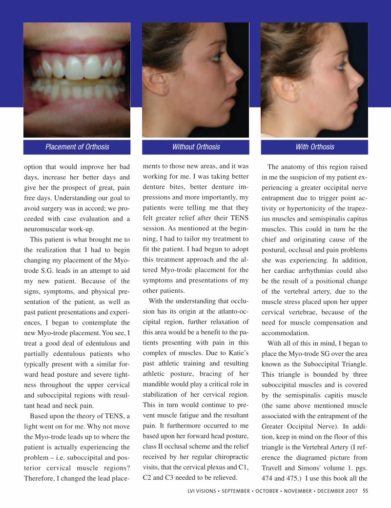
LVI VISIONS • SEPTEMBER • OCTOBER • NOVEMBER • DECEMBER 2007 55
option that would improve her bad
days, increase her better days and
give her the prospect of great, pain
free days. Understanding our goal to
avoid surgery was in accord; we pro-
ceeded with case evaluation and a
neuromuscular work-up.
This patient is what brought me to
the realization that I had to begin
changing my placement of the Myo-
trode S.G. leads in an attempt to aid
my new patient. Because of the
signs, symptoms, and physical pre-
sentation of the patient, as well as
past patient presentations and experi-
ences, I began to contemplate the
new Myo-trode placement. You see, I
treat a good deal of edentulous and
partially edentulous patients who
typically present with a similar for-
ward head posture and severe tight-
ness throughout the upper cervical
and suboccipital regions with resul-
tant head and neck pain.
Based upon the theory of TENS, a
light went on for me. Why not move
the Myo-trode leads up to where the
patient is actually experiencing the
problem – i.e. suboccipital and pos-
terior cervical muscle regions?
Therefore, I changed the lead place-
ments to those new areas, and it was
working for me. I was taking better
denture bites, better denture im-
pressions and more importantly, my
patients were telling me that they
felt greater relief after their TENS
session. As mentioned at the begin-
ning, I had to tailor my treatment to
fit the patient. I had begun to adopt
this treatment approach and the al-
tered Myo-trode placement for the
symptoms and presentations of my
other patients.
With the understanding that occlu-
sion has its origin at the atlanto-oc-
cipital region, further relaxation of
this area would be a benefit to the pa-
tients presenting with pain in this
complex of muscles. Due to Katie’s
past athletic training and resulting
athletic posture, bracing of her
mandible would play a critical role in
stabilization of her cervical region.
This in turn would continue to pre-
vent muscle fatigue and the resultant
pain. It furthermore occurred to me
based upon her forward head posture,
class II occlusal scheme and the relief
received by her regular chiropractic
visits, that the cervical plexus and C1,
C2 and C3 needed to be relieved.
The anatomy of this region raised
in me the suspicion of my patient ex-
periencing a greater occipital nerve
entrapment due to trigger point ac-
tivity or hypertonicity of the trapez-
ius muscles and semispinalis capitus
muscles. This could in turn be the
chief and originating cause of the
postural, occlusal and pain problems
she was experiencing. In addition,
her cardiac arrhythmias could also
be the result of a positional change
of the vertebral artery, due to the
muscle stress placed upon her upper
cervical vertebrae, because of the
need for muscle compensation and
accommodation.
With all of this in mind, I began to
place the Myo-trode SG over the area
known as the Suboccipital Triangle.
This triangle is bounded by three
suboccipital muscles and is covered
by the semispinalis capitis muscle
(the same above mentioned muscle
associated with the entrapment of the
Greater Occipital Nerve). In addi-
tion, keep in mind on the floor of this
triangle is the Vertebral Artery (I ref-
erence the diagramed picture from
Travell and Simons' volume 1. pgs.
474 and 475.) I use this book all the
Placement of Orthosis Without Orthosis With Orthosis
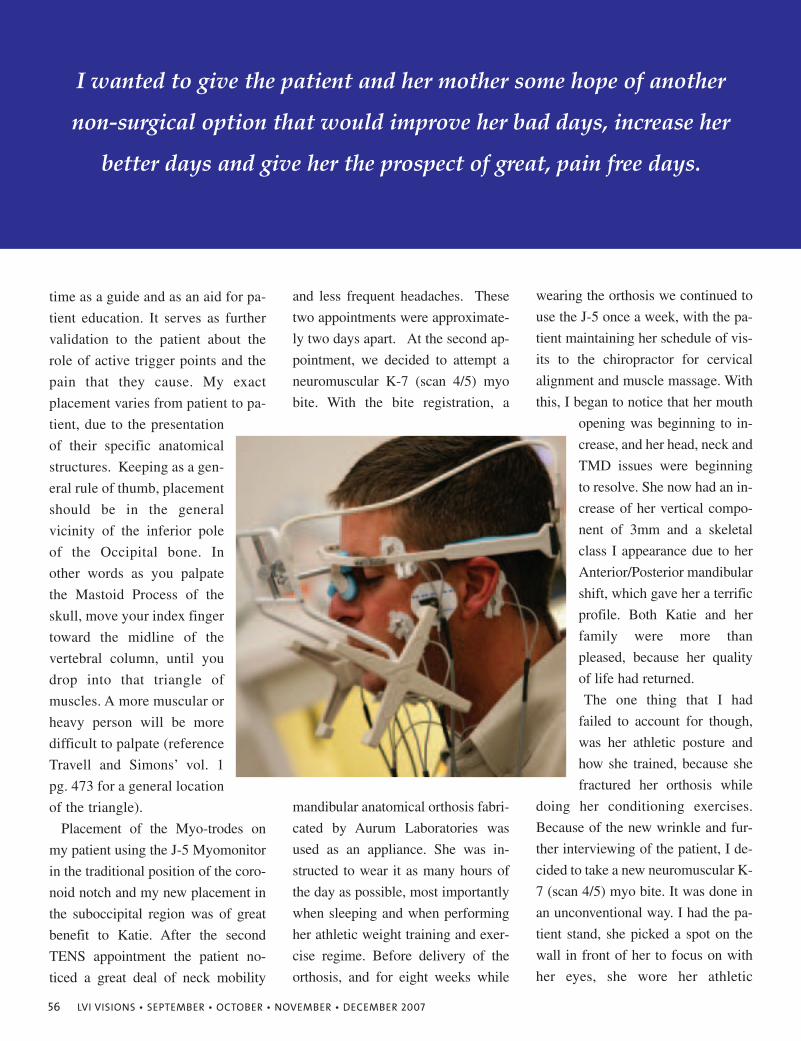
56 LVI VISIONS • SEPTEMBER • OCTOBER • NOVEMBER • DECEMBER 2007
time as a guide and as an aid for pa-
tient education. It serves as further
validation to the patient about the
role of active trigger points and the
pain that they cause. My exact
placement varies from patient to pa-
tient, due to the presentation
of their specific anatomical
structures. Keeping as a gen-
eral rule of thumb, placement
should be in the general
vicinity of the inferior pole
of the Occipital bone. In
other words as you palpate
the Mastoid Process of the
skull, move your index finger
toward the midline of the
vertebral column, until you
drop into that triangle of
muscles. A more muscular or
heavy person will be more
difficult to palpate (reference
Travell and Simons’ vol. 1
pg. 473 for a general location
of the triangle).
Placement of the Myo-trodes on
my patient using the J-5 Myomonitor
in the traditional position of the coro-
noid notch and my new placement in
the suboccipital region was of great
benefit to Katie. After the second
TENS appointment the patient no-
ticed a great deal of neck mobility
and less frequent headaches. These
two appointments were approximate-
ly two days apart. At the second ap-
pointment, we decided to attempt a
neuromuscular K-7 (scan 4/5) myo
bite. With the bite registration, a
mandibular anatomical orthosis fabri-
cated by Aurum Laboratories was
used as an appliance. She was in-
structed to wear it as many hours of
the day as possible, most importantly
when sleeping and when performing
her athletic weight training and exer-
cise regime. Before delivery of the
orthosis, and for eight weeks while
wearing the orthosis we continued to
use the J-5 once a week, with the pa-
tient maintaining her schedule of vis-
its to the chiropractor for cervical
alignment and muscle massage. With
this, I began to notice that her mouth
opening was beginning to in-
crease, and her head, neck and
TMD issues were beginning
to resolve. She now had an in-
crease of her vertical compo-
nent of 3mm and a skeletal
class I appearance due to her
Anterior/Posterior mandibular
shift, which gave her a terrific
profile. Both Katie and her
family were more than
pleased, because her quality
of life had returned.
The one thing that I had
failed to account for though,
was her athletic posture and
how she trained, because she
fractured her orthosis while
doing her conditioning exercises.
Because of the new wrinkle and fur-
ther interviewing of the patient, I de-
cided to take a new neuromuscular K-
7 (scan 4/5) myo bite. It was done in
an unconventional way. I had the pa-
tient stand, she picked a spot on the
wall in front of her to focus on with
her eyes, she wore her athletic
I wanted to give the patient and her mother some hope of another
non-surgical option that would improve her bad days, increase her
better days and give her the prospect of great, pain free days.
Neuromuscular Dentistry
www.myotronics.com
The Benefi ts of a Pleasing Smile.
Cosm
etic
Den
tist
ry b
y Ja
mes
Klim
, DD
S, S
anta
Ros
a, C
A
Some Things You Should Know About
COSMETICDENTISTRY
Call Myotronics at (800) 426-0316 or email [email protected] to learn more about the benefi ts of Neuromuscular Dentistry.
Neuromuscular Dentistry by Prabu Raman, DDS, Kansas City, MO
Dentists worldwide are discovering the science and the benefi ts of Neuromuscular Dentistry with Myotronics technology. Evaluate and success- fully treat occlusal, restorative and cosmetic cases with reliable, reproducible clinical data never seen before.
NMD LVI ad.indd 1 5/31/07 3:57:59 PM
58 LVI VISIONS • SEPTEMBER • OCTOBER • NOVEMBER • DECEMBER 2007
footwear and I corrected for her head
and neck posture according to the
EMG readings from the K-7, then we
took the neuromuscular bite just like
always, but instead of sitting, she was
in the standing position. I found out
from my patient that she spent more
time standing and running than she
did sitting. Another orthosis was de-
signed and fabricated from a material
called Natural flex; it is anatomical
and very strong. My patient has no
problems wearing it. We were able to
match her tooth shade to the material,
and she now wears it while eating.
By wearing her orthosis constantly
her TMD and cervical neck issues
were much improved. Additionally,
came a change in her head and neck
posture for the better, because she
was now experiencing no signs or
symptoms relating to her undiag-
nosed cardiac arrhythmia. With the
new bite came another increase in
her VDO by 1mm, making the total a
4mm change.
I began treating Katie in December
of 2005 and she says her relief can-
not be explained by words. She is
able to eat again without pain and she
is regaining the opening of her mouth
(approx. 38mm). She does not suffer
from her head and neck aches and is
excited about not having to worry
about that old part of her life. She is
beginning to turn the corner and has
nice muscle stabilization. She does
not mind wearing her orthosis, be-
cause she is painfully aware of the
consequences. She is very happy that
the TM joint surgery was avoided. I
believe that orthodontics with a com-
ponent for expansion of her arches is
in her future. She realizes that there
will be a next step and is looking for-
ward to that part.
As an aside, I have also found that
when performing longer dental pro-
cedures placement of the J-4 or J-5
Myomonitor at a subthreshold to
threshold amplitude improves patient
comfort at the completion of their
appointment. I believe that it is help-
ing the posturing muscles of the
neck, which in turn reduces the feed-
back to the muscle spindles. Also,
keep in mind that many patients have
years of poor occlusion, posture, nu-
trition, oxygen intake, proper muscle
activity, etc. Because of these factors,
I caution you to not promise the heal-
ing effects of TENS to them in one
appointment. I have found with my
patients, it may take several appoint-
ments to unwind and detangle the
muscles and associated nerves before
they start to feel relief and the bene-
fit of TENS therapy.
My goal in writing this article was
not to go through the process of
showing EMG's or Scans or endless
pictures of jaw tracings, it was to
open our minds in order to best help
our patients. The practice of neuro-
muscular dental medicine for me has
been a wonderful transformation. My
knowledge and education (obtained
from LVI Global) has given me the
greatest chance to start my patients
down the road to muscle harmony
and stability which will ultimately
improve their quality of life.
Dr. Pawlowicz graduated from the
University of Pittsburgh School of
Dental Medicine in 1993. He cur-
rently has completed over 1000
hours of advanced study and course-
work. He is a fellow in the Interna-
tional College of Craniomandibular
Orthopedics. An LVI Global Instruc-
tor for both adhesive aesthetics and
occlusion courses, Dr. Pawlowicz is
a Delegate of the Pennsylvania Den-
tal Association, Director of the West-
ern Pennsylvania Dental Society and
a consultant to Dentsply.
My goal was not to go through the process of showing EMG’s
or Scans or endless pictures of jaw tracings,
it was to open our minds to best help our patients.
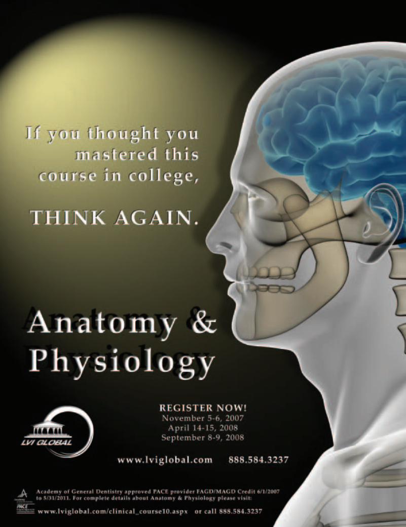
60 LVI VISIONS • SEPTEMBER • OCTOBER • NOVEMBER • DECEMBER 2007
SuePortland, Maine
Sue,What you are describing is a HANDOFF. The doctor hands off the patient to you, and
you hand off the patient to the receptionist. Think of it as a relay race. You are runningwith the baton and you catch up to then next person on your team and hand them thatbaton. Now, what happens if you drop the baton? RACE OVER! The same thing is truein the situation you described. Our job is to bring the patient through the office in a fluidway, handing them off where necessary and not “dropping the baton”. Are you follow-ing me? Great, I know you are! So let’s now relate this to your office. When you ap-proach the front desk with the hand written instructions for the next visit and the re-ceptionist does not clearly understand what is written this causes confusion, possiblemisunderstandings and so forth. You have to walk away to find out the correct infor-mation and by this time your “race is over”. The patient did not have a very good flowof information and understanding. Try This: The next time the doctor has somethingelse to schedule for the patient, find out what it is, walk the patient up and verbally tellthe scheduler, in front of the patient, exactly what the doctor would like scheduled next.Now there is not a question to the patient as to what is needed, and you are HANDINGOFF the patient with a transfer of information that they hear again another time. Yourcase acceptance will improve dramatically as you “hold on to that baton”.
You’ll win the race with proper handoffs!Heidi
Dear Heidi,
I’d like to say that we are “paperless” however we are not entirely ready for that. Weput all our treatment notes in the computer and do all of our billing through the com-puter but, our doctor still writes down the next procedure on paper and we physicallygo to the front desk to schedule it. The problem is many patients do not schedule atthat time. I know this sounds silly but, sometimes we can’t read the doctor’s writingand when we go back to see what he wants done next, the patient is out of there! Howcan we improve in this area?
SuePortland, Maine
LVI VISIONS • SEPTEMBER • OCTOBER • NOVEMBER • DECEMBER 2007 61
Dr. M,Your question is excellent. Many doctors have shared this same issue with me. It is a
tough one to answer because I do not know the details of the arrangement with your as-sociate. If she is being primed to take over your practice and your existing patients, thenshe should become busy as you transition your patients to her. In saying that, I haveseen a lot of dentists who have a hard time letting go of “their” patients and only tosswork over to the new guy when they either don’t really like the patient, or don’t wantto do a particular procedure. Only you can soul search and figure that one out.
But, let’s say your arrangement is that she is to grow the practice with you and bringon her own patients. Then she needs to be encouraged to work “on the practice” not just“in the practice”. You should mentor her in a way that will help bring clients in the door.For example, the associate should get involved in community associations and events,schools, and similar things that will form new business and personal relationships. Mar-keting that you have a wonderful new associate by placing an “announcement” in yourlocal paper is another way of introducing the associate and what she is all about to yourcommunity. What I am saying is the associate should be encouraged to turn down timeinto an opportunity to grow the practice. Let her know that she is not confined to thefour walls of your office. She can go out and creatively make things happen!
Hope this helps,Heidi
Dear Heidi,
I have a new associate who has a lot of down time. I try to come up with things forher to do but I am running out of ideas. She is frequently in our personal office killingtime on her computer. What can you suggest?
Dr. MAtlanta, Georgia

62 LVI VISIONS • SEPTEMBER • OCTOBER • NOVEMBER • DECEMBER 2007
LVI VISIONS • SEPTEMBER • OCTOBER • NOVEMBER • DECEMBER 2007 63
Thank you for staying tuned to read part two of
Controlling Overhead. No other subject is more
important to your business. Financially successful
dentists will implement change to achieve a
better bottom line. I promised to offer you some
ideas to help whittle down your overhead. Your
largest expenses are supply, lab, team and
marketing. Here are some ideas for reducing your
expense in each of the first three categories.
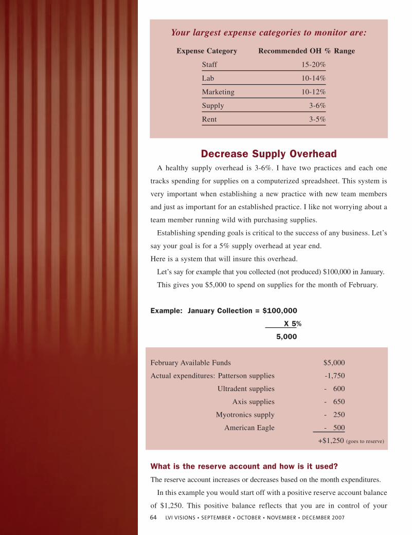
Decrease Supply OverheadA healthy supply overhead is 3-6%. I have two practices and each one
tracks spending for supplies on a computerized spreadsheet. This system is
very important when establishing a new practice with new team members
and just as important for an established practice. I like not worrying about a
team member running wild with purchasing supplies.
Establishing spending goals is critical to the success of any business. Let’s
say your goal is for a 5% supply overhead at year end.
Here is a system that will insure this overhead.
Let’s say for example that you collected (not produced) $100,000 in January.
This gives you $5,000 to spend on supplies for the month of February.
Example: January Collection = $100,000
X 5%
5,000
February Available Funds $5,000
Actual expenditures: Patterson supplies -1,750
Ultradent supplies - 600
Axis supplies - 650
Myotronics supply - 250
American Eagle - 500
+$1,250 (goes to reserve)
What is the reserve account and how is it used?
The reserve account increases or decreases based on the month expenditures.
In this example you would start off with a positive reserve account balance
of $1,250. This positive balance reflects that you are in control of your
Your largest expense categories to monitor are:
Expense Category Recommended OH % Range
Staff 15-20%
Lab 10-14%
Marketing 10-12%
Supply 3-6%
Rent 3-5%
64 LVI VISIONS • SEPTEMBER • OCTOBER • NOVEMBER • DECEMBER 2007
Establishing
spending goals
is critical to the
success of any
business.
spending. If you had spent more than $5,000 you would be over your goal of
5%. Having a negative reserve is not bad as long as you can correct your spend-
ing in future months.
The reserve account has a continually changing balance. You don’t actu-
ally open an account with this name – it is simply a number in your com-
puter that reflects your ability to control your spending.
The reserve account should build and at the end of the year be used for a
major purchase.
Major purchases could include a new sterilizer, digital sensor, etc. Under-
stand that if your reserve account does not build you will have an overhead
greater than 5% for supplies.
March expenditures will be based on February collections.
Say for example you collect $120,000 in February. You now have $6,000
(120,000 x .05 = 6,000) to spend on supplies in March.
This responsibility is best given to one team member. Preferably one who is
good with math. Dentists – you should not be spending your time with this task!
Decrease Lab Overhead
Raise your fees
Your crown, onlay or veneer fee should be 6-7 times your lab fee. If your
lab fee is $300, for example, your fee should range from $1,800 - $2,100 per
unit. A $2,100 fee with a $300 lab fee creates a 14.2% lab overhead. Re-
member that a healthy range for lab overhead is 10-14%. If you are utiliz-
ing a high end lab then reconsider your fee. Most dentists undervalue their
services and could raise their fees without resistance from their patients. If
you don’t think this is true – try it - how else will you ever know?
Wax up or no wax up?
I love the contouring aspect of a smile enhancement. For this reason I do
not order a wax up for an eight, ten or twenty unit veneer case. We only order
a wax up when we change the vertical dimension. Heather, my assistant, and
I do the mock up directly in the mouth the same day as our preparation. We
determine shape and length prior to anesthetic and always get the guest in-
volved in the process. Most people enjoy being involved in the decision mak-
ing process regarding shape, length, translucency, surface texture, etc.
Solving this piece of the puzzle in the beginning lessens problems on the
LVI VISIONS • SEPTEMBER • OCTOBER • NOVEMBER • DECEMBER 2007 65
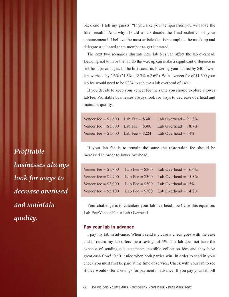
back end. I tell my guests, “If you like your temporaries you will love the
final result.” And why should a lab decide the final esthetics of your
enhancement? I believe the most artistic dentists complete the mock up and
delegate a talented team member to get it started.
The next two scenarios illustrate how lab fees can affect the lab overhead.
Deciding not to have the lab do the wax up can make a significant difference in
overhead percentages. In the first scenario, lowering your lab fee by $40 lowers
lab overhead by 2.6% (21.3% - 18.7% = 2.6%). With a veneer fee of $1,600 your
lab fee would need to be $224 to achieve a lab overhead of 14%.
If you decide to keep your veneer fee the same you should explore a lower
lab fee. Profitable businesses always look for ways to decrease overhead and
maintain quality.
Veneer fee = $1,600 Lab Fee = $340 Lab Overhead = 21.3%
Veneer fee = $1,600 Lab Fee = $300 Lab Overhead = 18.7%
Veneer fee = $1,600 Lab Fee = $224 Lab Overhead = 14%
If your lab fee is to remain the same the restoration fee should be
increased in order to lower overhead.
Veneer fee = $1,800 Lab Fee = $300 Lab Overhead = 16.6%
Veneer fee = $1,900 Lab Fee = $300 Lab Overhead = 15.8%
Veneer fee = $2,000 Lab Fee = $300 Lab Overhead = 15%
Veneer fee = $2,100 Lab Fee = $300 Lab Overhead = 14.2%
Your challenge is to calculate your lab overhead now! Use this equation:
Lab Fee/Veneer Fee = Lab Overhead
Pay your lab in advance
I pay my lab in advance. When I send my case a check goes with the case
and in return my lab offers me a savings of 5%. The lab does not have the
expense of sending out statements, possible collection fees and they have
great cash flow! Isn’t it nice when both parties win! In order to send in your
check you must first be paid at the time of service. Check with your lab to see
if they would offer a savings for payment in advance. If you pay your lab bill
Profitable
businesses always
look for ways to
decrease overhead
and maintain
quality.
66 LVI VISIONS • SEPTEMBER • OCTOBER • NOVEMBER • DECEMBER 2007
with a credit card expect their answer to be no. When you send a check the lab
does not have to pay credit card transaction fees. A 5% savings on a $300 lab
fee is $15. While this amount is small initially it adds up quickly on a large
case. Some offices have $200,000 in lab fees at year end. Five percent of
$200,000 is $10,000. If you collected 1.5 million, with a $200,000 lab
expense, $10,000 in lab savings would equate to a .7% reduction in lab over-
head. Are you going to ask your lab? You won’t get a yes unless you ask!
Create a warranty policy
Dentists are notorious for giving their dentistry away. If a patient breaks a
crown during the first year of service it should be warranted differently than if
it breaks during year six. Your dentistry is not meant to last forever. So when
someone asks you, “How long will that last?” you should respond, “Nothing
lasts forever and to be fair to you and me I have created a warranty policy that
explains your responsibility and mine.” Also ask how or why something failed.
Fracture of a crown from a car accident should be handled differently than a
fracture from chewing food. During the first year we replace the crown at no fee
and then from years two-five our responsibility for the cost of the remake goes
down and the patient’s goes up. Most importantly - be flexible and fair. Deliver
your warranty policy at the seat appointment and document this in your chart.
For a copy of our warranty policy e-mail [email protected].
Decrease Team Overhead
Fewer employees
So many dentists overstaff. I have a friend that has been overstaffed for
years. He knows it and does not have the heart to let anyone go. Understand
that if you can function as efficient or more efficiently with one less person
you are directly affecting your bottom line (your take home pay). Walter
Hailey used to say, “Get rid of the turkeys.”
If you help the turkeys move on you win and they do too. So what if she
is not a turkey?
Someone is still gobbling up your food! Move it or lose it! In an LVI style prac-
tice that performs more elective procedures you need fewer employees. Your over-
head for team should decrease since the need for additional personnel decreases.
“Nothing lasts
forever and to be
fair to you and
me I have created
a warranty policy
that explains your
responsibility
and mine.”
LVI VISIONS • SEPTEMBER • OCTOBER • NOVEMBER • DECEMBER 2007 67
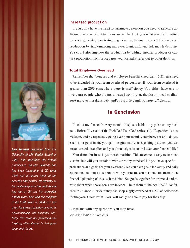
Increased production
If you don’t have the heart to terminate a position you need to generate ad-
ditional income to justify the expense. But I ask you what is easier – letting
someone go lovingly or trying to generate additional income? Increase your
production by implementing more quadrant, arch and full mouth dentistry.
You could also improve the production by adding another producer or cap-
ture production from procedures you normally refer out to other dentists.
Total Employee Overhead
Remember that bonuses and employee benefits (medical, 401K, etc) need
to be included in your team overhead percentage. If your team overhead is
greater than 20% somewhere there is inefficiency. You either have one or
two extra people who are not always busy or you, the doctor, need to diag-
nose more comprehensively and/or provide dentistry more efficiently.
In Conclusion
I look at my financials every month. It’s just a habit – my pulse on my busi-
ness. Robert Kiyosaki of the Rich Dad Poor Dad series said, “Repetition is how
we learn, and by repeatedly going over your monthly numbers, not only do you
establish a good habit, you gain insights into your spending patterns, you can
make corrections earlier, and you ultimately take control over your financial life.”
Your dental business is your cash machine. This machine is easy to start and
sustain. But will you sustain it with a healthy mindset? Do you have specific
projections and goals for your overhead? Do you have goals for yearly and daily
collection? You must talk about it with your team. You must include them in the
financial planning of this cash machine. Set goals together for overhead and re-
ward them when those goals are reached. Take them to the next IACA confer-
ence in Orlando, Florida if they can keep supply overhead at 4-5% of collections
for the year. Guess what – you will easily be able to pay for their trip!
E-mail me with any questions you may have!
Lori Kemmet graduated from The
University of MN Dental School in
1989. She maintains two private
practices in Boulder, Colorado. Lori
has been instructing at LVI since
1998 and attributes much of her
success and passion for dentistry to
her relationship with the dentists she
has met at LVI and her Incredible
Smiles team. She was the recipient
of the LVIM award in 2004. Lori has
a fee for service practice devoted to
neuromuscular and cosmetic den-
tistry. She loves our profession and
inspiring other dentist to feel great
about their future.
68 LVI VISIONS • SEPTEMBER • OCTOBER • NOVEMBER • DECEMBER 2007
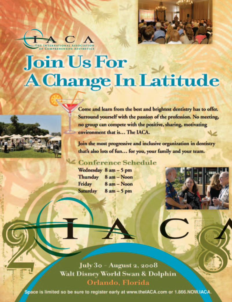
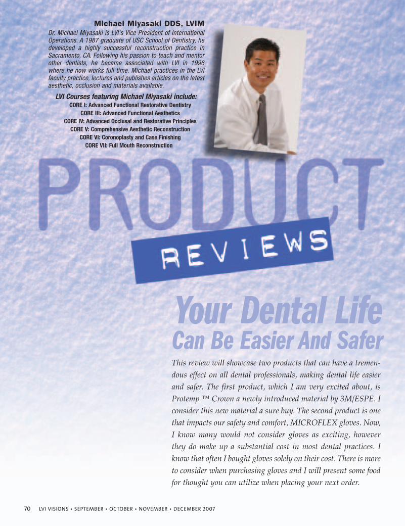
70 LVI VISIONS • SEPTEMBER • OCTOBER • NOVEMBER • DECEMBER 2007
Michael Miyasaki DDS, LVIM
Your Dental Life Can Be Easier And Safer
Dr. Michael Miyasaki is LVI's Vice President of InternationalOperations. A 1987 graduate of USC School of Dentistry, hedeveloped a highly successful reconstruction practice inSacramento, CA. Following his passion to teach and mentorother dentists, he became associated with LVI in 1996where he now works full time. Michael practices in the LVIfaculty practice, lectures and publishes articles on the latestaesthetic, occlusion and materials available.
LVI Courses featuring Michael Miyasaki include: CORE I: Advanced Functional Restorative Dentistry
CORE III: Advanced Functional AestheticsCORE IV: Advanced Occlusal and Restorative Principles
CORE V: Comprehensive Aesthetic ReconstructionCORE VI: Coronoplasty and Case Finishing
CORE VII: Full Mouth Reconstruction
This review will showcase two products that can have a tremen-dous effect on all dental professionals, making dental life easierand safer. The first product, which I am very excited about, isProtemp ™ Crown a newly introduced material by 3M/ESPE. Iconsider this new material a sure buy. The second product is onethat impacts our safety and comfort, MICROFLEX gloves. Now,I know many would not consider gloves as exciting, howeverthey do make up a substantial cost in most dental practices. Iknow that often I bought gloves solely on their cost. There is moreto consider when purchasing gloves and I will present some foodfor thought you can utilize when placing your next order.
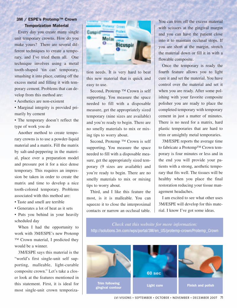
LVI VISIONS • SEPTEMBER • OCTOBER • NOVEMBER • DECEMBER 2007 71
3M / ESPE’s Protemp™ CrownTemporization Material
Every day you create many single
unit temporary crowns. How do you
make yours? There are several dif-
ferent techniques to create a tempo-
rary, and I’ve tried them all. One
technique involves using a metal
tooth-shaped ‘tin can’ temporary,
smashing it into place, cutting off the
excess metal and filling it with tem-
porary cement. Problems that can de-
velop from this method are:
• Aesthetics are non-existent
• Marginal integrity is provided pri-
marily by cement
• The temporary doesn’t reflect the
type of work you do
Another method to create tempo-
rary crowns is to use a powder-liquid
material and a matrix. Fill the matrix
by salt-and-peppering in the materi-
al, place over a preparation model
and pressure pot it for a nice dense
temporary. This requires an impres-
sion be taken in order to create the
matrix and time to develop a nice
tooth-colored temporary. Problems
associated with this method are:
• Taste and smell are terrible
• Generates a lot of heat as it sets
• Puts you behind in your heavily
scheduled day
When I had the opportunity to
work with 3M/ESPE’s new Protemp
™ Crown material, I predicted they
would be a winner.
3M/ESPE says this material is the
“world’s first single-unit self sup-
porting, malleable, light-curable
composite crown.” Let’s take a clos-
er look at the features mentioned in
this statement. First, it is ideal for
most single-unit crown temporiza-
tion needs. It is very hard to beat
this new material that is quick and
easy to use.
Second, Protemp ™ Crown is self
supporting. You measure the space
needed to fill with a disposable
measure, get the appropriately sized
temporary (nine sizes are available)
and you’re ready to begin. There are
no smelly materials to mix or mix-
ing tips to worry about.
Second, Protemp ™ Crown is self
supporting. You measure the space
needed to fill with a disposable mea-
sure, get the appropriately sized tem-
porary (9 sizes are available) and
you’re ready to begin. There are no
smelly materials to mix or mixing
tips to worry about.
Third, and I like this feature the
most, is it is malleable. You can
squeeze it to close the interproximal
contacts or narrow an occlusal table.
You can trim off the excess material
with scissors at the gingival margin
and you can have the patient close
into it to maintain occlusal stops. If
you are short at the margin, stretch
the material down or fill it in with a
flowable composite.
Once the temporary is ready the
fourth feature allows you to light
cure it and set the material. You have
control over the material and set it
when you are ready. After some pol-
ishing with your favorite composite
polisher you are ready to place the
completed temporary with temporary
cement in just a matter of minutes.
There is no need for a matrix, hard
plastic temporaries that are hard to
trim or unsightly metal temporaries.
3M/ESPE reports the average time
to fabricate a Protemp™ Crown tem-
porary is four minutes or less and in
the end you will provide your pa-
tients with a strong, aesthetic tempo-
rary that fits well. The tissues will be
healthy when you place the final
restoration reducing your tissue man-
agement headaches.
I am excited to see what other uses
3M/ESPE will develop for this mate-
rial. I know I’ve got some ideas.
Trim following gingival contour
Light cure
Check out this website for more information:http://solutions.3m.com/wps/portal/3M/en_US/protemp-crown/Protemp_Crown
Finish and polish
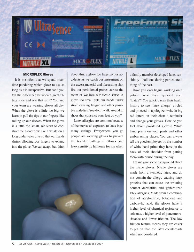
72 LVI VISIONS • SEPTEMBER • OCTOBER • NOVEMBER • DECEMBER 2007
MICROFLEX GlovesIt is not often that we spend much
time pondering which glove to use as
long as it is inexpensive. But can’t you
tell the difference between a great fit-
ting shoe and one that isn’t? You and
your team are wearing gloves all day.
When the glove is a little too big, we
learn to pull the tips to our fingers, like
rolling up our sleeves. When the glove
is a little too small, we learn to con-
strict the blood flow like a whale on a
long underwater dive so that our hands
shrink allowing our fingers to extend
into the glove. We can adapt, but think
about this: a glove too large invites ac-
cidents as we catch our instrument on
the excess material and like a sling shot
fire our periodontal probes across the
room or we lose our tactile sense. A
glove too small puts our hands under
strain causing fatigue and other possi-
ble maladies. You don’t walk around in
shoes that constrict your feet do you?
Latex allergies are common because
of the increased exposure to latex in so
many settings. Everywhere you go
people are wearing gloves to prevent
the transfer pathogens. Gloves and
latex sensitivity hit home for me when
a family member developed latex sen-
sitivity - balloons during parties are a
thing of the past.
Have you ever begun working on a
patient who then queried you,
“Latex?”You quickly scan their health
history to see ‘latex allergy’ circled
and proceed to apologize, write in big
red letters on their chart a reminder
and change your gloves. How do you
feel about powdered gloves? White
hand prints on your pants and other
embarrassing places. You can always
tell the good employees by the number
of white hand prints they have on the
back of their shoulder from patting
them with praise during the day.
Let me give some background about
the nitrile gloves. Nitrile gloves are
made from a synthetic latex, and do
not contain the allergy causing latex
proteins that can cause the irritating
contact dermatitis and generalized
latex allergies. Made from a combina-
tion of acrylonitrile, butadiene and
carboxylic acid, the gloves have a
higher level of chemical resistance to
solvents, a higher level of puncture re-
sistance and lower friction. The low
friction feature means they are easier
to put on than the latex counterparts
when not powdered.
74 LVI VISIONS • SEPTEMBER • OCTOBER • NOVEMBER • DECEMBER 2007
Common sense would suggest to
find a powder-free, non-latex glove
that fits well and offers great tactile
feeling when holding the small items
we work with in dentistry. Some old
nitrile gloves I have used in the past
fit terribly and I hated using them.
Today I use the MICROFLEX
FreeForm SE (Standard Exam) pow-
der-free, nitrile gloves. They are
easy to put on and are extremely
comfortable. I can wear them during
long procedures and my hands do
fine, and they come in my favorite
color, blue.
Factors to consider when selecting
gloves should be:
• latex or no latex
• powder or powder-free
• the overall quality of the product
• the fit on your hand
• the cost
MICROFLEX highlights the following features:
• Exclusive, patented* "super-soft"
formulation feels like latex
• Micro-textured fingertips for a se-
cure grip in wet and dry conditions
• Modulated elasticity allowing a full
range of motion helping to minimize
hand stress
• Polymer-coated to aid in quick
and easy donning, even in wet con-
ditions
One potential drawback is cost.
Nitrile gloves are, in general, more
expensive than latex gloves, but I
prefer not dealing with the risks of
causing a latex reaction in my pa-
tients or myself. You and your pa-
tients deserve the best health.
I would encourage you to go to
http://www.microflex.com/products/c
atalog.asp and review the several dif-
ferent types of gloves offered by MI-
CROFLEX. I’m sure there is a glove
in their line that will suit you well.
ConclusionI hope you try Protemp™ Crown a
new material by 3M/ESPE and con-
sider the importance of the gloves
you wear every day. MICROFLEX
has an extensive line of products that
I am sure will fit your needs. Our
work is demanding and I would en-
courage you to use products that
make your days both easier and more
comfortable. You deserve both,
hence, these recommendations. As
always I enjoy your feedback and
urge you to continue to submit infor-
mation on the products that are work-
ing well for you.
Take at look at these products. After you do I’d love to hear your comments. Your suggestions always are welcome – please send them to me at: [email protected]
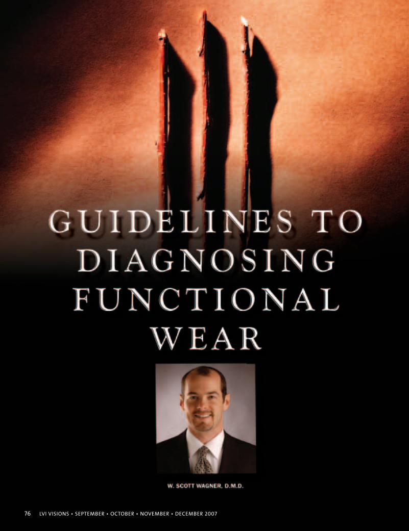
76 LVI VISIONS • SEPTEMBER • OCTOBER • NOVEMBER • DECEMBER 2007
LVI VISIONS • SEPTEMBER • OCTOBER • NOVEMBER • DECEMBER 2007 77
Given the rapid changes that are occurring in
dentistry today—in technology, vision, es-
thetics, diagnosis, and treatment—I am
aware of the need for a simple, user friendly
way of communicating the condition of a pa-
tient’s mouth upon examination. Beyond the
basics of occlusal classes, skeletal classes, TMJ classes, I have
realized there are very few ways to communicate the health of
the actual dentition.
In a comprehensive new patient exam, dentists will chart caries,
periodontal disease, missing teeth, and restorations. Further details
range from fractures, recession, mobility, and abfractions, to fur-
cations, wear facets, and even Shimbashi measurements.
Today, the ability to communicate the status of a patient’s mouth
clearly and concisely in our peer group is somewhat difficult. It is
even more challenging to express this to patients. Fractures, wear
facets, and loss of vertical are components that are sometimes hard
to see and show. Even with x-rays, periodontal charting, digital
photos, and models, there is a lot of information not entirely evi-
dent without direct examination.
Thus appears the void. I created and have been using this system
for about two years now, and have not found many discrepancies
or crossover. It is NOT designed to be a strict system, but rather a
system that allows for a general classification and easy communi-
cation. Think of this as a system to evaluate the functional wear
status of the teeth regardless of any occlusal class, skeletal class,
periodontal condition or caries.
It is NOT designed to be a strict system,
but rather a system that allows for a general
classification and easy communication.
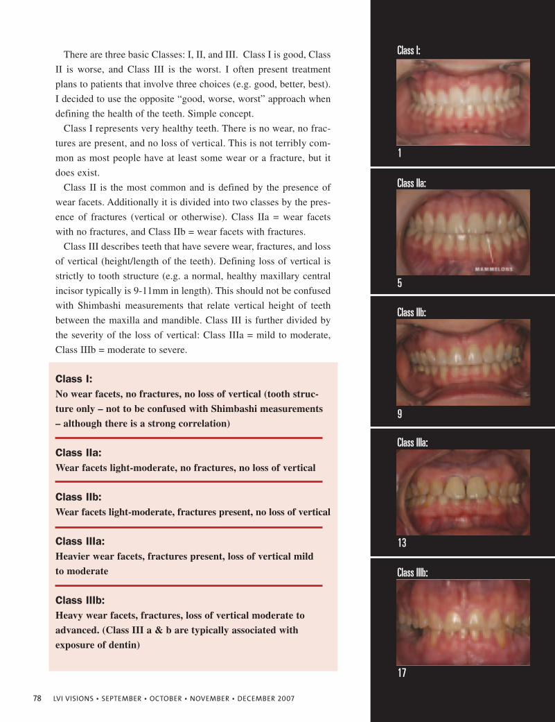
78 LVI VISIONS • SEPTEMBER • OCTOBER • NOVEMBER • DECEMBER 2007
There are three basic Classes: I, II, and III. Class I is good, Class
II is worse, and Class III is the worst. I often present treatment
plans to patients that involve three choices (e.g. good, better, best).
I decided to use the opposite “good, worse, worst” approach when
defining the health of the teeth. Simple concept.
Class I represents very healthy teeth. There is no wear, no frac-
tures are present, and no loss of vertical. This is not terribly com-
mon as most people have at least some wear or a fracture, but it
does exist.
Class II is the most common and is defined by the presence of
wear facets. Additionally it is divided into two classes by the pres-
ence of fractures (vertical or otherwise). Class IIa = wear facets
with no fractures, and Class IIb = wear facets with fractures.
Class III describes teeth that have severe wear, fractures, and loss
of vertical (height/length of the teeth). Defining loss of vertical is
strictly to tooth structure (e.g. a normal, healthy maxillary central
incisor typically is 9-11mm in length). This should not be confused
with Shimbashi measurements that relate vertical height of teeth
between the maxilla and mandible. Class III is further divided by
the severity of the loss of vertical: Class IIIa = mild to moderate,
Class IIIb = moderate to severe.
Class I:No wear facets, no fractures, no loss of vertical (tooth struc-
ture only – not to be confused with Shimbashi measurements
– although there is a strong correlation)
Class IIa:Wear facets light-moderate, no fractures, no loss of vertical
Class IIb:Wear facets light-moderate, fractures present, no loss of vertical
Class IIIa:Heavier wear facets, fractures present, loss of vertical mild
to moderate
Class IIIb:Heavy wear facets, fractures, loss of vertical moderate to
advanced. (Class III a & b are typically associated with
exposure of dentin)
Class I:
1
5
9
13
17
Class IIa:
Class IIb:
Class IIIa:
Class IIIb:
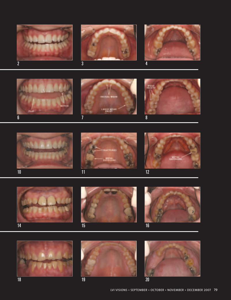
LVI VISIONS • SEPTEMBER • OCTOBER • NOVEMBER • DECEMBER 2007 79
2 3 4
6 7 8
10 11 12
14 15 16
18 19 20
So we have defined three main categories and their sub-groups.
We now have a tool to communicate the relative condition of the
dentition. What additional information can we gain by being aware
of a patient’s Functional Wear Class? Given we know that enamel
is the hardest natural substance in the body, we can also surmise
that the worse the Functional Wear Classification, the more we
must be aware of the forces that potentially caused each condition.
The most obvious problem contributing to fractures, wear, and
loss of vertical, is a parafunctional grinding habit (day and/or
night). But we must not forget to leave out a host of potential caus-
es other than bruxism: medications (most commonly anti-depres-
sants), a high-stress lifestyle, chronic ice or gum chewing, acid re-
flux, drug use, old amalgam restorations (which expand or contract
depending on the % mix of alloys), nail biting, trauma, caries, poor
airway development which can lead to abnormal archform devel-
opment, and abnormal posture which can cause strain on muscles
of the jaws, head, and neck, etc. The list of contributing factors is
long. A critical aspect to performing a good service for someone is
to be able to ask the question, “What caused the teeth to be in this
condition?” Regardless of how simple or complex you anticipate a
case being, if you do not ask questions about how they arrived in
their condition, you may create problems for yourself in the future.
A thorough exam that identifies any warning signs creates a non-
threatening informative session for the doctor and the patient. Doc-
umentation of a patient's Functional Wear Class can open a discus-
sion where patients can acknowledge and own their problem. Your
job is to provide solutions and choices. Often times when I am
doing a new patient exam or a cosmetic consult, I find myself say-
ing, “Mr. Jones, I just want to let you know about the severe wear
and fractures that are present in your mouth. You can choose any
treatment option you like, but unless we attempt to treat the cause
of the damage, I cannot assure that you will not continue to experi-
ence similar future problems even if you wear a biteguard.”
This simple statement and acknowledgement with a written re-
lease prior to treatment is a powerful tool I use especially when pa-
tients deny treatment that would correct the cause. They confirm
that we are attempting to maintain their comfort and stability in a
bite position that I have already told them is pathologic. Addition-
ally, if you use high quality intra- and extra-oral photos during
case and treatment presentation, the patient’s understanding of
what we see as practitioners becomes even more powerful.
The purpose of this five second diagnostic tool is communica-
tion. Patients will be impressed by your thoroughness, more con-
fident in your ability, and as I have found, are grateful because I
80 LVI VISIONS • SEPTEMBER • OCTOBER • NOVEMBER • DECEMBER 2007
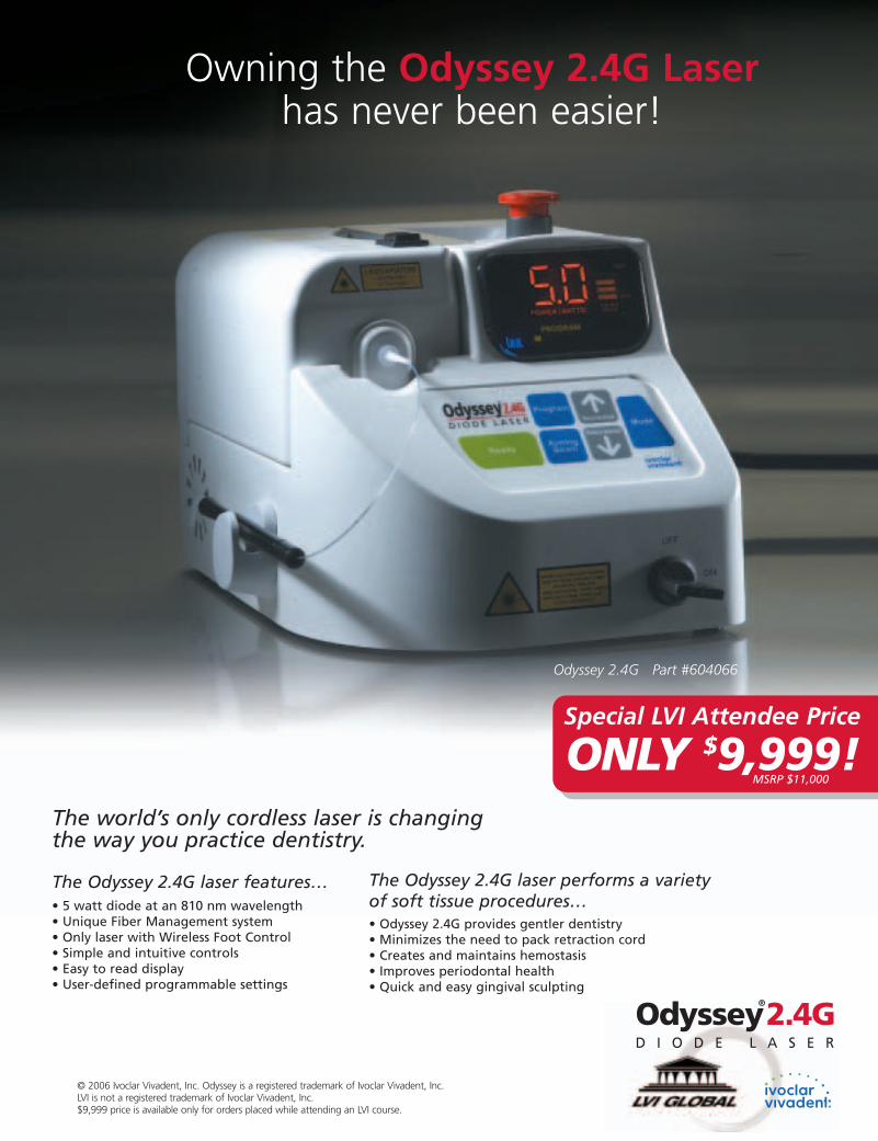
© 2006 Ivoclar Vivadent, Inc. Odyssey is a registered trademark of Ivoclar Vivadent, Inc. LVI is not a registered trademark of Ivoclar Vivadent, Inc.$9,999 price is available only for orders placed while attending an LVI course.
Owning the Odyssey 2.4G Laserhas never been easier!
The world’s only cordless laser is changingthe way you practice dentistry.
The Odyssey 2.4G laser features… • 5 watt diode at an 810 nm wavelength • Unique Fiber Management system• Only laser with Wireless Foot Control• Simple and intuitive controls• Easy to read display• User-defined programmable settings
The Odyssey 2.4G laser performs a varietyof soft tissue procedures…• Odyssey 2.4G provides gentler dentistry • Minimizes the need to pack retraction cord• Creates and maintains hemostasis • Improves periodontal health• Quick and easy gingival sculpting
Odyssey 2.4G Part #604066
Odyssey®
2.4GD I O D E L A S E R
Special LVI Attendee Price
ONLY $9,999!MSRP $11,000
am often the first to communicate why their mouths are in such
condition. In my experience, this type of doctor-patient relation-
ship has opened doors and led to patients moving forward with
more comprehensive treatment options.
I must clarify; the best way to diagnose these Functional Wear
Classes is by direct vision. Accurate models can show wear facets
and loss of vertical/tooth structure; however, they do not show
many of the marginal ridge hairline fractures that are often present.
It has been my experience that these types of fractures are critical
to diagnose as they are often the precursors of decay, large frac-
tures, pulp exposure or worse. High resolution intra-oral photos
are helpful, but one must be able to ‘zoom in’ on the teeth to be
able to see these fractures photographically. Remember not to con-
fuse normal developmental grooves as one of these hairline frac-
tures. To show how easy these guidelines are used, I will walk
through the five examples that I have provided and how each one
is defined.
Functional Wear Class I: The first things to notice are the rem-
nants of the mammelons on 9 & 10 (photo 2). The mammelons
have no diagnostic value here, but I know these two teeth are in
pristine condition. There are no wear facets on the canines or other
anterior teeth indicative of bruxing or other pathologic occlusal
forces. Examination of the posterior teeth reveals no wear facets,
fractures, or other occlusal pathology.
Functional Wear Class IIa: Starting with the anterior teeth, the in-
cisal wear on #’s 8, 9, 24, and 25 is obvious. Take care to notice the
notch on the disto-incisal of #11, and the flattened incisal of #27. It
is also fun to notice the perfect mammelons on #23 (it is completely
out of occlusion); however, diagnosis is based on the whole mouth,
not one tooth. Photos 6 and 7 show the incisal wear on the anteriors.
Make note of the large wear facet on the ML cusp of the first molar
(right side of photo 7). The intra-oral mandibular photo shows large
wear facets on the first and second molars (left side of photo 8). No
vertical fractures are present. There is no loss of vertical.
Functional Wear Class IIb: All the conditions in the case are es-
sentially the same as Class IIa (e.g. anterior and posterior wear
facets are readily evident in all images 9-12). I even pay attention
to the chipped porcelain on the occlusal of the maxillary and
mandibular first molars which has worn through to the metal sub-
structure. However, the key diagnostic difference between Class
IIa and IIb is the presence of vertical fractures. Some are hairline,
others are profound in this case. You can easily see the mesial frac-
tures of the maxillary 1st and 2nd premolars in the upper left of
photo 11. There is no loss of vertical.
Patients will be
impressed by your
thoroughness,
more confident
in your ability,
and as I have found,
are grateful because
I am often
the first to
communicate why
their mouths
are in such
condition.
82 LVI VISIONS • SEPTEMBER • OCTOBER • NOVEMBER • DECEMBER 2007
84 LVI VISIONS • SEPTEMBER • OCTOBER • NOVEMBER • DECEMBER 2007
Functional Wear Class III (a & b): Encountering a patient with
this amount of pathology should raise an eyebrow at the very least
(see photos 13-16 & 17-20). Both cases have loss of vertical
(height of tooth structure)—the key diagnostic point in a Func-
tional Wear Class III patient. Both have fractures and wear facets.
The only difference is the severity of wear (IIIb being obviously
greater). In my clinical experience, both IIIa & b are associated
with profound exposure of dentin. Many times these patients have
lost teeth. Vertical fractures are sometimes so significant, that de-
spite a patient’s efforts to save them via root canal & crown, they
lose the tooth. Without proper diagnosis and acknowledgement of
their condition, these patients routinely develop anxiety, fear, and
mistrust of the dental profession. Any money they have spent to
save teeth has been, as I often hear, “all for nothing.”
Other times, these Class III patients have no pain or discomfort.
They only present with severe wear, micro-fractures, and lots of
exposed dentin. They typically have a type-A, intense personality.
Diagnosis and communication with these patients is key, especial-
ly if they opt against ideal treatment. If left undiagnosed, that tooth
that you restored which subsequently broke is your fault and not
the patient’s.
It all comes back to vision, taking the five seconds for diagnosis,
and then communication. Among our peer group, a discussion re-
garding the challenge of treating a Functional Wear Class IIIb pa-
tient now paints a very vivid image. Obviously, using that type of
language with a patient is non-productive, but the mere fact that you
have a diagnosis of their condition allows one to use layman terms
to open the discussion. The goal is that patients own their condition.
I did not see a need for this type of diagnostic system until I
started learning about, and practicing, aesthetic neuromuscular
dentistry. The rewarding aspect for me is that this system applies
beyond neuromuscular dentistry. It is a simple system that can be
implemented easily and used by all specialties in dentistry. The
only requirements are vision and time. A dentist must have the vi-
sion (or experience) to see conditions as they appear, and it only
takes a few seconds to diagnose and chart.
The implications of this tool and how it can immediately im-
prove communication between all practitioners in dentistry is the
primary advantage. I believe that this is a small step that we can all
take to elevate our communication and performance in dentistry.
W. Scott Wagner, D.M.D., P.A.
www.jaxsmile.com
It is a simple
system that can be
implemented easily
and used by all
specialties in
dentistry. The only
requirements are
vision and time.
Scott Wagner graduated with Honorsfrom the University Of Florida CollegeOf Dentistry in 1998. He maintains aprivate practice in Jacksonville, Flori-da dedicated to Aesthetic & Neuro-muscular Dentistry. Scott began hisjourney at LVI in early 2003 and com-pleted the Graduate core curriculumin 2005. He attributes much of hissuccess to the clinical skills, vision,and passion that is LVI.
Built to celebrate the engineering and science of its age, the Eiffel Tower,
constructed in 1889, is the world’s most widely recognized and visited
landmark — attracting over 6 million visitors a year. It’s considered to be
one of the most remarkable pieces of architectural art today. Zimmer Dental
gives you the power to create something remarkable with its Tapered
Screw-Vent® Implant System. Delivering fully integrated single-stage and
two-stage solutions, the system gives you the flexibility to make a choice
at the time of surgery. Whether you choose the new Zimmer®One-Piece
Implant, or the renowned Tapered Screw-Vent or AdVent® Implants with
off-the-shelf or custom abutments, you can have total confidence that all
the pieces are in sync for the best possible patient outcome.
Zimmer Dental. Confidence in your hands.™
Remarkable things are created with all the right pieces.When a system is in sync, there are no limitations.
©2007 Zimmer Dental Inc. All rights reserved. 7449, Rev. 6/06.www.zimmerdental.com
Create something remarkable today. Visit us online atwww.zimmerdental.com/MC0381.asp or call us at +1 760 929 4300 for more information. Please reference code MC0381.
To receive our eNews visit us atwww.zimmerdental.com/news_eNewsLetterSignUp.aspx
www. aurumgroup.com
®
1-800-661-1169
Dr. Lynn Locklear, CEO Life-Enhancing DentistryWashington, DC
“For exceptional esthetics caseafter case, we rely on AurumCeramic. Their craftsmanshipand commitment to excellencetruly makes a difference in de-livering ‘life-changing’ cosmetic results.”
Create “scintillatingsmiles” on every casewith Aurum Ceramic.
Dentistry courtesy of Dr. Lynn Locklear. Restorations fabricated by Aurum Ceramic. Photography courtesy of Terrence Carter.
Call our Advanced Esthetic Hotline
LVI Designated Laboratory since its inception.