senior thesis draft - notre dame sites
TRANSCRIPT
Biochemical Features of Anti-Amyloidogenic Activity of Gastrokine-1
Cayla Bales
Submitted in partial fulfillment of the requirements for the Glynn Family Honors Program at the
University of Notre Dame
Advised by Dr. David Boone
Department of Biological Sciences
__________________
April 5th, 2019
ABSTRACT
Gastrokine 1 (Gkn1) is an 18 kDa protein produced and secreted into the
lumen of the stomach. It is stable and protease-resistant, which allows it to resist
degradation in the gastrointestinal (GI) tract. To examine the function of Gkn1 in
vivo, Gkn1-/- mice were previously generated. Gkn1-/- mice are healthy and have a
normal lifespan. However, Gkn1-/- mice have markedly reduced body fat compared
to wild type (WT) littermates, and they are resistant to weight gain on a high fat diet.
Gkn1-/- mice are not diabetic, have normal appetite and physical activity, and do not
malabsorb calories. We hypothesize that Gkn1 influences phenotype through
regulation of the intestinal microbiota. Gkn1 prevents a fundamental amyloid function
of bacteria, namely biofilm formation. This study investigates the biochemical
features of Gkn1 that mediate its anti-amyloidogenic effects, in particular its
BRICHOS domain, which is associated with amyloid fiber binding. For in vitro
experimentation, a Kluyveromyces lactis yeast model was created for generation of
large quantities of Gkn1. Site-directed mutagenesis was then used to change
specific conserved amino acids in the BRICHOS domain for subsequent generation
of mutant protein. To compare the abilities of Gkn1 and mutant Gkn1 to inhibit
biofilm formation, biofilm assays were performed with bacterial strain LF82.
Preliminary results suggest that certain amino acids in the BRICHOS domain, while
conserved in evolution, are not critical for the anti-amyloidogenic activity of Gkn1.
We are currently testing other mutant forms of Gkn1 to further elucidate the
biochemistry of this function.
2
Table of Contents 1. INTRODUCTION
1.1. Obesity 1.2. Metabolism and the Gut Microbiota 1.3. Biofilms and Amyloid Fibers 1.4. The BRICHOS Domain and Gastrokine-1 1.5. Site-Directed Mutagenesis
2. MATERIALS AND METHODS
2.1. Site-directed mutagenesis 2.2. Generation of yeast expression model 2.3. Polymerase Chain Reaction 2.4. Gastrokine-1 production and purification 2.5. Western blot 2.6. Ammonium sulfate protein precipitation and Coomassie stain 2.7. Biofilm assay
3. RESULTS 3.1. Confirmation of successful site-directed mutagenesis 3.2. Confirmation of mGkn1 generation 3.3. Gastrokine-1 inhibits biofilm formation 3.4. mGkn1 mutant 383 shows similar level of biofilm inhibition as mGkn1
4. DISCUSSION AND FUTURE DIRECTIONS 5. ACKNOWLEDGMENTS 6. REFERENCES
3
INTRODUCTION 1.1 Obesity
The worldwide obesity epidemic has been designated as one of the greatest
public health challenges of our time. Recent studies indicate that approximately one-
third of the United States population is obese, which is defined by a body mass
index (BMI) of greater than 30.0 (Flegal et al. 2010). This excess weight has
negative consequences for the health of individuals and the economic health of the
nation, since physical impairments and medical conditions associated with obesity
decrease the labor supply (Renna and Thakur 2010). The cost of obesity is also
reflected in healthcare expenditures. Between 0.7% and 2.8% of worldwide
healthcare spending is attributable to the condition, since obesity is associated with
various comorbidities such as cardiovascular disease, type 2 diabetes, stroke, and
certain types of cancer (Shea et al. 2012). In 2016, the average annual medical
costs of an obese person were approximately 42 percent higher than a person of
normal weight (Goforth 2016). This gap will likely continue to widen, since per capita
health care spending has grown faster for individuals in above-normal weight
categories in recent decades (Duchovney and Baker 2010).
The obesity problem has led to research efforts in both prevention and
treatment. Many doctors prescribe diet and exercise as a solution, yet studies have
shown that long term, sustained weight loss is difficult to maintain (Bray 2012). In
addition, behavioral interventions frequently fail to reach certain segments of the
population, such as those with low incomes (Anthes 2014). Other treatment options
include surgery and weight loss drugs. During bariatric surgeries, doctors shrink the
4
size of the stomach and reroute digestion past the early parts of the small intestine,
which restricts intake and limits absorption of food (Weight-control Information
Network 2011). Despite the general effectiveness of the procedures, they are costly
and risky, and some patients still suffer from weight regain (Ghaferi and Varban
2018).
Prescription drugs may be a practical solution to the obesity epidemic.
Researchers have found that weight loss drugs, when used in combination with
lifestyle interventions, caused patients to lose 3-9% more weight than patients
receiving a placebo (Yanovski and Yanovski 2014). However, medications can have
serious side effects. Two popular appetite-suppressants, fenfluramine and
dexfenfluramine, were pulled from the market in 1997 after being linked to the
development of valvular heart disease and primary pulmonary hypertension
(National Institute 2004). Few drugs are currently approved by the FDA. For years,
the mainstays of treatment were phentermine, an amphetamine that suppresses
appetite, and orlistat, a lipase inhibitor that interferes with the body’s ability to absorb
fat (Shonders et al. 2018). In 2012, two new drugs were approved: a
phentermine/topiramate extended release capsule (Qsymia) and lorcaserin, which
promotes satiety by activating a particular serotonin receptor in the brain (Anthes
2014). Most recently, the FDA approved naltrexone sustained release
(SR)/bupropion SR and liraglutide in 2014 (Shonders et al. 2018). Each of these
medications comes with a variety of adverse effects and risks. For instance,
topiramate causes the development of oral clefts in fetuses of pregnant patients,
while liraglutide has been linked to acute and chronic pancreatitis (The Lancet 2012;
5
Jensen, Saha, and Steinberg 2015). While no drug is expected to be a magic bullet,
it is obvious that identification of new drug targets is vital. One promising area is the
gut microbiome, which has been shown to play a critical role in host adiposity and
energy metabolism (Martin et al. 2015, Mazidi et al. 2016).
1.2 Metabolism and the Gut Microbiota
Microbiota transplantation experiments have connected the composition of
the gut microbiota with obesity. These studies use germ-free mice, which have no
contact with bacteria and are raised in a sterile environment. When the germ-free
mice are colonized with microbes from obese donors, they gain weight and fat mass
without a significant increase in chow consumption (Turnbaugh et al. 2006). Germ-
free mice are also protected from diet induced obesity when placed on a high-fat,
high-sugar diet, indicating that alterations in the gut microbiome are an important
player in the fat accumulation induced by a Western-style diet (Backhed et al. 2007).
Furthermore, weight loss and metabolism changes after bariatric surgery may be
partly mediated by changes in the microbiota (Tremaroli et al. 2015). Germ-free mice
were transplanted with fecal microbiota of obese patients and bariatric surgery
patients, and the mice receiving the bacteria from patients post bariatric surgery
gained significantly less body fat (Tremaroli et al. 2015).
Gut microbes influence host energy balance by impacting energy harvest and
caloric extraction from the diet (Backhed et al. 2004). Some dietary calories come
from indigestible carbohydrate fibers and proteins, and bacteria in the gut are able to
break down these macromolecules into smaller particles that the host can absorb.
The bacteria can also affect obesity by producing metabolites. The breakdown of
6
dietary fiber produces short chain fatty acids (SCFAs) such as propionate and
butyrate, which promote metabolic benefits in energy homeostasis by activating
intestinal gluconeogenesis (De Vadder et al. 2014). Bacteria also metabolize primary
bile acids into secondary bile salts, which may mitigate against weight gain and
benefit host metabolic health (Sayin et al. 2013).
Recent studies have analyzed the differences in gut flora between obese and
lean individuals. While the microbiota varies between individuals, the vast majority of
bacteria are from the phyla Firmicutes and Bacteriodetes. A higher ratio of
Firmicutes:Bacteriodetes is associated with obesity, while the opposite is associated
with leanness, indicating that the composition of the microbiome impacts host
phenotype (Turnbaugh et al. 2006). Some studies have aimed to determine which
specific bacterial family, genera, or species may be linked to obesity (Bianchi et al.
2019). The genus Bifidobacterium has been shown to be more abundant in lean
subjects compared to overweight groups and negatively correlate with visceral fat
(Reyes et al. 2016; Aoki et al. 2017). Other analyses divide patients into two
microbial enterotypes: the Prevotella P-type or Bacteriodetes B-type. It was shown
that the Prevotella enterotype is predominant in individuals consuming lots of
carbohydrates and fiber, while the Bacteriodetes enterotype was more common in
individuals consuming more protein and animal fat (Christensen et al. 2018). This
study, among others, provides evidence that dietary ingredients can modulate the
microbiota.
Adjusting the gut flora with diet or drugs could induce healthy changes in
patients with metabolic dysregulation or pathogenic intestinal conditions. A large
7
number of non-antibiotic human-targeted drugs have been shown to impact specific
bacterial strains, indicating that some drugs could be repurposed as specific
microbiome modulators (Maier et al. 2018). Furthermore, metformin, a common
medication for treatment of type II diabetes, has already been observed to positively
influence host metabolism through the promotion of butyrate-producing taxa
(Forslund et al. 2015). Medications could also be used to target entire microbial
communities and higher order bacterial structures known as biofilms.
1.3 Biofilms and Amyloid Fibers
Biofilms are communities of surface-adherent organisms that are embedded
in complex extracellular matrices of self-produced polysaccharides. The colonies
adhere to surfaces, and many microbes have developed systems to bind to mucin,
the glycoprotein that is the major component of mucosa (de Vos 2015). The bacterial
populations undergo frequent horizontal gene transfer and behave similarly to
eukaryotic cells or multifaceted ecosystems; this cooperation allows the bacterial
cells to grow and survive in hostile environments (Costerton et al. 1995). As a result,
biofilms are inherently resistant to both antimicrobial agents and host defenses. A
particular problem is their formation on therapeutic medical devices like protheses
and catheters, which leads to persistent bacterial infections (Fux 2003).
Furthermore, many recent studies have associated biofilm formation with disease
states such as bacterial endocarditis, cystic fibrosis, and the two main types of
inflammatory bowel disease (IBD): ulcerative colitis and Crohn’s disease (Hall-
Stoodley 2004). Studies have shown that patients with IBD have higher
concentrations of adherent mucosal bacteria, indicating that the presence of
8
intestinal biofilms may play a role in disease pathogenesis (Swidinski et al. 2005).
While the general composition of the microbiota is important, the spatial organization
of the gut flora and the presence of biofilm structures may have an additional impact
on microbiome function and host phenotype.
One crucial component of microbial biofilms is amyloid fibers, which are
polymeric fibrils composed of folded b-sheets stacked perpendicularly. While they
are frequently associated with protein misfolding and neurogenerative diseases such
as Alzheimer’s and Parkinson’s, ‘functional’ amyloids are manufactured by various
organisms to fulfill physiological functions (Blanco et al. 2011). These common
building block structures are resistant to protease digestion and denaturation, so
bacteria use them as structural materials and adhesions, as well as for protection
against host defensive mechanisms (Schwartz 2013). Amyloid fibers, as the main
proteinaceous component of biofilms, may also play a role in the pathogenesis of
diseases. It has been suggested that leakage of microbiome amyloids through
compromised gastrointestinal tract or blood-brain barriers could contribute to
neuroamyloidogensis and promote the characteristic protein aggregation in
Alzheimer’s disease (Zhao and Lukiw 2015). On the other hand, amyloids could be
therapeutic candidates. Oppong and coworkers showed that presence of curli fibers
from the microbiota resulted in the production of an immunomodulatory cytokine that
reduced inflammation in a mouse colitis model (2015). Finally, amyloids could serve
as targets in the fight against biofilms. Several molecules that inhibit amyloid fiber
formation and interfere with biofilm production or assembly are under investigation
(Taglialegna et al. 2016). If these compounds have the potential to modulate

9
microbial biofilms and microbiota organization, they could consequently impact host
phenotype.
1.4 The BRICHOS domain and Gastrokine-1
Certain protein domains are also known to have anti-amyloidogenic
properties. One such domain is the BRICHOS domain, a domain of ~ 100 amino
acids that was first described in 2002 (Sánchez-Pulido, Devos, and Valencia 2002).
Its name comes from three proteins: Bri2, chondromodulin-I, and prosurfactant
protein C (proSP-C), which are members of the diverse superfamily of BRICHOS-
containing proteins including the gastrokines, tenomodulins, arencins, and the group
C proteins (Knight et al. 2013). The proteins in this superfamily share a common
overall architecture and secondary structure (figure 1), despite the fact that there is
low amino acid sequence conservation within the BRICHOS domain (Willander et al.
2011). However, two of the three amino acids conserved across the entire family are
cysteines that form a disulfide bridge present in all proteins (Willander et al. 2012).
After identification, it was proposed that the BRICHOS domain may have a
chaperone-like function which prevents b-sheet aggregation and amyloid fibril
formation. Consequently, the domain has been linked to a variety of amyloid
diseases such as familial dementia and respiratory distress syndrome (Knight et al.
2013).
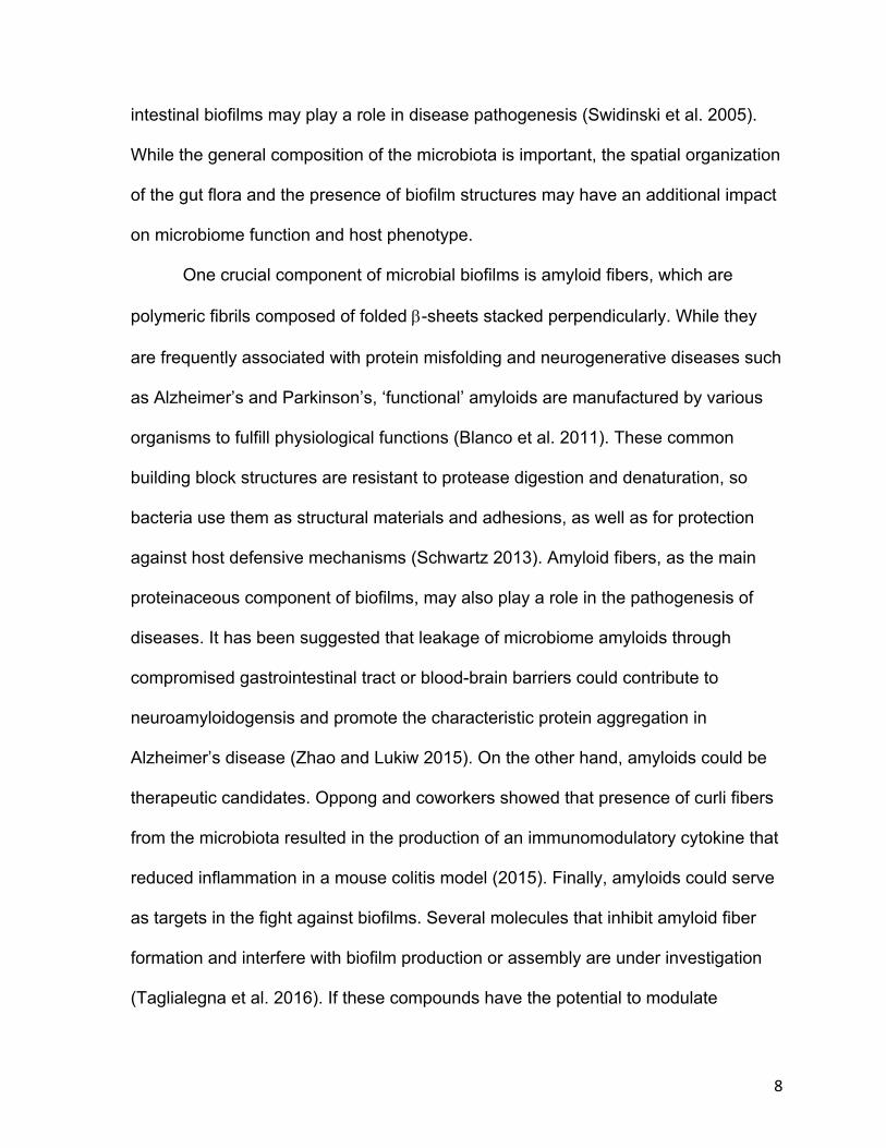
10
Figure 1. Ribbon diagram illustrating the fold of the proSP-C BRICHOS domain. a-helices and b-
sheets are labeled (Knight et al. 2015).
Like other members of the family, Gastrokine-1 (Gkn1), has been shown to
have anti-amyloidogenic properties that prevent the aggregation of amyloid fibrils
and therefore biofilm formation (Altieri et al. 2014). Gkn-1 was initially identified in
2003 and originally named antral mucosal protein (AMP)-18, since it is expressed in
the stomach antrum and is 18 kilodaltons in size (T.E. Martin et al. 2003). It is highly
conserved across mammals, including human and mouse, and is abundantly
expressed by the mucus secreting cells of the stomach antrum. Nearly 1-5% of
stomach RNA is thought to be Gkn1 (T.E. Martin et al. 2003; Oien et al. 2004). Two
main functions of Gkn1 have been identified. First, studies have shown that Gkn1 is
downregulated in gastric carcinoma, and it may play a role in gastric cancer
suppression through the promotion of apoptosis of cancerous cells (Oien et al. 2004;
Mao et al. 2012). Gkn1 also functions to protect the colonic mucosal barrier from
injury and promote intestinal epithelial cell growth. In one study, administration of
exogenous Gkn1 limited the extent of colonic mucosal injury in mice by increasing
the accumulation of tight junction proteins (Walsh-Reitz et al. 2005). While the
11
protective functions of Gkn1 suggest its importance for pathologies like inflammatory
bowel disease, the protein likely also plays a role in obesity.
To advance understanding of Gkn1, Dr. David Boone and coworkers use
Gkn1 knockout mice (Gkn1-/- mice) for in vivo experiments. Gkn1-/- mice are healthy
and have a normal lifespan, but they have markedly reduced body fat compared to
wild type (WT) mice and are resistant to weight gain on a high fat diet. It was found
that they are not diabetic, have normal appetite and physical activity, and do not
malabsorb calories. Instead, altered fat metabolism or storage may cause the
difference in adipose accumulation. Notably, WT mice given chicken anti-Gkn1
antibodies gained less fat and weight than control mice on a high fat diet, indicating
that blocking of Gkn1 activity can mediate against weight gain (Bakke 2016). As a
result, modulation of the activity of this protein could serve as a potential treatment
against obesity. Further understanding of Gkn1 is crucial for development of these
potential treatments. In this thesis, I investigate the mechanism of action of Gkn1.
We hypothesize that Gkn1 exerts its function via its anti-amyloidogenic properties,
which allow it to modulate the gut microbiome and influence adiposity. Two aims
were defined: 1) Successful production of Gkn1 for use in assays. 2) Investigation of
the impact of Gkn1 on biofilm formation in vitro.
1.5 Site Directed Mutagenesis
A third aim was also defined: 3) Use of site-directed mutagenesis to identify
critical amino acids and elucidate the importance of the BRICHOS domain on Gkn1
function. BRICHOS-domain containing proteins like Gkn1 have a similar pattern.
They all contain a short cytosolic region, a hydrophobic domain, a linker region, the
12
BRICHOS domain, and a C-terminal region (Knight et al. 2015). Previous research
on the tumor suppressive mechanism of Gkn1 utilized deletion and point mutant
forms of the protein in order to examine which regions were responsible for the
protein’s effects (Yoon et al. 2013). In the present study, point mutants are
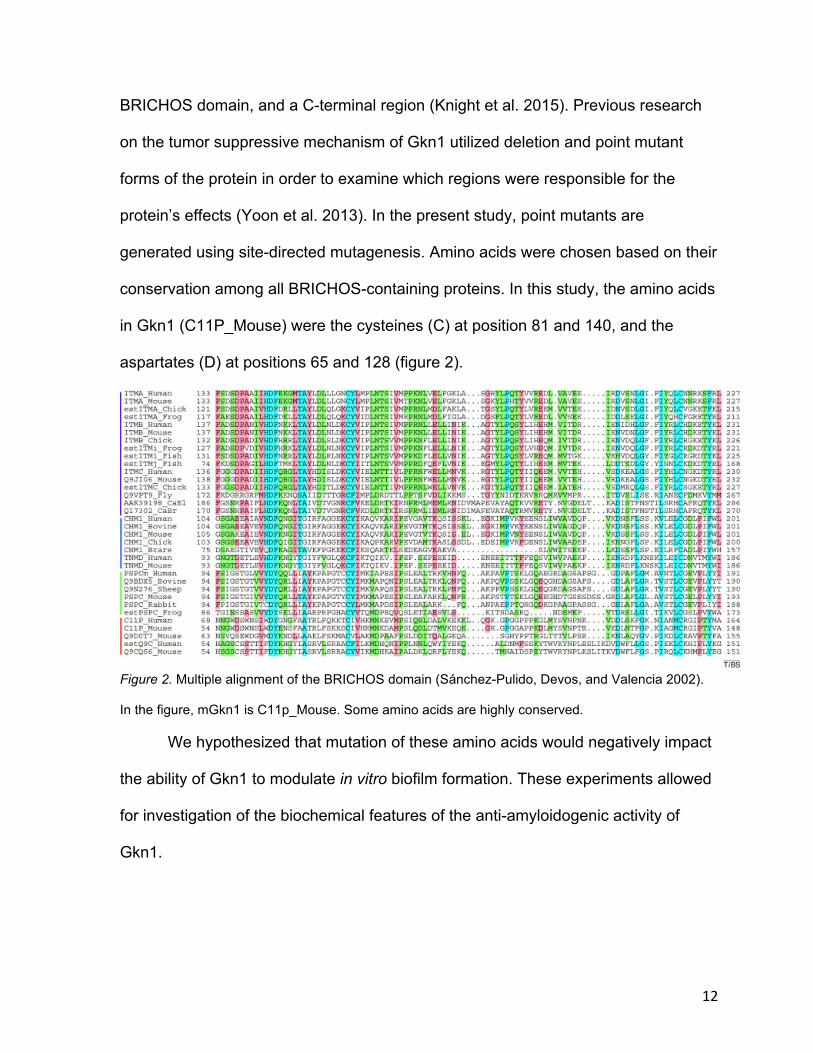
generated using site-directed mutagenesis. Amino acids were chosen based on their
conservation among all BRICHOS-containing proteins. In this study, the amino acids
in Gkn1 (C11P_Mouse) were the cysteines (C) at position 81 and 140, and the
aspartates (D) at positions 65 and 128 (figure 2).
Figure 2. Multiple alignment of the BRICHOS domain (Sánchez-Pulido, Devos, and Valencia 2002).
In the figure, mGkn1 is C11p_Mouse. Some amino acids are highly conserved.
We hypothesized that mutation of these amino acids would negatively impact
the ability of Gkn1 to modulate in vitro biofilm formation. These experiments allowed
for investigation of the biochemical features of the anti-amyloidogenic activity of
Gkn1.
13
MATERIALS AND METHODS
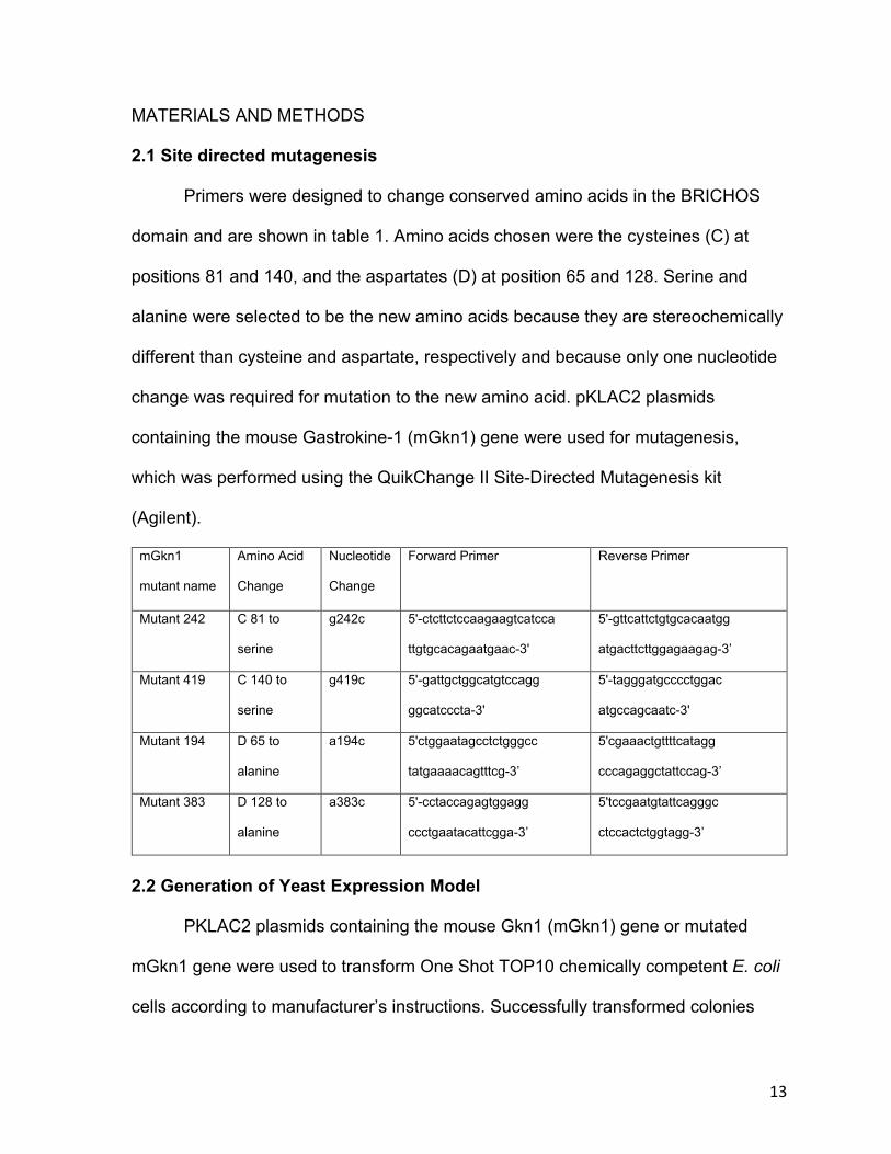
2.1 Site directed mutagenesis
Primers were designed to change conserved amino acids in the BRICHOS
domain and are shown in table 1. Amino acids chosen were the cysteines (C) at
positions 81 and 140, and the aspartates (D) at position 65 and 128. Serine and
alanine were selected to be the new amino acids because they are stereochemically
different than cysteine and aspartate, respectively and because only one nucleotide
change was required for mutation to the new amino acid. pKLAC2 plasmids
containing the mouse Gastrokine-1 (mGkn1) gene were used for mutagenesis,
which was performed using the QuikChange II Site-Directed Mutagenesis kit
(Agilent).
mGkn1
mutant name
Amino Acid
Change
Nucleotide
Change
Forward Primer Reverse Primer
Mutant 242 C 81 to
serine
g242c 5'-ctcttctccaagaagtcatcca
ttgtgcacagaatgaac-3'
5'-gttcattctgtgcacaatgg
atgacttcttggagaagag-3’
Mutant 419 C 140 to
serine
g419c 5'-gattgctggcatgtccagg
ggcatcccta-3'
5'-tagggatgcccctggac
atgccagcaatc-3'
Mutant 194 D 65 to
alanine
a194c 5'ctggaatagcctctgggcc
tatgaaaacagtttcg-3’
5'cgaaactgttttcatagg
cccagaggctattccag-3’
Mutant 383 D 128 to
alanine
a383c 5'-cctaccagagtggagg
ccctgaatacattcgga-3’
5'tccgaatgtattcagggc
ctccactctggtagg-3’
2.2 Generation of Yeast Expression Model PKLAC2 plasmids containing the mouse Gkn1 (mGkn1) gene or mutated
mGkn1 gene were used to transform One Shot TOP10 chemically competent E. coli
cells according to manufacturer’s instructions. Successfully transformed colonies
14
were cultured in LB broth (Bertani et al. 1951). and miniprepped using the
PureLinkTM HIPure Plasmid Miniprep Kit (Invitrogen) for sequencing. 3 µg pKLAC2
plasmid DNA was linearized using 30 units SacII in 50 µL 1X CutSmart Buffer at
37°C for 2 hours (NEB). Before transformation, restriction digests were purified
through the addition of an equal volume of phenol:chloroform (1:1, v/v) and isolation
of the top (aqueous) phase. This step was repeated with only chloroform before
addition of 10 µg glycogen and 1/10 volume sodium acetate. After mixing, an equal
volume of 100% isopropanol was added, and the mixture was incubated for 10min.
Tubes were microcentrifuged for 15min at 12,000 x g, then pellets were rinsed once
with 70% ethanol before resuspension in 25 µL TE buffer (10 mM Tris-HCl, 1 mM
EDTA, pH 8.0). Nanodrop was used to measure DNA concentration. K. lactis GG799
competent cells were then transformed according to the K. lactis Protein Expression
Kit (NEB). Colonies were maintained on YCB Agar Medium plates containing 5 mM
acetamide per kit protocol.
2.3 Polymerase Chain Reaction
Transformants were tested using polymerase chain reaction (PCR) to verify
integration of the pKLAC expression fragment. From YCB Agar Medium Plates
containing 5 mM acetamide, cells were harvested from an area approximately 1 mm2
and incubated in 25µL of 1 M sorbitol with 2mg/mL lyticase for 60min at 30°C and
10min at 98°C. 50 µL of master mix (2.5µL DNTP, 5µL 10X DreamTaq Green Buffer,
32µL DI H20, 0.5µL DreamTaq polymerase, 5µL 1X Integration primer 1, 5µL 1X
Integration primer 2 from K. lactis Protein Expression Kit) was then added to each
tube and samples were thermocycled at 94°C for 30sec, 50°C for 30sec, and 72°C
15
for 2min for 30 rounds before final extension at 72°C for 10min. Amplification
reactions were run on 1% agarose gels before visualization using Gel Logic 1500
Imaging System and KODAK MI software.
2.4 Gastrokine-1 production and purification
Cells containing the integrated Gkn1 gene were harvested and resuspended
in YPGal medium in a sterile culture tube, then incubated at 30°C with 250 rpm
shaking. After centrifugation, supernatant was filter sterilized. Anti-hemagglutinin
(Anti-HA) immunoprecipitation was performed for purification.
2.5 Western blot
Supernatant samples were boiled in Laemmli buffer for 5 minutes and loaded
into 12% polyacrylamide gels to run at 200 V for 50min. Gels were then rinsed and
placed in a transfer apparatus to transfer proteins onto polyvinylidene difluoride
(PVDF) membrane. Transfer was performed on ice for 60min at 30 V. The
membrane was rinsed briefly in water and blocked with 5% non-fat dry milk (NFDM)
for 60min, then incubated overnight in 1:200 anti Gkn-1 rabbit antibody in 5% NFDM
(Proteintech). After overnight incubation, membrane was washed three times for
15min by rocking in PBS-T. Membrane was then incubated in 5% NFDM with 1:2000
dilution of horse radish peroxidase-tagged anti-rabbit secondary antibody. After
washing three more times in PBS-T, the membrane was incubated in WesternSure
Chemiluminescent reagent and imaged using a c-digit blot scanner (Li-Cor).
2.6 Ammonium sulfate protein precipitation and Coomassie stain
Ammonium sulfate was added to supernatant samples while vortexing at low
speed to reach 50% saturation. Samples were incubated on ice for 30min before
16
centrifugation at 16,000 x g to pellet insoluble material. Supernatant was poured off
and pellets were resuspended in 10 µL Laemmli buffer (0.125 M Tris-HCl (pH 6.8),
4% SDS, 20% glycerol, 10% DTT, 0.004% bromphenol blue) for each mL of initial
supernatant. After running, gels were rinsed and stained with SimplyBlue SafeStain
(Thermo Fisher) per manufacturer’s protocol.
2.7 Biofilm Assay
Biofilm Assays were performed according to the protocol of Chassaing and
Darfeuille-Michaud (2013) with modifications. mGkn1 protein concentration was first
determined via comparison with proteinase K standards after running on 12% Bis-
tris gel and staining with SimplyBlue Safestain. mGkn1, mutant mGkn1, and control
HA peptide were UV sterilized using a GS GeneLinker UV Chamber (Bio-Rad) prior
to use in assays. Plates were incubated for 8 hours before washing, then wells were
entirely filled with 0.1% crystal violet in milliQ water. After crystal violet staining, wells
were dried overnight and solubilized with 260 µL of absolute ethanol before reading
using a SpectraMax Pro 384 (Molecular Devices).
17
RESULTS
3.1 Confirmation of successful site-directed mutagenesis
After site directed mutagenesis and successful transformation of One Shot
TOP10 chemically competent E. coli cells, plasmid DNA was sequenced to confirm
successful mutation. Sequencing results were translated using ExPASy and amino
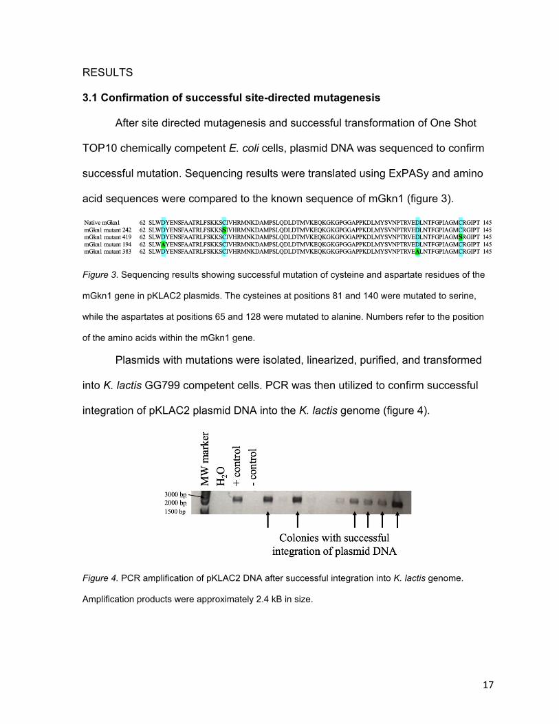
acid sequences were compared to the known sequence of mGkn1 (figure 3).
Figure 3. Sequencing results showing successful mutation of cysteine and aspartate residues of the
mGkn1 gene in pKLAC2 plasmids. The cysteines at positions 81 and 140 were mutated to serine,
while the aspartates at positions 65 and 128 were mutated to alanine. Numbers refer to the position
of the amino acids within the mGkn1 gene.
Plasmids with mutations were isolated, linearized, purified, and transformed
into K. lactis GG799 competent cells. PCR was then utilized to confirm successful
integration of pKLAC2 plasmid DNA into the K. lactis genome (figure 4).
Figure 4. PCR amplification of pKLAC2 DNA after successful integration into K. lactis genome.
Amplification products were approximately 2.4 kB in size.
18
3.2 Confirmation of mGkn1 generation
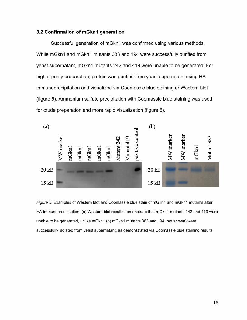
Successful generation of mGkn1 was confirmed using various methods.
While mGkn1 and mGkn1 mutants 383 and 194 were successfully purified from
yeast supernatant, mGkn1 mutants 242 and 419 were unable to be generated. For
higher purity preparation, protein was purified from yeast supernatant using HA
immunoprecipitation and visualized via Coomassie blue staining or Western blot
(figure 5). Ammonium sulfate precipitation with Coomassie blue staining was used
for crude preparation and more rapid visualization (figure 6).
Figure 5. Examples of Western blot and Coomassie blue stain of mGkn1 and mGkn1 mutants after
HA immunoprecipitation. (a) Western blot results demonstrate that mGkn1 mutants 242 and 419 were
unable to be generated, unlike mGkn1 (b) mGkn1 mutants 383 and 194 (not shown) were
successfully isolated from yeast supernatant, as demonstrated via Coomassie blue staining results.
19
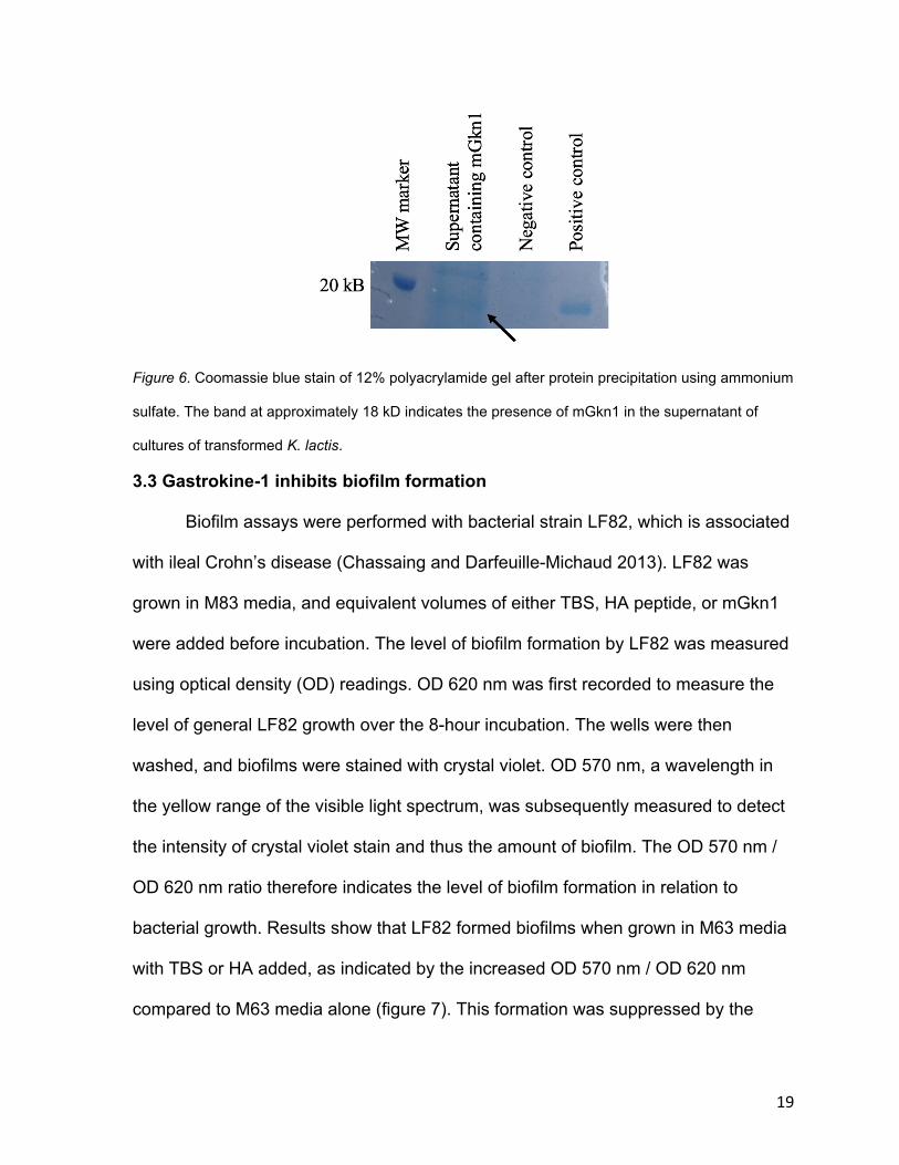
Figure 6. Coomassie blue stain of 12% polyacrylamide gel after protein precipitation using ammonium
sulfate. The band at approximately 18 kD indicates the presence of mGkn1 in the supernatant of
cultures of transformed K. lactis.
3.3 Gastrokine-1 inhibits biofilm formation
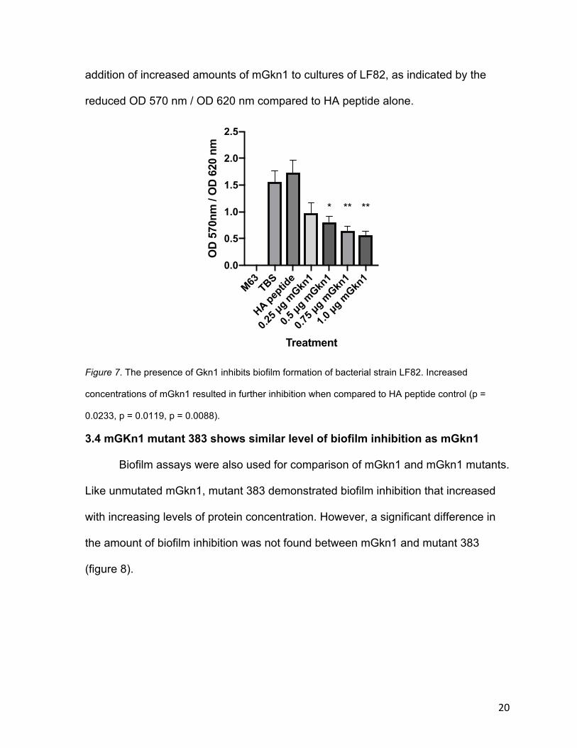
Biofilm assays were performed with bacterial strain LF82, which is associated
with ileal Crohn’s disease (Chassaing and Darfeuille-Michaud 2013). LF82 was
grown in M83 media, and equivalent volumes of either TBS, HA peptide, or mGkn1
were added before incubation. The level of biofilm formation by LF82 was measured
using optical density (OD) readings. OD 620 nm was first recorded to measure the
level of general LF82 growth over the 8-hour incubation. The wells were then
washed, and biofilms were stained with crystal violet. OD 570 nm, a wavelength in
the yellow range of the visible light spectrum, was subsequently measured to detect
the intensity of crystal violet stain and thus the amount of biofilm. The OD 570 nm /
OD 620 nm ratio therefore indicates the level of biofilm formation in relation to
bacterial growth. Results show that LF82 formed biofilms when grown in M63 media
with TBS or HA added, as indicated by the increased OD 570 nm / OD 620 nm
compared to M63 media alone (figure 7). This formation was suppressed by the
20
addition of increased amounts of mGkn1 to cultures of LF82, as indicated by the
reduced OD 570 nm / OD 620 nm compared to HA peptide alone.
Figure 7. The presence of Gkn1 inhibits biofilm formation of bacterial strain LF82. Increased
concentrations of mGkn1 resulted in further inhibition when compared to HA peptide control (p =
0.0233, p = 0.0119, p = 0.0088).
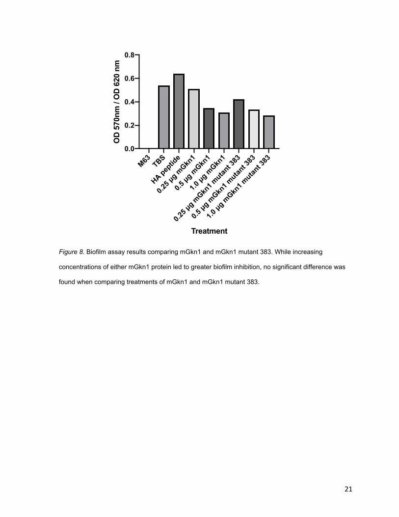
3.4 mGKn1 mutant 383 shows similar level of biofilm inhibition as mGkn1
Biofilm assays were also used for comparison of mGkn1 and mGkn1 mutants.
Like unmutated mGkn1, mutant 383 demonstrated biofilm inhibition that increased
with increasing levels of protein concentration. However, a significant difference in
the amount of biofilm inhibition was not found between mGkn1 and mutant 383
(figure 8).
M63 TBS
HA peptid
e
0.25 µ
g mGkn
1
0.5 µg m
Gkn1
0.75 µ
g mGkn
1
1.0 µg m
Gkn1
0.0
0.5
1.0
1.5
2.0
2.5
Treatment
OD
570
nm /
OD
620
nm
* ** **
21
Figure 8. Biofilm assay results comparing mGkn1 and mGkn1 mutant 383. While increasing
concentrations of either mGkn1 protein led to greater biofilm inhibition, no significant difference was
found when comparing treatments of mGkn1 and mGkn1 mutant 383.
M63 TBS
HA peptid
e
0.25 µ
g mGkn
1
0.5 µg m
Gkn1
1.0 µg m
Gkn1
0.25 µ
g mGkn
1 mutan
t 383
0.5 µg m
Gkn1 m
utant 3
83
1.0 µg m
Gkn1 m
utant 3
830.0
0.2
0.4
0.6
0.8
Treatment
OD
570
nm /
OD
620
nm
22
DISCUSSION AND FUTURE DIRECTIONS
While some mutant versions of mGkn1 were successfully generated, other
versions were unable to be isolated from yeast supernatant. These results suggest
that the transformed K. lactis was either unable to produce that version or unable to
secrete the final folded protein. Notably, the common feature of each failure was the
site directed mutagenesis of a cysteine residue. All BRICHOS domain containing
proteins contain a conserved pair of cysteine residues that are predicted to form a
disulfide bridge. Previous research on proSP-C, another protein in the BRICHOS
superfamily, has suggested that these residues are vital for the trafficking and
processing of the proprotein (Mulugeta et al. 2005). While mutant versions of proSP-
C with point mutations of conserved cysteines are expressed, they are misfolded
and mistargeted, leading to retainment and aggregation in aggresomes (Kabore et
al. 2001). As such, it is unsurprising that our similarly mutated versions of mGkn1
were unable to be produced and secreted. Further experimentation would involve
the analysis of cell lysates of K. lactis producing mGkn1 cysteine mutants, to identify
possible misfolding, mistrafficking, or degradation of cysteine mutant Gkn1.
Furthermore, the BRICHOS domain mutants of proSP-C are linked to
proliferative and interstitial lung diseases, since the misfolded proteins cause cell
injury and death through the deposition of toxic aggregates and induction of ER
stress (Mulugeta et al. 2005). Most proteins in the BRICHOS superfamily are
associated with degenerative diseases, and Gkn1 may play a role in the
development of inflammatory bowel disease (IBD). Perhaps patients suffering from
Crohn’s disease or ulcerative colitis have mutations in Gkn1 that prevent the protein
23
from being secreted, leaving the mucosa more susceptible to injury. Furthermore, it
has been shown that proSP-C and mutant proSP-C undergo heteromeric assembly,
and the formation of these multimers produces a dominant-negative effect,
exacerbating the effect of the mutations (Wang et al. 2002). To examine the impact
of Gkn1 mutations on mucosal health, samples from patients suffering from IBD and
control patients could be collected, and the amino acid sequence would be analyzed
to potentially pinpoint a Gkn1 mutation. Currently, most information on genetic
susceptibility to IBD is limited to genome-wide association studies which identify
genetic regions that contain risk modifying alleles. These studies have not identified
the Gkn1 region as a risk modifying region for IBD. However, rare variants that
cause disease remain to be fully explored, as there have been only limited published
studies on whole genome sequencing or exome sequencing in IBD. Although
genetic mutations in Gkn1 associated with IBD or other diseases have not been
found, it remains possible that such mutations could exist in rare cases. In addition,
since Gkn1 is resistant to degradation in the GI tract, there is potential for oral
administration of Gkn1 as a treatment method for IBD. Purified Gkn1 protein could
be fed to mice to assess its effect on promoting or restoring mucosal health.
We hypothesize that Gkn1 exerts its protective and obesogenic functions via
its anti-amyloidogenic properties and ability to modulate the microbiota. Biofilm
assay results show an inverse relationship between the concentration of Gkn1 and
the level of biofilm formation; these results support earlier research on the anti-
amyloidogenic effects of Gkn1 (Altieri et al. 2014). Further research on these effects
will involve in vitro protein assays. In these assays, the formation of amyloid fibers
24
can be traced in real time. These assays will offer more substantial evidence that
inhibition of amyloids fibrils leads to the loss of biofilm formation in our biofilm
assays. The comparison between mGkn1 and mGkn1 mutants could also be
performed using future in vitro protein assays.
Surprisingly, the mGkn1 mutant 383 displayed a similar level of inhibition
compared to WT mGkn1, which indicates that this amino acid, while conserved in
evolution, is not critical for the anti-biofilm activity of Gkn1 and therefore not likely
critical for the anti-amyloidogenic activity of Gkn1. The creation of different mutants
is underway in the hopes of pinpointing specific crucial amino acids and elucidating
the biochemical interaction between the protein and the amyloid fibers. This
information could allow for the development of anti-amyloid drugs that mimic the
function of Gkn1. As mentioned previously, oral administration of Gkn1 could be a
potential treatment option for digestive diseases. Synthesis of drugs that mimic its
properties would be more efficient and cost-effective to produce when compared to
synthesis of the whole protein.
On the other hand, loss of Gkn1 results in resistance to adiposity, as seen in
the Gkn1-/- mice. Selectively inhibiting or fully inhibiting this protein in the GI tract
could be a potential treatment for obesity. Since mice models fed anti-Gkn1 antibody
gained less fat and weight than control mice (Bakke 2016), drugs that inhibit Gkn1
could also be developed. However, since Gkn1 has multiple functions, selective
inhibition may be required for successful obesity treatment without compromising the
health of the GI tract (Bakke 2016). More information is still needed about the
mechanism, or mechanisms, of action of Gkn1. It is possible that different parts of
25
the protein, or different parts of the BRICHOS domain, have varying importance with
regards to carrying out each function. Knowledge about specific biochemical
features could allow for the development of drugs with higher specificity that target,
for example, the obesogenic activity of Gkn1, but not the IBD suppressing activity of
Gkn1.
It is also possible that Gkn1 exerts multiple functions because the microbes or
metabolites that promote epithelial cell health are different than those promoting
weight gain. In a broad sense, the specifics of the modulation of the gut microbiome
by Gkn1 are still unknown. While prevention of biofilm formation may alter the
organization or structure of microbial communities, we lack information on
compositional changes of the microbiota in the presence or absence of Gkn1. Many
previous research studies have shown that the microbial composition of the GI tract
plays a critical role in the phenotype of the host. To investigate the impact of Gkn1
on microbiome composition, a comparative analysis of 16S rRNA from multiple
different sections of the GI tract of WT and Gkn1-/- mice should be performed. This
information may show large differences in the microbiome, and particular bacterial
species could be associated with the weight gain caused by Gkn1. Knowledge of the
biochemistry of the interactions between Gkn1 and particular bacteria may permit
the development of drugs that target specific bacterial species and health conditions.
Overall, our data show that Gkn1 exerts anti-amyloidogenic activity but that
not all conserved amino acids in the BRICHOS domain are crucial for this function.
Further experimentation on the biochemistry of the mechanism of action of Gkn1 is

26
still required. Ultimately, this information may be used to create specific drugs that
can target and treat obesity and its related health problems.

27
ACKNOWLEDGEMENTS
It is important to acknowledge that this research work is part of a larger
Gastrokine-1 project, which has been contributed to by many skilled researchers and
students in the Boone Lab. I am thankful for all their assistance, guidance, and
support. Particular thanks to Toni Boger, for her patience and know-how, Hunter
Hoffman, for his encouragement and answers to my many questions, and Dr. Anne-
Marie Overstreet, for teaching me practically everything I know. Finally, thank you
most importantly to Dr. David Boone, who welcomed me into his laboratory and has
guided my development as a researcher.
I would also like to thank the Notre Dame College of Science and the Glynn
Family Honors Program for partially funding my research during the summer of
2017. Thank you also to my family and friends, who have listened to me talk
excitedly about Gastrokine-1 for four years now.
28
REFERENCES
Altieri F, Di Stadio CS, Severino V, Sandomenico A, Minopoli G. (2014). Anti-
amyloidogenic property of human gastrokine 1. Biochimie 106: 91-100.
Anthes E. (2014). Treatment: Marginal gains. Nature 508(7496): S54.
Aoki R, Kamikado K, Suda W, Takii H, Mikami Y. (2017). A proliferative probiotic
Bifidobacteriu strain in the gut ameliorates progression of metabolic disorders
via microbiota modulation and acetate elevation. Sci Rep 7: 43522
Backhed F, Ding H, Wang T, Hooper LV, Koh GY, Nagy A, Semenkovitch CF,
Gordon JI. (2004). Gut microbiota as an environmental factor that regulates
fat storage. Proceedings of the National Academy of Sciences of the United
States of America 101(44): 15718-15723.
Bäckhed F, Manchester J, Semenkovich C, Gordon J. (2007). Mechanisms
Underlying the Resistance to Diet-Induced Obesity in Germ-Free Mice.
Proceedings of the National Academy of Sciences of the United States of
America 104(3), 979-984.
Bakke D. (2016). Gastrokine 1 protects against gastrointestinal inflammation and
regulates host metabolism [dissertation]. [Chicago, Illinois]: The University of
Chicago.
Bertani G. (1951). Studies on lysogenesis. I. The mode of phage liberation by
lysogenic Escherichia coli. J. Bacteriol 62: 293-300.
Bianchi F, Duque A, Saad L, Sivieri R. (2019). Gut microbiome approaches to treat
obesity in humans. Applied Microbiology and Biotechnology 103(3): 1081-
1094.
29
Blanco LP, Evans ML, Smith DR, Badtke MP, & Chapman MR. (2011). Diversity,
biogenesis and function of microbial amyloids. Trends in Microbiology 20(2):
66-73.
Bray G. (2012). Diet and Exercise for Weight Loss. JAMA 307(24): 2641-2642.
Chassaing B, Darfeuille-Michaud A. (2013). Adherent-invasive Escherichia
coli Biofilm Formation Assays. Bio-protocol 3(23): e982.
Christensen L, Roager HM, Astrup A, Hjorth MF. (2018). Microbial enterotypes in
personalized nutrition and obesity management, The American Journal of
Clinical Nutrition 108(4): 645–651.
Costerton JW, Lewandowski ZE, Caldwell DR, Korber DM, Lappin-Scott H. (1995).
Microbial biofilms. Annual Review of Microbiology 49: 711-745.
De Vadder F, Kovatcheva-Datchary P, Goncalves D, Vinera J, Zitoun C. (2014).
Microbiota-Generated Metabolites Promote Metabolic Benefits via Gut-Brain
Neural Circuits. Cell 156: 84-96.
de Vos WM. (2015). Microbial biofilms and the human intestinal microbiome. NPJ
Biofilms and Microbiomes 1: 2055-5008.
Duchovny NJ, Baker C. (2010). How does obesity in adults affect spending on health
care? (Economic and budget issue brief). Washington, D.C.]: Congressional
Budget Office.
Flegal K, Carroll M, Ogden C, Curtin L. (2010). Prevalence and trends in obesity
among US adults, 1999-2008. JAMA 303(3): 235-241.
Forslund K, Hildebrand F, Nielsen T, Falony G, Le Chatelier E, Sunagawa S, Prifti E,
Vieria-Silva S, Gudmundsdottir V, Pedersen HK, Arumugam M, Kristiansen K,
30
Voigt AY, Vestergaard H, Hercog R, Costea PI, Kultima JR, Li J, Jorgensen
T, Levenez F, Dore J, Nielsen HB, Brunak S, Raes J, Hansen T, Wang J,
Ehrlich SD, Bork P, Pedersen O. (2015). Disentangling type 2 diabetes and
metformin treatment signatures in the human gut microbiota. Nature
528(7581): 266-273.
Fux C, Stoodley P, Hall-Stoodley L, Costerton J. (2003). Bacterial biofilms: A
diagnostic and therapeutic challenge. Expert Review of Anti-infective Therapy
1(4): 667-683.
Garcia M, Lee J, Ramsook C, Alsteens D, Dufrêne Y, Lipke P, Nielsen K. (2011). A
Role for Amyloid in Cell Aggregation and Biofilm Formation (Amyloids in Cell
Aggregation and Biofilms). PLoS ONE 6(3): E17632.
Ghaferi A, Varban O. (2018). Setting Appropriate Expectations After Bariatric
Surgery: Evaluating Weight Regain and Clinical Outcomes. JAMA 320(15):
1543-1544.
Goforth A. (2016). “The high costs of obesity.” Benefits PRO June 2016: 12.
Business Insights: Essentials. Web. 2 Mar 2019.
Hall-Stoodley L, Costerton JW, Stoodley P. (2004). Bacterial biofilms: From the
Natural environment to infectious diseases. Nature Reviews Microbiology
2(2): 95-108.
Jensen TM, Saha K, Steinberg WM. (2015). Is there a link between liraglutide and
pancreatitis? A post hoc review of pooled and patient-level data from
completed liraglutide type 2 diabetes clinical trials. Diabetes Care 38: 1058–
66.
31
Kabore AF, Wang WJ, Russo SJ, Beers MF. (2001). Biosynthesis of surfactant
protein C: characterization of aggresome formation by EGFP chimeras
containing propeptide mutants lacking conserved cysteine residues. J. Cell
Sci. 114(293): LP-302.
Knight S, Presto J, Linse S, Johansson J. (2013). The BRICHOS domain, amyloid
fibril formation, and their relationship. Biochemistry 52(43): 7523-31.
Maier L, Pruteanu M, Kuhn M, Zeller G, Telzerow A, Anderson EE, Typas A. (2018).
Extensive impact of non-antibiotic drugs on human gut bacteria. Nature
555(7698), 623-625.
Mao W, Chen J, Peng TL, Yin XF, Chen LZ, Chen MH (2012). Downregulation of
gastrokine-1 in gastric cancer tissues and restoration of its expression
induced gastric cancer cells to apoptosis. Journal of experimental & clinical
cancer research 31(1): 49.
Martin KA, Mani MV, Mani A. (2015). New targets to treat obesity and the metabolic
syndrome. European Journal of Pharmacology 763(Pt A): 64-74.
Martin TE, Powell CT, Wang Z, Bhattacharyya S, Walsh-Reitz MM, Agarwal K,
Toback FG. (2003). A Novel Mitogenic Protein That is Highly Expressed in
Cells of the Gastric Antrum Mucosa. American Journal of Physiology.
Gastrointestinal and Liver Physiology 285(2): G332-43.
Mazidi M, Rezaie P, Kengne AP, Mobarhan MG, Ferns GA. (2016). Gut microbiome
and metabolic syndrome. Diabetes & Metabolic Syndrome: Clinical Research
& Reviews 10(2): S150-S157.
32
Mulugeta S, Nguyen V, Russo SJ, Muniswamy M, Beers MF. (2005). A surfactant
protein C precursor protein BRICHOS domain mutation causes endoplasmic
reticulum stress, proteasome dysfunction, and caspase 3
activation. American Journal of Respiratory Cell and Molecular Biology 32(6):
521-30.
National Institute of Diabetes Digestive Kidney Diseases [U.S.], & Weight-control
Information Network [U.S.]. (2004). Prescription medications for the treatment
of obesity (NIH publication; no. 04-4191). Bethesda, MD]: National Institute of
Diabetes and Digestive and Kidney Diseases, National Institutes of Health.
Oien KA, McGregor F, Butler S, Ferrier RK, Downie I, Bryce S, Burns S, Keith WN.
(2004). Gastrokine 1 is abundantly and specifically expressed in superficial
gastric epithelium, down-regulated in gastric carcinoma, and shows high
evolutionary conservation. J. Pathol 203: 789-797.
Oien KA, McGregor F, Butler S, Ferrier RK, Downie I. (2004). Gastrokine 1 is
abundantly and specifically expressed in superficial gastric epithelium, down-
regulated in gastric carcinoma, and shows high evolutionary conservation.
The Journal of Pathology 203(3): 789-797.
Oppong GO, Rapsinski GJ, Tursi SA, Biesecker SG, Klein-Szanto AJ, Goulian M,
McCauley C, Healy C, Wilson RP, Tükel C. (2015). Biofilm-associated
bacterial amyloids dampen inflammation in the gut: oral treatment with curli
fibres reduces the severity of hapten-induced colitis in mice. NPJ Biofilms and
Microbiomes 1: 15019.
33
Renna F, Thakur N. (2010). Direct and indirect effects of obesity on U.S. labor
market outcomes of older working age adults. Social Science & Medicine
71(2): 405-413.
Reyes LM, Vázquez RG, Arroyo SMC, Avalos AM, Castillo PAR, Pérez DAC,
Terrones IR, Ibáñez NR, Magallanes MMR, Langella P, Humarán LB,
Espinosa AA. (2016). Correlation between diet and gut bacteria in a
population of young adults. Int J Food Sci Nutr 67: 470-478.
Rosenvinge EC, O'May GA, Macfarlane S, Macfarlane GT, Shirtliff ME. (2013).
Microbial biofilms and gastrointestinal diseases. Pathogens and Disease
67(1): 25–38.
Sanchez-Pulido L, Devos D, Valencia A. (2002). BRICHOS: a conserved domain in
proteins associated with dementia, respiratory distress and cancer. Trends in
Biochemical Sciences 27(7): 329e332.
Saunders K, Shukla A, Igel L, Aronne L. (2018). Obesity: When to consider
medication: These 4 cases illustrate how weight loss drugs--including the 4
newest--can be integrated into a treatment plan that includes diet, exercise,
and behavior modification. OBG Management 30(8): 41.
Sayin SI, Wahlström A, Felin J, Jäntti S, Marschall H, Bamberg K, Angelin B,
Hyotylainen T, Oresic M, Bäckhed F. (2013). Gut Microbiota Regulates Bile
Acid Metabolism by Reducing the Levels of Tauro-beta-muricholic Acid, a
Naturally Occurring FXR Antagonist. Cell Metabolism 17(2): 225-235.
Schwartz K, Boles BR. (2013). Microbial amyloids - functions and interactions within
the host. Current opinion in microbiology 16(1): 93-99.
34
Shea J, Diamandis E, Sharma A, Després J, Ezzat S, Greenway F. (2012). The
obesity epidemic. Clinical Chemistry 58(6): 968-73.
Swidsinski A, Weber J, Loening-Baucke V, Hale LP, Lochs H. (2005). Spatial
Organization and Composition of the Mucosal Flora in Patients with
Inflammatory Bowel Disease. Journal of Clinical Microbiology 43(7): 3380-
3389.
Taglialegna A, Lasa I, Valle J. (2016). Amyloid Structures as Biofilm Matrix
Scaffolds. Journal of Bacteriology 198(19): 2579-88.
The Lancet. (2012). New weight-loss drugs and the US obesity epidemic. The
Lancet 380(9839): 308.
Tremaroli V, Karlsson F, Werling M, Ståhlman M, Kovatcheva-Datchary P, Olbers T,
Fandricks L, Le roux CW, Nielsen J, Bäckhed F. (2015). Roux-en-Y Gastric
Bypass and Vertical Banded Gastroplasty Induce Long-Term Changes on the
Human Gut Microbiome Contributing to Fat Mass Regulation. Cell Metabolism
22(2): 228-238.
Turnbaugh PJ, Ley RE, Mahowald MA, Magrini V, Mardis ER. (2006). An obesity-
associated gut microbiome with increased capacity for energy harvest.
Nature 444(7122): 1027-1031.
Walsh-Reitz M, Huang E, Musch M, Chang E, Martin T, Kartha S, Toback F. (2005).
AMP-18 protects barrier function of colonic epithelial cells: Role of tight
junction proteins. American Journal of Physiology-Gastrointestinal And Liver
Physiology 289(1): G163-G171.
35
Wang WJ, Russo SJ, Mulugeta S, Beers MF. (2002). Biosynthesis of surfactant
protein C (SP-C): sorting of SP-C proprotein involves homomeric association
via a signal anchor domain. J Biol Chem 277:19929–19937.
Weight-control Information Network [U.S.], & National Institute of Diabetes Digestive
Kidney Diseases [U.S.]. (2011). Bariatric surgery for severe obesity (Updated
June 2011, NIH publication; no. 08-4006). Bethesda, MD: U.S. Dept. of
Health and Human Services, National Institutes of Health, National Institute of
Diabetes and Digestive and Kidney Diseases.
Willander H, Askarieh G, Landreh M, Westermark P, Nordling K, Keränen H,
Hermansson E, Hamvas A, Nogee LM, Bergman T, Saenz A, Casals C,
Åqvistg J, Jörnvall H, Berglund H, Presto J, Knight SD, Johansson J. (2012).
High-resolution structure of a BRICHOS domain and its implications for anti-
amyloid chaperone activity on lung surfactant protein C. Proceedings of the
National Academy of Sciences of the United States of America 109(7): 2325-
9.
Willander H, Hermansson E, Johansson J, Presto J. (2011). BRICHOS domain
associated with lung fibrosis, dementia and cancer – a chaperone that
prevents amyloid fibril formation?. The FEBS Journal 278: 3893-3904.
Yanovski SZ, Yanovski JA. (2014). Long-term Drug Treatment for Obesity. JAMA
311(1): 74-86.
Yoon JH, Choi YJ, Choi WS, Nam SW, Lee JY, Park WS. (2013). Functional
analysis of the NH 2 -terminal hydrophobic region and BRICHOS domain of
36
GKN1. Biochemical and Biophysical Research Communications 440(4): 689-
695.
Zhao Y, Lukiw WJ. (2015). Microbiome-generated amyloid and potential impact on
amyloidogenesis in Alzheimer's disease (AD). Journal of nature and
science 1(7): e138.