selected hematologic and biochemical measurements in african hiv-infected and uninfected pregnant...
TRANSCRIPT

BioMed CentralBMC Pediatrics
ss
Open AcceResearch articleSelected hematologic and biochemical measurements in African HIV-infected and uninfected pregnant women and their infants: the HIV Prevention Trials Network 024 protocolKasonde Mwinga1,2, Sten H Vermund*3, Ying Q Chen4, Anthony Mwatha4, Jennifer S Read5, Willy Urassa6, Nicole Carpenetti7, Megan Valentine8 and Robert L Goldenberg9Address: 1Department of Paediatrics of the University Teaching Hospital and the University of Zambia School of Medicine, and the Centre for Infectious Disease Research in Zambia, Lusaka, Zambia, 2Now with the World Health Organization, Lusaka, 3Institute for Global Health and Department of Pediatrics, Vanderbilt University School of Medicine, Nashville, TN, USA, 4Statistical Center for HIV/AIDS Research and Prevention, Fred Hutchinson Cancer Research Center, Seattle, WA, USA, 5National Institute of Child Health and Human Development, National Institutes of Health, Bethesda, MD, 6Muhimbili University, Dar-es-Salaam, Tanzania, 7College of Medicine – Johns Hopkins University Research Project, Blantyre, Malawi, 8Family Health International, Chapel Hill, NC, USA and 9Department of Obstetrics and Gynecology, Drexel University College of Medicine, Philadelphia, PA, USA
Email: Kasonde Mwinga - [email protected]; Sten H Vermund* - [email protected]; Ying Q Chen - [email protected]; Anthony Mwatha - [email protected]; Jennifer S Read - [email protected]; Willy Urassa - [email protected]; Nicole Carpenetti - [email protected]; Megan Valentine - [email protected]; Robert L Goldenberg - [email protected]
* Corresponding author
AbstractBackground: Reference values for hematological and biochemical assays in pregnant women andin newborn infants are based primarily on Caucasian populations. Normative data are limited forpopulations in sub-Saharan Africa, especially comparing women with and without HIV infection, andcomparing infants with and without HIV infection or HIV exposure.
Methods: We determined HIV status and selected hematological and biochemical measurementsin women at 20–24 weeks and at 36 weeks gestation, and in infants at birth and 4–6 weeks of age.All were recruited within a randomized clinical trial of antibiotics to prevent chorioamnionitis-associated mother-to-child transmission of HIV (HPTN024). We report nearly completelaboratory data on 2,292 HIV-infected and 367 HIV-uninfected pregnant African women who wererepresentative of the public clinics from which the women were recruited. Nearly all the HIV-infected mothers received nevirapine prophylaxis at the time of labor, as did their infants after birth(always within 72 hours of birth, but typically within just a few hours at the four study sites inMalawi (2 sites), Tanzania, and Zambia.
Results: HIV-infected pregnant women had lower red blood cell counts, hemoglobin, hematocrit,and white blood cell counts than HIV-uninfected women. Platelet and monocyte counts werehigher among HIV-infected women at both time points. At the 4–6-week visit, HIV-infected infantshad lower hemoglobin, hematocrit and white blood cell counts than uninfected infants. Plateletcounts were lower in HIV-infected infants than HIV-uninfected infants, both at birth and at 4–6
Published: 7 August 2009
BMC Pediatrics 2009, 9:49 doi:10.1186/1471-2431-9-49
Received: 19 September 2008Accepted: 7 August 2009
This article is available from: http://www.biomedcentral.com/1471-2431/9/49
© 2009 Mwinga et al; licensee BioMed Central Ltd. This is an Open Access article distributed under the terms of the Creative Commons Attribution License (http://creativecommons.org/licenses/by/2.0), which permits unrestricted use, distribution, and reproduction in any medium, provided the original work is properly cited.
Page 1 of 14(page number not for citation purposes)

BMC Pediatrics 2009, 9:49 http://www.biomedcentral.com/1471-2431/9/49
weeks of age. At 4–6 weeks, HIV-infected infants had higher alanine aminotransferase measuresthan uninfected infants.
Conclusion: Normative data in pregnant African women and their newborn infants are needed toguide the large-scale HIV care and treatment programs being scaled up throughout the continent.These laboratory measures will help interpret clinical data and assist in patient monitoring in a sub-Saharan Africa context.
Trial Registration: nicalTrials.gov Identifier NCT00021671.
IntroductionHematological parameters are affected by many factors,including age, sex, diet, recent nutritional status, and con-sumption of medications or illicit drugs.[1-4] Referencevalues for hematological and biochemical assays in preg-nant women and in infants are based largely on data fromCaucasian populations. Normative data have beenreported from only a few populations living in sub-Saha-ran Africa.[2,4-10] Fewer studies still highlight expectedvalues from women and infants with HIV infection orexposure.[3,11-13] Within a large clinical trial of the pre-vention of mother-to-child transmission of HIV, we tookadvantage of the fact that we had laboratories that werecertified by the National Institutes of Health performinglaboratory assessments with a high degree of oversightand quality control. All laboratories were subject to rigor-ous monitoring, including receipt of proficiency panels.Thus, we were in an excellent position to provide labora-tory data from both pregnant women and their newbornoffspring within a high quality laboratory environment infour African cities. These data were collected to assess thesafety of the antibiotic intervention of the parent clinicaltrial and the status of the patient vis-à-vis HIV infection orrisk. We describe hematological and biochemical meas-ures and trends over time in a large cohort of pregnantAfrican women and infants with and without humanimmunodeficiency virus type 1 (HIV) infection. Given themagnitude of the current efforts to identify and treat HIVdisease in Africa, we believe that data from our large sam-ple will prove helpful to service providers seeking to inter-pret data from their own pregnant clinical subjects andtheir infants.
Patients and MethodsThe HIV Prevention Trials Network Protocol 024 TrialThe HIV Prevention Trials Network Protocol 024(HPTN024) study was a Phase III randomized, doubleblind, placebo-controlled clinical trial of antibiotics toreduce chorioamnionitis-associated mother-to-childtransmission of HIV. The trial was conducted in four Afri-can sites: Blantyre, Malawi, Lilongwe, Malawi; Dar-es-Salaam, Tanzania; and Lusaka, Zambia. Prior to initiationof the trial, approval was received from institutionalreview boards or ethics committees at all participating
sites and universities. The findings of the HPTN024 trialitself have been published showing that an antepartumand peripartum antibiotic regimen did not reduce the riskof MTCT of HIV-1 in African women with high prevalencerates of bacterial vaginosis and subclinical chorioamnio-nitis.[14] HIV-infected and HIV-uninfected women wereenrolled at 20–24 weeks gestation from antenatal clinicsfrom July 2001 to February 2003. By trial design, in threeof the four sites, there were five pregnant HIV-infectedwomen enrolled in the study for each pregnant HIV-unin-fected woman. The principal exclusion criteria wererelated to serious illness that would prevent the womanfrom participating in a research study. Otherwise, subjectswere highly representative of the public sector clinics fromwhich they were recruited. All laboratory data indicatedfor the parent protocol were obtained on each subject,except that viral loads were not obtained if the subject wasHIV-uninfected.
Enrolled women were randomized to receive either met-ronidazole 250 mg and erythromycin 250 mg every 8hours for 7 days at 20–24 weeks, followed by metronida-zole 250 mg and ampicillin 500 mg every 4 hours withonset of premature rupture of membranes or labor, oridentically appearing placebos. Follow-up study visitsoccurred at 28 weeks and 36 weeks gestation. Infant studyvisits were conducted at birth and at 4–6 weeks of age. Allwomen received a conventional iron-containing vitamin/mineral preparation designed for pregnant women (Tish-con Corporation, Baltimore, MD) daily from enrolmentand until delivery. The supplement included: 30 mg iron,400 mcg folic acid, 5000 IU vitamin A, 400 IU vitamin D,30 IU vitamin E, 50 mg vitamin C, 2 mg vitamin B1, 3 mgvitamin B2, 3 mg vitamin B6, 5 mcg vitamin B12, 20 mgniacin, 250 mg calcium, 150 mcg iodine, 100 mg magne-sium, and 15 mg zinc.
HIV-infected women were offered single dose nevirapine(NVP) prophylaxis at delivery for prevention of mother-to-child transmission of HIV. Infants of HIV-infectedwomen received NVP within 72 hours of birth. No otherantiretroviral drugs were used by mothers and infants inthe trial, as these drugs were unavailable in the study set-tings at the time.
Page 2 of 14(page number not for citation purposes)

BMC Pediatrics 2009, 9:49 http://www.biomedcentral.com/1471-2431/9/49
All seroconverters had their baseline stored specimenssampled by PCR so that persons who were acutely infectedat baseline could be excluded. Hence, we are certain thatour seronegative women were not acutely infected. Fur-thermore, these baseline data were collected within amonth of enrolment, and often on the same day as enrol-ment.
Since the clinicians were blinded to the randomized treat-ment assignment, women were treated for all infectionsper the local treatment guidelines. The receipt of non-study antibiotics and all other medications received by thewomen was recorded using open-ended questions in theconcomitant medications log form (data not presented).
We used structured questionnaires to collect informationon maternal demographics as well as medical, obstetricand sexual histories. We estimated infant gestational agethrough the neuromuscular and physical maturity indicesof the new Ballard examination. Trained nurses assessedbirth weights.
For infants born to the study women, only the first liveborn infant was included in this analysis. If twins wereborn, only the firstborn twin was included (since labora-tory outcomes for twins are likely to be correlated).Women could not be enrolled for subsequent pregnanciesif they had already enrolled previously.
Laboratory ProceduresVenous blood was collected from pregnant women atboth the 20- to 24-week visit and the 36-week visit. Bloodfor complete blood counts (CBCs) was collected in ethyl-enediaminetetraacetic acid (EDTA) vacutainers and ana-lyzed at local laboratories on Coulter machines. Blood foralanine aminotransferase (ALT) assays was stored inEDTA vacutainers. Infant blood was collected by heelstick. CBCs and lymphocyte subsets (counts and percent-ages) were determined at both visits using HPTN CentralLab-approved site-specific procedures. Analyses for CD4+and CD8+ T lymphocyte cell counts were done using FAC-SCount™ flow cytometer in three sites (Dar-es-Salaam,Lilongwe and Lusaka) while one site (Blantyre) used FAC-Scan™ flow cytometer. All analyses were done according tothe manufacturer's procedures. For infants, CBCs wereobtained at both the birth visit and the 4–6-week visit. Atthe 4–6-week visit, ALT assays were performed with chem-istry analyzers according to manufacturer's protocols.
Blood samples from the pregnant women were screenedfor HIV at local laboratories using two different enzymelinked immunosorbent assays (ELISA), and confirmedwith Western blot assays. The HIV infection status ofinfants at birth and 4–6 weeks of age was determined byanalyzing dried blood spots, using a polymerase chain
reaction (PCR)-based HIV RNA assay. Results from ELISAassays (for women) and HIV RNA assays (for infants) wereconfirmed centrally.
Infants were categorized as HIV-uninfected at birth if theyhad a negative HIV RNA assay at birth, or if they weremissing an HIV RNA result from the birth visit, but had anegative HIV RNA test at 4–6 weeks of age. Infants whohad a negative HIV RNA test at birth, but a positive HIVRNA test at 4–6 weeks were categorized as HIV-uninfectedat birth, and as HIV-infected at 4–6 weeks of age.
The HPTN Central Laboratory at Johns Hopkins Univer-sity reviewed and certified all local laboratories before theinitiation of the trial. The Central Laboratory verified viro-logical, serological, hematological, immunological, andbiochemical tests based on proficiency panels provided bythe College of American Pathology (CAP) and UnitedKingdom (U.K.) National External Quality AssessmentService (UKNEQAS) on a periodic basis throughout thetrial.
Statistical AnalysisWe summarized all laboratory values by their means, 95%confidence intervals (2.5th and 97.5th percentiles), and cal-culated their associated standard deviations for HIV-infected and HIV-uninfected women and infants sepa-rately. Two-sample comparisons were done by hypothesistesting on continuous and categorical outcomes using theStudent's t-test and the Chi-square test (or the Fisher'sexact test, when indicated), respectively. Comparison onchanges in laboratory measures between visits (20–24weeks and 36 weeks) was done by hypothesis testingusing paired sample t-tests. Similar analyses were also per-formed for infants (birth visit and 4–6 week visit).
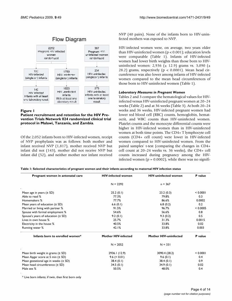
ResultsSize and Characteristics of the Study PopulationWe enrolled 2,659 eligible pregnant women intoHPTN024. Of these 2,659 women, 2,292 (86%) wereHIV-infected and 367 (14%) were uninfected (Figure 1).There were 2,382 live born infants (2,052 born to HIV-infected women and 331 born to uninfected women). Themean gestational age at birth (n = 2382) was 38.4 (± 0.06)weeks using the new Ballard score. The mean birth weightin the study was 2,957 (± 11.8) grams. Infants born toHIV-infected mothers had a 37-gram lower mean birthweight compared to infants born to HIV-uninfectedmothers (2980 grams ± 505; 95% ci: 2957–3005 vs. 3079grams ± 515; 95% ci: 3023–3135; respectively; p = 0.002by Student's t test). The mean head circumference was34.6 (± 0.05) cm. The mean Apgar score at 1 minute was8.2 (± 0.03) and was 9.6 (± 0.03) at 5 minutes.
Page 3 of 14(page number not for citation purposes)
BMC Pediatrics 2009, 9:49 http://www.biomedcentral.com/1471-2431/9/49
Of the 2,052 infants born to HIV-infected women, receiptof NVP prophylaxis was as follows: both mother andinfant received NVP (1,817), mother received NVP butinfant did not (143), mother did not receive NVP butinfant did (52), and neither mother nor infant received
NVP (40 pairs). None of the infants born to HIV-unin-fected mothers was exposed to NVP.
HIV-infected women were, on average, two years olderthan HIV-uninfected women (p < 0.001); education levelswere comparable (Table 1). Infants of HIV-infectedwomen had lower birth weights than those born to HIV-uninfected women: 2,936 (± 12.9) grams vs. 3,090 (±28.2) grams, respectively (p < 0.0001). Mean head cir-cumference was also lower among infants of HIV-infectedwomen compared to the mean head circumferences ofthose born to HIV-uninfected women (Table 1).
Laboratory Measures in Pregnant WomenTables 2 and 3 compare the hematological values for HIV-infected versus HIV-uninfected pregnant women at 20–24weeks (Table 2) and at 36 weeks (Table 3). At both 20–24weeks and 36 weeks, HIV-infected pregnant women hadlower red blood cell (RBC) counts, hemoglobin, hemat-ocrit, and WBC counts than HIV-uninfected women.Platelet counts and the monocyte differential counts werehigher in HIV-infected women than in HIV-uninfectedwomen at both time points. The CD4+ T lymphocyte cellcounts (CD4+ cell count) were lower in HIV-infectedwomen compared to HIV-uninfected women. From thepaired samples' t-test (comparing the changes in CD4+cell count at 20–24 weeks vs. 36 weeks), the CD4+ cellcounts increased during pregnancy among the HIV-infected women (p = 0.0002), while there was no signifi-
Patient recruitment and retention for the HIV Prevention Trials Network 024 randomized clinical trial protocol in Malawi, Tanzania, and ZambiaFigure 1Patient recruitment and retention for the HIV Pre-vention Trials Network 024 randomized clinical trial protocol in Malawi, Tanzania, and Zambia.
Table 1: Selected characteristics of pregnant women and their infants according to maternal HIV infection status
Pregnant women in antenatal care HIV-infected women HIV-uninfected women P value
N = 2292 n = 367
Mean age in years (± SD) 25.2 (0.1) 23.2 (0.3) < 0.0001Able to read % 77.3% 79.8% 0.3Homemakers % 77.7% 86.6% 0.0002Mean years of education (± SD) 6.6 (0.1) 6.8 (0.2) 0.2Married or living with partner % 91.5% 96.7% < 0.0005Spouse with formal employment % 54.6% 56.6% 0.8Spouse's years of education (± SD) 9.2 (0.1) 9.3 (0.2) 0.5Lives in own house % 25.7% 31.3% 0.0015Electricity in the house % 40.5% 33.8% 0.02Running water % 42.1% 33.8% 0.003
Infants born to enrolled women* Mother HIV-infected Mother HIV-uninfected P value
N = 2052 N = 331
Mean birth weight in grams (± SD) 2936.1 (12.9) 3090.4 (28.2) < 0.0001Mean Apgar score at 5 min (± SD) 9.6 (< 0.01) 9.6 (0.1) 0.4Mean gestational age in weeks (± SD) 38.4 (0.1) 38.4 (0.1) 0.9Mean head circumference (± SD) 34.5 (0.1) 34.9 (0.1) 0.02Male sex % 50.5% 48.0% 0.4
* Live born infants; if twin, then first born only
Page 4 of 14(page number not for citation purposes)

BMC Pediatrics 2009, 9:49 http://www.biomedcentral.com/1471-2431/9/49
cant change in this measure among the HIV-uninfectedwomen. The mean of the paired differences of CD4+ cellcounts among HIV-infected women was +14.8 cells/μLfrom the 20–24-week visit compared to the 36-week ante-natal visit.
About one-quarter of the women who had been seen atthe 20–24-week visit were not seen at the 36-week visit.The mean maternal age for women who were seen only at20–24 weeks was 24.5 ± 5.0 years, while women seenboth at gestational age 20–24 weeks as well as at 36 weekswere six months older, on average (mean age of 25.1 ± 4.8years; p = 0.004). Log viral load of women who were seenonly at 20–24 weeks was 4.39 ± 0.78 log10 copies/mLwhile women seen both at gestational age 20–24 weeks aswell as at 36 weeks had a 0.15 log10 copies/mL lower
mean viral load of 4.24 ± 0.82 log10 copies/mL (p =0.0002). We highlight the relevance of this observation,related to the CD4+ cell count changes in the discussion.
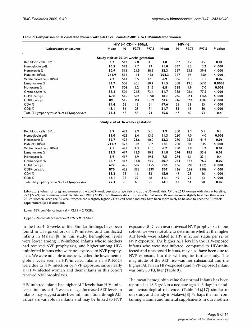
We found the differences between HIV-infected and unin-fected pregnant women to persist, for the most part,throughout the range of CD4+ cell counts (Tables 4, 5, 6and 7). For example, even women with HIV infection andCD4+ cell counts >500/μL had lower hemoglobins andhigher platelet counts than did HIV-uninfected women(Table 7). As one would expect, hematogical parameterswere more aberrant in women with lowest CD4+ cellcounts (Table 4).
Tables 8 and 9 give the paired sample t-tests for the labo-ratory values between the two time points in pregnancy,
Table 2: Laboratory values among HIV-infected and HIV-uninfected pregnant women at 20–24 weeks gestation
Laboratory Parameter HIV-infected HIV-uninfected p value*n Mean ± SD 95% CI n Mean ± SD 95% CI
Lower Upper Lower Upper
Red blood cell count 106/μL 2266 3.6 ± 0.5 2.56 4.65 367 3.8 ± 0.6 2.65 4.92 < 0.0001Hemoglobin g/dl 2262 10.1 ± 1.4 7.1 12.7 367 11.0 ± 1.3 8.2 13.2 < 0.0001Hematocrit % 2265 29.9 ± 4.2 21.4 37.7 367 32.3 ± 4.6 23.8 39.4 < 0.0001Platelets 103/μL 2267 231.6 ± 79.9 102 410 367 204.3 ± 64.9 97 350 < 0.0001White blood cell count 103/μL 2266 6.1 ± 2.0 2.9 10.3 366 6.9 ± 1.9 3.3 11.1 < 0.0001Lymphocytes % 2197 31.0 ± 9.9 16.4 58.1 358 31.5 ± 8.7 19 57 0.4Monocytes % 2197 8.1 ± 5.0 1.9 20.9 358 6.8 ± 4.4 1.9 17 < 0.0001Granulocytes % 2198 60.7 ± 12.5 22.8 78.6 358 61.7 ± 10.8 28.6 77.5 0.1CD4+ T lymphocyte count cells/μL 2072 374.4 ± 214.6 64 899 246 809.6 ± 257.9 344 1366 < 0.0001CD8+ T lymphocyte count cells/μL 2072 771.1 ± 357.6 276 1674 246 513.5 ± 196.8 262 1002 < 0.0001CD4+ T lymphocyte % 287 23.3 ± 10.7 5 46 55 47.6 ± 8.4 33 65 < 0.0001CD8+ T lymphocyte % 287 55.4 ± 15.3 26 83 55 31.7 ± 8.4 18 50 < 0.0001Total T lymphocytes as a % of all lymphocytes 235 74.5 ± 14.4 21 94 47 75.6 ± 8.3 60 93 0.6
*P values by Student's t test comparing laboratory values of HIV-infected and HIV-uninfected pregnant women at 20–24 weeks gestation.
Table 3: Laboratory values among HIV-infected and HIV-uninfected pregnant women at 36 weeks gestation
Laboratory Parameter HIV-infected HIV-uninfected p valuen Mean ± SD 95% CI n Mean ± SD 95% CI
Lower Upper Lower Upper
Red blood cell count 106/μL 1643 3.8 ± 0.6 2.78 4.94 280 3.9 ± 0.7 2.865 5.34 < 0.0001Hemoglobin g/dl 1642 10.7 ± 1.4 7.9 13.2 280 11.3 ± 1.3 9 13.95 < 0.0001Hematocrit % 1643 31.7 ± 4.3 23.3 39.5 280 33.3 ± 4.2 25.9 41.85 < 0.0001Platelets 103/μL 1642 209.8 ± 79.1 88 402 280 182.9 ± 64.8 86.5 344.5 < 0.0001White blood cell count 103/μL 1639 6.1 ± 2.0 3.1 10.2 280 6.7 ± 2.1 3.8 11.2 < 0.0001Lymphocytes % 1633 31.4 ± 8.7 16.7 50.5 274 31.8 ± 8.4 18.1 53.6 0.5Monocytes % 1633 8.4 ± 5.1 2.1 21.9 274 7.5 ± 5.3 1.1 23.1 0.01Granulocytes % 1632 60.1 ± 11.0 32.1 77.8 274 60.7 ± 10.8 32.6 76.5 0.4CD4+ T lymphocyte count cells/μμL 1459 399.8 ± 222.7 65 901 166 786.0 ± 266.4 368 1325 < 0.0001CD8+ T lymphocyte count cells/μL 1459 776.2 ± 336.3 293 1618 166 506.8 ± 218.5 216 1106 < 0.0001CD4+ T lymphocyte % 185 24.2 ± 10.4 4 43 49 45.0 ± 9.3 28 66 < 0.0001CD8+ lymphocyte % 185 54.0 ± 12.5 32 79 49 31.1 ± 7.4 21 43 < 0.0001Total T lymphocytes as a % of all lymphocytes 173 77.5 ± 9.7 57 92 43 74.1 ± 9.6 54 90 0.04
Note: Indicates t-test P values comparing laboratory values between HIV-infected and HIV-uninfected women at 36 weeks gestation
Page 5 of 14(page number not for citation purposes)

BMC Pediatrics 2009, 9:49 http://www.biomedcentral.com/1471-2431/9/49
among HIV-infected women and HIV-uninfected women,respectively. In both groups, the RBC count, hemoglobin,hematocrit and differential monocyte count increasedbetween the 20–24-week and the 36-week visits.
Laboratory Measures in InfantsWe compared hematological measures of HIV-infectedversus HIV-uninfected infants at birth (Table 10) and at4–6 weeks of age (Table 11). At birth, there were no signif-icant differences in hemoglobin, hematocrit and whiteblood cell (WBC) count between HIV-infected and HIV-uninfected infants. At birth, HIV-infected infants hadlower platelet counts than HIV-uninfected infants. At the4–6-week visit, HIV-infected infants had lower hemo-globin and hematocrit values, lower platelet counts, andhigher ALT values than HIV-uninfected infants. Table 12
provides the difference between the laboratory values forHIV-uninfected and HIV-infected infants at birth versus4–6 weeks of age. Among the infants, hemoglobin, hema-tocrit and WBC counts decreased in the first 4–6 weeks oflife, though platelet counts increased.
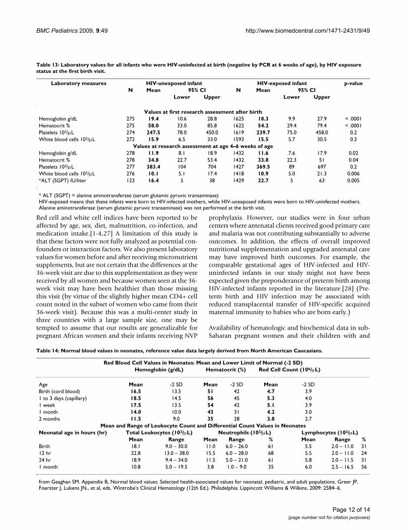
We categorized HIV-uninfected infants as to whether theywere born to HIV-infected mothers (termed HIV-exposedinfants) or to HIV-uninfected mothers (termed HIV-unex-posed infants). HIV-exposed, but uninfected infants hadlower hemoglobin and hematocrit levels than HIV-unex-posed infants both at birth (both p < 0.0001) and at 4–6weeks (both p < 0.05; Table 13). Platelet counts did notdiffer, while at the visit at 4–6 weeks of infant age, bothwhite blood cells and ALT were higher among exposedinfants (Table 13).
Table 4: Comparison of HIV-infected women with CD4+ cell counts <200/μL to HIV-uninfected women
HIV (+) CD4 < 200/μL HIV (-)Laboratory measures Mean N P2.75 P97.5 Mean N P2.75 P97.5 P value
Study visit at 20–24 weeks gestationRed blood cells 106/μL 3.4 448 2.4 4.6 3.8 367 2.9 4.9 < .0001Hemoglobin g/dL 9.6 447 7.0 12.0 11.0 367 8.2 13.2 < .0001Hematocrit % 28.5 447 21.3 35.9 32.3 367 23.8 39.4 < .0001Platelets 103/μL 226.0 449 100 391 204.3 367 97 350 < .0001White blood cells 103/μL 5.2 449 2.7 9 6.9 366 3.3 11.1 < .0001Lymphocytes % 28.0 435 12.8 55.1 31.5 358 19 57 < .0001Monocytes % 8.9 435 2.4 23.8 6.8 358 1.9 17 < .0001Granulocytes % 63.0 436 24 81.8 61.7 358 28.6 77.5 0.1CD4+ cells/μL 130 456 26 198 809.6 246 344 1366 < .0001CD8+ cells/μL 661 456 192 1615 513.5 246 262 1002 < .0001CD4 % 14.0 89 3 29 47.6 55 33 65 < .0001CD8 % 59.7 89 15 88 31.7 55 18 50 < .0001Total T-Lymphocytes as % of all lymphocytes 69.6 76 17 92 75.6 47 60 93 0.04
Study visit at 36 weeks gestation
Red blood cells 106/μL 3.7 279 2.6 4.9 3.9 280 2.9 5.3 < .0001Hemoglobin g/dL 10.4 279 7.4 13.2 11.3 280 9.0 14.0 < .0001Hematocrit % 30.8 279 22.6 39.0 33.3 280 25.9 41.9 < .0001Platelets 103/μL 208.8 279 77 419 182.9 280 87 345 < .0001White blood cells 103/μL 5.0 279 2.4 8.5 6.7 280 3.8 11.2 < .0001Lymphocytes % 28.7 279 13.5 51.6 31.8 274 18.1 53.6 < .0001Monocytes % 8.8 279 2.6 21.9 7.5 274 1.1 23.1 0.003Granulocytes % 62.5 279 35.6 80.4 60.7 274 32.6 76.5 0.06CD4+ cells/μL 131 280 19 196 786 166 368 1325 < .0001CD8+ cells/μL 656 280 215 1524 507 166 216 1106 < .0001CD4 % 13.8 49 3 29 45.0 49 28 66 < .0001CD8 % 60.2 49 34 88 31.1 49 21 43 < .0001Total T-Lymphocytes as % of all lymphocytes 74.9 47 50 93 74.1 43 54 90 0.7
Laboratory values for pregnant women at the 20–24-week gestational age visit and at the 36-week visit. Of the 2633 women with data at baseline, 727 (27.6%) were missing week 36 data and 1906 (72.4%) had 36-week data. It is possible that week 36 women were slightly healthier than week 20–24 women, since the 36 week women had a slightly higher CD4+ cell count and may have been more likely to be able to keep the 36-week appointment (see discussion).
Lower 95% confidence interval = P2.75 = 2.75%ile
Upper 95% confidence interval = P97.5 = 97.5%ile
Page 6 of 14(page number not for citation purposes)

BMC Pediatrics 2009, 9:49 http://www.biomedcentral.com/1471-2431/9/49
Among HIV-uninfected infants (by PCR at 6 weeks ofage), HIV-exposed infants had a 37-gram lower meanbirth weight compared to HIV-unexposed infants (2980 ±505 grams; 95% ci: 2957–3005 vs. 3079 ± 515 grams;95% ci: 3023–3135, respectively; p = 0.002).
Discussion and ConclusionA four-site clinical trial in Malawi, Tanzania, and Zambiaenabled us to document hematological, biochemical, andimmunological measures in a large cohort of pregnantAfrican women with and without HIV infection, and intheir infants. The infants were either HIV-infected, HIV-exposed but uninfected, or were uninfected and unex-posed to HIV. HIV-infected pregnant women had lowerred blood cell counts, hemoglobins, hematocrits andWBC counts than the HIV-uninfected cohort. However,
platelet and monocyte counts were higher in HIV-infectedwomen than in the HIV-uninfected women. Although val-ues were similar at birth, HIV-infected infants had lowerhemoglobin, hematocrit, and platelet counts at 4–6 weeksof age, but had higher ALT values, compared to HIV-unin-fected infants. Among the HIV-uninfected infants, theHIV-exposed babies had lower hemoglobins/hematocrits,but had higher white blood cell counts and ALT levelsthan did the HIV-unexposed infants at 4–6 weeks of age.
As one might expect, HIV-infected women were morelikely to be anemic (hemoglobin <12 g/dL) than HIV-uninfected women.[1] One possible cause of anemia inthese women could be the effect of HIV infection onhematopoiesis. The mean maternal hemoglobin levels inboth HIV-infected and HIV-uninfected women in this
Table 5: Comparison of HIV-infected women with CD4+ cell counts 200–349/μL to HIV-uninfected women
HIV (+) CD4 200–349/μL HIV (-)Laboratory measures Mean N P2.75 P97.5 Mean N P2.75 P97.5 P value
Study visit at 20–24 weeks gestationRed blood cells 106/μL 3.5 608 2.53 4.52 3.8 367 2.65 4.92 < .0001Hemoglobin g/dL 10.0 607 6.7 12.2 11.0 367 8.2 13.2 < .0001Hematocrit % 29.5 608 21 36.4 32.3 367 23.8 39.4 < .0001Platelets 103/μL 231.9 608 101 413 204.3 367 97 350 < .0001White blood cells 103/μL 5.7 608 2.6 9.1 6.9 366 3.3 11.1 < .0001Lymphocytes % 30.4 597 16.4 58.1 31.5 358 19 57 0.0778Monocytes % 8.4 597 2.1 21.4 6.8 358 1.9 17 < .0001Granulocytes % 60.8 597 21.1 78.7 61.7 358 28.6 77.5 0.3CD4 cells/μL 272.8 612 202 346 809.6 246 344 1366 < .0001CD8 cells/μL 737.0 612 284 1589 513.5 246 262 1002 < .0001CD4 % 22.7 92 12 37 47.6 55 33 65 < .0001CD8 % 57.3 92 31 82 31.7 55 18 50 < .0001Total T-Lymphocytes as % of all lymphocytes 76.4 75 48 98 75.6 47 60 93 0.7
Study visit at 36 weeks gestation
Red blood cells 106/μL 3.7 391 2.75 4.87 3.9 280 2.865 5.34 < .0001Hemoglobin g/dL 10.5 391 7.9 13 11.3 280 9 13.95 < .0001Hematocrit % 31.1 391 22.4 38.5 33.3 280 25.9 41.85 < .0001Platelets 103/μL 201.6 391 88 356 182.9 280 86.5 344.5 0.0005White blood cells 103/μL 5.6 391 3.3 8.9 6.7 280 3.8 11.2 < .0001Lymphocytes % 30.8 387 16.2 50.7 31.8 274 18.1 53.6 0.1Monocytes % 8.8 387 2.4 22.5 7.5 274 1.1 23.1 0.0020Granulocytes % 60.2 387 30.4 78.5 60.7 274 32.6 76.5 0.6CD4+ cells/μL 274.8 395 202 346 786.0 166 368 1325 < .0001CD8+ cells/μL 757.2 395 313 1731 506.8 166 216 1106 < .0001CD4 % 24.3 66 13 42 45.0 49 28 66 < .0001CD8 % 54.1 66 32 73 31.1 49 21 43 < .0001Total T-Lymphocytes as % of all lymphocytes 78.0 63 61 91 74.1 43 54 90 0.02
Laboratory values for pregnant women at the 20–24-week gestational age visit and at the 36-week visit. Of the 2633 women with data at baseline, 727 (27.6%) were missing week 36 data and 1906 (72.4%) had 36-week data. It is possible that week 36 women were slightly healthier than week 20–24 women, since the 36 week women had a slightly higher CD4+ cell count and may have been more likely to be able to keep the 36-week appointment (see discussion).
Lower 95% confidence interval = P2.75 = 2.75%ile
Upper 95% confidence interval = P97.5 = 97.5%ile
Page 7 of 14(page number not for citation purposes)

BMC Pediatrics 2009, 9:49 http://www.biomedcentral.com/1471-2431/9/49
study were lower than the values reported elsewhere forAfrican pregnant women (mean = 12.1 g/dL).[1] The lev-els of hemoglobin in this study are similar to thosereported previously for pregnant Indian women (mean =11.1 ± 1.6 g/dL).[15] Lower hemoglobin levels in thisstudy may be seen in individuals with malarial infection,but no recruited women had suspected malaria and allwere recruited at urban sites with low prevalence rates ofmalaria. A Nigerian study found that anemia was twice ascommon in HIV-infected subjects who had malaria para-sites, compared to non-parasitized controls.[16] In thisstudy, routine malaria blood smears were not done at thetime of laboratory testing. No women were on zidovudineas potent antiretroviral therapy and zidovudine forPMTCT were not in use at the time of the study. Womenwere supplemented with iron during pregnancy, so the
true magnitude of anemia may be greater than what wereport from this clinical trial.
Mean WBC counts were within the ranges of those previ-ously described.[1] The WBC count for normal male andfemale adults is 4,500–11,000/μL (range is estimate of95% confidence limits).[17] One study reported thatalthough changes in leukocyte counts during pregnancyin African women were similar to those reported in Cau-casian women, the total WBC counts were lower in theAfrican women.[5]
Although hematological parameters were comparable atbirth between HIV-infected infants and HIV-uninfectedinfants, HIV-infected infants showed a greater decrease inhemoglobin concentrations than HIV-uninfected infants
Table 6: Comparison of HIV-infected women with CD4+ cell counts 350–500/μL to HIV-uninfected women
HIV (+) CD4 350–500/μL HIV (-)Laboratory measures Mean N P2.75 P97.5 Mean N P2.75 P97.5 P value
Study visit at 20–24 weeks gestationRed blood cells 106/μL 3.6 488 2.7 4.6 3.8 367 2.7 4.9 < .0001Hemoglobin g/dL 10.3 486 7.1 12.7 11.0 367 8.2 13.2 < .0001Hematocrit % 30.2 488 21.6 37.4 32.3 367 23.8 39.4 < .0001Platelets 103/μL 227.8 488 103 399 204.3 367 97 350 < .0001White blood cells 103/μL 6.2 488 2.8 9.5 6.9 366 3.3 11.1 < .0001Lymphocytes % 32.2 481 19.0 62.5 31.5 358 19.0 57.0 0.3Monocytes % 7.7 481 1.7 17.7 6.8 358 1.9 17.0 0.004Granulocytes % 59.9 481 21.4 75.5 61.7 358 28.6 77.5 0.03CD4+ cells/μL 418 489 352 495 810 246 344 1366 < .0001CD8+ cells/μL 788 489 349 1602 514 246 262 1002 < .0001CD4 % 28.6 49 17 52 47.6 55 33 65 < .0001CD8 % 51.7 49 22 77 31.7 55 18 50 < .0001Total T-Lymphocytes as % of all lymphocytes 77.0 40 46 96.5 75.6 47 60 93 0.5
Study visit at 36 weeks gestation
Red blood cells 106/μL 3.8 356 2.8 4.9 3.9 280 2.9 5.3 0.002Hemoglobin g/dL 10.7 356 8 13.5 11.3 280 9 14.0 < .0001Hematocrit % 31.7 356 23.3 40 33.3 280 25.9 41.9 < .0001Platelets 103/μL 214.0 356 86 448 182.9 280 87 345 < .0001White blood cells 103/μL 6.1 356 3.3 9.9 6.7 280 3.8 11.2 < .0001Lymphocytes % 31.6 356 19.3 53.4 31.8 274 18.1 53.6 0.8Monocytes % 8.6 356 2.5 23.5 7.5 274 1.1 23.1 0.01Granulocytes % 59.6 356 29.5 75.0 60.7 274 32.6 76.5 0.2CD4+ cells/μL 418 359 353 495 786 166 368 1325 < .0001CD8+ cells/μL 779 359 340 1515 507 166 216 1106 < .0001CD4 % 28.2 36 12 50 45.0 49 28 66 < .0001CD8 % 51.8 36 20 76 31.1 49 21 43 < .0001Total T-Lymphocytes as % of all lymphocytes 78.4 34 57 92 74.1 43 54 90 0.05
Laboratory values for pregnant women at the 20–24-week gestational age visit and at the 36-week visit. Of the 2633 women with data at baseline, 727 (27.6%) were missing week 36 data and 1906 (72.4%) had 36-week data. It is possible that week 36 women were slightly healthier than week 20–24 women, since the 36 week women had a slightly higher CD4+ cell count and may have been more likely to be able to keep the 36-week appointment (see discussion).
Lower 95% confidence interval = P2.75 = 2.75%ile
Upper 95% confidence interval = P97.5 = 97.5%ile
Page 8 of 14(page number not for citation purposes)
BMC Pediatrics 2009, 9:49 http://www.biomedcentral.com/1471-2431/9/49
in the first 4–6 weeks of life. Similar findings have beenfound in a large cohort of HIV-infected and uninfectedinfants in Malawi.[8] In this study, hemoglobin levelswere lower among HIV-infected infants whose mothershad received NVP prophylaxis, and higher among HIV-uninfected infants who were not exposed to NVP prophy-laxis. We were not able to assess whether the lower hemo-globin levels seen in HIV-infected infants in HPTN024were due to HIV infection or NVP exposure, since nearlyall HIV-infected women and their infants in this cohortreceived NVP prophylaxis.
HIV-infected infants had higher ALT levels than HIV-unin-fected infants at 4–6 weeks of age. Increased ALT levels ininfants may suggest acute liver inflammation, though ALTvalues are variable in infants and may be linked to NVP
exposure.[8] Given near universal NVP prophylaxis in ourcohort, we were not able to determine whether the higherALT levels were related to HIV infection status per se, orNVP exposure. The higher ALT level in the HIV-exposedinfants who were not infected, compared to HIV-unin-fected and unexposed infants, may also have been due toNVP exposure, but this will require further study. Themagnitude of the ALT rise was not substantial and thehighest ALT in an HIV-exposed (and NVP-exposed) infantwas only 63 IU/liter (Table 9).
The mean hemoglobin value for normal infants has beenreported as 18.5 g/dL in a neonate ages 1–3 days in stand-ard hematological references (Table 14),[17] similar toour study and a study in Malawi.[8] Perhaps the iron-con-taining vitamin and mineral supplements in our mothers
Table 7: Comparison of HIV-infected women with CD4+ cell counts >500/μL to HIV-uninfected women
HIV (+) CD4 > 500/μL HIV (-)Laboratory measures Mean N P2.75 P97.5 Mean N P2.75 P97.5 P value
Study visit at 20–24 weeks gestationRed blood cells 106/μL 3.7 512 2.8 4.8 3.8 367 2.7 4.9 0.01Hemoglobin g/dL 10.5 512 7.7 13 11.0 367 8.2 13.2 < .0001Hematocrit % 30.9 512 21.5 38.5 32.3 367 23.8 39.4 < .0001Platelets 103/μL 242.9 512 111 453 204.3 367 97 350 < .0001White blood cells 103/μL 7.2 513 3.5 12.0 6.9 366 3.3 11.1 0.02Lymphocytes % 33.7 506 20.1 60.1 31.5 358 19.0 57.0 0.0005Monocytes % 7.7 506 1.2 21.2 6.8 358 1.9 17.0 0.008Granulocytes % 58.2 506 21.5 73.4 61.7 358 28.6 77.5 < .0001CD4+ cells/μL 670 515 504 1090 810 246 344 1366 < .0001CD8+ cells/μL 893 515 364 1919 514 246 262 1002 < .0001CD4 % 34.4 56 16 51 47.6 55 33 65 < .0001CD8 % 48.1 56 28 71 31.7 55 18 50 < .0001Total T-Lymphocytes as % of all lymphocytes 77.4 43 55 94 75.6 47 60 93 0.4
Study visit at 36 weeks gestation
Red blood cells 106/μL 3.9 422 2.9 5.0 3.9 280 2.9 5.3 0.3Hemoglobin g/dl 11.0 422 8.4 13.2 11.3 280 9.0 14.0 0.003Hematocrit % 32.7 422 23.6 40.0 33.3 280 25.9 41.9 0.06Platelets 103/μL 212.2 422 104 382 183 280 87 345 < .0001White blood cells 103/μL 7.1 421 4.3 11.0 6.7 280 3.8 11.2 0.01Lymphocytes % 33.3 417 18.5 50.3 31.8 274 18.1 53.6 0.01Monocytes % 7.9 417 1.9 19.1 7.5 274 1.1 23.1 0.4Granulocytes % 58.7 417 33.8 74.2 60.7 274 32.6 76.5 0.02CD4+ cells/μL 677 425 507 1155 786 166 368 1325 < .0001CD8+ cells/μL 870 425 395 1639 507 166 216 1106 < .0001CD4 % 35.2 33 16 53 45.0 49 28 66 < .0001CD8 % 47.1 33 29 68 31.1 49 21 43 < .0001Total T-Lymphocytes as % of all lymphocytes 79.3 28 60 91 74.1 43 54 90 0.02
Laboratory values for pregnant women at the 20–24-week gestational age visit and at the 36-week visit. Of the 2633 women with data at baseline, 727 (27.6%) were missing week 36 data and 1906 (72.4%) had 36-week data. It is possible that week 36 women were slightly healthier than week 20–24 women, since the 36 week women had a slightly higher CD4+ cell count and may have been more likely to be able to keep the 36-week appointment (see discussion).
Lower 95% confidence interval = P2.75 = 2.75%ile
Upper 95% confidence interval = P97.5 = 97.5%ile
Page 9 of 14(page number not for citation purposes)

BMC Pediatrics 2009, 9:49 http://www.biomedcentral.com/1471-2431/9/49
improved infants' hemoglobin levels, as noted in a Tanza-nian study.[11] These values are 2 g/dL higher than thosereported in two other African studies done in Malawi andNigeria that suggested that the hemoglobin values at birthwere lower in African than Caucasian children.[2,6] Thehemoglobin levels in this study were also higher thanthose found in a study of Italian neonates in which themean hemoglobin level in term, appropriate-for-ageneonates was only 14.4 (± 4.4) g/dL.[18] Two studies inIndia and one in Turkey also reported lower hemoglobinlevels than found in studying our African new-borns.[15,19,20] Newborn hemoglobin and hematocritlevels may be influenced by many factors, including themode of delivery.[21] For example, if a midwife holds ababy a bit lower than the level of the mother's pelvis andthe not-yet-delivered placenta immediately after birth and
before the cord is clamped, the baby's hemoglobin can beraised. WBC counts in infants in HPTN024 are within thenormal ranges of standard references for largely Caucasianinfants whose mean WBC value for normal infants at birthis 18.1/μL (Table 14).[17]
Hematological and biochemical measurements can alsobe useful for clinical monitoring of HIV-infected individ-uals when viral load testing and CD4+ cell count monitor-ing are not readily available.[22] Whether they could beuseful surrogates for pregnant women or young childrenis not known. There are findings that will require specificstudy, including the higher platelet counts noted in ourHIV-infected mothers, but lower platelet counts in theHIV-infected infants, for which we do not have a readyexplanation.[2,9,23-26]
Table 8: Comparison of laboratory values at 20–24 weeks vs. 36 weeks gestation among HIV-infected women
Laboratory Parameter Number of Pairs Mean (*) of the paired differences 95% CI p value*Lower Upper
Red blood cell count 106/μL 1626 0.17 0.23 < 0.0001Hemoglobin g/dl 1623 0.5 0.4 0.6 < 0.0001Hematocrit % 1626 1.5 1.3 1.7 < 0.0001Platelets 103/μL 1625 -23.9 -27.5 -20.4 < 0.0001White blood cell count 103/μL 1623 -0.1 -0.2 0.1 0.3Lymphocytes % 1570 0.5 0.0 1.0 0.1Monocytes % 1570 0.5 0.2 0.9 0.002Granulocytes % 1569 -0.8 -1.6 -0.1 0.02CD4+ T lymphocyte count cells/μL 1343 14.8 7.1 22.5 0.0002CD8+ T lymphocyte count cells/μL 1343 8.5 -6.6 23.5 0.3CD4+ T lymphocyte % 107 -2.3 -3.7 -0.8 0.003CD8+ T lymphocyte % 107 -2.5 -4.6 -0.4 0.02Total T lymphocytes as a % of all lymphocytes 74 -1.3 -3.7 1.1 0.3
* For each pair, the 20–24 week laboratory value is subtracted from the 36 week value. Hence, a negative value indicates a decrease over time in the laboratory parameter.
Table 9: Comparison of laboratory values at 20–24 weeks vs. 36 weeks gestation among HIV-uninfected women
Laboratory Parameter Number of Pairs Mean (*) of the paired differences 95% CI p value*Lower Upper
Red blood cells count 106/μL 280 0.2 0.1 0.3 < 0.0001Hemoglobin g/dl 280 0.2 0.1 0.4 0.01Hematocrit % 280 0.7 0.1 1.3 0.01Platelets 103/μL 280 -17.6 -25.1 -10.0 < 0.0001White blood cell count 103/μL 279 -0.2 -0.4 0.1 0.2Lymphocytes % 266 0.1 -1.1 1.2 0.9Monocytes % 266 0.9 0.1 1.7 0.02Granulocytes % 266 -0.9 -2.5 0.6 0.2CD4+ T lymphocyte count cells/μL 134 -24.2 -71.9 23.6 0.3CD8+ T lymphocyte count cells/μL 134 -12.5 -52.5 27.5 0.5CD4+ T lymphocyte % 25 -2.5 -6.2 1.3 0.2CD8+ T lymphocyte % 25 -1.2 -3.6 1.3 0.3Total T lymphocytes as a % of all lymphocytes 19 1.1 -3.1 5.2 0.6
(*) For each pair, the 20–24 week laboratory value is subtracted from the 36 week value. Hence, a negative value indicates a decrease over time in the laboratory parameter.
Page 10 of 14(page number not for citation purposes)

BMC Pediatrics 2009, 9:49 http://www.biomedcentral.com/1471-2431/9/49
Page 11 of 14(page number not for citation purposes)
Table 10: Laboratory values among HIV-infected and HIV-uninfected infants (both exposed and unexposed to HIV) at birth
Laboratory Parameter
HIV-uninfected Infants HIV-infected Infants P value
n Mean ± Standard Deviation
95% CI n Mean ± Standard Deviation
95% CI
Lower Upper Lower Upper
Hemoglobin g/dl 1900 18.5 ± 4.3 10.1 28.2 149 18.0 ± 3.7 10.4 24.6 0.1
Hematocrit % 1897 54.8 ± 12.1 29.7 80.2 149 53.5 ± 10.8 29.8 72.3 0.2
Platelets 103/μL 1893 240.8 ± 102.7 76 458 147 207.0 ± 97.3 37 419 < 0.0001
White blood cell count 103/μL
1865 15.5 ± 6.2 5.8 30.6 145 15.4 ± 10.5 3.4 39.7 0.8
Table 11: Laboratory values among HIV-infected and HIV-uninfected infants at 4–6 weeks of age
Laboratory Parameter
HIV-uninfected Infants HIV-infected Infants P value
n Mean ± Standard Deviation
95% CI n Mean ± Standard Deviation
95% CI
Lower Upper Lower Upper
Hemoglobin g/dl 1710 11.6 ± 2.6 7.7 18 269 11.2 ± 2.8 6.6 18.6 < 0.007
Hematocrit % 1710 34.0 ± 7.3 22.4 51.5 269 32.9 ± 8.5 18 55.2 < 0.02
Platelets 103/μL 1704 371.8 ± 156.3 90 697 269 306.5 ± 150.7 55 626 < 0.0001
White blood cell Count 103/μL
1694 10.7 ± 4.2 5 20.9 268 11.6 ± 5.3 4.5 25.2 < 0.002
*ALT (SGPT) IU/L 1552 22.2 ± 24.1 5 62 272 30.8 ± 33.9 5 133 < 0.001
*ALT (SGPT) = alanine aminotransferase (serum glutamic pyruvic transaminase)
Table 12: Comparison of laboratory values at birth vs. 4–6 weeks of age among HIV-infected and HIV-uninfected infants
Laboratory Parameter
HIV-infected Infants HIV-uninfected Infants p value
Number of Pairs
Mean (*) of the paired
differences
95% CI Number of Pairs
Mean (*) of the paired
differences
95% CI
Lower Upper Lower Upper
Hemoglobin g/dl 124 -7.1 -7.8 -6.4 1301 -7.0 -7.2 -6.8 < 0.0001Hematocrit % 124 -21.0 -23.0 -19.0 1298 -20.9 -21.6 -20.3 < 0.0001Platelets 103/μL 122 89.2 64.0 114.5 1292 125.1 115.9 134.3 < 0.0001White blood cell count 103/μL
119 -2.0 -3.4 0.5 1263 -4.8 -5.2 -4.5 0.009
(*) For each pair, the birth laboratory value is subtracted from the 4–6 week value. Hence a negative value indicates a decrease over time in the laboratory parameter.
BMC Pediatrics 2009, 9:49 http://www.biomedcentral.com/1471-2431/9/49
Red cell and white cell indices have been reported to beaffected by age, sex, diet, malnutrition, co-infection, andmedication intake.[1-4,27] A limitation of this study isthat these factors were not fully analyzed as potential con-founders or interaction factors. We also present laboratoryvalues for women before and after receiving micronutrientsupplements, but are not certain that the differences at the36-week visit are due to this supplementation as they werereceived by all women and because women seen at the 36-week visit may have been healthier than those missingthis visit (by virtue of the slightly higher mean CD4+ cellcount noted in the subset of women who came from their36-week visit). Because this was a multi-center study inthree countries with a large sample size, one may betempted to assume that our results are generalizable forpregnant African women and their infants receiving NVP
prophylaxis. However, our studies were in four urbancenters where antenatal clients received good primary careand malaria was not contributing substantially to adverseoutcomes. In addition, the effects of overall improvednutritional supplementation and upgraded antenatal caremay have improved birth outcomes. For example, thecomparable gestational ages of HIV-infected and HIV-uninfected infants in our study might not have beenexpected given the preponderance of preterm birth amongHIV-infected infants reported in the literature.[28] (Pre-term birth and HIV infection may be associated withreduced transplacental transfer of HIV-specific acquiredmaternal immunity to babies who are born early.)
Availability of hematologic and biochemical data in sub-Saharan pregnant women and their children with and
Table 14: Normal blood values in neonates, reference value data largely derived from North American Caucasians.
Red Blood Cell Values in Neonates: Mean and Lower Limit of Normal (-2 SD)Hemoglobin (g/dL) Hematocrit (%) Red Cell Count (106/μL)
Age Mean -2 SD Mean -2 SD Mean -2 SDBirth (cord blood) 16.5 13.5 51 42 4.7 3.91 to 3 days (capillary) 18.5 14.5 56 45 5.3 4.01 week 17.5 13.5 54 42 5.1 3.91 month 14.0 10.0 43 31 4.2 3.02 months 11.5 9.0 35 28 3.8 2.7
Mean and Range of Leukocyte Count and Differential Count Values in NeonatesNeonatal age in hours (hr) Total Leukocytes (103/μL) Neutrophils (103/μL) Lymphocytes (103/μL)
Mean Range Mean Range % Mean Range %Birth 18.1 9.0 – 30.0 11.0 6.0 – 26.0 61 5.5 2.0 – 11.0 3112 hr 22.8 13.0 – 38.0 15.5 6.0 – 28.0 68 5.5 2.0 – 11.0 2424 hr 18.9 9.4 – 34.0 11.5 5.0 – 21.0 61 5.8 2.0 – 11.5 311 month 10.8 5.0 – 19.5 3.8 1.0 – 9.0 35 6.0 2.5 – 16.5 56
from Geaghan SM. Appendix B, Normal blood values: Selected health-associated values for neonatal, pediatric, and adult populations. Greer JP, Foerster J, Lukens JN., et al, eds. Wintrobe's Clinical Hematology (12th Ed.). Philadelphia: Lippincott Williams & Wilkins, 2009: 2584–6.
Table 13: Laboratory values for all infants who were HIV-uninfected at birth (negative by PCR at 6 weeks of age), by HIV exposure status at the first birth visit.
Laboratory measures HIV-unexposed infant HIV-exposed infant p-valueN Mean 95% CI N Mean 95% CI
Lower Upper Lower Upper
Values at first research assessment after birthHemoglobin g/dL 275 19.4 10.6 28.8 1625 18.3 9.9 27.9 < .0001Hematocrit % 275 58.0 33.0 85.8 1622 54.2 29.4 79.4 < .0001Platelets 103/μL 274 247.5 78.0 450.0 1619 239.7 75.0 458.0 0.2White blood cells 103/μL 272 15.9 6.5 33.0 1593 15.5 5.7 30.5 0.3
Values at research assessment at age 4–6 weeks of ageHemoglobin g/dL 278 11.9 8.1 18.9 1432 11.6 7.6 17.9 0.02Hematocrit % 278 34.8 22.7 53.4 1432 33.8 22.3 51 0.04Platelets 103/μL 277 383.4 104 704 1427 369.5 89 697 0.2White blood cells 103/μL 276 10.1 5.1 17.4 1418 10.9 5.0 21.3 0.006*ALT (SGPT) IU/liter 123 16.4 5 38 1429 22.7 5 63 0.005
* ALT (SGPT) = alanine aminotransferase (serum glutamic pyruvic transaminase)HIV-exposed means that these infants were born to HIV-infected mothers, while HIV-unexposed infants were born to HIV-uninfected mothers. Alanine aminotransferase (serum glutamic pyruvic transaminase) was not performed at the birth visit.
Page 12 of 14(page number not for citation purposes)

BMC Pediatrics 2009, 9:49 http://www.biomedcentral.com/1471-2431/9/49
without HIV infection can assist in clinical program devel-opment, clinical research design and planning, training ofboth health care providers, and community education.Our large study of poor pregnant women who receivedvitamin and nutrient supplementation and their offspringin urban Africa provides new normative data that will beuseful comparison values for clinicians managing similarpatients with and without HIV.
Competing interestsThe authors declare that they have no competing interests.
Authors' contributionsKM, SHV, and RLG conceptualized the manuscript,designed the analyses, and drafted the manuscript. YQCand AM participated in the design of analyses, crafted thetables from the dataset, and performed the statistical anal-yses. JSR and MV oversaw the parent clinical trial and pro-vided writing and editing input to the manuscript drafts.KM, WU, and NC collected the data and helped conceptu-alize the analysis. All authors read and approved the finalmanuscript. Each author has participated sufficiently inthe work to take public responsibility for appropriate por-tions of the content.
AcknowledgementsThe authors thank the participants in the HPTN 024 study and the following members of the Protocol Team: Protocol Co-Chairs: Taha E. Taha, MD, PhD (Johns Hopkins University Bloomberg School of Public Health); Robert Goldenberg, MD (University of Alabama at Birmingham); In-Country Co-Chairs/Investigators of Record: Newton Kumwenda, PhD, George Kafula-fula, MBBS, FCOG (Blantyre, Malawi); Francis Martinson, MD, PhD (Lilongwe, Malawi); Gernard Msamanga, MD, ScD (Dar es Salaam, Tanza-nia); Moses Sinkala, MD, MPH, Jeffrey Stringer, MD (Lusaka, Zambia); US Co-Chairs: Irving Hoffman, PA, MPH (University of North Carolina, Chapel Hill); Wafaie Fawzi, MD, DrPH (Harvard School of Public Health); In-Coun-try Investigators, Consultants and Key Site Personnel: Robin Broadhead, MBBS, FRCP, George Liomba, MBBS, FRCPath, Johnstone Kumwenda, MBChB, MRCP, Tsedal Mebrahtu, ScM, Pauline Katunda, MHS, Maysoon Dahab, MHS (Blantyre, Malawi); Peter Kazembe, MBChB, David Chilongozi CO, MPH, Charles Chasela CO, MPH, George Joaki, MD, Willard Dzinyemba, Sam Kamanga (Lilongwe, Malawi); Elgius Lyamuya, MD, PhD, Charles Kilewo, MD, MMed, Karim Manji, MD, MMed, Sylvia Kaaya, MD, MS, Said Aboud, MD, MMed, Muhsin Sheriff MD, MPH, Elmar Saathoff, PhD, Priya Satow, MPH, Illuminata Ballonzi, SRN, Gretchen Antelman, ScD, Edgar Basheka, BPharm (Dar-es-Salaam, Tanzania); Victor Mudenda, MD, Christine Kaseba, MD, Maureen Njobvu, MD, Makungu Kabaso, MD, Muz-ala Kapina, MD, Anthony Yeta, MD, Seraphine Kaminsa, MD, MPH, Con-stantine Malama, MD, Dara Potter, MBA, Maclean Ukwimi, RN, Alison Taylor, BSc, Patrick Chipaila, MSc, Bernice Mwale, BPharm (Lusaka, Zam-bia); U.S. Investigators, Consultants and Key Site Personnel: Priya Joshi, BS, Ada Cachafeiro, BS, Shermalyn Greene, PhD, Marker Turner, BS, Melissa Kerkau, BS, Paul Alabanza, BS, Amy James, BS, Som Siharath, BS, Tiffany Tribull, MS (UNC-CH); Saidi Kapiga, MD, ScD, George Seage, PhD (HSPH); Sten H. Vermund, MD, PhD, William Andrews, PhD, MD, Deedee Lyon, BS, MT(ASCP) (UAB); NIAID Medical Officer: Samuel Adeniyi-Jones, MD; NICHD Medical Officer: Jennifer S. Read, MD, MS, MPH, DTM&H; Proto-col Pharmacologist: Scharla Estep, RPh, MS; Protocol Statisticians: Elizabeth R. Brown, ScD, Thomas R. Fleming, PhD, Anthony Mwatha, MS, Lei Wang,
PhD, Ying Q. Chen, PhD; Protocol Virologist: Susan Fiscus, PhD; Protocol Operations Coordinator: Lynda Emel, PhD; Data Coordinators: Debra J. Lands, Ed.M, Ceceilia J. Dominique; Systems Analyst Programmers: Alice H. Fisher, BA, Martha Doyle; Protocol Specialist: Megan Valentine, PA-C, MS. Meredith Bortz contributed editorial expertise.
Sources of Support:
This study was supported by the HIV Network for Prevention Trials (HIVNET) and sponsored by the U.S. National Institute of Allergy and Infec-tious Diseases (NIAID), National Institutes of Health, Department of Health and Human Services, through contract N01-AI-035173 with Family Health International; contract N01-AI-045200 with Fred Hutchinson Can-cer Research Center; and subcontract N01-AI-035173-117/412 with Johns Hopkins University. In addition, this work was supported by the HIV Pre-vention Trials Network (HPTN) and sponsored by the National Institute of Allergy and Infectious Diseases, National Institute of Child Health and Human Development, National Institute on Drug Abuse, National Institute of Mental Health, and Office of AIDS Research, of the National Institutes of Health, U.S. Department of Health and Human Services, Harvard Univer-sity (U01-AI-048006), Johns Hopkins University (U01-AI-048005), the Uni-versity of Alabama at Birmingham (U01-AI-047972), and Family Health International (U01-AI-068619). Nevirapine (Viramune®) was provided for the HPTN024 protocol by Boehringer Ingelheim Pharmaceuticals, Inc. The conclusions and opinions expressed in this paper are those of the authors and do not necessarily reflect those of the funding agencies and participating institutions.
References1. Beers MH, Porter RS, Jones TV, Kaplan JL, Berkwits M: Approach to
the patient with anemia: Hematology and Oncologyin the Merck Manual ofDiagnosis and Therapy Volume Chapter 3. 18th edition. Issue Section 11Merck Research Laboratories, Division of Merck & Co., Inc. White-house Station NJ; 2006:1031-1033.
2. Mukiibi JM, Mtimavalye LAR, Broadhead R, et al.: Some hematolog-ical parameters in Malawian neonates. East Afr Med J 1995,72(1):10-14.
3. Miller MF, Stoltzfus RJ, Iliff PJ, et al.: Effect of maternal and neona-tal vitamin A supplementation and other postnatal factorson anemia in Zimbabwean infants: a prospective, rand-omized study. Am J Clin Nutr 2006, 84(1):212-222.
4. Quintó L, Aponte JJ, Sacarlal J, et al.: Hematological and biochem-ical indices in young African children: in search of referenceintervals. Trop Med Int Health 2006, 11(11):1741-1748.
5. Fleming AF, Harrison KA: Leukocyte counts during pregnancyand the puerperium and at birth in Nigerians. East Afr Med J1985, 62(3):175-184.
6. Scott-Emuakpor AB, Okolo AA, Omene JA, Ukpe SI: NormalHematological Values in the African Neonate. Blut 1985,51(1):11-18.
7. Stancheva VP, Sherman GG, Avent M, Cory BJ, Ballot DE, Cooper PA:Hematological reference ranges in black very low birthweight infants. Pediatr Hematol Oncol 2002, 19(2):91-94.
8. Taha TE, Kumwenda N, Gibbons A, et al.: Effect of HIV-1 antiret-roviral prophylaxis on hepatic and hematological parame-ters of African infants. AIDS 2002, 16(6):851-858.
9. Lugada ES, Mermin J, Kaharuza F, et al.: Downing R: Population-based hematologic and immunologic reference values for ahealthy Ugandan population. Clin Diagn Lab Immunol 2004,11(1):29-34.
10. Gomo E, Vennervald BJ, Ndhlovu P, Kaestel P, Nyazema N, Friis H:Predictors and reference values of CD4 and CD8 T lym-phocyte counts in pregnancy: a cross sectional study amongHIV-negative women in Zimbabwe. Cent Afr J Med 2004, 50(1–2):10-19.
11. Fawzi WW, Msamanga GI, Kupka R, et al.: Multivitamin supple-mentation improves hematologic status in HIV-infectedwomen and their children in Tanzania. Am J Clin Nutr 2007,85(5):1335-1343.
Page 13 of 14(page number not for citation purposes)

BMC Pediatrics 2009, 9:49 http://www.biomedcentral.com/1471-2431/9/49
Publish with BioMed Central and every scientist can read your work free of charge
"BioMed Central will be the most significant development for disseminating the results of biomedical research in our lifetime."
Sir Paul Nurse, Cancer Research UK
Your research papers will be:
available free of charge to the entire biomedical community
peer reviewed and published immediately upon acceptance
cited in PubMed and archived on PubMed Central
yours — you keep the copyright
Submit your manuscript here:http://www.biomedcentral.com/info/publishing_adv.asp
BioMedcentral
12. Embree J, Bwayo J, Nagelkerke N, et al.: Lymphocyte subsets inhuman immunodeficiency virus type 1-infected and unin-fected children in Nairobi. Pediatr Infect Dis J 2001,20(4):397-403.
13. Moodley D, Bobat RA, Coovadia HM, Doorasamy T, Munsamy S,Gouws E: Lymphocyte subset changes between 3 and 15months of age in infants born to HIV-seropositive women inSouth Africa. Trop Med Int Health 1997, 2(5):415-421.
14. Taha TE, Brown ER, Hoffman IF, et al.: A phase III clinical trial ofantibiotics to reduce chorioamnionitis-related perinatalHIV-1 transmission. AIDS 2006, 20(9):1313-1321.
15. Devi SB, Singh KJ, Devi YL, Singh WG: Maternal and neonatalanthropometric and haematological parameters in ManipuriPopulation. Indian Pediatr 1989, 26(7):673-677.
16. Erhabor O, Babatunde S, Uko K: Some hematological parame-ters in plasmodial parasitized HIV-infected Nigerians. Niger JMed 2006, 15(1):52-55.
17. Geaghan SM: Appendix B, Normal blood values: Selectedhealth-associated values for neonatal, pediatric, and adultpopulations. In Wintrobe's Clinical Hematology 12th edition. Editedby: Greer JP, Foerster J, Lukens JN, et al. Philadelphia: Lippincott Wil-liams & Wilkins; 2009:2584-6.
18. Ozyurek E, Cetintas S, Ceylan T, et al.: Complete blood countparameters for healthy, small-for-gestational-age, full-termnewborns. Clin Lab Haem 2006, 28(2):97-104.
19. Maconi M, Rolfo A, Cardaropoli S, Brini M, Danise P: Hematologi-cal values in health and small for gestational age newborns.Lab Hematol 2005, 11(2):152-156.
20. Marwaha N, Marwaha RK, Narang A, Thusu K, Garewal G, BhakooON: Routine haematological values in term newborns. IndianPediatr 1992, 29(9):1095-1099.
21. Redźko S, Przepiesć J, Zak J, Urban J, Wysocka J: Influence of peri-natal factors on hematological variables in umbilical cordblood. J Perinat Med 2005, 33(1):42-45.
22. Chen RY, Westfall AO, Hardin JM, et al.: Complete blood cellcount as a surrogate CD4+ cell marker for HIV monitoringin resource-limited settings. J Acquir Immune Defic Syndr 2007,44(5):525-530.
23. Mukiibi JM, Nkrumah FK, Kaur M, Akino V, Nhembe M: Neonatalhaematology in Zimbabwe. I: The platelet parameters. CentAfr J Med 1994, 40(4):80-83.
24. Ogala WN: Platelet counts in healthy Nigerian neonates andinfants. East Afr Med J 1986, 63(9):592-594.
25. Abdurrahman MB, Adekoje MA: Haematological values inNorthern Nigerian neonates. Trans R Soc Trop Med Hyg 1983,77(6):786-788.
26. Effiong CE, Usanga EA, Mellits ED: Platelet count in healthy full-term Nigerian neonates. Trop Geogr Med 1976, 28(4):329-332.
27. Modjarrad K, Chamot E, Vermund SH: Impact of small reductionsin plasma HIV RNA levels on the risk of heterosexual trans-mission and disease progression. AIDS 2008, 22(16):2179-2185.
28. Risk factors for mother-to-child transmission of HIV-1: EuropeanCollaborative Study. Lancet 1992, 339(8800):1007-1012.
Pre-publication historyThe pre-publication history for this paper can be accessedhere:
http://www.biomedcentral.com/1471-2431/9/49/prepub
Page 14 of 14(page number not for citation purposes)