se incorporated into zeolite mordenite-na: a single-crystal x-ray study
TRANSCRIPT

ARTICLE IN PRESS
www.elsevier.com/locate/micromeso
Microporous and Mesoporous Materials 71 (2004) 185–198
Se incorporated into zeolite mordenite-Na: a single-crystalX-ray study
Petra Simoncic, Thomas Armbruster *
Laboratorium f€ur Chemische und Mineralogische Kristallographie, University of Bern, Freiestrasse 3, CH-3012 Bern, Switzerland
Received 9 February 2004; received in revised form 24 March 2004; accepted 29 March 2004
Abstract
Single crystals of self-synthesized mordenite-Na were used for incorporation of elemental selenium. The mordenite sample was
first dehydrated at 280 �C and selenium was subsequently incorporated as gas phase at 450 �C for 72 h. Bright orange-colored Se-
loaded mordenite was quantitatively analyzed by an electron microprobe yielding Na6Al6Si42O96 � [Se7:9]. X-ray data collection of
mordenite-Na and Se-loaded mordenite-Na single-crystals were performed at 120 K with synchrotron radiation (k ¼ 0:79946 �A)
using the single-crystal diffraction line at SNBL (ESRF, Grenoble), where diffracted intensities were registered with a MAR image
plate. The structures of mordenite-Na and Se-mordenite-Na were both refined in the monoclinic space group Cc converging at
R1¼ 5.25% (mordenite-Na), and R1¼ 6.65% (Se-mordenite-Na). A strongly broadened Raman band at approximately 254 cm�1
confirmed the existence of Se chains in the 12-membered channels along the c-axis. Several, low-populated, disordered Se chains
with a length up to 10 �A and seven Se atoms were located in the large mordenite channels. During structure refinement nearest and
next nearest neighbor Se–Se distances were fixed at 2.34 and 3.62 �A, respectively. Other distances and angles remained uncon-
strained. Because of electrostatic interaction with the framework and influence of extraframework occupants such as Naþ and H2O
molecules, the chains show different geometrical Se arrangement with highly variable dihedral angles. Any other Se species such as
Se6 or Se8 rings were neither confirmed by structure refinement nor by Raman spectroscopy. There was no indication of a trigonal
Se chain geometry within the 12-membered ring channel.
� 2004 Published by Elsevier Inc.
Keywords: Zeolites; Mordenite; Single-crystal X-ray study; Semiconductor material; Selenium
1. Introduction
Microporous materials as zeolites are more and more
applied for the design and development of new materi-
als. With their channels and cavities of several ang-
stroms in size, they allow a spatial arrangement and
stabilization of individual atoms, clusters or molecules.
One aspect of modern technology and new materials are
one-dimensional systems and miniaturized electronic
devices with fast response, high selectivity, and effi-ciency, e.g. semiconductors organized as quantum dots
or chains in zeolites channels. Semiconductor materials
were encapsulated in synthetic zeolites such as AlPO4-5,
zeolites A and Y [1–3] with their uniform and large
channels (>7 �A), but also the naturally occurring
*Corresponding author. Fax: +41-31-631-3996.
E-mail address: [email protected] (T. Armbrus-
ter).
1387-1811/$ - see front matter � 2004 Published by Elsevier Inc.
doi:10.1016/j.micromeso.2004.03.031
mordenite with its compressed channels (7 · 6.5 �A) was
successfully used for molecular organization of atomsand molecules.
The structure of mordenite can be described as built
by edge-sharing 5-membered rings of tetrahedra (sec-
ondary building unit 5-1) forming chains along the c-axis [4]. However, the mordenite framework can also be
more comprehensibly envisioned as formed by puckered
sheets parallel to (1 0 0), assembled of 6-membered rings
of tetrahedra [5,6]. These sheets are interlinked by 4-membered rings (Fig. 1) in a way that large, ellipsoidal
12-membered (12MRc: aperture 7 · 6.5 �A) and strongly
compressed 8-membered rings (8MRc: aperture 5.7 · 2.6�A) define channels parallel to the c-axis. Another set of
compressed 8-membered rings (8MRb: aperture 3.4 · 4.8�A) connects the wide channels with the strongly com-
pressed channels parallel to the b-axis.Elemental selenium incorporated into mordenite is a
good example of a one-dimensional semiconductor and

Fig. 1. Tetrahedral framework structure of mordenite with unit-cell
outlines. The structure can be envisioned as built by puckered sheets
(light gray shading) parallel to (1 0 0) formed by 6-membered rings of
tetrahedra. These sheets are connected along a by 4-membered ring
pillars (dark gray shading) in a way that 12-memebered ring channels
(12MRc) and compressed 8-membered ring channels (8MRc) are
formed, both extending along c.
186 P. Simoncic, T. Armbruster / Microporous and Mesoporous Materials 71 (2004) 185–198
ARTICLE IN PRESS
this host–guest system has widely been investigated.
Two methods have been established for encapsulation of
Se in mordenite: (1) adsorption in gas phase and (2)
molten selenium injection under pressure. Elemental
selenium exists in different solid modification: two
monoclinic, both characterized by Se8 rings, a trigonal,
an amorphous, and a cubic modification. The trigonalphase is the only stable phase under ambient conditions.
The trigonal Se modification builds helical chains.
Typical bond lengths to nearest and next nearest
neighbors are 2.373(5) and 3.72 �A, respectively. The Se–
Se–Se bond angle is 103�±0.02 [7].
The channel wall, as well as extraframework cations
and H2O molecules affect Se incorporated into morde-
nite. Therefore, it can be expected that the form of ahelical chain will be influenced by the zeolite structure
and symmetry and will be different to the trigonal helical
chain known from the Se polytype. Structure and
arrangement of Se atoms in zeolites were mainly inves-
tigated by Raman spectroscopy, X-ray adsorption,
EXAFS, XANES and electron microscopy where helical
Se chains and their forms were verified in mordenite
channels.A slightly inclined selenium chain within the large
channels of mordenite along c is composed of approxi-
mately six Se atoms yielding the periodicity of the
mordenite c translation (7.5 �A). Thus, if the channels are
completely occupied by Se chains, the maximum Se
concentrations amounts to approximately 12 Se per
formula unit (p.f.u.).
First experiments concerning elemental Se in morde-
nite were done by Bogomolov et al. [8]. They incorpo-
rated Se and Te by gas phase as well as by injection fromSe melt under pressure and reported optical density
spectra of Se- and Te-mordenite. They postulated that
the symmetry of the chains in the mordenite channels
differs from the symmetry of elemental Se and Te (D3),
due to variation of the dihedral angle but with constant
bond length and bond angle. In a second study, Bo-
gomolov et al. [9] compared Raman spectra of trigonal
and monoclinic Se with those of Se incorporated intomordenite. They observed a clear shift of the Raman
active A1 symmetric stretching mode to higher frequency
for Se in mordenite. Due to similar modes in Se-mord-
enite compared with the symmetric bond-bending modes
of Se8, they proposed ring- and chain-like Se fragments
in the mordenite channel distinct by variation of the
dihedral angle. They expected that the dihedral angle
could deviate up to 30� from the preferred value of 102�.Poborchii and co-authors performed series of exper-
iments [10–15] with Se loaded in different zeolites
(mordenite, cancrinite, AlPO4-5) applying EXAFS and
Raman spectroscopy. They discussed the influence of
ion exchange and different incorporation methods of Se
in mordenite on the form of the Se in the channels.
Poborchii and co-authors [10–15] observed that Se
chains are more regular if Se is incorporated by vaporadsorption. In addition to Se chains, they postulated
also 6-membered Se rings located in the mordenite
channel. The relative concentration of rings or chains is
not dependent of the incorporation method but of type
of extraframework cations in mordenite. The Se chain
concentration is decreasing compared to Se6-rings and
Se–Se bond lengths increase if extraframework Na-ions
are substituted by other monovalent cations. Poborchii[10] discussed also the geometry of Se chains. Based on
the work of Bogolomov et al. [8,9], Poborchii derived a
Se chain arrangement in the mordenite channel from
stretching and bending modes of Raman spectra.
Terasaki et al. [16] performed an electron microscopy
study on synthetic Hþ-exchanged mordenite, modified
with vapor-induced Se. High-resolution images showed
that channels are patch-wise filled with selenium.Based on EXAFS experiments, bond lengths and
angles of Se chains in mordenite were discussed by
Khouchaf et al. [17], Parise et al. [18] and Katayama
et al. [19]. The nearest neighbor distance between Se
atoms was determined by all groups to 2.34 �A, next
nearest neighbor distance to 3.62 �A with a bond angle of
102�. Nearest and next nearest distances of Se atoms in
mordenite are therefore shorter than in trigonal Se. Thethird neighbor distance was more difficult to determine.
Khouchaf et al. [17] and Katayama et al. [19] derived
from EXAFS data a corresponding distance of 4.3 �A.

Table 1
Synthesis conditions for mordenite single-crystals
Mordenite
Batch composition
4.32Na2O, 19SiO2, 1Al2O3, 293.6H2O
Source materials
Distilled water
Sodium hydroxide (H€anseler)Sodium aluminate (Riedel-de Haen, anhydrous, technical)
Silica gel (Aldrich, grade 62, 60–200 mesh, 150 �A;
preheated 24 h at 850 �C under air)
Ethanol
Batch preparation
(1) [0.1858 g sodium hydroxide+ 1.8277 g distilled water],
stir until dissolved
(2) [0.1147 g sodium aluminate and 1.8277 g distilled water],
stir until dissolved
(3) [(1) + (2) + 0.7989 g preheated silica gel + 2 ml ethanol],
mix and stir for 30 min
Crystallization
Teflon autoclave, 50 ml
Temperature, 175 �CTime, 96 h
Product recovery
(1) Filter and wash<pH 10
(2) Dry at 100 �C
P. Simoncic, T. Armbruster / Microporous and Mesoporous Materials 71 (2004) 185–198 187
ARTICLE IN PRESS
This distance was assumed [17] to be due to an overlap
of two chains (interchain distance), whereas Katayama
et al. [19] interpreted it as an intrachain distance to the
third neighbor. Thus the details of the third neighbor
distance, which is crucial for the form of the Se chain inmordenite, remained unclear.
Ikawa and Fukutome [20,21] calculated semiempiri-
cal models for the electronic and lattice structure of
isolated Se chains in mordenite channels and discussed
internal defects of Se chains and interactions with ca-
tions, H2O molecules, and framework oxygen. They
postulated that incorporated Se is highly influenced by
the zeolite host. The modeled chains show differentgeometries due to variation of the dihedral angle, which
are distinct from the trigonal Se chains.
Whereas the existence of rings and chains in morde-
nite channels is confirmed by different spectroscopic
methods, the exact structure of encapsulated Se has not
been solved yet. In particular, the form of Se chains is
controversially discussed. Single-crystal X-ray diffrac-
tion is a suitable method for investigating Se-modifiedmordenite. The aims of this study are (1) incorporation
of Se in self-synthesized, large mordenite single-crystals;
(2) study of location and bonding of Se in mordenite
channels by single-crystal X-ray diffraction.
ReferenceWarzywoda et al. [22]
Fig. 2. Scanning electron microscopic image of synthetic, platy
mordenite of Na6Al6:02Si42:02O96 � 19H2O composition. Notice that the
(0 0 1) face is rough and slightly curved. The large channels (Fig. 1) run
perpendicular to this face.
2. Experimental
2.1. Sample
Pure mordenite-Na crystals were synthesized hydro-
thermally in the home lab after a modified method by
Warzywoda et al. [22]. The exact synthesis conditions
are summarized in Table 1. The crystallization products
were 100% mordenite with platy and uniform mor-
phology. The run products were studied with a polar-izing microscope and the single crystals were examined
with a scanning electron microscope and showed well-
defined, but slightly curved faces and no apparent
twinning. Average size of the mordenite crystals was
about 0.06 · 0.04 · 0.05 mm (Fig. 2). Previous ion-ex-
change experiments with cationic dyes [23] showed that
the used sample is a large port mordenite, and therefore
suitable for incorporation of Se chains.
2.2. Selenium incorporation
Self-synthesized mordenite-Na was used for encap-
sulation of elemental selenium. Mordenite single-crys-
tals (�5 mg) were dehydrated at 280 �C in an open glass
tube (0.3 ml, outer diameter: 5 mm, wall thickness: 1
mm) for 2 h. Previous thermo-gravimetric (TG) analysisshowed that mordenite-Na is completely dehydrated
after this treatment and no extraframework H2O is
present in mordenite-Na before Se incorporation. After
dehydration the temperature was rapidly lowered to 150
�C and approximately 10 mg Se (Aldrich, 99.5%) was
added into the glass tube, which was immediately sealed.The sealed tube was subsequently heated in an oven at
450 �C for 72 h and then cooled down in the oven to
room temperature. The crystals were examined under an
optical microscope showing a clear pleochroism chang-
ing from orange to yellow. Successful incorporation was
188 P. Simoncic, T. Armbruster / Microporous and Mesoporous Materials 71 (2004) 185–198
ARTICLE IN PRESS
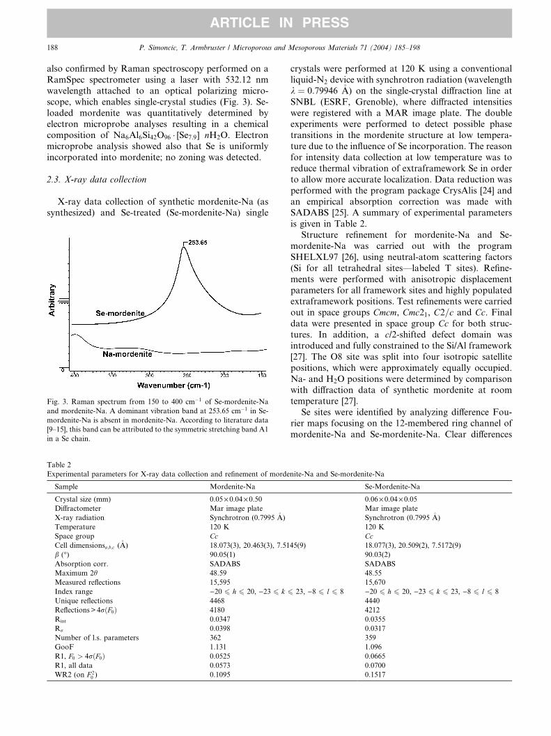
also confirmed by Raman spectroscopy performed on a
RamSpec spectrometer using a laser with 532.12 nm
wavelength attached to an optical polarizing micro-
scope, which enables single-crystal studies (Fig. 3). Se-
loaded mordenite was quantitatively determined byelectron microprobe analyses resulting in a chemical
composition of Na6Al6Si42O96 � [Se7:9] Æ nH2O. Electron
microprobe analysis showed also that Se is uniformly
incorporated into mordenite; no zoning was detected.
2.3. X-ray data collection
X-ray data collection of synthetic mordenite-Na (assynthesized) and Se-treated (Se-mordenite-Na) single
Fig. 3. Raman spectrum from 150 to 400 cm�1 of Se-mordenite-Na
and mordenite-Na. A dominant vibration band at 253.65 cm�1 in Se-
mordenite-Na is absent in mordenite-Na. According to literature data
[9–15], this band can be attributed to the symmetric stretching band A1
in a Se chain.
Table 2
Experimental parameters for X-ray data collection and refinement of morde
Sample Mordenite-Na
Crystal size (mm) 0.05· 0.04· 0.50Diffractometer Mar image plate
X-ray radiation Synchrotron (0.7995 �A)
Temperature 120 K
Space group CcCell dimensionsa;b;c (�A) 18.073(3), 20.463(3), 7.51
b (�) 90.05(1)
Absorption corr. SADABS
Maximum 2h 48.59
Measured reflections 15,595
Index range )20 6 h 6 20, )23 6 k 6
Unique reflections 4468
Reflections> 4rðF0Þ 4180
Rint 0.0347
Rr 0.0398
Number of l.s. parameters 362
GooF 1.131
R1, F0 > 4rðF0Þ 0.0525
R1, all data 0.0573
WR2 (on F 20 ) 0.1095
crystals were performed at 120 K using a conventional
liquid-N2 device with synchrotron radiation (wavelength
k ¼ 0:79946 �A) on the single-crystal diffraction line at
SNBL (ESRF, Grenoble), where diffracted intensities
were registered with a MAR image plate. The doubleexperiments were performed to detect possible phase
transitions in the mordenite structure at low tempera-
ture due to the influence of Se incorporation. The reason
for intensity data collection at low temperature was to
reduce thermal vibration of extraframework Se in order
to allow more accurate localization. Data reduction was
performed with the program package CrysAlis [24] and
an empirical absorption correction was made withSADABS [25]. A summary of experimental parameters
is given in Table 2.
Structure refinement for mordenite-Na and Se-
mordenite-Na was carried out with the program
SHELXL97 [26], using neutral-atom scattering factors
(Si for all tetrahedral sites––labeled T sites). Refine-
ments were performed with anisotropic displacement
parameters for all framework sites and highly populatedextraframework positions. Test refinements were carried
out in space groups Cmcm, Cmc21, C2=c and Cc. Finaldata were presented in space group Cc for both struc-
tures. In addition, a c/2-shifted defect domain was
introduced and fully constrained to the Si/Al framework
[27]. The O8 site was split into four isotropic satellite
positions, which were approximately equally occupied.
Na- and H2O positions were determined by comparisonwith diffraction data of synthetic mordenite at room
temperature [27].
Se sites were identified by analyzing difference Fou-
rier maps focusing on the 12-membered ring channel of
mordenite-Na and Se-mordenite-Na. Clear differences
nite-Na and Se-mordenite-Na
Se-Mordenite-Na
0.06· 0.04· 0.05Mar image plate
Synchrotron (0.7995 �A)
120 K
Cc45(9) 18.077(3), 20.509(2), 7.5172(9)
90.03(2)
SADABS
48.55
15,670
23, )8 6 l 6 8 )20 6 h 6 20, )23 6 k 6 23, )8 6 l 6 8
4440
4212
0.0355
0.0317
359
1.096
0.0665
0.0700
0.1517

Fig. 4. Difference Fourier maps of Se-mordenite-Na and mordenite-Na, contours at 0.25 electron interval for all maps. Upper line Se-mordenite-
Na––(A) (0 0 1) section of 12-membered ring channel at z ¼ �0:64: Strong peaks shifted �x from the center of the channel, as well as one strong peak
shifted þy from the center. (B) (0 0 1) section at z ¼ �0:51: Strong peaks shifted �x and þy from the channel center. (C) (0 0 1) section at z ¼ �0:38:
strong, smeared electron density rhombus like around the center. Lower line mordenite-Na––(D) (0 0 1) section of 12-membered ring channel at
z ¼ �0:64 and (E) (0 0 1) section at z ¼ �0:51: only weak electron density inside the channel. (F) (0 0 1) section at )0.38: Two peaks shifted �y fromthe center, attributed to Na3.
P. Simoncic, T. Armbruster / Microporous and Mesoporous Materials 71 (2004) 185–198 189
ARTICLE IN PRESS
of electron density in the channels are visible (Fig. 4).
High electron density (3–5 e/�A3) in the 12-membered
ring channel of the Se-mordenite-Na is concentratedaround the channel center, whereas only low electron
density occurs in the corresponding mordenite-Na
channel.
3. Results
3.1. Structure refinements
The single-crystal X-ray diffraction pattern of mord-
enite-Na and Se-treated mordenite-Na exhibited no
difference concerning diffuse scattering features. In
particular, there were no indications for super-structure
reflections in the Se-treated mordenite-Na. The cell
dimensions of both samples are very similar (Table 2)
and the Se-treated crystal displayed only a significantexpansion (0.2%) along the b-axis, the direction where
the large channel with elliptical cross section is oblate.
The results of the structure refinement, comprising
atomic coordinates, populations, and isotropic dis-
placement parameters are given in Table 3 for Se-
mordenite-Na. Atomic coordinates, populations and
isotropic displacement parameters of the mordenite-Na
framework are identical within two standard deviations,and therefore not displayed. Both samples show within
two standard deviations the same mean T–O bond
lengths (Tables 4 and 5). Mean T–O distances vary be-
tween 1.596 for T2c–O and 1.640 for T3a–O. Shortest
bond lengths are in both samples around T2a–d. No
estimations were made about Si/Al ordering, but based
on T–O bond lengths a slight Al enrichment is assumedfor tetrahedra in the 4-membered rings (T3 and T4),
which is in agreement with literature data [27,28].
In mordenite-Na four Na sites were located, whereas
in Se-mordenite-Na only three sites were found. Highest
occupied Na1 (2.64 p.f.u. in Se-mordenite-Na, 2.32
p.f.u. in mordenite-Na) is situated in the center of the
compressed 8-membered ring channel along c. Na2 is
located at the intersection of the large 12-membered ringchannel along c to the 8-membered ring channel along b.Na2 in the Se-mordenite-Na is occupied with 2.28 p.f.u.,
whereas the Na2 site in mordenite-Na was split in two
satellite positions Na2a (0.52 p.f.u.) and Na2b (0.76
p.f.u.). The third Na site is located in the 12-membered
ring channel but shifted from the center along b. Na3 in
Se-mordenite-Na has the lowest occupation with 1.76
p.f.u. In mordenite-Na this site is also split in two sa-tellite positions Na3a (1.36 p.f.u.) and Na3b (1.16
p.f.u.). In mordenite-Na, another site (Na4) is located in
the center of the 12-membered ring channel along c,which is missing in the Se-mordenite-Na. Comparing
extraframework cation distributions in synthetic mord-
enite-Na at room temperature [27] and 120 K, positions
and occupations of the other Na sites are almost iden-
tical, whereas the Na3 site in low-temperature mordeniteis slightly higher occupied than at room temperature. By
comparison of the extraframework cation distribution in
Se-mordenite-Na and mordenite-Na, it is obvious that
the Na2 site in Se-mordenite-Na is higher occupied than

Table 3
Atomic coordinates and Beq values for synthetic Se-mordenite-Na (space group Cc)
Atom Population x y z Beq [�A2]
T1a 0.970(1) 0.30252(4)1 0.07286(9)2 0.1683(3)3 1.03(3)
T1b 0.970(1) 0.30252(4)1 )0.07286(9)2 0.0835(6) 1.03(3)
T1c 0.970(1) )0.30252(4)1 0.07286(9)2 0.1683(3)3 1.11(4)
T1d 0.970(1) )0.30252(4)1 )0.07286(9)2 0.0837(3) 1.11(4)
T2a 0.970(1) 0.19671(4)4 0.19037(9)5 0.6698(4) 1.11(3)
T2b 0.970(1) 0.19671(4)4 0.19037(9)5 0.0789(3)6 1.11(3)
T2c 0.970(1) )0.19671(4)4 0.19037(9)5 )0.3304(3) 1.18(4)
T2d 0.970(1) )0.19671(4)4 0.19037(9)5 0.0789(3)6 1.18(4)
T3a 0.970(1) 0.4132(2) )0.11776(5)7 0.3743(4)8 1.10(3)
T3b 0.970(1) )0.4122(2) )0.11776(5)7 0.3743(4)8 0.84(3)
T4a 0.970(1) 0.0865(2) 0.22740(5)9 0.3766(4)10 0.84(3)
T4b 0.970(1) )0.0877(2) 0.22740(5)9 0.3766(4)10 1.10(3)
O1a 0.970(1) 0.3778(1)11 )0.0845(1)12 0.1989(7)13 2.62(5)
O1b 0.970(1) 0.3778(1)11 )0.0845(1)12 0.5558(6)14 2.62(5)
O1c 0.970(1) )0.3778(1)11 )0.0845(1)12 0.1989(7)13 2.62(5)
O1d 0.970(1) )0.3778(1)11 )0.0845(1)12 0.5558(6)14 2.62(5)
O2a 0.970(1) 0.1224(1)15 0.1946(1)16 0.2021(7)17 2.1(1)
O2b 0.970(1) 0.1224(1)15 0.1946(1)16 0.5558(6)14 2.1(1)
O2c 0.970(1) )0.1224(1)15 0.1946(1)16 0.2021(7)17 2.3(1)
O2d 0.970(1) )0.1224(1)15 0.1946(1)16 0.5558(6)14 2.3(1)
O3a 0.970(1) 0.2380(1)18 0.1228(1)19 0.639(1) 3.04(7)
O3b 0.970(1) 0.2380(1)18 0.1228(1)19 0.119(1) 2.90(7)
O3c 0.970(1) )0.2380(1)18 )0.1228(1)19 0.615(1) 3.04(7)
O3d 0.970(1) )0.2380(1)18 )0.1228(1)19 0.142(1) 2.90(7)
O4a 0.970(1) )0.3991(4) )0.1960(3) 0.3578(9) 2.01(5)
O4b 0.970(1) 0.4015(4) )0.1953(3) 0.3572(9) 2.05(7)
O5a 0.970(1) 0.1665(5) 0.1912(4) 0.876(1) 2.75(8)
O5b 0.970(1) )0.1746(5) 0.1970(4) )0.122(1) 2.75(8)
O6a 0.970(1) 0.3192(4) 0.0821(4) 0.3736(9)20 2.01(5)
O6b 0.970(1) )0.3302(4) 0.0772(4) 0.3736(9)20 2.01(5)
O7a 0.970(1) 0.2700(4) 0.0014(3)21 0.1148(9)22 1.90(6)
O7b 0.970(1) )0.2740(4) 0.0014(3)21 0.1148(9)22 1.90(6)
O8a 0.38(5) 0.244(1) )0.256(1) 0.169(4) 2.4
O8b 0.61(5) 0.2569(9) )0.2435(8) 0.120(3) 2.4
O8c 0.60(7) 0.2494(8) )0.2570(6) )0.399(3) 2.4
O8d 0.39(7) 0.262(2) )0.247(1) )0.344(6) 2.4
O9 0.970(1) 0.4999(5) )0.0983(2) 0.386(1) 2.01(5)
O10 0.970(1) 0 0.2097(2) 0.379(2) 2.05(7)
T1Ba 0.029(1) )0.30252(4) )0.07286(9) 0.1683(3) 2.37
T1Bb 0.029(1) )0.30252(4) 0.07286(9) 0.0835(6) 2.37
T1Bc 0.029(1) 0.30252(4) )0.07286(9) 0.1683(3) 2.37
T1Bd 0.029(1) 0.30252(4) 0.07286(9) 0.0837(3) 2.37
T2Ba 0.029(1) )0.19671(4) )0.19037(9) 0.6698(4) 2.37
T2Bb 0.029(1) )0.19671(4) )0.19037(9) 0.0789(3) 2.37
T2Bc 0.029(1) 0.19671(4) )0.19037(9) )0.3304(3) 2.37
T2Bd 0.029(1) 0.19671(4) )0.19037(9) 0.0789(3) 2.37
T3Ba 0.029(1) )0.4132(2) 0.11776(5) 0.3743(4) 2.37
T3Bb 0.029(1) 0.4122(2) 0.11776(5) 0.3743(4) 2.37
T4Ba 0.029(1) 0.0877(2) )0.22740(5) 0.3766(4) 2.37
T4Bb 0.029(1) )0.0865(2) )0.22740(5) 0.3766(4) 2.37
O1Ba 0.029(1) )0.3778(1) 0.0845(1) 0.1989(7) 2.37
O1Bb 0.029(1) )0.3778(1) 0.0845(1) 0.5558(6) 2.37
O1Bc 0.029(1) 0.3778(1) 0.0845(1) 0.5558(6) 2.37
O1Bd 0.029(1) 0.3778(1) 0.0845(1) 0.1989(7) 2.37
O2Ba 0.029(1) )0.1224(1) )0.1946(1) 0.2021(7) 2.37
O2Bb 0.029(1) )0.1224(1) )0.1946(1) 0.5558(6) 2.37
O2Bc 0.029(1) 0.1224(1) )0.1946(1) 0.2021(7) 2.37
O2Bd 0.029(1) 0.1224(1) )0.1946(1) 0.5558(6) 2.37
O3Ba 0.029(1) )0.2380(1) )0.1228(1) 0.639(1) 2.37
O3Bb 0.029(1) )0.2380(1) )0.1228(1) 0.119(1) 2.37
O3Bc 0.029(1) 0.2380(1) 0.1228(1) 0.615(1) 2.37
O3Bd 0.029(1) 0.2380(1) 0.1228(1) 0.142(1) 2.37
190 P. Simoncic, T. Armbruster / Microporous and Mesoporous Materials 71 (2004) 185–198
ARTICLE IN PRESS

Table 3 (continued)
Atom Population x y z Beq [�A2]
O4Ba 0.029(1) 0.3991(4) 0.1960(3) 0.3578(9) 2.37
O4Bb 0.029(1) 0.1015(4) )0.3053(3) 0.3572(9) 2.37
O5Ba 0.029(1) )0.1665(5) )0.1912(4) 0.876(1) 2.37
O5Bb 0.029(1) 0.1746(5) )0.1970(4) )0.122(1) 2.37
O6Ba 0.029(1) )0.3192(4) )0.0821(4) 0.3736(9) 2.37
O6Bb 0.029(1) 0.3302(4) )0.0772(4) 0.3736(9) 2.37
O7Ba 0.029(1) )0.2700(4) )0.0014(3) 0.1148(9) 2.37
O7Bb 0.029(1) 0.2740(4) )0.0014(3) 0.1148(9) 2.37
O9B 0.029(1) )0.4999(5) 0.0983(2) 0.386(1) 2.37
O10B 0.029(1) 0 )0.2097(2) 0.379(2) 2.37
Na1 0.66(1) 0.5002(8) 0.0006(6) 0.125(1) 4.1(1)
Na2 0.57(1) 0.001(1) 0.1845(5) 0.848(2) 7.90
Na3 0.44(2) 0.0011(9) 0.1252(9) 0.647(2) 7.90
W1 1.0 0.5005(8) )0.0648(4) 0.877(2) 5.2(2)
W2 0.25(2) 0.000(3) )0.174(3) 0.517(8) 7.90
W3 0.42(1) 0.002(2) 0.092(1) 0.934(3) 7.90
W5 0.55(2) )0.545(1) )0.189(1) 0.871(3) 7.90
W11 0.29(2) )0.504(2) )0.217(2) 0.950(5) 7.90
Se chain A
A1 0.075(1)a 0.0570(9) 0.0198(9) 0.126(2) 3.48(8)g
A2 0.075(1)a 0.1275(8) )0.0184(8) 0.366(2) 3.48(8)g
A3 0.075(1)a 0.0626(9) 0.0240(6) 0.611(1) 3.48(8)g
A4 0.075(1)a )0.0059(8) )0.0675(6) 0.698(2) 3.48(8)g
A5 0.075(1)a )0.0059(8) )0.0604(7) 0.007(2) 3.48(8)g
A6 0.075(1)a )0.102(1) 0.0146(8) 0.055(2) 3.48(8)g
A7 0.075(1)a )0.132(1) )0.0066(9) 0.353(2) 3.48(8)g
Se chain B
B1 0.056(1)b 0.006(1) 0.0580(9) 0.364(3) 3.48(8)g
B2 0.056(1)b 0.105(1) )0.010(1) 0.438(2) 3.48(8)g
B3 0.056(1)b 0.0994(9) )0.007(1) 0.750(2) 3.48(8)g
B4 0.056(1)b )0.012(1) )0.062(1) 0.800(2) 3.48(8)g
B5 0.056(1)b )0.0121(9) )0.0711(8) 0.109(2) 3.48(8)g
B6 0.056(1)b )0.103(1) 0.005(1) 0.188(2) 3.48(8)g
B7 0.056(1)b )0.105(1) )0.007(1) 0.498(2) 3.48(8)g
Se chain C
C1 0.027(1)c 0.091(2) )0.003(3) 0.393(6) 3.48(8)g
C2 0.027(1)c 0.037(2) )0.047(2) 0.647(4) 3.48(8)g
C3 0.027(1)c )0.072(2) 0.014(2) 0.673(3) 3.48(8)g
C4 0.027(1)c )0.057(2) 0.063(1) 0.949(3) 3.48(8)g
C5 0.027(1)c )0.093(3) )0.019(2) 0.145(4) 3.48(8)g
C6 0.027(1)c )0.12361 0.039(2) 0.400(3) 3.48(8)g
Se chain D
D1 0.041(1)d 0.092(2) 0.044(1) 0.293(2) 3.48(8)g
D2 0.041(1)d 0.105(2) )0.033(1) 0.517(2) 3.48(8)g
D3 0.041(1)d 0.100(1) 0.033(1) 0.773(2) 3.48(8)g
D4 0.041(1)d 0.0761(9) )0.046(1) 0.991(2) 3.48(8)g
D5 0.041(1)d )0.0531(9) )0.047(1) 0.997(2) 3.48(8)g
D6 0.041(1)d )0.075(1) )0.0569(9) 0.303(2) 3.48(8)g
D7 0.041(1)d )0.096(2) 0.052(1) 0.383(3) 3.48(8)g
Se chain E
E1 0.030(1)e )0.065(1) )0.036(2) 0.533(4) 3.48(8)g
E2 0.030(1)e 0.063(1) )0.045(2) 0.560(3) 3.48(8)g
E3 0.030(1)e 0.087(1) 0.019(1) 0.811(3) 3.48(8)g
E4 0.030(1)e 0.044(2) )0.048(1) 0.041(3) 3.48(8)g
E5 0.030(1)e )0.064(2) 0.008(2) 0.125(3) 3.48(8)g
E6 0.030(1)e )0.104(2) )0.056(1) 0.365(3) 3.48(8)g
E7 0.030(1)e )0.108(2) 0.021(2) 0.594(3) 3.48(8)g
Se chain F
F1 0.074(1)f 0.102(1) 0.0031(9) 0.054(2) 3.48(8)g
F2 0.074(1)f 0.1120(6) 0.0194(9) 0.363(2) 3.48(8)g
(continued on next page)
P. Simoncic, T. Armbruster / Microporous and Mesoporous Materials 71 (2004) 185–198 191
ARTICLE IN PRESS

Table 3 (continued)
Atom Population x y z Beq [�A2]
F3 0.074(1)f )0.0083(7) 0.0508(7) 0.434(2) 3.48(8)g
F4 0.074(1)f )0.0467(8) )0.0385(6) 0.613(1) 3.48(8)g
F5 0.074(1)f )0.1056(9) 0.0174(9) 0.846(1) 3.48(8)g
F6 0.074(1)f )0.0561(9) )0.0375(7) 0.094(1) 3.48(8)g
F7 0.074(1)f )0.1038(9) 0.0270(9) 0.327(2) 3.48(8)g
Various Se atoms
G1 0.095(3) 0.0962(9) 0.0041(8) 0.678(2) 3.48(8)g
G2 0.128(4) )0.1084(6) 0.0078(6) 0.910(2) 3.48(8)g
G3 0.101(3) )0.0980(7) )0.0080(7) 0.252(2) 3.48(8)g
G4 0.036(2) 0.002(2) )0.069(1) 0.216(4) 3.48(8)g
1–22Coordinates with the same superscript were constrained to be equal. All atomic parameters (coordinates, population, isotropic displacement
factors) labeled B are fully constrained and belong to a domain of mordenite shifted c/2 relative to the main part of the structure [27]. a–gParameters
of the Se-sites with the same superscript were constrained to be equal.* Starred atoms with standard deviation were refined isotropically, those without standard deviations were fixed. Anisotropically refined atoms are
given in the form of the isotropic equivalent thermal parameter defined as Beq ¼ 8/3 p2P
i (P
j (Uijai aj ai � aj)).
Table 4
T–O distances (�A) of Se-mordenite-Na with standard deviations in parentheses
Bond Bond Bond
T1a–O6a 1.592(7) T2a–O3a 1.604(3) T3a–O4b 1.600(7)
T1a–O3b 1.595(3) T2a–O2b 1.601(4) T3a–O1a 1.620(5)
T1a–O1b 1.620(3) T2a–O8 (ave) 1.61(1) T3a–O9 1.630(9)
T1a–O7a 1.614(7) T2a–O5a 1.630(9) T3a–O1b 1.652(5)
Mean 1.605 Mean 1.612 Mean 1.625
T1b–O3a 1.604(3) T2b–O3b 1.603(3) T3b–O1d 1.615(5)
T1b–O6a 1.618(7) T2b–O5a 1.630(9) T3b–O4a 1.630(8)
T1b–O1a 1.626(3) T2b–O2a 1.626(4) T3b–O9 1.629(9)
T1b–O7a 1.662(7) T2b–O8 (ave) 1.66(1) T3b–O1c 1.646(5)
Mean 1.627 Mean 1.631 Mean 1.630
T1c–O1c 1.621(3) T2c–O8 (ave) 1.58(1) T4a–O4a 1.599(7)
T1c–O7b 1.576(7) T2c–O2d 1.602(4) T4a–O2a 1.628(5)
T1c–O3c 1.612(3) T2c–O3d 1.594(3) T4a–O10 1.596(3)
T1c–O6b 1.631(7) T2c–O5b 1.611(9) T4a–O2b 1.630(4)
Mean 1.610 Mean 1.596 Mean 1.613
T1d–O7b 1.626(7) T2d–O5b 1.568(9) T4b–O2c 1.610(5)
T1d–O1d 1.632(3) T2d–O3c 1.598(3) T4b–O4b 1.624(7)
T1d–O6b 1.649(7) T2d–O2c 1.625(4) T4b–O10 1.638(3)
T1d–O3d 1.620(3) T2d–O8 (ave) 1.60(1) T4b–O2d 1.613(4)
Mean 1.632 Mean 1.597 Mean 1.621
192 P. Simoncic, T. Armbruster / Microporous and Mesoporous Materials 71 (2004) 185–198
ARTICLE IN PRESS
in mordenite-Na, whereas the Na3 position situated in
the 12-membered ring channel, is significantly lower
occupied in Se-mordenite-Na. This can be explained by
a partial displacement of Na by the Se chains and the
partially dehydrated status of Se-mordenite-Na.
3.2. Raman spectroscopy
Se incorporated under different conditions and in
different types of mordenite has been widely investigated
by Raman spectroscopy [9–15]. Prominent vibration
bands in Se-modified mordenites lie between 200 and
300 cm�1. Raman spectra of Se-mordenite-Na can be
compared with the vibration spectra of elemental Se.
The most active Raman band in helical, trigonal Se is
the A1 symmetric stretching mode at 237 cm�1, which
strongly depends on chain interactions [29]. The most
dominant vibrational band in our Se-mordenite-Na was
detected at 254 cm�1. According to the literature, the
254 cm�1 band in Se-mordenite-Na can be attributed to
the A1 symmetric stretching mode in elemental trigonal
Se [9–15]. The difference between the A1 mode of ele-mental Se and Se in mordenite can be interpreted as
interaction of Se with channel wall atoms, but also as a
missing chain interaction in Se-mordenite-Na because
the single chains are laterally separated through the
mordenite channels. Bands characteristic of other forms
than chains, e.g. Se6 or Se8 rings, were not observed.

Table 5
T–O distances (�A) of mordenite-Na with standard deviations in parentheses
Bond Bond Bond
T1a–O6a 1.614(6) T2a–O3a 1.604(2) T3a–O4b 1.632(7)
T1a–O3b 1.614(3) T2a–O2b 1.594(3) T3a–O1a 1.617(6)
T1a–O1b 1.629(4) T2a–O8 (ave) 1.612(8) T3a–O9 1.644(7)
T1a–O7a 1.593(6) T2a–O5a 1.595(7) T3a–O1b 1.667(5)
Mean 1.613 Mean 1.601 Mean 1.640
T1b–O3a 1.602(3) T2b–O3b 1.595(3) T3b–O1d 1.597(5)
T1b–O6a 1.649(6) T2b–O5a 1.658(7) T3b–O4a 1.598(5)
T1b–O1a 1.652(5) T2b–O2a 1.623(3) T3b–O9 1.617(7)
T1b–O7a 1.621(6) T2b–O8 (ave) 1.612(8) T3b–O1c 1.658(5)
Mean 1.631 Mean 1.622 Mean 1.618
T1c–O1c 1.583(5) T2c–O8 (ave) 1.612(8) T4a–O4a 1.636(5)
T1c–O7b 1.621(6) T2c–O2d 1.594(3) T4a–O2a 1.625(4)
T1c–O3c 1.614(3) T2c–O3d 1.596(3) T4a–O10 1.580(2)
T1c–O6b 1.596(6) T2c–O5b 1.574(7) T4a–O2b 1.654(4)
Mean 1.603 Mean 1.594 Mean 1.624
T1d–O7b 1.626(6) T2d–O5b 1.613(7) T4b–O2c 1.591(4)
T1d–O1d 1.616(5) T2d–O3c 1.589(3) T4b–O4b 1.590(7)
T1d–O6b 1.630(6) T2d–O2c 1.625(3) T4b–O10 1.660(3)
T1d–O3d 1.649(3) T2d–O8 (ave) 1.611(8) T4b–O2d 1.621(4)
Mean 1.630 Mean 1.610 Mean 1.615
P. Simoncic, T. Armbruster / Microporous and Mesoporous Materials 71 (2004) 185–198 193
ARTICLE IN PRESS
Our results from Raman spectroscopy are completely
consistent with other spectroscopy studies according
to incorporation conditions, methods, and used mord-
enite samples. The results clearly suggest a chain like
arrangement of incorporated Se. Compared to the sharp
237 cm�1 stretching mode in trigonal Se [30], the 254
cm�1 band of Se-mordenite-Na is strongly broadened.
This broadening can be interpreted as an overlay due toSe chains with different geometries. To check the exis-
tence of SeO species (SeO2, selenites, selenates), the
Raman spectra of Se-mordenite-Na was compared with
Se–O stretching modes as they occur in SeO species. The
vibrational behaviour of SeO2 is well established [31]
and shows prominent modes between 500 and 900 cm�1.
Our Raman spectra displays no bands in this region.
Therefore, it can be concluded to that no moietiescontaining Se–O bonds are present in the structure.
3.3. Se atoms
Six chains (all only partly occupied) consisting of up
to seven Se atoms were found, all located in the large 12-
membered ring channel along the c-axis. Because 1st
neighbor and 2nd neighbor distances in Se chains arewell established, these distances were fixed to 2.34 �A (1st
neighbor) and 3.62 �A (2nd neighbor) [17–19], resulting
in bond angles of 102�. Such constrains were necessary
to handle the overlay of partly occupied chain frag-
ments. Distances of 3rd and further neighbors were not
fixed resulting in bonds of different lengths (4–6 �A to the
3rd neighbor) and different dihedral angles. The popu-
lation was constrained to be equal within a chain, and
isotropic displacement parameters were constrained to
be equal for all Se atoms.
Chain A has a length of 9.85 �A and is made up of
seven Se atoms. The chain has a total population of 2.1
Se p.f.u. The closest distance to channel oxygen is 3.14 �Afrom Se atom A7 to O7b. Third neighbor distances are
A1–A4 4.79 �A, A2–A5 5.45 �A, A3–A6 4.47 �A and A4–A7 5.56 �A with torsion angles of 99.4� (A1–A2–A3–A4),
142.9� (A2–A3–A4–A5), 81.8� (A3–A4–A5–A6) and
159.8� (A4–A5–A6–A7).
ChainB contains also seven Se atoms and is 8.84�A long
with a total population of 1.8 Se p.f.u. The shortest dis-
tance to the channel wall is between Se at B4 andO10with
3.08 �A. Third neighbor distances are B1–B4 4.10 �A, B2–
B5 5.62 �A, B3–B6 4.93 �A and B4–B7 5.61 �A with torsionangles of 67.3� (B1–B2–B3–B4), 172.9� (B2–B3–B4–B5),107.1� (B3–B4–B5–B6) and 178.4� (B4–B5–B6–B7).
Se chain C consists of six atoms with a length of 8.5 �Aand has a low occupancy of 0.6 Se p.f.u. The closest
framework oxygen is O3d to C6 with a distance of 3.24�A. Third neighbor distances are C1–C4 5.117 �A, C2–C5
4.48 �A and C3–C6 5.57 �A with torsion angles of )112.6�(C1–C2–C3–C4), )75.6� (C2–C3–C4–C5) and )147.5�(C3–C4–C5–C6).
Chain D with seven Se atoms has a length of 8.9 �Aand is populated with 1.12 Se p.f.u. Closest distance to
channel oxygen is 3.12 �A from Se atom D2 to O3b.
Third neighbor distances are D1–D4 5.57 �A, D2–D5
4.61 �A, D3–D6 5.41 �A and D4–D7 4.73 �A with torsion
angles of 163.1� (D1–D2–D3–D4), )90.9� (D2–D3–D4–

194 P. Simoncic, T. Armbruster / Microporous and Mesoporous Materials 71 (2004) 185–198
ARTICLE IN PRESS
D5), )140.2� (D3–D4–D5–D6) and 97.5� (D4–D5–D6–
D7).
Chain E has a length of 8.1 �A and a low occupancy of
0.84 Se p.f.u. O7b is nearest to E7 with a bond length of
3.04 �A. Third neighbor distances are E1–E4 4.3 �A, E2–E5 4.94 �A, E3–E6 5.62 �A and E4–E7 5.18 �A with torsion
angles of 73.7� (E1–E2–E3–E4), )107.7� (E2–E3–E4–
Table 6
Chain lengths (�A), 3rd neighbor distances (�A) and torsion angles (�) of Se c
Atom 1 Atom 2 Distance Atom 1 A
Third neighbor Torsion angle
A1 A4 4.80(2) A1 A
A2 A5 5.46(2) A2 A
A3 A6 4.47(2) A3 A
A4 A7 5.57(2) A4 A
Chain length
A1 A7 9.85(2)
Third neighbor Torsion angle
B1 B4 4.11(3) B1 B
B2 B5 5.62(2) B2 B
B3 B6 4.93(3) B3 B
B4 B7 5.62(2) B4 B
Chain length
B1 B7 8.85(3)
Third neighbor Torsion angle
C1 C4 5.12(5) C1 C
C2 C5 4.48(5) C2 C
C3 C6 5.56(4) C3 C
Chain length
C1 C6 8.54(5)
Third neighbor Torsion angle
D1 D4 5.57(3) D1 D
D2 D5 4.61(3) D2 D
D3 D6 5.41(3) D3 D
D4 D7 4.73(3) D4 D
Chain length
D1 D7 8.87(3)
Third neighbor Torsion angle
E1 E4 4.30(4) E1 E
E2 E5 4.94(3) E2 E
E3 E6 5.62(4) E3 E
E4 E7 5.18(4) E4 E
Chain length
E1 E7 8.10(4)
Third neighbor Torsion angle
F1 F4 5.06(2) F1 F
F2 F5 5.35(2) F2 F
F3 F6 5.35(2) F3 F
F4 F7 5.63(2) F4 F
Chain length
F1 F7 10.27(2)
First and second neighbor distances and bond angle are not displayed becaus
and angle are 2.34(3) (1st neighbor), 3.62(2) (2nd neighbor) and 102(1)� (bo
E5), )176.9� (E3–E4–E5–E6) and 121.8� (E4–E5–E6–
E7).
Chain F is the longest chain with 10.3 �A and has a
high population of 2.1 p.f.u. F1 is closely located to O7a
(3.07 �A). Third neighbor distances are F1–F4 5.06 �A,F2–F5 5.35 �A, F3–F6 5.35 �A and F4–F7 5.63 �A with
torsion angles of 113.8� (F1–F2–F3–F4), 136.2� (F2–
hains
tom 2 Atom 3 Atom 4 Angle
2 A3 A4 99.415(7)
3 A4 A5 142.908(6)
4 A5 A6 81.796(9)
5 A6 A7 159.746(4)
2 B3 B4 67.342(9)
3 B4 B5 172.961(1)
4 B5 B6 107.146(9)
5 B6 B7 178.421(2)
2 C3 C4 )112.606(1)3 C4 C5 )75.639(7)4 C5 C6 147.487(5)
2 D3 D4 163.132(3)
3 D4 D5 )90.874(8)4 D5 D6 )140.340(4)5 D6 D7 97.531(2)
2 E3 E4 73.686(8)
3 E4 E5 )107.649(9)4 E5 E6 )176.911(7)5 E6 E7 121.863(7)
2 F3 F4 113.806(6)
3 F4 F5 136.173(7)
4 F5 F6 )134.121(5)5 F6 F7 173.326(8)
e they were fixed during refinement. Corresponding mean bond lengths
nd angle).

P. Simoncic, T. Armbruster / Microporous and Mesoporous Materials 71 (2004) 185–198 195
ARTICLE IN PRESS
F3–F4–F5), )134.1� (F3–F4–F5–F6) and 173.3� (F4–
F5–F6–F7). Four additional low occupied Se positions
were located in the 12-membered ring channel but could
not be assigned to a chain. Distances and angles are
summarized in Table 6.
Fig. 6. Extraframework cation and H2O molecule positions in Se-
mordenite-Na. Small circles with numbers correspond to H2O posi-
tions. Se positions are not displayed.
4. Discussion
4.1. H2O in Se-loaded mordenite-Na
Dehydration of natural and cation exchanged
mordenites is widely discussed in the literature [32–36].The framework of mordenite shows almost no lattice
deformation upon dehydration. Total water loss in
natural mordenite was predicted at 300 �C [33], for
mordenite-Ca at 500 �C, respectively [34]. The H2O site,
which is most resistant to dehydration, coordinates the
extraframework cations within the compressed 8-mem-
bered ring channels. In natural mordenite Ca usually
occupies this site [27], which bonds considerably stron-ger to H2O than Na in our synthetic mordenite-Na. This
explains the lower dehydration temperature (�280 �C)for mordenite-Na. Compared to mordenite-Na (19 H2O
p.f.u., Fig. 5), 10 H2O molecules p.f.u. were detected in
Se-mordenite-Na. This H2O content can be explained by
rehydration due to subsequent storage of the sample at
ambient humidity conditions. Even if the large channels
are plugged by Se chains, H2O molecules find appro-priate diffusion pathways along the compressed 8-
membered ring channels along b and c. A reaction of
this re-incorporated H2O and Se within the structure at
ambient conditions cannot be expected. The H2O mol-
ecules in Se-mordenite-Na are located at five different
sites: (1) a highly populated position in the 8-membered
ring channel along c, which is coordinated to Na1; (2)
two positions in the 8-membered ring channel along b;(3) two positions in the 12-membered ring channel along
c close to the Na2 and Na3 positions (Fig. 6).
Fig. 5. Extraframework cation and H2O molecule positions in mord-
enite-Na. Small circles with numbers correspond to H2O positions.
4.2. The mordenite framework
The mordenite structure at room temperature is
usually refined in space group Cmcm or Cmc21, respec-tively [1,28]. Simoncic and Armbruster [27] proposed a
domain like structure where about 3% of the Si/Al
framework is shifted by c=2. In spite of these studies, thesymmetry of the framework is generally handled as
pseudosymmetry and represents only a fair approxima-
tion. Lower symmetries or micro-twin models are also
supposed [37] but not proven by experiments. Several
space groups were tested during structural refinement:
orthorhombic Cmcm and Cmc21, and monoclinic C2=cand Cc. For mordenite-Na and Se-mordenite-Na at 120
K the space group Cc represents the most suitablemodel. Because the Cc model fits both structures, it can
be ruled out that a phase transition from Cmc21 to Ccoccurred due to Se incorporation.
Application of low temperature (120 K), chosen for
an improved resolution of extraframework occupants,
led also to a better resolution of the complex disordered
microstructure within the tetrahedral framework [37].
However, we were not able to develop a correct inter-growth/twinning model for the description of the tetra-
hedral framework. After refinements in space group
Cmcm and Cmc21 indicated only poor agreement be-
tween structure model and diffraction data, we have
chosen as a ‘‘best’’ compromise the monoclinic space
group Cc. Furthermore, to reduce correlation problems
due to pseudosymmetry, related tetrahedral sites and
their atomic displacement parameters were constrainedto each other. As will be shown below, Se chains may be
longer than the periodicity of the framework parallel to
the channel axis. The fact that we did not observe any
superstructure reflection or diffuse scattering evidence
for the Se-treated mordenite-Na, different to mordenite-
Na, indicates that the distribution of Se chains along the
c-axis is rather random.
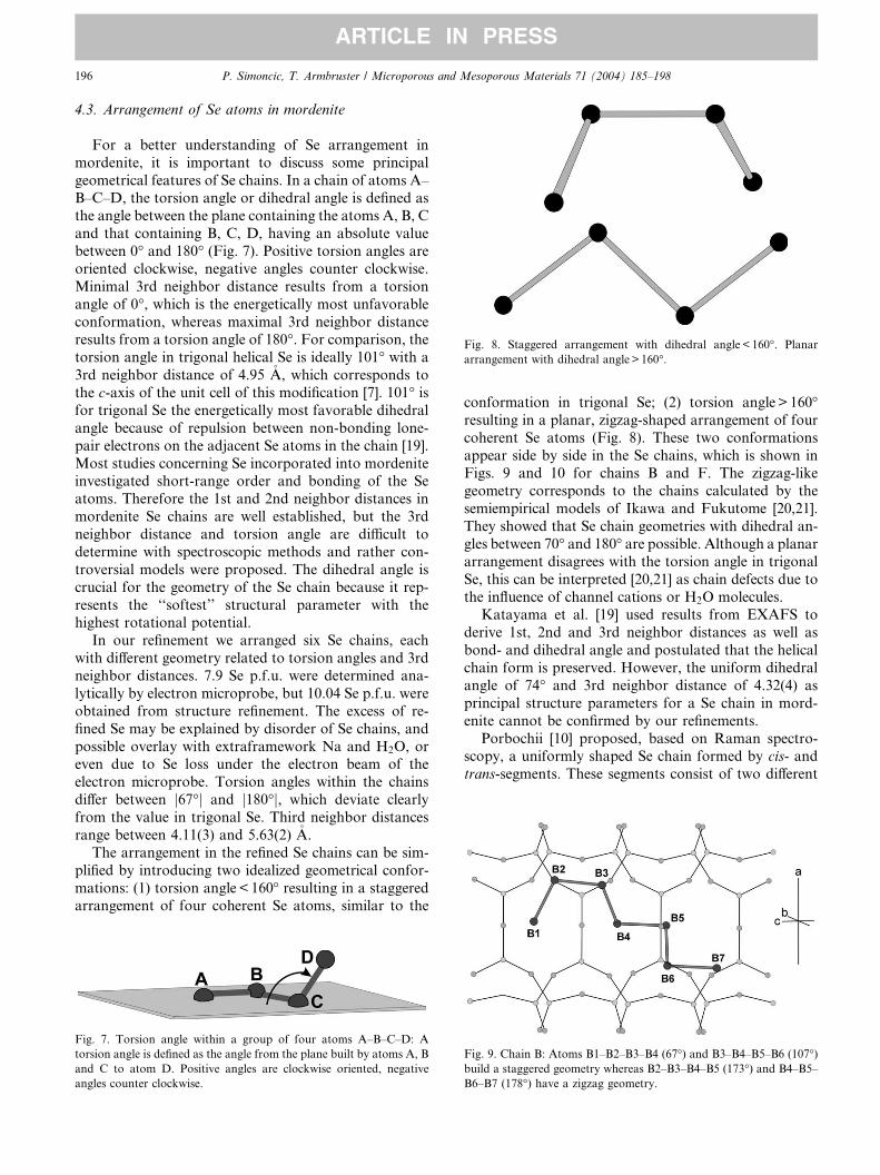
Fig. 8. Staggered arrangement with dihedral angle < 160�. Planar
arrangement with dihedral angle > 160�.
196 P. Simoncic, T. Armbruster / Microporous and Mesoporous Materials 71 (2004) 185–198
ARTICLE IN PRESS
4.3. Arrangement of Se atoms in mordenite
For a better understanding of Se arrangement in
mordenite, it is important to discuss some principal
geometrical features of Se chains. In a chain of atoms A–B–C–D, the torsion angle or dihedral angle is defined as
the angle between the plane containing the atoms A, B, C
and that containing B, C, D, having an absolute value
between 0� and 180� (Fig. 7). Positive torsion angles are
oriented clockwise, negative angles counter clockwise.
Minimal 3rd neighbor distance results from a torsion
angle of 0�, which is the energetically most unfavorable
conformation, whereas maximal 3rd neighbor distanceresults from a torsion angle of 180�. For comparison, the
torsion angle in trigonal helical Se is ideally 101� with a
3rd neighbor distance of 4.95 �A, which corresponds to
the c-axis of the unit cell of this modification [7]. 101� isfor trigonal Se the energetically most favorable dihedral
angle because of repulsion between non-bonding lone-
pair electrons on the adjacent Se atoms in the chain [19].
Most studies concerning Se incorporated into mordeniteinvestigated short-range order and bonding of the Se
atoms. Therefore the 1st and 2nd neighbor distances in
mordenite Se chains are well established, but the 3rd
neighbor distance and torsion angle are difficult to
determine with spectroscopic methods and rather con-
troversial models were proposed. The dihedral angle is
crucial for the geometry of the Se chain because it rep-
resents the ‘‘softest’’ structural parameter with thehighest rotational potential.
In our refinement we arranged six Se chains, each
with different geometry related to torsion angles and 3rd
neighbor distances. 7.9 Se p.f.u. were determined ana-
lytically by electron microprobe, but 10.04 Se p.f.u. were
obtained from structure refinement. The excess of re-
fined Se may be explained by disorder of Se chains, and
possible overlay with extraframework Na and H2O, oreven due to Se loss under the electron beam of the
electron microprobe. Torsion angles within the chains
differ between j67�j and j180�j, which deviate clearly
from the value in trigonal Se. Third neighbor distances
range between 4.11(3) and 5.63(2) �A.
The arrangement in the refined Se chains can be sim-
plified by introducing two idealized geometrical confor-
mations: (1) torsion angle < 160� resulting in a staggeredarrangement of four coherent Se atoms, similar to the
Fig. 7. Torsion angle within a group of four atoms A–B–C–D: A
torsion angle is defined as the angle from the plane built by atoms A, B
and C to atom D. Positive angles are clockwise oriented, negative
angles counter clockwise.
conformation in trigonal Se; (2) torsion angle> 160�resulting in a planar, zigzag-shaped arrangement of four
coherent Se atoms (Fig. 8). These two conformations
appear side by side in the Se chains, which is shown inFigs. 9 and 10 for chains B and F. The zigzag-like
geometry corresponds to the chains calculated by the
semiempirical models of Ikawa and Fukutome [20,21].
They showed that Se chain geometries with dihedral an-
gles between 70� and 180� are possible. Although a planar
arrangement disagrees with the torsion angle in trigonal
Se, this can be interpreted [20,21] as chain defects due to
the influence of channel cations or H2O molecules.Katayama et al. [19] used results from EXAFS to
derive 1st, 2nd and 3rd neighbor distances as well as
bond- and dihedral angle and postulated that the helical
chain form is preserved. However, the uniform dihedral
angle of 74� and 3rd neighbor distance of 4.32(4) as
principal structure parameters for a Se chain in mord-
enite cannot be confirmed by our refinements.
Porbochii [10] proposed, based on Raman spectro-scopy, a uniformly shaped Se chain formed by cis- and
trans-segments. These segments consist of two different
Fig. 9. Chain B: Atoms B1–B2–B3–B4 (67�) and B3–B4–B5–B6 (107�)build a staggered geometry whereas B2–B3–B4–B5 (173�) and B4–B5–
B6–B7 (178�) have a zigzag geometry.
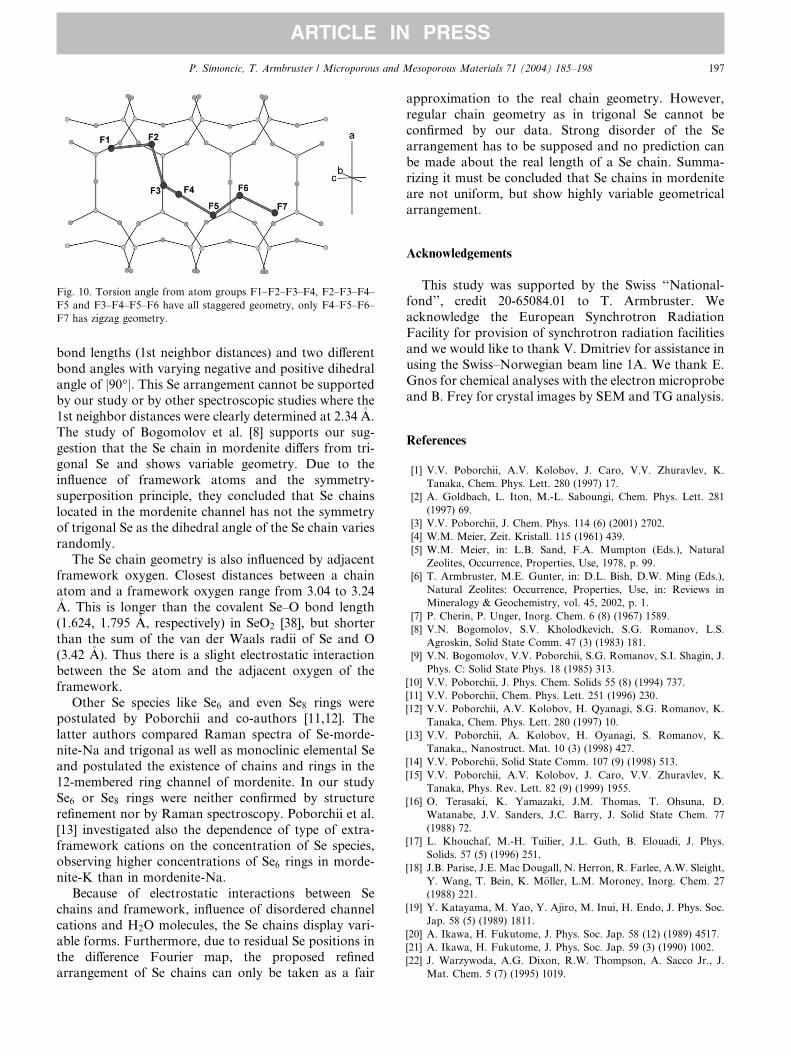
Fig. 10. Torsion angle from atom groups F1–F2–F3–F4, F2–F3–F4–
F5 and F3–F4–F5–F6 have all staggered geometry, only F4–F5–F6–
F7 has zigzag geometry.
P. Simoncic, T. Armbruster / Microporous and Mesoporous Materials 71 (2004) 185–198 197
ARTICLE IN PRESS
bond lengths (1st neighbor distances) and two different
bond angles with varying negative and positive dihedral
angle of j90�j. This Se arrangement cannot be supported
by our study or by other spectroscopic studies where the
1st neighbor distances were clearly determined at 2.34 �A.
The study of Bogomolov et al. [8] supports our sug-
gestion that the Se chain in mordenite differs from tri-gonal Se and shows variable geometry. Due to the
influence of framework atoms and the symmetry-
superposition principle, they concluded that Se chains
located in the mordenite channel has not the symmetry
of trigonal Se as the dihedral angle of the Se chain varies
randomly.
The Se chain geometry is also influenced by adjacent
framework oxygen. Closest distances between a chainatom and a framework oxygen range from 3.04 to 3.24�A. This is longer than the covalent Se–O bond length
(1.624, 1.795 �A, respectively) in SeO2 [38], but shorter
than the sum of the van der Waals radii of Se and O
(3.42 �A). Thus there is a slight electrostatic interaction
between the Se atom and the adjacent oxygen of the
framework.
Other Se species like Se6 and even Se8 rings werepostulated by Poborchii and co-authors [11,12]. The
latter authors compared Raman spectra of Se-morde-
nite-Na and trigonal as well as monoclinic elemental Se
and postulated the existence of chains and rings in the
12-membered ring channel of mordenite. In our study
Se6 or Se8 rings were neither confirmed by structure
refinement nor by Raman spectroscopy. Poborchii et al.
[13] investigated also the dependence of type of extra-framework cations on the concentration of Se species,
observing higher concentrations of Se6 rings in morde-
nite-K than in mordenite-Na.
Because of electrostatic interactions between Se
chains and framework, influence of disordered channel
cations and H2O molecules, the Se chains display vari-
able forms. Furthermore, due to residual Se positions in
the difference Fourier map, the proposed refinedarrangement of Se chains can only be taken as a fair
approximation to the real chain geometry. However,
regular chain geometry as in trigonal Se cannot be
confirmed by our data. Strong disorder of the Se
arrangement has to be supposed and no prediction can
be made about the real length of a Se chain. Summa-rizing it must be concluded that Se chains in mordenite
are not uniform, but show highly variable geometrical
arrangement.
Acknowledgements
This study was supported by the Swiss ‘‘National-fond’’, credit 20-65084.01 to T. Armbruster. We
acknowledge the European Synchrotron Radiation
Facility for provision of synchrotron radiation facilities
and we would like to thank V. Dmitriev for assistance in
using the Swiss–Norwegian beam line 1A. We thank E.
Gnos for chemical analyses with the electron microprobe
and B. Frey for crystal images by SEM and TG analysis.
References
[1] V.V. Poborchii, A.V. Kolobov, J. Caro, V.V. Zhuravlev, K.
Tanaka, Chem. Phys. Lett. 280 (1997) 17.
[2] A. Goldbach, L. Iton, M.-L. Saboungi, Chem. Phys. Lett. 281
(1997) 69.
[3] V.V. Poborchii, J. Chem. Phys. 114 (6) (2001) 2702.
[4] W.M. Meier, Zeit. Kristall. 115 (1961) 439.
[5] W.M. Meier, in: L.B. Sand, F.A. Mumpton (Eds.), Natural
Zeolites, Occurrence, Properties, Use, 1978, p. 99.
[6] T. Armbruster, M.E. Gunter, in: D.L. Bish, D.W. Ming (Eds.),
Natural Zeolites: Occurrence, Properties, Use, in: Reviews in
Mineralogy & Geochemistry, vol. 45, 2002, p. 1.
[7] P. Cherin, P. Unger, Inorg. Chem. 6 (8) (1967) 1589.
[8] V.N. Bogomolov, S.V. Kholodkevich, S.G. Romanov, L.S.
Agroskin, Solid State Comm. 47 (3) (1983) 181.
[9] V.N. Bogomolov, V.V. Poborchii, S.G. Romanov, S.I. Shagin, J.
Phys. C: Solid State Phys. 18 (1985) 313.
[10] V.V. Poborchii, J. Phys. Chem. Solids 55 (8) (1994) 737.
[11] V.V. Poborchii, Chem. Phys. Lett. 251 (1996) 230.
[12] V.V. Poborchii, A.V. Kolobov, H. Qyanagi, S.G. Romanov, K.
Tanaka, Chem. Phys. Lett. 280 (1997) 10.
[13] V.V. Poborchii, A. Kolobov, H. Oyanagi, S. Romanov, K.
Tanaka,, Nanostruct. Mat. 10 (3) (1998) 427.
[14] V.V. Poborchii, Solid State Comm. 107 (9) (1998) 513.
[15] V.V. Poborchii, A.V. Kolobov, J. Caro, V.V. Zhuravlev, K.
Tanaka, Phys. Rev. Lett. 82 (9) (1999) 1955.
[16] O. Terasaki, K. Yamazaki, J.M. Thomas, T. Ohsuna, D.
Watanabe, J.V. Sanders, J.C. Barry, J. Solid State Chem. 77
(1988) 72.
[17] L. Khouchaf, M.-H. Tuilier, J.L. Guth, B. Elouadi, J. Phys.
Solids. 57 (5) (1996) 251.
[18] J.B. Parise, J.E. Mac Dougall, N. Herron, R. Farlee, A.W. Sleight,
Y. Wang, T. Bein, K. M€oller, L.M. Moroney, Inorg. Chem. 27
(1988) 221.
[19] Y. Katayama, M. Yao, Y. Ajiro, M. Inui, H. Endo, J. Phys. Soc.
Jap. 58 (5) (1989) 1811.
[20] A. Ikawa, H. Fukutome, J. Phys. Soc. Jap. 58 (12) (1989) 4517.
[21] A. Ikawa, H. Fukutome, J. Phys. Soc. Jap. 59 (3) (1990) 1002.
[22] J. Warzywoda, A.G. Dixon, R.W. Thompson, A. Sacco Jr., J.
Mat. Chem. 5 (7) (1995) 1019.

198 P. Simoncic, T. Armbruster / Microporous and Mesoporous Materials 71 (2004) 185–198
ARTICLE IN PRESS
[23] P. Simoncic, T. Armbruster, in: P. Misaelides (Ed.), Zeolite’02, 6th
International Conference on the Occurrence, Properties and
Utilization of Natural Zeolites, 2002, p. 336.
[24] Oxford Diffraction Xcalibur System, User Manual, CrysAlis
Software Package, version 1.169, Oxfordshire, UK, 2001.
[25] G.M. Sheldrick, SADABS, version 2.06, Empirical Absorption
Correction Program, University of G€ottingen, Germany, 2002.
[26] G.M. Sheldrick, SHELX-97, University of G€ottingen, Germany,
1997.
[27] P. Simoncic, T. Armbruster, Am. Mineral. 89 (2004) 421.
[28] A. Alberti, P. Davoli, P.G. Vezzalini, Zeit. Kristall. 175 (1986) 249.
[29] G. Lucovsky, A. Mooradian, W. Taylor, G.B. Wright, Solid State
Comm. 5 (1967) 113.
[30] M.H. Brodsky, in: M. Cardona (Ed.), Light Scattering in Solids,
Springer, Berlin, 1975, p. 208.
[31] A. Anderson, A. Sanders, W. Smith, J. Raman Spec. 31 (2000)
403.
[32] L.P. van Reeuwijk, The Thermal Dehydration of Natural Zeolites,
Ph.D. Thesis, Landbouwhogeschool Wageningen, 1974, p. 22.
[33] A. Gottardi, E. Galli, Natural Zeolites, Springer, Berlin, 1985, p.
223.
[34] J. Elsen, G.S.D. King, W.J. Mortier, J. Phys. Chem. 91 (1987)
5800.
[35] A. Martucci, M. Sacerdoti, G. Cruciani, Surf. Sci. Catal. 135
(2001) 290.
[36] A. Martucci, M. Sacerdoti, G. Cruciani, C. Dalconi, Eur. J.
Mineral. 15 (2003) 485.
[37] R. Gramlich-Meier, Strukturparameter in Zeolithen der Morde-
nitfamilie, Dissertation ETH Z€urich, Nr. 6760 (1981).
[38] K. Stahl, J.P. Legros, J. Galy, Zeit. Kristall. 202 (1992) 99.