scenes from the past
TRANSCRIPT
2101EDUCATION EXHIBIT
Natale Villari, MD • Gino Fornaciari, MD • Donatella Lippi, PhD Marco Matucci Cerinic, MD • Andrea Ginestroni, MD Giannantonio Pellicanò, MD • Mario Mascalchi, MD, PhD
The remains of 12 members of the grand ducal (junior) branch of the Florentine Medici family were exhumed in 2003 as part of the Medici Project, a multidisciplinary study whose aim was to investigate the lifestyles, health status, and possible causes of death of members of one of the richest, most powerful families of the Italian Renaissance. Digital radiography and orthopantomography were performed on the skeletal remains of individuals who lived between 1562 and 1666. The observed bone malformations, deformities, and changes (degen-erative, metabolic, and dental) challenge traditional views, based on portraits and historical accounts, about the appearance and lifestyle of some family members. Moreover, the occurrence of a constellation of bone changes related to diabetes (osteoporosis, osteoarthritis, diffuse idiopathic skeletal hyperostosis, cranial hyperostosis, and crystalline arthropathy) suggests that this metabolic disease was common in the grand ducal branch of the Medici family.
IntroductionThe Medici were one of the richest and most powerful families of the Italian Renais-sance. Beginning in the 14th century, through careful management of banking ven-tures and skillful political action, the family rose to the pinnacle of social and politi-cal power in the city of Florence in the Tuscany region of Italy, which the Medici made the intellectual center of the Western world for the next two centuries. In par-ticular, the members of the senior branch of the Medici family, including Cosimo “the Elder,” Piero “the Gouty,” and Lorenzo “the Magnificent,” were lovers of the arts and sciences and were patrons of Michelangelo Buonarroti, Leonardo da Vinci, Sandro Botticelli, Galileo Galilei, and Benvenuto Cellini.
Scenes from the PastThe Medici Project: Radiographic Survey1
Abbreviation: DISH = diffuse idiopathic skeletal hyperostosis
RadioGraphics 2009; 29:2101–2114 • Published online 10.1148/rg.297085212 • Content Codes: 1From the Radiodiagnostic Section, Department of Clinical Physiopathology (N.V., A.G., G.P., M.M.), Department of Anatomy, Histology and Legal Medicine (D.L.), and Rheumatology Unit, Department of Internal Medicine (M.M.C.), University of Florence, Viale Morgagni 85, 50134 Florence, Italy; and Division of Paleopathology, Department of Oncology, Transplants and Advanced Technologies in Medicine, University of Pisa, Pisa, Italy (G.F.). Received September 10, 2008; revision requested October 1; final revision received May 13, 2009; accepted July 15. The Medici Project was funded by the MIUR-PRIN 2005 2-year research project, “Malattie, ambiente e società alla corte granducale di Firenze: studio storico, archeologico e paleopatologico delle deposizioni funebri dei Medici (secoli XVI-XVIII)” (prot. 2005067073). Address correspondence to A.G. (e-mail: [email protected]).
©RSNA, 2009 • radiographics.rsnajnls.org
See last page
TEACHING POINTS
Note: This copy is for your personal non-commercial use only. To order presentation-ready copies for distribution to your colleagues or clients, contact us at www.rsna.org/rsnarights.
2102 November-December 2009 radiographics.rsnajnls.org
The grand ducal (junior) branch of the fam-ily began in the 16th century when Cosimo I was born of the consanguineous marriage between Giovanni delle Bande Nere and Maria Salviati. The members of the grand ducal branch contin-ued to enrich the Medicean art collections, with Francesco I collecting many important pieces and displaying them in the Uffizi Gallery of Florence. They also gave a new shape to the city of Florence by building churches, palazzi, and villas, which can still be appreciated today. In addition, they fostered scientists such as Galileo and promoted the spread of scientific knowledge.
Since the 20th century, the Medici family has attracted considerable interest on the part of medi-cal doctors (1–9). In particular, although common health problems in Tuscany during the 15th and 16th centuries included endemic malaria and other infectious diseases as smallpox, tuberculo-sis, and syphilis, different hypotheses have been proposed for the arthritic disease of the peripheral and axial skeleton seen in several members of the Medici family, and which contemporary doctors comprehensively labeled as “gout” (1–9).
The Medici Project is a multi-institutional, multidisciplinary study that was started in 2003 and is based on the exhumation of the remains of 12 members of the grand ducal branch of the Medici family buried in the Medici Chapel of San Lorenzo Church in Florence, Italy. Studies are being carried out in the areas of paleopathol-ogy, immunohistochemistry, molecular biology, isotope analysis, toxicology, and diagnostic radi-ology to provide information on the lifestyles, illnesses, and possible causes of death of these family members (4–11).
In this article, we discuss and illustrate the imaging techniques and the resultant findings as-sociated with our study of the remains of these 12 Medici family members.
Materials and MethodsBetween May 2004 and June 2006, the remains of 22 individuals were found beneath the floor of the crypt of the Medici Chapel. Twelve individu-als were identified; the other 10, eight of whom were small children, could not be identified due to the absence of inscriptions or the poor state of preservation. Identification was made on the basis of (a) labels on the zinc boxes in which the bones had been placed after prior exhumations in 1559, 1791, 1857, and 1945–1948; and (b) consistency
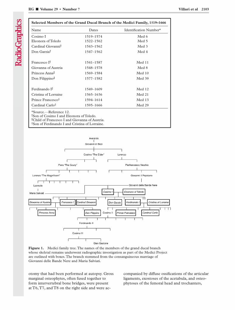
between the historical accounts and paleopatho-logic features. The 12 identified individuals were all members of the grand ducal branch of the Medici family who lived between 1519 and 1666 (Table), and their skeletal remains were examined with dig-ital radiography and orthopantomography as part of the Medici Project. Their familial relationships are illustrated in Figure 1. As a consequence of pri-or exhumations and of the flood that hit Florence on November 4th, 1966, only one skeleton (that of Don Filippino) was intact; the skeletal remains of the other individuals had been stored in the afore-mentioned zinc boxes and needed to be matched prior to radiographic assessment. The lack of soft tissue, muscles, and tendons made it difficult, and at times impossible, to correctly reassemble the various parts of the skeletons (13). Other difficul-ties arose from postmortem bone lesions caused by clumsy handling or the floodwaters.
All radiographic examinations were per-formed on a Siemens AXIOM Iconos R200 system (Siemens Medical Solutions, Malvern, Pa) at 48–65 kV and 4–32 mA with a “contact” technique, with the exception of skull radiogra-phy, for which a focal distance of 1 m was used. Orthopantomographic images were obtained with a Siemens ORTHOPANTOGRAPH 2 at 75 kV, 10 mA, and a focal distance of 30 cm.
Two senior radiologists interpreted the radio-graphs in the light of (a) historical data available about each individual’s lifestyle, medical history, and presumed cause of death; and (b) paleo-pathologic findings (5).
Results
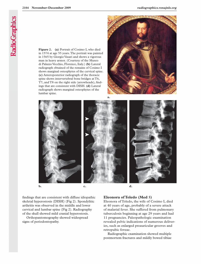
Cosimo I (Med 6)Cosimo I, son of Giovanni delle Bande Nere and Maria Salviati (Fig 1), was the first grand duke of Tuscany and the husband of Eleonora of Toledo. He died at age 55 years of pneumonia. Cosimo I survived several illnesses, including smallpox, ma-larial fevers at age 24–25 years, “gravel” (renal or bladder stones) at age 41–43 years, and bronchitis. Contemporary descriptions indicate that he also experienced several cerebrovascular accidents and suffered from an acute disorder of the right knee, termed gout by the court physician, at ages 49 and 52–53 years (14). Paleopathologic examination showed well-developed muscle-tendon insertions in line with historical descriptions of the great physical strength and robustness of Cosimo I and his reputation as a skilled horseman.
Radiographic examination showed multiple postmortem skeletal lesions and signs of crani-
TeachingPoint
TeachingPoint
RG ■ Volume 29 • Number 7 Villari et al 2103
Figure 1. Medici family tree. The names of the members of the grand ducal branch whose skeletal remains underwent radiographic investigation as part of the Medici Project are outlined with boxes. The branch stemmed from the consanguineous marriage of Giovanni delle Bande Nere and Maria Salviati.
Selected Members of the Grand Ducal Branch of the Medici Family, 1519–1666
Name Dates Identification Number*
Cosimo I 1519–1574 Med 6Eleonora of Toledo 1522–1562 Med 5
Cardinal Giovanni† 1543–1562 Med 3
Don Garzia† 1547–1562 Med 4
Francesco I† 1541–1587 Med 11
Giovanna of Austria 1548–1578 Med 8
Princess Anna‡ 1569–1584 Med 10
Don Filippino‡ 1577–1582 Med 39
Ferdinando I† 1549–1609 Med 12
Cristina of Lorraine 1565–1636 Med 21
Prince Francesco§ 1594–1614 Med 13
Cardinal Carlo§ 1595–1666 Med 29
*Source.—Reference 12. †Son of Cosimo I and Eleonora of Toledo. ‡Child of Francesco I and Giovanna of Austria. §Son of Ferdinando I and Cristina of Lorraine.
otomy that had been performed at autopsy. Gross marginal osteophytes, often fused together to form intervertebral bone bridges, were present at T6, T7, and T8 on the right side and were ac-
companied by diffuse ossifications of the articular ligaments, exostoses of the acetabula, and osteo-phytoses of the femoral head and trochanters,
2104 November-December 2009 radiographics.rsnajnls.org
Eleonora of Toledo (Med 5)Eleonora of Toledo, the wife of Cosimo I, died at 40 years of age, probably of a severe attack of malarial fever. She suffered from pulmonary tuberculosis beginning at age 29 years and had 11 pregnancies. Paleopathologic examination revealed pelvic indications of numerous deliver-ies, such as enlarged preauricular grooves and retropubic foveae.
Radiographic examination showed multiple postmortem fractures and mildly bowed tibiae
findings that are consistent with diffuse idiopathic skeletal hyperostosis (DISH) (Fig 2). Spondylitic arthritis was observed in the middle and lower cervical and lumbar spine (Fig 2). Radiography of the skull showed mild cranial hyperostosis.
Orthopantomography showed widespread signs of periodontopathy.
Figure 2. (a) Portrait of Cosimo I, who died in 1574 at age 55 years. The portrait was painted in 1565 by Giorgio Vasari and shows a vigorous man in heavy armor. (Courtesy of the Museo di Palazzo Vecchio, Florence, Italy.) (b) Lateral radiograph obtained of the remains of Cosimo I shows marginal osteophytes of the cervical spine. (c) Anteroposterior radiograph of the thoracic spine shows intervertebral bone bridges at T6, T7, and T8 on the right side (arrowheads), find-ings that are consistent with DISH. (d) Lateral radiograph shows marginal osteophytes of the lumbar spine.
RG ■ Volume 29 • Number 7 Villari et al 2105
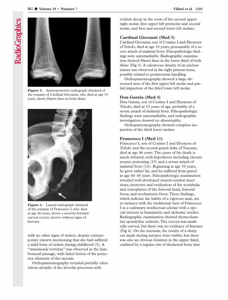
Figure 3. Anteroposterior radiograph obtained of the remains of Cardinal Giovanni, who died at age 19 years, shows Harris lines in both tibiae.
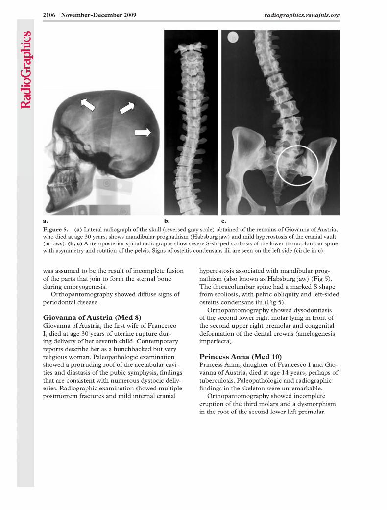
Figure 4. Lateral radiograph obtained of the remains of Francesco I, who died at age 46 years, shows a severely forward-curved coccyx (arrow) without signs of fracture.
with no other signs of rickets, despite contem-porary reports mentioning that she had suffered a mild form of rickets during childhood (5). A “transitional vertebra” was observed in the lum-bosacral passage, with failed fusion of the poste-rior elements of the sacrum.
Orthopantomography revealed partially eden-tulous atrophy of the alveolar processes with
evident decay in the roots of the second upper right molar, first upper left premolar and second molar, and first and second lower left molars.
Cardinal Giovanni (Med 3)Cardinal Giovanni, son of Cosimo I and Eleonora of Toledo, died at age 19 years, presumably of a se-vere attack of malarial fever. Paleopathologic find-ings were unremarkable. Radiographic examina-tion showed Harris lines in the lower third of both tibiae (Fig 3). A calcareous density of an unclear nature was observed in the right petrous bone, possibly related to postmortem handling.
Orthopantomography showed a large ab-scessed area of the first upper left molar and par-tial impaction of the third lower left molar.
Don Garzia (Med 4)Don Garzia, son of Cosimo I and Eleonora of Toledo, died at 15 years of age, probably of a severe attack of malarial fever. Paleopathologic findings were unremarkable, and radiographic investigation showed no abnormality.
Orthopantomography showed complete im-paction of the third lower molars.
Francesco I (Med 11)Francesco I, son of Cosimo I and Eleonora of Toledo and the second grand duke of Tuscany, died at age 46 years. The cause of his death is much debated, with hypotheses including chronic arsenic poisoning (15) and a severe attack of malarial fever (16). Beginning at age 35 years, he grew rather fat, and he suffered from gravel at age 44–45 years. Paleopathologic examination revealed well-developed muscle-tendon inser-tions; exostoses and ovalization of the acetabula; and osteophytes of the femoral head, femoral fovea, and trochanteric fovea. These findings, which indicate the habits of a vigorous man, are at variance with the traditional view of Francesco I as a sedentary intellectual scholar with a spe-cial interest in humanistic and alchemic studies. Radiographic examination showed thoracolum-bar spondylitic arthritis. The coccyx was mark-edly curved, but there was no evidence of fracture (Fig 4). On the sternum, the results of a sharp cut made during autopsy were visible, but there was also an obvious foramen in the upper third, outlined by a regular rim of thickened bone that
2106 November-December 2009 radiographics.rsnajnls.org
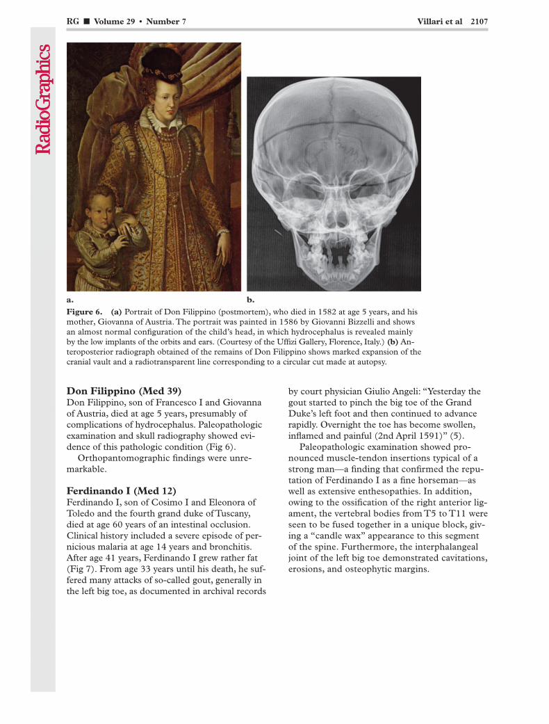
Figure 5. (a) Lateral radiograph of the skull (reversed gray scale) obtained of the remains of Giovanna of Austria, who died at age 30 years, shows mandibular prognathism (Habsburg jaw) and mild hyperostosis of the cranial vault (arrows). (b, c) Anteroposterior spinal radiographs show severe S-shaped scoliosis of the lower thoracolumbar spine with asymmetry and rotation of the pelvis. Signs of osteitis condensans ilii are seen on the left side (circle in c).
was assumed to be the result of incomplete fusion of the parts that join to form the sternal bone during embryogenesis.
Orthopantomography showed diffuse signs of periodontal disease.
Giovanna of Austria (Med 8)Giovanna of Austria, the first wife of Francesco I, died at age 30 years of uterine rupture dur-ing delivery of her seventh child. Contemporary reports describe her as a hunchbacked but very religious woman. Paleopathologic examination showed a protruding roof of the acetabular cavi-ties and diastasis of the pubic symphysis, findings that are consistent with numerous dystocic deliv-eries. Radiographic examination showed multiple postmortem fractures and mild internal cranial
hyperostosis associated with mandibular prog-nathism (also known as Habsburg jaw) (Fig 5). The thoracolumbar spine had a marked S shape from scoliosis, with pelvic obliquity and left-sided osteitis condensans ilii (Fig 5).
Orthopantomography showed dysodontiasis of the second lower right molar lying in front of the second upper right premolar and congenital deformation of the dental crowns (amelogenesis imperfecta).
Princess Anna (Med 10)Princess Anna, daughter of Francesco I and Gio-vanna of Austria, died at age 14 years, perhaps of tuberculosis. Paleopathologic and radiographic findings in the skeleton were unremarkable.
Orthopantomography showed incomplete eruption of the third molars and a dysmorphism in the root of the second lower left premolar.
RG ■ Volume 29 • Number 7 Villari et al 2107
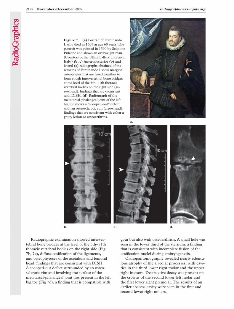
Figure 6. (a) Portrait of Don Filippino (postmortem), who died in 1582 at age 5 years, and his mother, Giovanna of Austria. The portrait was painted in 1586 by Giovanni Bizzelli and shows an almost normal configuration of the child’s head, in which hydrocephalus is revealed mainly by the low implants of the orbits and ears. (Courtesy of the Uffizi Gallery, Florence, Italy.) (b) An-teroposterior radiograph obtained of the remains of Don Filippino shows marked expansion of the cranial vault and a radiotransparent line corresponding to a circular cut made at autopsy.
by court physician Giulio Angeli: “Yesterday the gout started to pinch the big toe of the Grand Duke’s left foot and then continued to advance rapidly. Overnight the toe has become swollen, inflamed and painful (2nd April 1591)” (5).
Paleopathologic examination showed pro-nounced muscle-tendon insertions typical of a strong man—a finding that confirmed the repu-tation of Ferdinando I as a fine horseman—as well as extensive enthesopathies. In addition, owing to the ossification of the right anterior lig-ament, the vertebral bodies from T5 to T11 were seen to be fused together in a unique block, giv-ing a “candle wax” appearance to this segment of the spine. Furthermore, the interphalangeal joint of the left big toe demonstrated cavitations, erosions, and osteophytic margins.
Don Filippino (Med 39)Don Filippino, son of Francesco I and Giovanna of Austria, died at age 5 years, presumably of complications of hydrocephalus. Paleopathologic examination and skull radiography showed evi-dence of this pathologic condition (Fig 6).
Orthopantomographic findings were unre-markable.
Ferdinando I (Med 12)Ferdinando I, son of Cosimo I and Eleonora of Toledo and the fourth grand duke of Tuscany, died at age 60 years of an intestinal occlusion. Clinical history included a severe episode of per-nicious malaria at age 14 years and bronchitis. After age 41 years, Ferdinando I grew rather fat (Fig 7). From age 33 years until his death, he suf-fered many attacks of so-called gout, generally in the left big toe, as documented in archival records
2108 November-December 2009 radiographics.rsnajnls.org
Radiographic examination showed interver-tebral bone bridges at the level of the 5th–11th thoracic vertebral bodies on the right side (Fig 7b, 7c), diffuse ossification of the ligaments, and osteophytoses of the acetabula and femoral head, findings that are consistent with DISH. A scooped-out defect surrounded by an osteo-sclerotic rim and involving the surface of the metatarsal-phalangeal joint was present in the left big toe (Fig 7d), a finding that is compatible with
gout but also with osteoarthritis. A small hole was seen in the lower third of the sternum, a finding that is consistent with incomplete fusion of the ossification nuclei during embryogenesis.
Orthopantomography revealed nearly edentu-lous atrophy of the alveolar processes, with cavi-ties in the third lower right molar and the upper right incisors. Destructive decay was present on the crowns of the second lower left molar and the first lower right premolar. The results of an earlier abscess cavity were seen in the first and second lower right molars.
Figure 7. (a) Portrait of Ferdinando I, who died in 1609 at age 60 years. The portrait was painted in 1590 by Scipione Pulzone and shows an overweight man. (Courtesy of the Uffizi Gallery, Florence, Italy.) (b, c) Anteroposterior (b) and lateral (c) radiographs obtained of the remains of Ferdinando I show marginal osteophytes that are fused together to form rough intervertebral bone bridges at the level of the 5th–11th thoracic vertebral bodies on the right side (ar-rowhead), findings that are consistent with DISH. (d) Radiograph of the metatarsal-phalangeal joint of the left big toe shows a “scooped-out” defect with an osteosclerotic rim (arrowhead), findings that are consistent with either a gouty lesion or osteoarthritis.
RG ■ Volume 29 • Number 7 Villari et al 2109
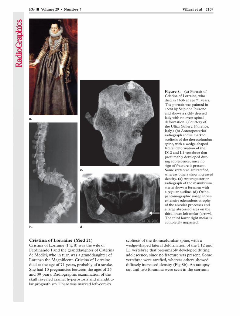
scoliosis of the thoracolumbar spine, with a wedge-shaped lateral deformation of the T12 and L1 vertebrae that presumably developed during adolescence, since no fracture was present. Some vertebrae were rarefied, whereas others showed diffusely increased density (Fig 8b). An autopsy cut and two foramina were seen in the sternum
Cristina of Lorraine (Med 21)Cristina of Lorraine (Fig 8) was the wife of Ferdinando I and the granddaughter of Caterina de Medici, who in turn was a granddaughter of Lorenzo the Magnificent. Cristina of Lorraine died at the age of 71 years, probably of a stroke. She had 10 pregnancies between the ages of 25 and 39 years. Radiographic examination of the skull revealed cranial hyperostosis and mandibu-lar prognathism. There was marked left-convex
Figure 8. (a) Portrait of Cristina of Lorraine, who died in 1636 at age 71 years. The portrait was painted in 1590 by Scipione Pulzone and shows a richly dressed lady with no overt spinal deformation. (Courtesy of the Uffizi Gallery, Florence, Italy.) (b) Anteroposterior radiograph shows marked scoliosis of the thoracolumbar spine, with a wedge-shaped lateral deformation of the D12 and L1 vertebrae that presumably developed dur-ing adolescence, since no sign of fracture is present. Some vertebrae are rarefied, whereas others show increased density. (c) Anteroposterior radiograph of the manubrium sterni shows a foramen with a regular outline. (d) Ortho-pantomographic image shows extensive edentulous atrophy of the alveolar processes and a large abscessed area on the third lower left molar (arrow). The third lower right molar is completely impacted.
2110 November-December 2009 radiographics.rsnajnls.org
RG ■ Volume 29 • Number 7 Villari et al 2111
and collapsed as a possible consequence of bone tuberculosis (Fig 9e, 9f). The appendicular bones were markedly osteoporotic, especially those of the lower limbs, presumably because of the car-dinal’s disability, which was especially prominent during the last years of his life (5). In addition, radiographic examination showed nearly sym-metric ankylosis of multiple joints, including the carpal bones, right sacroiliac joint, knees and rot-ulae, and tarsal and metatarsal bones (Fig 9g–9i).
Orthopantomography showed widespread, nearly edentulous atrophy of the alveolar pro-cesses and severe demineralization of the mandi-ble. Interdental decay was present in the second lower left incisor and between the lower right canine and the first premolar.
DiscussionThe radiographic findings in the 12 exhumed skeletons can be classified into malformations and deformities, degenerative and inflammatory changes, metabolic changes, and dental changes.
Malformations and DeformitiesIn the Renaissance, the appearance of members of the nobility was captured on canvas by court painters. Our study indicates that the painters often purposely hid overt imperfections, such as the facial asymmetry of Cardinal Carlo, the hydrocephalus of Don Filippino, and the severe scoliosis of Giovanna of Austria and Cristina of Lorraine.
Other bone malformations that we observed had no such cosmetic implications, including the various spinal abnormalities observed in the remains of Francesco I, Cardinal Carlo, and Eleonora of Toledo. In particular, the markedly forward-angled appearance of the coccyx in the remains of Francesco I is not attributable to an earlier traumatic event (eg, a fall from a horse) as was previously hypothesized (5), but to an ana-tomic variant (17).
A relatively common bone anomaly in our series was a sternal foramen, which was ob-served in the remains of both of Cosimo I’s sons, Francesco I and Ferdinando I, and (in the
(Fig 8c). The upper foramen, which involved the manubrium, was presumably of a vascular nature because of the regularity of its outline and its slightly sunken appearance.
Orthopantomography showed extensive eden-tulous atrophy of the alveolar processes and the presence of a large abscessed area on the third lower left molar. The third lower right molar was completely impacted (Fig 8d).
Prince Francesco (Med 13)Prince Francesco, son of Ferdinando I and Cristina of Lorraine, died at age 20 years, proba-bly of abdominal typhus. He entered the military when he was 15 years of age. Paleopathologic findings were unremarkable. Radiographic ex-amination showed a slash lesion on the left hu-meral head made by the blade that was used to open the box containing the remains, signs of craniotomy performed during autopsy, and signs of arthritis of the great joints. A small subcortical bone fibroma was present in the distal end of the right femoral diaphysis.
Orthopantomography showed widespread signs of periodontopathy with atrophy of the in-terdental septa.
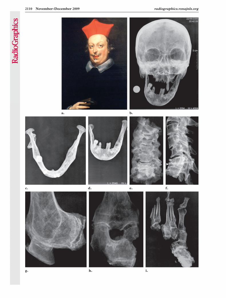
Cardinal Carlo (Med 29)Cardinal Carlo (Fig 9), son of Ferdinand I and Cristina of Lorraine, became a cardinal at 20 years of age and died at age 71 years of bron-chopneumonia. At age 8 years, he suffered from tuberculosis of the cervical and thoracic spine (Pott disease), which caused a cervical fistula (known as “scrofula”) and as a result of which he remained hunchbacked and had to wear a corset for the rest of his life (5). Beginning at age 24 years, he suffered from a recurrent acute joint disorder of the feet, hands, and knees, which was diagnosed as gout. Radiographic examina-tion showed marked asymmetry of the mandible, which appeared smaller on the right side with a compensatory large mastoid process (Fig 9b–9d). There was a presumably congenital fusion of the atlanto-occipital joint and of the posterior ele-ments of the cervical vertebrae from C1 to C5. A second vertebral block was seen to involve the C6 and C7 vertebrae, appearing wedge shaped
Figure 9. (a) Detail from a portrait of Cardinal Carlo, who died in 1666 at age 71 years. The portrait was painted by Justus Sustermans in about 1640 and shows minimal facial asymmetry. (Courtesy of the Poldi Pezzoli Museum, Milan, Italy.) (b–d) Anteroposterior radiograph of the skull (b) and superoinferior (c) and anteroposterior (d) ra-diographs show severe asymmetry of the mandible due to hypoplasia of the ascending branch of the right hemiman-dible with no fracture. (e, f) Anteroposterior (e) and lateral (f) radiographs of the cervical spine reveal fusion of the posterior vertebral elements from C1 to C5. A second vertebral block involving C6 and C7 is also seen. The block appears wedge shaped and collapsed (arrowhead in f), possibly as a consequence of bone tuberculosis. (g–i) Lateral radiograph of the right knee (g), anteroposterior radiograph of the left knee (h), and anteroposterior radiograph of the left tarsal and metatarsal bones (i) show nearly symmetric ankylosing disease of multiple joints.
TeachingPoint
2112 November-December 2009 radiographics.rsnajnls.org
In our opinion, the ankylosing polyarthritis with its recurrent course seen in the remains of Cardinal Carlo may be due to psoriatic arthritis, but there is no portrait or contemporary medical report to support this hypothesis. DNA analysis is currently in progress, with search being made for the presence of the human leukocyte antigen (HLA)–B27 gene. This gene is usually associated with some autoimmune disorders, such as anky-losing spondylitis and psoriatic arthritis.
Two other types of inflammatory bone change were observed in our series: osseous tubercu-losis in the remains of Cardinal Carlo and left-sided osteitis condensans ilii in the remains of Giovanna of Austria, the latter finding possibly representing a consequence of multiple pregnan-cies and deliveries (postpartum stress osteitis).
Metabolic ChangesThe hypothesis that the Medici suffered from gout is based on historical literary sources. Actually, contemporary physicians used the term gout to identify any disease that caused joint pain. The term was introduced in the 13th century and de-rives from the Latin word gutta (meaning “drop”), referring to the belief that an excess of one of the four “humors”—whose balance was thought to maintain health—would, under certain circum-stances, drop or flow into a joint, causing pain and inflammation. Gout has traditionally been associated with rich foods and excessive alcohol consumption. Because gout is clearly associated with a lifestyle that, at least in the past, could only be afforded by the wealthiest people, it has been referred to as the “disease of kings,” and, presum-ably for this reason, the painful arthritic conditions of the Medici were comprehensively labeled as gout, as indicated by the nickname “the Gouty” attributed to Piero. As a matter of fact, a distinc-tion between gout and rheumatism was not made until the 17th century (27).
Costa and Weber (1) pathologically examined the remains of four members of the senior branch of the Medici family (Cosimo the Elder; Piero the Gouty; Lorenzo the Magnificent; and Giuliano, Duke of Nemours) but did not find any bone changes that might be due to chronic tophaceous gout. They concluded that the Medici probably suffered from neurogenic arthropathy, ankylosing spondylitis, and osteoarthritis rather than gout, claiming a familial association of these diseases.
In our series of 12 members of the grand ducal branch, only the left foot lesion found in the remains of Ferdinando I was compatible with gout. Although gout is typically seen in elderly males and our series contained several young and
manubrium sterni) in the remains of Cristina of Lorraine, the consanguineous wife of Ferdinando I. Sternal foramina are incidental anomalies that occur in about 5% of the general population and are due to the incomplete fusion of the multiple ossification centers (18). No soft-tissue anomalies associated with sternal foramina (19) were men-tioned in or could be suspected on the basis of the contemporary reports.
Degenerative and Inflammatory ChangesThe lifestyle of the adult males in the Medici family was characterized by intense physical activity, such as riding horseback for long dis-tances, wearing heavy armor that overloaded the spinal column, and brandishing hefty swords and shields—all of which had significant conse-quences for the large joints. Signs of spondyloar-thritis were seen in all of the adult male Medici family members whose remains were examined, although this finding was more pronounced in the elderly subjects. In the remains of two adult males in our series—a father (Cosimo I) and son (Ferdinando I)—these changes were combined with (a) coarse secondary osteophytes fused together to form thoracic intervertebral bone bridges; and (b) other extraspinal osteophyte production, featuring a condition that today is known as DISH or Forestier disease (20–22) and in the past was variably termed moniliform hy-perostosis, ossifying ligamental spondylitis, senile ankylosing spondylitis, or dysmetabolic hyperos-totic polyenthesopathy.
The clinical features of DISH can be subtle if limited to stiffness and mild pain (23), which can account for the lack of references to the disease in the contemporary reports.
DISH is a common disorder of unknown cause, although genetic, metabolic, endocrino-logic, anatomic, environmental, and toxic fac-tors have all been suggested as playing a possible pathogenetic role in the new bone growth that characterizes DISH. The observation of DISH in the remains of a father and son in our series points to possible inherited or genetic predispos-ing factors, although no such factors have yet been identified. On the other hand, type II diabe-tes, obesity, dyslipidemia, and arterial hyperten-sion are common findings and potential risk fac-tors in patients with DISH (23–25). Interestingly, an association between DISH and gout arthropa-thy, as was apparently observed in the case of Ferdinando I, is also known (23,26).
female subjects, the results of our radiographic investigation confirmed the impression that the prevalence of gout in the Medici family might be overestimated. This impression is based on the
TeachingPoint
RG ■ Volume 29 • Number 7 Villari et al 2113
dence of tooth decay, and many abscess cavities. The calcium depletion associated with her 11 pregnancies can be hypothesized to have played a contributory role in the severe dental disease, contracted at a relatively young age, observed in the remains of Eleonora of Toledo.
ConclusionsRadiographic investigation of the exhumed skel-etal remains of several members of the grand ducal branch of the Medici family demonstrated multiple findings that challenge traditional views, based on portraits and historical accounts, con-cerning the appearance, lifestyle, and medical history of a number of these individuals. The oc-currence of a constellation of bone changes for which diabetes represents a general risk factor, including osteoporosis, osteoarthritis, DISH, in-tracranial hyperostosis, and crystalline arthropa-thy, suggests the possibility that this metabolic disease was common in the grand ducal branch of the family.
References 1. Costa A, Weber G. Le alterazioni morbose del
sistema scheletrico in Cosimo dei Medici il Vecchio, in Pietro il Gottoso, in Lorenzo il Magnifico, in Giu-liano Duca di Nemours. Arch Vecchi 1956;23:1–69.
2. Ferri M. I medici riesumano i Medici. Florence, Italy: Nuova Toscana Editrice, 2005.
3. Fornaciari G, Brier B, Fornaciari A. Secrets of the Medici. Archaeology 2005;58:36–41.
4. Fornaciari G, Vitiello A, Giusiani S, Giuffra V, Fornaciari A. The “Medici Project”: first results of the explorations of the Medici tombs in Florence (15th–18th centuries). Paleopathol Newsletter 2006;133:15–22.
5. Fornaciari G, Vitiello A, Giusiani S, Giuffra V, For-naciari A, Villari N. The “Medici Project”: first an-thropological and paleopathological results of the exploration of the Medici tombs in Florence. Med Secoli 2007;19:521–543.
6. Lippi D, Von Engelhardt D. “Progetto Medici”: Analyse der Leichname der Medicifamilie in der Ba-silica von San Lorenzo in Florenz. Focus Mul 2006; 23:142–148.
7. Lippi D. The “Medici Project.” Herald Eur 2006;3: 122–125.
8. Lippi D, Fornaciari G, Gensini GF. Evidence based history of medicine: the experience of the Florence Medical School. Proceedings of the 40th Interna-tional Congress on the History of Medicine, Buda-pest, August 25–30, 2006; 633–636.
9. Lippi D. Illacrimate sepolture: curiosità e ricerca scientifica nella storia delle riesumazioni dei Medici. Florence, Italy: Florence University Press, 2006.
10. Fornaciari G. Food and disease at the Renaissance courts of Naples and Florence: a paleonutritional study. Appetite 2008;51:10–14.
lack of pathologic evidence of chronic tophaceous gout, even in Piero the Gouty (1), and on the rec-ognition that contemporary physicians tended to label as gout every acute condition of the joints, especially in wealthy individuals (27).
In the remains of Cosimo I and his daughters, Giovanna of Austria and Cristina of Lorraine, we observed hyperostosis of the skull. This is a poorly understood benign condition of presum-ably hormonal etiopathogenesis possibly cor-related with latent or overt diabetes (28,29). Interestingly, one of the daughters of Giovanna of Austria (Maria) probably died of a diabetic coma (30). Unfortunately, the reason for the occur-rence of diabetes in the Medici family members whose remains we examined cannot be further ascertained due to the paucity of clinical data in contemporary records and the intrinsic limita-tions of paleopathologic and radiographic exami-nation. In our opinion, however, the obesity re-ported in some family members and our findings of (a) a constellation of bone changes (including osteoporosis, osteoarthritis, DISH, crystalline arthropathy) for which diabetes represents a gen-eral risk factor (31), and (b) intracranial hyper-ostosis represent indirect clues to the possibility that diabetes was common in this family branch. We recognize that this is merely a hypothesis and needs to be supported by further evidence.
The bowed legs that characterized the remains of Eleonora of Toledo were the only finding in our series that is consistent with rickets. It has been hypothesized that rickets in noble families of the Renaissance was likely due to children’s insufficient exposure to light in the environment of the court (5).
Harris lines in the tibiae in the remains of Cardinal Giovanni can be considered signs of malnourishment during bone development, but the easy access to food that the Medici family presumably enjoyed indicates that these findings were probably due to intercurrent diseases.
Dental ChangesThe eating habits of the Medici lords and ladies implied free access to meat, especially game, which presumably left abundant interdental remains. Because the oral hygiene of the time left much to be desired, the Medici family suf-fered the unpleasant consequences. In fact, we observed frequent loss of teeth, widespread evi-
TeachingPoint
2114 November-December 2009 radiographics.rsnajnls.org
20. Resnick D, Shaul S, Robins JM. Diffuse idiopathic skeletal hyperostosis (DISH): Forestier’s disease with extraspinal manifestations. Radiology 1975; 115:513–524.
21. Resnick D, Niwayama G. Radiographic and patho-logic features of spinal involvement in diffuse id-iopathic skeletal hyperostosis (DISH). Radiology 1976;119:559–568.
22. Resnick D, Guerra J Jr, Robinson CA, Vint VC. As-sociation of diffuse idiopathic skeletal hyperostosis (DISH) and calcification and ossification of the pos-terior longitudinal ligament. AJR Am J Roentgenol 1978;131:1049–1053.
23. Sarzi-Puttini P, Atzeni F. New developments in our understanding of DISH (diffuse idiopathic skel-etal hyperostosis). Curr Opin Rheumatol 2004;16: 287–292.
24. Kiss C, Szilágyi M, Paksy A, Poór G. Risk factors for diffuse idiopathic skeletal hyperostosis: a case-con-trol study. Rheumatology (Oxford) 2002;41:27–30.
25. Mader R, Lavi I. Diabetes mellitus and hyperten-sion as risk factors for early diffuse idiopathic skel-etal hyperostosis (DISH). Osteoarthr Cart 2009;17: 825–828.
26. Littlejohn GO, Hall S. Diffuse idiopathic skeletal hyperostosis and new bone formation in male gouty subjects. Rheumatol Int 1982;2:83–86.
27. Nuki G, Simkin PA. A concise history of gout and hyperuricemia and their treatment. Arthritis Res Ther 2006;8(suppl 1):S1.
28. Appel W. Hyperostosis of the skull in diabetics. Dtsch Arch Klin Med 1951;198:61–70.
29. Streda A, Hajkova Z, Skrha F. Endocranial hyperos-tosis, ankylosing spinal changes and diabetes. Acta Diabetol Lat 1971;8:479–499.
30. De Leeuw I. Did Queen Maria de’ Medici have type 2 diabetes? [in Dutch]. Vlaams Tijdschr Diabetol 2008;1:37–38.
31. Burner TW, Rosenthal AK. Diabetes and rheumatic diseases. Curr Opin Rheumatol 2009;21:50–54.
11. Fornaciari G, Giuffra V, Giusiani S, Fornaciari A, Villari N, Vitiello A. The ‘gout’ of the Medici, Grand Dukes of Florence: a paleopathological study. Rheu-matology (Oxford) 2009;48:375–377.
12. Sommi Picenardi G. Esumazione e Ricognizione delle Ceneri dei Principi Medicei fatta nell’anno 1857. In: Processo verbale e note, Archivio Storico Italiano, serie V, tomo I–II, Cellini M, Firenze, 1888.
13. Ortner DJ. Identification of pathological conditions in human skeletal remains. 2nd ed. New York, NY: Smithsonian Institution Press, 2003.
14. Pieraccini G. La stirpe di Cafaggiolo. Vol 2. Flor-ence, Italy: Nardini Editore, 1986.
15. Mari F, Polettini A, Lippi D, Bertol E. The mysteri-ous death of Francesco I de’ Medici and Bianca Cappello: an arsenic poisoning? BMJ 2006;333: 1299–1301.
16. Fornaciari G. The mystery of beard hairs. Available at: http://www.bmj.com/cgi/eletters/333/7582/1299. Published December 29, 2006.
17. Kohler A, Schmidt H, Zimmer EA, Hol W, Frey-schmidt J. Borderlands of normal and early patho-logic findings in skeletal radiography. New York, NY: Thieme Medical Publishers, 1993.
18. Yekeler E, Tunaci M, Tunaco A, Dursun M, Acunas G. Frequency of sternal variations and anomalies evaluated by MDCT. AJR Am J Roentgenol 2006; 186:956–960.
19. Wheeler DS, St Louis JD. Pentalogy of Cantrell associated with hypoplastic left heart syndrome. Pe-diatr Cardiol 2007;28:311–313.
RG Volume 29 Number 7 November-December 2009 Villari et al
Scenes from the Past The Medici Project: Radiographic Survey
Natale Villari, MD, et al
Page 2102
Since the 20th century, the Medici family has attracted considerable interest on the part of medical
doctors.
Page 2102
The Medici Project is a multi-institutional, multidisciplinary study that was started in 2003 and is
based on the exhumation of the remains of 12 members of the grand ducal branch of the Medici
family buried in the Medici Chapel of San Lorenzo Church in Florence, Italy. Studies are being
carried out in the areas of paleopathology, immunohistochemistry, molecular biology, isotope
analysis, toxicology, and diagnostic radiology to provide information on the lifestyles, illnesses, and
possible causes of death of these family members.
Page 2111
Our study indicates that the painters often purposely hid overt imperfections, such as the facial
asymmetry of Cardinal Carlo, the hydrocephalus of Don Filippino, and the severe scoliosis of
Giovanna of Austria and Cristina of Lorraine.
Page 2112
In our series of 12 members of the grand ducal branch, only the left foot lesion found in the remains
of Ferdinando I was compatible with gout. Although gout is typically seen in elderly males and our
series contained several young and female subjects, the results of our radiographic investigation
confirmed the impression that the prevalence of gout in the Medici family might be overestimated.
Page 2113 The occurrence of a constellation of bone changes for which diabetes represents a general risk factor, including osteoporosis, osteoarthritis, DISH, intracranial hyperostosis, and crystalline arthropathy, suggests the possibility that this metabolic disease was common in the grand ducal branch of the family.
RadioGraphics 2009; 29:2101–2114 • Published online 10.1148/rg.297085212 • Content Codes: