sars-cov-2 vocs immune evasion from previously elicited
TRANSCRIPT
1
SARS-CoV-2 VOCs immune evasion from previously elicited neutralizing
antibodies is mainly driven by lower cross-reactivity due to Spike RBD
electrostatic surface changes
Matheus V. F. Ferraza,b, Emerson G. Moreiraa,b, Danilo F. Coêlhoa,b, Gabriel Luz Wallaua and
Roberto D. Linsa,* on behalf of Fiocruz COVID-19 Genomic Surveillance Network
aAggeu Magalhães Institute, Oswaldo Cruz Foundation, Recife, PE, Brazil
bDepartment of Fundamental Chemistry, Federal University of Pernambuco, Recife, PE, Brazil
*Corresponding author: [email protected]
SARS-CoV-2 VOCs immune evasion is mainly due to lower cross-reactivity from previously
elicited antibodies, while increased affinity to hACE2 plays a minor role. Low affinity
between those antibodies and SARS-CoV-2 VOCs are caused by remodeling in the
electrostatic surface potential of the Spike RBDs. P.3 variant is a putative VOC.
Introduction
The COVID-19 pandemic has dramatically impacted the world population since 2019
and currently accounts for more than 2 million deaths.1 The genome evolution of its
etiological agent, the severe acute respiratory syndrome coronavirus 2 (SARS-CoV-2),
has been closely monitored since the rapid sharing of the first genomic sequences in
December 2019.2 SARS-CoV-2 presents a relatively low mutation rate compared to
other RNA viruses,3 and hence few genomic sites accumulated mutations and were
fixed until the second quarter of 2020.4 However, a substantially different scenario
emerged between September-December 2020 with the detection of independent
variants of concern (VOCs - B.1.1.7,5 B.1.351,6 and P.17) lineages bearing multiple
amino acid replacements (K417T, E484K, and N501Y) and indels in the Spike protein,8
in which some researchers hypothesized to have occurred due to a “global shift in the
2
SARS-CoV-2 selective landscape”.9 Although large-scale immunological studies are not
available so far, the main hypothesis to explain such a global shift takes into account
the rising population immunity, which would naturally select escape mutants with a
higher fitness compared to previous circulating lineages. To support this hypothesis,
some evidence could be mentioned, such as the increasing number of reinfection cases
with VOCs and variants of interest (VOI) carrying some of the same amino acid
mutation (E484K),10 the continuum emergence of new VOIs carrying E484K and N501Y
during the first months of 202111 and the recurrent emergence of some of those Spike
amino acid changes in SARS-CoV-2 experimental evolution settings challenged with
monoclonal and polyclonal antibodies.12
To enter the host cell, SARS-CoV-2 makes use of the glycoprotein Spike (S). Protein
S is a homotrimer and each monomer has two subunits, S1 and S2. The S1 subunit
contains the receptor-binding domain (RBD), which binds to the human receptor
angiotensin-converting enzyme 2 (hACE2), thus allowing the fusion of membranes and
entry into the cell.13 Among the 29 SARS-CoV-2 encoded proteins, the S protein has
been investigated more thoroughly due to its key role in hACE2 binding, and because
the RBD region is one of the main targets of neutralizing antibodies (nAbs) produced
from the human immunological response against the SARS-CoV-2.14 Deep mutational
scanning of the RBD region has identified that most amino acid changes are deleterious
for hACE2 binding, whereas a few marginally enhance the affinity to hACE2,15 including
some that have been detected in VOCs, such as N501Y in the more transmissible and
mortal B.1.1.7 lineage.16 On the other hand, amino acid changes such as K417N, E484K,
and N501Y found in VOCs P.117 and B.1.3518 have been shown to increase viral fitness
by lowering the effectiveness of neutralizing monoclonal and/or polyclonal
antibodies.11 Therefore, the emergence and spread of more fit VOCs lineages may be
driven by a more complex mechanism other than the often-proposed affinity increase
between hACE2 and SARS-CoV-2 Spike protein.
Results and Discussion
Despite the extensive description of mutations occurring in the SARS-CoV-2 RBD, little is
known about their impact on receptor recognition, namely hACE2. In this regard, Starr et al15
3
have systematically measured the impact of every amino acid in the RBD, by replacing for the
20 amino acids in each position, towards hACE2 binding affinity, expressed as the 𝛥𝑙𝑜𝑔(KD),
in which KD represents the dissociation constant. Changes in the KD upon single-point
mutations were obtained from a deep mutational scanning library using the yeast-surface
display technique. We have converted the 𝛥𝑙𝑜𝑔(KD) into the change in the mutational Gibbs
free energy of binding 𝛥𝛥𝐺 for all sites of interaction between SARS-CoV-2 RBD and hACE2
(more details in Electronic Supplementary Information) and observed that whereas most of
the single mutations on the RBD tend to be deleterious (i.e, 𝛥𝛥𝐺>0), none of the mutations
would dramatically enhance the affinity for the hACE2 (Figure S1). Since the wild-type (WT)
RBD (from Wuhan reference genome) binds the hACE2 in the nanomolar range, variations in
the KD on the order of 1E+1, resulting in approximately 0.5-1.5 kcal.mol-1, do not have the
potential to significantly impact the binding affinity. However, it is important to stress that
deep mutational scanning binding 𝛥𝛥𝐺 values were reported for single mutations only and
the novel SARS-CoV-2 VOCs often involve more than one mutation on the RBD.
Aiming to investigate whether multiple mutations could lead to a significant increase in
affinity to hACE2, variation in binding free energy calculations were performed between the
RBDs of selected VOCs and hACE2. For 4 out of 7 lineages evaluated, the binding 𝛥𝛥𝐺 values
fell within the mean absolute error (MAE) associated with the method used (ca. ± 1.7 REU,
by testing the 𝛥𝛥𝐺 calculations considering the 4,000 mutations from the deep mutational
scanning data, i.e, a dataset containing a similar system to the calculations we have
performed; see methodology section of the ESI), whereas the remaining values have positive
values and are boardliners to the error limit (Figure 1B). This finding is in agreement with data
from the above discussed deep mutational scanning experiments15 and bio-layer
interferometry assays,18 where all measurements have shown KD values for different RBDs to
hACE2 within 10-8 to 10-9 M. Therefore, assumptions that higher transmissibility is mainly
associated with mutations in the RBD that leads to an enhanced affinity to hACE2 must be
revisited, as both experiments and calculations do not show substantial evidence
corroborating this hypothesis.
On the other hand, a plethora of studies has demonstrated antibody evasion for some of
the novel VOCs.19-28 However, data correlating immune evasion of SARS-CoV-2 VOCs with
binding free energies are currently not available. In order to evaluate the thermodynamics
contribution of such low cross-reactivity, we calculated the variation in binding free energy
4
values for a selected set of SARS-CoV-2 VOCs RBDs complexed to 21 known nAbs for which
atomic coordinates were made publicly available (Table S2). These nAbs bind to two distinct
regions of the viral RBD. As illustrated in Figure 1A, all nAbs block the RBD access to hACE2;
however, classes I and II have a higher spatial overlap with the hACE2 binding site compared
to classes III and IV.29 The variations in binding free energies are shown in Figure 1B. The data
suggest that binding is highly compromised between most nAbs elicited against RBD of
previous circulating non-VOCs lineages when compared to VOCs RBD, with the only exception
being lineage B.1.429. It is important to note that this variant was identified in Portugal and
California (USA)30 at a similar time frame as VOCs B.1.1.7, B.1.351, and P.1, but it has not
spread and increased in frequency significantly as recognized VOCs did. Interestingly, the
calculated variations in binding free energies is able to capture the trend, i.e., variants that
emerged and rapidly reached higher frequencies outcompeting other circulating lineages
(VOCs) by having a larger number of nAbs that would not bind to the VOCs. Our calculations
also corroborate with available experimental data for nAbs in our dataset. nAbs STE90-C11,31
and C10229 were verified to continue to bind to RBD containing the E484K mutation (lineage
P.2). The calculated 𝛥𝛥𝐺 for these nAbs against P.2 are within the MAE (Table S2). On the
other hand, nAb 15033 moderately loses its neutralization power against a pseudo-virus
containing the E484K mutation,32 a finding supported by a positive 𝛥𝛥𝐺 variation above the
MAE (Table S2). As for nAb BD-629, a neutralization assay was performed with the pseudo-
virus containing the L452R mutation,33 which corresponds to lineage B.1.429. It showed that
even after the mutation, the nAb continues to neutralize the virus, which is also corroborated
by our calculations (Table S2). Moreover, our data also suggest that variants B.1.526 and P.2
would have limited spread as a large number of nAbs are still capable of binding to their RBD
and, hence neutralizing it. Conversely, most of the tested nAbs are expected not to bind
efficiently to P.3 RBD, a recently reported lineage detected in the Philippines,34 similar to
recognized VOCs B.1.1.7, B.1.351 and P.1, suggesting that this variant is a putative VOC with
a high spreading potential in a population with medium to high community immunity against
previous circulating lineages due to immune evasion. (We would like to note that similar
results were obtained when using FoldX35 (Figure S2), a different software package/algorithm,
in which its empirical energy function provides an MAE of 0.46 kcal.mol-1,36 therefore
providing further support to these observations).
5
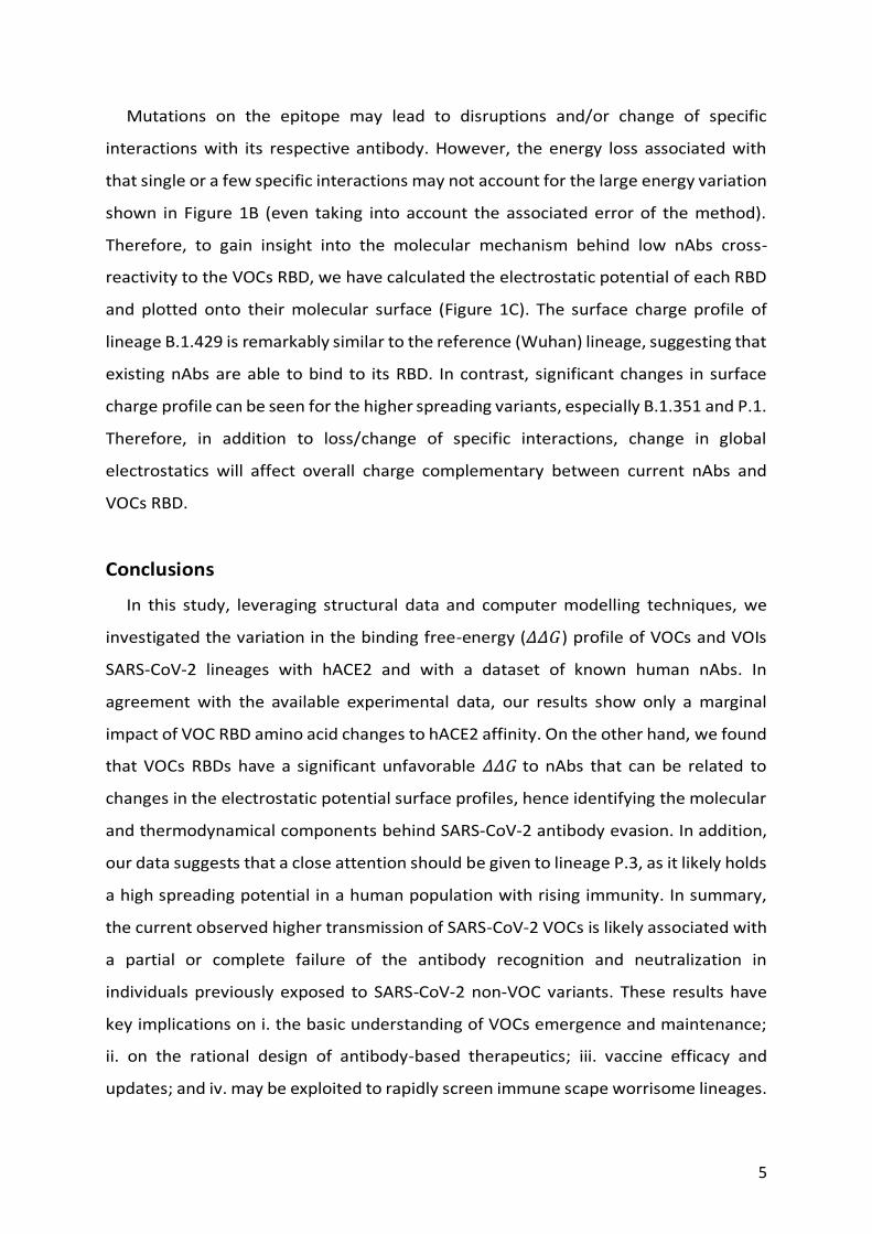
Mutations on the epitope may lead to disruptions and/or change of specific
interactions with its respective antibody. However, the energy loss associated with
that single or a few specific interactions may not account for the large energy variation
shown in Figure 1B (even taking into account the associated error of the method).
Therefore, to gain insight into the molecular mechanism behind low nAbs cross-
reactivity to the VOCs RBD, we have calculated the electrostatic potential of each RBD
and plotted onto their molecular surface (Figure 1C). The surface charge profile of
lineage B.1.429 is remarkably similar to the reference (Wuhan) lineage, suggesting that
existing nAbs are able to bind to its RBD. In contrast, significant changes in surface
charge profile can be seen for the higher spreading variants, especially B.1.351 and P.1.
Therefore, in addition to loss/change of specific interactions, change in global
electrostatics will affect overall charge complementary between current nAbs and
VOCs RBD.
Conclusions
In this study, leveraging structural data and computer modelling techniques, we
investigated the variation in the binding free-energy (𝛥𝛥𝐺) profile of VOCs and VOIs
SARS-CoV-2 lineages with hACE2 and with a dataset of known human nAbs. In
agreement with the available experimental data, our results show only a marginal
impact of VOC RBD amino acid changes to hACE2 affinity. On the other hand, we found
that VOCs RBDs have a significant unfavorable 𝛥𝛥𝐺 to nAbs that can be related to
changes in the electrostatic potential surface profiles, hence identifying the molecular
and thermodynamical components behind SARS-CoV-2 antibody evasion. In addition,
our data suggests that a close attention should be given to lineage P.3, as it likely holds
a high spreading potential in a human population with rising immunity. In summary,
the current observed higher transmission of SARS-CoV-2 VOCs is likely associated with
a partial or complete failure of the antibody recognition and neutralization in
individuals previously exposed to SARS-CoV-2 non-VOC variants. These results have
key implications on i. the basic understanding of VOCs emergence and maintenance;
ii. on the rational design of antibody-based therapeutics; iii. vaccine efficacy and
updates; and iv. may be exploited to rapidly screen immune scape worrisome lineages.
6
Figure 1. Title. A) Cartoon representation of the SARS-CoV-2 RBD (gray) bound to
hACE2 (green) and representative nAbs from classes I and II (blue) and II and IV (red);
B) Calculated relative binding free energies of RBDs complexed to hACE and nAbs (RBD
from Wuhan lineage is taken as reference); C) Electrostatic surface potential of SARS-
CoV-2 RBD reference and selected variants.
RDL and GW designed the study. MVFF, EGM and DFC performed all modeling, calculations
and data analysis. The manuscript was written with the input from all authors.
The authors have declared no competing interests. We thank all the health care workers and
scientists who have worked hard to deal with this pandemic threat, the GISAID team, and all
the EpiCoV database's submitters, GISAID acknowledgment table containing sequences used
in this study are attached to this post (Table S1). This study was supported by grants from
FACEPE, CAPES, CNPq and INCT-FCx and FIOCRUZ COVID-19 Genomic Surveillance Network.
Computer allocation was partly granted by the Brazilian National Scientific Computing Center
(LNCC). GLW and RDL are supported by the CNPq through their productivity research
fellowships (303902/2019-1 and 425997/2018-9).
Notes and References
1. E. Dong, H. Du and L. Gardner, Lancet Infect Dis, 2020, 20, 533-534.
+2-2
kcal•mol-1•e-1Wuhan
B.1.526
B.1.1.7 P.1
B.1.351
P.2 B.1.429 P.3
A B
C
RBD
hACE2
nAb Classes I, II
nAb Classes III, IV
7
2. P. Zhou, X. L. Yang, X. G. Wang, B. Hu, L. Zhang, W. Zhang, H. R. Si, Y. Zhu, B. Li, C. L. Huang, H. D. Chen, J. Chen, Y. Luo, H. Guo, R. D. Jiang, M. Q. Liu, Y. Chen, X. R. Shen, X. Wang, X. S. Zheng, K. Zhao, Q. J. Chen, F. Deng, L. L. Liu, B. Yan, F. X. Zhan, Y. Y. Wang, G. F. Xiao and Z. L. Shi, Nature, 2020, 579, 270-273.
3. L. van Dorp, M. Acman, D. Richard, L. P. Shaw, C. E. Ford, L. Ormond, C. J. Owen, J. Pang, C. C. S. Tan, F. A. T. Boshier, A. T. Ortiz and F. Balloux, Infect Genet Evol, 2020, 83, 104351.
4. D. Mercatelli and F. M. Giorgi, Front Microbiol, 2020, 11, 1800. 5. A. Rambaut, N. Loman, O. Pybus, W. Barclay, J. Barrett, A. Carabelli, T. Connor, T.
Peacock, D. L. Robertson and E. Volz, Genom. Epidemiol, 2020. 6. H. Tegally, E. Wilkinson, M. Giovanetti, A. Iranzadeh, V. Fonseca, J. Giandhari, D.
Doolabh, S. Pillay, E. J. San, N. Msomi, K. Mlisana, A. von Gottberg, S. Walaza, M. Allam, A. Ismail, T. Mohale, A. J. Glass, S. Engelbrecht, G. Van Zyl, W. Preiser, F. Petruccione, A. Sigal, D. Hardie, G. Marais, M. Hsiao, S. Korsman, M.-A. Davies, L. Tyers, I. Mudau, D. York, C. Maslo, D. Goedhals, S. Abrahams, O. Laguda-Akingba, A. Alisoltani-Dehkordi, A. Godzik, C. K. Wibmer, B. T. Sewell, J. Lourenço, L. C. J. Alcantara, S. L. K. Pond, S. Weaver, D. Martin, R. J. Lessells, J. N. Bhiman, C. Williamson and T. de Oliveira, medRxiv, 2020, DOI: 10.1101/2020.12.21.20248640, 2020.2012.2021.20248640.
7. N. R. Faria, I. M. Claro, D. Candido, L. A. Moyses Franco, P. S. Andrade, T. M. Coletti, C. A. M. Silva, F. C. Sales, E. R. Manuli and R. S. Aguiar, Virological. org, 2021, 586.
8. H. Tegally, E. Wilkinson, M. Giovanetti, A. Iranzadeh, V. Fonseca, J. Giandhari, D. Doolabh, S. Pillay, E. J. San, N. Msomi, K. Mlisana, A. von Gottberg, S. Walaza, M. Allam, A. Ismail, T. Mohale, A. J. Glass, S. Engelbrecht, G. Van Zyl, W. Preiser, F. Petruccione, A. Sigal, D. Hardie, G. Marais, M. Hsiao, S. Korsman, M. A. Davies, L. Tyers, I. Mudau, D. York, C. Maslo, D. Goedhals, S. Abrahams, O. Laguda-Akingba, A. Alisoltani-Dehkordi, A. Godzik, C. K. Wibmer, B. T. Sewell, J. Lourenco, L. C. J. Alcantara, S. L. Kosakovsky Pond, S. Weaver, D. Martin, R. J. Lessells, J. N. Bhiman, C. Williamson and T. de Oliveira, Nature, 2021, DOI: 10.1038/s41586-021-03402-9.
9. D. P. Martin, S. Weaver, H. Tegally, E. J. San, S. D. Shank, E. Wilkinson, J. Giandhari, S. Naidoo, Y. Pillay, L. Singh, R. J. Lessells, S. A. Ngs, C.-G. UK, R. K. Gupta, J. O. Wertheim, A. Nekturenko, B. Murrell, G. W. Harkins, P. Lemey, O. A. MacLean, D. L. Robertson, T. de Oliveira and S. L. K. Pond, medRxiv, 2021, DOI: 10.1101/2021.02.23.21252268.
10. N. Felipe, C. Cristiano da, N. Valdinete, S. Victor, C. André, N. Fernanda, C. Ágatha, D. Débora, S. George, M. Matilde, P. Karina, G. Luciana, B. Maria Júlia, J. Michele, S. Marineide, M. Tirza, A. Lígia, S. João Hugo, C.-F. Rubens, S. Tsuyoshi, I. Kentaro, H. Masanori, K. Makoto, S. Marilda Mendonça, W. Gabriel Luz, D. Edson, G. Tiago, B. Gonzalo and R. Paola Cristina, Research Square, 2021, DOI: 10.21203/rs.3.rs-318392/v1.
11. A. J. Greaney, A. N. Loes, K. H. D. Crawford, T. N. Starr, K. D. Malone, H. Y. Chu and J. D. Bloom, Cell Host Microbe, 2021, 29, 463-476 e466.
12. Z. Wang, F. Schmidt, Y. Weisblum, F. Muecksch, C. O. Barnes, S. Finkin, D. Schaefer-Babajew, M. Cipolla, C. Gaebler, J. A. Lieberman, T. Y. Oliveira, Z. Yang, M. E. Abernathy, K. E. Huey-Tubman, A. Hurley, M. Turroja, K. A. West, K. Gordon, K. G. Millard, V. Ramos, J. Da Silva, J. Xu, R. A. Colbert, R. Patel, J. Dizon, C. Unson-O'Brien, I. Shimeliovich, A. Gazumyan, M. Caskey, P. J. Bjorkman, R. Casellas, T. Hatziioannou,
8
P. D. Bieniasz and M. C. Nussenzweig, Nature, 2021, DOI: 10.1038/s41586-021-03324-6.
13. X. Chi, R. Yan, J. Zhang, G. Zhang, Y. Zhang, M. Hao, Z. Zhang, P. Fan, Y. Dong, Y. Yang, Z. Chen, Y. Guo, J. Zhang, Y. Li, X. Song, Y. Chen, L. Xia, L. Fu, L. Hou, J. Xu, C. Yu, J. Li, Q. Zhou and W. Chen, Science, 2020, 369, 650-655.
14. M. McCallum, A. De Marco, F. A. Lempp, M. A. Tortorici, D. Pinto, A. C. Walls, M. Beltramello, A. Chen, Z. Liu, F. Zatta, S. Zepeda, J. di Iulio, J. E. Bowen, M. Montiel-Ruiz, J. Zhou, L. E. Rosen, S. Bianchi, B. Guarino, C. S. Fregni, R. Abdelnabi, S. C. Foo, P. W. Rothlauf, L. M. Bloyet, F. Benigni, E. Cameroni, J. Neyts, A. Riva, G. Snell, A. Telenti, S. P. J. Whelan, H. W. Virgin, D. Corti, M. S. Pizzuto and D. Veesler, Cell, 2021, DOI: 10.1016/j.cell.2021.03.028.
15. T. N. Starr, A. J. Greaney, S. K. Hilton, D. Ellis, K. H. D. Crawford, A. S. Dingens, M. J. Navarro, J. E. Bowen, M. A. Tortorici, A. C. Walls, N. P. King, D. Veesler and J. D. Bloom, Cell, 2020, 182, 1295-1310 e1220.
16. N. G. Davies, C. I. Jarvis, C. C.-W. Group, W. J. Edmunds, N. P. Jewell, K. Diaz-Ordaz and R. H. Keogh, Nature, 2021, DOI: 10.1038/s41586-021-03426-1.
17. N. Felipe, N. Valdinete, S. Victor, C. André, N. Fernanda, S. George, C. Ágatha, D. Débora, P. Karina, M. Matilde, B. Maria, J. Michele, G. Luciana, C. Cristiano da, S. Vanderson, B. Daniel, S. Marineide, M. Tirza, P. Gemilson, A. Ligia, S. João, A. Ighor, D. Filipe, S. Marilda, W. Gabriel, R. Paola, D. Edson, G. Tiago and B. Gonzalo, Nature Portfolio, 2021, DOI: 10.21203/rs.3.rs-275494/v1.
18. X. Zhu, D. Mannar, S. S. Srivastava, A. M. Berezuk, J.-P. Demers, J. W. Saville, K. Leopold, W. Li, D. S. Dimitrov, K. S. Tuttle, S. Zhou, S. Chittori and S. Subramaniam, bioRxiv, 2021, DOI: 10.1101/2021.01.11.426269, 2021.2001.2011.426269.
19. D. M. Altmann, R. J. Boyton and R. Beale, Science, 2021, 371, 1103-1104. 20. D. Zhou, W. Dejnirattisai, P. Supasa, C. Liu, A. J. Mentzer, H. M. Ginn, Y. Zhao, H. M.
E. Duyvesteyn, A. Tuekprakhon, R. Nutalai, B. Wang, G. C. Paesen, C. Lopez-Camacho, J. Slon-Campos, B. Hallis, N. Coombes, K. Bewley, S. Charlton, T. S. Walter, D. Skelly, S. F. Lumley, C. Dold, R. Levin, T. Dong, A. J. Pollard, J. C. Knight, D. Crook, T. Lambe, E. Clutterbuck, S. Bibi, A. Flaxman, M. Bittaye, S. Belij-Rammerstorfer, S. Gilbert, W. James, M. W. Carroll, P. Klenerman, E. Barnes, S. J. Dunachie, E. E. Fry, J. Mongkolsapaya, J. Ren, D. I. Stuart and G. R. Screaton, Cell, 2021, DOI: 10.1016/j.cell.2021.02.037.
21. R. Wang, Q. Zhang, J. Ge, W. Ren, R. Zhang, J. Lan, B. Ju, B. Su, F. Yu, P. Chen, H. Liao, Y. Feng, X. Li, X. Shi, Z. Zhang, F. Zhang, Q. Ding, T. Zhang, X. Wang and L. Zhang, bioRxiv, 2021, DOI: 10.1101/2021.03.09.434497, 2021.2003.2009.434497.
22. C. K. Wibmer, F. Ayres, T. Hermanus, M. Madzivhandila, P. Kgagudi, B. Oosthuysen, B. E. Lambson, T. de Oliveira, M. Vermeulen, K. van der Berg, T. Rossouw, M. Boswell, V. Ueckermann, S. Meiring, A. von Gottberg, C. Cohen, L. Morris, J. N. Bhiman and P. L. Moore, Nat Med, 2021, DOI: 10.1038/s41591-021-01285-x.
23. W. F. Garcia-Beltran, E. C. Lam, K. St Denis, A. D. Nitido, Z. H. Garcia, B. M. Hauser, J. Feldman, M. N. Pavlovic, D. J. Gregory, M. C. Poznansky, A. Sigal, A. G. Schmidt, A. J. Iafrate, V. Naranbhai and A. B. Balazs, Cell, 2021, DOI: 10.1016/j.cell.2021.03.013.
24. D. Focosi and F. Maggi, Rev Med Virol, 2021, DOI: 10.1002/rmv.2231. 25. C. Rees-Spear, L. Muir, S. A. Griffith, J. Heaney, Y. Aldon, J. L. Snitselaar, P. Thomas, C.
Graham, J. Seow, N. Lee, A. Rosa, C. Roustan, C. F. Houlihan, R. W. Sanders, R. Gupta,
9
P. Cherepanov, H. Stauss, E. Nastouli, K. J. Doores, M. J. van Gils and L. E. McCoy, bioRxiv, 2021, DOI: 10.1101/2021.01.15.426849, 2021.2001.2015.426849.
26. T. N. Starr, A. J. Greaney, A. Addetia, W. W. Hannon, M. C. Choudhary, A. S. Dingens, J. Z. Li and J. D. Bloom, Science, 2021, 371, 850.
27. A. J. Greaney, T. N. Starr, P. Gilchuk, S. J. Zost, E. Binshtein, A. N. Loes, S. K. Hilton, J. Huddleston, R. Eguia, K. H. D. Crawford, A. S. Dingens, R. S. Nargi, R. E. Sutton, N. Suryadevara, P. W. Rothlauf, Z. Liu, S. P. J. Whelan, R. H. Carnahan, J. E. Crowe and J. D. Bloom, Cell Host & Microbe, 2021, 29, 44-57.e49.
28. B. Luan and T. Huynh, bioRxiv, 2021, DOI: 10.1101/2021.02.06.430088, 2021.2002.2006.430088.
29. C. O. Barnes, C. A. Jette, M. E. Abernathy, K. A. Dam, S. R. Esswein, H. B. Gristick, A. G. Malyutin, N. G. Sharaf, K. E. Huey-Tubman, Y. E. Lee, D. F. Robbiani, M. C. Nussenzweig, A. P. West, Jr. and P. J. Bjorkman, Nature, 2020, 588, 682-687.
30. W. Zhang, B. D. Davis, S. S. Chen, J. M. S. Martinez, J. T. Plummer and E. Vail, medRxiv, 2021, DOI: 10.1101/2021.01.18.21249786, 2021.2001.2018.21249786.
31. F. Bertoglio, V. Fühner, M. Ruschig, P. A. Heine, U. Rand, T. Klünemann, D. Meier, N. Langreder, S. Steinke, R. Ballmann, K.-T. Schneider, K. D. R. Roth, P. Kuhn, P. Riese, D. Schäckermann, J. Korn, A. Koch, S. Zock-Emmenthal, M. Becker, M. Scholz, G. M. S. G. Moreira, E. V. Wenzel, G. Russo, H. S. P. Garritsen, S. Casu, A. Gerstner, G. Roth, A. Hermann, T. Schirrmann, S. Dübel, A. Frenzel, J. Van den Heuvel, L. Čičin-Šain, M. Schubert and M. Hust, bioRxiv, 2020, DOI: 10.1101/2020.12.03.409318, 2020.2012.2003.409318.
32. S. Miersch, Z. Li, R. Saberianfar, M. Ustav, J. B. Case, L. Blazer, C. Chen, W. Ye, A. Pavlenco, M. Gorelik, J. G. Perez, S. Subramania, S. Singh, L. Ploder, S. Ganaie, R. E. Chen, D. W. Leung, P. P. Pandolfi, G. Novelli, G. Matusali, F. Colavita, M. R. Capobianchi, S. Jain, J. B. Gupta, G. K. Amarasinghe, M. S. Diamond, J. Rini and S. S. Sidhu, bioRxiv, 2020, DOI: 10.1101/2020.10.31.362848.
33. S. Du, Y. Cao, Q. Zhu, P. Yu, F. Qi, G. Wang, X. Du, L. Bao, W. Deng, H. Zhu, J. Liu, J. Nie, Y. Zheng, H. Liang, R. Liu, S. Gong, H. Xu, A. Yisimayi, Q. Lv, B. Wang, R. He, Y. Han, W. Zhao, Y. Bai, Y. Qu, X. Gao, C. Ji, Q. Wang, N. Gao, W. Huang, Y. Wang, X. S. Xie, X. D. Su, J. Xiao and C. Qin, Cell, 2020, 183, 1013-1023 e1013.
34. F. A. Tablizo, K. M. Kim, C. M. Lapid, M. J. R. Castro, M. S. L. Yangzon, B. A. Maralit, M. E. C. Ayes, E. M. Cutiongco-de la Paz, A. R. De Guzman, J. M. C. Yap, J.-H. S. Llames, S. M. M. Araiza, K. P. Punayan, I. C. A. Asin, C. F. B. Tambaoan, A. L. U. Chong, K. S. A. R. Padilla, R. P. S. Cruz, E. K. D. Morado, J. G. A. Dizon, R. N. M. Hao, A. A. Zamora, D. R. Pacial, J. A. R. Magalang, M. Alejandria, C. Carlos, A. Ong-Lim, E. M. Salvaña, J. Q. Wong, J. C. Montoya, M. R. Singh-Vergeire and C. P. Saloma, medRxiv, 2021, DOI: 10.1101/2021.03.03.21252812, 2021.2003.2003.21252812.
35. J. Schymkowitz, J. Borg, F. Stricher, R. Nys, F. Rousseau and L. Serrano, Nucleic Acids Res, 2005, 33, W382-388.
36. R. Guerois, J. E. Nielsen and L. Serrano, J Mol Biol, 2002, 320, 369-387.
10
Electronic Supplementary Information
SARS-CoV-2 VOCs immune evasion from previously elicited neutralizing antibodies is
mainly driven by lower cross-reactivity due to Spike RBD electrostatic surface changes
Matheus V. F. Ferraza,b, Emerson G. Moreiraa,b, Danilo F. Coêlhoa,b, Gabriel Luz Wallaua and
Roberto D. Linsa,* on behalf of Fiocruz COVID-19 Genomic Surveillance Network
aAggeu Magalhães Institute, Oswaldo Cruz Foundation, Recife, PE, Brazil
bDepartment of Fundamental Chemistry, Federal University of Pernambuco, Recife, PE, Brazil
*Corresponding author: [email protected]
1. Experimental values for ΔΔGBind between RBD and hACE2 upon single mutations ........ 11 2. Computational Procedure ................................................................................................ 12 2.1. Systems Preparation ........................................................................................................ 12 2.2. Binding free-energy calculations with Rosetta ................................................................ 13 2.3. Command-lines and XML scripts for the calculations with the Rosetta package ........... 15 2.3.1. ..................................................................................................... Rosetta Command-lines 15 2.3.2. .................................................................................................................... RosettaScripts 15 2.4. Binding free-energy calculation with FoldX ..................................................................... 18 2.5. Electrostatic potential calculations .................................................................................. 19 3. Antibodies dataset used for ΔΔGBind computational calculations ................................... 19 4. Associated References ..................................................................................................... 21
11
1. Experimental values for ΔΔGBind between RBD and hACE2 upon single mutations
Starr et al.1 obtained deep mutational scan data for all sites of interaction between SARS-CoV-
2 RBD and hACE2, transformed to values of Δlog(KD) according to the following relation:
∆𝑙𝑜𝑔(𝐾𝐷) = 𝑙𝑜𝑔(𝐾𝐷,𝑊𝑇| ) − 𝑙𝑜𝑔(𝐾𝐷,𝑀𝑈𝑇| ) eq. 1
where KD is the dissociation constant and WT are values for wild-type and MUT for mutated
variant. To transform the values into differences in binding free-energy, the following
mathematical relationship was used:
∆∆𝐺𝐵𝑖𝑛𝑑 = ∆𝐺𝑀𝑈𝑇 − ∆𝐺𝑀𝑈𝑇 = 𝑅𝑇[𝑙𝑛(𝐾𝐷,𝑀𝑈𝑇) − 𝑙𝑛(𝐾𝐷,𝑊𝑇)] = 𝑅𝑇𝑙𝑛 (𝐾𝐷,𝑀𝑈𝑇
𝐾𝐷,𝑊𝑇) =
−𝑅𝑇𝑙𝑛 (𝐾𝐷,𝑊𝑇
𝐾𝐷,𝑀𝑈𝑇) eq. 2
From eq. 1 we have:
∆𝑙𝑜𝑔(𝐾𝐷) = 𝑙𝑜𝑔 (𝐾𝐷,𝑊𝑇
𝐾𝐷,𝑀𝑈𝑇) ∴
𝐾𝐷,𝑊𝑇
𝐾𝐷,𝑀𝑈𝑇= 10∆𝑙𝑜𝑔(𝐾𝐷) eq. 3
Substituting eq.3 in eq. 2, we then have:
∆∆𝐺𝐵𝑖𝑛𝑑 = −𝑅𝑇𝑙𝑛(10∆𝑙𝑜𝑔(𝐾𝐷)) = −∆𝑙𝑜𝑔(𝐾𝐷) ∙ 𝑅𝑇𝑙𝑛(10| ) eq. 4
Using T = 298 K (25 ºC) and R = 1.987 kcal⋅K−1⋅mol−1 we transformed all ∆𝑙𝑜𝑔(𝐾𝐷)
experimental values into ∆∆𝐺𝐵𝑖𝑛𝑑 using eq. 4. Figure S1 shows the heat map plot for the
values.
12
Figure S.1 - Heat map plot of Deep Mutational Scan ∆∆𝑮𝑩𝒊𝒏𝒅 values converted from
experimental data retrieved from Starr et al.1 Horizontal-axis shows amino acid position in
RBD and correspondent native amino acid. The vertical axis shows the identity of mutation
amino acid. Values range from highly unfavorable changes (red squares) to marginally
favorable change (blue), whereas black squares denote mutation to native amino acid (no
experimental data available).
2. Computational Procedure
2.1. Systems Preparation
Atomic coordinates for the three-dimensional structure of the complex hACE2- SARS-CoV-2
RBD were retrieved from the Protein Data Bank (PDB) under accession code 6M0J.2 27 crystal
structures of nAbs bound to SARS-Cov-2 RBD were downloaded from the PDB. The structures
were cleaned by removing crystallographic water molecules, ligands, and ions. Only
structures without missing residues in the interface between RBD and the respective antibody
were considered, resulting in a total of 22 structures (accession code in Table S2). The missing
residues lying far from the binding interface were modeled using Swiss-Model.3, 4 Then, 4
steps of the FastRelax protocol, as implemented in the Rosetta package v. 3.12,5 were used
13
to pack and minimize the side-chains and backbone's conformations. Recognized VOCs
(B.1.1.7, B.1.351 and P.1) and VOIs (P.2, B.1.429, B.1.526) with known epidemiological
importance and bearing a number of key mutations in the RBD of the S protein (https://cov-
lineages.org/ and https://www.cdc.gov/coronavirus/2019-ncov/cases-updates/variant-
surveillance/variant-info.html) (Table S1)6 that were previously evaluated in experimental
studies were recovered from the GISAID database (https://www.gisaid.org/) and used to
estimate binding affinities with hACE2 and nAbs. Moreover, we also included a recent
emerged variant7 (P.3 - https://cov-lineages.org/lineages/lineage_P.3.html) bearing
additional Spike mutations to investigate its binding affinities to the hACE2 and nAbs as well.
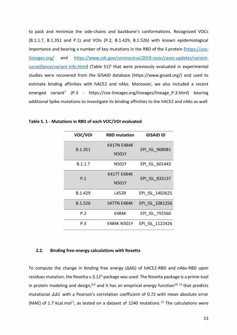
Table S. 1 - Mutations in RBD of each VOC/VOI evaluated
VOC/VOI RBD mutation GISAID ID
B.1.351 K417N E484K
N501Y EPI_ISL_968081
B.1.1.7 N501Y EPI_ISL_601443
P.1 K417T E484K
N501Y EPI_ISL_833137
B.1.429 L452R EPI_ISL_1402625
B.1.526 S477N E484K EPI_ISL_1081256
P.2 E484K EPI_ISL_792560
P.3 E484K N501Y EPI_ISL_1122426
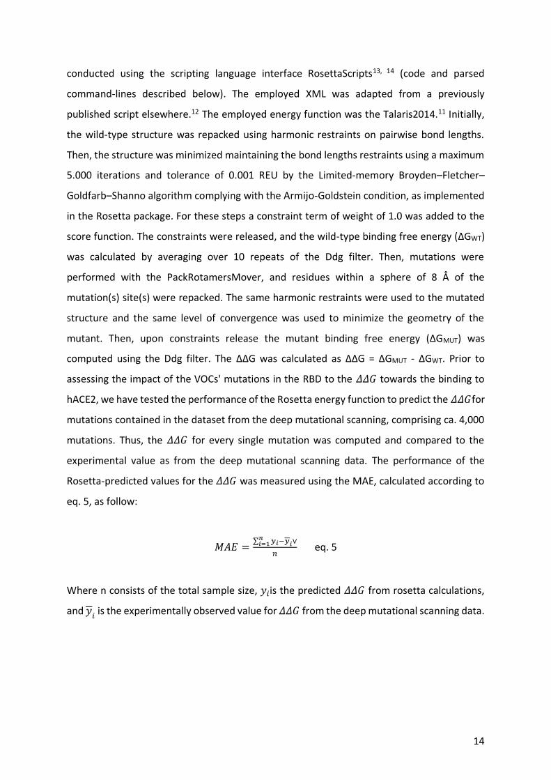
2.2. Binding free-energy calculations with Rosetta
To compute the change in binding free energy (ΔΔG) of hACE2-RBD and nAbs-RBD upon
residues mutation, the Rosetta v.3.125 package was used. The Rosetta package is a prime-tool
in protein modeling and design,8,9 and it has an empirical energy function10, 11 that predicts
mutational 𝛥𝛥𝐺 with a Pearson's correlation coefficient of 0.72 with mean absolute error
(MAE) of 1.7 kcal.mol-1, as tested on a dataset of 1240 mutations.12 The calculations were
14
conducted using the scripting language interface RosettaScripts13, 14 (code and parsed
command-lines described below). The employed XML was adapted from a previously
published script elsewhere.12 The employed energy function was the Talaris2014.11 Initially,
the wild-type structure was repacked using harmonic restraints on pairwise bond lengths.
Then, the structure was minimized maintaining the bond lengths restraints using a maximum
5.000 iterations and tolerance of 0.001 REU by the Limited-memory Broyden–Fletcher–
Goldfarb–Shanno algorithm complying with the Armijo-Goldstein condition, as implemented
in the Rosetta package. For these steps a constraint term of weight of 1.0 was added to the
score function. The constraints were released, and the wild-type binding free energy (ΔGWT)
was calculated by averaging over 10 repeats of the Ddg filter. Then, mutations were
performed with the PackRotamersMover, and residues within a sphere of 8 Å of the
mutation(s) site(s) were repacked. The same harmonic restraints were used to the mutated
structure and the same level of convergence was used to minimize the geometry of the
mutant. Then, upon constraints release the mutant binding free energy (ΔGMUT) was
computed using the Ddg filter. The ΔΔG was calculated as ΔΔG = ΔGMUT - ΔGWT. Prior to
assessing the impact of the VOCs' mutations in the RBD to the 𝛥𝛥𝐺 towards the binding to
hACE2, we have tested the performance of the Rosetta energy function to predict the 𝛥𝛥𝐺for
mutations contained in the dataset from the deep mutational scanning, comprising ca. 4,000
mutations. Thus, the 𝛥𝛥𝐺 for every single mutation was computed and compared to the
experimental value as from the deep mutational scanning data. The performance of the
Rosetta-predicted values for the 𝛥𝛥𝐺 was measured using the MAE, calculated according to
eq. 5, as follow:
𝑀𝐴𝐸 =∑ 𝑦𝑖−𝑦𝑖∨𝑛
𝑖=1
𝑛 eq. 5
Where n consists of the total sample size, 𝑦𝑖is the predicted 𝛥𝛥𝐺 from rosetta calculations,
and 𝑦𝑖 is the experimentally observed value for 𝛥𝛥𝐺 from the deep mutational scanning data.
15
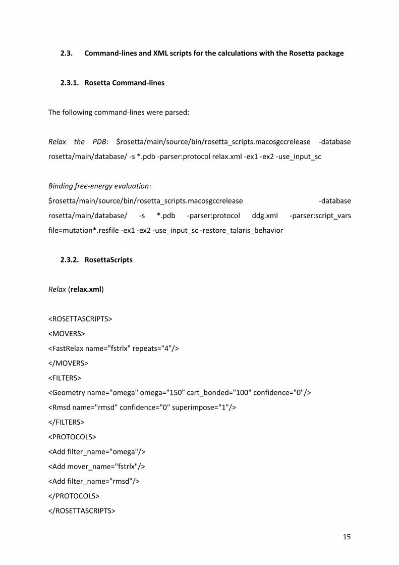
2.3. Command-lines and XML scripts for the calculations with the Rosetta package
2.3.1. Rosetta Command-lines
The following command-lines were parsed:
Relax the PDB: $rosetta/main/source/bin/rosetta_scripts.macosgccrelease -database
rosetta/main/database/ -s *.pdb -parser:protocol relax.xml -ex1 -ex2 -use_input_sc
Binding free-energy evaluation:
$rosetta/main/source/bin/rosetta_scripts.macosgccrelease -database
rosetta/main/database/ -s *.pdb -parser:protocol ddg.xml -parser:script_vars
file=mutation*.resfile -ex1 -ex2 -use_input_sc -restore_talaris_behavior
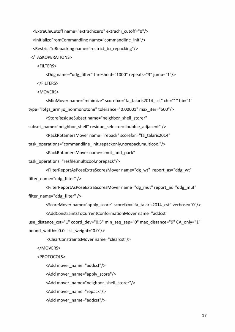
2.3.2. RosettaScripts
Relax (relax.xml)
<ROSETTASCRIPTS>
<MOVERS>
<FastRelax name="fstrlx" repeats="4"/>
</MOVERS>
<FILTERS>
<Geometry name="omega" omega="150" cart_bonded="100" confidence="0"/>
<Rmsd name="rmsd" confidence="0" superimpose="1"/>
</FILTERS>
<PROTOCOLS>
<Add filter_name="omega"/>
<Add mover_name="fstrlx"/>
<Add filter_name="rmsd"/>
</PROTOCOLS>
</ROSETTASCRIPTS>
16
Binding free-energy evaluation (ddg.xml)
<ROSETTASCRIPTS>
<SCOREFXNS>
<ScoreFunction name="fa_talaris2014" weights="talaris2014"/>
<ScoreFunction name="fa_talaris2014_cst" weights="talaris2014">
<Reweight scoretype="atom_pair_constraint" weight="1.0"/>
<Set fa_max_dis="9.0"/>
</ScoreFunction>
</SCOREFXNS>
<TASKOPERATIONS>
<InitializeFromCommandline name="init"/>
<ReadResfile name="resfile" filename="%%file%%" />
</TASKOPERATIONS>
<RESIDUE_SELECTORS>
<Task name="resselector" fixed="0" packable="0" designable="1"
task_operations="resfile"/>
<Neighborhood name="bubble" selector="resselector" distance="8.0"/>
<PrimarySequenceNeighborhood name="bubble_adjacent" selector="bubble" lower="1"
upper="1"/>
<StoredResidueSubset name="restore_neighbor_shell" subset_name="neighbor_shell"/>
<Not name="everythingelse" selector="restore_neighbor_shell"/>
</RESIDUE_SELECTORS>
<TASKOPERATIONS>
<OperateOnResidueSubset name="repackonly" selector="restore_neighbor_shell">
<RestrictToRepackingRLT/>
</OperateOnResidueSubset>
<OperateOnResidueSubset name="norepack" selector="everythingelse">
<PreventRepackingRLT/>
</OperateOnResidueSubset>
<UseMultiCoolAnnealer name="multicool" states="6"/>
17
<ExtraChiCutoff name="extrachizero" extrachi_cutoff="0"/>
<InitializeFromCommandline name="commandline_init"/>
<RestrictToRepacking name="restrict_to_repacking"/>
</TASKOPERATIONS>
<FILTERS>
<Ddg name="ddg_filter" threshold="1000" repeats="3" jump="1"/>
</FILTERS>
<MOVERS>
<MinMover name="minimize" scorefxn="fa_talaris2014_cst" chi="1" bb="1"
type="lbfgs_armijo_nonmonotone" tolerance="0.00001" max_iter="500"/>
<StoreResidueSubset name="neighbor_shell_storer"
subset_name="neighbor_shell" residue_selector="bubble_adjacent" />
<PackRotamersMover name="repack" scorefxn="fa_talaris2014"
task_operations="commandline_init,repackonly,norepack,multicool"/>
<PackRotamersMover name="mut_and_pack"
task_operations="resfile,multicool,norepack"/>
<FilterReportAsPoseExtraScoresMover name="dg_wt" report_as="ddg_wt"
filter_name="ddg_filter" />
<FilterReportAsPoseExtraScoresMover name="dg_mut" report_as="ddg_mut"
filter_name="ddg_filter" />
<ScoreMover name="apply_score" scorefxn="fa_talaris2014_cst" verbose="0"/>
<AddConstraintsToCurrentConformationMover name="addcst"
use_distance_cst="1" coord_dev="0.5" min_seq_sep="0" max_distance="9" CA_only="1"
bound_width="0.0" cst_weight="0.0"/>
<ClearConstraintsMover name="clearcst"/>
</MOVERS>
<PROTOCOLS>
<Add mover_name="addcst"/>
<Add mover_name="apply_score"/>
<Add mover_name="neighbor_shell_storer"/>
<Add mover_name="repack"/>
<Add mover_name="addcst"/>
18
<Add mover_name="minimize"/>
<Add mover_name="clearcst"/>
<Add mover_name="dg_wt"/>
<Add mover_name="mut_and_pack"/>
<Add mover_name="addcst"/>
<Add mover_name="minimize"/>
<Add mover_name="clearcst"/>
<Add mover_name="dg_mut"/>
</PROTOCOLS>
</ROSETTASCRIPTS>
2.4. Binding free-energy calculation with FoldX
For the calculations using FoldX, the relaxed input PDBs were geometry-optimized using the
FoldX force field and the RepairPDB command. The binding energy of the wild-type complex
(ΔGWT) was calculated using the AnalyseComplex command and was assumed as being
corresponding to the interaction energy term. Then, mutations in the RBD for the different
variants were introduced using a single run of the BuildModel. The binding energy of the
mutated complex (ΔGMUT) was calculated with the AnalyseComplex, and the ΔΔGBind was
taken as ΔΔGBind = (ΔGMUT - ΔGWT). For all FoldX calculations, the pH was set as 7.0 and a
solution ionic strength of 0.05M. The full command-lines were as following:
Repair PDB: $foldx --command=RepairPDB --pdb=*.pdb --ionStrength=0.05 --pH=7 --
vdwDesign=2 --pdbHydrogens=false –water=predict
Mutation: $foldx --command=BuildModel --pdb=*Repaired.pdb --mutant-
file=mutations_list.txt --ionStrength=0.05 --pH=7 --water=predict --vdwDesign=2
--pdbHydrogens=false
Binding free-energy evaluation: $foldx --command=AnalyseComplex
--analyseComplexChains=A,E --pdb=*.pdb
19
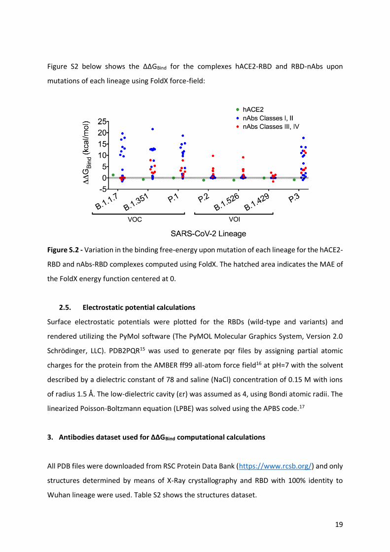
Figure S2 below shows the ΔΔGBind for the complexes hACE2-RBD and RBD-nAbs upon
mutations of each lineage using FoldX force-field:
Figure S.2 - Variation in the binding free-energy upon mutation of each lineage for the hACE2-
RBD and nAbs-RBD complexes computed using FoldX. The hatched area indicates the MAE of
the FoldX energy function centered at 0.
2.5. Electrostatic potential calculations
Surface electrostatic potentials were plotted for the RBDs (wild-type and variants) and
rendered utilizing the PyMol software (The PyMOL Molecular Graphics System, Version 2.0
Schrödinger, LLC). PDB2PQR15 was used to generate pqr files by assigning partial atomic
charges for the protein from the AMBER ff99 all-atom force field16 at pH=7 with the solvent
described by a dielectric constant of 78 and saline (NaCl) concentration of 0.15 M with ions
of radius 1.5 Å. The low-dielectric cavity (εr) was assumed as 4, using Bondi atomic radii. The
linearized Poisson-Boltzmann equation (LPBE) was solved using the APBS code.17
3. Antibodies dataset used for ΔΔGBind computational calculations
All PDB files were downloaded from RSC Protein Data Bank (https://www.rcsb.org/) and only
structures determined by means of X-Ray crystallography and RBD with 100% identity to
Wuhan lineage were used. Table S2 shows the structures dataset.
20
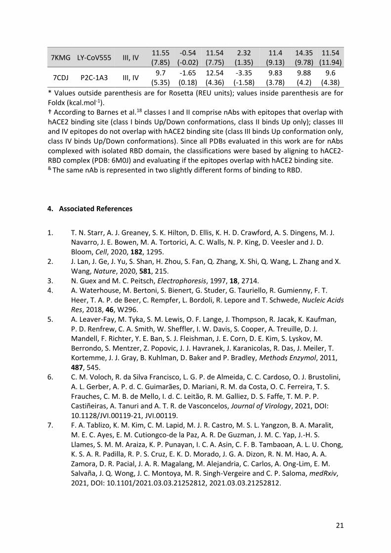
Table S2. Antibodies dataset used for computational calculations
ΔΔGBind*
PDB ID nAb Name nAb
Class† B.1.351 B.1.1.7 P.1 B.1.429 B.1.526 P.2 P.3
6XC4 CC12.3 I, II 0.25
(0.92) -0.59 (0.35)
-0.51 (1.11)
-0.55 (0.01)
-2.9 (0.09)
-1.6 (0.05)
-0.419 (1.12)
6XKQ CV07-250 I, II 0.6
(-0.24) -0.95 (0.99)
0.06 (-0.25)
-0.83 (0.02)
0.39 (0.13)
-0.96 (0.39)
-0.49 (2.1)
7B3O STE90-C11 I, II 16.85
(12.33) 15.8
(16.98) 15.57 (9.68)
-0.45 (0.02)
-0.52 (0.01)
-0.76 (0.03)
15.65 (9.49)
7BEI COVOX-150 I, II 15.99
(12.63) 15.26
(11.76) 16.5
(10.69) -0.23
(-0.02) 0.45
(0.81) 0.46
(0.71) 15.84
(11.98)
7BEJ COVOX-
158& I, II
6.79 (7.64)
6.36 (10.19)
6.71 (11.29)
-0.25 (0.01)
-0.23 (0.15)
-0.12 (-0.03)
6.26 (6.41)
7BEK COVOX-
158& I, II
14.16 (12.77)
14.96 (13.01)
13.65 (14.89)
0.08 (0.01)
-0.01 (0.03)
-0.01 (0.03)
15.17 (13.75)
7BEM COVOX-269 I, II 14.53
(12.69) 12
(13.71) 14.74
(13.42) -1.1
(-0.10) -0.63
(-0.01) -1.26 (0.02)
11.92 (11.03)
7C01 CB6 I, II 1.54
(0.71) -1.8
(-0.89) 1.07
(1.75) -1.02 (0.08)
-0.25 (-0.21)
-0.98 (-0.2)
-1.72 (-0.91)
7CH4 BD-604 I, II 12.16
(12.99) 9.11
(19.69) 10.84
(15.32) -0.35
(-0.02) 0.11
(-0.05) -0.45
(-0.03) 8.93
(13.56)
7CH5 BD-629 I, II 1.41
(0.06) 1.05
(0.97) 1.29
(-0.13) -0.35
(0) -0.94 (0.15)
-0.07 (0.35)
1.08 (1.24)
7CHB BD-236 I, II 21.79
(12.35) 17.39 (9.56)
20.68 (11.77)
-0.96 (-0.09)
-0.56 (-0.03)
-0.47 (-0.01)
17.4 (10.01)
7K8M C102 I, II 6.86
(21.6) 15.25
(17.76) 14.43
(18.71) -0.03 (0.02)
0.67 (-0.16)
-0.66 (-0.06)
15.31 (17.7)
7KLG 15033 I, II 1.03
(0.49) -0.94
(-0.69) 2.97
(0.37) -0.64
(-0.01) 2.742 (0.74)
2.99 (0.72)
3.13 (0.12)
7KLH 15033-7 I, II 4.5
(0.62) 0.3
(-1.51) 3.8
(0.8) -0.4
(-0.03) 2.3
(1.22) 2.6
(0.94) 2.68
(-0.22)
7CDI P2C-1F11 I, II 0.96
(4.91) -0.23 (3.62)
0.83 (4.37)
-0.28 (-0.04)
0.25 (0.3)
-0.21 (0.32)
0.04 (3.93)
7BZ5 B38 I, II 0.79
(-0.39) -0.59 (0.52)
0.85 (0.29)
-1.25 (0)
-0.94 (0.01)
-0.58 (0.02)
-0.88 (0.55)
6XKP CV07-270 III, IV 4.72
(3.84) -1.32
(0) 4.31
(3.26) 4.2
(2.3) 4.51
(3.84) 4.67
(3.18) 4.55
(3.56)
7BEH COVOX-316 III, IV 8.45
(2.38) -0.55 (-0.8)
8.59 (2.78)
-1.49 (0.07)
8.46 (3.08)
8.2 (3.05)
8.34 (2.25)
7BWJ P2B-2F6 III, IV 6.29 (2.1)
-0.9 (0)
0.09 (1.75)
-1.11 (1.04)
0.07 (1.38)
0.17 (1.65)
0.02 (11.1)
7L5B 2-15 III, IV 6.92
(2.66) -2.59 (0.16)
6.37 (0.55)
-1.2 (0.7)
7.77 (0.35)
7.61 (0.37)
6.08 (3.53)
21
7KMG LY-CoV555 III, IV 11.55 (7.85)
-0.54 (-0.02)
11.54 (7.75)
2.32 (1.35)
11.4 (9.13)
14.35 (9.78)
11.54 (11.94)
7CDJ P2C-1A3 III, IV 9.7
(5.35) -1.65 (0.18)
12.54 (4.36)
-3.35 (-1.58)
9.83 (3.78)
9.88 (4.2)
9.6 (4.38)
* Values outside parenthesis are for Rosetta (REU units); values inside parenthesis are for Foldx (kcal.mol-1). † According to Barnes et al.18 classes I and II comprise nAbs with epitopes that overlap with hACE2 binding site (class I binds Up/Down conformations, class II binds Up only); classes III and IV epitopes do not overlap with hACE2 binding site (class III binds Up conformation only, class IV binds Up/Down conformations). Since all PDBs evaluated in this work are for nAbs complexed with isolated RBD domain, the classifications were based by aligning to hACE2-RBD complex (PDB: 6M0J) and evaluating if the epitopes overlap with hACE2 binding site. & The same nAb is represented in two slightly different forms of binding to RBD.
4. Associated References
1. T. N. Starr, A. J. Greaney, S. K. Hilton, D. Ellis, K. H. D. Crawford, A. S. Dingens, M. J.
Navarro, J. E. Bowen, M. A. Tortorici, A. C. Walls, N. P. King, D. Veesler and J. D. Bloom, Cell, 2020, 182, 1295.
2. J. Lan, J. Ge, J. Yu, S. Shan, H. Zhou, S. Fan, Q. Zhang, X. Shi, Q. Wang, L. Zhang and X. Wang, Nature, 2020, 581, 215.
3. N. Guex and M. C. Peitsch, Electrophoresis, 1997, 18, 2714. 4. A. Waterhouse, M. Bertoni, S. Bienert, G. Studer, G. Tauriello, R. Gumienny, F. T.
Heer, T. A. P. de Beer, C. Rempfer, L. Bordoli, R. Lepore and T. Schwede, Nucleic Acids Res, 2018, 46, W296.
5. A. Leaver-Fay, M. Tyka, S. M. Lewis, O. F. Lange, J. Thompson, R. Jacak, K. Kaufman, P. D. Renfrew, C. A. Smith, W. Sheffler, I. W. Davis, S. Cooper, A. Treuille, D. J. Mandell, F. Richter, Y. E. Ban, S. J. Fleishman, J. E. Corn, D. E. Kim, S. Lyskov, M. Berrondo, S. Mentzer, Z. Popovic, J. J. Havranek, J. Karanicolas, R. Das, J. Meiler, T. Kortemme, J. J. Gray, B. Kuhlman, D. Baker and P. Bradley, Methods Enzymol, 2011, 487, 545.
6. C. M. Voloch, R. da Silva Francisco, L. G. P. de Almeida, C. C. Cardoso, O. J. Brustolini, A. L. Gerber, A. P. d. C. Guimarães, D. Mariani, R. M. da Costa, O. C. Ferreira, T. S. Frauches, C. M. B. de Mello, I. d. C. Leitão, R. M. Galliez, D. S. Faffe, T. M. P. P. Castiñeiras, A. Tanuri and A. T. R. de Vasconcelos, Journal of Virology, 2021, DOI: 10.1128/JVI.00119-21, JVI.00119.
7. F. A. Tablizo, K. M. Kim, C. M. Lapid, M. J. R. Castro, M. S. L. Yangzon, B. A. Maralit, M. E. C. Ayes, E. M. Cutiongco-de la Paz, A. R. De Guzman, J. M. C. Yap, J.-H. S. Llames, S. M. M. Araiza, K. P. Punayan, I. C. A. Asin, C. F. B. Tambaoan, A. L. U. Chong, K. S. A. R. Padilla, R. P. S. Cruz, E. K. D. Morado, J. G. A. Dizon, R. N. M. Hao, A. A. Zamora, D. R. Pacial, J. A. R. Magalang, M. Alejandria, C. Carlos, A. Ong-Lim, E. M. Salvaña, J. Q. Wong, J. C. Montoya, M. R. Singh-Vergeire and C. P. Saloma, medRxiv, 2021, DOI: 10.1101/2021.03.03.21252812, 2021.03.03.21252812.
22
8. K. W. Kaufmann, G. H. Lemmon, S. L. Deluca, J. H. Sheehan and J. Meiler, Biochemistry, 2010, 49, 2987.
9. J. K. Leman, B. D. Weitzner, S. M. Lewis, J. Adolf-Bryfogle, N. Alam, R. F. Alford, M. Aprahamian, D. Baker, K. A. Barlow, P. Barth, B. Basanta, B. J. Bender, K. Blacklock, J. Bonet, S. E. Boyken, P. Bradley, C. Bystroff, P. Conway, S. Cooper, B. E. Correia, B. Coventry, R. Das, R. M. De Jong, F. DiMaio, L. Dsilva, R. Dunbrack, A. S. Ford, B. Frenz, D. Y. Fu, C. Geniesse, L. Goldschmidt, R. Gowthaman, J. J. Gray, D. Gront, S. Guffy, S. Horowitz, P. S. Huang, T. Huber, T. M. Jacobs, J. R. Jeliazkov, D. K. Johnson, K. Kappel, J. Karanicolas, H. Khakzad, K. R. Khar, S. D. Khare, F. Khatib, A. Khramushin, I. C. King, R. Kleffner, B. Koepnick, T. Kortemme, G. Kuenze, B. Kuhlman, D. Kuroda, J. W. Labonte, J. K. Lai, G. Lapidoth, A. Leaver-Fay, S. Lindert, T. Linsky, N. London, J. H. Lubin, S. Lyskov, J. Maguire, L. Malmstrom, E. Marcos, O. Marcu, N. A. Marze, J. Meiler, R. Moretti, V. K. Mulligan, S. Nerli, C. Norn, S. O'Conchuir, N. Ollikainen, S. Ovchinnikov, M. S. Pacella, X. Pan, H. Park, R. E. Pavlovicz, M. Pethe, B. G. Pierce, K. B. Pilla, B. Raveh, P. D. Renfrew, S. S. R. Burman, A. Rubenstein, M. F. Sauer, A. Scheck, W. Schief, O. Schueler-Furman, Y. Sedan, A. M. Sevy, N. G. Sgourakis, L. Shi, J. B. Siegel, D. A. Silva, S. Smith, Y. Song, A. Stein, M. Szegedy, F. D. Teets, S. B. Thyme, R. Y. Wang, A. Watkins, L. Zimmerman and R. Bonneau, Nat Methods, 2020, 17, 665.
10. R. F. Alford, A. Leaver-Fay, J. R. Jeliazkov, M. J. O'Meara, F. P. DiMaio, H. Park, M. V. Shapovalov, P. D. Renfrew, V. K. Mulligan, K. Kappel, J. W. Labonte, M. S. Pacella, R. Bonneau, P. Bradley, R. L. Dunbrack, Jr., R. Das, D. Baker, B. Kuhlman, T. Kortemme and J. J. Gray, J Chem Theory Comput, 2017, 13, 3031.
11. A. Bazzoli, S. P. Kelow and J. Karanicolas, PLoS One, 2015, 10, e0140359. 12. K. A. Barlow, O. C. S, S. Thompson, P. Suresh, J. E. Lucas, M. Heinonen and T.
Kortemme, J Phys Chem B, 2018, 122, 5389. 13. S. J. Fleishman, A. Leaver-Fay, J. E. Corn, E. M. Strauch, S. D. Khare, N. Koga, J.
Ashworth, P. Murphy, F. Richter, G. Lemmon, J. Meiler and D. Baker, PLoS One, 2011, 6, e20161.
14. B. J. Bender, A. Cisneros, 3rd, A. M. Duran, J. A. Finn, D. Fu, A. D. Lokits, B. K. Mueller, A. K. Sangha, M. F. Sauer, A. M. Sevy, G. Sliwoski, J. H. Sheehan, F. DiMaio, J. Meiler and R. Moretti, Biochemistry, 2016, 55, 4748.
15. T. J. Dolinsky, J. E. Nielsen, J. A. McCammon and N. A. Baker, Nucleic Acids Res, 2004, 32, W665.
16. A. Spasic, J. Serafini and D. H. Mathews, J Chem Theory Comput, 2012, 8, 2497. 17. N. A. Baker, D. Sept, S. Joseph, M. J. Holst and J. A. McCammon, Proc Natl Acad Sci U
S A, 2001, 98, 10037. 18. C. O. Barnes, C. A. Jette, M. E. Abernathy, K.-M. A. Dam, S. R. Esswein, H. B. Gristick,
A. G. Malyutin, N. G. Sharaf, K. E. Huey-Tubman, Y. E. Lee, D. F. Robbiani, M. C. Nussenzweig, A. P. West and P. J. Bjorkman, Nature, 2020, 588, 682.