s41467-021-25012-9.pdf - nature
TRANSCRIPT
ARTICLE
Genomic characterization of co-existing neoplasiaand carcinoma lesions reveals distinct evolutionarypaths of gallbladder cancerJianzhen Lin1,2,7, Xinxin Peng3,7, Kun Dong4,7, Junyu Long1,7, Xuejiao Guo3, Hongyue Li3, Yi Bai1, Xu Yang1,
Dongxu Wang1, Xin Lu1, Yilei Mao1, Xinting Sang1, Xuwo Ji3, Haitao Zhao1✉ & Han Liang 5,6✉
Gallbladder carcinoma is the most common cancer of the biliary tract with dismal survival
largely due to delayed diagnosis. Biliary tract intraepithelial neoplasia (BilIN) is the common
benign tumor that is suspected to be precancerous lesions. However, the genetic and evo-
lutionary relationships between BilIN and carcinoma remain unclear. Here we perform whole-
exome sequencing of coexisting low-grade BilIN (adenoma), high-grade BilIN, and carcinoma
lesions, and normal tissues from the same patients. We identify aging as a major factor
contributing to accumulated mutations and a critical role of CTNNB1 mutations in these
tumors. We reveal two distinct carcinoma evolutionary paths: carcinoma can either diverge
earlier and evolve more independently or form through the classic adenoma/dysplasia-car-
cinoma sequence model. Our analysis suggests that extensive loss-of-heterozygosity and
mutation events in the initial stage tend to result in a cancerous niche, leading to the
subsequent BilIN-independent path. These results reframes our understanding of tumor
transformation and the evolutionary trajectory of carcinogenesis in the gallbladder, laying a
foundation for the early diagnosis and effective treatment of gallbladder cancer.
https://doi.org/10.1038/s41467-021-25012-9 OPEN
1 Department of Liver Surgery, State Key Laboratory of Complex Severe and Rare Diseases, Peking Union Medical College Hospital, Chinese Academy ofMedical Sciences & Peking Union Medical College (CAMS & PUMC), Beijing, China. 2 Pancreas Center, The First Affiliated Hospital of Nanjing MedicalUniversity, Pancreas Institute, Nanjing Medical University, Nanjing, China. 3 Precision Scientific (Beijing) Co. Ltd., Beijing, China. 4 Key Laboratory ofCarcinogenesis and Translational Research (Ministry of Education), Department of Pathology, Peking University Cancer Hospital & Institute, Beijing, China.5 Department of Bioinformatics and Computational Biology, The University of Texas MD Anderson Cancer Center, Houston, TX, USA. 6 Department ofSystems Biology, The University of Texas MD Anderson Cancer Center, Houston, TX, USA. 7These authors contributed equally: Jianzhen Lin, Xinxin Peng,Kun Dong, Junyu Long. ✉email: [email protected]; [email protected]
NATURE COMMUNICATIONS | (2021) 12:4753 | https://doi.org/10.1038/s41467-021-25012-9 |www.nature.com/naturecommunications 1
1234
5678
90():,;
Gallbladder carcinoma (GBC) is the most common cancerof the biliary tract1,2. Its incidence rate shows a widegeographic variance, and in some countries such as Chile
and India, the incidence rate is nearly 30/100000 in women3.Unlike other cancer types, only 30% of GBC patients are diag-nosed or suspected preoperatively, whereas other cases are diag-nosed based on postoperative incidental findings1. In mostpatients, the tumor is recognized either at the time of surgery orby the pathologist examining the surgical specimen. Mostsymptomatic GBC patients have an incurable tumor, and the riskof GBC recurrence is 64% at 5 years, even after R0 resection. Theclinical outcome of GBC is very poor: although a 5-year survivalrate of 75% can be achieved in early-stage (T1) disease, the overall5-year survival rate is <5%4. Therefore, there is an urgent clinicalneed to identify high-risk patients and have their gallbladdersremoved before the development of GBC.
When considering the potential risk for gallbladder cancer, it isimportant to understand the relationship between gallbladderpolypoid lesions and GBC, thereby managing them wisely andtimely. The population prevalence of gallbladder polyps is ~5%,accounting for 2–12% of the cholecystectomy specimens5,6. Low-grade biliary tract intraepithelial neoplasia (LG-BilIN) and high-grade biliary tract intraepithelial neoplasia (HG-BilIN), twocommon benign neoplastic polyps, have attracted much attentiondue to their proposed premalignant behaviors. Specifically, mor-phological studies suggest the histologic transition of LG-BilIN(adenoma) into GBC, with or without neighboring HG-BilIN7.Molecular analyses show that p53 and KRAS are commonlymutated in LG-BilIN and GBC8, and the dysregulation of p16/cyclin-D1/CDK4 cell cycle pathway is associated with both gall-bladder dysplasia and cancer cells9. Furthermore, there is a pro-gressive increase in the average age of patients with LG-BilIN, LG-BilIN with malignant changes, and invasive GBC10,11. Theseobservations suggest that LG-BilIN is a precancerous lesion thatgradually progresses to GBC through a step-wise evolution, whichis roughly analogous to colorectal carcinogenesis from adenoma12.However, some histological and genomic observations challengethe LG-BilIN-GBC sequential model. The most direct evidence isthe relative rarity of LG-BilIN as compared to the frequency ofGBC in patients undergoing cholecystectomy13. Also, LG-BilINgenerally accompanies HG-BilIN rather than GBC14. Morpholo-gically, it is uncommon for advanced GBC foci to co-exist in thevicinity of LG-BilIN, even for early or well-differentiated cancercells10. HG-BilIN, rather than LG-BilIN, exhibits morphologicalfeatures that are thought to facilitate progression to an infiltratingGBC15. The altered signaling pathways in LG-BilIN are distinctfrom those altered in GBC16. Additionally, the KRAS codon-12mutation is detected more frequently in the de novo gallbladdercarcinomas compared to carcinoma-in-pyloric-gland-type LG-BilIN17. Thus, the genetic and evolutionary relationships betweenLG-BilIN, HG-BilIN, and GBC, and the molecular events drivinggallbladder carcinogenesis, remain unclear18. A comprehensivecharacterization of neighboring LG-BilIN, HG-BilIN, and GBCcoexisting in the same patients will provide a unique opportunityto elucidate their evolutionary relationships by removing inter-tumor heterogeneity, a major confounding factor in previousstudies7,19,20.
In this work, we perform laser microdissections on tissuesections to isolate tissues of the normal gallbladder, LG-BilIN,HG-BilIN, and GBC from the same individuals. Using whole-exome sequencing, we systematically investigated the GBCgenomic landscape and the evolution of the carcinoma from aprecancerous stage to GBC. Our study provides insights intothe biology of gallbladder tumors and their evolutionarytrajectories.
ResultsGenomic characteristics of BilIN-related gallbladder carci-noma. After a rigorous pathological review of a large number ofgallbladder tumor samples, we collected tissue samples, includingnormal ones, from 11 patients diagnosed with T1 tumors, inwhom GBC, LG-BilIN (gallbladder adenoma with low-gradedysplasia), and HG-BilIN (high-grade dysplasia or carcinomain situ) lesions geographically coexisted (average age, 62). Despitethe limited sample size, we noted more female patients in ourcohort (female: 9; male: 2), which echoes the gender biasuncovered in epidemiological studies21. Age at diagnosis for nineof the 11 enrolled patients was above 60 years, except for patientsP03 and P11, both diagnosed at 38. Through laser microdissec-tion, we separated GBC, LG-BilIN, HG-BilIN, and normal gall-bladder tissues and performed whole-exome sequencing on44 samples in total (mean depth: normal 107×; GBC 171×; LG-BilIN 188×; HG-BilIN 206×) (Fig. 1a and Supplementary Data 1).We employed a well-established, multi-caller-based MC3approach to call single nucleotide variant (SNV) mutations andsmall indels, and this approach has been used to generate thehigh-quality mutation data of The Cancer Genome Atlas(TCGA)22. Importantly, we performed a rescue step to callmutations across different samples from the same patient toreduce false negatives. Across the 33 tumor samples from the 11patients, we identified an average of 214 SNVs (range: 69-725)and 9 indels (range: 3-48) (Supplementary Fig. 1a and Supple-mentary Data 2). To assess the accuracy of our mutation calls, weperformed independent Sanger sequencing and reached a vali-dation rate of 95% (42 out of 44 mutations) (Supplementary Fig. 2and Supplementary Data 3). To assess the relative mutationburden of GBC, we compared our GBC samples with stage-Isamples of 16 other cancer types from TCGA and found thatGBC exhibited a moderate level of tumor mutation burden(TMB), similar to that of liver hepatocellular carcinoma (LIHC)(t-test, p= 0.79) but significantly higher than that of cholangio-carcinoma (CHOL) (t-test, p= 7.3 × 10⁻⁴, Fig. 1b).
In order to identify the mutagenesis mechanisms underlyingthe mutations and evolution of these GBC samples and elucidatethe common/specific contributing factors, we decomposed themutational spectrum for the 16 TCGA cancer types (stage-I only)as well as our GBC samples, based on 30 established mutationalsignatures (COSMIC mutational signature v2)23. This analysisconfirmed some known signature patterns such as smoking(Signature 4) for lung cancers (Fig. 1c). We found that in mostcancer types, the mutations in stage-I tumors were mainly causedby age, especially for colorectal cancer (COAD/READ), pancrea-tic adenocarcinoma (PAAD), and stomach cancer (STAD). In theGBC samples, age-related Signature 1 contributed to >75% of themutations and APOBEC-related Signature 2 contributed ~10%(Fig. 1c). These results indicate the importance of mutationsaccumulated by the aging process for GBC development andprovide the first view of mutational characteristics of GBCcoexisting with LG/HG-BilIN.
Somatic alteration feature comparison of gallbladder tumors.In addition to the above-mentioned comparison among malignanttumor types, we compared the mutational features among thethree gallbladder tumor types. We found that GBC, LG-BilIN, andHG-BilIN showed similar mutation rates (paired t-test, p > 0.05,Fig. 2a), but the tumor samples from the two young patients (P03and P11) showed much fewer mutations than those from olderpatients, regardless of tumor types (mean number, 101 vs. 273, t-test, p < 7 × 10⁻⁵, Fig. 2b). We further quantified the microsatelliteinstability (MSI) status of these tumor samples based on a large set
ARTICLE NATURE COMMUNICATIONS | https://doi.org/10.1038/s41467-021-25012-9
2 NATURE COMMUNICATIONS | (2021) 12:4753 | https://doi.org/10.1038/s41467-021-25012-9 | www.nature.com/naturecommunications
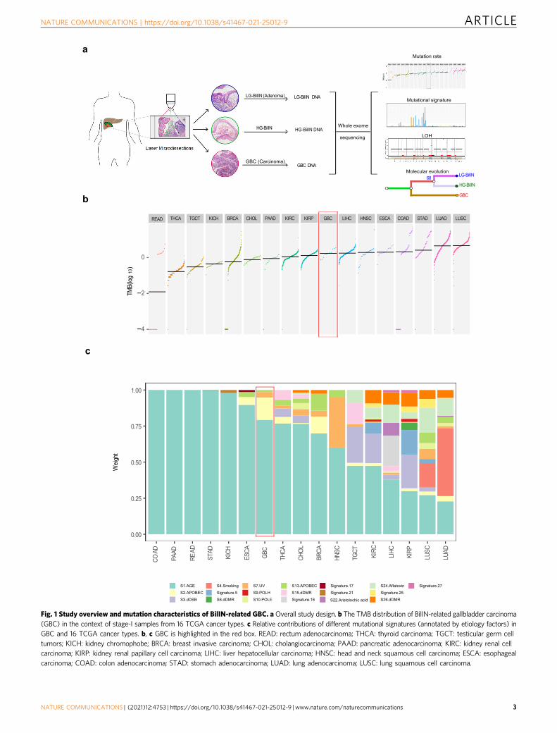
Fig. 1 Study overview and mutation characteristics of BilIN-related GBC. aOverall study design. b The TMB distribution of BilIN-related gallbladder carcinoma(GBC) in the context of stage-I samples from 16 TCGA cancer types. c Relative contributions of different mutational signatures (annotated by etiology factors) inGBC and 16 TCGA cancer types. b, c GBC is highlighted in the red box. READ: rectum adenocarcinoma; THCA: thyroid carcinoma; TGCT: testicular germ celltumors; KICH: kidney chromophobe; BRCA: breast invasive carcinoma; CHOL: cholangiocarcinoma; PAAD: pancreatic adenocarcinoma; KIRC: kidney renal cellcarcinoma; KIRP: kidney renal papillary cell carcinoma; LIHC: liver hepatocellular carcinoma; HNSC: head and neck squamous cell carcinoma; ESCA: esophagealcarcinoma; COAD: colon adenocarcinoma; STAD: stomach adenocarcinoma; LUAD: lung adenocarcinoma; LUSC: lung squamous cell carcinoma.
NATURE COMMUNICATIONS | https://doi.org/10.1038/s41467-021-25012-9 ARTICLE
NATURE COMMUNICATIONS | (2021) 12:4753 | https://doi.org/10.1038/s41467-021-25012-9 |www.nature.com/naturecommunications 3
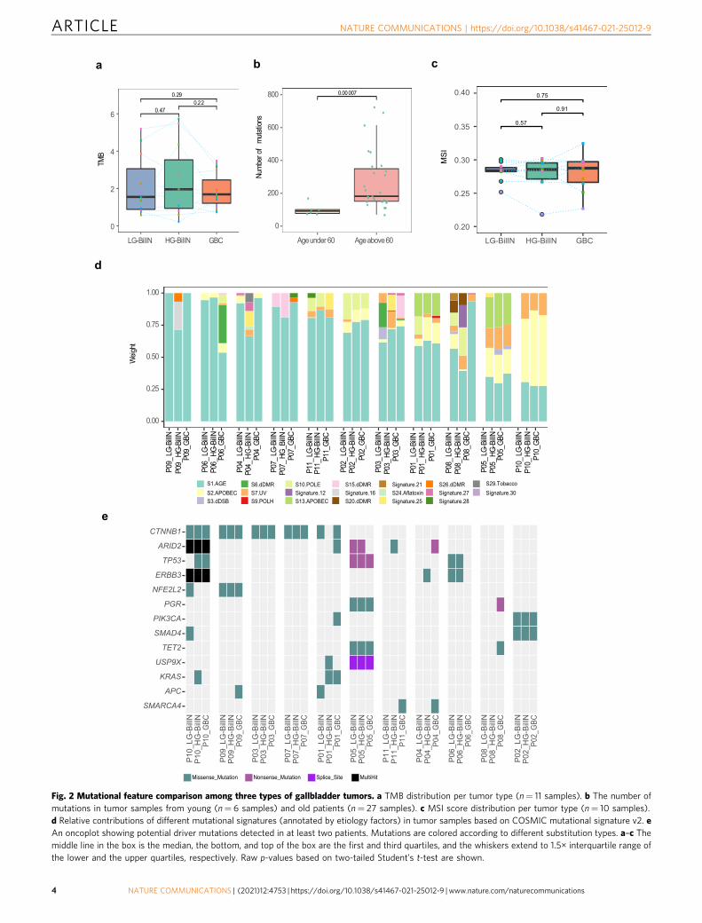
Fig. 2 Mutational feature comparison among three types of gallbladder tumors. a TMB distribution per tumor type (n= 11 samples). b The number ofmutations in tumor samples from young (n= 6 samples) and old patients (n= 27 samples). c MSI score distribution per tumor type (n= 10 samples).d Relative contributions of different mutational signatures (annotated by etiology factors) in tumor samples based on COSMIC mutational signature v2. eAn oncoplot showing potential driver mutations detected in at least two patients. Mutations are colored according to different substitution types. a–c Themiddle line in the box is the median, the bottom, and top of the box are the first and third quartiles, and the whiskers extend to 1.5× interquartile range ofthe lower and the upper quartiles, respectively. Raw p-values based on two-tailed Student’s t-test are shown.
ARTICLE NATURE COMMUNICATIONS | https://doi.org/10.1038/s41467-021-25012-9
4 NATURE COMMUNICATIONS | (2021) 12:4753 | https://doi.org/10.1038/s41467-021-25012-9 | www.nature.com/naturecommunications
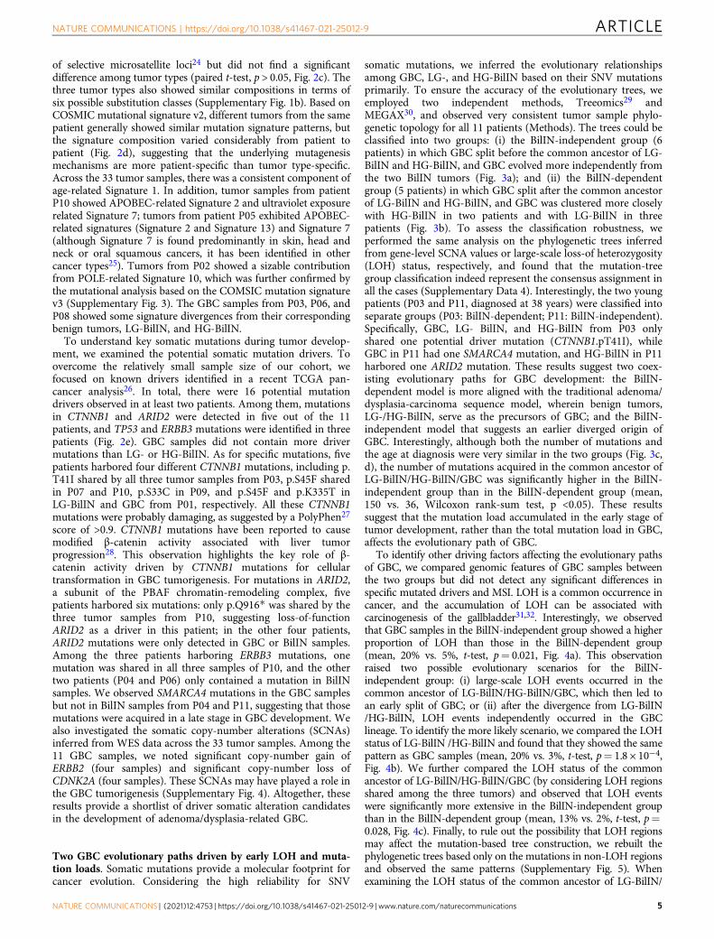
of selective microsatellite loci24 but did not find a significantdifference among tumor types (paired t-test, p > 0.05, Fig. 2c). Thethree tumor types also showed similar compositions in terms ofsix possible substitution classes (Supplementary Fig. 1b). Based onCOSMIC mutational signature v2, different tumors from the samepatient generally showed similar mutation signature patterns, butthe signature composition varied considerably from patient topatient (Fig. 2d), suggesting that the underlying mutagenesismechanisms are more patient-specific than tumor type-specific.Across the 33 tumor samples, there was a consistent component ofage-related Signature 1. In addition, tumor samples from patientP10 showed APOBEC-related Signature 2 and ultraviolet exposurerelated Signature 7; tumors from patient P05 exhibited APOBEC-related signatures (Signature 2 and Signature 13) and Signature 7(although Signature 7 is found predominantly in skin, head andneck or oral squamous cancers, it has been identified in othercancer types25). Tumors from P02 showed a sizable contributionfrom POLE-related Signature 10, which was further confirmed bythe mutational analysis based on the COMSIC mutation signaturev3 (Supplementary Fig. 3). The GBC samples from P03, P06, andP08 showed some signature divergences from their correspondingbenign tumors, LG-BilIN, and HG-BilIN.
To understand key somatic mutations during tumor develop-ment, we examined the potential somatic mutation drivers. Toovercome the relatively small sample size of our cohort, wefocused on known drivers identified in a recent TCGA pan-cancer analysis26. In total, there were 16 potential mutationdrivers observed in at least two patients. Among them, mutationsin CTNNB1 and ARID2 were detected in five out of the 11patients, and TP53 and ERBB3 mutations were identified in threepatients (Fig. 2e). GBC samples did not contain more drivermutations than LG- or HG-BilIN. As for specific mutations, fivepatients harbored four different CTNNB1 mutations, including p.T41I shared by all three tumor samples from P03, p.S45F sharedin P07 and P10, p.S33C in P09, and p.S45F and p.K335T inLG-BilIN and GBC from P01, respectively. All these CTNNB1mutations were probably damaging, as suggested by a PolyPhen27
score of >0.9. CTNNB1 mutations have been reported to causemodified β-catenin activity associated with liver tumorprogression28. This observation highlights the key role of β-catenin activity driven by CTNNB1 mutations for cellulartransformation in GBC tumorigenesis. For mutations in ARID2,a subunit of the PBAF chromatin-remodeling complex, fivepatients harbored six mutations: only p.Q916* was shared by thethree tumor samples from P10, suggesting loss-of-functionARID2 as a driver in this patient; in the other four patients,ARID2 mutations were only detected in GBC or BilIN samples.Among the three patients harboring ERBB3 mutations, onemutation was shared in all three samples of P10, and the othertwo patients (P04 and P06) only contained a mutation in BilINsamples. We observed SMARCA4 mutations in the GBC samplesbut not in BilIN samples from P04 and P11, suggesting that thosemutations were acquired in a late stage in GBC development. Wealso investigated the somatic copy-number alterations (SCNAs)inferred from WES data across the 33 tumor samples. Among the11 GBC samples, we noted significant copy-number gain ofERBB2 (four samples) and significant copy-number loss ofCDNK2A (four samples). These SCNAs may have played a role inthe GBC tumorigenesis (Supplementary Fig. 4). Altogether, theseresults provide a shortlist of driver somatic alteration candidatesin the development of adenoma/dysplasia-related GBC.
Two GBC evolutionary paths driven by early LOH and muta-tion loads. Somatic mutations provide a molecular footprint forcancer evolution. Considering the high reliability for SNV
somatic mutations, we inferred the evolutionary relationshipsamong GBC, LG-, and HG-BilIN based on their SNV mutationsprimarily. To ensure the accuracy of the evolutionary trees, weemployed two independent methods, Treeomics29 andMEGAX30, and observed very consistent tumor sample phylo-genetic topology for all 11 patients (Methods). The trees could beclassified into two groups: (i) the BilIN-independent group (6patients) in which GBC split before the common ancestor of LG-BilIN and HG-BilIN, and GBC evolved more independently fromthe two BilIN tumors (Fig. 3a); and (ii) the BilIN-dependentgroup (5 patients) in which GBC split after the common ancestorof LG-BilIN and HG-BilIN, and GBC was clustered more closelywith HG-BilIN in two patients and with LG-BilIN in threepatients (Fig. 3b). To assess the classification robustness, weperformed the same analysis on the phylogenetic trees inferredfrom gene-level SCNA values or large-scale loss-of heterozygosity(LOH) status, respectively, and found that the mutation-treegroup classification indeed represent the consensus assignment inall the cases (Supplementary Data 4). Interestingly, the two youngpatients (P03 and P11, diagnosed at 38 years) were classified intoseparate groups (P03: BilIN-dependent; P11: BilIN-independent).Specifically, GBC, LG- BilIN, and HG-BilIN from P03 onlyshared one potential driver mutation (CTNNB1.pT41I), whileGBC in P11 had one SMARCA4 mutation, and HG-BilIN in P11harbored one ARID2 mutation. These results suggest two coex-isting evolutionary paths for GBC development: the BilIN-dependent model is more aligned with the traditional adenoma/dysplasia-carcinoma sequence model, wherein benign tumors,LG-/HG-BilIN, serve as the precursors of GBC; and the BilIN-independent model that suggests an earlier diverged origin ofGBC. Interestingly, although both the number of mutations andthe age at diagnosis were very similar in the two groups (Fig. 3c,d), the number of mutations acquired in the common ancestor ofLG-BilIN/HG-BilIN/GBC was significantly higher in the BilIN-independent group than in the BilIN-dependent group (mean,150 vs. 36, Wilcoxon rank-sum test, p <0.05). These resultssuggest that the mutation load accumulated in the early stage oftumor development, rather than the total mutation load in GBC,affects the evolutionary path of GBC.
To identify other driving factors affecting the evolutionary pathsof GBC, we compared genomic features of GBC samples betweenthe two groups but did not detect any significant differences inspecific mutated drivers and MSI. LOH is a common occurrence incancer, and the accumulation of LOH can be associated withcarcinogenesis of the gallbladder31,32. Interestingly, we observedthat GBC samples in the BilIN-independent group showed a higherproportion of LOH than those in the BilIN-dependent group(mean, 20% vs. 5%, t-test, p= 0.021, Fig. 4a). This observationraised two possible evolutionary scenarios for the BilIN-independent group: (i) large-scale LOH events occurred in thecommon ancestor of LG-BilIN/HG-BilIN/GBC, which then led toan early split of GBC; or (ii) after the divergence from LG-BilIN/HG-BilIN, LOH events independently occurred in the GBClineage. To identify the more likely scenario, we compared the LOHstatus of LG-BilIN /HG-BilIN and found that they showed the samepattern as GBC samples (mean, 20% vs. 3%, t-test, p= 1.8 × 10⁻⁴,Fig. 4b). We further compared the LOH status of the commonancestor of LG-BilIN/HG-BilIN/GBC (by considering LOH regionsshared among the three tumors) and observed that LOH eventswere significantly more extensive in the BilIN-independent groupthan in the BilIN-dependent group (mean, 13% vs. 2%, t-test, p=0.028, Fig. 4c). Finally, to rule out the possibility that LOH regionsmay affect the mutation-based tree construction, we rebuilt thephylogenetic trees based only on the mutations in non-LOH regionsand observed the same patterns (Supplementary Fig. 5). Whenexamining the LOH status of the common ancestor of LG-BilIN/
NATURE COMMUNICATIONS | https://doi.org/10.1038/s41467-021-25012-9 ARTICLE
NATURE COMMUNICATIONS | (2021) 12:4753 | https://doi.org/10.1038/s41467-021-25012-9 |www.nature.com/naturecommunications 5
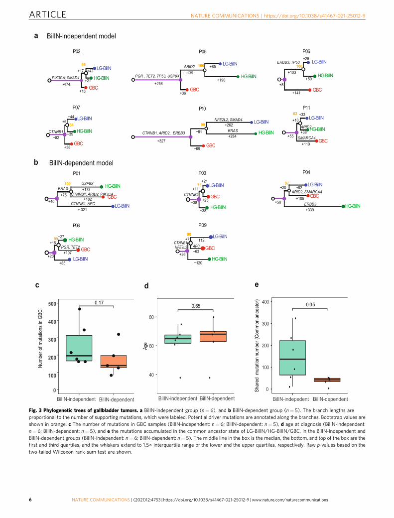
Fig. 3 Phylogenetic trees of gallbladder tumors. a BilIN-independent group (n= 6), and b BilIN-dependent group (n= 5). The branch lengths areproportional to the number of supporting mutations, which were labeled. Potential driver mutations are annotated along the branches. Bootstrap values areshown in orange. c The number of mutations in GBC samples (BilIN-independent: n= 6; BilIN-dependent: n= 5), d age at diagnosis (BilIN-independent:n= 6; BilIN-dependent: n= 5), and e the mutations accumulated in the common ancestor state of LG-BilIN/HG-BilIN/GBC, in the BilIN-independent andBilIN-dependent groups (BilIN-independent: n= 6; BilIN-dependent: n= 5). The middle line in the box is the median, the bottom, and top of the box are thefirst and third quartiles, and the whiskers extend to 1.5× interquartile range of the lower and the upper quartiles, respectively. Raw p-values based on thetwo-tailed Wilcoxon rank-sum test are shown.
ARTICLE NATURE COMMUNICATIONS | https://doi.org/10.1038/s41467-021-25012-9
6 NATURE COMMUNICATIONS | (2021) 12:4753 | https://doi.org/10.1038/s41467-021-25012-9 | www.nature.com/naturecommunications
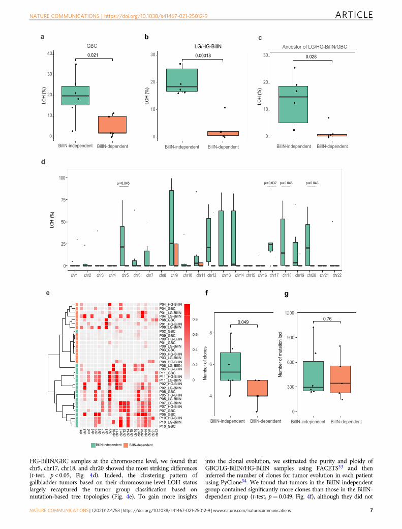
HG-BilIN/GBC samples at the chromosome level, we found thatchr5, chr17, chr18, and chr20 showed the most striking differences(t-test, p < 0.05, Fig. 4d). Indeed, the clustering pattern ofgallbladder tumors based on their chromosome-level LOH statuslargely recaptured the tumor group classification based onmutation-based tree topologies (Fig. 4e). To gain more insights
into the clonal evolution, we estimated the purity and ploidy ofGBC/LG-BilIN/HG-BilIN samples using FACETS33 and theninferred the number of clones for tumor evolution in each patientusing PyClone34. We found that tumors in the BilIN-independentgroup contained significantly more clones than those in the BilIN-dependent group (t-test, p= 0.049, Fig. 4f), although they did not
NATURE COMMUNICATIONS | https://doi.org/10.1038/s41467-021-25012-9 ARTICLE
NATURE COMMUNICATIONS | (2021) 12:4753 | https://doi.org/10.1038/s41467-021-25012-9 |www.nature.com/naturecommunications 7
contain more somatic mutations (t-test, p= 0.76, Fig. 4g). We alsoinvestigated the possible seeding patterns for GBC, based oninferred cancer cell fractions (CCF) of mutations35 and found thatall 11 GBC cases were polyclonal seeding (Supplementary Data 5).
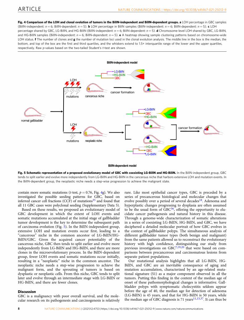
Based on these results, we proposed an evolutionary model ofGBC development in which the extent of LOH events andsomatic mutations accumulated at the initial stage of gallbladdertumor development is the key to determine the subsequent pathof carcinoma evolution (Fig. 5). In the BilIN-independent group,extensive LOH and mutation events occur first, leading to a“cancerous” niche in the common ancestor of LG-BilIN/HG-BilIN/GBC. Given the acquired cancer potentiality of thecancerous niche, GBC then tends to split earlier and evolve moreindependently from LG-BilIN and HG-BilIN, and there are moreclones in the microevolutionary process. In the BilIN-dependentgroup, fewer LOH events and somatic mutations occur initially,resulting in a “neoplastic” niche in the common ancestor. Theneoplastic niche needs a step-wise progression to achieve themalignant form, and the sprouting of tumors is based ondysplastic or neoplastic cells. From this niche, GBC tends to splitlater and evolve through an intermediate stage with LG-BilIN orHG-BilIN, and there are fewer clones.
DiscussionGBC is a malignancy with poor overall survival, and the mole-cular research on its pathogenesis and carcinogenesis is relatively
rare. Like most epithelial cancer types, GBC is preceded by aseries of precancerous histological and molecular changes thatevolve possibly over a period of several decades18. Adenoma andhyperplastic changes progressing to dysplasia are often assumedto be the usual form of GBC36, offering the opportunity to elu-cidate cancer pathogenesis and natural history in this disease.Through a genome-wide characterization of somatic alterationsin a series of coexisting LG-BilIN, HG-BilIN, and GBC, we havedeciphered a detailed molecular portrait of how GBC evolves inthe context of gallbladder polyps. The simultaneous analysis ofdifferent gallbladder tumor types (both benign and malignant)from the same patients allowed us to reconstruct the evolutionaryhistory with high confidence, distinguishing our study fromprevious investigations on GBC7,19,20 that were based on com-parisons between precancerous and carcinomatous lesions fromseparate patient populations.
Our mutational analysis highlights that all LG-BilIN, HG-BilIN, and GBC are an inevitable consequence of aging andmutation accumulation, characterized by an age-related muta-tional signature (S1) as a major component observed in all thetumors. Putting this finding in the context of the median age ofonset of these pathomorphological changes is informative. Gall-bladder polyps with symptomatic cholecystitis seldom appearbefore the age of 40, the median age for detection of adenoma(LG-BilIN) is 45 years, and that for HG-BilIN is 50 years, whilethe median age of GBC diagnosis is 71 years11,13,37. It can thus be
Fig. 4 Comparison of the LOH and clonal evolution of tumors in the BilIN-independent and BilIN-dependent groups. a LOH percentage in GBC samples(BilIN-independent: n= 6; BilIN-dependent: n= 5). b LOH percentage in BilIN samples (BilIN-independent: n= 6; BilIN-dependent: n= 5). c LOHpercentage shared by GBC, LG-BilIN, and HG-BilIN (BilIN-independent: n= 6; BilIN-dependent: n= 5). d Chromosome-level LOH shared by GBC, LG-BilIN,and HG-BilIN samples (BilIN-independent: n= 6; BilIN-dependent: n= 5). e A heatmap showing sample clustering patterns based on chromosome-wideLOH status. f The number of clones and g the number of mutation loci used in the clonal evolution analysis. The middle line in the box is the median, thebottom, and top of the box are the first and third quartiles, and the whiskers extend to 1.5× interquartile range of the lower and the upper quartiles,respectively. Raw p-values based on the two-tailed Student’s t-test are shown.
GBC
LG-BilIN
HG-BilINGBC
normal gallbladder
neoplastic niche
cancerous nichecancer formation
cancer formation
LOH
mutations
LOH
mutations
LG-BilIN
HG-BilINGBC
BilIN-independent model
BilIN-dependent model
HG-BilINLG-BilIN
Fig. 5 Schematic representation of a proposed evolutionary model of GBC with coexisting LG-BilIN and HG-BilIN. In the BilIN-independent group, GBCtends to split earlier and evolve more independently from LG-BilIN and HG-BilIN in the cancerous niche that harbors extensive LOH and mutation events. Inthe BilIN-dependent group, the neoplastic niche needs a step-wise progression to achieve the malignant state.
ARTICLE NATURE COMMUNICATIONS | https://doi.org/10.1038/s41467-021-25012-9
8 NATURE COMMUNICATIONS | (2021) 12:4753 | https://doi.org/10.1038/s41467-021-25012-9 | www.nature.com/naturecommunications
speculated that the cancerous lesions start as small clusters ofneoplastic epithelial cells at an early stage that then increases insize and number over the years. At the time of cancerous tran-sition, mutation-driven uncontrollable proliferation usuallyselects the clones with oncogenic potential. We observed severalrecurrent driver mutations in GBC, including TP53, KRAS,ARID2, SMAD4, PI3KCA, and ERBB3, similar to a previousstudy38. Notably, our results suggest a critical role of CTNNB1mutations in the cellular transformation from normal cell growthinto a state of uncontrollable proliferation. Previous studiesreported that adenoma or LG-BilIN expresses significantly higherβ-catenin than GBC, and 62.5% of LG-BilIN samples harboredmutations in CTNNB1 exon 3, but this rate was only 4.8% in theGBC39,40. In our cohort, 5 of the 11 patients harbored CTNNB1mutations; in 4 of these 5 cases, GBC and its adjacent LG-BilINshared the same mutations in CTNNB1, suggesting that CTNNB1mutations play a more active role in the formation of carcinomain the context of adenoma/dysplasia than GBC alone.
Two major developmental paths have been suggested for GBC:(i) de novo development when only GBC is present; and (ii) theadenoma/dysplasia-carcinoma sequence model when GBC coex-ists with LG-BilIN or HG-BilIN2. However, it remains unclearhow the GBC developmental paths are determined and whetherLG-BilIN or HG-BilIN represents a major transition state in thelatter model. Through a comprehensive, multilayer analysis of thecancer genome, our study suggests that the relationship betweenGBC and adjacent LG-BilIN/HG-BilIN can be divided into twoevolutionary paths, the BilIN-independent path, and the BilIN-dependent path. The BilIN-independent model is more alignedwith the de novo development path wherein the carcinomainitiates before the divergence of LG-BilIN and HG-BilIN, andevolves more independently. In contrast, the BilIN-dependentdivergence model is similar to the adenoma/dysplasia-carcinomasequence where carcinoma evolves from LG-BilIN or HG-BilINthrough a step-wise progressive manner. The key determinant isthe accumulation of LOH and somatic mutation events in theinitial stage that is sufficient to form a cancerous niche for sub-sequent GBC development. Interestingly, besides somatic muta-tions, we uncovered contrasting patterns of LOH events betweenthe two evolutionary paths, which highlights the importance oflosing functional tumor suppressor genes in the niche-definingprocess. Among individual chromosomes enriched for LOHevents that presumably promote the cancerous niche, LOH onchr.5q and chr.7p have been associated with early carcinogenicchanges of the gallbladder31, and importantly, LOH events arerare in the normal gallbladder32. Clinically, our results suggestthat proactive intervention needs to focus on identifying patientswith LOH-rich gallbladder tissue through noninvasive detectionmethods, so that earlier or more aggressive cholecystectomy maybe adopted as a primary prevention approach for GBC. Fur-thermore, our results confirm that both dysplasia-carcinomasequence (HG-BilIN-GBC) and adenoma-carcinoma sequence(LG-BilIN-GBC) exist in the GBC development. We did notobserve significant bias towards LG-BilIN or HG-BilIN, despitethe small sample size of our BilIN-dependent group (HG-BilIN-GBC, n= 2; LG-BilIN-GBC, n= 3). It remains unclear whetherresidual dysplasia at the cystic duct margin after cholecystectomyis associated with an increased incidence of recurrentcarcinoma41–43. Thus, timely detection of GBC with neoplasticniches is critical in deciding the need for a BilIN-free surgicalmargin.
One limitation of this study is the representativeness of ourcases. Since the cases (carcinomas coexisting with both adenomaand BilIN) surveyed are rare among gallbladder cancer with aprevalence from 1.8% to 6.4%36,44,45, further efforts are requiredto examine the evolutionary paths of more common gallbladder
cancer. Other limitations may be the relatively small cohort sizeand the selection of manual sampling locations. To mitigate these,we have made extensive efforts in searching for such cases from alarge tumor tissue bank, and have performed a careful patholo-gical examination of all the sections of the resected gallbladder.We also ensured that every normal control sample was tumor-,adenoma-, and dysplasia-free mucosa, thereby removing thepotential bias in phylogenetic tree inference. The mutations incancer driver genes identified in our cohort are similar to pre-vious findings, supporting their representativeness38. Furtherefforts are needed to confirm our evolutionary model usingindependent cohorts, especially prospective studies with sequen-tial samplings from the normal gallbladder, LG-BilIN, emergingHG-BilIN, and GBC formation, over the disease course. Themechanisms through which different LOH events and mutationloads (high vs. low) could drive normal gallbladder mucosa intodistinct states of gallbladder cancerous/neoplastic “niche” remainunclear. Further multi-omics characterization of epigeneticalterations, aneuploidy, or transcriptomes will provide moreinsights into these oncogenic transformations.
MethodsPatient recruitment and sample cohort. All samples were collected with theapproval of the Institutional Review Board (IRB) from Peking Union MedicalCollege Hospital (PUMCH), Beijing, China. Informed and written consent wasobtained from each patient. We enrolled 11 patients who underwent surgicalresection of gallbladder polyps at PUMCH from April 2008 to December 2016 andhad pathologically confirmed geographically coexisting GBC, LG-BilIN, and HG-BilIN lesions, according to the 2019 WHO classification of tumors of the digestivesystem46. For LG-BilIN, we only included gallbladder adenoma with low-gradedysplasia, referring to the International Classification of Diseases for Oncology(ICD-O) code as 8503/0. The HG-BilIN indicated the regions with high-gradedysplasia or carcinoma in situ (CIS), referring to ICD-0 code as 8503/2. All GBClesions were T1 stage tumors, including tumors invaded lamina propria and tumorsinfiltrating muscle layer. The inclusion of samples required GBC, LG-BilIN, HG-BilIN, and normal gallbladder tissues for each patient. Independent pathologistsevaluated the pathology of all the samples in this study and obtained consistentresults. To minimize the cancerization effect on the normal samples, we onlyincluded normal gallbladder tissues in a paraffin block that contained no cancer.All clinical data were obtained from the hospital records.
Whole-exome sequencing. Serial consecutive 5-μm-thick sections were cut fromrepresentative deparaffinized blocks and stained with hematoxylin. Multiplepathologists independently confirmed the diagnosis, different histological features,and adequate cellularity. Four components, GBC, LG-BilIN, HG-BilIN, and normaltissue, were separately laser-microdissected using a Leica CTR 6000 Microsystem(Wetzlar, Germany) from the slides of each patient. Genomic DNA (gDNA) wasextracted using the QIAamp DNA FFPE Tissue Kit (Qiagen, Germany) accordingto the manufacturer’s protocol. Then, 0.1 µg DNA was sheared into 200–300 bpfragments using a Covaris sonicator (Covaris, MA, USA). The resulting DNAfragments were repaired and 3’ A-tailed. Adaptors were ligated to both ends of thefragments, followed by size selection. Size-selected fragments were amplified viapolymerase chain reaction (PCR). Exome capture was performed using SureSelectHuman All Exon V6 (Agilent) according to the manufacturer’s protocol, followedby PCR amplification. Libraries were sequenced on Illumina NovaSeq.
Analysis of somatic mutations. Whole-exome sequencing read pairs were trim-med, and only read pairs with <3% N bases and >50% high-quality bases were keptfor subsequent analyses. The resulting high-quality reads were aligned to thehuman reference genome (Homo_sapiens_assembly19) using Burrows-WheelerAligner (0.7.17)47. To improve the alignment accuracy, we used Genome AnalysisToolkit (GATK, version 3.8.1)48 to process BAM files, including marking dupli-cates, base quality recalibration, and local realignment around high-confidenceinsertions and deletions. Based on ~7000 high-frequency SNP sites, the identicalcall rate among GBC, LG-BilIN, HG-BilIN, and normal tissues was >95% usingBAM-matcher49, confirming that these lesions were indeed from the same patients.We used the variant calling pipeline developed by TCGA MC3 project22. Briefly,this pipeline employed five callers to call SNV mutations, and three callers toidentify small indels, with detailed annotation. We only focused on SNV mutationsand indels meeting the following criteria: (1) site depth ≥10× in both normal andtumor samples; (2) supported by at least two callers; (3) located in regions targetedby WES probes. Since all the GBC patients were diagnosed at T1, to make a faircomparison with other cancer types, we employed a mutation dataset of ~1200stage-I samples from 16 TCGA cancer types that were generated with the samebioinformatics pipeline. To calculate the TMB values, we only used missense/
NATURE COMMUNICATIONS | https://doi.org/10.1038/s41467-021-25012-9 ARTICLE
NATURE COMMUNICATIONS | (2021) 12:4753 | https://doi.org/10.1038/s41467-021-25012-9 |www.nature.com/naturecommunications 9
nonsense mutations or ORF-shift mutations in the overlapped targeted regions inthis study with those defined in the TCGA MC3 project. For the GBC/LG-BilIN/HG-BilIN analysis, we further reconciled their mutation calls from the threesamples of the same patient to increase the sensitivity of mutation detection.Specifically, for a specific mutation identified in only one or two samples in apatient, we rescued it in the remaining sample(s) of the same patient even ifonly one caller supported the mutation. We used MANTIS24 to call the MSIstatus for each sample based on 2539 loci from the mSINGS package50. Formutational signatures, we employed deconstructSigs51 to compute the relativecontributions of the 30 known mutational signatures defined by the COSMICdatabase (version 2)23. We also repeated the mutational signature analysis based onCOSMIC mutational signature version 3. To identify potential driver mutations,we examined the mutation status of driver genes identified through TCGA Pan-CanAtlas across 33 cancer types26, including missense mutations, nonsensemutations, splice sites, and nonstop mutations.
Experimental validation of somatic mutations. Using the same genomic DNAsubjected to WES, we performed Sanger sequencing on 44 SNV mutations withVAF >10% from four patients. We designed universally tagged primers (SangonBiotech Co., Ltd., Shanghai) using Primer3Plus52 and compared them with thosefrom public databases to avoid SNPs and nonspecific amplification. SupplementaryData 3 lists the primer sequences.
Analysis of somatic copy-number alteration and loss of heterozygosity. Basedon paired tumor-normal WES data, we first identified SCNAs using CNVkit(v0.9.3)53 with default parameters. Then we generated the pooled segmentationfrom all 33 samples of 11 patients using GISTIC254, which applied both low-level(cutoff, +/–1) and high-level (cutoff, +/–2) thresholds to define the gene-levelSCNAs. To identify potential SCNA drivers, we focused on a well-defined set ofgenes comprising frequently amplified oncogenes or deleted tumor suppressorgenes55. We performed the LOH analysis using the SNP-pipeline followed byFACETS33 based on WES data. The germline information was generated byHaplotypeCaller from GATK3.848.
Phylogenetic tree construction. To infer the evolutionary relationships betweenGBC, LG-BilIN, and HG-BilIN samples from the same patients, we employedTreeomics29 and the maximum parsimony method from MEGAX30. Based onthe status of SNV mutations, we obtained consistent phylogenetic topologies in10 of the 11 patients. For the remaining patient, we obtained a consistentphylogenetic tree when including mutations detected outside the exome-targetedregions. We also performed the phylogenetic tree reconstruction based on gene-level SCNA values and large-scale LOH status as described above, respectively(Supplementary Data 4). According to the phylogenetic trees, we classified the 11patients into two groups based on whether GBC split before or after the commonancestor of LG-BilIN and HG-BilIN: one group was labeled as BilIN-independent divergence (six patients), and the other was labeled as BilIN-dependent divergence (five patients). To remove the potential bias of LOHregions on mutation calling, we repeated the tree constructions using only themutations in the non-LOH regions.
Analysis of clonal evolution. We employed PyClone34, a statistical model basedon a Bayesian clustering method, to infer the cancer cell fractions (CCF) foreach SNV mutation with a Beta Binomial emission. The purity and ploidy formost samples were successfully estimated by FACETS based on segmentedgenome33 except for those from P03 and P07, whose genome showed fewalterations. The samples from these two patients were further estimated byControl-FREEC56 with a ploidy set to 2. For each SNV mutation, we extractedits major and minor copy numbers generated by FACETS. Based on PyCloneestimation, we only counted those clones (mutation clusters) with ≥10 muta-tions. To infer the tumor seeding pattern for GBC patients, we compared theCCFs between sample pairs and calculated the Jaccard similarity index (JSI) foreach pair as previously described35 and the JSI cutoff of 0.3 to distinguishmonoclonal versus polyclonal seeding.
Reporting summary. Further information on research design is available in the NatureResearch Reporting Summary linked to this article.
Data availabilityThe raw WES data used in this study are available in the Genome Sequence Archive(GSA) database under accession code HRA00029 and the European Genome-PhenomeArchive (EGA) database under the accession code EGAS00001005402. The person whowants to access the data can contact The Data Access Committee (DAC) listed at thewebsites. The access applications should be sent by email with a detailed introduction onthe research intended to carry out based on the data. Somatic mutation data are alsoavailable in the European Variation Archive (EVA) database under the accession codePRJEB44269. Source data are available as a Source Data file. The remaining data areavailable within the Article and Supplementary Information. Source data are providedwith this paper.
Received: 31 July 2020; Accepted: 16 July 2021;
References1. Varshney, S., Butturini, G. & Gupta, R. Incidental carcinoma of the
gallbladder. Eur. J. Surg. Oncol. 28, 4–10 (2002).2. Wistuba, I. I. & Gazdar, A. F. Gallbladder cancer: lessons from a rare tumour.
Nat. Rev. Cancer 4, 695–706 (2004).3. Hundal, R. & Shaffer, E. A. Gallbladder cancer: epidemiology and outcome.
Clin. Epidemiol. 6, 99–109 (2014).4. Goetze, T. O. & Paolucci, V. Adequate extent in radical re-resection of
incidental gallbladder carcinoma: analysis of the German Registry. Surg.Endosc. 24, 2156–2164 (2010).
5. Heitz, L., Kratzer, W., Grater, T., Schmidberger, J. & group, E. S. Gallbladderpolyps - a follow-up study after 11 years. BMC Gastroenterol. 19, 42 (2019).
6. Inui, K., Yoshino, J. & Miyoshi, H. Diagnosis of gallbladder tumors. Intern.Med. 50, 1133–1136 (2011).
7. Meirelles-Costa, A. L. et al. Are histological alterations observed in thegallbladder precancerous lesions? Clin. (Sao Paulo) 65, 143–150 (2010).
8. Kim, Y. T. et al. Genetic alterations in gallbladder adenoma, dysplasia andcarcinoma. Cancer Lett. 169, 59–68 (2001).
9. Feng, Z. et al. The risk factor of gallbladder cancer: hyperplasia of mucousepithelium caused by gallstones associates with p16/CyclinD1/CDK4 pathway.Exp. Mol. Pathol. 91, 569–577 (2011).
10. Kozuka, S., Tsubone, N., Yasui, A. & Hachisuka, K. Relation of adenoma tocarcinoma in the gallbladder. Cancer 50, 2226–2234 (1982).
11. Sun, Y., Yang, Z., Lan, X. & Tan, H. Neoplastic polyps in gallbladder: aretrospective study to determine risk factors and treatment strategy forgallbladder polyps. Hepatobiliary Surg. Nutr. 8, 219–227 (2019).
12. Fearon, E. R. & Vogelstein, B. A genetic model for colorectal tumorigenesis.Cell 61, 759–767 (1990).
13. Lee, S. R., Kim, H. O. & Shin, J. H. Reasonable cholecystectomy of gallbladderpolyp - 10 years of experience. Asian J. Surg. 42, 332–337 (2019).
14. Katabi, N. Neoplasia of gallbladder and biliary epithelium. Arch. Pathol. Lab.Med. 134, 1621–1627 (2010).
15. Vicent, S. et al. Experimental models to unravel the molecular pathogenesis,cell of origin and stem cell properties of cholangiocarcinoma. Liver Int. 39,79–97 (2019).
16. Pai, R. K., Mojtahed, K. & Pai, R. K. Mutations in the RAS/RAF/MAP kinasepathway commonly occur in gallbladder adenomas but are uncommon ingallbladder adenocarcinomas. Appl. Immunohistochem. Mol. Morphol. 19,133–140 (2011).
17. Itoi, T. et al. APC, K-ras codon 12 mutations and p53 gene expression incarcinoma and adenoma of the gall-bladder suggest two genetic pathways ingall-bladder carcinogenesis. Pathol. Int. 46, 333–340 (1996).
18. Barreto, S. G., Dutt, A. & Chaudhary, A. A genetic model for gallbladdercarcinogenesis and its dissemination. Ann. Oncol. 25, 1086–1097 (2014).
19. Roa, I. et al. Preneoplastic lesions and gallbladder cancer: an estimate of theperiod required for progression. Gastroenterology 111, 232–236 (1996).
20. Akita, M. et al. Intracholecystic papillary neoplasms are distinct from papillarygallbladder cancers: a clinicopathologic and exome-sequencing study. Am. J.Surgical Pathol. 43, 783–791 (2019).
21. Lazcano-Ponce, E. C. et al. Epidemiology and molecular pathology ofgallbladder cancer. CA Cancer J. Clin. 51, 349–364 (2001).
22. Ellrott, K. et al. Scalable open science approach for mutation calling of tumorexomes using multiple genomic pipelines. Cell Syst. 6, 271–281 e7 (2018).
23. Alexandrov, L. B. et al. Clock-like mutational processes in human somaticcells. Nat. Genet. 47, 1402–1407 (2015).
24. Kautto, E. A. et al. Performance evaluation for rapid detection of pan-cancermicrosatellite instability with MANTIS. Oncotarget 8, 7452–7463 (2017).
25. Adnan Awad, S. et al. Mutation accumulation in cancer genes relates tononoptimal outcome in chronic myeloid leukemia. Blood Adv. 4, 546–559 (2020).
26. Bailey, M. H. et al. Comprehensive characterization of cancer driver genes andmutations. Cell 173, 371–385 e18 (2018).
27. Adzhubei, I., Jordan, D. M. & Sunyaev, S. R. Predicting functional effect ofhuman missense mutations using PolyPhen-2. Curr. Protoc. Hum. Genet.Chapter 7, Unit7.20 (2013).
28. Rebouissou, S. et al. Genotype-phenotype correlation of CTNNB1 mutationsreveals different ss-catenin activity associated with liver tumor progression.Hepatology 64, 2047–2061 (2016).
29. Reiter, J. G. et al. Reconstructing metastatic seeding patterns of humancancers. Nat. Commun. 8, 14114 (2017).
30. Kumar, S., Stecher, G., Li, M., Knyaz, C. & Tamura, K. MEGA X: molecularevolutionary genetics analysis across computing platforms. Mol. Biol. Evol. 35,1547–1549 (2018).
ARTICLE NATURE COMMUNICATIONS | https://doi.org/10.1038/s41467-021-25012-9
10 NATURE COMMUNICATIONS | (2021) 12:4753 | https://doi.org/10.1038/s41467-021-25012-9 | www.nature.com/naturecommunications
31. Chang, H. J., Kim, S. W., Kim, Y. T. & Kim, W. H. Loss of heterozygosity indysplasia and carcinoma of the gallbladder. Mod. Pathol.: Off. J. U. S. Can.Acad. Pathol., Inc. 12, 763–769 (1999).
32. Jain, K. et al. Sequential occurrence of preneoplastic lesions and accumulationof loss of heterozygosity in patients with gallbladder stones suggest causalassociation with gallbladder cancer. Ann. Surg. 260, 1073–1080 (2014).
33. Shen, R. & Seshan, V. E. FACETS: allele-specific copy number and clonalheterogeneity analysis tool for high-throughput DNA sequencing. NucleicAcids Res. 44, e131 (2016).
34. Roth, A. et al. PyClone: statistical inference of clonal population structure incancer. Nat. Methods 11, 396–398 (2014).
35. Hu, Z., Li, Z., Ma, Z. & Curtis, C. Multi-cancer analysis of clonality and thetiming of systemic spread in paired primary tumors and metastases. Nat.Genet. 52, 701–708 (2020).
36. Roa, I., de Aretxabala, X., Araya, J. C. & Roa, J. Preneoplastic lesions ingallbladder cancer. J. Surgical Oncol. 93, 615–623 (2006).
37. Szpakowski, J.-L. & Tucker, L.-Y. Outcomes of gallbladder polyps and theirassociation with gallbladder cancer in a 20-year cohort. JAMA Netw. Open 3,e205143 (2020).
38. Li, M. et al. Whole-exome and targeted gene sequencing of gallbladdercarcinoma identifies recurrent mutations in the ErbB pathway. Nat. Genet. 46,872–876 (2014).
39. Chang, H. J., Jee, C. D. & Kim, W. H. Mutation and altered expression of beta-catenin during gallbladder carcinogenesis. Am. J. Surgical Pathol. 26, 758–766(2002).
40. Yanagisawa, N., Mikami, T., Saegusa, M. & Okayasu, I. More frequent beta-catenin exon 3 mutations in gallbladder adenomas than in carcinomasindicate different lineages. Cancer Res. 61, 19–22 (2001).
41. Akki, A. S. et al. Systematic selective sampling of cholecystectomy specimens isadequate to detect incidental gallbladder adenocarcinoma. Am. J. SurgicalPathol. 43, 1668–1673 (2019).
42. Bickenbach, K. A. et al. High-grade dysplasia of the cystic duct margin in theabsence of malignancy after cholecystectomy. HPB: Off. J. Int. HepatoPancreato Biliary Assoc. 13, 865–868 (2011).
43. Moslim, M. A., Tang, A. & Morris-Stiff, G. Management of high-gradedysplasia of the cystic duct after cholecystectomy. BMJ Case Rep. 2017,bcr2016218994 (2017).
44. Adsay, V. et al. Intracholecystic papillary-tubular neoplasms (ICPN) of thegallbladder (neoplastic polyps, adenomas, and papillary neoplasms that are>/=1.0 cm): clinicopathologic and immunohistochemical analysis of 123cases. Am. J. Surg. Pathol. 36, 1279–1301 (2012).
45. Lee, S. H. et al. [Histopathologic analysis of adenoma and adenoma-relatedlesions of the gallbladder]. Korean J. Gastroenterol. 55, 119–126 (2010).
46. Nagtegaal, I. D. et al. The 2019 WHO classification of tumours of the digestivesystem. Histopathology 76, 182–188 (2020).
47. Li, H. & Durbin, R. Fast and accurate short read alignment with Burrows-Wheeler transform. Bioinformatics 25, 1754–1760 (2009).
48. DePristo, M. A. et al. A framework for variation discovery and genotypingusing next-generation DNA sequencing data. Nat. Genet. 43, 491–498 (2011).
49. Wang, P. P., Parker, W. T., Branford, S. & Schreiber, A. W. BAM-matcher: atool for rapid NGS sample matching. Bioinformatics 32, 2699–2701 (2016).
50. Salipante, S. J., Scroggins, S. M., Hampel, H. L., Turner, E. H. & Pritchard, C.C. Microsatellite instability detection by next generation sequencing. Clin.Chem. 60, 1192–1199 (2014).
51. Rosenthal, R., McGranahan, N., Herrero, J., Taylor, B. S. & Swanton, C.DeconstructSigs: delineating mutational processes in single tumorsdistinguishes DNA repair deficiencies and patterns of carcinoma evolution.Genome Biol. 17, 31 (2016).
52. Untergasser, A. et al. Primer3–new capabilities and interfaces. Nucleic AcidsRes. 40, e115 (2012).
53. Talevich, E., Shain, A. H., Botton, T. & Bastian, B. C. CNVkit: genome-widecopy number detection and visualization from targeted DNA sequencing.PLoS Comput. Biol. 12, e1004873 (2016).
54. Mermel, C. H. et al. GISTIC2.0 facilitates sensitive and confident localizationof the targets of focal somatic copy-number alteration in human cancers.Genome Biol. 12, R41 (2011).
55. Zack, T. I. et al. Pan-cancer patterns of somatic copy number alteration. Nat.Genet. 45, 1134–1140 (2013).
56. Boeva, V. et al. Control-FREEC: a tool for assessing copy number and alleliccontent using next-generation sequencing data. Bioinformatics 28, 423–425(2012).
AcknowledgementsThis study is supported by a faculty scholar award from the University of Texas MDAnderson Cancer Center (to H. Liang), an International Science and TechnologyCooperation Project (2016YFE0107100 to H. Zhao), CAMS Innovation Fund for MedicalScience (CIFMS, 2017-I2M-4-003 to H. Zhao), National Ten-thousand Talent Program(to H. Zhao), Beijing Science and Technology Cooperation Special Award SubsidyProject (to H. Zhao), and CAMS Initiative for Innovative Medicine (CAMS-2018-I2M-3-001, to H. Zhao). We gratefully acknowledge the support from the Suzhou New District,Jiangsu Province, China (to X.J.). We thank Kamalika Mojumdar for editorial assistance.
Author contributionsH. Liang. and H.Z. conceived of and designed the research. J.L., K.D., J.L. contributed tothe collection of specimens and discussion of clinical significance; X.P. led the dataanalysis; X.G., H. Li, and X.J. contributed to the data analysis; Y.B., X.Y., D.W., X.L.,Y.M., and X.S. contributed to the collection of specimens and performed the experi-ments. X.P., J.L., H.Z., and H. Liang wrote the manuscript with input from all authors.
Competing interestsX.P., X.G., H. Li., and X.J. are full-time employees, and H. Liang is a shareholder andscientific advisor of Precision Scientific Ltd. All other authors declare that they have nocompeting interests.
Additional informationSupplementary information The online version contains supplementary materialavailable at https://doi.org/10.1038/s41467-021-25012-9.
Correspondence and requests for materials should be addressed to H.Z. or H.L.
Peer review information Nature Communications thanks Zheng Hu and the other,anonymous, reviewer(s) for their contribution to the peer review of this work.
Reprints and permission information is available at http://www.nature.com/reprints
Publisher’s note Springer Nature remains neutral with regard to jurisdictional claims inpublished maps and institutional affiliations.
Open Access This article is licensed under a Creative CommonsAttribution 4.0 International License, which permits use, sharing,
adaptation, distribution and reproduction in any medium or format, as long as you giveappropriate credit to the original author(s) and the source, provide a link to the CreativeCommons license, and indicate if changes were made. The images or other third partymaterial in this article are included in the article’s Creative Commons license, unlessindicated otherwise in a credit line to the material. If material is not included in thearticle’s Creative Commons license and your intended use is not permitted by statutoryregulation or exceeds the permitted use, you will need to obtain permission directly fromthe copyright holder. To view a copy of this license, visit http://creativecommons.org/licenses/by/4.0/.
© The Author(s) 2021
NATURE COMMUNICATIONS | https://doi.org/10.1038/s41467-021-25012-9 ARTICLE
NATURE COMMUNICATIONS | (2021) 12:4753 | https://doi.org/10.1038/s41467-021-25012-9 |www.nature.com/naturecommunications 11