rutin decreases lipopolysaccharide-induced acute lung injury via inhibition of oxidative stress and...
TRANSCRIPT

Original Contribution
Rutin decreases lipopolysaccharide-induced acute lung injury viainhibition of oxidative stress and the MAPK–NF-κB pathway
Chung-Hsin Yeh a,b, Jiann-Jou Yang c, Ming-Ling Yang d, Yi-Ching Li e, Yu-Hsiang Kuan e,f,n
a Department of Neurology, Show Chwan Memorial Hospital, Changhua, Taiwanb Department of Nursing, College of Medicine & Nursing, Hung Kuang University, Taichung, Taiwanc Department of Biomedical Sciences, Chung Shan Medical University, Taichung, Taiwand Department of Anatomy, School of Medicine, Chung Shan Medical University, Taichung, Taiwane Department of Pharmacology, School of Medicine, Chung Shan Medical University, Taichung, Taiwanf Department of Pharmacy, Chung Shan Medical University Hospital, Taichung 402, Taiwan
a r t i c l e i n f o
Article history:Received 1 September 2013Received in revised form17 January 2014Accepted 22 January 2014Available online 28 January 2014
Keywords:RutinAcute lung injuryLipopolysaccharideSODCATGPxHO-1NF-κBMAPKFree radicals
a b s t r a c t
Acute lung injury (ALI) is a serious disease with unacceptably high mortality and morbidity rates. Up tonow, no effective therapeutic strategy for ALI has been established. Rutin, quercetin-3-rhamnosylglucoside, expresses a wide range of biological activities and pharmacological effects, such as anti-inflammatory, antihypertensive, anticarcinogenic, vasoprotective, and cardioprotective activities. Pre-treatment with rutin inhibited not only histopathological changes in lung tissues but also infiltration ofpolymorphonuclear granulocytes into bronchoalveolar lavage fluid in lipopolysaccharide (LPS)-inducedALI. In addition, LPS-induced inflammatory responses, including increased secretion of proinflammatorycytokines and lipid peroxidation, were inhibited by rutin in a concentration-dependent manner.Furthermore, rutin suppressed phosphorylation of NF-κB and MAPK and degradation of IκB, an NF-κBinhibitor. Decreased activities of antioxidative enzymes such as superoxide dismutase, catalase,glutathione peroxidase, and heme oxygenase-1 caused by LPS were reversed by rutin. At the same time,we found that ALI amelioration by chelation of extracellular metal ions with rutin is more efficaciousthan with deferoxamine. These results indicate that the protective mechanism of rutin is throughinhibition of MAPK–NF-κB activation and upregulation of antioxidative enzymes.
& 2014 Elsevier Inc. All rights reserved.
Acute lung injury (ALI) and its most severe form, the acuterespiratory distress syndrome, are critical illnesses whose clinicalsymptoms include rapid onset of respiratory failure with bilateralpulmonary infiltrations associated with either intra- or extrapulmon-ary risk factors. The distinguishing features of ALI are noncardiogenicedema, severe systemic hypoxemia, alveolar hemorrhage, hyalinemembrane formation, increasing thickness of the alveolar wall, andpulmonary inflammation characterized by the upregulation of alveo-lar capillary permeability, polymorphonuclear neutrophil (PMN)infiltration, and secretion of proinflammatory cytokines and tran-scription factors [1]. ALI often results from sepsis, shock, aspiration,and blood transfusion and presents a high mortality rate of 18 to54.7% [2]. In fact, sepsis is the main cause of ALI in humans.Administration of lipopolysaccharide (LPS), also called endotoxin,
has been used for simulating sepsis-induced ALI in several animalmodels, such as mouse, rat, and sheep [3].
LPS, which exists in the cell wall of gram-negative bacteria, notonly is the cause of the serious global problem of sepsis but also isthe most potent bioactivator of the immunological system, espe-cially in innate immunity [4]. Administration of LPS to micethrough the respiratory tract stimulates the release of proinflam-matory cytokines such as tumor necrosis factor (TNF)-α, inter-leukin (IL)-1β, and IL-6 by the airway epithelial cells and alveolarmacrophages [1]. Expression of proinflammatory cytokines isregulated by the activation of the transcription factor nuclearfactor (NF)-κB and the three mitogen-activated protein kinase(MAPK) pathways, which are extracellular signal-regulated kinase(ERK), c-Jun NH2-terminal kinase (JNK), and p38 [5]. LPS andproinflammatory cytokines induce the generation of proinflam-matory mediators and apoptosis of epithelial and endothelial cells,which result in damage of the alveolar capillary barrier and hencean increase in permeability. These conditions lead to the activationof peripheral PMNs followed by their transmigration into the lung,crucial events in the early development of ALI [6]. Activated PMNs
Contents lists available at ScienceDirect
journal homepage: www.elsevier.com/locate/freeradbiomed
Free Radical Biology and Medicine
http://dx.doi.org/10.1016/j.freeradbiomed.2014.01.0280891-5849 & 2014 Elsevier Inc. All rights reserved.
n Corresponding author at: Chung Shan Medical University and Chung ShanMedical University Hospital, Department of Pharmacology and Department ofPharmacy and Department of Pharmacy, School of Medicine, No.110, Sec. 1, JianguoN. Road, Taichung 402, Taiwan.
E-mail address: [email protected] (Y.-H. Kuan).
Free Radical Biology and Medicine 69 (2014) 249–257
contribute to the killing of infectious pathogens through therespiratory burst and degranulation. However, overactivation ofPMNs can mediate inflammatory responses and cause tissuedamage through oxidative stress [6], whose primary sources arerespiratory burst and degranulation. The oxidants include super-oxide anion (O2
��) and its toxic metabolites, such as hydrogenperoxide (H2O2), hydroxyl radical, and hypochlorous acid (HOCl).In vivo, the lung tissue is protected against oxidative damage byantioxidative enzymes (AOEs), including superoxide dismutase(SOD), catalase (CAT), glutathione peroxidase (GPx), and hemeoxygenase (HO)-1 [7].
Rutin, quercetin-3-rhamnosyl glucoside, is a natural polyphenolicflavanoid found in buckwheat seeds, citrus fruits, vegetables, andplant-derived beverages such as wine and tea. Rutin has awide rangeof biological activities and pharmacological effects, such as anti-inflammatory, antihypertensive, anticarcinogenic, vasoprotective, andcardioprotective activities [8–10]. These beneficial effects of rutin aredue to its high radical-scavenging activity and antioxidative capacity,thus it is also known as vitamin P [11]. However, there is no evidenceshowing a preventive effect of rutin in LPS-induced ALI. At present,the therapeutic options for ALI are still restricted and the majority oftreatment strategies are just supportive interventions [12]. The aimof this study was to investigate the anti-inflammatory effect of rutinin LPS-induced ALI in an in vivo animal model and the mechanisminvolved for consideration of rutin as a potential therapeutic medica-tion for ALI.
Materials and methods
Materials
Lipopolysaccharide (Escherichia coli, serotype 0111:B4), dimethylsulfoxide (DMSO), and other reagents, unless specifically stated else-where, were purchased from Sigma–Aldrich (St. Louis, MO, USA). Thefinal volume of DMSO in the reaction mixture was o0.5%. Antibodiesagainst IκB, phospo-p65, phospho-ERK, phospho-JNK, p65, HO-1, p38MAPK, JNK, ERK, and β-actin were purchased from Santa CruzBiotechnology (Santa Cruz, CA, USA). Antibodies against phospho-p38 MAPK were purchased from Cell Signaling Technology (Beverly,MA, USA). Secondary antibodies were obtained from Jackson Immu-noResearch Laboratories (Baltimore, MD, USA). Myeloperoxidase(MPO) content assay kit and CAT, SOD, and GPx activity assay kitswere products of Cayman Chemical (Ann Arbor, MI, USA); malondial-dehyde (MDA) assay kit was manufactured by ZeptoMetrix (Buffalo,NY, USA). All these assay kits were obtained from the same distributor,Excel Biomedical (Taipei, Taiwan).
Animals
Adult male ICR mice (25–30 g) were purchased from BioLASCO(Taipei, Taiwan). All mice were fed a standard laboratory diet andwater ad libitum and housed under standard laboratory conditionsof controlled lighting (12 h light, 12 h dark) and temperature(2171 1C). All animal care and studies were approved by theInstitutional Animal Ethics Committee of Chung Shan MedicalUniversity in accordance with the principles and guidelines of theU.S. National Institutes of Health Guide for the Care and Use ofLaboratory Animals.
Murine model and grouping of LPS-induced ALI
The procedures for induction of ALI by LPS in mice wereperformed as described in previous studies [7,13,14]. Forty-eightmice were randomly divided into six groups, which were a controland five treatment groups. The mice of the control group first
received vehicle intraperitoneally (i.p.) for 30 min followed byintratracheal (i.t.) instillation of 50 μl saline, whereas the mice ofthe treatment groups were injected with 0, 1, 10, or 100 μmol/kgrutin or 1 mg/kg dexamethasone (Dex), or 20 mg/kg desferriox-amine (DFX), i.p., for 30 min, followed by i.t. instillation of 100 μg/50 μl LPS. After 6 h, to collect the tissue samples, the mice weresacrificed using sodium pentobarbital (30 mg/kg) i.p. In eachgroup, the right lung was collected from 4 animals for Westernblot assay and the left lung for lipid peroxidation, MPO content,and CAT, SOD, and GPx activity assay. For the other 4 animals, theright lung was collected for histopathological assay, and bronch-oalveolar lavage fluid (BALF) was collected from the left lung forcytokine and leukocyte content assays.
Histopathological analysis
After midsternal thoracotomy, the lung tissues of mice wereexcised and fixed via tracheal cannula with 4% isotonic parafor-maldehyde in situ. The samples were dehydrated with a gradedethanol series and xylene and then embedded in paraffin at 60 1C.Serial paraffin sections (3 μm) were procured using a rotatorymicrotome and stained with hematoxylin and eosin using stan-dard histological techniques. Evaluation of lung injury, such asalveolar congestion, hemorrhage, infiltration of leukocytes, changein thickness of the alveolar wall, and formation of hyalinemembrane, was then performed using these histological prepara-tions [13].
Bronchoalveolar lavage and cell counting
Bronchoalveolar lavage was performed as previously described[7,13,14]. After euthanasia, the lungs were lavaged three timeswith 1 ml sterile saline each through a tracheal cannula. BALF wascollected on ice and centrifuged at 800 g for 10 min at 4 1C. Thecell-free supernatant was stored at �20 1C for cytokine concen-tration assay. Total leukocyte content was determined by countingthe cells in the pellet with Giemsa stain.
Measurement of MPO contents
MPO content assay was performed as previously described[7,14]. MPO was extracted from the lungs with phosphate buffercontaining cetyltrimethylammonium bromide in the presence ofguaiacol. Reaction was started by the addition of H2O2. A standardcurve was generated in the same fashion using commerciallyavailable pure MPO. The absorbance of the sample was measuredat 470 nm using a microplate reader. The specific activities of theMPO in the lung were expressed as U/mg of the tissue.
Measurement of lipid peroxidation
A thiobarbituric acid-reactive substances assay kit was used tomeasure the lipid peroxidation products, the MDA equivalents,according to the manufacturer's instructions and previous study[14]. Briefly, the lungs were homogenized in phosphate-bufferedsaline (PBS) containing reaction buffer (provided by the kit) andheated at 95 1C for 60 min. The homogenates were then cooled inan ice bath and centrifuged at 2500 g for 15 min. The absorbanceof the supernatant was measured at 532 nm using a microplatereader. The lipid peroxidation products are expressed in terms ofMDA equivalents.
Measurement of antioxidative enzyme activities
CAT, SOD, and GPx activities were determined using commer-cially available assay kits [14]. After tissue homogenization,
C.-H. Yeh et al. / Free Radical Biology and Medicine 69 (2014) 249–257250

activities of these enzymes were determined following the proce-dures provided by the respective manufacturer. The measurementof CAT activity was based on the peroxidation of CAT withmethanol in the presence of an optimal concentration of H2O2.The generated formaldehyde was assayed spectrophotometricallywith 4-amino-3-hydrazino-5-mercapto-1,2,4-triazole as chromo-gen. The measurement of SOD activity utilized a tetrazolium saltfor detection of superoxide radicals generated by xanthine oxidaseand hypoxanthine. The GPx activity was measured indirectly by itscoupled reaction with reductase. Oxidized glutathione, producedupon reduction of hydroperoxide by GPx, was recycled to itsreduced state by reductase and NADPH. The oxidation of NADPHto NADPþ was accompanied by a decrease in absorbance at340 nm. Under conditions in which the GPx activity was ratelimiting, the rate of decrease at A340 was directly proportional tothe GPx activity. The specific activities of the various enzymes inthe lung are expressed in mol/mg of the respective protein.
Measurement of cytokine concentrations
The levels of TNF-α, IL-1β, and IL-6 in BALF were measuredusing a commercially available ELISA kit (R&D Systems, Minneapolis,MN, USA). These concentrations were extrapolated from the standardcurves for recombinant TNF-α, IL-1β, and IL-6.
Western blot analysis of lung tissue
The lungs were harvested, with extrapulmonary structuresremoved, and then frozen in liquid nitrogen immediately untilhomogenization. Tissue extracts were homogenized in tissue proteinextraction solution (T-PER; Pierce, Rockford, IL, USA) containing 1%proteinase inhibitor cocktail. After centrifugation, the protein concen-tration in the supernatant was determined by Bradford assay. Eachwell was loaded with 100 μg of protein, separated by SDS–PAGE, andelectrophoretically transferred to polyvinylidene difluoride membrane.The membranes were blocked with 5% (w/v) nonfat dried milk for 1 hat room temperature to reduce nonspecific binding, washed with PBScontaining 0.1% Tween 20 (PBST), and then probed with antibodiesincluding β-actin, IκB, HO-1, and phosphorylated and nonphosphory-lated forms of p65, p38 MAPK, ERK, and JNK. After the membraneswere washed again with PBST, a 1:10,000 (v/v) dilution of horseradish
peroxidase-labeled IgG was added at room temperature for 1 h, andthe blots were developed using ECL Western blotting reagents [14].
Statistical analysis
Statistical analyses were performed using ANOVA followed bythe Bonferroni t test for multigroup comparisons; po0.05 wasconsidered significant for all tests. Data are expressed as the mean7 standard deviation.
Results
Effects of rutin on lung histopathology in LPS-induced ALI
To evaluate the effect of rutin on ALI, we observed the histopatho-logical changes that occurred in the lung of LPS-administered micepretreated with or without rutin. In the control group, the lungspresented normal structure; no histopathological change was seenunder the light microscope (Fig. 1A). In contrast, lung tissues from theLPS-administered groups without rutin pretreatment showed exten-sive morphological damage, such as hemorrhage, interstitial edema,thickening of the alveolar wall, and infiltration of PMNs into theparenchyma and alveolar spaces of lung (Fig. 1B). With 30 min of rutinpretreatment, LPS-induced histopathological damage was attenuatedin a concentration-dependent manner (Fig. 1C, D, and E). In thepositive control group, pretreatment with dexamethasone alsoreduced LPS-induced histopathological damage (Fig. 1F). The resultsindicated that rutin has the ability to improve the histopathologicalconditions of lung caused by LPS-induced ALI.
Effects of rutin on the infiltration and activation of PMNsin LPS-induced ALI
Infiltration of PMNs into the lung is an important feature ofLPS-induced ALI [6]. To evaluate the effects of rutin on ALI, weevaluated the alterations in leukocyte infiltration occurring in thelung of LPS-administered mice with or without rutin pretreatment.The number of infiltrating leukocytes was counted by performingGiemsa stain on BALF. As shown in Fig. 2A, administration of LPSfor 6 h without rutin pretreatment caused extensive leukocyteinfiltration, whereas with 30 min rutin pretreatment, LPS-inducedleukocyte infiltration was inhibited in a concentration-dependent
Fig. 1. Effects of rutin pretreatment on histopathological changes in lung tissues in LPS-induced ALI (original magnification 100� ). (A) Control, (B) LPS, (C) 1 μmol/kg rutin þLPS, (D) 10 μmol/kg rutin þLPS, (E) 100 μmol/kg rutin þ LPS, (F) 1 mg/kg dexamethasone þ LPS.
C.-H. Yeh et al. / Free Radical Biology and Medicine 69 (2014) 249–257 251

manner; a significant inhibitory effect began at 10 μmol/kg(po0.05). Furthermore, we measured the ability of rutin toinfluence MPO activity, which is a marker for PMN infiltrationand activation, and found that MPO activity was significantlyupregulated in lung tissue by the LPS challenge. On the otherhand, the upregulation of MPO activity was prevented in animalspretreated with rutin (Fig. 2B). These results demonstrated thatrutin had a protective effect on LPS-induced ALI mice by inhibitingPMN infiltration and activation.
Effects of rutin on lipid peroxidation in LPS-induced ALI
MDA, a product of lipid peroxidation, is the biomarker forestimating the status of oxidative stress. The generation of MDAwas induced by LPS in ALI. The lipid peroxidation in lungs wassignificantly higher in the LPS-treated group compared with thecontrol (po0.05). Pretreatment with rutin reduced LPS-inducedaccumulation of MDA in a concentration-dependent manner.At both 10 and 100 μmol/kg, rutin significantly attenuated lipidperoxidation in lung tissue (po0.05; Fig. 3B). These resultsindicated that rutin inhibited LPS-induced lipid peroxidation.
Effects of rutin on TNF-α, IL-1β, and IL-6 production inLPS-induced ALI
Proinflammatory cytokines, including TNF-α, IL-1β, and IL-6,are major mediators involved in recruitment of PMNs into thelungs in LPS-induced pulmonary injury [1]. After LPS treatment,the concentrations of TNF-α, IL-1β, and IL-6 were increased inBALF. The responses were inhibited by rutin in a concentration-
dependent manner (Fig. 4). These results indicated that rutinreduced the expression of proinflammatory cytokines, which inturn improved the lung damage caused by LPS-induced ALI.
Effects of rutin on NF-κB phosphorylation and IκB degradationin LPS-induced ALI
Phosphorylation of NF-κB prompts the transcription of mostproinflammatory cytokines including TNF-α, IL-1β, and IL-6, thusplaying a pivotal role in the pathogenesis of ALI [15]. The effects ofrutin on phosphorylation of NF-κB p65 on serine-536 weremeasured by Western blotting. The serine phosphorylation ofNF-κB p65 in lung increased significantly after LPS administrationcompared with the control group. Pretreatment with rutinreduced LPS-induced serine phosphorylation of NF-κB p65 in aconcentration-dependent manner; significant inhibitory effectsstarted at 10 μmol/kg (po0.05). In parallel with serine phosphor-ylation of NF-κB p65, the effect of rutin on IκB degradation wasalso investigated. Similar to the serine phosphorylation on NF-κBp65, LPS administration induced an elevation in degradation ofIκB. The increase degradation was significantly attenuated by rutinin a concentration-dependent manner; significant inhibitoryeffects also started at 10 μmol/kg (po0.05; Fig. 5).
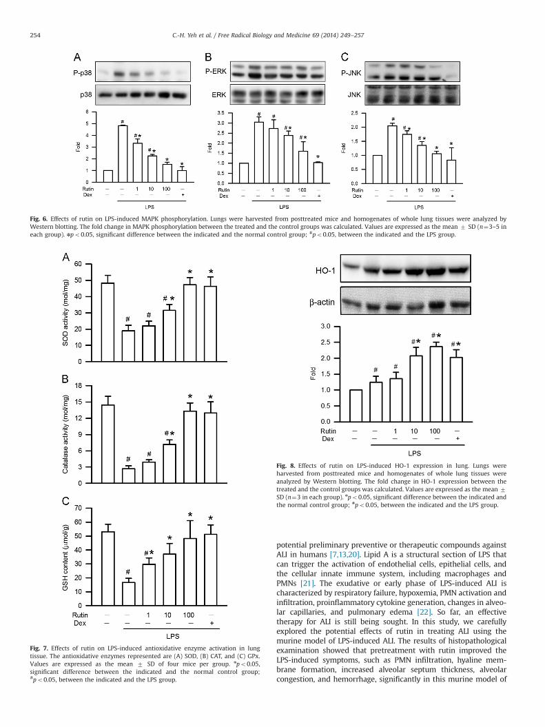
Effects of rutin on MAPK activation in LPS-induced ALI
The three MAPK pathways, ERK, p38 MAPK, and JNK, have beendemonstrated to participate in NF-κB activation in LPS-inducedALI [5]. The effect of rutin on phosphorylation of ERK (on residuetyrosine-204), JNK (on residues threonine-183 and tyrosine-185),and p38 MAPK (on residues threonine-180 and tyrosine-182) inLPS-induced ALI was analyzed by Western blotting. The resultsshowed that LPS stimulation significantly increased MAPK phos-phorylation and rutin inhibited the LPS-induced phosphorylationof p38 MAPK and JNK in a concentration-dependent manner.The significant inhibitory effect started at 1 μmol/kg (po0.05;Fig. 6A and C). In addition, rutin also inhibited LPS-inducedphosphorylation of ERK in a concentration-dependent manner,but with significant inhibitory effect started at 10 μmol/kg (po0.05;Fig. 6B). These data indicated that rutin relieved LPS-induced ALI byinhibiting the activity of MAPK pathways.
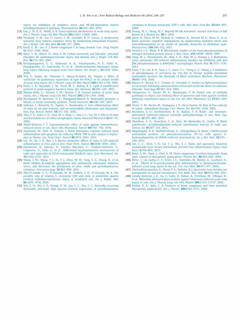
Effects of rutin on antioxidative enzymes in LPS-induced ALI
Oxidative stress exerted by activated PMNs is critically impor-tant in the pathophysiology of ALI. Antioxidative enzymes, such as
Fig. 2. Effects of rutin on LPS-induced leukocyte infiltration into BALF. PBS or1–100 μmol/kg rutin was intraperitoneally injected for 30 min before intratrachealinstillation of LPS (100 μg/50 μl saline) or saline into the mice. Six hours later, themice were anesthetized and BALF was collected for (A) PMN count and (B) MPOactivation in lung tissue. Values are expressed as the mean 7 SD (n¼4 in eachgroup).
n
po0.05, significant difference between the indicated and the normalcontrol group; #po0.05, between the indicated and the LPS group.
Fig. 3. Effects of rutin on LPS-induced MDA formation in lung tissue. Values areexpressed as the mean 7 SD of four mice per group. npo0.05, significantdifference between the indicated and the normal control group; #po0.05, betweenthe indicated and the LPS group.
C.-H. Yeh et al. / Free Radical Biology and Medicine 69 (2014) 249–257252

SOD, CAT, and GPx, are consumed during amelioration of ALI [16].SOD, CAT, and GPx activities were significantly decreased in LPS-treated mice compared to the untreated group (po0.05). Pre-treatment with rutin recovered the LPS-induced reduction in theactivation of these antioxidative enzymes in a concentration-dependent manner; significant inhibitory effect on SOD and CATstarted at 10 μmol/kg (po0.05; Fig. 7A and B), whereas on GPx, asignificant inhibitory effect started at 1 μmol/kg (po0.05; Fig. 7C).
Effects of rutin on HO-1 expression in LPS-induced ALI
HO-1, an antioxidative protein, is expressed during ameliora-tion of ALI [17]. The effect of rutin on HO-1 expression in LPS-induced ALI was analyzed by Western blotting. Whereas LPSstimulation significantly increased HO-1 expression, rutin furtherenhanced LPS-induced HO-1 expression in a concentration-dependent manner. A significant effect started at 1 μmol/kg(po0.05; Fig. 8). This result suggested the ability of rutin toreduce LPS-induced oxidative stress.
Comparison of the effects of rutin and desferrioxamine on LPS-induced ALI
DFX is an iron chelator that reduces oxidative stress andprovides beneficial effects on LPS-induced ALI [18,19]. AlthoughLPS-induced PMN infiltration was reduced by both rutin and DFX,rutin worked at a higher efficiency than DFX (Fig. 9A). In addition,rutin could reduce not only the secretion of TNF-α, IL-1β, and IL-6but also the activity of CAT, whereas DFX had no effect (Fig. 9B andC). Nevertheless, SOD and GPx activities were recovered by bothrutin and DFX. These results suggested the amelioration by rutin isbetter than that by DFX on LPS-induced ALI.
Discussion
The major course of ALI correlates with infection withLPS-containing gram-negative bacteria [1]. In the mouse modelof LPS-induced ALI, the syndromes presented are similar to thepathological characteristics of ALI in humans [3]. Therefore,the mouse model for the development of ALI induced by wayof intratracheal LPS administration is well suited to the study of
Fig. 4. Effects of rutin on LPS-induced proinflammatory cytokine secretion in BALF.Values are expressed as the mean 7 SD (n¼3 or 4 in each group). npo0.05,significant difference between the indicated and the normal control group;#po0.05, between the indicated and the LPS group.
Fig. 5. Effects of rutin on LPS-induced NF-κB p65 phosphorylation and IκB degradation in lung. Lungs were harvested from posttreated mice and homogenates of whole lungtissues were analyzed by Western blotting. The fold change in NF-κB p65 phosphorylation and IκB degradation between the treated and the control groups was calculated.Values are expressed as the mean 7 SD (n¼3–5 in each group). npo0.05, significant difference between the indicated and the normal control group; #po0.05, between theindicated and the LPS group.
C.-H. Yeh et al. / Free Radical Biology and Medicine 69 (2014) 249–257 253
potential preliminary preventive or therapeutic compounds againstALI in humans [7,13,20]. Lipid A is a structural section of LPS thatcan trigger the activation of endothelial cells, epithelial cells, andthe cellular innate immune system, including macrophages andPMNs [21]. The exudative or early phase of LPS-induced ALI ischaracterized by respiratory failure, hypoxemia, PMN activation andinfiltration, proinflammatory cytokine generation, changes in alveo-lar capillaries, and pulmonary edema [22]. So far, an effectivetherapy for ALI is still being sought. In this study, we carefullyexplored the potential effects of rutin in treating ALI using themurine model of LPS-induced ALI. The results of histopathologicalexamination showed that pretreatment with rutin improved theLPS-induced symptoms, such as PMN infiltration, hyaline mem-brane formation, increased alveolar septum thickness, alveolarcongestion, and hemorrhage, significantly in this murine model of
Fig. 6. Effects of rutin on LPS-induced MAPK phosphorylation. Lungs were harvested from posttreated mice and homogenates of whole lung tissues were analyzed byWestern blotting. The fold change in MAPK phosphorylation between the treated and the control groups was calculated. Values are expressed as the mean 7 SD (n¼3–5 ineach group). npo0.05, significant difference between the indicated and the normal control group; #po0.05, between the indicated and the LPS group.
Fig. 7. Effects of rutin on LPS-induced antioxidative enzyme activation in lungtissue. The antioxidative enzymes represented are (A) SOD, (B) CAT, and (C) GPx.Values are expressed as the mean 7 SD of four mice per group. npo0.05,significant difference between the indicated and the normal control group;#po0.05, between the indicated and the LPS group.
Fig. 8. Effects of rutin on LPS-induced HO-1 expression in lung. Lungs wereharvested from posttreated mice and homogenates of whole lung tissues wereanalyzed by Western blotting. The fold change in HO-1 expression between thetreated and the control groups was calculated. Values are expressed as the mean 7SD (n¼3 in each group). npo0.05, significant difference between the indicated andthe normal control group; #po0.05, between the indicated and the LPS group.
C.-H. Yeh et al. / Free Radical Biology and Medicine 69 (2014) 249–257254

LPS-induced ALI. This indicated the potential therapeutic effect ofrutin against ALI.
ALI is one of the inflammatory disorders in lung caused bypneumonia, sepsis, trauma, or aspiration [1]. In ALI, PMNs are themajor component to participate in the inflammation and patho-genesis processes [6]. Therefore, PMNs are the predominant celltype in BALF and histological specimens from patients with ALI [1].In the murine model, PMNs are rapidly activated and migrate intothe alveolar space and interalveolar septum in response tointratracheal administration of LPS. MPO, stored in azurophilicgranules of naive PMNs, can serve as a marker for content andactivation of PMNs in tissue [7,23]. Chemotaxis of PMNs inducedby formyl-L-methionyl-L-leucyl-L-phenylalanine or phorbol 12-myristate 13-acetate in in vitro assays is reduced by rutin [24].In a murine model of surgically induced esophagitis, rutin inhib-ited MPO activation in the esophagus, which means the infiltrationof PMNs is inhibited by rutin [25]. Furthermore, pretreatment withrutin significantly inhibits PMN infiltration into mucosal tissues in
indomethacin-induced gastric ulcers [26]. Here, we also found thatpretreatment with rutin prevented PMN infiltration into pulmon-ary tissue in LPS-induced ALI.
At the inflammatory site, proinflammatory cytokines such asTNF-α, IL-1β, and IL-6 are secreted from pulmonary cells andalveolar macrophages. These cytokines play an important role inLPS-induced ALI [7,13,14]. TNF-α and IL-1β are early responsecytokines generated by activated alveolar macrophages thatappear in BALF and plasma in ALI. The secretion of TNF-α andIL-1β in turn stimulates the neighboring cells to generate moreeffective proinflammatory cytokines and chemokines, such as IL-6,monocyte chemotactic protein, macrophage inflammatory protein,keratinocyte-derived chemokine, cytokine-induced neutrophilchemoattractant, and macrophage inflammatory protein-2, whichsubsequently mediate the recruitment of PMNs, macrophages, andlymphocytes [1]. Rutin reduces the generation of TNF-α in humanumbilical vein endothelial cells (HUVECs) induced by LPS, in acuteliver damage in CCl4-intoxicated mice, and in cisplatin-inducedrenal inflammation [27–29]. Rutin inhibits the production ofTNF-α and IL-1β induced by β-amyloid 42 in microglia, by brainischemia/reperfusion injury in rat serum, and by high glucosein human monocytic THP-1 cells [30–32]. In a murine model ofLPS-induced ALI, rutin reduced the production of TNF-α, IL-1β, andIL-6 in BALF. The results indicate that rutin reduced leukocyteinfiltration into lung by decreasing the expression of proinflam-matory cytokines.
The transcription factor NF-κB is the crucial signal factormodulating proinflammatory cytokines in LPS-induced ALI [15].There are five subunits, p65 (RelA), RelB, c-Rel, p50, and p52, toconstruct the homo- and heterodimers of NF-κB. The mostabundant NF-κB in mammalian cells is the p50/p65 heterodimer.In unstimulated cells, the inactive NF-κB dimers are sequestered inthe cytosol via noncovalent interactions with IκB, an inhibitorprotein. After stimulation, IκB is phosphorylated by activated IκBkinases, then the phosphorylated IκB is degraded by ubiquitin-dependent proteasomes. During the process NF-κB is released,allowing rapid phosphorylation of the p65 subunit, and subse-quently translocates into the nucleus [33]. Rutin suppresses bothexpression and activation of NF-κB and phosphorylation anddegradation of IκB in LPS-stimulated HUVECs, cisplatin-inducednephrotoxicity, CCl4-induced liver damage, and intracerebroven-tricular–streptozotocin-infused rat brain [25–27,34]. Here, wefound that pretreatment with rutin prevented the phosphorylationof NF-κB p65 and degradation of IκB in the lung of LPS-inducedALI. Furthermore, dual phosphorylation of MAPK, one of theupstream kinases in the NF-κB p65 phosphorylation pathway[5], at threonine and tyrosine residues within the kinase's activa-tion loop is required for MAPK activation [35]. Phosphorylation ofp38 MAPK and JNK induced by LPS is inhibited by Boehmeria niveaextract, 40% of which is made up of rutin [36]. High-glucose-induced phosphorylation of ERK and p38 MAPK is inhibited byrutin in human monocytic THP-1 cells [32]. The present studydemonstrated that intratracheal LPS instillation in mice resulted inphosphorylation of ERK, p38 MAPK, and JNK in lung tissue, andrutin pretreatment prevented these manifestations. Parallel trendswere observed between suppression of both MAPK and NF-κB p65phosphorylation. Therefore, the reduction in NF-κB p65 phosphor-ylation in lung resulting from rutin pretreatment was associatedwith MAPK activation.
MAPK also plays an important role in PMN activation, such astransmigration, degranulation, and respiratory burst [37]. During therespiratory burst, the amount of oxygen consumed is converted intosuperoxide anions through nicotinamide adenine dinucleotide phos-phate oxidase. SOD, which exists in the cytoplasm, can catalyze thereduction of superoxide anions into oxygen and hydrogen peroxide.MPO released via PMN degranulation can catalyze hydrogen peroxide
Fig. 9. Comparison of the effects of rutin and DFX on LPS-induced ALI. (A) Effects ofrutin (100 μmol/kg) and DFX (20 mg/kg) on LPS-induced leukocyte infiltration intoBALF. (B) Effects of rutin and DFX on LPS-induced proinflammatory cytokinesecretion in BALF. (C) Effects of rutin and DFX on LPS-induced antioxidativeenzyme activation in lung tissue. Values are expressed as the mean 7 SD of fourmice per group. npo0.05, significant difference between the indicated and thenormal control group; #po0.05, between the indicated and the LPS group.
C.-H. Yeh et al. / Free Radical Biology and Medicine 69 (2014) 249–257 255

and chloride anions to form hypochlorous acid. These reactive oxygenspecies (ROS), including superoxide anions, hydrogen peroxide, andhypochlorous acid, cause oxidative stress, which leads to tissue injuryvia lipid peroxidation, protein oxidation, and DNA damage [38]. MDAis the end-product of lipid peroxidation, indicating the destructionand damage of cell membranes caused by ROS [40]. In animal studies,rutin functions against MDA accumulation induced by ischemia/reperfusion in rat brain or kidney and by cyclophosphamide in rattestes [11,39]. In addition, rutin decreases the formation of MDA inSH-SY5Y neuroblastoma cells treated with β-amyloid 42 [30]. In thepresent study, we observed that with rutin pretreatment, MDAcontent in lung tissue was decreased compared with the controlgroup in LPS-induced ALI.
Furthermore, the formation of MDA acts as a marker foroxidative stress [38]. Under normal physiological conditions,improvement of oxidative damage is provided by antioxidativeenzymes, such as SOD, CAT, and GPx. Superoxide anions areconverted to hydrogen peroxide by SOD, which is then metabo-lized to water by CAT or GPx [39]. Rutin increases SOD and CATlevels in cerebral ischemia/reperfusion injury in rats [40]. Theactivation of SOD, CAT, and GPx is elevated by rutin in 6-hydroxydopamine-induced neurotoxicity, ethanol- and CCl4-induced liver damage, cyclophosphamide-induced reproductivetoxicity, and cadmium-induced testicular injury [41–44]. In thisstudy, we demonstrated that pretreatment with rutin raised theactivation levels of SOD, CAT, and GPx in LPS-induced ALI.
Heat shock protein 32, also called HO-1, is an inducible defenseenzyme opposed to oxidative stress. In the process of oxidativedegradation of heme to carbon monoxide, bilirubin, and ferrousion, HO-1 is the rate-limiting enzyme [45]. Carbon monoxide andbilirubin express cytoprotective effects. However, ferrous ioninduces oxidative stress in mammalian cells [46]. In the rat modelof LPS-induced ALI, the inflammatory responses evoked aredecreased by the combination of N-acetylcysteine, an ROS scaven-ger and glutathione precursor, plus DFX, an iron chelator. Never-theless, the use of isolated N-acetylcysteine or DFX has no effecton LPS-induced ALI [18]. Although rutin is a nonpermeativeextracellular iron chelator that exerts most of its antioxidativeeffects via inhibition of Fenton reactions catalyzed by labile iron[47], our data clearly suggest that extracellular labile iron has arole in ALI, perhaps after release from necrotic inflammatory cells.At present, we also found the amelioration provided by rutin isbetter than that by DFX. A previous study had suggested thatbiliverdin, the marker of HO-1 activation, could improve injuryoccurring in LPS-induced ALI, such as lung permeability, lungalveolitis, and generation of IL-6 [48]. Nuclear factor-erythroid-2-related-factor-2 (Nrf2), a transcription factor that mediates HO-1induction, has been demonstrated to diminish damages caused byinflammation and oxidative stress [49]. Rutin reduces Nrf2 andHO-1 expression in CCl4-injured liver in BALB/c N mice [27]. Ourdata demonstrate that rutin prompted expression of HO-1 in LPS-induced ALI. These observations suggest that rutin is capable ofreducing serious lung damage through AOEs.
In conclusion, we demonstrated that rutin effectively attenu-ated LPS-induced ALI by inhibiting histopathological changes andinfiltration of leukocytes in lung. The mechanisms underlying thisprotective effect include (1) reduction in MPO activity; (2) decreasein proinflammatory cytokines, such as TNF-α, IL-1β, and IL-6secretion; (3) inhibition of NF-κB phosphorylation and IκB degra-dation; (4) inhibition of MAPK phosphorylation; (5) diminution oflipid peroxidation and MDA formation; (6) elevation of activity ofantioxidative enzymes, such as SOD, CAT, and GPx; (7) increasedexpression of HO-1; and (8) chelation of extracellular iron (Fig. 10).Experimental findings support the potential use of rutin as atherapeutic agent for prevention of ALI associated with directinfection by gram-negative bacteria.
Acknowledgments
The authors thank the National Science Council of the Republicof China, Taiwan, for financially supporting this research underContract NSC102-2320-B-040-009. We also thank the ShowChwan Health Care System of the Republic of China, Taiwan, forfinancially supporting this research under Contract RD101060.
References
[1] Ware, L. B.; Matthay, M. A. The acute respiratory distress syndrome. N. Engl. J.Med. 342:1334–1349; 2000.
[2] Brun-Buisson, C.; Fartoukh, M.; Lechapt, E.; Honoré, S.; Zahar, J. R.; Cerf, C.;et al. Contribution of blinded, protected quantitative specimens to thediagnostic and therapeutic management of ventilator-associated pneumonia.Chest 128:533–544; 2005.
[3] Rojas, M.; Woods, C. R.; Mora, A. L.; Xu, J.; Brigham, K. L. Endotoxin-inducedlung injury in mice: structural, functional, and biochemical responses. Am. J.Physiol. Lung Cell. Mol. Physiol 288:L333–L341; 2005.
[4] Bhattacharyya, J.; Biswas, S.; Datta, A. G. Mode of action of endotoxin: role offree radicals and antioxidants. Curr. Med. Chem. 11:35–368; 2004.
[5] Khan, S.; Choi, R. J.; Shehzad, O.; Kim, H. P.; Islam, M. N.; Choi, J. S.; et al.Molecular mechanism of capillarisin-mediated inhibition of MyD88/TIRAPinflammatory signaling in in vitro and in vivo experimental models. J.Ethnopharmacol. 145:626–637; 2013.
[6] Grommes, J.; Soehnlein, O. Contribution of neutrophils to acute lung injury.Mol. Med. 17:293–307; 2011.
[7] Kuo, M. Y.; Liao, M. F.; Chen, F. L.; Li, Y. C.; Yang, M. L.; Lin, R. H.; et al. Luteolinattenuates the pulmonary inflammatory response involves abilities of anti-oxidation and inhibition of MAPK and NFκB pathways in mice withendotoxin-induced acute lung injury. Food Chem. Toxicol. 49:2660–2666; 2011.
[8] Guo, R.; Wei, P. Studies on the antioxidant effect of rutin in the microenviron-ment of cationic micelles. Microchim. Acta 161:233–239; 2008.
[9] Lee, S.; Suh, S.; Kim, S. Protective effects of the green tea polyphenol(�)-epigallocatechin gallate against hippocampal neuronal damage aftertransient global ischemia in gerbils. Neurosci. Lett 287:191–194; 2000.
[10] Novakovic, A.; Gojkovic-Bukarica, L.; Peric, M.; Nezic, D.; Djukanovic, B.;Markovic-Lipkovski, J.; et al. The mechanism of endothelium-independentrelaxation induced by the wine polyphenol resveratrol in human internalmammary artery. J. Pharmacol. Sci. 101:85–90; 2006.
[11] Korkmaz, A.; Kolankaya, D. Protective effect of rutin on the ischemia/reperfu-sion induced damage in rat kidney. J. Surg. Res. 164:309–315; 2010.
[12] Ferguson, N. D.; Frutos-Vivar, F.; Esteban, A.; Gordo, F.; Honrubia, T.; Penuelas,O.; et al. Clinical risk conditions for acute lung injury in the intensive care unitand hospital ward: a prospective observational study. Crit. Care 11:R96; 2007.
[13] Li, Y. C.; Yeh, C. H.; Yang, M. L.; Kuan, Y. H. Luteolin suppresses inflammatorymediator expression by blocking the Akt/NFκB pathway in acute lung injuryinduced by lipopolysaccharide in mice. Evid. Based Complement. Alternat. Med2012:383608; 2012.
[14] Huang, C. H.; Yang, M. L.; Tsai, C. H.; Li, Y. C.; Lin, Y. J.; Kuan, Y. H. Ginkgo bilobaleaves extract (EGb 761) attenuates lipopolysaccharide-induced acute lung
Fig. 10. Scheme of the mechanisms in the protective effect of rutin on LPS-inducedALI. The shaded parts indicate the molecules affected by rutin.
C.-H. Yeh et al. / Free Radical Biology and Medicine 69 (2014) 249–257256
injury via inhibition of oxidative stress and NF-κB-dependent matrixmetalloproteinase-9 pathway. Phytomedicine 20:303–309; 2013.
[15] Fan, J.; Ye, R. D.; Malik, A. B. Transcriptional mechanisms of acute lung injury.Am. J. Physiol. Lung Cell. Mol. Physiol 281:L1037–L1050; 2001.
[16] Hosakote, Y. M.; Liu, T.; Castro, S. M.; Garofalo, R. P.; Casola, A. Respiratorysyncytial virus induces oxidative stress by modulating antioxidant enzymes.Am. J. Respir. Cell. Mol. Biol. 41:348–357; 2009.
[17] Raval, C. M.; Lee, P. J. Heme oxygenase-1 in lung disease. Curr. Drug Targets11:1532–1540; 2010.
[18] Ryter, S. W.; Morse, D.; Choi, A. M. Carbon monoxide and bilirubin: potentialtherapies for pulmonary/vascular injury and disease. Am. J. Respir. Cell Mol.Biol. 36:175–182; 2007.
[19] Kostopanagiotou, G. G.; Kalimeris, K. A.; Arkadopoulos, N. P.; Pafiti, A.;Panagopoulos, D.; Smyrniotis, V.; et al. Desferrioxamine attenuates minorlung injury following surgical acute liver failure. Eur. Respir. J. 33:1429–1436;2009.
[20] Wu, Y.; Singer, M.; Thouron, F.; Alaoui-El-Azher, M.; Touqui, L. Effect ofsurfactant on pulmonary expression of type IIA PLA(2) in an animal modelof acute lung injury. Am. J. Physiol. Lung Cell. Mol. Physiol 282:L743–L750; 2002.
[21] Raetz, C. R.; Reynolds, C. M.; Trent, M. S.; Bishop, R. E. Lipid A modificationsystems in gram-negative bacteria. Annu. Rev. Biochem. 76:295–329; 2007.
[22] Matute-Bello, G.; Frevert, C. W.; Martin, T. R. Animal models of acute lunginjury. Am. J. Physiol. Lung Cell. Mol. Physiol 295:L379–L399; 2008.
[23] Borregaard, N; Sørensen, O. E.; Theilgaard-Mönch, K. Neutrophil granules: alibrary of innate immunity proteins. Trends Immunol. 28:340–345; 2007.
[24] Selloum, L.; Bouriche, H.; Tigrine, C.; Boudoukha, C. Anti-inflammatory effectof rutin on rat paw oedema, and on neutrophils chemotaxis and degranula-tion. Exp. Toxicol. Pathol. 54:313–318; 2003.
[25] Shin, Y. K.; Sohn, U. D.; Choi, M. S.; Kum, C.; Sim, S. S.; Lee, M. Y. Effects of rutinand harmaline on rat reflux oesophagitis. Auton. Autacoid Pharmacol 22:47–55;2002.
[26] Abdel-Raheem, I. T. Gastroprotective effect of rutin against indomethacin-induced ulcers in rats. Basic Clin. Pharmacol. Toxicol 107:742–750; 2010.
[27] Arjumand, W.; Seth, A.; Sultana, S. Rutin attenuates cisplatin induced renalinflammation and apoptosis by reducing NFκB, TNF-α and caspase-3 expres-sion in Wistar rats. Food Chem. Toxicol 49:2013–2021; 2011.
[28] Lee, W.; Ku, S. K.; Bae, J. S. Barrier protective effects of rutin in LPS-inducedinflammation in vitro and in vivo. Food Chem. Toxicol. 50:3048–3055; 2012.
[29] Domitrović, R.; Jakovac, H.; Vasiljev Marchesi, V.; Vladimir-Knežević, S.;Cvijanović, O.; Tadić, Z.; et al. Differential hepatoprotective mechanisms ofrutin and quercetin in CCl(4)-intoxicated BALB/cN mice. Acta Pharmacol. Sin.33:1260–1270; 2012.
[30] Wang, S. W.; Wang, Y. J.; Su, Y. J.; Zhou, W. W.; Yang, S. G.; Zhang, R.; et al.Rutin inhibits β-amyloid aggregation and cytotoxicity, attenuates oxidativestress, and decreases the production of nitric oxide and proinflammatorycytokines. Neurotoxicology 33:482–490; 2012.
[31] Abd-El-Fattah, A. A.; El-Sawalhi, M. M.; Rashed, E. R.; El-Ghazaly, M. A. Thepossible role of vitamin E, coenzyme Q10 and rutin in protection againstcerebral ischemia/reperfusion injury in irradiated rats. Int. J. Radiat. Biol.86:1070–1078; 2010.
[32] Wu, C. H.; Wu, C. F.; Huang, H. W.; Jao, Y. C.; Yen, G. C. Naturally occurringflavonoids attenuate high glucose-induced expression of proinflammatory
cytokines in human monocytic THP-1 cells. Mol. Nutr. Food Res. 53:984–995;2009.
[33] Huang, W. C.; Hung, M. C. Beyond NF-κB activation: nuclear functions of IκBkinase α. J. Biomed Sci. 20:3; 2013.
[34] Javed, H.; Khan, M. M.; Ahmad, A.; Vaibhav, K.; Ahmad, M. E.; Khan, A.; et al.Rutin prevents cognitive impairments by ameliorating oxidative stress andneuroinflammation in rat model of sporadic dementia of Alzheimer type.Neuroscience 210:340–352; 2012.
[35] Ferrell Jr J. E.; Bhatt, R. R. Mechanistic studies of the dual phosphorylation ofmitogen-activated protein kinase. J. Biol. Chem. 272:19008–19016; 1997.
[36] Sung, M. J.; Davaatseren, M.; Kim, S. H.; Kim, M. J.; Hwang, J. T. Boehmerianivea attenuates LPS-induced inflammatory markers by inhibiting p38 andJNK phosphorylations in RAW264.7 macrophages. Pharm. Biol. 51:1131–1136;2013.
[37] Kuan, Y. H.; Lin, R. H.; Tsao, L. T.; Chen, Y. L.; Tzeng, C. C.; Wang, J. P. Inhibitionof phospholipase D activation by CYL-26z in formyl peptide-stimulatedneutrophils involves the blockade of RhoA activation. Biochem. Pharmacol.70:901–910; 2005.
[38] Manca, D.; Ricard, A. C.; Trottier, B.; Chevalier, G. Studies on lipid peroxidationin rat tissues following administration of low and moderate doses of cadmiumchloride. Toxicology 67:303–323; 1991.
[39] Annapurna, A.; Ansari, M. A.; Manjunath, P. M Partial role of multiplepathways in infarct size limiting effect of quercetin and rutin against cerebralischemia–reperfusion injury in rats. Eur. Rev. Med. Pharmacol. Sci. 17:491–500;2013.
[40] Victor, V. M.; Rocha, M.; Esplugues, J. V.; De la Fuente, M. Role of free radicalsin sepsis: antioxidant therapy. Curr. Pharm. Des 11:3141–3158; 2005.
[41] Abarikwu, S. O.; Iserhienrhien, B. O.; Badejo, T. A. Rutin- and selenium-attenuated cadmium-induced testicular pathophysiology in rats. Hum. Exp.Toxicol. 32:395–406; 2013.
[42] Abarikwu, S. O.; Otuechere, C. A.; Ekor, M; Monwuba, K.; Osobu, D. Rutinameliorates cyclophosphamide-induced reproductive toxicity in male rats.Toxicol. Int 19:207–214; 2012.
[43] Magalingam, K. B.; Radhakrishnan, A.; Haleagrahara, N. Rutin, a bioflavonoidantioxidant protects rat pheochromocytoma (PC-12) cells against 6-hydroxydopamine (6-OHDA)-induced neurotoxicity. Int. J. Mol. Med. 32:235–-240; 2013.
[44] Lee, C. C.; Shen, S. R.; Lai, Y. J.; Wu, S. C. Rutin and quercetin, bioactivecompounds from tartary buckwheat, prevent liver inflammatory injury. FoodFunct 4:794–802; 2013.
[45] Ryter, S. W.; Alam, J.; Choi, A. M. Heme oxygenase-1/carbon monoxide: frombasic science to therapeutic applications. Physiol. Rev. 86:583–650; 2006.
[46] Ritter, C.; da Cunha, A. A.; Echer, I. C.; Andrades, M.; Reinke, A.; Lucchiari, N.;et al. Effects of N-acetylcysteine plus deferoxamine in lipopolysaccharide-induced acute lung injury in the rat. Crit. Care Med. 34:471–477; 2006.
[47] Vlachodimitropoulou, E.; Sharp, P. A.; Naftalin, R. J. Quercetin–iron chelates aretransported via glucose transporters. Free Radic. Biol. Med. 50:934–944; 2011.
[48] Sarady-Andrews, J. K.; Liu, F.; Gallo, D.; Nakao, A.; Overhaus, M.; Ollinger, R.;et al. Biliverdin administration protects against endotoxin-induced acute lunginjury in rats. Am. J. Physiol. Lung Cell. Mol. Physiol 289:L1131–L1137; 2005.
[49] Kirkby, K. A.; Adin, C. A. Products of heme oxygenase and their potentialtherapeutic applications. Am. J. Physiol. 290:F563–F571; 2006.
C.-H. Yeh et al. / Free Radical Biology and Medicine 69 (2014) 249–257 257