role of quantitative planar thallium-201 imaging for determining viability in patients with acute...
TRANSCRIPT

viable myocardium in patients with AMI. In this study,the relationship between the extent ofviable myocardiumand thallium uptake on the delayed images was established in three ways. First, average thallium activitywithin the occluded bed was correlated with regional wallmotion in that bed at baseline. Second, the increase inthallium uptake within the occluded bed after attemptedangioplasty of the IRA was correlated with improvementin regional function in that bed. Finally, pre-angioplastythallium uptake in the occluded bed was correlated withwall motion score in that bed after attempted angioplasty.
METHODS
PatientPopulationandStudyProtocolFifty-sevenconsecutive patients with recent AMI who were
found to have a totally occluded IRA at cardiac catheterizationwere included in this study. Their characteristics are depicted inTable 1. The protocol was approved by the Human InvestigationCommittee at the University of Virginia and all patients gaveinformed consent. They underwent thallium imaging and twodimensional echocardiography prior to and 1 mo after attemptedangioplasty of the IRA.
Thallium ImagingBaseline thallium imagingwas performed after exercise in 23
patients (40%) and during rest in 34 (60%), whereas 1 mo postangioplastyimagingwas performedafter exercise in all patients.Approximately 2.0 mCi of thallium (Dupont Medical Products,No. Billerica, MA) was injected 5 mm prior to obtaining theinitial images; the delayed images were obtained 2 hr later.Images were obtained in the anterior and 45°and 70°left anterioroblique projections and were analyzed using a previously described computer-assisted approach (1). After interpolativebackground subtraction, thalliumactivity in each of the preyously described 11segments in the three views was expressed asa percent of activity in the 9 x 9 pixel region with maximalactivity in that view (2).
The IRA bed was defined in the pre-angioplasty initial imagesas the segments showing hypoperfusion. Thallium activity wasmeasured in these same segments in the delayed images bothprior to and after attempted angioplasty. The activity in allsegmentswithinthe IRAbed shownon the delayedimageswereaveraged and activity in the segment with the least thallium
We studied57 patientswith a recent infarctionand an occluded infarct-relatedartery to test the hypothesisthat theamountof @°1Tlon delayedplanarimagescorrelateswiththeextent of viable myocardiumafter acute myocardialinfarction.Therewasa significant(p < 0.001)correlationbetweenmean @°1Tlactivity in the infarct zone and regional wall motionscore in that zone bothat baseline(r = —0.60,n = 57)and 1 mo after attemptedangioplasty(r = —0.67,n = 48),with better function being associated with greater 201Tluptakein the delayedimages.Therewasno correlationbetween the number of segments showing redistribution andthe wall motion score. We concludethat in patients withrecent myocardialinfarctionand an occludedinfarct-relatedartery, the average20111activitywithin the infarctzone ondelayed planar imaging correlates well with the extent ofviable myocardium in that zone. The presence or absence ofredistribution does not influence these results.
J NucIMed1993;34:728—736
n patients with acute myocardial infarction (AMI), theuptake of 20111within the infarct zone may depend on theamount of blood flow to and the extent of viable myocardium within that zone. When the infarct-related artery(IRA)is totally occluded,flowto the infarctzone canemanate only from collateral vessels from other myocardial beds. Moreover, since patients with AMI have amixture of normal, ischemic and necrotic tissue, the overall thallium kinetics will be determined by how much ofthe IRA bed consists of each of these types of tissues.Finally, because of possibility of redistribution, the relative thallium activity within a hypoperfused but viablezone may be maximal on the delayed image.
Based on these considerations, we hypothesized thatthe amount of thallium within an occluded bed on thedelayed planar images would correlate with the extent of
Received Oct. 8, 1992; revisionaccepted Jan. 18, 1993.For correspondence or reprints contact: Sanjiv Kaul, MD, DMS@n of Car
diology. Box 158. UniversIty of Wginia, Char1ottes@, VA 22908.
728 The Journalof Nuclear Medicine•Vol. 34 •No. 5 •May 1993
Role of Quantitative Planar Thallium-201Imaging for Determining Viability in Patientswith Acute Myocardial Infarction and a TotallyOccluded Infarct-Related ArteryPeter J. Sabia, Eric R. Powers, Michael Ragosta, William H. Smith, Denny D. Watson and Sanjiv Kaul
Division of Cardiology, Department ofMedicine, University of Virginia School ofMedicine, Charlottesville, Virginia
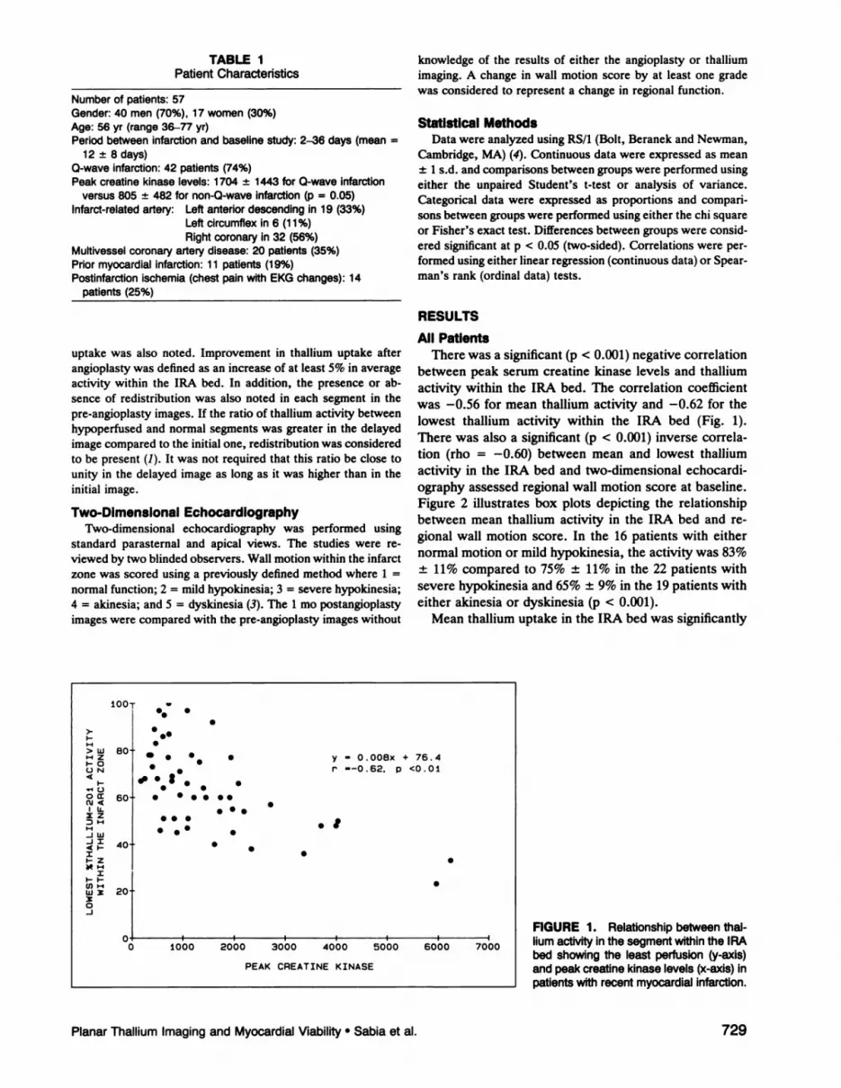
TABLE IPatientCharacteristics
Number of patients: 57Gender:40 men(70%),17 women(30%)Age: 56 yr (range 36—77yr)Periodbetweeninfarctionandbaselinestudy:2—36days(mean=
12 ±8 days)Q-waveInfarction:42 patients(74%)Peak creatine kinase levels: 1704 ±1443 for 0-wave Infarction
versus805 ±482fornon-Q-waveinfarction(p = 0.05)Infarct-relatedartery: Leftanteriordescendingin 19 (33%)
Left circumflex in 6 (11%)Right coronary in 32 (56%)
Multivesselcoronaryarterydisease:20 patients(35%)Priormyocardialinfarction:11 patients(19%)Postlnfarctionischemia(chestpainwith EKGchanges):14
patients(25%)
knowledgeof the results of either the angioplasty or thalliumimaging. A change in wall motion score by at least one gradewas considered to represent a change in regionalfunction.
Statistical MethodsData were analyzedusingRS/1(Bolt, Beranek and Newman,
Cambridge,MA)(4). Continuousdata were expressed as mean± 1 s.d. and comparisons between groups were performed using
either the unpaired Student's t-test or analysis of variance.Categorical data were expressed as proportions and compariSons between groups were performed using either the chi squareor Fisher's exact test. Differences between groups were considered significant at p < 0.05 (two-sided). Correlations were performedusingeither linearregression(continuousdata)or Spearman's rank (ordinal data) tests.
RESULTS
All PatientsThere was a significant (p < 0.001) negative correlation
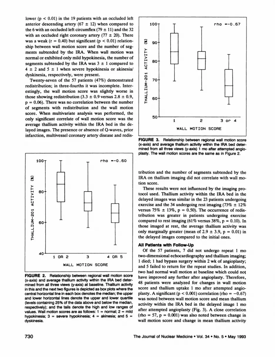
between peak serum creatine kinase levels and thalliumactivity within the IRA bed. The correlation coefficientwas —0.56for mean thallium activity and —0.62for thelowest thallium activity within the IRA bed (Fig. 1).There was also a significant (p < 0.001) inverse correlation (rho = —0.60)between mean and lowest thalliumactivity in the IRA bed and two-dimensional echocardiography assessed regional wall motion score at baseline.Figure 2 illustrates box plots depicting the relationshipbetween mean thallium activity in the IRA bed and regional wall motion score. In the 16 patients with eithernormal motion or mild hypokinesia, the activity was 83%±11% compared to 75% ±11% in the 22 patients withsevere hypokinesia and 65% ±9% in the 19 patients witheither akinesia or dyskinesia (p < 0.001).
Mean thallium uptake in the IRA bed was significantly
FIGURE 1. Relationship between thaihum activity in the segment within the IRAbed showing the least perfusion (y-axis)andpeakcreatinekinaselevels(x-axls)inpatientswithrecentmyocardialinfarction.
uptake was also noted. Improvement in thallium uptake afterangioplasty was defined as an increase of at least 5% in averageactivity within the IRA bed. In addition, the presence or ab
sence of redistribution was also noted in each segment in thepre-angioplasty images. If the ratio of thallium activity betweenhypoperfused and normal segments was greater in the delayedimage compared to the initial one, redistribution was consideredto be present (1). It was not required that this ratio be close tounity in the delayed image as long as it was higher than in theinitial image.
Two-Dimensional EchocardlographyTwo-dimensional echocardiography was performed using
standard parasternal and apical views. The studies were reviewed by two blinded observers. Wall motion within the infarctzone was scored using a previously defined method where 1 =normal function; 2 = mild hypokinesia; 3 = severe hypokinesia;4 = akinesia; and 5 = dyskinesia (3). The 1 mo postangioplastyimages were compared with the pre-angioplasty images without
100 ••w•
•S>-
>w,-IzI-0ON4
I-@10
Ohc'j<Li.
:D@-iN-Jw-ii4'-I
I
CONClixx0-J
80 . • •S
S •#. I
S •S ••
. y —0.008x + 76.4
C ——0.62. p <0.01
60 S •S
S ••S@S
S •• S
S
5:
40 SS
S
20
0 1000 2000 3000 4000 5000 6000 7000
PEAK CREATINE KINASE
PlanarThalliumImagingand MyocardialViability•Sabia et al. 729
lower (p < 0.01) in the 19 patients with an occluded leftanterior descending artery (67 ±12) when compared tothe 6 with an occluded left circumflex (79 ±11) and the 32with an occluded right coronary artery (77 ±20). Therewas a weak (r = 0.40) but significant (p < 0.01) relationship between wall motion score and the number of segments subtended by the IRA. When wall motion wasnormal or exhibited only mild hypokinesia, the number ofsegments subtended by the IRA was 3 ±1 compared to4 ±2 and 5 ±1 when severe hypokinesia or akinesia/dyskinesia, respectively, were present.
Twenty-seven of the 57 patients (47%) demonstratedredistribution; in three-fourths it was incomplete. Interestingly, the wall motion score was slightly worse inthose showing redistribution (3.3 ±0.9 versus 2.8 ±0.9,p = 0.06). There was no correlation between the numberof segments with redistribution and the wall motionscore. When multivariate analysis was performed, theonly significant correlate of wall motion score was theaverage thallium activity within the IRA bed in the delayed images. The presence or absence of 0-waves, priorinfarction, multivessel coronary artery disease and redis
FiGURE 2. Relationshipbetweenregionalwailmotionscore(x-axis) and average thallium activity within the IRA bed determined from all three views (y-axis)at baseline.Thallium activityinthisandthenexttwofiguresisdepictedas boxplotswherethecentralhorizontallineineachboxdenotesthemedian;theupperand lowerhorizontallinesdenotethe upperand lowerquartile(levelscontaining25% ofthedataaboveandbelowthemedian,respectively);and the tails denote the high and low ranges ofvalues.Wallmotionscoresare as follows:I = normal;2 = mildhypokinesia; 3 = severe hypokinesia; 4 = akinesia; and 5 =dyskinesia.
100@ rho ——0.67
@9o,>-I.I@1>1-4I-.04
0(@J 70@
I:DI-4-I-J4II-
50I 2 3or4
WALL MOTION SCORE
FIGURE 3. Relationshipbetweenregionalwallmotionscore(x-axis)andaveragethalliumactivItywithinthe IRAbeddeterminedfrom all three views (y-axis) I mo after attemptedangioplasty.Thewallmotionscoresarethesameas in Figure2.
tribution and the number of segments subtended by theIRA on thallium imaging did not correlate with wall motion score.
These results were not influenced by the imaging protocol used. Thallium activity within the IRA bed in thedelayed images was similar in the 23 patients undergoingexercise and the 34 undergoing rest imaging (73% ±12%versus 75% ±13%, p = 0.50). The occurrence of redistribution was greater in patients undergoing exercisecompared to rest imaging (61% versus 38%, p = 0.10). Inthose imaged at rest, the average thallium activity wasonly marginally greater (mean of 2.9 ±3.9, p = 0.01) inthe delayed images compared to the initial ones.
All Patients with Follow-UpOf the 57 patients, 7 did not undergo repeat 1 mo
two-dimensional echocardiography and thallium imaging;1 died; 1 had bypass surgery within 2 wk of angioplasty;and 5 failed to return for the repeat studies. In addition,two had normal wall motion at baseline which could no@have improved any further after angioplasty. Therefore,48 patients were analyzed for changes in wall motionscore and thallium uptake 1 mo after attempted angioplasty. A significant (p < 0.001) correlation (rho = —0.67)was noted between wall motion score and mean thalliumactivity within the IRA bed in the delayed image 1 moafter attempted angioplasty (Fig. 3). A close correlation(rho = 57, p = 0.001) was also noted between change inwall motion score and change in mean thallium activity
ioO rho ——0.60
>-I-
@ GO
:@ 60‘-I-J-J4II-.
4010R2 3 40R5
WALL MOTION SCORE
730 The Journalof Nuclear Medicine•Vol. 34 •No. 5 •May 1993

Pre PostpVariableangioplastyangioplastyvalue
SuccessfulUnsuccessfulangioplastyangioplastypVariables
analyzed (n = 38)(n = 10)value
LAD = left anteriordescendingartery and Seg. = numberofsegments(totalof 11).
TABLE 3Comparisonof Wail MotionScore and ThalliumActivity
Withinthe InfarctBed at Baselineand 1 MonthAfterAttempted Angioplasty
30 rho —0.57
>.
@ :ii‘-4>‘-II-
@ 20@1
0oJ
I:D
‘:;10-J
I4II.
zI-'
wCDz4I0
—10
0 1 2
CHANGE IN WALL MOTION SCORE
Patients with unsuccessful angioplasty (n = 10)Wallmotionscore 3.3±0.8 3.3±0.8 0.99Meanthalliumactivity 69 ±8 67 ±8 0.14
Patients with successful angioplasty (n = 38)Wallmotionscore 3.1±0.9 1.9±0.9 0.0001Meanthalliumactivity 75 ±12 81 ±11 0.001
With lowerwall motionscores,there is betterregionalfunction.
baseline characteristics between these two groups of patients. Importantly, wall motion score and mean thalliumactivity were similar between the two groups.
Patients with Unsuccessful AngioplastyThere was no change in mean wall motion score 1 mo
later in the 10 patients with unsuccessful angioplasty (Table 3). In six patients, wall motion score did not change,while in two it improved by one grade and in two itworsened by one grade. Similarly, there was no overallchange in mean thallium activity within the IRA bed(Table 3). It remained unchanged in five patients, improved by > 5% in one and worsened by > 5% in four.
Patients with Successful AngioplastyUnlike those with unsuccessful angioplasty, the 38 pa
tients with successful angioplasty showed significant improvement in wall motion score 1 mo later (Table 3).Figure 5 illustrates wall motion score in each of thesepatients before and 1 mo after successful angioplasty.Compared to 27 patients (71%) with severe wall motion
FIGURE 5. Wallmotionscoresin38patientsbeforeand1moaftersuccessfulangioplasty.
FIGURE 4. Relationshipbetweenchangeinregionalwallmotion score (x-axis) and change in average thalliumactivitywithinthe IRA bed (y-axis) I mo after attempted angioplasty.
within the IRA bed, with greater improvement in thalliumactivity being associated with greater improvement inwall motion score (Fig. 4).
Of these 48 patients, 38 had successful angioplasty withsustained reperfusion, whereas 10 did not, 7 had initialunsuccessful angioplasty and 3 had reocclusion. Table 2shows that there were no significant differences in the
TABLE 2Comparisonof BaselineCharacteristicsBetweenPatients
With and WithoutSuccessfulAngioplasty
5—
4
3
2
0
Lii
00U)
z0I-0I
-J-I4
Age (yr)57 ±1052 ±90.12Males(%)63700.99Days
afterinfarction12 ±910 ±40.80Priorinfarction(%)16400.18Q-wave
infarction(%)711000.09Peakcreatinekinase1 232 ±11511 997 ±12030.08Multivessel
disease(%)26500.25OccludedLAD(%)42200.72Collateralgrade1 .8 ±0.91 .6 ±0.90.54Wall
motionscore3.1 ±0.93.3 ±0.70.43Meanthalliumactivity75 ±1269 ±80.19Seg.
withhypoperfusion3.9 ±1.64.9 ±2.00.10Redistribution(%)58500.73 PREANGIOPLASTY POSTANGIOPLASTY
PlanarThalliumImagingand MyocardialViability•Sabia et al. 731
. .@
FIGURE 6. Delayedthalliumimagesbefore(upperpanel)and I mo after (lowerpanel)successfulangioplastyof the leftanteriordescendingartery in a patientwith anteriorAMI. The leftpanel depicts the anterior projection,while the middle and rightpanelsdepictthe 45°and 70°left anteriorobliqueprojections,respectively.
abnormality prior to angioplasty (severe hypokinesia todyskinesia), only 7 (18%) had severe wall motion abnormality 1 mo after successful angioplasty (p < 0.001).Seventeen patients improved their wall motion score by 1grade and 14 patients improved it by more than 1 grade;in 7 patients there was no change in the wall motionscore.
The patients with successful angioplasty also showedimprovement in mean thallium activity 1 mo later (Table3): 22 showed an increase in activity by > 5%; 4 exhibiteda decrease in activity by > 5%; whereas in 12 patients theactivity remained unchanged. There was a fair but significant (p = 0.01) correlation between pre-angioplastymean thallium activity within the IRA bed and postangioplasty wall motion score (rho = —0.50)as well as between change in mean thallium activity and change inwall motion score (rho = —0.51).There was no association between the presence of redistribution before angioplasty and change in wall motion score after angioplasty.Wall motion score improved by 1.3 ±0.8 in patients withredistribution compared with 1.0 ±0.6 in those withoutredistribution (p = 0.30).
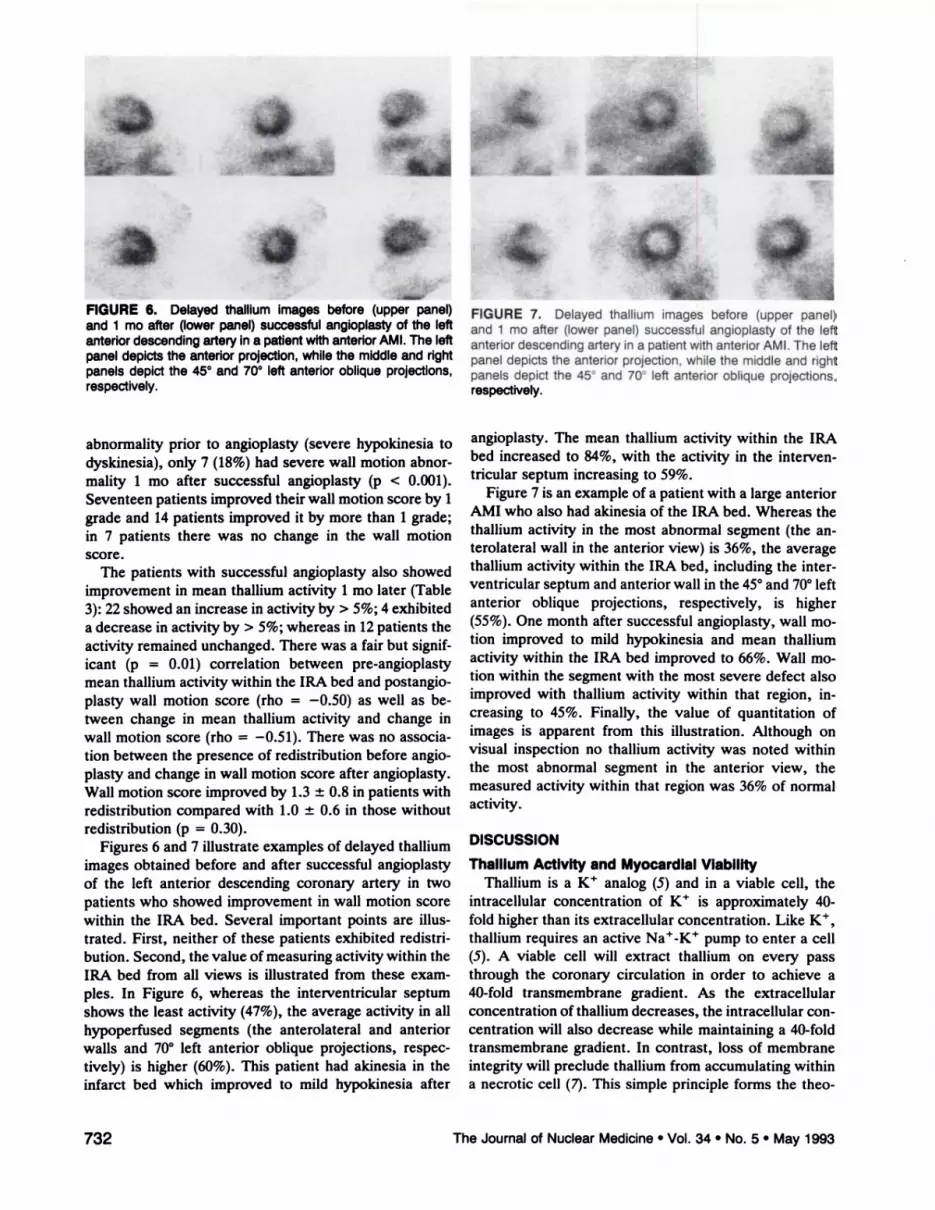
Figures 6 and 7 illustrate examples of delayed thalliumimages obtained before and after successful angioplastyof the left anterior descending coronary artery in twopatients who showed improvement in wall motion scorewithin the IRA bed. Several important points are illustrated. First, neither of these patients exhibited redistribution. Second, the value of measuring activity within theIRA bed from all views is illustrated from these examples. In Figure 6, whereas the interventricular septumshows the least activity (47%), the average activity in allhypoperfused segments (the anterolateral and anteriorwalls and 70°left anterior oblique projections, respectively) is higher (60%). This patient had akinesia in theinfarct bed which improved to mild hypokinesia after
732 The Journalof NuclearMedicine•Vol. 34 •No. 5 •May 1993
0;a@' iii
FIGURE 7. Delayed thalliumimages before (upper panel)and 1 mo after (lower panel) successful angioplastyof the leftanterior descending artery in a patient with anterior AMI. The leftpanel depicts the anterior projection,while the middle and rightpanels depict the 45°and 70°left anterior oblique projections,respectively.
angioplasty. The mean thallium activity within the IRAbed increased to 84%, with the activity in the interventricular septum increasing to 59%.
Figure 7 is an example of a patient with a large anteriorAMI who also had akinesiaof the IRA bed. Whereas thethallium activity in the most abnormal segment (the anterolateral wall in the anterior view) is 36%, the averagethallium activity within the IRA bed, including the interventricular septum and anterior wall in the 45°and 70°leftanterior oblique projections, respectively, is higher(55%). One month after successfulangioplasty, wall motion improved to mild hypokinesia and mean thalliumactivity within the IRA bed improved to 66%. Wall motion within the segment with the most severe defect alsoimproved with thallium activity within that region, increasing to 45%. Finally, the value of quantitation ofimages is apparent from this illustration. Although onvisual inspection no thallium activity was noted withinthe most abnormal segment in the anterior view, themeasured activity within that region was 36% of normalactivity.
DISCUSSION
Thallium Activity and MyocardialViabilityThallium is a K@ analog (5) and in a viable cell, the
intracellular concentration of K@ is approximately 40-fold higher than its extracellular concentration. Like K@,thallium requires an active Na@-K@pump to enter a cell(5). A viable cell will extract thallium on every passthrough the coronary circulation in order to achieve a40-fold transmembrane gradient. As the extracellularconcentration of thallium decreases, the intracellular concentration will also decrease while maintaining a 40-foldtransmembrane gradient. In contrast, loss of membraneintegrity will preclude thallium from accumulating withina necrotic cell (7). This simple principle forms the theo
B.A.
@rme1t@1ssue
average activity
infarcted tissue
1::@ma1t1ssue@1ssue
@ 600 04 4
@1
0 0Cu 40
I IDN N_J
@ 20
0I I
INITIAL DELAYED INITIAL DELAYEDIMAGE IMAGE IMAGE IMAGE
NORMAL MYOCARDIUM INFARCTED MYOCARDIUM
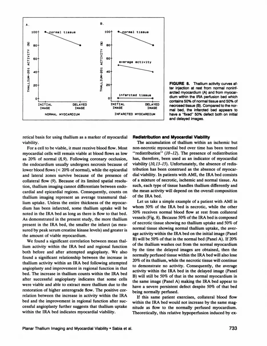
FIGURE 8. Thalliumactivitycurvesafter injectionat rest from normalnoninfarcted myocardlum(A) and from myocardium within the IRA perfusion bed whichcontains50%ofnormaltissueand50%ofnecrosedtissue(B).Comparedtothenormal bed, the infarctedbed appearstohavea “fixed―50% defectbothon initialanddelayedimages.
retical basis for using thallium as a marker of myocardialviability.
For a cell to be viable, it must receive blood flow. Mostmyocardial cells will remain viable at blood flows as lowas 20% of normal (8, 9). Following coronary occlusion,the endocardium usually undergoes necrosis because oflower blood flows (< 20% of normal), while the epicardialand lateral zones survive because of the presence ofcollateral flow (9). Because of its limited spatial resolution, thallium imaging cannot differentiate between endocardial and epicardial regions. Consequently, counts onthallium imaging represent an average transmural thalhum uptake. Unless the entire thickness of the myocardium has been infarcted, some thallium uptake will benoted in the IRA bed as long as there is flow to that bed.As demonstrated in the present study, the more thalliumpresent in the IRA bed, the smaller the infarct (as measured by peak serum creatine kinase levels) and greater isthe amount of viable myocardium.
We found a significant correlation between mean thalhum activity within the IRA bed and regional functionboth before and after attempted angioplasty. We alsofound a significant relationship between the increase inthallium activity within an IRA bed following attemptedangioplasty and improvement in regional function in thatbed. The increase in thallium counts within the IRA bedafter successful angioplasty indicates that some cellswere viable and able to extract more thallium due to therestoration of higher anterograde flow. The positive correlation between the increase in activity within the IRAbed and the improvement in regional function after successful angioplasty further suggests that thallium uptakewithin the IRA bed indicates myocardial viability.
Redistribution and Myocardial ViabilityThe accumulation of thallium within an ischemic but
non-necrotic myocardial bed over time has been termed“redistribution―(10—12).The presence of redistributionhas, therefore, been used as an indicator of myocardialviability (10, 13—15).Unfortunately, the absence of redistribution has been construed as the absence of myocardial viability. In patients with AMI, the IRA bed consistsof a mixture of necrotic, ischemic and normal tissue. Assuch, each type of tissue handles thallium differently andthe mean activity will depend on the overall compositionof the IRA bed.
Let us take a simple example of a patient with AMI inwhom 50% of the IRA bed is necrotic, while the other50% receives normal blood flow at rest from collateralvessels (Fig. 8). Because 50% of the IRA bed is composedof necrotic tissue showing no thallium uptake and 50% ofnormal tissue showing normal thallium uptake, the average activity within the IRA bed on the initial image (PanelB) will be 50%of that in the normalbed (Panel A). If 20%of the thallium washes out from the normal myocardiumby the time the delayed images are obtained, then thenormally perfused tissue within the IRA bed will also lose20% of its thallium, while the necrotic tissue will continueto demonstrate no activity. Consequently, the averageactivity within the IRA bed in the delayed image (PanelB) will still be 50% of that in the normal myocardium inthe same image (Panel A) making the IRA bed appear tohave a severe persistent defect despite 50% of that bedbeing normally perfused.
If this same patient exercises, collateral blood flowwithin the IRA bed would not increase by the same magnitude as flow to the normally perfused myocardium.Theoretically, this relative hypoperfusion induced by cx
Planar ThalliumImagingand MyocardialViability•Sabia et al. 733

ercise should reverse on the delayed image. The apparentreversal, however, depends on several factors. The degree of disparity in thaffium activity in the initial imageswill influence whether redistribution can be detected inthe delayed images. Therefore, the greater the degree ofdisparity in the initial images, the greater the likelihood ofseeing this disparity decrease in the delayed images. It isfor this reason that we noted redistribution twice as frequently during exercise than during rest. The timing ofthe initial image can also be crucial. If the initial imagesare obtained relatively late after thallium injection, significant redistribution may already have occurred. Finally, the noise inherent in thallium images can be asource of substantial error in determining whether achange has indeed occurred in the thaffium activity ratiobetween normal and hypoperfused images over time.
Quantitative Versus Visual Assessment of ThalliumImages
When only visual assessment of images is performed,not only can redistribution be missed, but the amount ofthallium within an IRA bed can be grossly underestimated (16). Therefore, it is not surprising to visuallydetermine myocardial segments as “dead―on thalliumimaging (dense defect and no redistribution), while demonstrating myocardial metabolism on positron emissiontomography (PET) (17). Myocardial cells cannot, however, be viable without blood flow and “absence―ofthallium on delayed images may merely indicate that itsuptake is being underestimated by visual inspection.
One reason for the underestimation of thallium activityby visual inspection may be due to the use of high-contrast x-ray film used for processing images. Figure 9illustrates the characteristic curve of a widely used singleemulsion film where the log relative exposure on thex-axis can be translated to thallium activity. Consequently, if a region of the myocardium receives maximalcounts so as to produce a log relative exposure of 24 unitsand an x-ray film density of close to 4 units, half thecounts (equal to a log relative exposure of 12) will hardlyproduce any density on the x-ray film. Furthermore, depending on which part of the curve is examined, the samedifference in the log relative exposure can result in variable differences in x-ray density. For instance, log relative exposures of 16 and 20 (difference of 4) will appear asdistinctly different densities on the film while those of 8and 12 (also a difference of 4) will look similar. Otherfactors, such as film exposure time, will also affect therelative densities on the x-ray film. It is only by quantitation of the digital background-subtracted data that actual counts within a myocardial bed can be determined.Even viewing images on computer screens is not adequate because of the inherent limitations of the humaneye in discerning grey shades.
Comparison with PrevIous StudiesIt has been previously demonstrated that patients with
“fixed―defects can improve regional function after Un
4S
S
S
S
3 5
>-I- SNU)z
@2 S
I—INLi@ S
I S
S
S•555S
C I I I I I I I I— —I@8 10 12 14 16 18 20 22 24
LOG RELATIVE EXPOSURE
FIGURE 9. Relationshipbetweenlogrelativeexposure(xaxis)and x-ray density (y-axis)on high-contrast,single emulsionx-rayfilmresultsin“severedefects―evenwhenthalliumactMtyis30%-50% of normal.
dergoing revascularization (18, 19). Such patients havebeen shown to have viable myocardium within regionswith “fixeddefects― on the basis of ‘8F-fluorodeoxyglucose uptake during PET (17). Based on this “fowlmetabolism mismatch,― it has been erroneously construed and popularized that markers of blood flow, suchas thallium, are poor indicators of viability and that demonstration of metabolic activity on PET is required todefine the presence of viable myocardium (17).
Because thallium cannot accumulate within nonviablecells, different imaging algorithms have been suggested toenhance the discrimination between nonviable and viablemyocardium using thallium. One such algorithm recommends that if redistribution is not noted on the conventional delayed image (2—4hr postinjection), then 24-hrimaging should be performed to give enough time forredistribution to occur (20). Using such an approach,regions that do not show fill-in on the routine delayedimages may sometimes show better fill-in on the 24 hrimages (20). We have found that redistribution occursalmost immediately after thallium injection and can bequantitatively determined 2 hr postinjection even if visualfill-in has not occurred (21). These same segments showvisual fill-in later (21).
Another recommended approach involves reinjectionof thallium if redistribution is not noted on the postexercisc delayed images (22,23). Reinjection at rest allowsmore thallium to enter myocardial tissue that may beviable but which shows a fixed defect 2—4hr after exercisc. Moreover, the problem with reduced count statistics
734 The Journalof Nuclear Medicine•Vol. 34 •No. 5 •May 1993
Maryland to Dr. Kaul. Dr. Ragosta was the recipient of a fellowship training grant from the Virginia Affiliate of the Amencan Heart Association, Glen Allen, Virginia. Dr. Kaul is anEstablished Investigator of the American Heart Association(National Center), Dallas, Texas. Presented in part at the 65thAnnual Scientific Session of the American Heart Association,November 1992, New Orleans, LA.
REFERENCESI. Berger BB, Watson DD, Taylor GJ, et at. Quantitative thallium-201cx
ercise scintigraphy for detection of coronary disease. J Nuci Med 1981;22:585—593.
2. VillanuevaFS, Kaul S. Smith WH, Watson DD, Varma D, Belier GA.Prevalence and correlates of increased lung/heart ratio of thallium-201duringdipyridamoiestress imaging.Am J Cardiol 1990;66:1324—1328.
3. Touchstone DA, Belier GA, Nygaard T, Tedesco C, Kaul S. Effects ofsuccessful intravenous reperfusion therapy on regional myocardial function and geometry in humans: a tomographic assessment using twodimensional echocardiography. J Am Coil Cardioi 1989;13:i506—15l3.
4. BBN Software Products Corp. RS/l user's guide book2. Cambridge, MA:1987:1—37.
5. Weast RC. Handbook of chemistry and physics. Boca Raton, FL: CRCPress; 1986—1987:3—38.
6. BelIer GA. Nuclear cardiology: current indications and clinical usefulness. Curr Probi Cardiol 1985;lO:i—76.
7. Goldhaber SZ, Newell JB, IngwallJS, Pohost GM, Alpert NM, FosselET. Effects of reduced coronary flow on thallium-201 accumulation andrelease in an in vitro rat heart preparation. Am J Cardiol 1983;51:891—896.
8. Reimer KA, Jennings RB. The “wavefrontphenomenon' â€of myocardialischemic cell death. II. Transmural progression of necrosis within theframeworkof ischemicbed size (myocardiumat risk) and collateral flow.Lab Invest 1979;40:633—644.
9. Jugdutt B!, Hutchins GM, Bulkley BM, Becker LC. Myocardial infarction in the conscious dog: three-dimensional mapping of infarct, collateralflow and region at risk. Circulation 1979;60:1141—1150.
10. Pohost GM, Zir LM, Moore RH, McKusick KA, Guiney TE, Belier GA.Differentiation of transiently ischemic from infarcted myocardium byserial imaging after a single dose ofthallium-201. Circulation l977;55:294—302.
11. BelIer GA, Watson DD, Ackell P. Pohost GM. Time course of thallium201 redistribution after transient myocardial ischemia. Circulation 1980;61:791—797.
12. Gerry JL, Becker LC, Flaherty iT, Weisfeldt ML. Evidence for a flowindependent contribution to the phenomenon of thallium redistribution.Am J Cardiol 1980;45:58—62.
13. Pohost GM, Okada RD. O'Keefe DB, et al. Thallium redistribution indogs with severe coronary artery stenosis of fixed caliber. Circ Res1981;48:439—446.
14. Leppo J, Rosenkrantz J, Rosenthal R, Bontemps R, Yipintsoi T. Quantitative thallium-201 redistribution with a fixed coronary stenosis in dogs.Circulation 1981;63:632—639.
15. Okada 1W, Leppo JA, Strauss HW, Boucher CA, Pohost GM. Mechanisms and time course for the disappearance of thallium-201defects atrest in dogs: relation oftime to peak activity ofmyocardiai blood flow. AmJ Cardiol 1982;49:699—706.
16. Kaul S. Chesler DA, Okada RD. Boucher CA. Computer versus visualanalysis of exercise thallium-201 images: a critical appraisal in 325 patients with chest pain. Am HeartJ 1987;114:1129—1l37.
17. Brunken RC, Kottori 5, Nienaber CA, et al. PET detection of viabletissue in myocardial segments with persistent defects at 201T1SPECT.Radiology 1989;172:65—73.
18. Gibson RS, Watson DD, Taylor GJ, et al. Prospective assessment ofregional myocardial perfusion after coronary revascularization surgery byquantitative thallium-201 scintigraphy. J Am Coil Cardiol 1983;1:804—815.
19. Liu P, Kiess MC, Okada RD. Block PC, Strauss HW, Pohost GM. Thepersistentdefecton exercise thalliumimagingand its fate after myocardialrevascularization: does it represent scar or ischemia? Am Heart J 1985;I 10:996—1001.
20. Kiat H, Berman DS, Maddahi J, et al. Late reversibility of tomographicmyocardial thallium-201 defects: an accurate marker of myocardial viability. I Am Coil Cardiol 1988;12:1456—1463.
inherent in 24 hr imaging is minimized. Once again, however, we have demonstrated that this method does notimprove upon the detection of redistribution usingquantitative techniques (24). Although fill-in is visuallymore apparent after reinjection, it invariably occurswithin regions that quantitatively demonstrate redistribution.
LimitationsoftheStudyWe did not have many patients with severe reduction
(< 35%)in averagethallium activity. If suchpatients hadbeen present in our study, we could have conceivablyestablished the “threshold―of thallium activity belowwhich the myocardium may not show recovery in function despite restoration of anterograde flow. On the otherhand, finding such a threshold may be elusive because thegamma camera's sensitivity may be too low to detectthallium in regions where blood flow is so low as to causetransmural necrosis. Therefore, it could be argued thatevidence of any thallium uptake within an IRA bed mayrepresent viable tissue. As has been demonstrated in ourstudy, the presence of greater amounts of thallium mdicate the presence of more viable myocardium.
Because it is not possible to register exactly myocardial segments on planar imaging and those on two-dimensional echocardiography, we believe that averaging activity within an IRA bed from all views is reasonable. It ispossible, however, that “shinethrough― from normaloverlying or underlying tissue could have affected ourresults. Thallium activity within the most hypoperfusedsegment in any single view, however, also correlated wellwith regional function.
Finally, in some patients we used rest images and inothers we used exercise images for the pre-angioplastydata analysis. However, since we analyzed only the delayed images, the results from both subsets of patientswere comparable. Similarly, we compared rest imagespre-angioplasty with exercise images postangioplasty insome patients. Since we only compared the relative thalhum activity in the delayed images, the different imagingprotocols would not have influenced our results.
Clinical ImplicationsOur results indicate that in patients with recent AMI
and a totally occluded IRA, the amount of thallium activity within the IRA bed on delayed planar thalliumimaging correlates with the extent of viable myocardiumwithin that bed. Greater amounts of thallium present inthese images indicate greater extents of viable myocardium. The presence or absence of redistribution does notinfluence these results. Further studies are required todefine the usefulness of this approach in the day-to-daymanagement of unselected patients with AMI.
ACKNOWLEDGMENTS
Supported in part by grants (R29-HL38345 and ROlHL48890) from the National Institutes of Health, Bethesda,
PlanarThalliumImagingand MyocardialViability•Sabiaet al. 735

21. Watson DD, Smith WH, Lilywhite RC, Belier GA. Quantitative analysis 23. Dilsizian V, Smeltzer WR, Freedman NMT, Dextras R, Bonow RO.of 20―flredistribution at 24 hours compared to 2 and 4 hours postinjection Thallium reinjection after stress-redistribution imaging: does 24-hour de[Abstract].I Nucl Med 1990;31:763. layed imagingfollowingreinjection enhance detection of viable myocar
22. Dilsizian V, Rocco TP, Freedman NMT, Leon MB, Bonow RO. En- dium? Circulation 1991;83:1247—1255.hanced detection of ischemic but viable myocardium by the reinjection of 24. Watson DD, Thallium-20l reinjection: truth or artifact? J Nuci Biol Medthallium after stress redistribution. N Engl I Med 1990;323:141—146. 1992;36:15—21.
SELF-STUDY TEST
SkeletalNuclearMedicineQuestionsaretakenfromtheNuclearMedicineSelf-StudyProgram1,
publishedbyTheSocietyofNuclearMedicineu.ff@@i I iuur.@
Items1-3 consistof a questionor incompletestatementfollowedby five letteredanswersor completions.Selecttheone lettered answer or completion option that is best in each case. Answers may be found on page 814.
1. The “flare―phenomenonin bonescintigraphyreferstowhich one of the following?
A. Anincreaseinuptakeinhealingmetastasesfollowing therapy.
B. The extended pattern seen with primary bonetumors.
C. The flame-like edge seen in long-bone lesions ofPaget's disease.
D. The persisting minimaluptake seen in regressingmetastases.
E. The calvarial flame seen in the skull on obliqueviews.
2 . Which one of the followingmechanisms is most important in causing locally increased uptake of a boneseeking radiopharmaceutical in an osseous lesion?
A. Increasedbloodflow.B. Increasedcompactbonemass.C. The presence of excessive organic matrix.0. Increasedlocalalkalinephosphataseactivity.E. Increasedsurfaceareaof hydroxyapatitecrystals
per unit volume of bone.
3. A 65-year-oldmanwithnewlydiagnosedcarcinomaofthe prostateis referredfor skeletalscintigraphy.Figure1 is a posterior image of the peMs. Based on the scintigraphic findings, the most appropriate next step iswhich one of the following?
A. Obtaina plainradiographofthepelvis.B. Repeatthe pelvic scintigramafteradministrationof
furosemide.C. PerformSPECTof the pelvis.0. Perform67Gascintigraphy.E. Obtaina caudalscintigramof the pelvis.
True statementsregardingradionuclideimagingof fracturesincludewhichof the following?
4. Tibialstressfracturesand shinsplintsare generallyindistinguishable by @“Tcscintigraphy.
5. Infected and noninfected hypertrophic pseudoarthroses can be distinguished reliably by the findingof a high concentration of 67Gaat the fracture site.
6. The @“TcMDPscintigraphicabnormalitiesin a fractureoften develop more slowly in patients over 70 years ofage than in younger patients.
7. The finding of normal or minimally increased @‘TcMDP uptake at a site of delayed union indicates a highlikelihood of healing with piezoelectric stimulation.
8. Vertebralcompressionfracturesusuallyhavereturnedto a normal scintigraphic appearance by 6 monthsafter injury.
Three-phase bone scintigraphy reliably distinguishes9. cellulitisfromosteomyelitis.
10. periarticularcellulitisfromsepticarthritis.I I . acuteosteomyelitisfromrecentfracture.12. osteomyelitisfromosteoid osteoma.13. activePaget'sdiseasefromacuteosteomyelitis.
True statements concerning the scintigraphic diagnosis ofosteomyelitis include which of the following?14. Intensefocal concentrationof 67Gain a knownregion
of chronic osteomyelitis is highly suggestive of activeinfection.
15. Imagingwith 1111n-labeledleukocytesis less sensitivefor detecting chronic than acute osteomyelitis.
Figure1
I R
(continuedon page 814)
POST
736 The Journal of Nuclear Medicine •Vol. 34 •No. 5 •May 1993