role of oxidant stress in the permeability transition induced in rat hepatic mitochondria by...
TRANSCRIPT
Role of Oxidant Stress in the PermeabilityTransition Induced in Rat Hepatic Mitochondria
by Hydrophobic Bile Acids
RONALD J. SOKOL, MICHAEL S. STRAKA, ROLF DAHL, MICHAEL W. DEVEREAUX,BARUCH YERUSHALMI, ERIC GUMPRICHT, NANCY ELKINS, AND GREGORY EVERSON
Pediatric Liver Center, Section of Pediatric Gastroenterology, Hepatology and Nutrition, Department ofPediatrics [R.J.S., M.S.S., R.D., M.W.D., B.Y., E.G.], Section of Hepatology, Department of Medicine, and
the Hepatobiliary Research Center [G.E.], and the Pediatric General Clinical Research Center [R.J.S.],University of Colorado School of Medicine, Denver, Colorado 80262, U.S.A., and The Children’s Hospital,
Denver, Colorado 80218, U.S.A., and Webb-Waring Institute, Denver, Colorado 80262, U.S.A. [N.E.]
Hydrophobic bile acids may cause hepatocellular necrosisand apoptosis during cholestatic liver diseases. The mechanismfor this injury may involve mitochondrial dysfunction and thegeneration of oxidant stress. The purpose of this study was todetermine the relationship of oxidant stress and the mitochon-drial membrane permeability transition (MMPT) in hepatocytenecrosis induced by bile acids. The MMPT was measured spec-trophotometrically and morphologically in rat liver mitochondriaexposed to glycochenodeoxycholic acid (GCDC). Freshly iso-lated rat hepatocytes were exposed to GCDC and hepatocellularnecrosis was assessed by lactate dehydrogenase release, hy-droperoxide generation by dichlorofluorescein fluorescence, andthe MMPT in cells by JC1 and tetramethylrhodamine methyl-ester fluorescence on flow cytometry. GCDC induced the MMPTin a dose- and Ca21-dependent manner. Antioxidants signifi-cantly inhibited the GCDC-induced MMPT and the generation ofhydroperoxides in isolated mitochondria. Other detergents failedto induce the MMPT and a calpain-like protease inhibitor had noeffect on the GCDC-induced MMPT. In isolated rat hepatocytes,GCDC induced the MMPT, which was inhibited by antioxidants.Blocking the MMPT in hepatocytes reduced hepatocyte necrosisand oxidant stress caused by GCDC. Oxidant stress, and notdetergent effects or the stimulation of calpain-like proteases,
mediates the GCDC-induced MMPT in hepatocytes. We proposethat reducing mitochondrial generation of reactive oxygen spe-cies or preventing increases in mitochondrial Ca21 may protectthe hepatocyte against bile acid-induced necrosis.(Pediatr Res49: 519–531, 2001)
AbbreviationsCbz-Leu-Leu-Tyr, N-carbonylbenzyloxy-L-leucyl-L-leucyl-L-tyrosine, diazomethyl ketoneCHAPS, 3-[(3-cholamidopropyl)dimethyl, ammonio]-1-propanesulfonateCMC, critical micellar concentrationGCDC, glycochenodeoxycholic acidFACS, fluorescence-activated cell sorterFCCP, carbonyl cyanide p-trifluoromethoxyphenylhydrazoneMMPT, mitochondrial membrane permeability transitionROS, reactive oxygen speciesTBARS, thiobarbituric acid-reactive substancesTFP, trifluoperazineTMRM, tetramethylrhodamine methylesterTriton X-100, t-octylphenoxypolyethoxyethanolDc, mitochondrial electrochemical gradient
A factor implicated in the pathogenesis of cholestatic liverinjury is the hepatic retention of hydrophobic bile acids, such
as conjugates of chenodeoxycholic acid (CDC) (1–3). Al-though hydrophobic bile acids cause injury to isolated hepato-cytes (4), cultured hepatocytes (5), and the intact liver (6), themechanisms of this toxicity are not fully understood. Bothhepatocellular necrosis at higher bile acid concentrations (4)and apoptosis at lower concentrations (7) have been demon-strated and are proposed as playing a role in cholestatic liverinjury. Hepatocyte necrosis is characterized by cellular swell-ing, loss of mitochondrial respiratory function, depleted cellu-lar ATP levels, and formation of plasma membrane blebs thatrupture and release cellular contents (8, 9). In cholestatic liver
Received August 29, 2000; accepted November 24, 2000.Correspondence and reprint requests: Ronald J. Sokol, M.D., Professor of Pediatrics,
Pediatric Liver Center and Liver Transplantation Program, Box 290, The Children’sHospital, 1056 East 19th Avenue, Denver, CO 80218-1088, U.S.A.; e-mail:[email protected]
Supported in part by grants from the National Institutes of Health (RO1DK38446 andIP30 DK34914) and the Abbey Bennett Liver Research Fund.
Presented in part at the American Association for the Study of Liver Diseases AnnualMeeting, Chicago, IL, U.S.A., November 1996, and published in abstract form (Hepatol-ogy 1996: 24:237A).
0031-3998/01/4904-0519PEDIATRIC RESEARCH Vol. 49, No. 4, 2001Copyright © 2001 International Pediatric Research Foundation, Inc. Printed in U.S.A.
ABSTRACT
519
disorders of infancy, massive swelling of hepatocytes thatcontain accumulated bile and elevated serum hepatocellularaminotransferase enzymes are characteristic findings (10).Similar histologic changes in hepatocytes have been describedin liver from adults with cholestasis, so-called “feathery de-generation” (11). Thus, histologic features of hepatocyte ne-crosis seem to be common in the liver of humans with chole-static disorders.
Recent studies have suggested that oxidant stress may playan important role in the pathogenesis of hepatic injury duringcholestasis (6, 12–16). Supporting this proposed mechanism isthe observation thata-tocopherol, the major membrane-associated, lipid-soluble antioxidant, reduces both the genera-tion of ROS and injury to hepatocytes exposed to hydrophobicbile acids (17, 18) and in the intact rat infused with bile acids(6). Several lines of evidence also support hepatic mitochon-dria as a major source of the oxidant stress imposed byhydrophobic bile acids, including observations that hepaticmitochondria undergo lipid peroxidation during experimentalcholestasis and bile acid toxicity (6, 16), that hydrophobic bileacids impair respiration and electron transport in hepatic mi-tochondria (19), and that hydrophobic bile acids stimulate thegeneration of ROS by isolated hepatic mitochondria (18).
More recently, hydrophobic bile acids have been shown toinduce the MMPT in hepatic mitochondria (20, 21). TheMMPT is a rapid increase in the permeability of the innermitochondrial membrane to solutes of molecular mass,1500D that results in collapse of the electrochemical gradient (Dci)across the inner membrane, uncoupling of oxidative phosphor-ylation, and colloid-osmotic swelling of mitochondria (22–24).Induction of the MMPT precedes the onset of cell necrosis (22,23, 25, 26) and may also be central to the process of apoptosis(27, 28). The MMPT is mediated by the opening of a trans-membrane proteinaceous megachannel, the mitochondrial per-meability pore, which shares electrophysiological propertieswith the voltage-dependent anion channel (29, 30) and includesthe mitochondrial adenine nucleotide translocator as a keycomponent (31, 32). The peptide, cyclosporin A, has beenshown to bind to mitochondrial cyclophilin (28), which spe-cifically inhibits the opening of the permeability pore andprevents the MMPT (25, 32–34). The role of the MMPT in themechanistic pathway that leads to cellular necrosis may in-volve NADP(H) oxidation, a reduced capacity for oxidativephosphorylation and depletion of cellular ATP, alterations incellular calcium homeostasis, and plasma membrane structuralchanges (4, 26, 27). Several of these events have been observedin bile acid–induced hepatocyte necrosis (4, 8), however, theprecise mechanisms by which bile acids induce the MMPT andsubsequent hepatocyte necrosis are not well characterized. Thedeterminant of whether necrosis or apoptosis will occur afterinduction of the MMPT may be the residual cellular ATPlevels. If a large number of mitochondria in a given cellundergo the permeability transition and cellular ATP levelsbecome depleted, necrosis is favored, whereas maintenance ofATP levels when a fewer number of mitochondria undergo theMMPT will favor apoptosis (27). Recent studies suggest thatbile acid–induced apoptosis may also involve mitochondrialproteases (20), protein kinase C (35), the Fas signaling path-
way (36), translocation of Bax to mitochondria (37), or down-stream caspases (36). Several of these pathways may induce orregulate apoptosis by activation or inhibition of the MMPT.
In this study, we further explored the mechanistic role ofoxidant stress in hepatocyte necrosis caused by bile acids. Wepostulated that mitochondrial respiratory dysfunction causedby accumulated hydrophobic bile acids generated increasedROS that induced the MMPT, triggering irreversible eventsthat led to cellular necrosis. Therefore, the major objective ofthis study was to provide insight into the interrelationship ofROS generation and induction of the MMPT in mitochondriaand hepatocytes exposed to concentrations of hydrophobic bileacids that caused cellular necrosis. The specific aims of thisstudy were (1) to understand factors that regulate the MMPTinduced by GCDC, the hydrophobic bile acid implicated in thepathogenesis of cholestatic liver disease; (2) to determinewhether detergent properties of bile acids play a role in MMPTinduction; (3) to determine the role of oxidative stress in thebile acid-induced MMPT; and (4) to determine whether theMMPT occurred in hepatocytes undergoing bile acid-inducednecrosis and if blocking the MMPT protected isolated hepato-cytes from bile acid–induced necrosis. The results of this studystrongly support an important role for ROS generation in theinduction of the MMPT and cellular necrosis caused by hy-drophobic bile acids.
MATERIALS AND METHODS
Materials
All chemicals were obtained from Sigma Chemical Co. (St.Louis, MO, U.S.A.) and were of analytical grade, except whereotherwise noted.
Isolation of Rat Liver Mitochondria
Rat liver mitochondria were isolated from adult maleSprague Dawley rats (150–200 g weight), which were main-tained on a 12-h light-dark cycle and fed standard laboratoryrat chow. Humane care was given to all experimental animalsand this study was approved by the Institutional Animal Careand Use Committee of the University of Colorado HealthSciences Center. Mitochondria were isolated by differentialcentrifugation as previously described (16), with the followingmodifications: freshly isolated livers were rinsed in 250 mMsucrose, 1 mM EGTA, pH 7.4. Livers were homogenized inbuffer containing 220 mM mannitol, 70 mM sucrose, 10 mMHEPES, 1 mM EGTA, pH 7.4. Homogenates were centrifugedfor 10 min at 4003 g; the supernatant was then centrifuged at70003 g for 10 min. The resulting pellet was resuspended in1 mL of wash buffer and layered onto a preformed gradient of75% sucrose/25% Percoll and centrifuged at 36,0003 g for 26min and then washed two times in wash buffer. Wash bufferconsisted of 100 mM KCl, 5 mM 3-[N-morpholino] propane-sulfonic acid (MOPS), 1 mM EGTA, pH 7.4, treated with 1%(wt:vol) iminodiacetic acid immobilized on crosslinked poly-styrene (Chelex 100). The resultant purified mitochondria werethen resuspended in final buffer containing 125 mM sucrose, 50
520 SOKOL ET AL.
mM KCl, 5 mM HEPES, 2 mM KH2PO4, pH 7.4, treated with1% Chelex 100.
Measurement of the Mitochondrial MembranePermeability Transition
The MMPT was measured by the spectrophotometricmethod described by Pastorinoet al. (25) as modified by Botlaet al. (21) and by transmission electron microscopy. In thespectrophotometric assay, the MMPT was equated with rapid,high-amplitude swelling of mitochondria, taking advantage ofthe linear relationship between average mitochondrial volumeand the reciprocal of absorbance (38). Thus, mitochondrialswelling was monitored as a decrease in OD. Mitochondriawere diluted to 1 mg protein/mL in respiration buffer contain-ing 125 mM sucrose, 100 mM NaCl, and 10 mM MOPS, pH7.4, treated with 1% Chelex 100. The OD at 540 nm wasmonitored at 25°C for a total of 10 min in a Perkin-Elmer(Norwalk, CT, U.S.A.) model Lambda-2 spectrophotometer.Experiments were conducted using 1.5 mL mitochondrial sus-pension that were preincubated from t5 210 min to t5 25min with either 5mM cyclosporin A (Sandimmune, a gift ofthe Sandoz Research Institute, East Hanover, NJ, U.S.A.), thecalpain protease inhibitor Cbz-Leu-Leu-Tyr (MolecularProbes, Eugene, OR, U.S.A.), or the indicated concentrationsof the antioxidants R,R,R-a-tocopherol (Fisher Scientific,Pittsburgh, PA, U.S.A.), sodium ascorbate, the coenzyme Qanalogue, idebenone (Takeda Laboratories, Tokyo, Japan), theperoxidase ebselen (Sigma Chemical Co. St. Louis, MO,U.S.A.), or corresponding volumes of their respective solvents.At t 5 25 min, CaCl2 was added and respirationvia complexI was initiated by the addition of glutamate and malate (finalconcentration5 1 mM) or via complex II by the addition ofsodium succinate (5 mM). In the Ca21-dependence experi-ments, the final concentration of Ca21 varied from 0 to 150mM, whereas in all other experiments the final concentration ofCa21 was 100mM. At t 5 22 min in all experiments, rotenone(5 mM in dimethylformamide), an inhibitor of complex I, wasadded to the suspension, as described by others (7, 25). At t50 min, the sodium salt of GCDC dissolved in the respirationbuffer was added; in dose-response experiments, the finalconcentration of GCDC ranged from 25 to 400mM, and for allother experiments, 50–200mM GCDC was used. Controlexperiments demonstrated that corresponding volumes of sol-vent vehicles alone had no effect on the MMPT. In a separateseries of experiments, antioxidants or cyclosporin A wereadded to the mitochondrial suspension 1 min after the additionof GCDC rather than during the preincubation. In other exper-iments, the detergents CHAPS and Triton X-100 were used inplace of GCDC as inducers of the MMPT. Final concentrationsof CHAPS ranged from 0 to 800mM, whereas those of TritonX-100 were 0 to 200mM. In an additional set of experiments,the effect of oxygen-free buffer on the GCDC-induced MMPTwas determined. Hepatic mitochondria were isolated as above,however, they were resuspended in buffer treated with 100%nitrogen gas and maintained under an atmosphere of 100%nitrogen during induction of the MMPT, which was conductedas described above.
In three experiments, the MMPT was analyzed morpholog-ically by transmission electron microscopy. Aliquots of mito-chondria were removed at various time points throughout theMMPT experiment, fixed immediately in 2.5% glutaraldehyde,centrifuged into a pellet, and then postfixed in 2% OsO4. Afterdehydration, the mitochondria were embedded in epoxy resin.Ultrathin sections were stained with uranyl acetate and leadcitrate and examined by transmission electron microscopy witha Philips (Eindhoven, Netherlands) CM10 electron micro-scope. The pellet was sampled by systematically taking fourelectron micrographs, each of different regions of the pellet,starting with the bottom and sampling to the top. At least 100mitochondria per condition were measured. After electroni-cally scanning the micrographs, the cross-sectional area permitochondrion was calculated using the National Institutes ofHealth Image 1.60 software.
Measurement of Hydroperoxide Generation in IsolatedMitochondria
To determine whether GCDC induced significant generationof ROS during the time course of MMPT induction, hydroper-oxide generation was measured in isolated mitochondria by thefluorescent probe, dichlorofluorescein (DCFein), as previouslydescribed (20). Dichlorofluorescin-diacetate (DCF-DA) istaken up by mitochondria (or hepatocytes) and intramitochon-drial (or intracellular) esterases hydrolyze the acetate esters,trapping free dichlorofluorescin (DCF) inside mitochondria (orcells). The nonfluorescent DCF is converted to the fluorescentDCFein by intramitochondrial or intracellular hydroperoxides(hydrogen peroxide and lipid hydroperoxides). BecauseDCF-DA does not itself react with hydroperoxides, only intra-mitochondrial (or intracellular) hydroperoxides are detected bythis method. A solution of DCF-DA (27 mM) in dimethylform-amide (DMF) was made up fresh for each experiment. Thefinal mitochondrial pellet was resuspended in wash buffer,loaded with DCF-DA (8mM) at 28°C for 30 min, washedtwice with wash buffer and centrifuged at 10,0003 g for 10min, and then resuspended in 20 mL of final buffer. Aliquots ofmitochondria were removed, centrifuged, and resuspended in30 mL of respiration buffer. Mitochondria were then preincu-bated with the antioxidantsa-tocopherol (100mM), idebenone(10 mM), or sodium ascorbate (1 mM), or the MMPT blockerscyclosporin A (1mM) and bongkrekic acid (5mM), or theirrespective vehicles for 10 min. Mitochondria were then incu-bated with CaCl2 (100mM), succinate (5 mM), and rotenone (5mM) in the same manner as for the MMPT assay, followed bythe addition of GCDC to a final concentration of 0–100mM.Aliquots of mitochondria (3 m.) were removed at 0, 1, 3, 5, and10 min, and DCFein fluorescence (490 nm excitation and 520nm emission wavelengths) was recorded on a Perkin-ElmerMPF-66 fluorimeter as a measure of hydroperoxide generation(18). Results were compared with a standard curve using2'7'-dichlorofluorescein as the standard and were expressed aspmol of DCFein per milligram mitochondrial protein present ateach time point. The average protein content of the finalmitochondrial suspension, determined by the bicinchoninicacid protein assay (Sigma Chemical Co.), was>1.0 mg/mL.
521ANTIOXIDANTS AND BILE ACID-INDUCED MMPT
To determine the effect of varying concentrations of cyclo-sporin A on the MMPT and ROS generation, isolated mito-chondria were preincubated with cyclosporin A (0, 0.0025,0.005, 0.01, 0.05, 0.125, 0.250, 0.50, 1.0, and 5.0mM) and theMMPT initiated by 100 mM GCDC during succinate-stimulated respiration. MMPT was measured by the absor-bance method and ROS generation by the DCFein assay at 0,1, 3, 5, and 10 min after the addition of the GCDC.
Effect of MMPT Blockers on Hepatocyte Toxicity andROS Generation
Isolated hepatocyte studies.To determine whether blockingthe MMPT reduced cell necrosis and oxidant stress of hepato-cytes exposed to hydrophobic bile acids, experiments wereconducted in fresh hepatocytes isolated from adult SpragueDawley rats (weight 150–200 g) by a recirculating collagenaseperfusion technique previously described (17, 18), and main-tained in suspension in incubation buffer of Kreb-RingersHEPES (KRH) buffer (115 mM NaCl, 5 mM KCl, 1 mMKH2PO4, 1.2 mM MgSO4, and 25 mM Na1HEPES, pH 7.4)containing 0.2% BSA. Freshly isolated rat hepatocytes, ratherthan primary cultured hepatocytes or hepatoma cell lines, wereused in this study so that bile acid uptake (39) and antioxidantdefenses of the cells would be preserved (40), thus representinghepatocytes in the intact liver. Following isolation, hepatocyteviability was greater than 95% by trypan blue exclusion (17).Cells were then preincubated with the MMPT blockers, cyclo-sporin A (5 mM), and trifluoperazine (TFP; 10mM), or theappropriate vehicle for 10 min and then exposed to 0 or 500mM GCDC for 4 h. Aliquots of cells were removed hourly for4 h and then analyzed for cellular necrosis by release of lactatedehydrogenase (LDH) (18), lipid peroxidation by the thiobar-bituric acid reacting substances (TBARS) assay (17, 18), andROS (hydroperoxides) generation by the DCFein assay (18).For the TBARS assay, 0.2 mL of hepatocyte suspension wasadded to 0.5 mL of trichloroacetic acid (10% wt/vol) and 50mL of butylated hydroxytoluene (2% wt/vol), vortexed, andcentrifuged at 12953 g for 10 min. The supernatant was addedto 1 mL of TBA (0.67% wt/vol) and heated in a water bath to100°C for 15 min. After cooling to room temperature andcentrifugation (10003 g for 10 min), absorbance at 532 nmwas determined on the supernatant and compared with astandard curve using 1,1,3,3-tetraethoxypropane as the stan-dard. TBARS were expressed as nmol/106 cells.
For the hydroperoxide assay (18), hepatocytes were pre-loaded with 8mM DCF-DA at 37°C for 30 min, washed twiceby centrifugation at 503 g for 1 min, and resuspended inincubation buffer (KRH1 0.2% BSA). The cells were thenpreincubated with and without the MMPT blockers for 10 min,and then the indicated concentrations of GCDC were added.Aliquots of cells (0.8 mL) were removed each hour, added to2.2 mL of incubation buffer and analyzed for DCF fluorescenceas described for mitochondria, and expressed as pmol/106
hepatocytes compared with a standard curve generated by2'7'-dichlorofluorescein.
Bile acid analysis.To determine whether cyclosporin A hadany significant effect on the uptake or retention of GCDC by
the isolated hepatocytes, concentrations of conjugated andunconjugated species of chenodeoxycholic acid (CDC) weremeasured in aliquots of hepatocytes after 0, 1, 2, and 4 h ofincubation with 0 or 500mM GCDC with and without cyclo-sporin A and TFP. Briefly, 203 106 hepatocytes were re-moved at the indicated time points, washed rapidly with ice-cold buffer and centrifuged at 403 g for 1 min three times(only initial wash contained 2% BSA to remove adherent bileacids) and then stored at270°C. Free and conjugated bileacids were then measured by gas chromatography–mass spec-trometry by modification of previously described techniques(41). To each sample of hepatocytes was added an internalstandard (7-alpha, 12 alpha, dihydroxy-5 beta cholanic acid) inbutanol. The samples and appropriate standards were incubatedwith 2N sodium hydroxide at 80°C for 1.5 h, cooled to roomtemperature, pH adjusted to 8.0, and trypsinized at 37°C for2 h. Cooled samples were then eluted through C18 Sep-paks(Waters Corp., Milford, MA, U.S.A.) using 85% methanol inwater, the eluent was then evaporated by N2 gas in a 60°Cwater bath. Conjugates from the residue were then hydrolyzedby fresh cholylglycine hydrolase at 37°C overnight. Free bileacids were then extracted in diethyl ether after acidification,followed by methylation and the formation of trimethylsilylethers. The residue was extracted in hexane and injected into aHewlett-Packard 5790 gas chromatograph (Hewlett-PackardCo., Wilmington, DE, U.S.A.) with a flame ionization detectorequipped with a 30 m DB-1 capillary column (J&W Scientific,Folsom, CA, U.S.A.) with internal diameter of 0.25 mm and afilm thickness of 0.25mm at 215–290°C. Selected ion moni-toring was performed on a Hewlett-Packard 5970-A massselective detector. Results were expressed as nanmole of CDCper milligram cellular protein.
Flow cytometry studies.Finally, FACS analysis, using thefluorescent mitochondrial probes, TMRM, and JC-1, were usedto verify that GCDC dissipated the mitochondrial membraneDc in isolated hepatocytes, indicating the opening of thepermeability pore, and the effect of antioxidants on theDc.TMRM accumulates in mitochondria in proportion to themitochondrial membraneDc. JC-1, at relatively low concen-trations, exists in a monomeric form that fluoresces at 527 nm;when concentrated by actively respiring mitochondria, JC-1aggregates form that fluoresce at 590 nm (42). The intensity offluorescence at 590 nm is proportional to theDc, whichindicates a closed permeability transition pore. Upon inductionof the MMPT, dissipation of theDc prevents the formation ofJC-1 aggregates and diminishes the fluorescence at 590 nm.
Hepatocytes were exposed to 500mM GCDC in KRH 10.2% BSA, aliquots were removed at 0, 1, 2, 3, and 4 h andthen loaded with JC-1 (7.6mM) or TMRM (1 mM) in the samebuffer for 15 min at 22°C in the dark. After washing the cellswith buffer at 4°C, the hepatocytes were analyzed by flowcytometry and fluorescence of the probes was measured at theappropriate wavelengths with a Bectin Dickinson FACS Cali-bur (Bectin Dickinson Immunocytometry Systems, San Jose,CA, U.S.A.) using CELLQuest software. Ten thousand cellswere analyzed at each time point. The JC-1 monomers weredetected at a peak fluorescence of 530 nm, and JC-1 aggregatesand TMRM at 575 nm. All were excited with the 488 nm line
522 SOKOL ET AL.
of an argon ion laser at 15 mW. In some experiments, cellswere preincubated for 15 min witha-tocopherol (250mM) oridebenone (100mM) before exposure to GCDC and TMRMloading. That the peak of fluorescence measured was due tomitochondrial accumulation of each fluorescent probe wasconfirmed by experiments that showed that 3 h of exposure toFCCP (250 nM) or valinomycin (100 nM), two compoundsthat dissipate mitochondrialDc, resulted in the loss of fluo-rescence of JC-1 and TMRM.
Statistical Analysis
Statistical comparisons among experimental groups wereconducted by the ANOVA with the Schefe test or thet test. Ap value of ,0.05 was considered statistically significant. Allvalues are expressed as the mean6 SE.
RESULTS
The GCDC-Induced MMPT is Concentration,Respiratory Substrate, Ca21, and Oxygen-Dependent
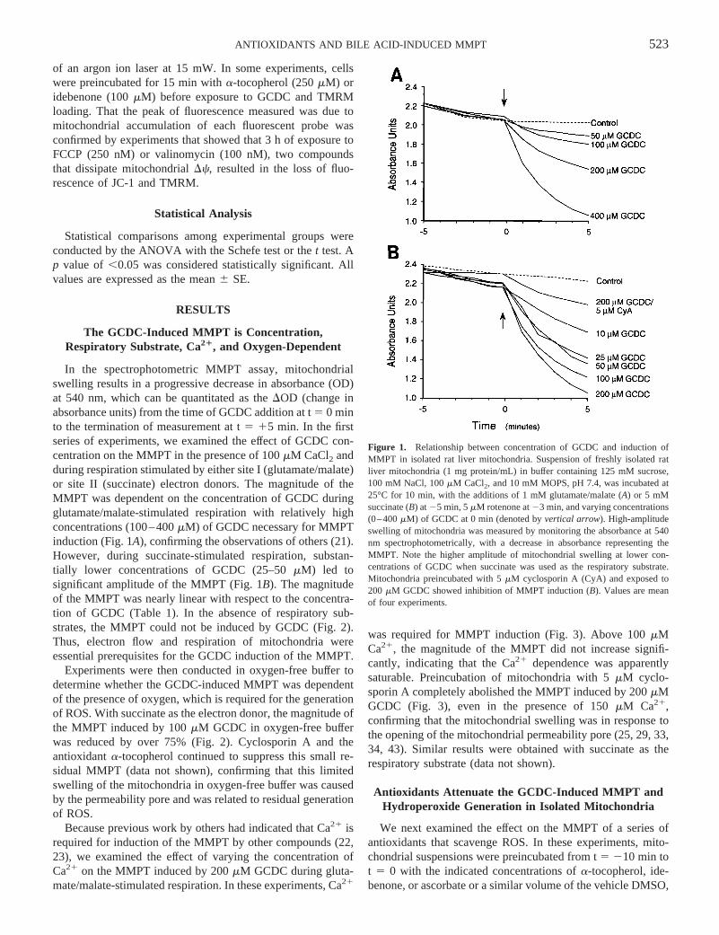
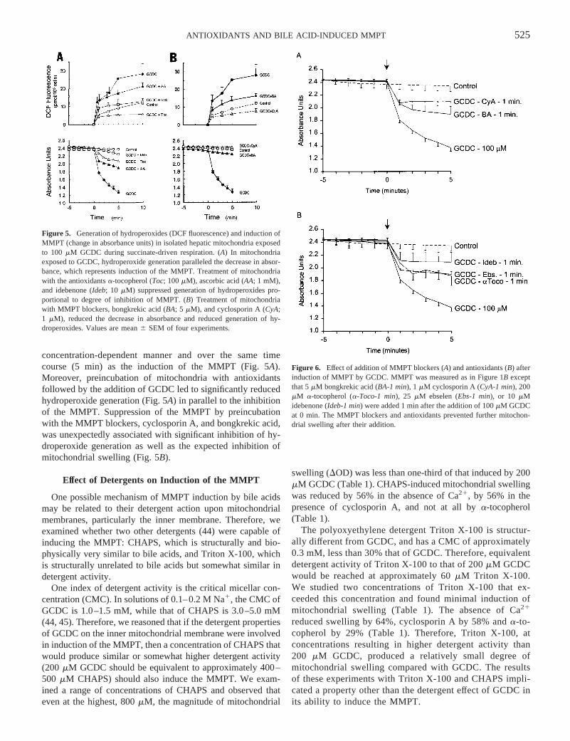
In the spectrophotometric MMPT assay, mitochondrialswelling results in a progressive decrease in absorbance (OD)at 540 nm, which can be quantitated as theDOD (change inabsorbance units) from the time of GCDC addition at t5 0 minto the termination of measurement at t5 15 min. In the firstseries of experiments, we examined the effect of GCDC con-centration on the MMPT in the presence of 100mM CaCl2 andduring respiration stimulated by either site I (glutamate/malate)or site II (succinate) electron donors. The magnitude of theMMPT was dependent on the concentration of GCDC duringglutamate/malate-stimulated respiration with relatively highconcentrations (100–400mM) of GCDC necessary for MMPTinduction (Fig. 1A), confirming the observations of others (21).However, during succinate-stimulated respiration, substan-tially lower concentrations of GCDC (25–50mM) led tosignificant amplitude of the MMPT (Fig. 1B). The magnitudeof the MMPT was nearly linear with respect to the concentra-tion of GCDC (Table 1). In the absence of respiratory sub-strates, the MMPT could not be induced by GCDC (Fig. 2).Thus, electron flow and respiration of mitochondria wereessential prerequisites for the GCDC induction of the MMPT.
Experiments were then conducted in oxygen-free buffer todetermine whether the GCDC-induced MMPT was dependentof the presence of oxygen, which is required for the generationof ROS. With succinate as the electron donor, the magnitude ofthe MMPT induced by 100mM GCDC in oxygen-free bufferwas reduced by over 75% (Fig. 2). Cyclosporin A and theantioxidanta-tocopherol continued to suppress this small re-sidual MMPT (data not shown), confirming that this limitedswelling of the mitochondria in oxygen-free buffer was causedby the permeability pore and was related to residual generationof ROS.
Because previous work by others had indicated that Ca21 isrequired for induction of the MMPT by other compounds (22,23), we examined the effect of varying the concentration ofCa21 on the MMPT induced by 200mM GCDC during gluta-mate/malate-stimulated respiration. In these experiments, Ca21
was required for MMPT induction (Fig. 3). Above 100mMCa21, the magnitude of the MMPT did not increase signifi-cantly, indicating that the Ca21 dependence was apparentlysaturable. Preincubation of mitochondria with 5mM cyclo-sporin A completely abolished the MMPT induced by 200mMGCDC (Fig. 3), even in the presence of 150mM Ca21,confirming that the mitochondrial swelling was in response tothe opening of the mitochondrial permeability pore (25, 29, 33,34, 43). Similar results were obtained with succinate as therespiratory substrate (data not shown).
Antioxidants Attenuate the GCDC-Induced MMPT andHydroperoxide Generation in Isolated Mitochondria
We next examined the effect on the MMPT of a series ofantioxidants that scavenge ROS. In these experiments, mito-chondrial suspensions were preincubated from t5 210 min tot 5 0 with the indicated concentrations ofa-tocopherol, ide-benone, or ascorbate or a similar volume of the vehicle DMSO,
Figure 1. Relationship between concentration of GCDC and induction ofMMPT in isolated rat liver mitochondria. Suspension of freshly isolated ratliver mitochondria (1 mg protein/mL) in buffer containing 125 mM sucrose,100 mM NaCl, 100mM CaCl2, and 10 mM MOPS, pH 7.4, was incubated at25°C for 10 min, with the additions of 1 mM glutamate/malate (A) or 5 mMsuccinate (B) at25 min, 5mM rotenone at23 min, and varying concentrations(0–400mM) of GCDC at 0 min (denoted byvertical arrow). High-amplitudeswelling of mitochondria was measured by monitoring the absorbance at 540nm spectrophotometrically, with a decrease in absorbance representing theMMPT. Note the higher amplitude of mitochondrial swelling at lower con-centrations of GCDC when succinate was used as the respiratory substrate.Mitochondria preincubated with 5mM cyclosporin A (CyA) and exposed to200 mM GCDC showed inhibition of MMPT induction (B). Values are meanof four experiments.
523ANTIOXIDANTS AND BILE ACID-INDUCED MMPT
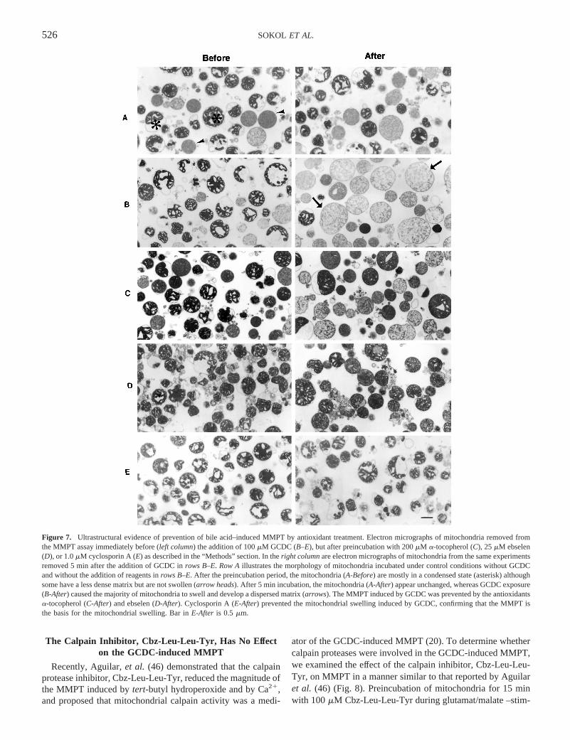
and the protocol for the GCDC induction of the MMPT wasfollowed.a-Tocopherol inhibited the magnitude of the MMPT(DOD) in a dose-dependent manner during succinate- (Fig. 4)and glutamate/malate- (Table 1) stimulated respiration. In mi-tochondria exposed to 25 and 50mM GCDC, preincubationwith 200 mM a-tocopherol had a similar effect of reducingMMPT by 50–60% (data not shown). A series of other anti-oxidants also significantly inhibited the MMPT when preincu-bated before exposure of mitochondria to 100mM GCDC (Fig.5A). Of particular interest was the suppression of furtherchanges in absorbance when the antioxidants were added afterthe GCDC had initiated the onset of the MMPT (Fig. 6).
In three experiments, transmission electron microscopy wasperformed on mitochondria after isolation and before the ad-dition of GCDC and again 5 min after the addition of 100mMGCDC. GCDC induced significant swelling of mitochondriawith decreased matrix density and loss of visible cristae (Fig.7) and an increase in cross-sectional area per mitochondrion(Table 2). This swelling was significantly inhibited by pretreat-ment of the mitochondria with cyclosporin A and with theantioxidantsa-tocopherol and ebselen (Fig. 7, Table 2). Thesemorphologic data confirmed the findings of the spectrophoto-metric assay for mitochondrial swelling.
Experiments were conducted to determine the time course ofROS generation as related to the MMPT induced by GCDC.Employing the DCFein probe as an indicator of hydroperoxidegeneration, GCDC stimulated the generation of ROS in a
Figure 2. Effect of oxygen-free buffer, and absence of respiratory substrates,on MMPT induced by 100mM GCDC. MMPT experiment was conductedusing succinate as the respiratory substrate in buffer under ambient atmosphere(Control andGCDC-100mM) and in oxygen-free buffer with GCDC (GCDC-O2-free), and using no respiratory substrates with GCDC under ambientatmosphere (GCDC-Succ-Free). GCDC induction of the MMPT was depen-dent on the presence of oxygen and respiratory substrates. Values are mean ofthree experiments.
Figure 3. Induction of the MMPT by GCDC (200mM) requires calcium.Absorbance of mitochondrial suspension was monitored at 540 nm as in Figure1B, except that the concentration of calcium chloride (added at t5 25 min)was varied between 0 and 150mM and the concentration of GCDC added att 5 0 min (vertical arrow) was 200mM for all experiments. Values shown aremean of four experiments.
Figure 4. a-Tocopherol (Toc) inhibits the MMPT induced by GCDC duringsuccinate-stimulated respiration. MMPT experiment was conducted as inFigure 1B with the following modifications: Mitochondria were preincubatedfrom t 5 210 min to t 5 0 min with 0–200mM R, R, R-a-tocopheroldissolved in 0.5% DMSO, or the volume of 0.5% DMSO used for the highestconcentration ofa-tocopherol. At t5 0 min, 100 mM GCDC was added.a-Tocopherol significantly inhibited the MMPT in a concentration-dependentmanner. Values shown are the mean of three experiments.
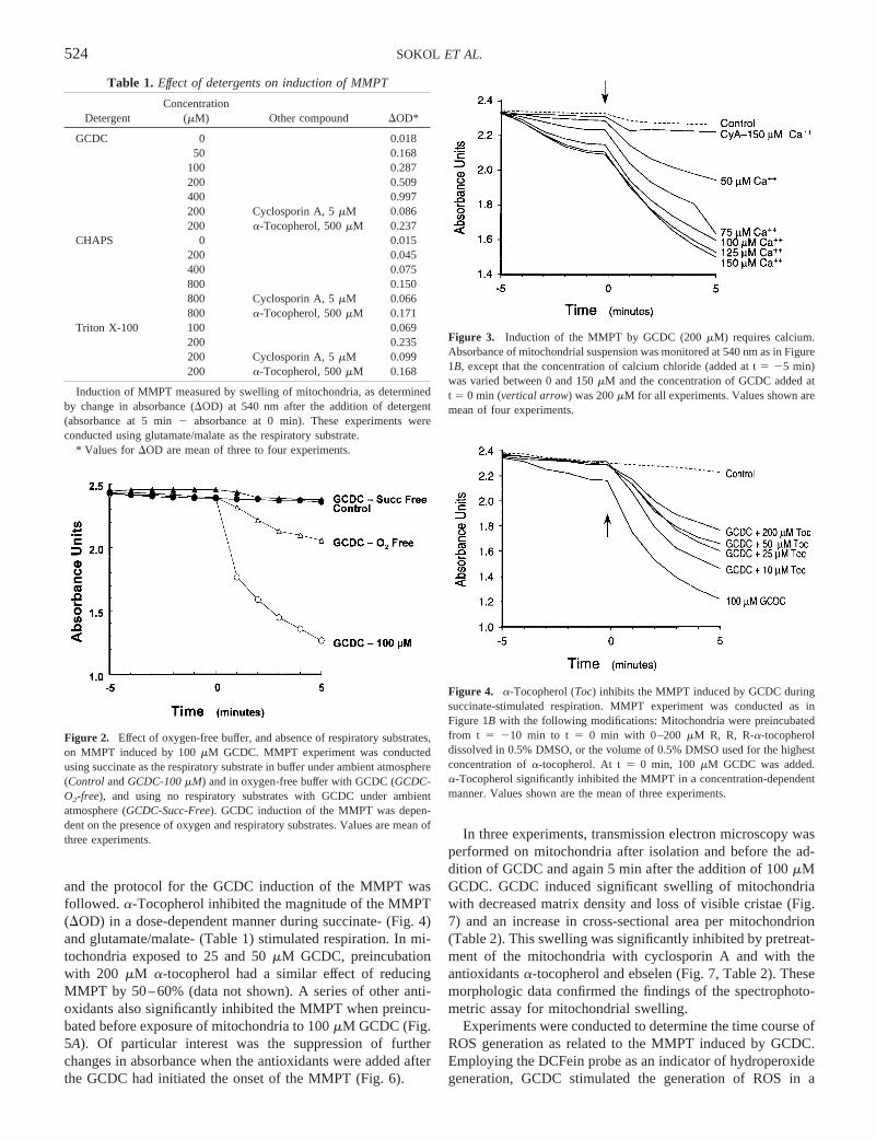
Table 1. Effect of detergents on induction of MMPT
DetergentConcentration
(mM) Other compound DOD*
GCDC 0 0.01850 0.168
100 0.287200 0.509400 0.997200 Cyclosporin A, 5mM 0.086200 a-Tocopherol, 500mM 0.237
CHAPS 0 0.015200 0.045400 0.075800 0.150800 Cyclosporin A, 5mM 0.066800 a-Tocopherol, 500mM 0.171
Triton X-100 100 0.069200 0.235200 Cyclosporin A, 5mM 0.099200 a-Tocopherol, 500mM 0.168
Induction of MMPT measured by swelling of mitochondria, as determinedby change in absorbance (DOD) at 540 nm after the addition of detergent(absorbance at 5 min2 absorbance at 0 min). These experiments wereconducted using glutamate/malate as the respiratory substrate.
* Values for DOD are mean of three to four experiments.
524 SOKOL ET AL.
concentration-dependent manner and over the same timecourse (5 min) as the induction of the MMPT (Fig. 5A).Moreover, preincubation of mitochondria with antioxidantsfollowed by the addition of GCDC led to significantly reducedhydroperoxide generation (Fig. 5A) in parallel to the inhibitionof the MMPT. Suppression of the MMPT by preincubationwith the MMPT blockers, cyclosporin A, and bongkrekic acid,was unexpectedly associated with significant inhibition of hy-droperoxide generation as well as the expected inhibition ofmitochondrial swelling (Fig. 5B).
Effect of Detergents on Induction of the MMPT
One possible mechanism of MMPT induction by bile acidsmay be related to their detergent action upon mitochondrialmembranes, particularly the inner membrane. Therefore, weexamined whether two other detergents (44) were capable ofinducing the MMPT: CHAPS, which is structurally and bio-physically very similar to bile acids, and Triton X-100, whichis structurally unrelated to bile acids but somewhat similar indetergent activity.
One index of detergent activity is the critical micellar con-centration (CMC). In solutions of 0.1–0.2 M Na1, the CMC ofGCDC is 1.0–1.5 mM, while that of CHAPS is 3.0–5.0 mM(44, 45). Therefore, we reasoned that if the detergent propertiesof GCDC on the inner mitochondrial membrane were involvedin induction of the MMPT, then a concentration of CHAPS thatwould produce similar or somewhat higher detergent activity(200 mM GCDC should be equivalent to approximately 400–500 mM CHAPS) should also induce the MMPT. We exam-ined a range of concentrations of CHAPS and observed thateven at the highest, 800mM, the magnitude of mitochondrial
swelling (DOD) was less than one-third of that induced by 200mM GCDC (Table 1). CHAPS-induced mitochondrial swellingwas reduced by 56% in the absence of Ca21, by 56% in thepresence of cyclosporin A, and not at all bya-tocopherol(Table 1).
The polyoxyethylene detergent Triton X-100 is structur-ally different from GCDC, and has a CMC of approximately0.3 mM, less than 30% that of GCDC. Therefore, equivalentdetergent activity of Triton X-100 to that of 200mM GCDCwould be reached at approximately 60mM Triton X-100.We studied two concentrations of Triton X-100 that ex-ceeded this concentration and found minimal induction ofmitochondrial swelling (Table 1). The absence of Ca21
reduced swelling by 64%, cyclosporin A by 58% anda-to-copherol by 29% (Table 1). Therefore, Triton X-100, atconcentrations resulting in higher detergent activity than200 mM GCDC, produced a relatively small degree ofmitochondrial swelling compared with GCDC. The resultsof these experiments with Triton X-100 and CHAPS impli-cated a property other than the detergent effect of GCDC inits ability to induce the MMPT.
Figure 5. Generation of hydroperoxides (DCF fluorescence) and induction ofMMPT (change in absorbance units) in isolated hepatic mitochondria exposedto 100 mM GCDC during succinate-driven respiration. (A) In mitochondriaexposed to GCDC, hydroperoxide generation paralleled the decrease in absor-bance, which represents induction of the MMPT. Treatment of mitochondriawith the antioxidantsa-tocopherol (Toc; 100mM), ascorbic acid (AA; 1 mM),and idebenone (Ideb; 10 mM) suppressed generation of hydroperoxides pro-portional to degree of inhibition of MMPT. (B) Treatment of mitochondriawith MMPT blockers, bongkrekic acid (BA; 5 mM), and cyclosporin A (CyA;1 mM), reduced the decrease in absorbance and reduced generation of hy-droperoxides. Values are mean6 SEM of four experiments.
Figure 6. Effect of addition of MMPT blockers (A) and antioxidants (B) afterinduction of MMPT by GCDC. MMPT was measured as in Figure 1B exceptthat 5mM bongkrekic acid (BA-1 min), 1 mM cyclosporin A (CyA-1 min), 200mM a-tocopherol (a-Toco-1 min), 25 mM ebselen (Ebs-1 min), or 10 mMidebenone (Ideb-1 min) were added 1 min after the addition of 100mM GCDCat 0 min. The MMPT blockers and antioxidants prevented further mitochon-drial swelling after their addition.
525ANTIOXIDANTS AND BILE ACID-INDUCED MMPT
The Calpain Inhibitor, Cbz-Leu-Leu-Tyr, Has No Effecton the GCDC-induced MMPT
Recently, Aguilar,et al. (46) demonstrated that the calpainprotease inhibitor, Cbz-Leu-Leu-Tyr, reduced the magnitude ofthe MMPT induced bytert-butyl hydroperoxide and by Ca21,and proposed that mitochondrial calpain activity was a medi-
ator of the GCDC-induced MMPT (20). To determine whethercalpain proteases were involved in the GCDC-induced MMPT,we examined the effect of the calpain inhibitor, Cbz-Leu-Leu-Tyr, on MMPT in a manner similar to that reported by Aguilaret al. (46) (Fig. 8). Preincubation of mitochondria for 15 minwith 100mM Cbz-Leu-Leu-Tyr during glutamat/malate –stim-
Figure 7. Ultrastructural evidence of prevention of bile acid–induced MMPT by antioxidant treatment. Electron micrographs of mitochondria removed fromthe MMPT assay immediately before (left column) the addition of 100mM GCDC (B–E), but after preincubation with 200mM a-tocopherol (C), 25mM ebselen(D), or 1.0mM cyclosporin A (E) as described in the “Methods” section. In theright columnare electron micrographs of mitochondria from the same experimentsremoved 5 min after the addition of GCDC inrows B–E. Row Aillustrates the morphology of mitochondria incubated under control conditions without GCDCand without the addition of reagents inrows B–E. After the preincubation period, the mitochondria (A-Before) are mostly in a condensed state (asterisk) althoughsome have a less dense matrix but are not swollen (arrow heads). After 5 min incubation, the mitochondria (A-After) appear unchanged, whereas GCDC exposure(B-After) caused the majority of mitochondria to swell and develop a dispersed matrix (arrows). The MMPT induced by GCDC was prevented by the antioxidantsa-tocopherol (C-After) and ebselen (D-After). Cyclosporin A (E-After) prevented the mitochondrial swelling induced by GCDC, confirming that the MMPT isthe basis for the mitochondrial swelling. Bar inE-After is 0.5 mm.
526 SOKOL ET AL.
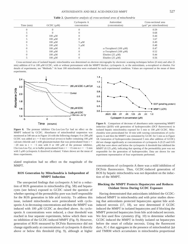
ulated respiration had no effect on the magnitude of theMMPT.
ROS Generation by Mitochondria is Independent ofMMPT Induction
The unexpected findings that cyclosporin A led to a reduc-tion of ROS generation in mitochondria (Fig. 5B) and hepato-cytes (see below) exposed to GCDC raised the question ofwhether opening of the permeability pore was itself responsiblefor the ROS generation in bile acid toxicity. To address thisissue, isolated mitochondria were preincubated with cyclo-sporin A in decreasing concentrations and then the MMPT wasinduced with 100mM GCDC as described above. As cyclo-sporin A concentrations were reduced, a clear threshold wasreached in four separate experiments, below which there wasno inhibition of the GCDC-induced MMPT (Fig. 9). However,generation of ROS measured by DCFein fluorescence did notchange significantly at concentrations of cyclosporin A directlyabove or below this threshold (Fig. 9), although at higher
concentrations of cyclosporin A there was a mild inhibition ofDCFein fluorescence. Thus, GCDC-induced generation ofROS by hepatic mitochondria was not dependent on the induc-tion of the MMPT.
Blocking the MMPT Protects Hepatocytes and ReducesOxidant Stress During GCDC Exposure
Having demonstrated that antioxidants inhibited the GCDC-induced MMPT in mitochondria and with prior studies show-ing that antioxidants protected hepatocytes against bile acid-induced necrosis (17, 18), we next determined if GCDCinduced the MMPT in isolated hepatocytes and if blocking theMMPT protected hepatocytes from bile acid-induced necrosis.We first used flow cytometry (Fig. 10) to determine whetherGCDC induced the MMPT in freshly isolated rat hepatocytesbefore the onset of cellular necrosis. Using the fluorescentdyes, JC-1 that aggregates in the presence of mitochondrialDcand TMRM which accumulates in mitochondria proportional
Table 2. Quantitative analysis of cross-sectional area of mitochondria
Time (min) GCDC (mM)Cyclosporin Aconcentration
Antioxidant(concentration)
Cross-sectional area(mm2 per mitochondrion)
0 0 0 0.595 0 0 0.680 100mM 0 0.445 100mM 0 1.670 100mM 5 mM 0.495 100mM 5 mM 0.480 100mM 0 a-Tocopherol (100mM) 0.475 100mM 0 a-Tocopherol (100mM) 0.520 100mM 0 Ebselen (25mM) 0.445 100mM 0 Ebselen (25mM) 0.56
Cross-sectional area of isolated hepatic mitochondria was determined on electron micrographs by electronic scanning techniques before (0 min) andafter (5min) addition of 0 or 100mM GCDC, with or without pretreatment with the MMPT blocker, cyclosporin A, or the antioxidants,a-tocopherol or ebselen. Fordetails of experiments, see “Methods.” At least 100 mitochondria were evaluated for each experimental condition. Values are expressed at the mean ofthreeexperiments.
Figure 8. The protease inhibitor Cbz-Leu-Leu-Tyr had no effect on theMMPT induced by GCDC. Absorbance of mitochondrial suspension wasmonitored at 540 nm as in Figure 1A with the following changes: 0 or 200mMGCDC was added at t5 0 min (vertical arrow) to buffer containing 100mMcalcium chloride and 1 mM glutamate/malate that was preincubated from t5220 min to t 5 25 min with 0 or 100 mM of the protease inhibitor,Cbz-Leu-Leu-Tyr, or to buffer preincubated from t5 215 min to t5 25 minwith 5 mM cyclosporin A dissolved in ethanol. Values shown are the mean ofthree experiments.
Figure 9. Comparison of decrease of absorbance units representing MMPTinduction (DOD) with generation of hydroperoxides (DCF fluorescence) inisolated hepatic mitochondria exposed for 5 min to 100mM GCDC. Mito-chondria were preincubated for 10 min with varying concentrations of cyclo-sporin A and then the MMPT was stimulated by GCDC for 5 min as in Figure1B. Generation of hydroperoxides (measured 5 min after addition of GCDC)did not change significantly at concentrations of cyclosporin A (0.25 and 0.05mM) that were above and below the cyclosporin A threshold that inhibited theMMPT (0.125mM), indicating that opening of the permeability pore was notresponsible for the generation of hydroperoxides. Data are shown for oneexperiment representative of four experiments performed.
527ANTIOXIDANTS AND BILE ACID-INDUCED MMPT
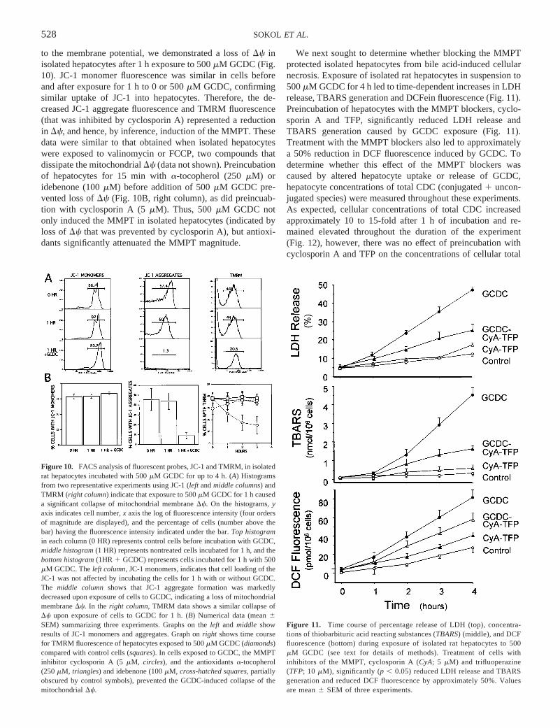
to the membrane potential, we demonstrated a loss ofDc inisolated hepatocytes after 1 h exposure to 500mM GCDC (Fig.10). JC-1 monomer fluorescence was similar in cells beforeand after exposure for 1 h to 0 or 500mM GCDC, confirmingsimilar uptake of JC-1 into hepatocytes. Therefore, the de-creased JC-1 aggregate fluorescence and TMRM fluorescence(that was inhibited by cyclosporin A) represented a reductionin Dc, and hence, by inference, induction of the MMPT. Thesedata were similar to that obtained when isolated hepatocyteswere exposed to valinomycin or FCCP, two compounds thatdissipate the mitochondrialDc (data not shown). Preincubationof hepatocytes for 15 min witha-tocopherol (250mM) oridebenone (100mM) before addition of 500mM GCDC pre-vented loss ofDc (Fig. 10B, right column), as did preincuab-tion with cyclosporin A (5mM). Thus, 500mM GCDC notonly induced the MMPT in isolated hepatocytes (indicated byloss ofDc that was prevented by cyclosporin A), but antioxi-dants significantly attenuated the MMPT magnitude.
We next sought to determine whether blocking the MMPTprotected isolated hepatocytes from bile acid-induced cellularnecrosis. Exposure of isolated rat hepatocytes in suspension to500mM GCDC for 4 h led to time-dependent increases in LDHrelease, TBARS generation and DCFein fluorescence (Fig. 11).Preincubation of hepatocytes with the MMPT blockers, cyclo-sporin A and TFP, significantly reduced LDH release andTBARS generation caused by GCDC exposure (Fig. 11).Treatment with the MMPT blockers also led to approximatelya 50% reduction in DCF fluorescence induced by GCDC. Todetermine whether this effect of the MMPT blockers wascaused by altered hepatocyte uptake or release of GCDC,hepatocyte concentrations of total CDC (conjugated1 uncon-jugated species) were measured throughout these experiments.As expected, cellular concentrations of total CDC increasedapproximately 10 to 15-fold after 1 h of incubation and re-mained elevated throughout the duration of the experiment(Fig. 12), however, there was no effect of preincubation withcyclosporin A and TFP on the concentrations of cellular total
Figure 10. FACS analysis of fluorescent probes, JC-1 and TMRM, in isolatedrat hepatocytes incubated with 500mM GCDC for up to 4 h. (A) Histogramsfrom two representative experiments using JC-1 (left andmiddle columns) andTMRM (right column) indicate that exposure to 500mM GCDC for 1 h causeda significant collapse of mitochondrial membraneDc. On the histograms,yaxis indicates cell number,x axis the log of fluorescence intensity (four ordersof magnitude are displayed), and the percentage of cells (number above thebar) having the fluorescence intensity indicated under the bar.Top histogramin each column (0 HR) represents control cells before incubation with GCDC,middle histogram(1 HR) represents nontreated cells incubated for 1 h, and thebottom histogram(1HR 1 GCDC) represents cells incubated for 1 h with 500mM GCDC. Theleft column, JC-1 monomers, indicates that cell loading of theJC-1 was not affected by incubating the cells for 1 h with or without GCDC.The middle columnshows that JC-1 aggregate formation was markedlydecreased upon exposure of cells to GCDC, indicating a loss of mitochondrialmembraneDc. In the right column, TMRM data shows a similar collapse ofDc upon exposure of cells to GCDC for 1 h. (B) Numerical data (mean6SEM) summarizing three experiments. Graphs on theleft and middle showresults of JC-1 monomers and aggregates. Graph onright shows time coursefor TMRM fluorescence of hepatocytes exposed to 500mM GCDC (diamonds)compared with control cells (squares). In cells exposed to GCDC, the MMPTinhibitor cyclosporin A (5mM, circles), and the antioxidantsa-tocopherol(250mM, triangles) and idebenone (100mM, cross-hatched squares, partiallyobscured by control symbols), prevented the GCDC-induced collapse of themitochondrialDc.
Figure 11. Time course of percentage release of LDH (top), concentra-tions of thiobarbituric acid reacting substances (TBARS) (middle), and DCFfluorescence (bottom) during exposure of isolated rat hepatocytes to 500mM GCDC (see text for details of methods). Treatment of cells withinhibitors of the MMPT, cyclosporin A (CyA; 5 mM) and trifluoperazine(TFP; 10 mM), significantly (p , 0.05) reduced LDH release and TBARSgeneration and reduced DCF fluorescence by approximately 50%. Valuesare mean6 SEM of three experiments.
528 SOKOL ET AL.
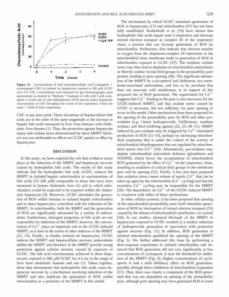
CDC at any time point. These elevations of hepatocellular bileacids are in the order of the same magnitude as the increase inhepatic bile acids measured in liver from humans with chole-static liver disease (1). Thus, the protection against hepatocyteinjury and oxidant stress demonstrated by these MMPT block-ers was not attributable to effects on GCDC uptake or efflux byhepatocytes.
DISCUSSION
In this study, we have explored the role that oxidative stressplays in the induction of the MMPT and hepatocyte necrosiscaused by hydrophobic bile acids. The results of this studyindicate that the hydrophobic bile acid, GCDC, induces theMMPT in isolated hepatic mitochondria at concentrations ofbile acids (25–200mM) comparable to those that have beenmeasured in human cholestatic liver (1) and to which mito-chondria would be expected to be exposed within the choles-tatic hepatocyte (4). Moreover, GCDC stimulates the genera-tion of ROS within minutes in isolated hepatic mitochondriaand in intact hepatocytes, coincident with the induction of theMMPT. In mitochondria, both the MMPT and the generationof ROS are significantly attenuated by a variety of antioxi-dants. Furthermore, detergent properties of bile acids are notresponsible for induction of the MMPT, however, the concen-tration of Ca21 plays an important role in the GCDC-inducedMMPT, as it does in the action of other inducers of the MMPT(22, 23). Finally, in freshly isolated rat hepatocytes, GCDCinduces the MMPT and hepatocellular necrosis, antioxidantsinhibit the MMPT and blockers of the MMPT provide strongprotection against cellular necrosis caused by exposure toGCDC. The bile acid concentrations achieved in these hepa-tocytes exposed to 500mM GCDC for 4 h are in the range ofliver from cholestatic humans and rats (1). Taken together,these data demonstrate that hydrophobic bile acids cause he-patocyte necrosis by a mechanism involving induction of theMMPT and also implicate the generation of ROS withinmitochondria as a promoter of the MMPT in this model.
The mechanism by which GCDC stimulates generation ofROS in hepatocytes (17) and mitochondria (47) has not beenfully established. Krahenbuhlet al. (19) have shown thathydrophobic bile acids impair state 3 respiration and interruptnormal electron transport at complex III of the respiratorychain, a process that can increase generation of ROS bymitochondria. Preliminary data indicate that electron transferto oxygen from the ubquinone-complex III interaction in themitochondrial inner membrane leads to generation of ROS inmitochondria exposed to GCDC (47). The resultant oxidantstress may then lead to depletion of mitochondrial antioxidantsor directly oxidize vicinal thiol groups in the permeability poreprotein, leading to pore opening (48). The significant attenua-tion of the MMPT bya-tocopherol and idebenone, two mem-brane-associated antioxidants, and less so by ascorbate thatdoes not associate with membranes, is in support of thisproposed site of ROS generation. The requirement for Ca21
suggests that Ca21 binding to the pore is also necessary for theGCDC-induced MMPT, and that oxidant stress caused byGCDC is necessary, but not sufficient, for pore opening tooccur in this model. Other mechanisms have been proposed forthe opening of the permeability pore by ROS and other pro-oxidants (e.g. t-butyl hydroperoxide, Fe(II)citrate, xanthineoxidase, and thiol-oxidizing agents) (22, 23, 49–51). MMPTinduced by pro-oxidants may be triggered by Ca21-stimulatedproduction of ROS (51–53), perhaps by increasing mitochon-drial respiration that is under the control of the activity ofmitochondrial dehydrogenases that are regulated by mitochon-drial matrix free Ca21 (54). Alternatively, pro-oxidants maydeplete mitochondrial antioxidant defenses (glutathione andNADPH), which favors the accumulation of mitochondrialROS generated by the effect of Ca21 on the respiratory chain,resulting in oxidation of critical thiol groups in the membranepore and its opening (52). Finally, it has also been proposedthat oxidative stress causes release of matrix Ca21 that can betaken up again by the mitochondria (Ca21 cycling) and that theexcessive Ca21 cycling may be responsible for the MMPT(50). The dependence on Ca21 of the GCDC-induced MMPTis consistent with either of these mechanisms.
In other cellular systems, it has been proposed that openingof the mitochondrial permeability pore itself stimulates gener-ation of ROS by interruption of normal electron transport (55)caused by the release of mitochondrial cytochromec to cytosol(56). In our studies, chemical blockade of the MMPT inhepatocytes exposed to GCDC resulted in a modest reductionof hydroperoxide generation in association with protectionagainst necrosis (Fig. 11). In addition, ROS generation inisolated mitochondria paralleled the opening of the MMPT(Fig. 5). We further addressed this issue by performing adose-response experiment in isolated mitochondria and ob-served that ROS generation did not vary significantly at lowconcentrations of cyclosporin A near the threshold for inhibi-tion of the MMPT (Fig. 9). Higher concentrations of cyclo-sporin A had a mild inhibitory effect on ROS generation,possibly through direct inhibition of mitochondrial respiration(57). Thus, there was clearly a component of the ROS gener-ated that was not dependent on opening of the permeabilitypore, although pore opening may have generated ROS to some
Figure 12. Concentrations of total chenodeoxycholic acid (conjugated1unconjugated CDC) in isolated rat hepatocytes exposed to 500mM GCDCover 4 h. CDC concentrations were measured by gas chromatography–massspectrometry as detailed in “Methods.” Treatment of cells with 5mM cyclo-sporin A (CyA) and 10mM trifluoperazine (TFP) did not reduce hepatocyteconcentrations of CDC throughout the course of the experiment. Values aremean6 SEM of three experiments.
529ANTIOXIDANTS AND BILE ACID-INDUCED MMPT
extent, as well. In other models, stimulation of the MMPTpromotes ROS generation by mitochondria, possibly throughthe release of cytochromec (56). In mitochondria depleted incytochromec, the respiratory chain protein complexes up-stream of cytochromec become highly reduced and directlytransfer their electrons to oxygen, forming superoxide (56). Ithas been proposed that this autocatalytic mechanism mightexplain the synchronization of MMPT onset that has beenobserved by confocal microscopy in single hepatocytes afterexposure totert-butyl hydroperoxide (26).
We determined whether the detergent properties of GCDCon mitochondrial membrane structure could account for theinduction of the MMPT in our model system by testing twoother detergents. Inasmuch as hydrophobic bile acids are ca-pable of solubilizing both membrane lipids and proteins, wechose a detergent (CHAPS) that would solubilize primarilymembrane lipids and another (Triton X-100) that would solu-bilize both membrane lipids and proteins (44). Neither deter-gent induced significant high-amplitude mitochondrial swell-ing at concentrations equivalent to 200mM GCDC (based onrelative CMC equivalency), and did so minimally at higherconcentrations, suggesting that the detergent action of GCDCon the mitochondrial membrane plays a minor role, if any, inthe induction of the MMPT.
Aguilar et al. (46) recently reported the presence of calpain-like protease activity in hepatocyte mitochondria, whichseemed to be involved in the MMPT induced by Ca21 and byt-butyl hydroperoxide, an agent that produces oxidative stressand hepatocyte necrosis (46, 58). These investigators demon-strated that the cysteine protease inhibitor, Cbz-Leu-Leu-Tyr(100 mM), inhibited mitochondrial high-amplitude swellinginduced by both 100mM Ca21 and by 50mM t-butyl hydroper-oxide, and that preincubation with Cbz-Leu-Leu-Tyr delayedthe loss of the mitochondrial membrane potential and the onsetof hepatocyte necrosis caused byt-butyl hydroperoxide (46).To determine whether similar calpain-like activity played arole in the GCDC-induced MMPT observed in our experi-ments, we preincubated mitochondria for 15 min with Cbz-Leu-Leu-Tyr and then exposed the mitochondria to 200mMGCDC in the presence of 100mM Ca21. Contrary to theobservations of Aguilaret al.(46), we observed no effect of theprotease inhibitor on the MMPT in our model (Fig. 8). Thus, itis unlikely that mitochondrial calpain-like protease activityplays a major role in the MMPT induced by GCDC.
It is recognized that hepatocellular necrosis is not the onlymechanism of cell death induced by toxic bile acids. Patelet al.(7) have reported that low concentrations of hydrophobic bileacids can induce apoptosis in primary cultured rat hepatocytes.The Fas signaling (36) and protein kinase C pathways (35)have been implicated in bile acid–induced apoptosis. In apo-ptosis, it is possible that direct activation of Fas by bile acids(36) causes caspase 8 activation that truncates bid, a member ofthe bcl-2 family, that translocates to mitochondria where it mayopen the permeability pore by unknown mechanisms, inducingROS generation (59). Recent studies have suggested that theincreased activation of Fas by bile acids may be mediated bythe promotion of cytoplasmic transport of Fas to the cellsurface by a Golgi- and microtubule-dependent pathway (60).
Preliminary data from our laboratory demonstrate involvementof ROS generation and induction of the MMPT during bileacid–induced apoptosis (61, 62). The beneficial effects ofursodeoxycholic acid, a hydrophilic bile acid, in reducinghepatocyte apoptosis may also be related to reduction of ROSgeneration in mitochondria and inhibition of the MMPT (37).However, Benzet al. (63) recently provided evidence that theFas receptor pathway may not be involved in apoptosis stim-ulated by GCDC in human hepatocytes in primary culture.Thus, elucidating the relationships between oxidant stress andthe activation of the Fas receptor, bid and bax translocation tothe mitochondria, induction of the MMPT, cytochromec re-lease from mitochondria, and activation of caspases in bileacid–induced apoptosis requires further investigation.
In conclusion, in this study we have demonstrated thatconcentrations of GCDC representative of those that accumu-late in the cholestatic liver (1, 4) induce the MMPT in hepaticmitochondria by a mechanism dependent on the generation ofROS and the presence of Ca21. Because antioxidants havepreviously been shown to prevent hepatocellular necrosis andreduce oxidant stress in isolated hepatocytes exposed to hy-drophobic bile acids (17, 18), and, in the current study, toinhibit dissipation of mitochondrialDc, it is postulated thatinduction of the MMPT by ROS generated in hepatocytemitochondria (19, 47) is a critical event promoting bile acid–induced hepatocyte necrosis. Elevation of the cytosolic freeCa21 concentration induced by hydrophobic bile acids (64)may also be an important permissive factor that allows theoxidant stress to open the permeability pore. Thus, novelapproaches to reduce the generation of mitochondrial-derivedROS or to prevent increases in mitochondrial Ca21 concentra-tion may have a beneficial effect in human liver diseasesassociated with the accumulation of hydrophobic bile acids.The concentrations of bile acids achieved in the isolated hepa-tocyte experiments and used in the mitochondrial experimentsin this study are in the range of those measured in liver fromhumans with cholestasis (1, 65), making the findings reportedof potential clinical relevance. The inhibition of the MMPTdemonstrated in our study when antioxidants were added aftermitochondrial exposure to GCDC suggests that this therapeuticstrategy could be of potential benefit, even after the onset ofcholestasis and the hepatic accumulation of bile acids. Therelative contribution of cellular necrosisversusapoptosis toliver injury in clinical cholestasis has yet to be determined,however, prevention of oxidant stress and inhibition of theMMPT may be possible strategies to reduce both kinds ofcellular injury in cholestasis.
REFERENCES
1. Greim H, Czygan P, Schaffner F, Popper H 1973 Determination of bile acids in needlebiopsies of human liver. Biochem Med 8:280–286
2. Attili F, Angelico M, Cantafora A, Alvaro D, Capocaccia L 1986 Bile acid-inducedliver toxicity: relation to the hydophobic-hydrophilic balance of bile acids. MedHypothesis 19:57–69
3. Armstrong MJ, Carey MC 1982 The hydrophobic-hydrophilic balance of bile salts.Inverse correlation between reverse-phase high performance liquid chromatographicmobilities and micellar cholesterol-solubilizing capacities. J Lipid Res 23:70–80
4. Spivey JR, Bronk SG, Gores GJ 1993 Glycochenodeoxycholate-induced lethal cellinjury in rat hepatocytes. J Clin Invest 92:17–24
5. Galle PR, Theilmann L, Raedsch R, Otto G Stiehl A 1990 Ursodeoxycholate reduceshepatotoxicity of bile salts in primary human hepatocytes. Hepatology 12:486–491
530 SOKOL ET AL.
6. Sokol RJ, McKim Jr JM, Goff MC, Devereaux MW, Ruyle SZ, Han D, Packer L,Everson G 1998 Vitamin E reduces oxidant injury to mitochondria and hepatotoxicityof intravenous taurochenodeoxycholic acid in the rat. Gastroenterology 114:164–174
7. Patel T, Bronk SF, Gores GJ 1994 Increases of intracellular magnesium promoteglycodeoxycholate-induced apoptosis in rat hepatocytes. J Clin Invest 94:2183–2192
8. Rosser BG, Gores GJ 1995 Liver cell necrosis: cellular mechanisms and clinicalimplications. Gastroenterology 108:252–275
9. Gores GJ, Herman B, Lemasters JJ 1990 Plasma membrane bleb formation andrupture: a common feature of hepatocellular injury. Hepatology 11:690–698.
10. Phillips MJ 1994 Mechanisms and morphology of cholestasis. In: Suchy FJ (ed).Liver Disease in Children. CV Mosby, St. Louis, pp 129–144
11. Scheuer PJ 1980 Liver Biopsy Interpretation. Bailliere Tindall, London, pp 36–5912. Dahm LJ, Hewett JA, Roth RA 1988 Bile and bile salts potentiate superoxide anion
release from activated rat peritoneal neutrophils. Toxicol Appl Pharmacol 95:82–9213. Togashi H, Shinzawa H, Wakabayoshi H, Nakamura T, Yamada N, Takahashi T,
Ishikawa M 1990 Activities of free oxygen radical scavenger enzymes in human liver.J Hepatology 11:200–205
14. Iritani N, Fukuda E, Kitamura Y 1980 Effect of corn oil feeding on lipid peroxidationin rats. J Nutr 110:924–930
15. Lemonnier F, Cresteil D, Feueant M, Couturier M, Bernard O, Alagille D 1987Plasma lipid peroxides in cholestatic children. Acta Paediatr Scand 76:928–934
16. Sokol RJ, Devereaux M, Khandwala RA 1991 Effect of dietary lipid and vitamin Eon mitochondrial lipid peroxidation and hepatic injury in the bile duct-ligated rat. JLipid Res 32:1349–1357
17. Sokol RJ, Devereaux M, Khandwala R, O’Brien K 1993 Evidence for involvement ofoxygen free radicals in bile acid toxicity to isolated rat hepatocytes. Hepatology17:869–881
18. Sokol RJ, Winklhofer-Roob BM, Devereaux MW, McKim Jr JM 1995 Generation ofhydroperoxides in isolated rat hepatocytes and hepatic mitochondria exposed tohydrophobic bile acids. Gastroenterology 109:1249–1256
19. Krähenbühl S, Talos C, Fischer S, Reichen J 1994 Toxicity of bile acids on theelectron transport chain of isolated rat liver mitochondria. Hepatology 19:471–479
20. Gores GJ, Miyoshi H, Botla R, Aguilar HI, Bronk SF 1998 Induction of themitochondrial permeability transition as a mechanism of liver injury during cholesta-sis: a potential role for mitochondrial proteases. Bioch Biophys Acta 1366:167–175
21. Botla R, Spivey JR, Aguilar H, Bronk SF, Gores GJ 1995 Ursodeoxycholate (UDCA)inhibits the mitochondrial membrane permeability transition induced by glycocheno-deoxycholate: a mechanism of UDCA cytoprotection. J Pharmacol Exp Ther272:930–938
22. Gunter TE, Pfeiffer DR 1990 Mechanisms by which mitochondria transport calcium.Am J Physiol 258:C755–C786
23. Zoratti M, Szabo I 1995 The mitochondrial permeability transition. Biochim BiophysActa 1241:139–176
24. Bernardi P, Vassanelli S, Veronese P, Colonna R, Szabo I, Zoratti M 1992 Modula-tion of the mitochondrial permeability transition pore. J Biol Chem 267:2934–2939
25. Pastorino JG, Snyder JW, Serroni A, Hoek JB, Farber JL 1993 Cyclosporin andcarnitine prevent the anoxic death of cultured hepatocytes by inhibiting the mito-chondrial permeability transition. J Biol Chem 268:13791–13798
26. Nieminen A-L, Byrne AM, Herman B, Lemasters JJ 1997 Mitochondrial permeabilitytransition in hepatocytes induced by t-BuOOH: NAD(P)H and reactive oxygenspecies. Am J Physiol 272:C1286–C1294
27. Lemasters JJ, Nieminen AL, Qian T, Trost LC, Elmore SP, Nishimura Y, Crowe RA,Cascio WE, Bradham CA, Brenner DA, Herman B 1998 The mitochondrial perme-ability transition in cell death: a common mechanism in necrosis, apoptosis andautophagy. Biochim Biophys Acta 1366:177–196
28. Susin SA, Zamzami N, Kroemer G 1998 Mitochondria as regulators of apoptosis:doubt no more. Biochim Biophys Acta 1366:151–165
29. Szabo I, Zoratti M 1993 The mitochondrial permeability transition pore may compriseVDAC molecules I: binary structure and voltage dependence of the pore. FEBS Lett330:201–205
30. Szabo I, De Pinto V, Zoratti M 1993 The mitochondrial permeability transition poremay comprise VDAC molecules II: the electrophysiological properties of VDAC arecompatible with those of the mitochondrial megachannel. FEBS Lett 330:206–210
31. Brustovetsky N, Klingenberg M 1996 Mitochondrial ADP/ATP carrier can bereversibly converted into a large channel by Ca21. Biochemistry 35:8483–8488
32. Woodfield K, Ruck A, Brdiczka D, Halestrap AP 1998 Direct demonstration of aspecific interaction between cyclophilin-D and the adenine nucleotide translocaseconfirms their role in the mitochondrial permeability transition. Biochem J336:287–90
33. Fournier N, Ducet G, Crevat A 1987 Action of cyclosporin on mitochondrial calciumfluxes. J Bioenerg Biomembr 19:297–303
34. Broekemeier KM, Dempsey ME, Pfeiffer DR 1989 Cylcosporin A is a potent inhibitorof the inner membrane permeability transition in liver mitochondria. J Biol Chem264:7826–7830
35. Jones BA, Rao Y-P, Stravitz T, Gores GJ 1997 Bile salt-induced apoptosis ofhepatocytes involves activation of protein kinase C. Am J Physiol 272:G1109–G1115
36. Faubion WA, Guicciardi ME, Miyoshi H, Bronk SF, Roberts PJ, Svingen PA,Kaufmann SH, Gores GJ 1999 Toxic bile salts induce rodent hepatocyte apoptosis viadirect activation of Fas. J Clin Invest 103:137–145
37. Rodrigues CM, Fan G, Wong PY, Kren BT, Steer CJ 1998 Ursodeoxycholic acid mayinhibit deoxycholic acid-induced apoptosis by modulating mitochondrial transmem-brane potential and reactive oxygen species production. Mol Med 4:165–178
38. Massari S, Frigeri S, Azzone GF 1972 Permeability to water, dimension of surface,and structural changes during swelling in rat liver mitochondria. J Membr Biol9:67–70
39. Liang D, Hagenbuch B, Steiger B, Meier PJ 1993 Parallel decrease of Na1-taurocholate cotransport and its encoding mRNA in primary cultures of hepatocytes.Hepatology 18:1162–1166
40. Glascott PA, Glifor E, Farber JL 1992 Effects of vitamin E on the killing of culturedhepatocytes by t-butyl hydroperoxide. Mol Pharmac 41:1155–1162
41. Everson GT, Daggy B, McKinley C, Story JA 1992 Effects of psyllium hydrophilicmucilloid on LDL-cholesterol and bile acid synthesis in hypercholesterolemic man. JLipid Res 33:1183–1192
42. Salvioli S, Ardizzoni A, Franceschi C, Cassarizza A 1997 JC-1, but not DiOC6(3) orrhodamine 123, is a reliable fluorescent probe to assessDc changes in intact cells:implications for studies on mitochondrial functionality during apoptosis. FEBS Lett411:77–82
43. Bernardi P, Petronelli V 1996 The permeability transition pore as a mitochondrialcalcium release channel: a critical appraisal. J Bioenerg Biomemb. 268:1005–1010
44. Neugebauer JM 1990 Detergents: an overview. In: Deutscher MP (ed) Guide toProtein Purification. Academic Press, San Diego, pp 239–82
45. Cabral DJ, Small DM 1989 Physical chemistry of bile. In: Schultz SG, Forte JG,Rauner BB (eds) Handbook of Physiology—The Gastrointestinal System III. WaverlyPress, Baltimore, pp 621–662
46. Aguilar HI, Botla R, Arora AS, Bronk SF, Gores GJ 1996 Induction of the mito-chondrial permeability transition by protease activity in rats: a mechanism of hepa-tocyte necrosis. Gastroenterology 110:558–566
47. Winklhofer-Roob BM, McKim Jr JM, Devereaux MW, Sokol RJ 1996 Characteriza-tion of site of reactive oxygen species generation in mitochondria exposed toglycochenodeoxycholic acid. Hepatology 24:338A
48. Costantini P, Chernyak BV, Petronilli V, Bernardi P 1996 Modulation of themitochondrial permeability transition pore by pyridine nucleotides and dithiol oxida-tion at two separate sites. J Biol Chem 271:6746–6751
49. Fagian MM, Pereira-da-Silva L, Martins LS, Vercesi AE 1990 Membrane proteinthiol cross-linking associated with the permeabilization of the inner mitochondrialmembrane by Ca21 plus prooxidants. J Biol Chem 265:19955–19960
50. Takeyama N, Matsuo N, Tanaka T 1993 Oxidative damage to mitochondria ismediated by the Ca21-dependent inner membrane permeability transition. Biochem J294:719–725
51. Kowaltowski AJ, Castilho RF, Vercesi AE 1996 Opening of the mitochondrialpermeability transition pore by uncoupling or inorganic phosphate in the presence ofCa21 is dependent on mitochondrial-generated reactive oxygen species. FEBS Lett378:150–152
52. Castilho RF, Kowaltowski AJ, Meinicke AR, Bechara EJH, Vercesi AE 1995Permeabilization of the inner mitochondrial membrane by Ca21 ions is stimulated byt-butyl hydroperoxide and mediated by reactive oxygen species generated by mito-chondria. Free Rad Biol Med 18:479–486
53. Bernardes CF, Meyer-Fernandes JR, Basseres DS, Castilho RF, Vercesi AE 1994Ca21-dependent permeabilization of the inner mitochondrial membrane by 4,4'-diisothiocyanato-stilbene-2,2'disulfonic acid (DIDS). Biochim Biophys Acta1188:93–100
54. Denton RM, McCormack JG 1980 On the role of the calcium transport cycle in heartand other mammalian mitochondria. FEBS Lett 199:1–8
55. Kowaltowski AJ, Castilho RF, Vercesi AE 1995 Ca21-induced mitochondrial mem-brane permeabilization: role of coenzyme Q redox state. Am J Physiol 269:C141–C147
56. Cai J, Jones DP 1998 Superoxide in apoptosis. Mitochondrial generation triggered bycytochrome c loss. J Biol Chem 1273:11401–11404
57. Hokanson JF, Mercier JG, Brooks GA 1995 Cyclosporine A decreases rat skeletalmuscle mitochondrial respirationin vitro. Am J Respir Crit Care Med 151:1848–51
58. Imberti R, Nieminen A-L, Herman B, Lemasters JJ 1993 Mitochondrial and glyco-lytic dysfunction in lethal injury to hepatocytes by t-butylhydroperoxide: protectionby fructose, cyclosporin and trifluoperazine. J Pharm Exp Ther 265:392–400
59. Feldman G, Haouzi D, Moreau A, Durand-Schneider A-M, Bringuier A, Berson A,Mansouri A, Fau D, Pessayre D 2000 Opening of the mitochondrial permeabilitytransition pore causes matrix expansion and outer membrane rupture in Fas-mediatedhepatic apoptosis in mice. Hepatology 31:674–683
60. Sodeman T, Bronk SF, Roberts PJ, Miyoshi H, Gores GJ 2000 Bile salts mediatehepatocyte apoptosis by increasing cell surface trafficking of Fas. Am J PhysiolGastrointest Liver Physiol 278:G992–999
61. Sokol RJ, Dahl R, Gumpricht E, Devereaux M 1998 Generation of reactive oxygenspecies in hepatocytes undergoing bile acid-induced apoptosis. Hepatology28:325A(abstr)
62. Sokol RJ, Dahl R, Yerushalmi B, Gumpricht E, Devereaux MW 1999 Bile acid-induced hepatocyte apoptosis is inhibited by antioxidants and blockers of the mito-chondrial permeability transition. Hepatology 30(Pt. 2):398A(abstr)
63. Benz C, Angermuller S, Otto G, Sauer P, Stremmel W, Stiehl A 2000 Effect oftauroursodeoxycholic acid on bile acid-induced apoptosis in primary human hepato-cytes. Eur J Clin Invest 30:203–209
64. Anwer MS, Engelking LR, Nolan K, Sullivan D, Zimniak P, Lester R 1988 Hepa-totoxic bile acids increase cytosolic calcium activity of isolated rat hepatocytes.Hepatology 8:887–891
65. Shivaram KN, Winklhofer-Roob BW, Straka MS, Devereaux MW, Everson G,Mierau GW, Sokol RJ 1998 The effect of idebenone, a coenzyme Q analogue, onhydrophobic bile acid toxicity to isolated rat hepatocytes and hepatic mitochondria.Free Rad Biol Med 25:480–492
531ANTIOXIDANTS AND BILE ACID-INDUCED MMPT