role of collagens and perlecan in microvascular stability: exploring the mechanism of capillary...
TRANSCRIPT

Role of Collagens and Perlecan in Microvascular Stability:Exploring the Mechanism of Capillary Vessel Damage bySnake Venom MetalloproteinasesTeresa Escalante1, Natalia Ortiz1,2, Alexandra Rucavado1, Eladio F. Sanchez3, Michael Richardson3, Jay W.
Fox4, Jose Marıa Gutierrez1*
1 Instituto Clodomiro Picado, Facultad de Microbiologıa, Universidad de Costa Rica, San Jose, Costa Rica, 2 Departamento de Bioquımica, Escuela de Medicina, Universidad
de Costa Rica, San Jose, Costa Rica, 3 Centro de Pesquisa e Desenvolvimento, Fundacao Ezequiel Dias (FUNED), Belo Horizonte, Minas Gerais, Brazil, 4 Department of
Microbiology, Immunology, and Cancer Biology, University of Virginia School of Medicine, Charlottesville, Virginia, United States of America
Abstract
Hemorrhage is a clinically important manifestation of viperid snakebite envenomings, and is induced by snake venommetalloproteinases (SVMPs). Hemorrhagic and non-hemorrhagic SVMPs hydrolyze some basement membrane (BM) andassociated extracellular matrix (ECM) proteins. Nevertheless, only hemorrhagic SVMPs are able to disrupt microvessels; themechanisms behind this functional difference remain largely unknown. We compared the proteolytic activity of thehemorrhagic P-I SVMP BaP1, from the venom of Bothrops asper, and the non-hemorrhagic P-I SVMP leucurolysin-a (leuc-a),from the venom of Bothrops leucurus, on several substrates in vitro and in vivo, focusing on BM proteins. When incubatedwith Matrigel, a soluble extract of BM, both enzymes hydrolyzed laminin, nidogen and perlecan, albeit BaP1 did it at a fasterrate. Type IV collagen was readily digested by BaP1 while leuc-a only induced a slight hydrolysis. Degradation of BMproteins in vivo was studied in mouse gastrocnemius muscle. Western blot analysis of muscle tissue homogenates showed asimilar degradation of laminin chains by both enzymes, whereas nidogen was cleaved to a higher extent by BaP1, andperlecan and type IV collagen were readily digested by BaP1 but not by leuc-a. Immunohistochemistry of muscle tissuesamples showed a decrease in the immunostaining of type IV collagen after injection of BaP1, but not by leuc-a. Proteomicanalysis by LC/MS/MS of exudates collected from injected muscle revealed higher amounts of perlecan, and types VI and XVcollagens, in exudates from BaP1-injected tissue. The differences in the hemorrhagic activity of these SVMPs could beexplained by their variable ability to degrade key BM and associated ECM substrates in vivo, particularly perlecan and severalnon-fibrillar collagens, which play a mechanical stabilizing role in microvessel structure. These results underscore the keyrole played by these ECM components in the mechanical stability of microvessels.
Citation: Escalante T, Ortiz N, Rucavado A, Sanchez EF, Richardson M, et al. (2011) Role of Collagens and Perlecan in Microvascular Stability: Exploring theMechanism of Capillary Vessel Damage by Snake Venom Metalloproteinases. PLoS ONE 6(12): e28017. doi:10.1371/journal.pone.0028017
Editor: Rory Edward Morty, University of Giessen Lung Center, Germany
Received August 31, 2011; Accepted October 29, 2011; Published December 8, 2011
Copyright: � 2011 Escalante et al. This is an open-access article distributed under the terms of the Creative Commons Attribution License, which permitsunrestricted use, distribution, and reproduction in any medium, provided the original author and source are credited.
Funding: This study was supported by Vicerrectorıa de Investigacion, Universidad de Costa Rica (projects 741-A7-502 and 741-B0-606), the InternationalFoundation for Science (IFS) and the Organisation for the Prohibition of Chemical Weapons (OPCW) (projectF/4096-1), NeTropica (projects 2-N-2008 and 01N-2010), Brazilian agencies CNPq and FAPEMIG, and the University of Virginia School of Medicine (J.W.F.). The funders had no role in study design, data collectionand analysis, decision to publish, or preparation of the manuscript.
Competing Interests: The authors have declared that no competing interests exist.
* E-mail: [email protected]
Introduction
Zinc-dependent metalloproteinases are abundant components
in the proteomes of many snake venoms, especially in those of
species of the family Viperidae [1,2]. Snake venom metallopro-
teinases (SVMPs) are multi-domain proteins which have been
classified in various classes on the basis of their domain
composition [1]. Class P-I SVMPs comprise enzymes having, in
their mature protein, only the metalloproteinase domain, whereas
class P-II SVMPs present, in addition to the catalytic domain, a
disintegrin domain, which may be cleaved to generate disintegrins.
Enzymes of the P-III class have disintegrin-like and cysteine-rich
domains following the metalloproteinase domain [1].
SVMPs play key roles in envenomations by snakes of the
family Viperidae, and probably also in the case of some species
of the family Colubridae (sensu lato) [3–7]. One of the most
notorious effects of SVMPs is their ability to disrupt microvessels,
provoking local and systemic hemorrhage [3,8]. It has been
proposed that this effect is the consequence of the hydrolysis of
proteins forming the basement membrane (BM) of capillary
blood vessels, a phenomenon that has been consistently
demonstrated in vitro [9–17]. Although studies on BM damage
in vivo have been scarce, a number of observations support the
concept that capillary vessel BM is indeed affected by SVMPs
when injected in tissues [16,18–20]. A unified hypothesis, based
on a two-step mechanism, has been proposed to explain the
pathogenesis of hemorrhage by SVMPs [8,21]. Initially, SVMPs
hydrolyze key peptide bonds in BM and supporting extracellular
matrix (ECM) components, promoting the weakening of the
mechanical stability of BM. As a consequence, the hemodynamic
biophysical forces normally operating in the vasculature, such as
microvessel wall tension and shear stress, provoke the distention
of the weakened capillary, which leads to microvessel disruption
and extravasation [8].
PLoS ONE | www.plosone.org 1 December 2011 | Volume 6 | Issue 12 | e28017

Despite sharing a highly similar structure in their catalytic
domain, SVMPs greatly differ in their capacity to induce
hemorrhage [8,22]. In general, P-III SVMPs are more potent
hemorrhagic toxins than P-I SVMPs. This is likely to depend on
the presence, in the former, of extra domains in addition to the
catalytic one, since exosites in disintegrin-like and cysteine-rich
domains enable these enzymes to bind to relevant targets in the
extracellular matrix or in endothelial cells [1,20,23–26]. More-
over, P-III SVMPs are highly resistant to inhibition by the plasma
proteinase inhibitor a2-macroglobulin (a2M), whereas P-I SVMPs
are readily inhibited [27–32]. On the other hand, an intriguing
observation is that a significant variation in hemorrhagic potency
occurs also within the class P-I SVMPs [33,34]. Since these
enzymes comprise the metalloproteinase domain only, such
difference in hemorrhagic activity depends on variations within
this domain. Various proposals have been presented for identifying
key structural determinants for hemorrhagic activity in P-I SVMPs
[34–39], although this issue remains largely unsolved.
The functional differences between hemorrhagic and non-
hemorrhagic P-I SVMPs have not been clarified either, as both
groups are able to hydrolyze a variety of ECM components in vitro
[11–13,15–18,22,40,41]. The cleavage patterns of several SVMPs
on BM and plasma components in vitro have been investigated
[12,13,16,22,42], but a systematic comparison between hemor-
rhagic and non-hemorrhagic enzymes, particularly regarding their
ability to degrade BM and associated ECM components in vivo, is
warranted. In addition to providing novel clues for understanding
the pathogenesis of snake venom-induced hemorrhage, a highly
relevant effect of viperid snakebite envenomations, this informa-
tion will also shed light on the more general subject of the factors
that determine the stability of microvessels, particularly regarding
the mechanical supportive role played by BM and other ECM
components in capillary vessels. The importance of this subject lies
in the fact that many pathologies are associated with an
impairment in the mechanical stability of the microvasculature.
In the present study we compared two P-I SVMPs having
strikingly different hemorrhagic activity: BaP1 from the venom of
Bothrops asper, and leucurolysin-a from B. leucurus venom. The
comparison included their in vitro hydrolytic activity and the early
in vivo degradation of BM and related ECM components. Results
showed that hemorrhagic BaP1 induced a higher extent of
degradation in vivo of several proteins that play a critical role in the
mechanical stability of BM, thus supporting the concept that the
hemorrhagic potential of SVMPs is related to the destabilizing
consequences on BM structure of hydrolysis of selective ECM
substrates by these enzymes.
Methods
MetalloproteinasesBaP1 and leucurolysin-a (leuc-a) were isolated from the venoms
of Bothrops asper and B. leucurus, respectively, as previously described
[38,43,44].
Ethics statementAll in vivo experiments were performed in CD-1 mice (18–20 g).
The experimental protocols involving the use of animals in this
study were approved by the Institutional Committee for the Care
and Use of Laboratory Animals (CICUA) of the University of
Costa Rica (reference number 19–09).
Quantification of hemorrhagic activityHemorrhagic activity was assessed by the quantification of
hemoglobin present in muscle tissue, following a modification of
the method used by Gutierrez et al. [45]. Groups of four mice
were injected intramuscularly (i.m.), in the right gastrocnemius,
with either 50 mg of BaP1 or 50 mg of leuc-a, dissolved in 50 mL of
0.14 M NaCl, 0.04 M phosphate, pH 7.2 (PBS). Controls received
50 mL of PBS alone. After 15 min of injection, mice were
sacrificed by CO2 inhalation and the muscles were dissected out.
In order to extract the hemoglobin present in the muscle, samples
were placed individually in tubes containing 1.5 mL of distilled
water and incubated overnight at 4uC. Afterwards, an aliquot of
the solution was withdrawn, centrifuged at 5000 rpm for 5 min,
and diluted 1:2 with distilled water. Absorbance at 540 nm was
measured as quantitative assessment of hemoglobin concentration.
Proteolysis of azocasein and insulin B chainProteolytic activity of BaP1 and leuc-a on azocasein (Sigma–
Aldrich) was assessed according to Wang et al. [46], with
modifications. Briefly, various amounts of each SVMP, dissolved
in 20 mL of 25 mM Tris, 150 mM NaCl, 5 mM CaCl2, pH 7.4,
buffer were incubated with 100 mL of a 10 mg/mL solution of
azocasein in the same buffer. After an incubation of 90 min at
37uC, the reaction was stopped by the addition of 200 mL of 5%
trichloroacetic acid. Samples were then centrifuged at 2000 rpm,
an aliquot of 150 mL of the supernatant was mixed with 150 mL of
0.5 M NaOH, and the absorbance at 450 nm was recorded. BaP1
cleavage bond specificity on oxidized insulin B-chain was assessed
as previously described [44]. The substrate (0.75 mg) was dissolved
in 0.675 mL of 20 mM Tris-HCl, pH 8.1, buffer and incubated
with 0.4 mg BaP1 at 37uC at an enzyme:substrate (w/w) ratio of
1:200. At various time intervals (1, 5, 15, 30 and 60 min), aliquots
of 75 mL were withdrawn from the digestion mixture. The
reaction was stopped by adding 10 mL glacial acetic acid, and
samples were kept frozen until analysis. The peptides resulting
from the digestion of the insulin B-chain were separated by HPLC
in a column of Vydac C18 small pore (Vydac 2015P54) using a
gradient of acetonitrile (0–60%, v/v) in aqueous 0.1% (v/v)
trifluoroacetic acid during 60 min at a flow rate of 1 mL/min.
The amino acid sequences of the purified peptides were
determined using a Shimadzu PPSQ-21A protein sequencer.
The positions cleaved by the enzyme were deduced by comparing
the amino acid sequences with the known amino acid alignment of
the insulin B-chain. The cleavage bond specificity of BaP1 on
oxidized insulin B chain was compared with previously reported
data for leuc-a [44].
Inhibition of proteolytic activity by a2-macroglobulin(a2M)
Increasing amounts of a2M (2 to 16 mg) were incubated with a
constant amount of BaP1 or leuc-a in a 500-mL final volume of
50 mM Hepes, pH 7.5, buffer. Molar ratios of enzyme: a2M of
0.27, 0.55, 1.1 and 2.2 were used. Samples were incubated at 37uCfor 2 min and the proteinase activity was assayed on dimethylca-
sein as substrate, as described by Souza et al [47].
Proteolysis of MatrigelMatrigel (BD Biosciences) was used to assess the action of
SVMPs on BM components. Matrigel is a solubilized BM-like
composite from Engelbreth-Holm-Swarm sarcoma which is used
as a surrogate of BM [48]. It is mainly composed of laminin, type
IV collagen, nidogen and heparan sulphate proteoglycan. Matrigel
was incubated with either BaP1 or leuc-a, at an enzyme:substrate
ratio (w:w) of 1:50. Incubations were performed for 15 min, 1 h
and 3 h at 37uC. Reactions were stopped by the addition of
100 mL of RIPA buffer, containing 20 mM EDTA, and samples
Snake Venom Metalloproteinase-Induced Hemorrhage
PLoS ONE | www.plosone.org 2 December 2011 | Volume 6 | Issue 12 | e28017

were frozen at 270uC. Matrigel incubated without SVMPs, under
otherwise identical conditions and at the same time intervals, was
used as control. In order to identify which substrates were cleaved
preferentially by each SVMP, Matrigel mixtures were analyzed by
Western blot using antibodies against laminin, nidogen, type IV
collagen and perlecan. Briefly, 10 mg of each Matrigel mixture was
separated on a 4–15% polyacrylamide gradient gel, under
reducing or non-reducing conditions, transferred to a nitrocellu-
lose membrane (Bio-Rad), and incubated with 2% low-fat milk in
TBS. Some membranes were stained with Ponceau-S before
blockade, to visualize transferred proteins. After blocking,
membranes were incubated overnight at 4uC with one of the
following antibodies: rabbit polyclonal anti-laminin at a dilution of
1:1000 (Fitzgerald), rabbit polyclonal anti-type IV collagen at a
dilution of 1:2000 (Abcam), rabbit polyclonal anti-nidogen 1 at a
dilution of 1:6000 (Abcam), or rat monoclonal anti-perlecan at a
dilution of 1:300 (Millipore). Subsequently, membranes were
incubated with either peroxidase-conjugated anti-rabbit IgG or
peroxidase-conjugated anti-rat IgG (Jackson Immunoresearch),
and the reaction was developed with a chemiluminescent substrate
with the kit Novex (Invitrogen). Images were captured with
ChemiDoc XRS+ (BioRad) and analysis was performed with
ImageLab software (BioRad).
Proteolysis of BM substrates in vivoThe effects of BaP1 and leuc-a on BM proteins in vivo were studied
by injecting the enzymes, or PBS, in mouse gastrocnemius muscle.
Groups of four mice were injected i.m., in the right gastrocnemius,
with either 50 mg of BaP1 or 50 mg of leuc-a, dissolved in 50 mL of
PBS. A control group injected with 50 mL of PBS alone was included.
After 15 min of injection, mice were sacrificed by CO2 inhalation,
muscles were resected and placed either in liquid nitrogen, for
Western blot analysis, or in formalin free zinc fixative (BD
Biosciences), for histology and immunohistochemistry.
Western blot analysis of muscle homogenatesThe muscles frozen in liquid nitrogen were pulverized in a mortar
to the stage of fine particles. All muscles from each experimental
treatment were pulverized together in order to prepare a pool. Each
pool was resuspended in 1.5 mL of extraction buffer (8 M urea,
25 mM Tris-HCl, 150 mM NaCl, 1% Triton X-100, 0.1% SDS,
20 mM EDTA, pH 7.6) with a tablet of protease inhibitor cocktail
(Roche) per 10 mL of buffer, and incubated for 2 h on ice with mild
agitation. Samples were centrifuged and the supernatant was
distributed in aliquots and stored at 270uC until analysis. Protein
concentration was quantified with DC Protein Assay (BioRad). For
immunoblotting, 40 mg protein of each sample were separated,
under reducing or non-reducing conditions, on 4–15% Tris–HCl
polyacrylamide gradient gels, and transferred to nitrocellulose
membranes. Immunodetection was performed as described above,
incubating the membranes with either rabbit polyclonal anti-
laminin at a dilution of 1:500 (Abcam), rabbit polyclonal anti-type
IV collagen at a dilution of 1:500 (Abcam), rabbit polyclonal anti-
nidogen 1 at a dilution of 1:8000 (Abcam), or goat polyclonal anti-
endorepellin at a dilution of 1:200 (R&D Systems). An anti-GAPDH
antibody (Abcam), at a dilution of 1:400, was used as loading
control. The reaction was developed with a chemiluminescent
substrate with the kit Novex (Invitrogen). Images were captured
with ChemiDoc XRS+ (BioRad) and analysis was performed with
ImageLab software (BioRad).
Histology and immunohistochemistryResected muscles were placed in zinc fixative for 48 h, followed
by routine processing and embedding in paraffin; then, 5 mm
sections were mounted on positively charged slides (Thermo
Scientific). For each sample, three transversal non-sequential
sections were prepared. A set of slides was stained with
hematoxylin-eosin for histological assessment of tissue alterations.
Another set of slides was subjected to a double immunostaining,
with anti-VEGFR-2, for endothelial cells, and anti-type IV
collagen, for BM. Briefly, tissues were exposed to proteinase K
for 3 min followed by blocking solution for 30 min. The tissue
sections were then incubated with rat monoclonal anti-VEGFR-2
antibody (BD Biosciences), at a dilution of 1:200. The binding was
detected with a biotinylated anti-rat IgG (Dako), enhanced by a
Tyramide signal amplification kit (Perkin Elmer), and visualized
with streptavidin-Cy3 (Zymed Laboratories). The second immu-
nolabeling was performed using rabbit polyclonal anti-type IV
collagen at a dilution of 1:500 (Fitzgerald Industries) followed by a
biotinylated anti-rabbit IgG (Vector Laboratories). The remainder
of the procedure was performed as described above and the
reaction was visualized with streptavidin-Alexa 488 (Molecular
Probes). Images were captured with a Cool SNAP-Pro camera
(Media Cybernetics) and were analyzed with the software
ImagePro Plus (Media Cybernetics). Capillary vessels were
identified as round structures with a diameter of less than 10 mm
and double stained with anti-VEGFR2 and anti-type IV collagen.
In addition, skeletal muscle fibers were identified as cells of 40 mm
of diameter or more, surrounded by type IV collagen immuno-
staining, and were quantified in the same images. On this basis,
the capillary: muscle cell ratio was calculated.
Proteomic analysis of wound exudatesTo complement immunochemical techniques and to identify
additional extracellular matrix substrates degraded by SVMPs, a
proteomic analysis of the wound exudate induced by BaP1 or leuc-
a was performed, as previously described [49]. Groups of four
mice were injected in the right gastrocnemius with either 50 mg of
BaP1 or 50 mg of leuc-a, dissolved in 50 mL PBS. After 15 min of
injection, mice were sacrificed by CO2 inhalation, and a 5 mm
incision was made with a scalpel in the skin overlying the injected
muscle. Immediately, the sectioned skin was opened and a
heparinized capillary tube was introduced under the skin to
collect the wound fluid. An approximate volume of 20–50 ıL of
exudate was collected from each mouse. Exudate samples were
then pooled and lyophilized.
Lyophilized wound exudate samples were re-suspended in water
and protein quantification was performed using NanoOrange
protein kit (Invitrogen). Twenty micrograms of protein were
acetone precipitated, resuspended in Laemmli buffer, applied to a
12% precast electrophoresis gel (Bio-Rad), separated, and stained
with Coomassie Brilliant Blue. Gel lanes were cut in ten equal size
slices. Gel slices were destained for 2 h and the proteins reduced
(10 mM DTT) and alkylated (50 mM iodoacetamide) at room
temperature. Gel slices were washed with 100 mM ammonium
bicarbonate, dehydrated with acetonitrile and dried in a speed vac.
Hydration of the slices was performed with a solution of Promega
modified trypsin (20 ng/mL) in 50 mM ammonium bicarbonate
for 30 min on ice. Excess trypsin solution was removed and the
digestion was carried on for an additional 18 h at 37uC. Tryptic
peptides were twice extracted from gel slices with 30 mL of a 50%
acetonitrile/5% formic acid solution. The combined extracts were
dried to a volume of 15 mL for mass spectrometric analysis. LC/
MS/MS was performed using a Thermo Electron LTQ ion-trap
mass spectrometer. Analytical columns were fabricated in-house
by packing 7.5 cm Jupiter 10 mm C18 packing material (Phenom-
enex, Torrance, CA) into a 25 cm length of 360675 mm fused
silica (Polymicro Technologies, Phoenix, AZ) behind a bottleneck.
Snake Venom Metalloproteinase-Induced Hemorrhage
PLoS ONE | www.plosone.org 3 December 2011 | Volume 6 | Issue 12 | e28017

Samples were loaded directly onto these columns for the C18
analytical runs. In-gel digests (50% of each sample) were injected
into the mass spectrometer at 300 nL/min. Peptides were eluted
from the C18 column using an acetonitrile/0.1M acetic acid
gradient (2–90% acetonitrile). The instrument was programmed to
acquire a cycle of one mass spectrum followed by MS/MS on the
ten most abundant ions in a data-dependent mode. After MS/MS,
fragmentation was carried out on a particular parent ion and the
m/z was placed on an exclusion list for 2 min to enable greater
dynamic range and prevent repeat analysis of the same ions. The
electrospray voltage was set to 2.5 kV, and the capillary
temperature was 230uC.
The mass spectra were extracted and analyzed utilizing
Bioworks Sequest 3.11 software. Searches were performed against
a mouse IPI nonredundant database (http://www.ebi.ac.uk/IPI/).
Spectra generated on the LTQ were searched using 1.5 Da parent
tolerance and 1 Da fragment tolerance. All hits were required to
be fully tryptic. The results from the searches were exported to
Scaffold (version 2.2.03, Proteome Software Inc., Portland, OR).
Scaffold was used to validate MS/MS based peptide and protein
identifications and to visualize multiple datasets in a comprehen-
sive manner. Confidence of protein identification in Scaffold is
displayed as a Probability Legend with green coloration indicative
of over 95% confidence and yellow as 80% to 94% confidence.
Relative quantization of proteins was accomplished by summing
all data from the 10 gel slices for a particular sample in Scaffold
and then displaying the Quantitative Value from the program.
This number gives an average total of non-grouped spectral counts
for a protein divided by the total non-grouping spectral counts for
the 10 mass spectral runs from the gels slices from each lane
(http://www.proteomesoftware.com/). This format of presenta-
tion allows for a relative quantitative comparison between a
specific protein from different samples and to a certain degree
gives some measure of relative abundance between proteins
generated from the mass spectrometric analysis of the 10 gel slices
for a particular exudate sample. Some of the data were further
analyzed manually to determine if mass spectra were derived from
proteins migrating in the gel at their expected molecular mass or at
a lower mass.
Statistical analysisTo assess the statistical significance of the differences in the
mean values of experimental groups, an analysis of variance was
performed, followed by a Tukey–Kramer test to compare pairs of
means. P values ,0.05 were considered significant.
Results
Hemorrhagic activityAt the dose of 50 mg, BaP1 induced a conspicuous hemorrhage
in the gastrocnemius muscle of mice after 15 min of injection (Fig
1). In contrast, the same amount of leuc-a did not induce
hemorrhage, although there was conspicuous edema in the tissue.
Muscle injected with PBS did not show hemorrhage (Fig 1) or
edema.
Proteolysis of azocasein and insulin B chain, andinhibition by a2M
Proteolytic activity on azocasein was similar for BaP1 and leuc-a
(Fig 2A). Proteolytic activity of both enzymes was similarly
inhibited by a2M, with complete inhibition observed at an
enzyme/inhibitor molar ratio of 1:1.1 (Fig 2B). Both SVMPs
cleaved oxidized insulin B chain at positions Ala14-Leu15 and
Tyr16-Leu17; additionally, BaP1 cleaved the Ser9-His10 bond
(Fig 2C).
Proteolysis of MatrigelWhen tested on Matrigel, at various incubation times, both
enzymes degraded the predominant bands, with the appearance of
degradation products observed within 15 min of incubation (Fig 3).
At this time, BaP1 and leuc-a generated fragments of 100, 75 and
60 kDa. A higher extent of hydrolysis was observed for BaP1,
compared to leuc-a, at 1 and 3 h, with the observation of
additional proteolytic fragments of 155, 130, 40 and 30 kDa, and
a higher reduction in the intensity of the bands of ,300 and ,200 kDa (Fig 3).
Western blot analysis of BM degradation in vitro and invivo
Laminin. Immunodetection of laminin in Matrigel samples
by Western blot identified two main bands of ,310 and 200 kDa
in control samples, which are likely to correspond to laminin
chains a1 and b1, respectively. BaP1 and leuc-a hydrolyzed
laminin chains, generating fragments of 150, 95 and 60 kDa after
15 min of incubation (Fig 4A; Table 1). Nevertheless, BaP1
cleaved laminin more readily than leuc-a and caused a more
prominent reduction of the 310 and 200 kDa bands at 1 and 3 h
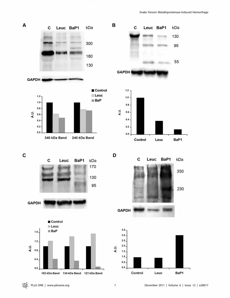
(Fig 4A). In muscle homogenates from control mice, anti-laminin
antibodies recognized four main bands of ,400, ,340, 240 and
180 kDa. Homogenates from tissue injected with either BaP1 or
leuc-a presented a similar reduction in the intensity of these bands
as compared with controls. In addition, BaP1 induced the
appearance of a degradation product of 285 kDa. (Fig 5A;
Table 1).
Nidogen. Western blot analysis of Matrigel samples showed
a rapid degradation of the 150 kDa nidogen band by both
SVMPs, with the generation of fragments of 95 and 50 kDa after
15 min of incubation; however, proteolysis by BaP1 occurred at a
faster rate (Fig 4B; Table 1). A 40 kDa degradation product was
Figue 1. Hemorrhagic activity of BaP1 and leuc-a in mousemuscle. Mice were injected intramuscularly, in the gastrocnemius, with50 mg of either BaP1 or leuc-a, dissolved in 50 mL PBS, or with 50 mL PBS(controls). After 15 min, mice were sacrificed and muscles weredissected out and placed in 1.5 mL of distilled water. Samples wereincubated overnight at 4uC and the absorbance at 540 nm wasrecorded in the supernatant as an indicator of muscle hemoglobincontent. BaP1 induced a profuse hemorrhage whereas leuc-a did notexert hemorrhagic activity. Results are presented as mean 6 SD (n = 4).*P,0.05 when compared to control and leuc-a treatments.doi:10.1371/journal.pone.0028017.g001
Snake Venom Metalloproteinase-Induced Hemorrhage
PLoS ONE | www.plosone.org 4 December 2011 | Volume 6 | Issue 12 | e28017

generated by BaP1, but not by leuc-a, at 3 h of incubation
(Fig 4B; Table 1). Immunodetection of nidogen in muscle samples
from mice treated with both SVMPs clearly showed hydrolysis of
this protein in vivo as early as 15 min of injection of the toxins,
with the appearance of degradation products of 100 and 50 kDa
(Fig 5B; Table 1). There was a higher extent of hydrolysis by
BaP1 than by leuc-a (Fig 5B).
Type IV collagen. Immunoblotting of Matrigel with anti-
type IV collagen antibodies revealed a 170 kDa band in control
and SVMP-treated samples (Fig 4C). Degradation of this band by
BaP1 was evident within 15 min of incubation and increased with
longer incubation times. In contrast, leuc-a treatment induced only
a slight reduction of the 170 kDa band after 3 h of incubation
(Fig 4C). Different proteolytic fragments were generated by the
two SVMPs: 140 and 120 kDa fragments by leuc-a, and 155, 140
and 100 kDa by BaP1, indicative of different cleavage sites (Fig 4C;
Table 1). Western blot of control muscle homogenates detected
three main bands of 155, 135 and 120 kDa (Fig 5C; Table 1). All
immunodetected bands were significantly reduced only by BaP1
treatment, and a degradation fragment of 110 kDa was detected
(Fig 5C; Table 1).
Figure 2. Proteolytic activity of BaP1 and leuc-a, and inhibition by a2-macroglobulin. (A) Hydrolytic activity of BaP1 and leuc-a onazocasein. Various amounts of each enzyme were incubated with azocasein for 90 min at 37uC. The reaction was stopped by the addition of 5%trichloroacetic acid, and the absorbances of the supernatants at 450 nm were recorded after centrifugation. Controls of azocasein without enzymewere run in parallel and their absorbance was subtracted from the sample values. Results are presented as mean 6 S.D. (n = 3). (B) Stoichiometry ofinhibition of BaP1 and leuc-a by a2M. The plasma inhibitor was incubated with BaP1 or leuc-a at various molar ratios, and proteolytic activity wastested on dimethylcasein. The remaining protease activity is expressed as percentage of the original activity measured in the absence of the inhibitor.Results are presented as mean 6 SD (n = 3). (C) Cleavage sites of BaP1 and leuc-a on oxidized insulin B-chain. After 30 min of digestion, peptides wereseparated by HPLC and identified by their amino acid sequence.doi:10.1371/journal.pone.0028017.g002
Figure 3. Hydrolysis of Matrigel proteins by BaP1 and leuc-a.Matrigel was incubated at 37uC with each enzyme at a 1:50 (w:w)enzyme:substrate ratio for 15 min, 1 h and 3 h. A control of Matrigelwithout enzymes (C) was included for each time interval. Matrigelsolutions were separated by SDS–PAGE under reducing conditionsusing a 4–15% gradient gel, and transferred to nitrocellulose membraneand stained with Ponceau-S.doi:10.1371/journal.pone.0028017.g003
Snake Venom Metalloproteinase-Induced Hemorrhage
PLoS ONE | www.plosone.org 5 December 2011 | Volume 6 | Issue 12 | e28017

Perlecan. Control samples of Matrigel incubated without
SVMPs showed a high molecular mass band (arrow in Fig 4D).
Several fragments of lower molecular mass were detected in Matrigel
samples incubated with either leuc-a or BaP1, demonstrating that
both enzymes can hydrolyze this BM component in vitro, generating
a similar pattern of degradation. BaP1 generated fragments of 170,
120, 35 and 30 kDa, whereas leuc-a generated fragments of 170,
120, 40 and 30 kDa (Fig 4D; Table 1). However, the rate of
degradation was faster with BaP1 since, at 15 min and 1 h, the
intensity of the bands corresponding to degradation products was
higher than in samples from leuc-a-treated mice (Fig 4D). The
monoclonal antibody against perlecan did not yield satisfactory
results when tested on muscle homogenates. Therefore, a polyclonal
antibody against endorepellin, the C-terminal domain of perlecan,
was used for detection of the proteoglycan in vivo. Immunoblotting
with this antibody demonstrated the presence of a diffuse band of
approximately 350 kDa in all samples analyzed, being more
pronounced in samples from muscle injected with BaP1 (Fig 5D).
Moreover, in samples from BaP1-injected mice, a 230 kDa band not
present in control or leuc-a samples was detected (Fig 5D; Table 1).
Histology and immunohistochemistryMuscle sections from control mice injected with PBS alone
showed a normal tissue structure with bundles of muscle fibers
surrounded by connective tissue (Fig 6A). Tissue sections from
mice injected with leuc-a showed a morphology similar to controls,
and there was no evidence of erythrocytes in the endomysium or
perimysium (Fig 6B). In contrast, BaP1-treated samples presented
hemorrhage, with masses of erythrocytes in the interstitial space
surrounding muscle fibers (Fig 6C).
Figure 4. Hydrolysis of basement membrane components in vitro. Hydrolysis of laminin (A), nidogen (B), type IV collagen (C) and perlecan (D)by BaP1 and leuc-a, as detected by Western blotting of Matrigel. Matrigel was incubated with either BaP1, leuc-a or PBS (control, lane C), as describedin the legend of Fig 3. Matrigel preparations were separated by SDS-PAGE under reducing conditions using a 4-15% gradient gel and transferred tonitrocellulose membranes. Immunodetection was performed with either anti-laminin, anti-nidogen, anti-type IV collagen or anti-perlecan antibodies.Reaction was developed with a chemiluminiscent substrate.doi:10.1371/journal.pone.0028017.g004
Table 1. Mol. mass of most abundant fragments of BMproteins generated by BaP1 and Leuc-a.
Matrigel in vitro In vivo
Protein Leuc-a BaP1 Leuc-a BaP1
Type IV Collagen 140, 120 155, 140, 100 -a 110
Laminin 150, 95, 60 150, 95, 60 -b 285
Nidogen 95, 50 95, 50, 40 100, 50 100, 50
Perlecan 170, 120, 40,30 170, 120, 35, 30 -c 230
aNo degradation bands were detected by immunoblotting, and the intensity ofthe main bands of type IV collagen was not reduced.
bNo degradation bands were detected by immunoblotting, but the mainlaminin bands were reduced as compared to control samples.
cNo degradation bands were detected by immunoblotting.doi:10.1371/journal.pone.0028017.t001
Snake Venom Metalloproteinase-Induced Hemorrhage
PLoS ONE | www.plosone.org 6 December 2011 | Volume 6 | Issue 12 | e28017
Snake Venom Metalloproteinase-Induced Hemorrhage
PLoS ONE | www.plosone.org 7 December 2011 | Volume 6 | Issue 12 | e28017

Since type IV collagen was the substrate which showed the
most significant differences in the degradation patterns between
the two SVMPs, detection of this protein by immunohisto-
chemistry was performed. Immunostaining for type IV collagen
and VEGFR-2, an endothelial cell marker, in sections of muscle
injected with PBS showed a normal distribution of capillaries
around the muscle fibers (Fig 6D). Sections from tissue injected
with leuc-a showed a pattern of immunostaining of these
markers which was similar to control samples (Fig 6E). In
contrast, BaP1 induced a reduction in the immunostaining for
type IV collagen and VEGFR-2 in capillaries (Fig 6F). The
capillary : muscle cell ratio in muscle was estimated after the
quantification, in tissue sections, of the numbers of capillaries
and muscle cells. The capillary : muscle cell ratio in muscle
injected with BaP1 was 0.760.3, which is significantly lower
(p,0.05) than in control muscle injected with PBS (Fig 6G),
whereas the capillary : muscle cell ratio in leuc-a treated muscle
did not show a significant drop, as compared with PBS-treated
muscle (p.0.05) (Fig 6G), in agreement with the lack of
hemorrhagic activity of this SVMP.
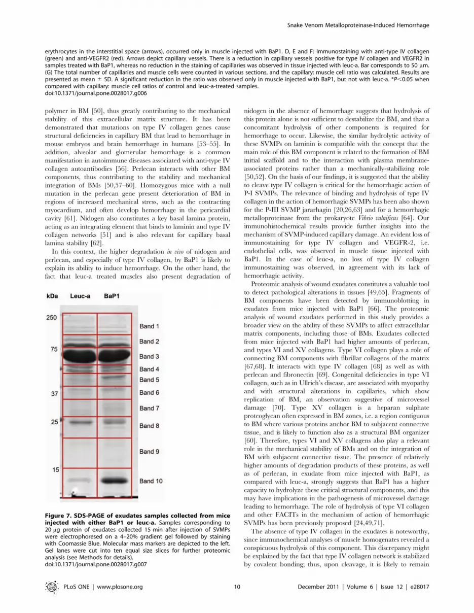
Proteomic analysis of wound exudatesThe electrophoretic profile of wound exudates collected from
mice injected with BaP1 and leuc-a is shown in Fig 7. Similar
profiles were observed with both enzymes, although exudate
collected from BaP1-injected mice showed a higher intensity in a
low molecular mass band which very likely corresponds to
hemoglobin, according to proteomic analysis (not shown), in
agreement with the hemorrhagic activity of this SVMP. From the
mass spectrometric analysis of the gel bands of these exudates,
more than 400 proteins were identified with XCorr Scores equal
of greater than 1.8, 2.3, 2.7 and 3.7 for +1, +2, +3 and +4 ions,
respectively, and protein identification probability above 95%.
Extracellular matrix proteins detected in the wound exudates are
listed in Table 2. Of the most abundant extracellular matrix
proteins identified, 9 showed differential abundances of greater
than three-fold between the two SVMPs. Of these, the fibrillar
type III collagen was more abundant in the exudate from leuc-a-
injected tissue. In contrast, the non-fibrillar types VI and XV
collagens, together with perlecan, were present in higher amounts
in exudates collected from mice injected with BaP1. No marked
differences were observed in laminin and nidogen between
treatments (Table 2).
Discussion
BM and associated ECM play a key role in the mechanical
stability of capillary blood vessels [50]. SVMPs constitute relevant
tools to explore the relevance of these ECM components in the
microvasculature, owing to the differential ability of these venom
components to damage capillary vessels. BaP1 and leuc-a are P-I
SVMPs which have a high sequence identity, and similar 3D
structure [34], enzymatic activity on azocasein in vitro, cleavage
pattern on insulin B chain, and inhibition by a2M. Moreover, both
have a clear preference for leucine in the P19 site [42]. Despite
these extensive similarities, BaP1 exerts hemorrhagic activity in
mice whereas leuc-a is unable to induce bleeding even when
injected at high doses. Our findings show that, in contrast with
their similar proteolytic activity on azocasein, these enzymes differ
in their capacity to hydrolyze BM proteins in vitro and, more
relevantly, in vivo. This observation may be central to their variable
hemorrhagic potential. Degradation of Matrigel revealed a similar
pattern of hydrolysis on laminin, nidogen and perlecan, although
BaP1 degraded these proteins at a faster rate than leuc-a. Despite
the fact that these observations correspond to in vitro experiments,
which need to be extrapolated with caution, they reveal a time-
dependent difference in the ability of these SVMPs to degrade BM
components, a finding that may be relevant in vivo from the
toxicokinetic standpoint. Once injected in the tissue, toxins diffuse
and are reabsorbed by several mechanisms, including lymphatic
drainage and, therefore, have a time frame within which they can
cleave BM components to induce hemorrhage. On the other hand,
a more interesting difference between these SVMPs was observed
in terms of hydrolysis of type IV collagen in Matrigel. BaP1 readily
cleaved this BM component, whereas leuc-a hydrolyzed it to a
much lesser extent. Earlier investigations demonstrated in vitro
degradation of type IV collagen by SVMPs [11,12,15], although
these studies did not evidence striking differences between SVMPs
in their ability to degrade this substrate.
Many works have analyzed the hydrolysis of BM components
by SVMPs in vitro. However, information on BM degradation in
vivo is scarce; immunohistochemical data have demonstrated the
ability of hemorrhagic SVMPs to degrade BM components in
mice [16,20]. Our present observations highlight marked differ-
ences in the ability of hemorrhagic and non-hemorrhagic P-I
SVMPs to hydrolyze type IV collagen and perlecan in tissue
homogenates of mouse gastrocnemius muscles injected with these
enzymes. In agreement, exudate analysis also revealed higher
amounts of perlecan in samples collected from mice injected with
BaP1 than in those receiving leuc-a. To the best of our knowledge,
this is the first report of degradation of perlecan by SVMPs both in
vitro and in vivo. Our results also demonstrate that BM proteins can
be readily hydrolyzed by SVMPs in vivo within a short time span,
in accordance with the rapid onset of hemorrhage induced by
these enzymes [18,19,21]. Of particular relevance is the observed
degradation of type IV collagen in vivo, since BaP1 readily cleaved
this protein within 15 min of injection, whereas no apparent
digestion was observed in the case of leuc-a. These observations
are relevant in the light of the role played by type IV collagen,
perlecan and nidogen in the scaffolding and mechanical stability of
capillary BM [50].
Current views on the assembly and structure of BM indicate
that the initial polymeric scaffold of BM is provided by laminin,
which is also responsible for early cell attachment of immature
BMs. Assembly is then completed with the integration of a
network of type IV collagen. Subsequently, other BM components,
such as nidogen, perlecan and other proteins are incorporated and
provide mechanical stability and complexity to the BM scaffold
[50–52]. Type IV collagen plays a key role in BM assembly and
stability, as it constitutes the only covalently-stabilized network
Figure 5. Hydrolysis of basement membrane components in vivo. Hydrolysis of laminin (A), nidogen (B), type IV collagen (C) and perlecan (D)by BaP1 and leuc-a, as detected by Western blotting of homogenates of injected mouse gastrocnemius muscle. Groups of mice were injected in thegastrocnemius muscle with either 50 mg BaP1, 50 mg leuc-a or PBS (lane C). After 15 min, mice were sacrificed and tissue was homogenized andcentrifuged to obtain the supernatant. Supernatants of muscle homogenates were separated by SDS-PAGE under reducing conditions, using a 4–15%gradient gel, and transferred to nitrocellulose membranes. Immunodetection was performed with either anti-laminin, anti-nidogen, anti-type IVcollagen or anti-endorepellin antibodies, and with anti-GAPDH as loading control in tissue homogenates. Reaction was developed with achemiluminiscent substrate. Densitometry was carried out in blots of tissue homogenates with ImageLab software; a relative quantification wasperformed adjusting each sample to the corresponding control.doi:10.1371/journal.pone.0028017.g005
Snake Venom Metalloproteinase-Induced Hemorrhage
PLoS ONE | www.plosone.org 8 December 2011 | Volume 6 | Issue 12 | e28017
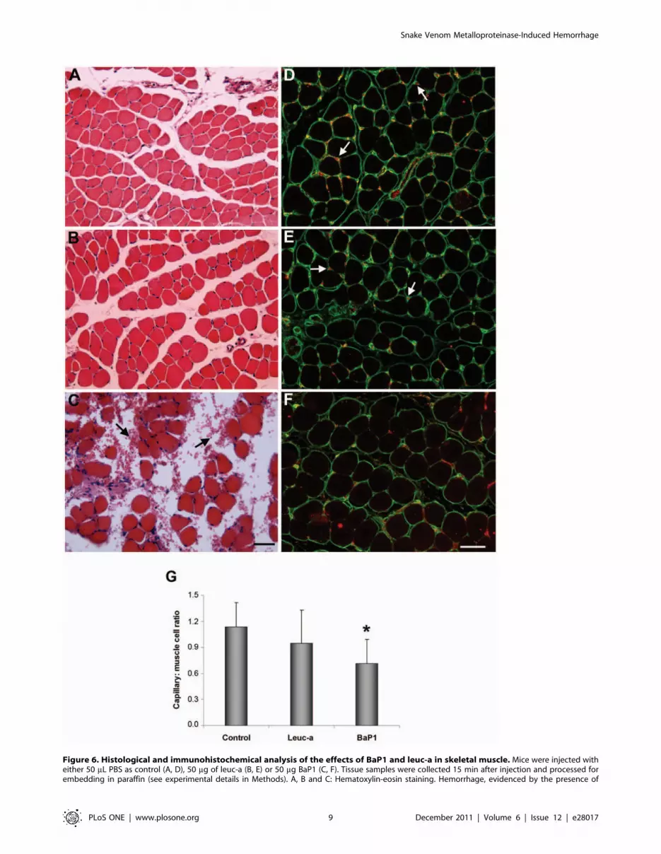
Figure 6. Histological and immunohistochemical analysis of the effects of BaP1 and leuc-a in skeletal muscle. Mice were injected witheither 50 mL PBS as control (A, D), 50 mg of leuc-a (B, E) or 50 mg BaP1 (C, F). Tissue samples were collected 15 min after injection and processed forembedding in paraffin (see experimental details in Methods). A, B and C: Hematoxylin-eosin staining. Hemorrhage, evidenced by the presence of
Snake Venom Metalloproteinase-Induced Hemorrhage
PLoS ONE | www.plosone.org 9 December 2011 | Volume 6 | Issue 12 | e28017
polymer in BM [50], thus greatly contributing to the mechanical
stability of this extracellular matrix structure. It has been
demonstrated that mutations on type IV collagen genes cause
structural deficiencies in capillary BM that lead to hemorrhage in
mouse embryos and brain hemorrhage in humans [53–55]. In
addition, alveolar and glomerular hemorrhage is a common
manifestation in autoimmune diseases associated with anti-type IV
collagen autoantibodies [56]. Perlecan interacts with other BM
components, thus contributing to the stability and mechanical
integration of BMs [50,57–60]. Homozygous mice with a null
mutation in the perlecan gene present deterioration of BM in
regions of increased mechanical stress, such as the contracting
myocardium, and often develop hemorrhage in the pericardial
cavity [61]. Nidogen also constitutes a key basal lamina protein,
acting as an integrating element that binds to laminin and type IV
collagen networks [51] and is also relevant for capillary basal
lamina stability [62].
In this context, the higher degradation in vivo of nidogen and
perlecan, and especially of type IV collagen, by BaP1 is likely to
explain its ability to induce hemorrhage. On the other hand, the
fact that leuc-a treated muscles also present degradation of
nidogen in the absence of hemorrhage suggests that hydrolysis of
this protein alone is not sufficient to destabilize the BM, and that a
concomitant hydrolysis of other components is required for
hemorrhage to occur. Likewise, the similar hydrolytic activity of
these SVMPs on laminin is compatible with the concept that the
main role of this BM component is related to the formation of BM
initial scaffold and to the interaction with plasma membrane-
associated proteins rather than a mechanically-stabilizing role
[50,52]. On the basis of our findings, it is suggested that the ability
to cleave type IV collagen is critical for the hemorrhagic action of
P-I SVMPs. The relevance of binding and hydrolysis of type IV
collagen in the action of hemorrhagic SVMPs has been also shown
for the P-III SVMP jararhagin [20,26,63] and for a hemorrhagic
metalloproteinase from the prokaryote Vibrio vulnificus [64]. Our
immunohistochemical results provide further insights into the
mechanism of SVMP-induced capillary damage. An evident loss of
immunostaining for type IV collagen and VEGFR-2, i.e.
endothelial cells, was observed in muscle tissue injected with
BaP1. In the case of leuc-a, no loss of type IV collagen
immunostaining was observed, in agreement with its lack of
hemorrhagic activity.
Proteomic analysis of wound exudates constitutes a valuable tool
to detect pathological alterations in tissues [49,65]. Fragments of
BM components have been detected by immunoblotting in
exudates from mice injected with BaP1 [66]. The proteomic
analysis of wound exudates performed in this study provides a
broader view on the ability of these SVMPs to affect extracellular
matrix components, including those of BMs. Exudates collected
from mice injected with BaP1 had higher amounts of perlecan,
and types VI and XV collagens. Type VI collagen plays a role of
connecting BM components with fibrillar collagens of the matrix
[67,68]. It interacts with type IV collagen [68] as well as with
perlecan and fibronectin [69]. Congenital deficiencies in type VI
collagen, such as in Ullrich’s disease, are associated with myopathy
and with structural alterations in capillaries, which show
replication of BM, an observation suggestive of microvessel
damage [70]. Type XV collagen is a heparan sulphate
proteoglycan often expressed in BM zones, i.e. a region contiguous
to BM where various proteins anchor BM to subjacent connective
tissue, and is likely to function also as a structural BM organizer
[60]. Therefore, types VI and XV collagens also play a relevant
role in the mechanical stability of BMs and on the integration of
BM with subjacent connective tissue. The presence of relatively
higher amounts of degradation products of these proteins, as well
as of perlecan, in exudate from mice injected with BaP1, as
compared with leuc-a, strongly suggests that BaP1 has a higher
capacity to hydrolyze these critical structural components, and this
may have implications in the pathogenesis of microvessel damage
leading to hemorrhage. The role of hydrolysis of type VI collagen
and other FACITs in the mechanism of action of hemorrhagic
SVMPs has been previously proposed [24,49,71].
The absence of type IV collagen in the exudates is noteworthy,
since immunochemical analyses of muscle homogenates revealed a
conspicuous hydrolysis of this component. This discrepancy might
be explained by the fact that type IV collagen network is stabilized
by covalent bonding; thus, upon cleavage, it is likely to remain
erythrocytes in the interstitial space (arrows), occurred only in muscle injected with BaP1. D, E and F: Immunostaining with anti-type IV collagen(green) and anti-VEGFR2 (red). Arrows depict capillary vessels. There is a reduction in capillary vessels positive for type IV collagen and VEGFR2 insamples treated with BaP1, whereas no reduction in the staining of capillaries was observed in tissue injected with leuc-a. Bar corresponds to 50 mm.(G) The total number of capillaries and muscle cells were counted in various sections, and the capillary: muscle cell ratio was calculated. Results arepresented as mean 6 SD. A significant reduction in the ratio was observed only in muscle injected with BaP1, but not with leuc-a. *P,0.05 whencompared with capillary: muscle cell ratios of control and leuc-a-treated samples.doi:10.1371/journal.pone.0028017.g006
Figure 7. SDS-PAGE of exudates samples collected from miceinjected with either BaP1 or leuc-a. Samples corresponding to20 mg protein of exudates collected 15 min after injection of SVMPswere electrophoresed on a 4–20% gradient gel followed by stainingwith Coomassie Blue. Molecular mass markers are depicted to the left.Gel lanes were cut into ten equal size slices for further proteomicanalysis (see Methods for details).doi:10.1371/journal.pone.0028017.g007
Snake Venom Metalloproteinase-Induced Hemorrhage
PLoS ONE | www.plosone.org 10 December 2011 | Volume 6 | Issue 12 | e28017

bound to the ECM and does not diffuse into the exudate. Overall,
BaP1 displays a higher ability to degrade not only BM
components, i.e. type IV collagen, perlecan, and nidogen, but
also type VI and XV collagens, which play a role in the integration
of BMs with their surrounding matrix. Another extracellular
matrix protein present in higher amounts in exudate collected
from mice injected with BaP1 is thrombospondin 1. This may be a
consequence of platelet aggregation secondary to microvessel
damage and hemorrhage [72]. This counteradhesive protein, in
turn, may contribute to an increase in vascular permeability and in
the separation of cells from the matrix, owing to their capacity to
inhibit cell-matrix and cell-cell interactions [72].
The structural basis for the functional differences described
remains unknown, since BaP1 and leuc-a have similar structures.
Analysis of the peptide cleavage consensus sequences on a
proteome-derived library, by using mass spectrometry, revealed
that these enzymes have a clear preference for leucine in the P19
site. However, leuc-a has a preference for aspartate in the P49 site,
whereas BaP1 has a preference for alanine in this position [42].
Thus, differences in the consensus sequences for the P4-P49 sites
between SVMPs may bear functional consequences related to
their ability to hydrolyze BM substrates, an issue that requires
further investigation. In addition, variations in protein backbone
flexibility of a loop located close to the active site of these enzymes
have been proposed to play a role in the hemorrhagic activity of P-
I SVMPs [34]. Further studies with a larger number of
hemorrhagic and non-hemorrhagic P-I SVMPs are necessary to
test these hypotheses and to discern the structural basis behind the
highly variable hemorrhagic potential of P-I SVMPs.
In conclusion, the present study provides clues to understand
the differences between the action of hemorrhagic and non-
hemorrhagic P-I SVMPs in vitro and in vivo, and offers insights into
the role of various ECM components in microvessel stability. BaP1
and leuc-a differ in their capacity to degrade key BM substrates,
mainly type IV collagen, perlecan and, to a lesser extent, nidogen,
as well as proteins which play a role in the integration of BMs with
the surrounding matrix, i.e. type VI and type XV collagens. The
drastic difference in type IV collagen hydrolysis between these
enzymes strongly suggests that degradation of this structurally-
relevant BM component is likely to represent a key step in the
Table 2. Proteomic analysis of extracellular matrix proteins in exudates collected from mice injected with BaP1 or Leuc-a.
Quantitative Value
Protein Accession # Mol. Weight BaP1 Leuc-a Fold change
Basement membrane-specific heparan sulfate proteoglycan core protein IPI00113824 398 kDa 24* 6 4.0
Biglycan IPI00123194 42 kDa 2 1 2.0
Collagen I alpha-1 chain (isoform 1) IPI00329872 138 kDa 20 42 2.1
Collagen I alpha-2 chain IPI00222188 130 kDa 19 32 1.7
Collagen III alpha-1 chain IPI00129571 139 kDa 8 25 3.1
Collagen VI alpha 3 chain IPI00830749 289 kDa 39 9 4.3
Collagen VI alpha 3 subunit IPI00845618 164 kDa 0 6 6.0
Collagen VI alpha3 (Fragment) IPI00877197 186 kDa 3 0 .3
Collagen VII alpha-1 chain IPI00134652 295 kDa 1 1 1.0
Collagen XI alpha-2 chain (isoform 7) IPI00138069 162 kDa 1 1 1.0
Collagen XII alpha-1 chain (isoform 1) IPI00121430 340 kDa 10 8 1.3
Collagen XIV alpha-1 chain (isoform 1) IPI00330632 193 kDa 24 19 1.3
Collagen XV alpha-1 chain IPI00409035 140 kDa 9 0 .9
Collagen XVIII alpha-1 chain (isoform 2) IPI00131476 156 kDa 2 3 1.5
Decorin IPI00123196 40 kDa 7 6 1.2
EGF-containing fibulin-like extracellular matrix protein 1 IPI00223457 55 kDa 3 10 3.3
Fibronectin IPI00113539 272 kDa 347 365 1.1
Fibulin-1 (isoform C) IPI00230432 75 kDa 3 2 1.5
Laminin subunit alpha-1 IPI00113726 338 kDa 1 2 2.0
Laminin subunit beta-3 IPI00117093 129 kDa 2 1 2.0
Laminin, gamma 2 IPI00117115 130 kDa 2 2 1.0
Lumican IPI00313900 38 kDa 47 44 1.1
Nidogen-1 IPI00111793 137 kDa 11 18 1.6
Nidogen-2 IPI00129903 154 kDa 1 2 2.0
Perlecan IPI00515360 470 kDa 3 0 .3
Tenascin (isoform 1) IPI00403938 232 kDa 2 1 2.0
Tenascin X IPI00130794 435 kDa 22 12 1.8
Thrombospondin-1 IPI00118413 130 kDa 20 1 20.0
Vitronectin IPI00129240 55 kDa 41 70 1.7
*Values underlined correspond to proteins whose quantitative value differed by more than 3 fold when comparing the two SVMPs.doi:10.1371/journal.pone.0028017.t002
Snake Venom Metalloproteinase-Induced Hemorrhage
PLoS ONE | www.plosone.org 11 December 2011 | Volume 6 | Issue 12 | e28017
mechanism of action of hemorrhagic SVMPs, and underscores the
key role played by this ECM protein in the mechanical stability of
capillary blood vessels.
Acknowledgments
The authors thank Bruno Lomonte for his insights on structural aspects of
SVMPs, and Llira Bonilla and Diego Morazan for their collaboration in
laboratory work. This study was carried out in partial fulfillment of the
requirements for the Ph.D. degree for T. Escalante at the University of
Costa Rica.
Author Contributions
Conceived and designed the experiments: TE AR JWF JMG. Performed
the experiments: TE NO AR EFS MR JWF JMG. Analyzed the data: TE
NO AR EFS MR JWF JMG. Contributed reagents/materials/analysis
tools: TE AR EFS JWF JMG. Wrote the paper: TE AR JWF JMG.
References
1. Fox JW, Serrano SMT (2005) Structural considerations of the snake venom
metalloproteinases, key members of the M12 reprolysin family of metallopro-
teinases. Toxicon 45: 969–985.
2. Calvete JJ, Sanz L, Angulo Y, Lomonte B, Gutierrez JM (2009) Venoms,
venomics, antivenomics. FEBS Lett 583: 1736–1743.
3. Gutierrez JM, Rucavado A, Escalante T (2010) Snake venom metalloprotei-
nases. Biological roles and participation in the pathophysiology of envenom-
ation. In Mackessy SP, ed. Handbook of Venoms and Toxins of Reptiles. Boca
Raton: CRC Press. pp 114–128.
4. Gutierrez JM, Rucavado A (2000) Snake venom metalloproteinases: Their role
in the pathogenesis of local tissue damage. Biochimie 82: 841–850.
5. Kamiguti AS (2005) Platelets as targets of snake venom metalloproteinases.
Toxicon 45: 1041–1049.
6. Moura-da-Silva AM, Serrano SMT, Fox JW, Gutierrez JM (2009) Snake venom
metalloproteinases. In de Lima ME, Pimenta AMC, Martin-Euclaire MF,
Zingali RB, Rochat H, eds. Animal Toxins:State of the Art. Perspectives in
Health and Biotechnology. Belo Horizonte: Editora UFMG. pp 525–546.
7. Mackessy SP (2002) Biochemistry and pharmacology of colubrid snake venoms.
J Toxicol Toxin Rev. 21: 43–83.
8. Gutierrez JM, Rucavado A, Escalante T, Dıaz C (2005) Hemorrhage induced by
snake venom metalloproteinases: biochemical and biophysical mechanisms
involved in microvessel damage. Toxicon 45: 997–1011.
9. Ohsaka A, Just M, Habermann E (1973) Action of snake venom hemorrhagic
principles on isolated glomerular basement membrane. Biochim Biophys Acta
323: 415–438.
10. Bjarnason JB, Hamilton D, Fox JW (1988) Studies on the mechanism of
hemorrhage production by five proteolytic hemorrhagic toxins from Crotalus atrox
venom. Biol Chem 369: 121–129.
11. Baramova EN, Shannon JD, Bjarnason JB, Fox JW (1989) Degradation of
extracellular matrix proteins by hemorrhagic metalloproteinases. Arch Biochem
Biophys 275: 63–71.
12. Baramova EN, Shannon JD, Bjarnason JB, Fox JW (1990) Identification of the
cleavage sites by a hemorrhagic metalloproteinase in type IV collagen. Matrix
10: 91–97.
13. Baramova EN, Shannon JD, Fox JW, Bjarnason JB (1991) Proteolytic digestion
of non-collagenous basement membrane proteins by the hemorrhagic metallo-
proteinase Ht-e from Crotalus atrox venom. Biomed Biochim Acta 50: 763–768.
14. Maruyama M, Sugiki M, Yoshida E, Shimaya K, Mihara H (1992) Broad
substrate specificity of snake venom fibrinolytic enzymes: possible role in
haemorrhage. Toxicon 30: 1387–1397.
15. Rucavado A, Lomonte B, Ovadia M, Gutierrez JM (1995) Local tissue damage
induced by BaP1, a metalloproteinase isolated from the Bothrops asper (terciopelo)
snake venom. Exp Mol Pathol 63: 186–199.
16. Escalante T, Shannon J, Moura-da-Silva AM, Gutierrez JM, Fox JW (2006)
Novel insights into capillary vessel basement membrane damage by snake venom
hemorrhagic metalloproteinases: a biochemical and immunohistochemical
study. Arch Biochem Biophys 455: 144–153.
17. Oliveira AK, Paes Leme AF, Asega AF, Camargo AC, Fox JW, et al. (2010) New
insights into the structural elements involved in the skin haemorrhage induced
by snake venom metalloproteinases. Thromb Haemost 104: 485–497.
18. Ownby CL, Bjarnason J, Tu AT (1978) Hemorrhagic toxins from rattlesnake
(Crotalus atrox) venom. Pathogenesis of hemorrhage induced by three purified
toxins. Am J Pathol 93: 201–208.
19. Moreira L, Borkow G, Ovadia M, Gutierrez JM (1994) Pathological changes
induced by BaH1, a hemorrhagic metalloproteinase isolated from Bothrops asper
(terciopelo) snake venom, on mouse capillary blood vessels. Toxicon 32:
977–987.
20. Baldo C, Jamora C, Yamanouye N, Zorn TM, Moura-da-Silva AM (2010)
Mechanisms of vascular damage by hemorrhagic snake venom metalloprotei-
nases: tissue distribution and in situ hydrolysis. PLoS Negl Trop Dis 4: e727.
21. Gutierrez JM, Nunez J, Escalante T, Rucavado A (2006) Blood flow is required
for rapid endothelial cell damage induced by a snake venom hemorrhagic
metalloproteinase. Microvasc Res 71: 55–63.
22. Bjarnason JB, Fox JW (1994) Hemorrhagic metalloproteinases from snake
venoms. Pharmacol Ther 62: 325–372.
23. Serrano SM, Jia LG, Wang D, Shannon JD, Fox JW (2005) Function of the
cysteine-rich domain of the haemorrhagic metalloproteinase atrolysin A:
targeting adhesion proteins collagen I and von Willebrand factor. Biochem J391: 69–76.
24. Serrano SM, Kim J, Wang D, Dragulev B, Shannon JD, et al. (2006) Thecysteine-rich domain of snake venom metalloproteinases is a ligand for von
Willebrand factor A domains: role in substrate targeting. J Biol Chem 281:39746–39756.
25. Serrano SM, Wang D, Shannon JD, Pinto AF, Polanowska-Grabowska RK,
et al. (2007) Interaction of the cysteine-rich domain of snake venommetalloproteinases with the A1 domain of von Willebrand factor promotes
site-specific proteolysis of von Willebrand factor and inhibition of vonWillebrand factor-mediated platelet aggregation. FEBS J 274: 3611–3621.
26. Tanjoni I, Evangelista K, Della-Casa MS, Butera D, Magalhaes GS, et al. (2010)Different regions of the class P-III snake venom metalloproteinase jararhagin are
involved in binding to a2b1 integrin and collagen Toxicon 55: 1093–1099.
27. Kurecki T, Kress LF (1985) Purification and partial characterization of a high
molecular weight metalloproteinase from the venom of Crotalus adamanteus
(eastern diamondback rattlesnake). Toxicon 23: 855–863.
28. Baramova EN, Shannon JD, Bjarnason JB, Gonias SL, Fox JW (1990)
Interaction of hemorrhagic metalloproteinases with human a2-macroglobulin.Biochemistry 29: 1069–1074.
29. Kamiguti AS, Desmond HP, Theakston RDG, Hay CRM, Zuzel M (1994)Ineffectiveness of the inhibition of the main haemorrhagic metalloproteinase
from Bothrops jararaca venom by its only plasma inhibitor, a2-macroglobulin.
Biochim Biophys Acta 1200: 307–314.
30. Escalante T, Nunez J, Moura da Silva A, Rucavado A, Theakston RDG, et al.
(2003) Pulmonary hemorrhage induced by jararhagin, a metalloproteinase fromBothrops jararaca snake venom: relationship to biodistribution and neutralization
by serum inhibitors. Toxicol Appl Pharmacol 193: 17–28.
31. Escalante T, Rucavado A, Kamiguti AS, Theakston RDG, Gutierrez JM (2004)
Bothrops asper metalloproteinase BaP1 is inhibited by a2-macroglobulin and
mouse serum and does not induce systemic hemorrhage or coagulopathy.Toxicon 43: 213–217.
32. Sanchez EF, Eble JA (2009) P-III metalloproteinase (Leucurolysin-B) fromBothrops leucurus venom: isolation and possible inhibition. In Supuran CT,
Winum JY, eds. Drug Design of Zinc-Enzyme Inhibitors; Functional, Structuraland Disease Applications. New Jersey: Wiley & Sons. pp 789–812.
33. Sanchez EF, Schneider SF, Yarleque A, Borges MH, Richardson M, et al. (2010)
The novel metalloproteinase atroxlysin-I from Peruvian Bothrops atrox (Jergon)snake venom acts both on blood vessel ECM and platelets. Arch Biochem
Biophys 496: 9–20.
34. Wallnoefer HG, Lingott T, Gutierrez JM, Merfort I, Liedl KR (2010) Backbone
flexibility controls the activity and specificity of a protein-protein interface:specificity in snake venom metalloproteinases. J Am Chem Soc 132:
10330–10337.
35. Tsai IH, Wang YM, Chiang TY, Chen YL, Huang RJ (2000) Purification,
cloning and sequence analyses for pro-metalloprotease-disintegrin variants from
Deinagkistrodon acutus venom and subclassification of the small venom metallo-proteases. Eur J Biochem 267: 1359–1367.
36. Bolger MB, Swenson S, Markland FS, Jr. (2001) Three-dimensional structure offibrolase, the fibrinolytic enzyme from southern copperhead venom, modeled
from the X-ray structure of adamalysin II and atrolysin C. AAPS PharmSci 3:
E16.
37. Ramos OH, Selistre-de-Araujo HS (2004) Comparative analysis of the catalytic
domain of hemorrhagic and non-hemorrhagic snake venom metallopeptidasesusing bioinformatic tools. Toxicon 44: 529–538.
38. Watanabe L, Shannon JD, Valente RH, Rucavado A, Alape-Giron A, et al.(2003) Amino acid sequence and crystal structure of BaP1, a metalloproteinase
from Bothrops asper snake venom that exerts multiple tissue-damaging activities.Protein Sci 12: 2273–2281.
39. Lingott T, Schleberger C, Gutierrez JM, Merfort I (2009) High-resolution crystal
structure of the snake venom metalloproteinase BaP1 complexed with apeptidomimetic: insight into inhibitor binding. Biochemistry 48: 6166–6174.
40. Rucavado A, Flores-Sanchez E, Franceschi A, Magalhaes A, Gutierrez JM(1999) Characterization of the local tissue damage induced by LHF-II, a
metalloproteinase with weak hemorrhagic activity isolated from Lachesis muta
muta snake venom. Toxicon 37: 1297–1312.
41. Rodrigues VM, Soares AM, Guerra-Sa R, Rodrigues V, Fontes MRM, et al.
(2000) Structural and functional characterization of neuwiedase, a non-
Snake Venom Metalloproteinase-Induced Hemorrhage
PLoS ONE | www.plosone.org 12 December 2011 | Volume 6 | Issue 12 | e28017

hemorrhagic fibrin(ogen)olytic metalloprotease from Bothrops neuwiedi snake
venom. Arch Biochem Biophys 381: 213–224.
42. Paes Leme AF, Escalante T, Pereira JGC, Sanchez EF, Gutierrez JM, et al.
(2011) High resolution analysis of snake venom metalloproteinase (SVMP)
peptide bond cleavage specificity using proteome based peptide libraries and
mass spectrometry. J Proteomics 74: 401–410.
43. Gutierrez JM, Romero M, Dıaz C, Borkow G, Ovadia M (1995) Isolation and
characterization of a metalloproteinase with weak hemorrhagic activity from the
venom of the snake Bothrops asper (terciopelo). Toxicon 33: 19–29.
44. Bello CA, Hermogenes AL, Magalhaes A, Veiga SS, Gremski LH, et al. (2006)
Isolation and biochemical characterization of a fibrinolytic proteinase from
Bothrops leucurus (white-tailed jararaca) snake venom. Biochimie 88: 189–200.
45. Gutierrez JM, Gene JA, Rojas G, Cerdas L (1985) Neutralization of proteolytic
and hemorrhagic activities of Costa Rican snake venoms by a polyvalent
antivenom. Toxicon 23: 887–893.
46. Wang WJ, Shih CH, Huang TF (2004) A novel P-I class metalloproteinase with
broad substrate-cleaving activity, agkislysin, from Agkistrodon acutus venom.
Biochem Biophys Res Comm 324: 224–230.
47. Souza CT, Moura MB, Magalhaes A, Heneine LG, Chaves-Olortegui C, et al.
(2001) Inhibition of mutalysin II, a metalloproteinase from bushmaster venom
by human a2-macroglobulin and rabbit immunoglobulin. Comp Biochem
Physiol B Biochem Mol Biol 130: 155–168.
48. LeBleu VS, MacDonald B, Kalluri R (2007) Structure and function of basement
membranes. Exp Biol Med 232: 1121–1129.
49. Escalante T, Rucavado A, Pinto AFM, Terra RMS, Gutierrez JM, et al. (2009)
Wound exudate as a proteomic window to reveal different mechanisms of tissue
damage by snake venom toxins. J Proteom Res 8: 5120–5131.
50. Yurchenco PD, Amenta PS, Patton BL (2004) Basement membrane assembly,
stability and activities observed through a developmental lens. Matrix Biol 22:
521–538.
51. Fox JW, Mayer U, Nischt R, Aumailley M, Reinhardt D, et al. (1991)
Recombinant nidogen consists of three globular domains and mediates binding
of laminin to collagen type IV. EMBO J 11: 3137–3146.
52. Hallmann R, Horn N, Selg M, Wendler O, Pausch F, et al. (2005) Expression
and function of laminins in the embryonic and mature vasculature. Physiol Rev
85: 979–1000.
53. Poschl E, Schlotzer-Schrehardt U, Brachvogel B, Saito K, Ninomiya Y, et al.
(2004) Collagen IV is essential for basement membrane stability but dispensable
for initiation of its assembly during early development. Development 131:
1619–1628.
54. Gould DB, Phalan FC, Breedveld GJ, van Mil SE, Smith RS, et al. (2005)
Mutations in Col4a1 cause perinatal cerebral hemorrhage and porencephaly.
Science 308: 1167–1171.
55. Bilguvar K, DiLuna ML, Bizzarro MJ, Bayri Y, Schneider KC, et al. (2009)
COL4A1 mutation in preterm intraventricular hemorrhage. J Pediatr 155:
743–745.
56. Specks U (2001) Diffuse alveolar hemorrhage syndromes. Curr Opin Rheumatol
13: 12–17.
57. Iozzo RV, Cohen IR, Grassel S, Murdoch AD (1994) The biology of perlecan:
the multifaceted heparan sulphate proteoglycan of basement membranes andpericellular matrices. Biochem J 302: 625–639.
58. Timpl R (1996) Macromolecular organization of basement membranes. Curr
Opin Cell Biol 8: 618–624.59. Kvansaluk M, Hopf M, Ries A, Timpl R, Hohenester E (2001) Structural basis
for the high affinity interaction of nidogen-1 with immunoglobulin-like domain 3of perlecan. EMBO J 20: 5342–5346.
60. Iozzo RV (2005) Basement membrane proteoglycans: from cellar to ceiling. Nat
Rev Mol Cell Biol 8: 646–656.61. Costell M, Gustafsson E, Aszodi A, Morgelin M, Bloch W, et al. (1999) Perlecan
maintains the integrity of cartilage and some basement membranes. J Cell Biol147: 1109–1122.
62. Mokkapati S, Baranowsky A, Mirancea N, Smyth N, Breitkreutz D, et al. (2008)Basement membranes in skin are differently affected by lack of nidogen 1 and 2.
J Invest Dermatol 128: 2259–2267.
63. Moura-da-Silva AM, Ramos OH, Baldo C, Niland S, Hansen U, et al. (2008)Collagen binding is a key factor for the hemorrhagic activity of snake venom
metalloproteinases. Biochimie 90: 484–492.64. Miyoshi S, Nakazawa H, Kawata K, Tomochika K, Tobe K, et al. (1998)
Characterization of the hemorrhagic reaction caused by Vibrio vulnificus
metalloprotease, a member of the thermolysin family. Infect Immun 66:4851–4855.
65. Ahn SM, Simpson RJ (2007) Body fluid proteomics: Prospects for biomarkerdiscovery. Proteomics Clin Appl 1: 1004–1015.
66. Rucavado A, Nunez J, Gutierrez JM (1998) Blister formation and skin damageinduced by BaP1, a haemorrhagic metalloproteinase from the venom of the
snake Bothrops asper. Int J Exp Pathol 79: 245–254.
67. Keene DR, Engvall E, Glanville RW (1988) Ultrastructure of type VI collagen inhuman skin and cartilage suggests an anchoring function for this filamentous
network. J Cell Biol 107: 1995–2006.68. Kuo HJ, Maslen CL, Keene DR, Glanville RW (1997) Type VI collagen
anchors endothelial basement membranes by interacting with type IV collagen.
J Biol Chem 272: 26522–26529.69. Tillet E, Wiedemann H, Golbik R, Pan TC, Zhang RZ, et al. (1994)
Recombinant expression and structural and binding properties of alpha 1(VI)and alpha 2(VI) chains of human collagen type VI. Eur J Biochem 221:
177–185.70. Niiyama T, Higuchi I, Hashiguchi T, Suehara M, Uchida Y, et al. (2003)
Capillary changes in skeletal muscle of patients with Ullrich’s disease with
collagen VI deficiency. Acta Neuropathol 106: 137–142.71. Pinto AF, Ma L, Dragulev B, Guimaraes JA, Fox JW (2007) Use of SILAC for
exploring sheddase and matrix degradation of fibroblasts in culture by the PIIISVMP atrolysin A: identification of two novel substrates with functional
relevance. Arch Biochem Biophys 465: 11–15.
72. Liu A, Mosher DF, Murphy-Ullrich JE, Goldblum SE (2009) The counter-adhesive proteins, thrombospondin 1 and SPARC/osteonectin, open the
tyrosine phosphorylation-responsive paracellular pathway in pulmonary vascularendothelia. Microv Res 77: 13–20.
Snake Venom Metalloproteinase-Induced Hemorrhage
PLoS ONE | www.plosone.org 13 December 2011 | Volume 6 | Issue 12 | e28017