riken ims annual report 2018
TRANSCRIPT
RIKEN IMSAnnual Report 2018
RIKEN Center for Integrative Medical Sciences
RIK
EN IM
S An
nu
al Rep
ort 2018 R
IKEN
Cen
ter for In
tegrative M
edical Scien
ces
Laboratory for Transcriptome Technology: Piero CarninciLaboratory for Cellular Function Conversion Technology: Harukazu SuzukiLaboratory for Genome Information Analysis: Chung Chau HonLaboratory for Applied Computational Genomics: Michel De HoonLaboratory for Single Cell Technologies: Piero CarninciLarge Scale Data Managing Unit: Takeya KasukawaLaboratory for Advanced Genomics Circuit: Jay W. ShinGenetic Diagnosis Technology Unit: Kengo UsuiEpigenome Technology Exploration Unit: Aki MinodaLaboratory for Comprehensive Genomic Analysis: Yasushi OkazakiLaboratory for Applied Regulatory Genomics Network Analysis: Erik Arner
Nucleic Acid Diagnostic System Development Unit: Kengo UsuiPreventive Medicine and Applied Genomics Unit: Hideya KawajiRIKEN-IFOM Joint Laboratory for Cancer Genomics: Yasuhiro MurakawaLaboratory for Genotyping Development: Yukihide MomozawaLaboratory for Statistical Analysis: Yoichiro KamataniLaboratory for Pharmacogenomics: Taisei MushirodaLaboratory for International Alliance on Genomic Research: Taisei MushirodaLaboratory for Bone and Joint Diseases: Shiro IkegawaLaboratory for Endocrinology, Metabolism and Kidney Diseases: Momoko HorikoshiLaboratory for Cardiovascular Diseases: Kaoru Ito
Division of Genomic Medicine
Laboratory for Autoimmune Diseases: Kazuhiko YamamotoLaboratory for Respiratory and Allergic Diseases: Kazuhiko YamamotoLaboratory for Cell Signaling: Takashi SaitoLaboratory for Lymphocyte Di�erentiation: Tomohiro KurosakiLaboratory for Transcriptional Regulation: Ichiro TaniuchiLaboratory for Immune Cell Systems: Shigeo Koyasu
Laboratory for Innate Immune Systems: Kazuyo MoroLaboratory for Immune Homeostasis: Taishin AkiyamaLaboratory for Immune Crosstalk: Hilde CheroutreLaboratory for In�ammatory Regulation: Takashi TanakaLaboratory for Cytokine Regulation: Masato Kubo
Division of Human Immunology
Laboratory for Developmental Genetics: Haruhiko KosekiLaboratory for Intestinal Ecosystem: Hiroshi OhnoLaboratory for Integrative Genomics: Osamu OharaLaboratory for Mucosal Immunity: Sidonia FagarasanLaboratory for Gut Homeostasis: Kenya HondaLaboratory for Skin Homeostasis: Masayuki Amagai
Laboratory for Tissue Dynamics: Takaharu OkadaLaboratory for Integrated Cellular Systems: Mariko OkadaLaboratory for Metabolomics: Makoto AritaLaboratory for Microbiome Sciences: Masahira HattoriDrug Discovery Antibody Platform Unit: Toshitada Takemori
Division of Disease Systems Biology
Laboratory for Immunogenetics: Tadashi YamamotoLaboratory for Medical Science Mathematics: Tatsuhiko TsunodaLaboratory for Cancer Genomics: Hidewaki Nakagawa
Laboratory for Immunotherapy: Shin-ichiro FujiiLaboratory for Human Disease Models: Fumihiko IshikawaLiver Cancer Prevention Research Unit: Soichi Kojima
Division of Cancer Immunology
Piero CarninciHaruhiko KosekiKazuhiko Yamamoto
Deputy Directors
Max Cooper (chair)Mark Lathrop (vice chair)Hiroyuki AburataniRudi BallingEwan BirneyRiccardo Dalla-FaveraMichel GeorgesRonald N. GermainYukiko GotohDavid A. HaflerHajime KarasuyamaYutaka KawakamiJuha KerePaul W. KincadeBernard MalissenJohn O'SheaFiona PowrieShimon SakaguchiSarah TeichmannArthur Weiss
Advisory Council
Genome Immunobiology RIKEN Hakubi Research Team: Nicholas Parrish
RIKEN Hakubi Research Team
Tadashi Yamamoto
Director
Shizuo Akira
Senior Advisor
Office of the Center Director
YCI Laboratory for Cellular Bioenergetic Network: Toshimori KitamiYCI Laboratory for Trans-omics: Katsuyuki YugiYCI Laboratory for Immunological Transcriptomics: Hideyuki Yoshida
YCI Laboratory for Next-Generation Proteomics: Yibo WuYCI Laboratory for Metabolic Epigenetics: Azusa Inoue
Young Chief Investigator Program
RIKEN Center for Integrative Medical Sciences Organization Chart
Laboratory for Transcriptome Technology: Piero CarninciLaboratory for Cellular Function Conversion Technology: Harukazu SuzukiLaboratory for Genome Information Analysis: Chung Chau HonLaboratory for Applied Computational Genomics: Michel De HoonLaboratory for Single Cell Technologies: Piero CarninciLarge Scale Data Managing Unit: Takeya KasukawaLaboratory for Advanced Genomics Circuit: Jay W. ShinGenetic Diagnosis Technology Unit: Kengo UsuiEpigenome Technology Exploration Unit: Aki MinodaLaboratory for Comprehensive Genomic Analysis: Yasushi OkazakiLaboratory for Applied Regulatory Genomics Network Analysis: Erik Arner
Nucleic Acid Diagnostic System Development Unit: Kengo UsuiPreventive Medicine and Applied Genomics Unit: Hideya KawajiRIKEN-IFOM Joint Laboratory for Cancer Genomics: Yasuhiro MurakawaLaboratory for Genotyping Development: Yukihide MomozawaLaboratory for Statistical Analysis: Yoichiro KamataniLaboratory for Pharmacogenomics: Taisei MushirodaLaboratory for International Alliance on Genomic Research: Taisei MushirodaLaboratory for Bone and Joint Diseases: Shiro IkegawaLaboratory for Endocrinology, Metabolism and Kidney Diseases: Momoko HorikoshiLaboratory for Cardiovascular Diseases: Kaoru Ito
Division of Genomic Medicine
Laboratory for Autoimmune Diseases: Kazuhiko YamamotoLaboratory for Respiratory and Allergic Diseases: Kazuhiko YamamotoLaboratory for Cell Signaling: Takashi SaitoLaboratory for Lymphocyte Di�erentiation: Tomohiro KurosakiLaboratory for Transcriptional Regulation: Ichiro TaniuchiLaboratory for Immune Cell Systems: Shigeo Koyasu
Laboratory for Innate Immune Systems: Kazuyo MoroLaboratory for Immune Homeostasis: Taishin AkiyamaLaboratory for Immune Crosstalk: Hilde CheroutreLaboratory for In�ammatory Regulation: Takashi TanakaLaboratory for Cytokine Regulation: Masato Kubo
Division of Human Immunology
Laboratory for Developmental Genetics: Haruhiko KosekiLaboratory for Intestinal Ecosystem: Hiroshi OhnoLaboratory for Integrative Genomics: Osamu OharaLaboratory for Mucosal Immunity: Sidonia FagarasanLaboratory for Gut Homeostasis: Kenya HondaLaboratory for Skin Homeostasis: Masayuki Amagai
Laboratory for Tissue Dynamics: Takaharu OkadaLaboratory for Integrated Cellular Systems: Mariko OkadaLaboratory for Metabolomics: Makoto AritaLaboratory for Microbiome Sciences: Masahira HattoriDrug Discovery Antibody Platform Unit: Toshitada Takemori
Division of Disease Systems Biology
Laboratory for Immunogenetics: Tadashi YamamotoLaboratory for Medical Science Mathematics: Tatsuhiko TsunodaLaboratory for Cancer Genomics: Hidewaki Nakagawa
Laboratory for Immunotherapy: Shin-ichiro FujiiLaboratory for Human Disease Models: Fumihiko IshikawaLiver Cancer Prevention Research Unit: Soichi Kojima
Division of Cancer Immunology
Piero CarninciHaruhiko KosekiKazuhiko Yamamoto
Deputy Directors
Max Cooper (chair)Mark Lathrop (vice chair)Hiroyuki AburataniRudi BallingEwan BirneyRiccardo Dalla-FaveraMichel GeorgesRonald N. GermainYukiko GotohDavid A. HaflerHajime KarasuyamaYutaka KawakamiJuha KerePaul W. KincadeBernard MalissenJohn O'SheaFiona PowrieShimon SakaguchiSarah TeichmannArthur Weiss
Advisory Council
Genome Immunobiology RIKEN Hakubi Research Team: Nicholas Parrish
RIKEN Hakubi Research Team
Tadashi Yamamoto
Director
Shizuo Akira
Senior Advisor
Office of the Center Director
YCI Laboratory for Cellular Bioenergetic Network: Toshimori KitamiYCI Laboratory for Trans-omics: Katsuyuki YugiYCI Laboratory for Immunological Transcriptomics: Hideyuki Yoshida
YCI Laboratory for Next-Generation Proteomics: Yibo WuYCI Laboratory for Metabolic Epigenetics: Azusa Inoue
Young Chief Investigator Program
I
Contents
Organization ............................................................................... IContents ......................................................................................II
Director’s report ....................................................................... IV
Massive genetic analyses link genes, cell types, molecules, and diseases .......................................................................... 2
Transcriptional landscape of Mycobacterium tuberculosis infection in macrophages ................................................... 3
Researchers uncover origin of virus-fighting plasma B cells..................................................................................... 4
Three transcriptional circuits found between hematopoietic stem cells and B cells ........................................................... 5
Genome sequencing analysis reveals genes associated with survival in biliary tract cancers ......................................... 6
Division of Genomic Medicine ........................................ 8Laboratory for Transcriptome Technology ...................... 9Laboratory for Cellular Function Conversion
Technology ................................................................... 10Laboratory for Genome Information Analysis ............. 11Laboratory for Applied Computational Genomics....... 12Laboratory for Single Cell Technologies ........................ 13Large Scale Data Managing Unit ..................................... 14Laboratory for Advanced Genomics Circuit ................. 15Genetic Diagnosis Technology Unit Nucleic Acid Diagnostic System Development Unit .... 16Epigenome Technology Exploration Unit ...................... 17Laboratory for Comprehensive Genomic Analysis ...... 18Laboratory for Applied Regulatory Genomics Network
Analysis ........................................................................ 19Preventive Medicine and Applied Genomics Unit ....... 20RIKEN-IFOM Joint Laboratory for Cancer
Genomics ..................................................................... 21Laboratory for Genotyping Development ..................... 22Laboratory for Statistical Analysis .................................. 23Laboratory for Pharmacogenomics ................................ 24Laboratory for Bone and Joint Diseases ......................... 25Laboratory for Endocrinology, Metabolism and
Kidney Diseases .......................................................... 26Laboratory for Cardiovascular Diseases ........................ 27
Division of Human Immunology ................................. 28Laboratory for Autoimmune Diseases ........................... 29Laboratory for Cell Signaling .......................................... 30Laboratory for Lymphocyte Differentiation .................. 31
Laboratory for Transcriptional Regulation .................... 32Laboratory for Immune Cell Systems ............................. 33Laboratory for Innate Immune Systems ........................ 34Laboratory for Immune Homeostasis ............................ 35Laboratory for Immune Crosstalk .................................. 36Laboratory for Inflammatory Regulation ...................... 37Laboratory for Cytokine Regulation ............................... 38
Division of Disease Systems Biology .......................... 39Laboratory for Developmental Genetics ........................ 40Laboratory for Intestinal Ecosystem ............................... 41Laboratory for Integrative Genomics ............................. 42Laboratory for Mucosal Immunity ................................. 43Laboratory for Gut Homeostasis ..................................... 44Laboratory for Skin Homeostasis .................................... 45Laboratory for Tissue Dynamics ..................................... 46Laboratory for Metabolomics .......................................... 47Laboratory for Microbiome Sciences ............................. 48Drug Discovery Antibody Platform Unit ...................... 49
Division of Cancer Immunology ................................... 50Laboratory for Immunogenetics ..................................... 51Laboratory for Medical Science Mathematics ............... 52Laboratory for Cancer Genomics ................................... 53Laboratory for Immunotherapy ...................................... 54Laboratory for Human Disease Models ......................... 55Liver Cancer Prevention Research Unit ......................... 56
Special Programs for Young Leaders ......................... 57Genome Immunobiology RIKEN Hakubi Research
Team ............................................................................. 58
Part 1Research Highlights
Part 2Lab Activities
II
Human Genome Meeting 2018 ............................................. 76Japan Prize awarded to Dr. Max D. Cooper ......................... 76RIKEN IMS-Stanford ISCBRM Joint Symposium .............. 77The IMS-JSI International Symposium on
Immunology 2018 ............................................................. 77ZPM-RIKEN Symposium ...................................................... 78RIKEN-KI-SciLifeLab Symposium 2018 ............................. 78RIKEN-Luxembourg Joint Symposium and Workshop ..... 79
Single Cell Symposium ........................................................... 79RIKEN - McGill Symposiums................................................ 80RIKEN -Tsinghua Joint Symposium ..................................... 80Harvard Summer School 2018 ............................................... 81International Conference on Innate Lymphoid Cells ......... 81IMS Retreat 2018 ..................................................................... 82Adjunct professorship programs ........................................... 82
Award winners 2018 ................................................................ 84Guest lectures 2018 ................................................................. 85Publications 2018 .................................................................... 87
Budget, personnel and patent ................................................ 95Original photos of the cover and front pages ...................... 96Access to RIKEN Yokohama Campus .................................. 97
ProjectsThe Human Cell Atlas project ......................................... 68FANTOM ........................................................................... 68Human genome analysis .................................................. 69SEAPharm for establishment of stratified medicine
in Asia ........................................................................... 69International Cancer Genome Consortium (ICGC)
and PCAWG ................................................................ 70eQTL project ...................................................................... 70Search for gut microbiota-associated biomarkers
involved in the pathogenesis of T2D ........................ 71
Medical Sciences Innovation Hub Program (MIH) ..... 71iPS project .......................................................................... 72Humanized mouse ............................................................ 72Cancer immunology ......................................................... 73Linkage to RIKEN Program for Drug Discovery and
Medical Technology Platforms (DMP) .................... 73Obituary
In memory of Dr. Kimishige Ishizaka ............................ 74
YCI Laboratory for Cellular Bioenergetic Network ..... 59YCI Laboratory for Trans-Omics .................................... 60YCI Laboratory for Immunological Transcriptomics .. 61Laboratory for Next-Generation Proteomics ................ 62YCI Laboratory for Metabolic Epigenetics .................... 63
Central facilitiesFACS Laboratory ............................................................... 64Confocal Laboratory ......................................................... 64
Genomics and related activities....................................... 65Animal Facility .................................................................. 65
Other programsRIKEN International Program Associate (IPA) ............ 66RIKEN Junior Research Associate (JRA) Program ...... 66RIKEN Special Postdoctoral Researcher (SPDR)
Program ........................................................................ 66
Part 3Research Projects
Part 4Events
Part 5Data and Statistics
III
A s the Director of the RIKEN Center for Integrative Medi-cal Sciences (IMS), I am happy and proud to report that
2018 was another exceptional year for our center.This year saw the official merger of the previous IMS center
and the Division of Genomic Technologies from the former RIKEN Center for Life Science Technologies. Under the new center schema, four divisions have been established. The Divi-sion of Genomic Medicine, led by Piero Carninci, aims to elu-cidate disease onset mechanisms originating from the genome. The Division of Human Immunology, led by Kazuhiko Yama-moto, aims to develop research platforms for human immunol-ogy and elucidate basic principles of the immune system. The Division of Disease Systems Biology, led by Haruhiko Koseki, aims to understand disease as a dynamic system in the context of environment versus the body. The Division of Cancer Im-munology, led by myself, aims to explore novel principles of the immune system, focusing on tumor cells. In addition, we have the RIKEN Hakubi Research Team and five Young Chief Investigator Teams. Together, we are working to create a new research and infrastructure platform that will contribute to the next generation of medical science by providing advanced research facilities and core technologies to researchers both within and outside of IMS. We have several ongoing efforts to incentivize research initiatives within our newly structured center in order to get to know each other and to establish col-laborations. Some examples are described later in this report.
In 2018, our investigators in the new IMS continued to perform outstanding research and published a total of 223 pa-pers, many using interdisciplinary approaches, in significant journals. Ichiro Taniuchi and colleagues reported on Runx-dependent and silencer-independent repression of a matura-tion enhancer in the Cd4 gene, which encodes a glycoprotein involved in recognition of antigenic peptides presented by major histocompatibility complex class II molecules (Nature Communications, 2018). Tomohiro Kurosaki and colleagues
reported on how T follicular helper cell-germinal center B cell interaction strength regulates entry into the plasma cell or re-cycling GC cell fate (Immunity, 2018). The paper ‘IgA regulates the composition and metabolic function of gut microbiota by promoting symbiosis between bacteria’ by Keiichiro Suzuki, Sidonia Fagarasan, and colleagues was one of the top 10 most-requested articles in 2018 in the Journal of Experimental Medi-cine.
We are driving innovative multidisciplinary science in other ways, including through the work led by Piero Carninci in the field of non-coding RNAs (ncRNAs). Under his leadership, scientists worldwide are constructing ncRNA datasets. His group, working with scientists from the FIRC Institute of Mo-lecular Oncology in Italy, developed a method called Target-Enrichment of small RNAs (TEsR), which enables targeted sequencing of rare small ncRNAs and diverse precursor and mature forms of small ncRNAs (Nature Protocols, 2018). In ad-dition, research led by Soichi Kojima demonstrated that acyclic retinoid prevents the recurrence of hepatocellular carcinoma (HCC) through selective targeting of one class of cancer stem cells. A phase 3 clinical trial of acyclic retinoid (or Peretinoin) is currently underway in at least three countries to test the drug’s ability to prevent HCC recurrence.
We continue to nurture young researchers, and 2018 was no exception. As part of the recently established RIKEN Hakubi Fellows Program, we welcomed Nicholas Parrish as leader of the Genome Immunobiology RIKEN Hakubi Research Team. As part of the IMS Young Chief Investigator (YCI) program, this year we welcomed Azusa Inoue as leader of the YCI Labo-ratory for Metabolic Epigenetics.
As part of our international collaborative efforts, several symposiums and workshops were held throughout the year. Collaborative partners include McGill University, Stanford University, the University of Luxembourg, Tubingen University, Tsinghua University, and others (see Part 4 for further details).
In 2019 and the years ahead, as we continue to develop our research and infrastructure platform aimed toward providing individuals the means for a healthy, long life, I expect we will see an increase in our center’s activities both internally and through outside collaborations.
Tadashi YamamotoDirectorRIKEN Center for Integrative Medical Sciences
Director’s report
IV
Part 1
Research Highlights
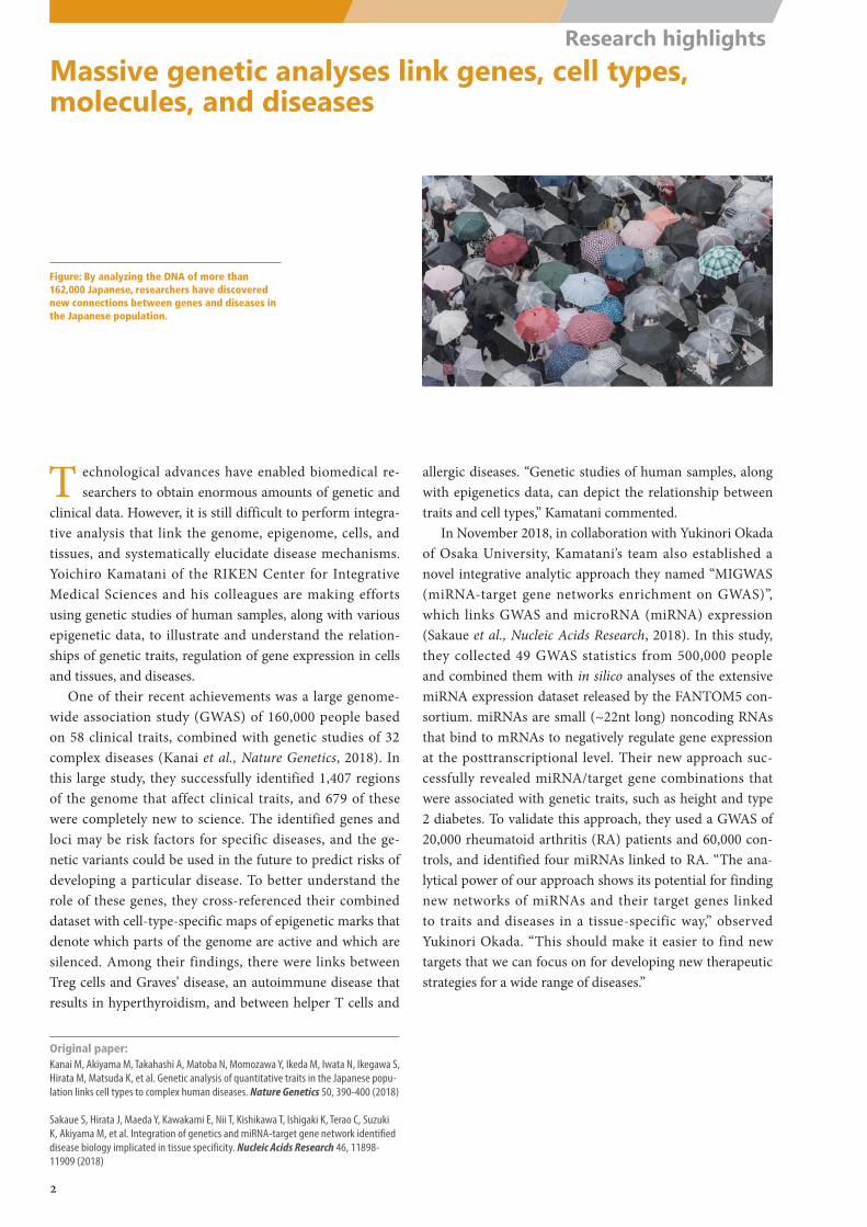
Massive genetic analyses link genes, cell types, molecules, and diseases
Figure: By analyzing the DNA of more than 162,000 Japanese, researchers have discovered new connections between genes and diseases in the Japanese population.
Original paper:Kanai M, Akiyama M, Takahashi A, Matoba N, Momozawa Y, Ikeda M, Iwata N, Ikegawa S, Hirata M, Matsuda K, et al. Genetic analysis of quantitative traits in the Japanese popu-lation links cell types to complex human diseases. Nature Genetics 50, 390-400 (2018)
Sakaue S, Hirata J, Maeda Y, Kawakami E, Nii T, Kishikawa T, Ishigaki K, Terao C, Suzuki K, Akiyama M, et al. Integration of genetics and miRNA-target gene network identified disease biology implicated in tissue specificity. Nucleic Acids Research 46, 11898-11909 (2018)
T echnological advances have enabled biomedical re-searchers to obtain enormous amounts of genetic and
clinical data. However, it is still difficult to perform integra-tive analysis that link the genome, epigenome, cells, and tissues, and systematically elucidate disease mechanisms. Yoichiro Kamatani of the RIKEN Center for Integrative Medical Sciences and his colleagues are making efforts using genetic studies of human samples, along with various epigenetic data, to illustrate and understand the relation-ships of genetic traits, regulation of gene expression in cells and tissues, and diseases.
One of their recent achievements was a large genome-wide association study (GWAS) of 160,000 people based on 58 clinical traits, combined with genetic studies of 32 complex diseases (Kanai et al., Nature Genetics, 2018). In this large study, they successfully identified 1,407 regions of the genome that affect clinical traits, and 679 of these were completely new to science. The identified genes and loci may be risk factors for specific diseases, and the ge-netic variants could be used in the future to predict risks of developing a particular disease. To better understand the role of these genes, they cross-referenced their combined dataset with cell-type-specific maps of epigenetic marks that denote which parts of the genome are active and which are silenced. Among their findings, there were links between Treg cells and Graves’ disease, an autoimmune disease that results in hyperthyroidism, and between helper T cells and
allergic diseases. “Genetic studies of human samples, along with epigenetics data, can depict the relationship between traits and cell types,” Kamatani commented.
In November 2018, in collaboration with Yukinori Okada of Osaka University, Kamatani’s team also established a novel integrative analytic approach they named “MIGWAS (miRNA-target gene networks enrichment on GWAS)”, which links GWAS and microRNA (miRNA) expression (Sakaue et al., Nucleic Acids Research, 2018). In this study, they collected 49 GWAS statistics from 500,000 people and combined them with in silico analyses of the extensive miRNA expression dataset released by the FANTOM5 con-sortium. miRNAs are small (~22nt long) noncoding RNAs that bind to mRNAs to negatively regulate gene expression at the posttranscriptional level. Their new approach suc-cessfully revealed miRNA/target gene combinations that were associated with genetic traits, such as height and type 2 diabetes. To validate this approach, they used a GWAS of 20,000 rheumatoid arthritis (RA) patients and 60,000 con-trols, and identified four miRNAs linked to RA. “The ana-lytical power of our approach shows its potential for finding new networks of miRNAs and their target genes linked to traits and diseases in a tissue-specific way,” observed Yukinori Okada. “This should make it easier to find new targets that we can focus on for developing new therapeutic strategies for a wide range of diseases.”
2
Research highlights
Nos2Gbp5Cxcl10Serpinb9ProcrIrf8AW112010Irf1
AcppCxcl11Cxcl9IL4IL13_M
tb_48h
IL4IL13_Mtb_24h
IL4IL13_Mtb_12h
IL4IL13_Mtb_4h
IFNg_M
tb_48h
IFNg_M
tb_24h
IFNg_M
tb_12h
IFNg_M
tb_4h
Mtb_48h
Mtb_24h
Mtb_12h
Mtb_4h
Unstim
ulated_48h
Unstim
ulated_24h
Unstim
ulated_12h
Unstim
ulated_4h
Unstim
ulated_0h
Transcriptional landscape of Mycobacterium tuberculosis infection in macrophages
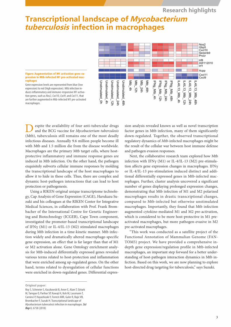
Figure: Augmentation of M1 activation gene ex-pression in Mtb-infected M1 pre-activated mac-rophagesGene expression levels are represented from blue (low expression) to red (high expression). Mtb infection in-duces inflammatory and immune-responsive M1 activa-tion genes, such as Nos2, Cxcl10, Cxcl9, and Cxcl11, that are further augmented in Mtb-infected M1 pre-activated macrophages.
Original paper:Roy S, Schmeier S, Kaczkowski B, Arner E, Alam T, Ozturk M, Tamgue O, Parihar SP, Kawaji H, Itoh M, Lassmann T, Carninci P, Hayashizaki Y, Forrest ARR, Guler R, Bajic VB, Brombacher F, Suzuki H. Transcriptional landscape of Mycobacterium tuberculosis infection in macrophages. Sci Rep 8, 6758 (2018)
D espite the availability of four anti-tubercular drugs and the BCG vaccine for Mycobacterium tuberculosis
(Mtb), tuberculosis still remains one of the most deadly infectious diseases. Annually 9.6 million people become ill with Mtb and 1.5 million die from the disease worldwide. Macrophages are the primary Mtb target cells, where host-protective inflammatory and immune response genes are induced in Mtb infection. On the other hand, the pathogen exquisitely subverts cellular immune responses by molding the transcriptional landscape of the host macrophages to allow it to hide in these cells. Thus, there are complex and dynamic host-pathogen interactions that can lead to host protection or pathogenesis.
Using a RIKEN-original unique transcriptome technolo-gy, Cap Analysis of Gene Expression (CAGE), Harukazu Su-zuki and his colleagues at the RIKEN Center for Integrative Medical Sciences, in collaboration with Prof. Frank Brom-bacher of the International Centre for Genetic Engineer-ing and Biotechnology (ICGEB), Cape Town component, investigated the promoter-based transcriptional landscape of IFNγ (M1) or IL-4/IL-13 (M2) stimulated macrophages during Mtb infection in a time-kinetic manner. Mtb infec-tion widely and dramatically altered macrophage-specific gene expression, an effect that is far larger than that of M1 or M2 activation alone. Gene Ontology enrichment analy-sis for Mtb-induced differentially expressed genes revealed various terms related to host-protection and inflammation that were enriched among up-regulated genes. On the other hand, terms related to dysregulation of cellular functions were enriched in down-regulated genes. Differential expres-
sion analysis revealed known as well as novel transcription factor genes in Mtb infection, many of them significantly down-regulated. Together, the observed transcriptional regulatory dynamics of Mtb-infected macrophages might be the result of the cellular war between host immune defense and pathogen evasion responses.
Next, the collaborative research team explored how Mtb infection with IFNγ (M1) or IL-4/IL-13 (M2) pre-stimula-tion affects gene expression changes in macrophages. IFNγ or IL-4/IL-13 pre-stimulation induced distinct and addi-tional differentially expressed genes in Mtb-infected mac-rophages. Further, cluster analysis uncovered a significant number of genes displaying prolonged expression changes, demonstrating that Mtb infection of M1 and M2 polarized macrophages results in drastic transcriptional changes, compared to Mtb-infected but otherwise unstimulated macrophages. Importantly, they found that Mtb infection augmented cytokine-mediated M1 and M2 pre-activation, which is considered to be more host-protective in M1 pre-activated macrophages, but more pathogen-evasive in M2 pre-activated macrophages.
“This work was conducted as a satellite project of the Functional Annotation of Mammalian Genome (FAN-TOM5) project. We have provided a comprehensive in-depth gene expression/regulation profile in Mtb-infected macrophages, an important step forward for a better under-standing of host-pathogen interaction dynamics in Mtb in-fection. Based on this work, we are now planning to explore host-directed drug targeting for tuberculosis,” says Suzuki.
3
Research highlights
Plasma cell
Ag
TFH GC-B
Bcl6loCD69hiBcl6hiCD69hi
Recycling
IRF4+
LZ
DZ
Strong Weak T-B interaction
Researchers uncover origin of virus-fighting plasma B cells
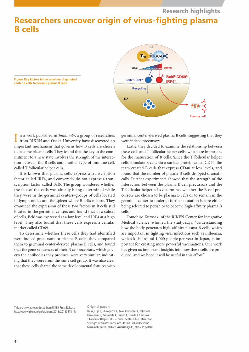
Figure: Key factors in the selection of germinal center B cells to become plasma B cells
This article was reproduced from RIKEN Press Releasehttp://www.riken.jp/en/pr/press/2018/20180418_1/
Original paper:Ise W, Fujii K, Shiroguchi K, Ito A, Kometani K, Takeda K, Kawakami E, Yamashita K, Suzuki K, Okada T, Kurosaki T. T Follicular Helper Cell-Germinal Center B Cell Interaction Strength Regulates Entry into Plasma Cell or Recycling Germinal Center Cell Fate. Immunity 48, 702-715 (2018)
I n a work published in Immunity, a group of researchers from RIKEN and Osaka University have discovered an
important mechanism that governs how B cells are chosen to become plasma cells. They found that the key to the com-mitment to a new state involves the strength of the interac-tion between the B cells and another type of immune cell, called T follicular helper cells.
It is known that plasma cells express a transcription factor called IRF4, and conversely do not express a tran-scription factor called Bcl6. The group wondered whether the fate of the cells was already being determined while they were in the germinal centers—groups of cells located in lymph nodes and the spleen where B cells mature. They examined the expression of these two factors in B cells still located in the germinal centers and found that in a subset of cells, Bcl6 was expressed at a low level and IRF4 at a high level. They also found that these cells express a cellular marker called CD69.
To determine whether these cells they had identified were indeed precursors to plasma B cells, they compared them to germinal center-derived plasma B cells, and found that the gene sequences of their B cell receptors, which gov-ern the antibodies they produce, were very similar, indicat-ing that they were from the same cell group. It was also clear that these cells shared the same developmental features with
germinal center-derived plasma B cells, suggesting that they were indeed precursors.
Lastly, they decided to examine the relationship between these cells and T follicular helper cells, which are important for the maturation of B cells. Since the T follicular helper cells stimulate B cells via a surface protein called CD40, the team created B cells that express CD40 at low levels, and found that the number of plasma B cells dropped dramati-cally. Further experiments showed that the strength of the interaction between the plasma B cell precursors and the T follicular helper cells determines whether the B cell pre-cursors are chosen to be plasma B cells or to remain in the germinal center to undergo further mutation before either being selected to perish or to become high-affinity plasma B cells.
Tomohiro Kurosaki of the RIKEN Center for Integrative Medical Science, who led the study, says, “Understanding how the body generates high-affinity plasma B cells, which are important in fighting viral infections such as influenza, which kills around 1,000 people per year in Japan, is im-portant for creating more powerful vaccinations. Our work has given us important insights into how these cells are pro-duced, and we hope it will be useful in this effort.”
4
Research highlights
Early (0.5-4hr) Mid (6-48hr) Late (72-168hr)
Tle2
Klf4
Relb
Klf2
Per2
Arid3a
Per1
Rel
Egr1 Rarb
JundAtf3Kdm6b
Egr2
Junb
Fos
Gfi1bNfil3
Nfatc1
Cebpa
FosbTead4 Klf5
Tead1Smad1
Vdr
Zfp57
Lhx2Srf
Pitx1
Tead2Yap1
Gata4Gata2
Gli3Msx1
Fosl2
NficMaff
Snai1
Cebpb
Nr2f2Pparg
Nfib
Fosl1
Tbx19Ets1
Mtf2
Stat4Gfi1 Ikzf3
ThrbPrdm9Parp1Wt1
Spib
Irf8
Phf19Lmnb1
Irf4
Men1
Ebf1Ncapg2
Nfe2
Lyl1
Tal1 EomesErg
E2f2
Ezh2
Foxo1Myb Pax5
Smarca4
Lmo2
Hematopoietic stem cell B cellDifferentiation
Three-step programming
Three transcriptional circuits found between hematopoietic stem cells and B cellsB cells pass through three distinct gene expression networks on their way from stem cells to B cells
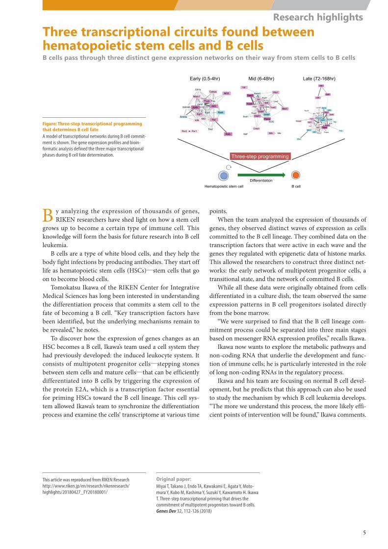
Figure: Three-step transcriptional programming that determines B cell fateA model of transcriptional networks during B cell commit-ment is shown. The gene expression profiles and bioin-formatic analysis defined the three major transcriptional phases during B cell fate determination.
This article was reproduced from RIKEN Researchhttp://www.riken.jp/en/research/rikenresearch/ highlights/20180427_FY20180001/
Original paper:Miyai T, Takano J, Endo TA, Kawakami E, Agata Y, Moto-mura Y, Kubo M, Kashima Y, Suzuki Y, Kawamoto H. Ikawa T. Three-step transcriptional priming that drives the commitment of multipotent progenitors toward B cells. Genes Dev 32, 112-126 (2018)
B y analyzing the expression of thousands of genes, RIKEN researchers have shed light on how a stem cell
grows up to become a certain type of immune cell. This knowledge will form the basis for future research into B cell leukemia.
B cells are a type of white blood cells, and they help the body fight infections by producing antibodies. They start off life as hematopoietic stem cells (HSCs)―stem cells that go on to become blood cells.
Tomokatsu Ikawa of the RIKEN Center for Integrative Medical Sciences has long been interested in understanding the differentiation process that commits a stem cell to the fate of becoming a B cell. “Key transcription factors have been identified, but the underlying mechanisms remain to be revealed,” he notes.
To discover how the expression of genes changes as an HSC becomes a B cell, Ikawa’s team used a cell system they had previously developed: the induced leukocyte system. It consists of multipotent progenitor cells―stepping stones between stem cells and mature cells―that can be efficiently differentiated into B cells by triggering the expression of the protein E2A, which is a transcription factor essential for priming HSCs toward the B cell lineage. This cell sys-tem allowed Ikawa’s team to synchronize the differentiation process and examine the cells’ transcriptome at various time
points.When the team analyzed the expression of thousands of
genes, they observed distinct waves of expression as cells committed to the B cell lineage. They combined data on the transcription factors that were active in each wave and the genes they regulated with epigenetic data of histone marks. This allowed the researchers to construct three distinct net-works: the early network of multipotent progenitor cells, a transitional state, and the network of committed B cells.
While all these data were originally obtained from cells differentiated in a culture dish, the team observed the same expression patterns in B cell progenitors isolated directly from the bone marrow.
“We were surprised to find that the B cell lineage com-mitment process could be separated into three main stages based on messenger RNA expression profiles,” recalls Ikawa.
Ikawa now wants to explore the metabolic pathways and non-coding RNA that underlie the development and func-tion of immune cells; he is particularly interested in the role of long non-coding RNAs in the regulatory process.
Ikawa and his team are focusing on normal B cell devel-opment, but he predicts that this approach can also be used to study the mechanism by which B cell leukemia develops. “The more we understand this process, the more likely effi-cient points of intervention will be found,” Ikawa comments.
5
Research highlights
Prognostic factors
Germline variants of cancer predisposing genes
TMA
Cell‐of‐origin prediction by WGS and epigenome
Liver origin
MUC17 expression
Genome sequencing analysis reveals genes associated with survival in biliary tract cancers
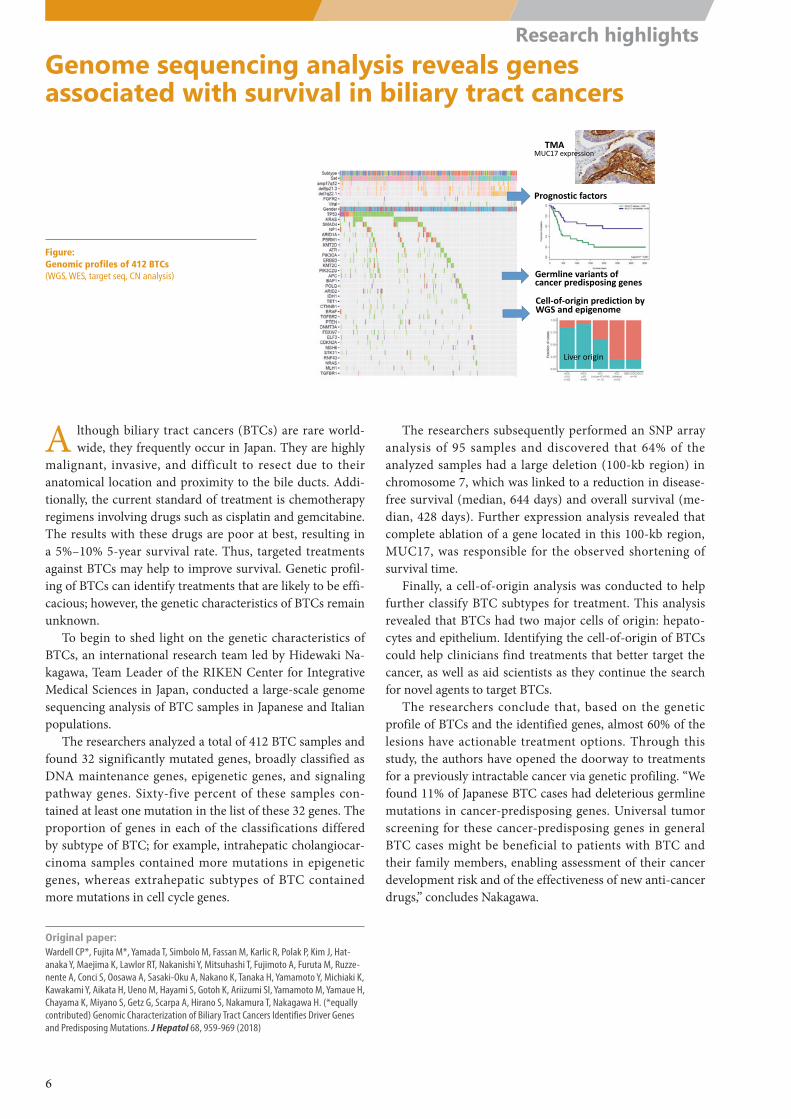
Figure: Genomic profiles of 412 BTCs (WGS, WES, target seq, CN analysis)
Original paper:Wardell CP*, Fujita M*, Yamada T, Simbolo M, Fassan M, Karlic R, Polak P, Kim J, Hat-anaka Y, Maejima K, Lawlor RT, Nakanishi Y, Mitsuhashi T, Fujimoto A, Furuta M, Ruzze-nente A, Conci S, Oosawa A, Sasaki-Oku A, Nakano K, Tanaka H, Yamamoto Y, Michiaki K, Kawakami Y, Aikata H, Ueno M, Hayami S, Gotoh K, Ariizumi SI, Yamamoto M, Yamaue H, Chayama K, Miyano S, Getz G, Scarpa A, Hirano S, Nakamura T, Nakagawa H. (*equally contributed) Genomic Characterization of Biliary Tract Cancers Identifies Driver Genes and Predisposing Mutations. J Hepatol 68, 959-969 (2018)
A lthough biliary tract cancers (BTCs) are rare world-wide, they frequently occur in Japan. They are highly
malignant, invasive, and difficult to resect due to their anatomical location and proximity to the bile ducts. Addi-tionally, the current standard of treatment is chemotherapy regimens involving drugs such as cisplatin and gemcitabine. The results with these drugs are poor at best, resulting in a 5%–10% 5-year survival rate. Thus, targeted treatments against BTCs may help to improve survival. Genetic profil-ing of BTCs can identify treatments that are likely to be effi-cacious; however, the genetic characteristics of BTCs remain unknown.
To begin to shed light on the genetic characteristics of BTCs, an international research team led by Hidewaki Na-kagawa, Team Leader of the RIKEN Center for Integrative Medical Sciences in Japan, conducted a large-scale genome sequencing analysis of BTC samples in Japanese and Italian populations.
The researchers analyzed a total of 412 BTC samples and found 32 significantly mutated genes, broadly classified as DNA maintenance genes, epigenetic genes, and signaling pathway genes. Sixty-five percent of these samples con-tained at least one mutation in the list of these 32 genes. The proportion of genes in each of the classifications differed by subtype of BTC; for example, intrahepatic cholangiocar-cinoma samples contained more mutations in epigenetic genes, whereas extrahepatic subtypes of BTC contained more mutations in cell cycle genes.
The researchers subsequently performed an SNP array analysis of 95 samples and discovered that 64% of the analyzed samples had a large deletion (100-kb region) in chromosome 7, which was linked to a reduction in disease-free survival (median, 644 days) and overall survival (me-dian, 428 days). Further expression analysis revealed that complete ablation of a gene located in this 100-kb region, MUC17, was responsible for the observed shortening of survival time.
Finally, a cell-of-origin analysis was conducted to help further classify BTC subtypes for treatment. This analysis revealed that BTCs had two major cells of origin: hepato-cytes and epithelium. Identifying the cell-of-origin of BTCs could help clinicians find treatments that better target the cancer, as well as aid scientists as they continue the search for novel agents to target BTCs.
The researchers conclude that, based on the genetic profile of BTCs and the identified genes, almost 60% of the lesions have actionable treatment options. Through this study, the authors have opened the doorway to treatments for a previously intractable cancer via genetic profiling. “We found 11% of Japanese BTC cases had deleterious germline mutations in cancer-predisposing genes. Universal tumor screening for these cancer-predisposing genes in general BTC cases might be beneficial to patients with BTC and their family members, enabling assessment of their cancer development risk and of the effectiveness of new anti-cancer drugs,” concludes Nakagawa.
6
Research highlights
Part 2
Lab Activities
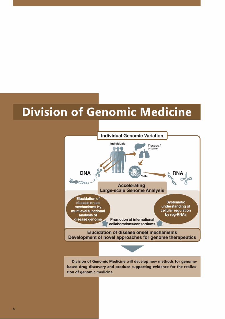
Division of Genomic Medicine
Division of Genomic Medicine will develop new methods for genome-based drug discovery and produce supporting evidence for the realiza-tion of genomic medicine.
8
A
C
B
Laboratory for
Transcriptome TechnologyTeam Leader: Piero Carninci
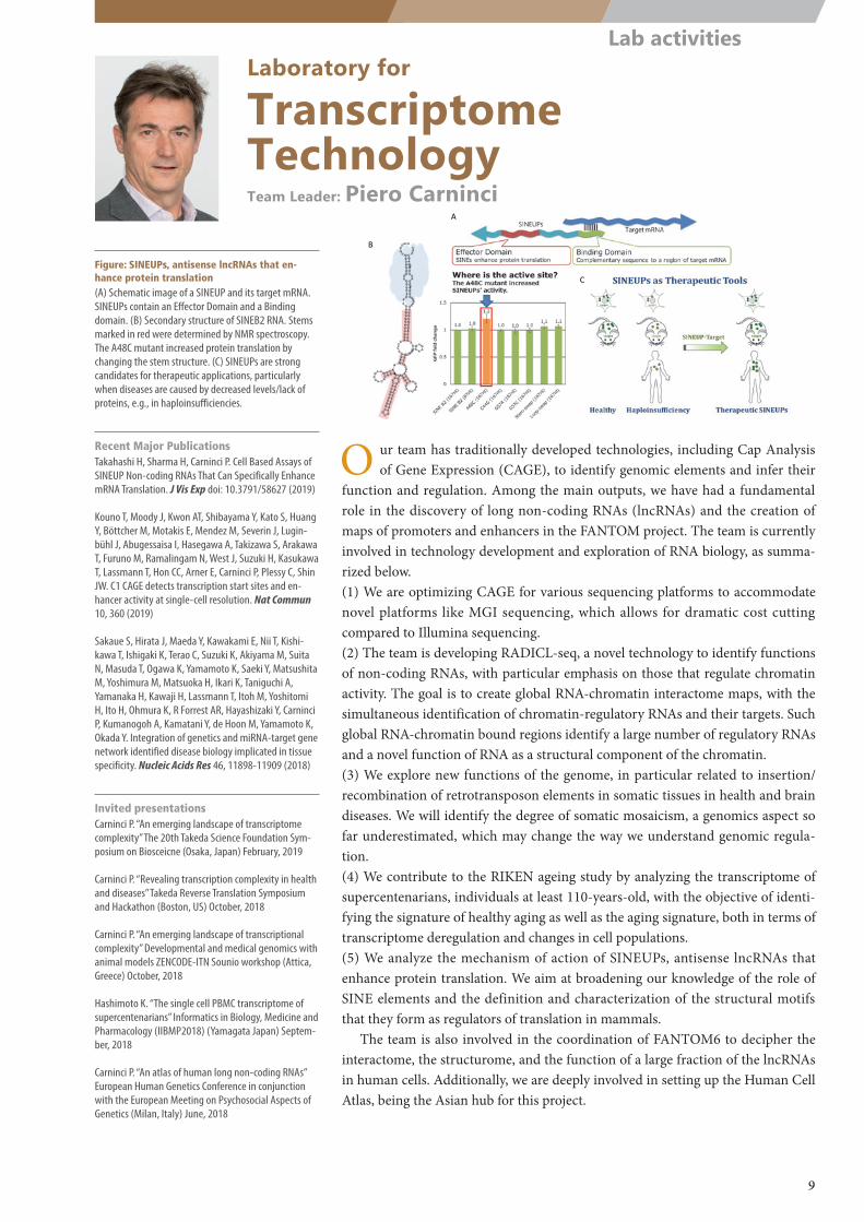
Figure: SINEUPs, antisense lncRNAs that en-hance protein translation(A) Schematic image of a SINEUP and its target mRNA. SINEUPs contain an Effector Domain and a Binding domain. (B) Secondary structure of SINEB2 RNA. Stems marked in red were determined by NMR spectroscopy. The A48C mutant increased protein translation by changing the stem structure. (C) SINEUPs are strong candidates for therapeutic applications, particularly when diseases are caused by decreased levels/lack of proteins, e.g., in haploinsufficiencies.
Recent Major PublicationsTakahashi H, Sharma H, Carninci P. Cell Based Assays of SINEUP Non-coding RNAs That Can Specifically Enhance mRNA Translation. J Vis Exp doi: 10.3791/58627 (2019)
Kouno T, Moody J, Kwon AT, Shibayama Y, Kato S, Huang Y, Böttcher M, Motakis E, Mendez M, Severin J, Lugin-bühl J, Abugessaisa I, Hasegawa A, Takizawa S, Arakawa T, Furuno M, Ramalingam N, West J, Suzuki H, Kasukawa T, Lassmann T, Hon CC, Arner E, Carninci P, Plessy C, Shin JW. C1 CAGE detects transcription start sites and en-hancer activity at single-cell resolution. Nat Commun 10, 360 (2019)
Sakaue S, Hirata J, Maeda Y, Kawakami E, Nii T, Kishi-kawa T, Ishigaki K, Terao C, Suzuki K, Akiyama M, Suita N, Masuda T, Ogawa K, Yamamoto K, Saeki Y, Matsushita M, Yoshimura M, Matsuoka H, Ikari K, Taniguchi A, Yamanaka H, Kawaji H, Lassmann T, Itoh M, Yoshitomi H, Ito H, Ohmura K, R Forrest AR, Hayashizaki Y, Carninci P, Kumanogoh A, Kamatani Y, de Hoon M, Yamamoto K, Okada Y. Integration of genetics and miRNA-target gene network identified disease biology implicated in tissue specificity. Nucleic Acids Res 46, 11898-11909 (2018)
Invited presentationsCarninci P. “An emerging landscape of transcriptome complexity” The 20th Takeda Science Foundation Sym-posium on Biosceicne (Osaka, Japan) February, 2019
Carninci P. “Revealing transcription complexity in health and diseases” Takeda Reverse Translation Symposium and Hackathon (Boston, US) October, 2018
Carninci P. “An emerging landscape of transcriptional complexity” Developmental and medical genomics with animal models ZENCODE-ITN Sounio workshop (Attica, Greece) October, 2018
Hashimoto K. “The single cell PBMC transcriptome of supercentenarians” Informatics in Biology, Medicine and Pharmacology (IIBMP2018) (Yamagata Japan) Septem-ber, 2018
Carninci P. “An atlas of human long non-coding RNAs” European Human Genetics Conference in conjunction with the European Meeting on Psychosocial Aspects of Genetics (Milan, Italy) June, 2018
O ur team has traditionally developed technologies, including Cap Analysis of Gene Expression (CAGE), to identify genomic elements and infer their
function and regulation. Among the main outputs, we have had a fundamental role in the discovery of long non-coding RNAs (lncRNAs) and the creation of maps of promoters and enhancers in the FANTOM project. The team is currently involved in technology development and exploration of RNA biology, as summa-rized below.(1) We are optimizing CAGE for various sequencing platforms to accommodate novel platforms like MGI sequencing, which allows for dramatic cost cutting compared to Illumina sequencing.(2) The team is developing RADICL-seq, a novel technology to identify functions of non-coding RNAs, with particular emphasis on those that regulate chromatin activity. The goal is to create global RNA-chromatin interactome maps, with the simultaneous identification of chromatin-regulatory RNAs and their targets. Such global RNA-chromatin bound regions identify a large number of regulatory RNAs and a novel function of RNA as a structural component of the chromatin.(3) We explore new functions of the genome, in particular related to insertion/recombination of retrotransposon elements in somatic tissues in health and brain diseases. We will identify the degree of somatic mosaicism, a genomics aspect so far underestimated, which may change the way we understand genomic regula-tion.(4) We contribute to the RIKEN ageing study by analyzing the transcriptome of supercentenarians, individuals at least 110-years-old, with the objective of identi-fying the signature of healthy aging as well as the aging signature, both in terms of transcriptome deregulation and changes in cell populations.(5) We analyze the mechanism of action of SINEUPs, antisense lncRNAs that enhance protein translation. We aim at broadening our knowledge of the role of SINE elements and the definition and characterization of the structural motifs that they form as regulators of translation in mammals.
The team is also involved in the coordination of FANTOM6 to decipher the interactome, the structurome, and the function of a large fraction of the lncRNAs in human cells. Additionally, we are deeply involved in setting up the Human Cell Atlas, being the Asian hub for this project.
9
Lab activities
Methylated Demethylated
GATA6_1FOS_1
TFAP2C_2−10g10P
150100500
12000
8000
4000
0
Laboratory for
Cellular Function Conversion TechnologyTeam Leader: Harukazu Suzuki
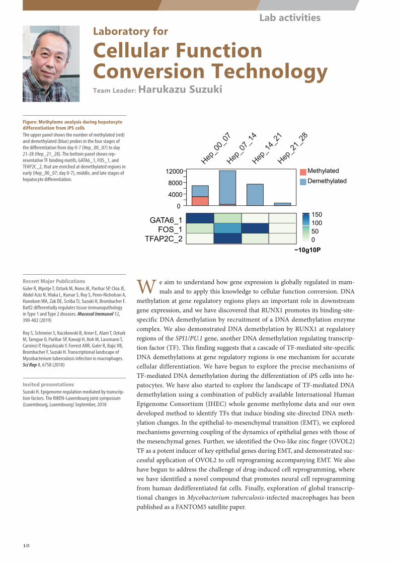
Figure: Methylome analysis during hepatocyte differentiation from iPS cellsThe upper panel shows the number of methylated (red) and demethylated (blue) probes in the four stages of the differentiation from day 0-7 (Hep_00_07) to day 21-28 (Hep_21_28). The bottom panel shows rep-resentative TF binding motifs, GATA6_1, FOS_1, and TFAP2C_2, that are enriched at demethylated regions in early (Hep_00_07; day 0-7), middle, and late stages of hepatocyte differentiation.
Recent Major PublicationsGuler R, Mpotje T, Ozturk M, Nono JK, Parihar SP, Chia JE, Abdel Aziz N, Hlaka L, Kumar S, Roy S, Penn-Nicholson A, Hanekom WA, Zak DE, Scriba TJ, Suzuki H, Brombacher F. Batf2 differentially regulates tissue immunopathology in Type 1 and Type 2 diseases. Mucosal Immunol 12, 390-402 (2019)
Roy S, Schmeier S, Kaczkowski B, Arner E, Alam T, Ozturk M, Tamgue O, Parihar SP, Kawaji H, Itoh M, Lassmann T, Carninci P, Hayashizaki Y, Forrest ARR, Guler R, Bajic VB, Brombacher F, Suzuki H. Transcriptional landscape of Mycobacterium tuberculosis infection in macrophages. Sci Rep 8, 6758 (2018)
Invited presentationsSuzuki H. Epigenome regulation mediated by transcrip-tion factors. The RIKEN-Luxembourg joint symposium (Luxembourg, Luxembourg) September, 2018
W e aim to understand how gene expression is globally regulated in mam-mals and to apply this knowledge to cellular function conversion. DNA
methylation at gene regulatory regions plays an important role in downstream gene expression, and we have discovered that RUNX1 promotes its binding-site-specific DNA demethylation by recruitment of a DNA demethylation enzyme complex. We also demonstrated DNA demethylation by RUNX1 at regulatory regions of the SPI1/PU.1 gene, another DNA demethylation regulating transcrip-tion factor (TF). This finding suggests that a cascade of TF-mediated site-specific DNA demethylations at gene regulatory regions is one mechanism for accurate cellular differentiation. We have begun to explore the precise mechanisms of TF-mediated DNA demethylation during the differentiation of iPS cells into he-patocytes. We have also started to explore the landscape of TF-mediated DNA demethylation using a combination of publicly available International Human Epigenome Consortium (IHEC) whole genome methylome data and our own developed method to identify TFs that induce binding site-directed DNA meth-ylation changes. In the epithelial-to-mesenchymal transition (EMT), we explored mechanisms governing coupling of the dynamics of epithelial genes with those of the mesenchymal genes. Further, we identified the Ovo-like zinc finger (OVOL2) TF as a potent inducer of key epithelial genes during EMT, and demonstrated suc-cessful application of OVOL2 to cell reprograming accompanying EMT. We also have begun to address the challenge of drug-induced cell reprogramming, where we have identified a novel compound that promotes neural cell reprogramming from human dedifferentiated fat cells. Finally, exploration of global transcrip-tional changes in Mycobacterium tuberculosis-infected macrophages has been published as a FANTOM5 satellite paper.
10
Lab activities
medulla oblongata brainstem pons thalamic complex cerebellum hippocampal formation caudate nucleus putamen striatum dorsal striatum frontal cortex middle frontal gyrus gyrus temporal lobe middle temporal gyrus metencephalon brain nervous system forebrain telencephalon brain gray matter occipital lobecerebral cortex parietal lobesubstantia nigraspinal cord nucleus of brain cerebral subcortextelencephalic nucleus
Nausea and vomitingSerum zinc measurementDementiaAutismAutistic disorderAutism spectrum disorderBipolar disorderBipolar affective disorderBehavioral abnormalityAuditory system diseaseCocaine dependenceAmyotrophic lateral sclerosisParkinsonian disordersObesityNicotine dependenceAbnormality of the CNSAttention deficit disorderAzoospermiaMental depressionGrowth abnormalityBody mass indexSchizophreniaMajor depressive disorderDepressionCognitive disorder Psychomotor performanceDyslexiaVascular calcification
leve
l of a
ssoc
iatio
n
123456
≥9
78
immune systemnervous systempigmented cellscardiovascular systemnon-immune blood cellshepato-intestinal system
cell-types (n=135)
traits
(n=2
77)
infec
tion a
nd im
munit
yne
urop
athy a
nd be
havio
rpig
menta
tion
card
iovas
cular
func
tion
blood
home
ostas
ishe
patic
func
tion
Laboratory for
Genome Information AnalysisTeam Leader: Chung-Chau Hon
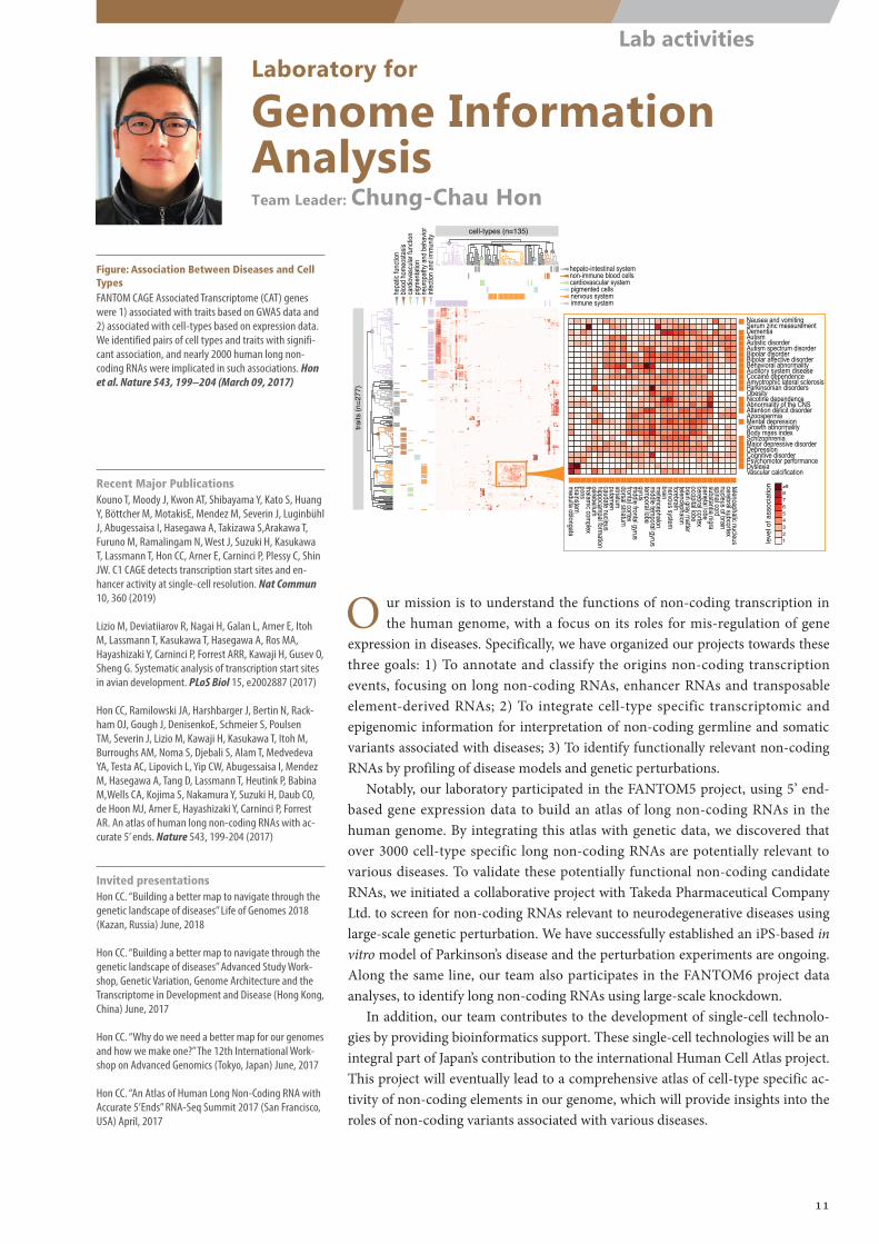
Figure: Association Between Diseases and Cell TypesFANTOM CAGE Associated Transcriptome (CAT) genes were 1) associated with traits based on GWAS data and 2) associated with cell-types based on expression data. We identified pairs of cell types and traits with signifi-cant association, and nearly 2000 human long non-coding RNAs were implicated in such associations. Hon et al. Nature 543, 199–204 (March 09, 2017)
O ur mission is to understand the functions of non-coding transcription in the human genome, with a focus on its roles for mis-regulation of gene
expression in diseases. Specifically, we have organized our projects towards these three goals: 1) To annotate and classify the origins non-coding transcription events, focusing on long non-coding RNAs, enhancer RNAs and transposable element-derived RNAs; 2) To integrate cell-type specific transcriptomic and epigenomic information for interpretation of non-coding germline and somatic variants associated with diseases; 3) To identify functionally relevant non-coding RNAs by profiling of disease models and genetic perturbations.
Notably, our laboratory participated in the FANTOM5 project, using 5’ end-based gene expression data to build an atlas of long non-coding RNAs in the human genome. By integrating this atlas with genetic data, we discovered that over 3000 cell-type specific long non-coding RNAs are potentially relevant to various diseases. To validate these potentially functional non-coding candidate RNAs, we initiated a collaborative project with Takeda Pharmaceutical Company Ltd. to screen for non-coding RNAs relevant to neurodegenerative diseases using large-scale genetic perturbation. We have successfully established an iPS-based in vitro model of Parkinson’s disease and the perturbation experiments are ongoing. Along the same line, our team also participates in the FANTOM6 project data analyses, to identify long non-coding RNAs using large-scale knockdown.
In addition, our team contributes to the development of single-cell technolo-gies by providing bioinformatics support. These single-cell technologies will be an integral part of Japan’s contribution to the international Human Cell Atlas project. This project will eventually lead to a comprehensive atlas of cell-type specific ac-tivity of non-coding elements in our genome, which will provide insights into the roles of non-coding variants associated with various diseases.
Recent Major PublicationsKouno T, Moody J, Kwon AT, Shibayama Y, Kato S, Huang Y, Böttcher M, MotakisE, Mendez M, Severin J, Luginbühl J, Abugessaisa I, Hasegawa A, Takizawa S,Arakawa T, Furuno M, Ramalingam N, West J, Suzuki H, Kasukawa T, Lassmann T, Hon CC, Arner E, Carninci P, Plessy C, Shin JW. C1 CAGE detects transcription start sites and en-hancer activity at single-cell resolution. Nat Commun 10, 360 (2019)
Lizio M, Deviatiiarov R, Nagai H, Galan L, Arner E, Itoh M, Lassmann T, Kasukawa T, Hasegawa A, Ros MA, Hayashizaki Y, Carninci P, Forrest ARR, Kawaji H, Gusev O, Sheng G. Systematic analysis of transcription start sites in avian development. PLoS Biol 15, e2002887 (2017)
Hon CC, Ramilowski JA, Harshbarger J, Bertin N, Rack-ham OJ, Gough J, DenisenkoE, Schmeier S, Poulsen TM, Severin J, Lizio M, Kawaji H, Kasukawa T, Itoh M, Burroughs AM, Noma S, Djebali S, Alam T, Medvedeva YA, Testa AC, Lipovich L, Yip CW, Abugessaisa I, Mendez M, Hasegawa A, Tang D, Lassmann T, Heutink P, Babina M,Wells CA, Kojima S, Nakamura Y, Suzuki H, Daub CO, de Hoon MJ, Arner E, Hayashizaki Y, Carninci P, Forrest AR. An atlas of human long non-coding RNAs with ac-curate 5’ ends. Nature 543, 199-204 (2017)
Invited presentationsHon CC. “Building a better map to navigate through the genetic landscape of diseases” Life of Genomes 2018 (Kazan, Russia) June, 2018
Hon CC. “Building a better map to navigate through the genetic landscape of diseases” Advanced Study Work-shop, Genetic Variation, Genome Architecture and the Transcriptome in Development and Disease (Hong Kong, China) June, 2017
Hon CC. “Why do we need a better map for our genomes and how we make one?” The 12th International Work-shop on Advanced Genomics (Tokyo, Japan) June, 2017
Hon CC. “An Atlas of Human Long Non-Coding RNA with Accurate 5’Ends” RNA-Seq Summit 2017 (San Francisco, USA) April, 2017
11
Lab activities
Celltype-1 …… …… Celltype-N
Fibroblast …… …… H1stemcell
lncRNA-1 FAM99B …… …… FAM99B
lncRNA-2 UBXN8 …… …… UBXN8…. …. …… …… ….…. …. …… …… ….…. …. …… …… ….
lncRNA-n SEMA3B …… …… SEMA3B
Hi-CinteractionsGenomicregion
Clustering
Genomicregioncluster-1
Genomicregioncluster-2
Annotationmapping
Genecluster-1
Genecluster-2
Func�onalanalysis:GOanalysiseQTLsGWAStraitsRNAbindingTranscriptionfactorsRNA-chromatininteraction
mRNAcodinglncRNA EnhancerGenomeconforma�onanalysis:
Skeletaldevelopment(Cellspecificfunction)
Chromatinorganization(Commonfunction)
Geneontology(GO)analysis:Databaseprovidingcelltype-specificfunc�onalannota�onsoflncRNAs:
Laboratory for
Applied Computational GenomicsTeam Leader: Michiel de Hoon
O ur laboratory applies computational methods to analyze transcriptome and other sequencing datasets to understand cellular regulation in general
and long non-coding RNAs (lncRNAs) in particular. We take a leading role in the bioinformatics analysis of data generated by the Functional Annotation of the Mammalian Genome 6 (FANTOM6) project, a pioneering effort to elucidate the function of long non-coding RNA. In particular, we focus on◦ The secondary and tertiary structures of lncRNAs, and the structure–function
relationship of lncRNAs;◦ The 3D structure of the genome as a framework for non-coding RNA-mediat-
ed regulatory interactions.In both aspects, we work together closely with other laboratories in our Cen-
ter, in particular, by sharing the analysis of the 3D structure of the genome to help understand GWAS SNPs identified in genetic studies performed by colleagues in our Center.
Additionally, we are finalizing projects remaining from FANTOM5 and from the pilot phase of FANTOM6. The FANTOM5 expression atlas of microRNAs was completed and published in 2017. A manuscript summarizing our FANTOM5 analysis of the conservation among vertebrate species of the coding and non-coding transcriptomes in primary cells is planned for submission within FY2018. The FANTOM6 pilot phase paper is currently being written together with Dr. Piero Carninci (Laboratory for Transcriptome Technology, RIKEN IMS) and Dr. Jay Shin (Laboratory for Advanced Genomics Circuit, RIKEN IMS).
Recent Major PublicationsSakaue S, Hirata J, Maeda Y, Kawakami E, Nii T, Kishi-kawa T, Ishigaki K, Terao C, Suzuki K, Akiyama M, Suita N, Masuda T, Ogawa K, Yamamoto K, Saeki Y, Matsushita M, Yoshimura M, Matsuoka H, Ikari K, Taniguchi A, Yamanaka H, Kawaji H, Lassmann T, Itoh M, Yoshitomi H, Ito H, Ohmura K, Forrest ARR, Hayashizaki Y, Carninci P, Kumanogoh A, Kamatani Y, De Hoon M, Yamamoto K, Okada Y. Integration of genetics and miRNA-target gene network identified disease biology implicated in tissue specificity. Nucleic Acids Res 46, 11898-11909 (2018)
De Rie D, Abugessaisa I, Alam T, Arner E, Arner P, Ashoor H, Åström G, Babina M, Bertin N, Burroughs AM, Carlisle AJ, Daub CO, Detmar M, Deviatiiarov R, Fort A, Gebhard C, Goldowitz D, Guhl S, Ha TJ, Harshbarger J, Hasegawa A, Hashimoto K, Herlyn M, Heutink P, Hitchens KJ, Hon CC, Huang E, Ishizu Y, Kai C, Kasukawa T, Klinken P, Lassmann T, Lecellier CH, Lee W, Lizio M, Makeev V, Mathelier A, Medvedeva YA, Mejhert N, Mungall CJ, Noma S, Ohshima M, Okada-Hatakeyama M, Persson H, Rizzu P, Roudnicky F, Sætrom P, Sato H, Severin J, Shin JW, Swoboda RK, Tarui H, Toyoda H, Vitting-Seerup K, Winteringham L, Yamaguchi Y, Yasuzawa K, Yoneda M, Yumoto N, Zabierowski S, Zhang PG, Wells CA, Summers KM, Kawaji H, Sandelin A, Rehli M, The FANTOM Con-sortium Hayashizaki Y, Carninci P, Forrest ARR, De Hoon MJL. An integrated expression atlas of miRNAs and their promoters in human and mouse. Nature Biotechnol 35, 872-878 (2017)
Hon CC, Ramilowski JA, Harshbarger J, Bertin N, Rack-ham OJ, Gough J, Denisenko E, Schmeier S, Poulsen TM, Severin J, Lizio M, Kawaji H, Kasukawa T, Itoh M, Bur-roughs AM, Noma S, Djebali S, Alam T, Medvedeva YA, Testa AC, Lipovich L, Yip CW, Abugessaisa I, Mendez M, Hasegawa A, Tang D, Lassmann T, Heutink P, Babina M, Wells CA, Kojima S, Nakamura Y, Suzuki H, Daub CO, De Hoon MJL, Arner E, Hayashizaki Y, Carninci P, Forrest ARR. An atlas of human long non-coding RNAs with accurate 5’ ends. Nature 543, 199-204 (2017)
Invited presentationsDe Hoon M. “Functional Annotation of Mammalian Genomes in the FANTOM projects” Annual Meeting of the Brazilian Bioinformatics and Computational Biology Association (X-Meeting) (São Pedro, Brazil) October, 2018
De Hoon M. “The FANTOM5 Integrated Expression Atlas of miRNAs and Their Promoters”. The 26th International KOGO Annual Conference: Genomics for the Future Biol-ogy and Medicine (Seoul, Korea) September, 2017
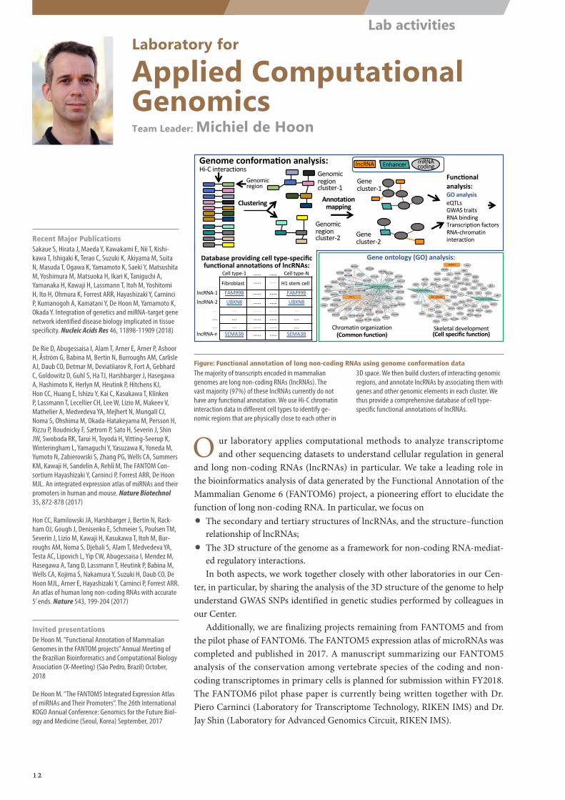
Figure: Functional annotation of long non-coding RNAs using genome conformation dataThe majority of transcripts encoded in mammalian genomes are long non-coding RNAs (lncRNAs). The vast majority (97%) of these lncRNAs currently do not have any functional annotation. We use Hi-C chromatin interaction data in different cell types to identify ge-nomic regions that are physically close to each other in
3D space. We then build clusters of interacting genomic regions, and annotate lncRNAs by associating them with genes and other genomic elements in each cluster. We thus provide a comprehensive database of cell type-specific functional annotations of lncRNAs.
12
Lab activities
A B
Collection of single cell barcoded cDNAs
Laboratory for
Single Cell TechnologiesTeam Leader: Piero Carninci
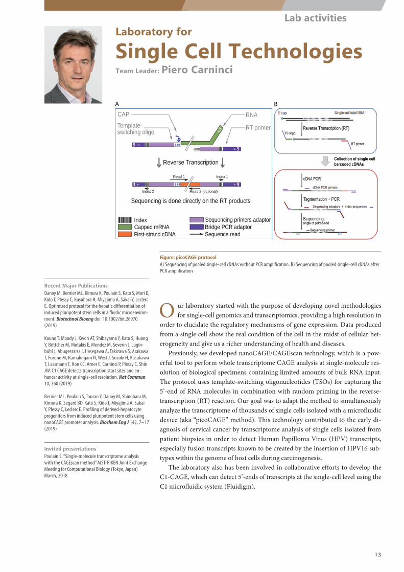
Figure: picoCAGE protocolA) Sequencing of pooled single-cell cDNAs without PCR amplification. B) Sequencing of pooled single-cell cDNAs after PCR amplification
Recent Major PublicationsDanoy M, Bernier ML, Kimura K, Poulain S, Kato S, Mori D, Kido T, Plessy C, Kusuhara H, Miyajima A, Sakai Y, Leclerc E. Optimized protocol for the hepatic differentiation of induced pluripotent stem cells in a fluidic microenviron-ment. Biotechnol Bioeng doi: 10.1002/bit.26970. (2019)
Kouno T, Moody J, Kwon AT, Shibayama Y, Kato S, Huang Y, Böttcher M, Motakis E, Mendez M, Severin J, Lugin-bühl J, Abugessaisa I, Hasegawa A, Takizawa S, Arakawa T, Furuno M, Ramalingam N, West J, Suzuki H, Kasukawa T, Lassmann T, Hon CC, Arner E, Carninci P, Plessy C, Shin JW. C1 CAGE detects transcription start sites and en-hancer activity at single-cell resolution. Nat Commun 10, 360 (2019)
Bernier ML, Poulain S, Tauran Y, Danoy M, Shinohara M, Kimura K, Segard BD, Kato S, Kido T, Miyajima A, Sakai Y, Plessy C, Leclerc E. Profiling of derived-hepatocyte progenitors from induced pluripotent stem cells using nanoCAGE promoter analysis. Biochem Eng J 142, 7–17 (2019)
Invited presentationsPoulain S. “Single-molecule transcriptome analysis with the CAGEscan method” AIST-RIKEN Joint Exchange Meeting for Computational Biology (Tokyo, Japan) March, 2018
O ur laboratory started with the purpose of developing novel methodologies for single-cell genomics and transcriptomics, providing a high resolution in
order to elucidate the regulatory mechanisms of gene expression. Data produced from a single cell show the real condition of the cell in the midst of cellular het-erogeneity and give us a richer understanding of health and diseases.
Previously, we developed nanoCAGE/CAGEscan technology, which is a pow-erful tool to perform whole transcriptome CAGE analysis at single-molecule res-olution of biological specimens containing limited amounts of bulk RNA input. The protocol uses template-switching oligonucleotides (TSOs) for capturing the 5’-end of RNA molecules in combination with random priming in the reverse-transcription (RT) reaction. Our goal was to adapt the method to simultaneously analyze the transcriptome of thousands of single cells isolated with a microfluidic device (aka “picoCAGE” method). This technology contributed to the early di-agnosis of cervical cancer by transcriptome analysis of single cells isolated from patient biopsies in order to detect Human Papilloma Virus (HPV) transcripts, especially fusion transcripts known to be created by the insertion of HPV16 sub-types within the genome of host cells during carcinogenesis.
The laboratory also has been involved in collaborative efforts to develop the C1-CAGE, which can detect 5’-ends of transcripts at the single-cell level using the C1 microfluidic system (Fluidigm).
13
Lab activities
A B
Large Scale Data Managing UnitUnit Leader: Takeya Kasukawa
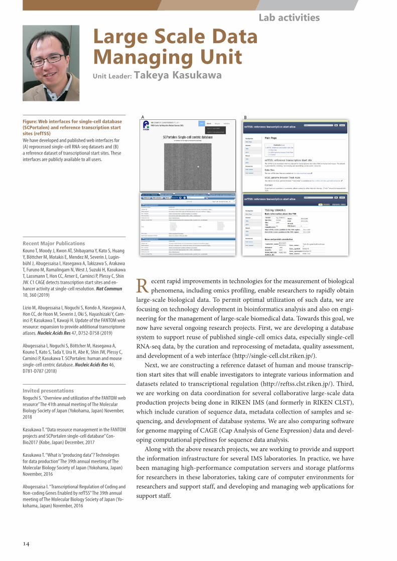
Figure: Web interfaces for single-cell database (SCPortalen) and reference transcription start sites (refTSS)We have developed and published web interfaces for (A) reprocessed single-cell RNA-seq datasets and (B) a reference dataset of transcriptional start sites. These interfaces are publicly available to all users.
R ecent rapid improvements in technologies for the measurement of biological phenomena, including omics profiling, enable researchers to rapidly obtain
large-scale biological data. To permit optimal utilization of such data, we are focusing on technology development in bioinformatics analysis and also on engi-neering for the management of large-scale biomedical data. Towards this goal, we now have several ongoing research projects. First, we are developing a database system to support reuse of published single-cell omics data, especially single-cell RNA-seq data, by the curation and reprocessing of metadata, quality assessment, and development of a web interface (http://single-cell.clst.riken.jp/).
Next, we are constructing a reference dataset of human and mouse transcrip-tion start sites that will enable investigators to integrate various information and datasets related to transcriptional regulation (http://reftss.clst.riken.jp/). Third, we are working on data coordination for several collaborative large-scale data production projects being done in RIKEN IMS (and formerly in RIKEN CLST), which include curation of sequence data, metadata collection of samples and se-quencing, and development of database systems. We are also comparing software for genome mapping of CAGE (Cap Analysis of Gene Expression) data and devel-oping computational pipelines for sequence data analysis.
Along with the above research projects, we are working to provide and support the information infrastructure for several IMS laboratories. In practice, we have been managing high-performance computation servers and storage platforms for researchers in these laboratories, taking care of computer environments for researchers and support staff, and developing and managing web applications for support staff.
Recent Major PublicationsKouno T, Moody J, Kwon AT, Shibayama Y, Kato S, Huang Y, Böttcher M, Motakis E, Mendez M, Severin J, Lugin-bühl J, Abugessaisa I, Hasegawa A, Takizawa S, Arakawa T, Furuno M, Ramalingam N, West J, Suzuki H, Kasukawa T, Lassmann T, Hon CC, Arner E, Carninci P, Plessy C, Shin JW. C1 CAGE detects transcription start sites and en-hancer activity at single-cell resolution. Nat Commun 10, 360 (2019)
Lizio M, Abugessaisa I, Noguchi S, Kondo A, Hasegawa A, Hon CC, de Hoon M, Severin J, Oki S, Hayashizaki Y, Carn-inci P, Kasukawa T, Kawaji H. Update of the FANTOM web resource: expansion to provide additional transcriptome atlases. Nucleic Acids Res 47, D752-D758 (2019)
Abugessaisa I, Noguchi S, Böttcher M, Hasegawa A, Kouno T, Kato S, Tada Y, Ura H, Abe K, Shin JW, Plessy C, Carninci P, Kasukawa T. SCPortalen: human and mouse single-cell centric database. Nucleic Acids Res 46, D781-D787 (2018)
Invited presentationsNoguchi S. “Overview and utilization of the FANTOM web resource” The 41th annual meeting of The Molecular Biology Society of Japan (Yokohama, Japan) November, 2018
Kasukawa T. “Data resource management in the FANTOM projects and SCPortalen single-cell database” Con-Bio2017 (Kobe, Japan) December, 2017
Kasukawa T. “What is “producing data”? Technologies for data production” The 39th annual meeting of The Molecular Biology Society of Japan (Yokohama, Japan) November, 2016
Abugessaisa I. “Transcriptional Regulation of Coding and Non-coding Genes Enabled by refTSS” The 39th annual meeting of The Molecular Biology Society of Japan (Yo-kohama, Japan) November, 2016
14
Lab activities
Laboratory for
Advanced Genomics CircuitTeam Leader: Jay W. Shin
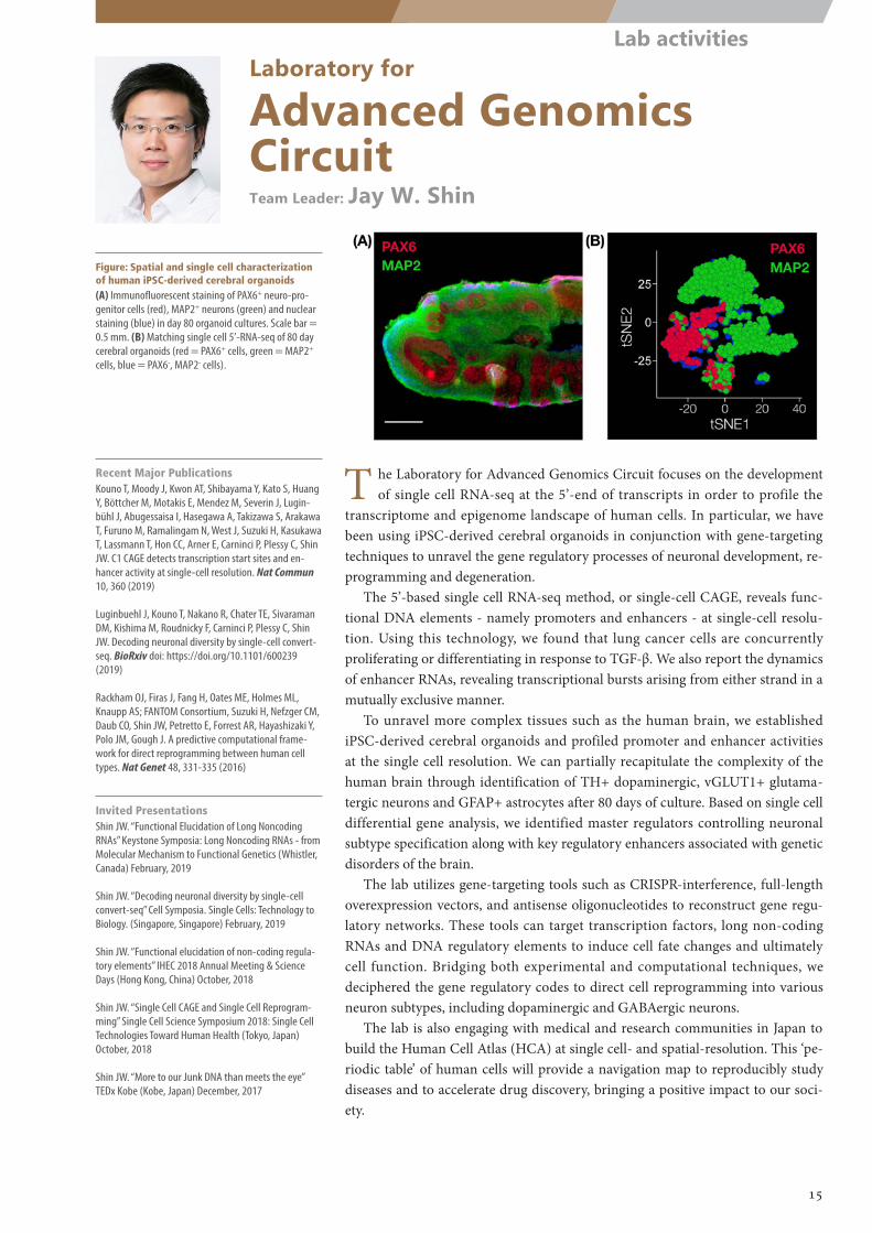
Figure: Spatial and single cell characterization of human iPSC-derived cerebral organoids(A) Immunofluorescent staining of PAX6+ neuro-pro-genitor cells (red), MAP2+ neurons (green) and nuclear staining (blue) in day 80 organoid cultures. Scale bar = 0.5 mm. (B) Matching single cell 5’-RNA-seq of 80 day cerebral organoids (red = PAX6+ cells, green = MAP2+ cells, blue = PAX6-, MAP2- cells).
T he Laboratory for Advanced Genomics Circuit focuses on the development of single cell RNA-seq at the 5’-end of transcripts in order to profile the
transcriptome and epigenome landscape of human cells. In particular, we have been using iPSC-derived cerebral organoids in conjunction with gene-targeting techniques to unravel the gene regulatory processes of neuronal development, re-programming and degeneration.
The 5’-based single cell RNA-seq method, or single-cell CAGE, reveals func-tional DNA elements - namely promoters and enhancers - at single-cell resolu-tion. Using this technology, we found that lung cancer cells are concurrently proliferating or differentiating in response to TGF-β. We also report the dynamics of enhancer RNAs, revealing transcriptional bursts arising from either strand in a mutually exclusive manner.
To unravel more complex tissues such as the human brain, we established iPSC-derived cerebral organoids and profiled promoter and enhancer activities at the single cell resolution. We can partially recapitulate the complexity of the human brain through identification of TH+ dopaminergic, vGLUT1+ glutama-tergic neurons and GFAP+ astrocytes after 80 days of culture. Based on single cell differential gene analysis, we identified master regulators controlling neuronal subtype specification along with key regulatory enhancers associated with genetic disorders of the brain.
The lab utilizes gene-targeting tools such as CRISPR-interference, full-length overexpression vectors, and antisense oligonucleotides to reconstruct gene regu-latory networks. These tools can target transcription factors, long non-coding RNAs and DNA regulatory elements to induce cell fate changes and ultimately cell function. Bridging both experimental and computational techniques, we deciphered the gene regulatory codes to direct cell reprogramming into various neuron subtypes, including dopaminergic and GABAergic neurons.
The lab is also engaging with medical and research communities in Japan to build the Human Cell Atlas (HCA) at single cell- and spatial-resolution. This ‘pe-riodic table’ of human cells will provide a navigation map to reproducibly study diseases and to accelerate drug discovery, bringing a positive impact to our soci-ety.
Recent Major PublicationsKouno T, Moody J, Kwon AT, Shibayama Y, Kato S, Huang Y, Böttcher M, Motakis E, Mendez M, Severin J, Lugin-bühl J, Abugessaisa I, Hasegawa A, Takizawa S, Arakawa T, Furuno M, Ramalingam N, West J, Suzuki H, Kasukawa T, Lassmann T, Hon CC, Arner E, Carninci P, Plessy C, Shin JW. C1 CAGE detects transcription start sites and en-hancer activity at single-cell resolution. Nat Commun 10, 360 (2019)
Luginbuehl J, Kouno T, Nakano R, Chater TE, Sivaraman DM, Kishima M, Roudnicky F, Carninci P, Plessy C, Shin JW. Decoding neuronal diversity by single-cell convert-seq. BioRxiv doi: https://doi.org/10.1101/600239 (2019)
Rackham OJ, Firas J, Fang H, Oates ME, Holmes ML, Knaupp AS; FANTOM Consortium, Suzuki H, Nefzger CM, Daub CO, Shin JW, Petretto E, Forrest AR, Hayashizaki Y, Polo JM, Gough J. A predictive computational frame-work for direct reprogramming between human cell types. Nat Genet 48, 331-335 (2016)
Invited PresentationsShin JW. “Functional Elucidation of Long Noncoding RNAs” Keystone Symposia: Long Noncoding RNAs - from Molecular Mechanism to Functional Genetics (Whistler, Canada) February, 2019
Shin JW. “Decoding neuronal diversity by single-cell convert-seq” Cell Symposia. Single Cells: Technology to Biology. (Singapore, Singapore) February, 2019
Shin JW. “Functional elucidation of non-coding regula-tory elements” IHEC 2018 Annual Meeting & Science Days (Hong Kong, China) October, 2018
Shin JW. “Single Cell CAGE and Single Cell Reprogram-ming” Single Cell Science Symposium 2018: Single Cell Technologies Toward Human Health (Tokyo, Japan) October, 2018
Shin JW. “More to our Junk DNA than meets the eye” TEDx Kobe (Kobe, Japan) December, 2017
15
Lab activities
Genetic Diagnosis Technology UnitNucleic Acid Diagnostic System Development UnitUnit Leader: Kengo Usui
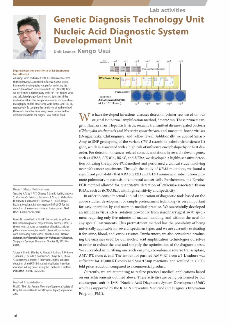
Figure: Detection sensitivity of RT-SmartAmp for influenzaAll assays were performed with A/California/07/2009 (H1N1pdm2009), a cultured influenza A virus strain. Immunochromatography was performed using the Alere™ BinaxNow® Influenza A & B Card (Abbott). First, we performed a plaque assay with 10-2-10-6 diluted virus and calculated plaque forming units (pfu)/ml of the viral culture fluid. The sample volumes for immunochro-matography and RT-SmartAmp were 100 µL and 180 µL, respectively. To compare the sensitivity of each method, the results from the three assays were normalized to viral dilutions from the original viral culture fluid. W e have developed infectious diseases detection primer sets based on our
original isothermal amplification method, SmartAmp. These primers tar-get influenza virus, Hepatitis B virus, sexually transmitted disease-related bacteria (Chlamydia trachomatis and Neisseria gonorrhoeae), and mosquito-borne viruses (Dengue, Zika, Chikungunya, and yellow fever). Additionally, we applied Smart-Amp to SNP genotyping of the variant CPT-2 (carnitine palmitoyltransferase II) gene, which is associated with a high risk of influenza encephalopathy or heat dis-order. For detection of cancer-related somatic mutations in several relevant genes, such as KRAS, PIK3CA, BRAF, and HER2, we developed a highly-sensitive detec-tion kit using the Eprobe-PCR method and performed a clinical study involving over 400 cancer specimens. Through the study of KRAS mutations, we found a significant probability that KRAS G12D and G13D amino acid substitutions pro-mote pulmonary metastasis of colorectal cancer cells. Furthermore, the Eprobe-PCR method allowed for quantitative detection of leukemia-associated fusion RNAs, such as BCR/ABL1, with high sensitivity and specificity.
In order to consider actual clinical application of diagnostic tools based on the above studies, development of sample pretreatment technology is very important for easy operation by end-users in medical practice. We successfully developed an influenza virus RNA isolation procedure from nasopharyngeal swab speci-mens requiring only five minutes of manual handling, and without the need for any special instruments. This pretreatment method has the possibility of being universally applicable for several specimen types, and we are currently evaluating it for urine, blood, and various tissues. Furthermore, we also considered produc-ing the enzymes used for our nucleic acid amplification technologies ourselves in order to reduce the cost and simplify the optimization of the diagnostic tests. We succeeded in purifying one such enzyme, recombinant reverse transcriptase, AMV-RT, from E. coli. The amount of purified AMV-RT from a 1 L-culture was sufficient for 10,000 RT-combined SmartAmp reactions, and resulted in a 100-fold price reduction compared to a commercial product.
Currently, we are attempting to realize practical medical applications based on our achievements outlined above. These activities are being performed by our counterpart unit in IMS, “Nucleic Acid Diagnostic System Development Unit”, which is supported by the RIKEN Preventive Medicine and Diagnosis Innovation Program (PMI).
Recent Major PublicationsTsuchiya K, Tabe Y, Ai T, Ohkawa T, Usui K, Yuri M, Misawa S, Morishita S, Takaku T, Kakimoto A, Yang H, Matsushita H, Hanami T, Yamanaka Y, Okuzawa A, Horii T, Hayas-hizaki Y, Ohsaka A. Eprobe-mediated RT-qPCR for the detection of leukemia-associated fusion genes. PLoS One 13, e0202429 (2018)
Gusev O, Hayashizaki Y, Usui K. Nucleic acid amplifica-tion-based diagnostics for pulmonary diseases: What is the current state and perspectives of nucleic acid am-plification technologies used in diagnostics associated with pulmonary diseases? In: Kaneko T. (ed), Clinical Relevance of Genetic Factors in Pulmonary Diseases, Singapore: Springer Singapore, Chapter 18, 333-344 (2018)
Takase Y, Usui K, Shimizu K, Kimura Y, Ichihara T, Ohkawa T, Atsumi J, Enokida Y, Nakazawa S, Obayashi K, Ohtaki Y, Nagashima T, Mitani Y, Takeyoshi I. Highly sensitive detection of a HER2 12-base pair duplicated insertion mutation in lung cancer using the Eprobe-PCR method. PLoS One 12, e0171225 (2017)
Invited PresentationsUsui K. “The 15th Annual Meeting of Japanese Society of Hospital General Medicine” (Urayasu, Japan) September 2017
16
Lab activities
Epigenome Technology Exploration UnitUnit Leader: Aki Minoda
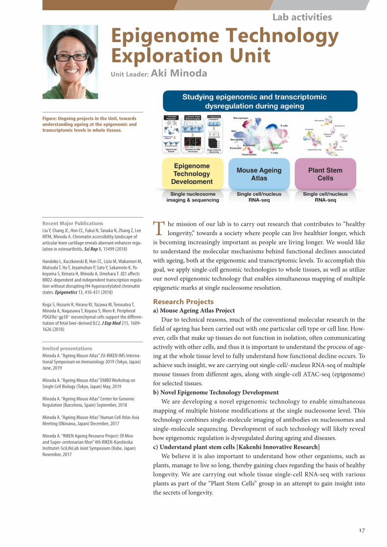
Figure: Ongoing projects in the Unit, towards understanding ageing at the epigenomic and transcriptomic levels in whole tissues.
Recent Major PublicationsLiu Y, Chang JC, Hon CC, Fukui N, Tanaka N, Zhang Z, Lee MTM, Minoda A. Chromatin accessibility landscape of articular knee cartilage reveals aberrant enhancer regu-lation in osteoarthritis. Sci Rep 8, 15499 (2018)
Handoko L, Kaczkowski B, Hon CC, Lizio M, Wakamori M, Matsuda T, Ito T, Jeyamohan P, Sato Y, Sakamoto K, Yo-koyama S, Kimura H, Minoda A, Umehara T. JQ1 affects BRD2-dependent and independent transcription regula-tion without disrupting H4-hyperacetylated chromatin states. Epigenetics 13, 410-431 (2018)
Koga S, Hozumi K, Hirano KI, Yazawa M, Terooatea T, Minoda A, Nagasawa T, Koyasu S, Moro K. Peripheral PDGFRα+gp38+ mesenchymal cells support the differen-tiation of fetal liver-derived ILC2. J Exp Med 215, 1609-1626 (2018)
Invited presentationsMinoda A. “Ageing Mouse Atlas” JSI-RIKEN IMS Interna-tional Symposium on Immunology 2019 (Tokyo, Japan) June, 2019
Minoda A. “Ageing Mouse Atlas” EMBO Workshop on Single Cell Biology (Tokyo, Japan) May, 2019
Minoda A. “Ageing Mouse Atlas” Center for Genomic Regulation (Barcelona, Spain) September, 2018
Minoda A. “Ageing Mouse Atlas” Human Cell Atlas Asia Meeting (Okinawa, Japan) December, 2017
Minoda A. “RIKEN Ageing Resource Project: Of Mice and Super-centenarian Men” 4th RIKEN-Karolinska Institutet-SciLifeLab Joint Symposium (Kobe, Japan) November, 2017
T he mission of our lab is to carry out research that contributes to “healthy longevity,” towards a society where people can live healthier longer, which
is becoming increasingly important as people are living longer. We would like to understand the molecular mechanisms behind functional declines associated with ageing, both at the epigenomic and transcriptomic levels. To accomplish this goal, we apply single-cell genomic technologies to whole tissues, as well as utilize our novel epigenomic technology that enables simultaneous mapping of multiple epigenetic marks at single nucleosome resolution.
Research Projectsa) Mouse Ageing Atlas Project
Due to technical reasons, much of the conventional molecular research in the field of ageing has been carried out with one particular cell type or cell line. How-ever, cells that make up tissues do not function in isolation, often communicating actively with other cells, and thus it is important to understand the process of age-ing at the whole tissue level to fully understand how functional decline occurs. To achieve such insight, we are carrying out single-cell/-nucleus RNA-seq of multiple mouse tissues from different ages, along with single-cell ATAC-seq (epigenome) for selected tissues.b) Novel Epigenome Technology Development
We are developing a novel epigenomic technology to enable simultaneous mapping of multiple histone modifications at the single nucleosome level. This technology combines single-molecule imaging of antibodies on nucleosomes and single-molecule sequencing. Development of such technology will likely reveal how epigenomic regulation is dysregulated during ageing and diseases.c) Understand plant stem cells [Kakenhi Innovative Research]
We believe it is also important to understand how other organisms, such as plants, manage to live so long, thereby gaining clues regarding the basis of healthy longevity. We are carrying out whole tissue single-cell RNA-seq with various plants as part of the “Plant Stem Cells” group in an attempt to gain insight into the secrets of longevity.
17
Lab activities
Laboratory for
Comprehensive Genomic AnalysisTeam Leader: Yasushi Okazaki
Figure: Future plansOmics analysis and functional analysis focusing mainly on mitochondrial and neurological diseases, direct reprogramming, and cancer. In the case of technologi-cal development for genome analysis, we will focus on target enrichment sequencing and modified RNA analysis using a Nanopore sequencer, and single tube Long Fragment Read (stLFR) long-read sequencing using the MGISEQ-2000 sequencer. The diagnostic rate of mitochondrial respiratory chain disorders using whole exome sequencing analysis is approximately 35%, while approximately 37% of cases have variants of uncertain significance. These variants should be evaluated to iden-tify disease causality. Approximately 30% of cases have no candidate genes. Whole genome sequencing, RNA sequencing, and other omics technologies are necessary to solve these difficult cases.
F rom FY2016 to FY2017, we provided research support for researchers inside and outside of RIKEN, such as cross-disciplinary projects within RIKEN and
national projects, as a Genome Network Analysis Support facility (GeNAS). Be-ginning in FY2018, the Laboratory for Comprehensive Genomic Analysis (CGA) was formed and it assumed some of the tasks of research and development from GeNAS.
The CGA laboratory conducts omics analyses to elucidate the pathophysiol-ogy of diseases, using functional genomics, and to understand the mechanisms of diseases that disrupt the homeostatic function of various cells and tissues, and to discover new drug targets. More precisely, we will focus our research activi-ties on identifying new disease genes and their functions and finding new drug targets, with the goal of realization of genome medical care. As initial targets, we start with genome and transcriptome analyses of mitochondrial and neurological diseases. In addition, capitalizing on the strengths of the technology developed by our research team, we contribute to ongoing IMS biomedical research. We ac-complish this through state-of-the-art genome and transcriptome analysis tech-nology, such as RIKEN’s original transcription start site analysis technology, Cap Analysis of Gene Expression (CAGE), and epigenome analysis. As an application using these technologies, we will first focus on single-cell analysis of taste recep-tor cells and also perform other collaborative studies. Furthermore, we will apply these technologies to define the molecular mechanisms of transcription factors that significantly enhance the direct reprogramming of fibroblasts into pancreatic beta cells.
Recent Major PublicationsBorna NN, Kishita Y, Kohda M, Lim SC, Shimura M, Wu Y, Mogushi K, Yatsuka Y, Harashima H, Hisatomi Y, Fushimi T, Ichimoto K, Murayama K, Ohtake A, Okazaki Y. Mi-tochondrial ribosomal protein PTCD3 mutations cause oxidative phosphorylation defects with Leigh syndrome. Neurogenetics (2019)
Noguchi S, et al. FANTOM5 CAGE profiles of human and mouse samples. Sci Data 4, 170112 (2017)
Kohda M, Tokuzawa Y, Kishita Y (Equally first author), et al, Okazaki Y. A comprehensive genomic analysis reveals the genetic landscape of mitochondrial respiratory chain complex deficiencies. PLOS Genet 12, e1005679 (2016)
Invited presentationsOkazaki Y. “MGISEQ-2000: the highly versatile platform to satisfy our multiple needs” The 13th International Conference on Genomics (Shenzhen, China) October, 2018
Okazaki Y. “Finding a way to establish Bahrain Genome Projects through the experiences in Japan” Bahrain Genome Symposium (Manama, Kingdom of Bahrain) October, 2018
Okazaki Y. “Genomic solution for ADPKD” The 61st An-nual Meeting of the Japanese Society of Nephrology (Niigata, Japan) June, 2018
Okazaki Y. “Mitochondrial cardiomyopathy” Cardiovascu-lar Disease Seminar (Osaka, Japan) May, 2018
Okazaki Y. “Genome medical research aimed at next- generation medical care - Comprehensive genome analysis of mitochondrial respiratory chain disorders” The 4th Nephrology Expert Conference (NEXT) (Tokyo, Japan) July, 2017
18
Lab activities
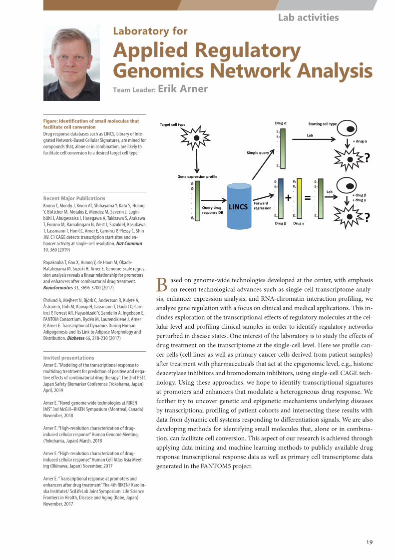
g1g2.....gn
g1g2.....gn
g1g2.....gn
Starting cell typeTarget cell type
LINCSQuery drugresponse DB
g1g2.....gn
+ =g1g2.....gn
Drug α
Drug β Drug γ
+ drug α
+ drug β+ drug γ
?
?
Lab
Lab
Simple query
Forward regression
Gene expression profile
Laboratory for
Applied Regulatory Genomics Network AnalysisTeam Leader: Erik Arner
Figure: Identification of small molecules that facilitate cell conversionDrug response databases such as LINCS, Library of Inte-grated Network-Based Cellular Signatures, are mined for compounds that, alone or in combination, are likely to facilitate cell conversion to a desired target cell type.
B ased on genome-wide technologies developed at the center, with emphasis on recent technological advances such as single-cell transcriptome analy-
sis, enhancer expression analysis, and RNA-chromatin interaction profiling, we analyze gene regulation with a focus on clinical and medical applications. This in-cludes exploration of the transcriptional effects of regulatory molecules at the cel-lular level and profiling clinical samples in order to identify regulatory networks perturbed in disease states. One interest of the laboratory is to study the effects of drug treatment on the transcriptome at the single-cell level. Here we profile can-cer cells (cell lines as well as primary cancer cells derived from patient samples) after treatment with pharmaceuticals that act at the epigenomic level, e.g., histone deacetylase inhibitors and bromodomain inhibitors, using single-cell CAGE tech-nology. Using these approaches, we hope to identify transcriptional signatures at promoters and enhancers that modulate a heterogeneous drug response. We further try to uncover genetic and epigenetic mechanisms underlying diseases by transcriptional profiling of patient cohorts and intersecting these results with data from dynamic cell systems responding to differentiation signals. We are also developing methods for identifying small molecules that, alone or in combina-tion, can facilitate cell conversion. This aspect of our research is achieved through applying data mining and machine learning methods to publicly available drug response transcriptional response data as well as primary cell transcriptome data generated in the FANTOM5 project.
Recent Major PublicationsKouno T, Moody J, Kwon AT, Shibayama Y, Kato S, Huang Y, Böttcher M, Motakis E, Mendez M, Severin J, Lugin-bühl J, Abugessaisa I, Hasegawa A, Takizawa S, Arakawa T, Furuno M, Ramalingam N, West J, Suzuki H, Kasukawa T, Lassmann T, Hon CC, Arner E, Carninci P, Plessy C, Shin JW. C1 CAGE detects transcription start sites and en-hancer activity at single-cell resolution. Nat Commun 10, 360 (2019)
Rapakoulia T, Gao X, Huang Y, de Hoon M, Okada-Hatakeyama M, Suzuki H, Arner E. Genome-scale regres-sion analysis reveals a linear relationship for promoters and enhancers after combinatorial drug treatment. Bioinformatics 33, 3696-3700 (2017)
Ehrlund A, Mejhert N, Björk C, Andersson R, Kulyté A, Åström G, Itoh M, Kawaji H, Lassmann T, Daub CO, Carn-inci P, Forrest AR, Hayashizaki Y, Sandelin A, Ingelsson E, FANTOM Consortium, Rydén M, Laurencikiene J, Arner P, Arner E. Transcriptional Dynamics During Human Adipogenesis and Its Link to Adipose Morphology and Distribution. Diabetes 66, 218-230 (2017)
Invited presentationsArner E. “Modeling of the transcriptional response to multidrug treatment for prediction of positive and nega-tive effects of combinatorial drug therapy”. The 2nd PSTC Japan Safety Biomarker Conference (Yokohama, Japan) April, 2019
Arner E. “Novel genome wide technologies at RIKEN IMS” 3rd McGill–RIKEN Symposium (Montreal, Canada) November, 2018
Arner E. “High-resolution characterization of drug-induced cellular response” Human Genome Meeting, (Yokohama, Japan) March, 2018
Arner E. “High-resolution characterization of drug-induced cellular response” Human Cell Atlas Asia Meet-ing (Okinawa, Japan) November, 2017
Arner E. “Transcriptional response at promoters and enhancers after drug treatment” The 4th RIKEN/ Karolin-ska Institutet/ SciLifeLab Joint Symposium: Life Science Frontiers in Health, Disease and Aging (Kobe, Japan) November, 2017
19
Lab activities
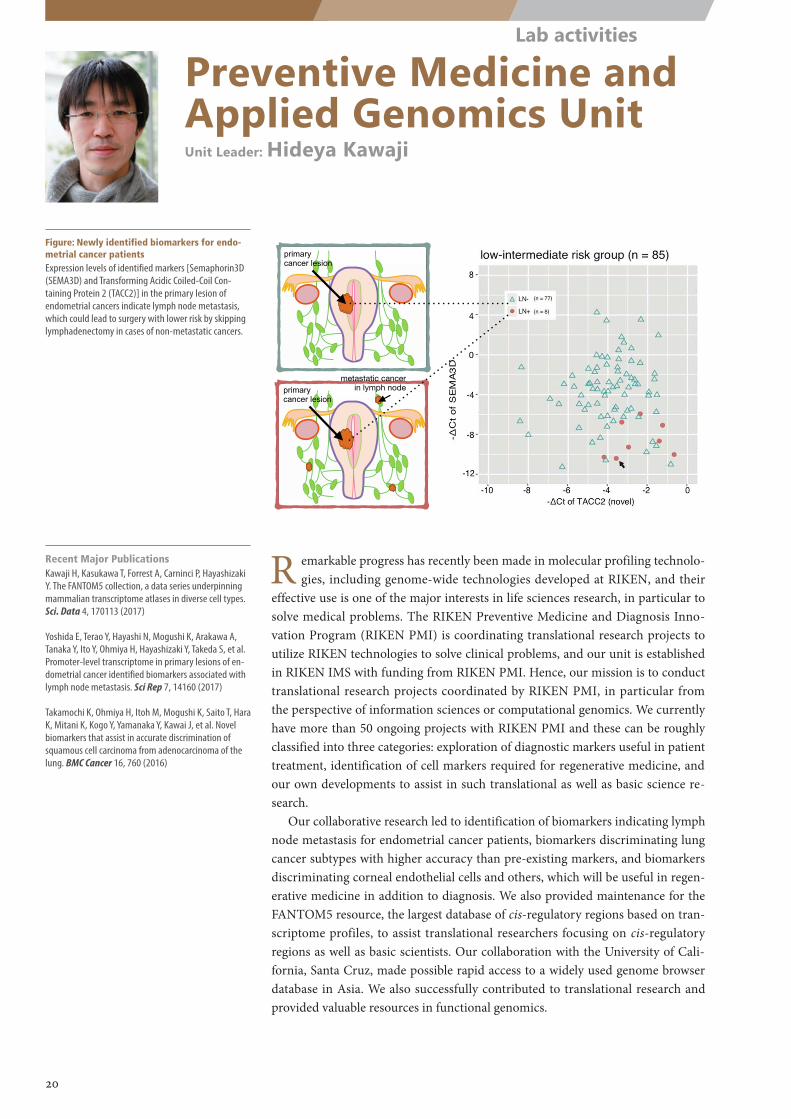
News & Information
図1:リンパ節転移診断マーカーの候補遺伝子の抽出
遺伝子の活性化状態を全ゲノムに渡って測定することができるCAGE法を用いて、リンパ節転移陽性の子宮体がんと陰性の子宮体がんで遺伝子の発現パターンを比較しました。この結果、リンパ節転移陽性群と陰性群で発現に差がある遺伝子がバイオマーカー候補として抽出されました。とても興味深いことに、候補遺伝子のひとつであるTACC2は、リンパ節転移陽性の子宮体がんにおいて既存のものと全く異なる部分から活性化されていたことを新しく発見しました。
散布図では、縦軸にSEMA3D、横軸にTACC2新規アイソフォームのそれぞれ相対発現量をとり、リンパ節転移との関連性を示しています。赤丸で示したリンパ節転移陽性群では、SEMA3Dの発現が低く、TACC2新規アイソフォームの発現が高い傾向を示しました。そこで2つの遺伝子の発現量差を算出し、この値におけるリンパ節転移陽性群と陰性群の識別精度を示したものがROC曲線になります。ROC曲線の下の部分の面積はAUCといい、識別能の尺度として用いられます。識別能がランダムであるときAUCは0.5となり、値が1に近いほど判別能が高いことを示しますが、SEMA3DとTACC2新規アイソフォームの発現量を組み合わせると0.929と非常に高い精度が示されました。
リンパ節転移陽性の子宮体がん リンパ節転移陰性の子宮体がん
CAGE法による網羅的遺伝子解析と発現量比較
リンパ節転移の有無によって発現差のある遺伝子群の抽出
リンパ節転移陰性の子宮体がん細胞 RNA
RNA
一つの遺伝子領域
これまでに知られていなかった部分より、遺伝子の活性化(RNAの合成)が始まっている
遺伝子は活性化されると、DNA塩基配列がRNAへとコピーされ、不要な部分(イントロン)が除去される
リンパ節転移陽性の子宮体がん細胞
No. 3
2017年 10月26日
図2:SEMA3DとTACC2新規アイソフォームとリンパ節転移の関係
リン
primary cancer lesion
転移
散布図
!
SEM
A3Dの相対発現量
8
4
0
-4
-8
-12-10 -8 -6 -4 -2 0
TACC2新規アイソフォームの相対発現量
リンパ節転移陰性
リンパ節転移陽性
ROC曲線
感度
1.0
0.8
0.6
0.4
0.2
0.00.0 0.2 0.4 0.6 0.8 1.0
1 –特異度
!
AUC = 0.929の高精度な識別能を示した
(TACC2新規アイソフォームの相対発現量) -(SEMA3Dの相対発現量)
News & Information
図1:リンパ節転移診断マーカーの候補遺伝子の抽出
遺伝子の活性化状態を全ゲノムに渡って測定することができるCAGE法を用いて、リンパ節転移陽性の子宮体がんと陰性の子宮体がんで遺伝子の発現パターンを比較しました。この結果、リンパ節転移陽性群と陰性群で発現に差がある遺伝子がバイオマーカー候補として抽出されました。とても興味深いことに、候補遺伝子のひとつであるTACC2は、リンパ節転移陽性の子宮体がんにおいて既存のものと全く異なる部分から活性化されていたことを新しく発見しました。
散布図では、縦軸にSEMA3D、横軸にTACC2新規アイソフォームのそれぞれ相対発現量をとり、リンパ節転移との関連性を示しています。赤丸で示したリンパ節転移陽性群では、SEMA3Dの発現が低く、TACC2新規アイソフォームの発現が高い傾向を示しました。そこで2つの遺伝子の発現量差を算出し、この値におけるリンパ節転移陽性群と陰性群の識別精度を示したものがROC曲線になります。ROC曲線の下の部分の面積はAUCといい、識別能の尺度として用いられます。識別能がランダムであるときAUCは0.5となり、値が1に近いほど判別能が高いことを示しますが、SEMA3DとTACC2新規アイソフォームの発現量を組み合わせると0.929と非常に高い精度が示されました。
リンパ節転移陽性の子宮体がん リンパ節転移陰性の子宮体がん
CAGE法による網羅的遺伝子解析と発現量比較
リンパ節転移の有無によって発現差のある遺伝子群の抽出
リンパ節転移陰性の子宮体がん細胞 RNA
RNA
一つの遺伝子領域
これまでに知られていなかった部分より、遺伝子の活性化(RNAの合成)が始まっている
遺伝子は活性化されると、DNA塩基配列がRNAへとコピーされ、不要な部分(イントロン)が除去される
リンパ節転移陽性の子宮体がん細胞
No. 3
2017年 10月26日
図2:SEMA3DとTACC2新規アイソフォームとリンパ節転移の関係
リンパ節転移
散布図
!
SEM
A3Dの相対発現量
8
4
0
-4
-8
-12-10 -8 -6 -4 -2 0
TACC2新規アイソフォームの相対発現量
リンパ節転移陰性
リンパ節転移陽性
ROC曲線
感度
1.0
0.8
0.6
0.4
0.2
0.00.0 0.2 0.4 0.6 0.8 1.0
1 –特異度
!
AUC = 0.929の高精度な識別能を示した
(TACC2新規アイソフォームの相対発現量) -(SEMA3Dの相対発現量)
transcription fromintronic promoter of TACC2
www.nature.com/scientificreports/
8SCIENTIFIC REPORTS | 7: 14160 | DOI:10.1038/s41598-017-14418-5
In cases of high risk of recurrence, ROC curves (Fig. 4F) indicate that SEMA3D expression discriminated between LN+and LN− status at a moderate level (AUC, 0.735), however the novel isoform of TACC2 made no contribution.
Optimal cut-off values dividing the cases into the groups with LN+and LN− were determined by maximiz-ing the Youden′s index from the ROC curves based on the expression of SEMA3D and the novel TACC2 isoforms in cases of low-intermediate risk of recurrence. Biomarker based on the expression difference between SEMA3D and the novel TACC2 isoforms had high specificities (87.0%), sensitivities (100%) and negative predict value(100%) at the optimal cut-off point (Supplementary Table 5).
Localization of SEMA3D mRNA and TACC2 protein in uterine cancer tissue. Last, we asked whether the biomarkers were signatures of cancer cells specifically. Although primary lesions of endometrial can-cer were collected for the experiments above, it is possible that non-cancer cells, such as stromal cells, also express the biomarkers. To address this point, we performed immunohistochemical analyses and FISH (fluorescent in situ hybridization) in primary tumor specimens. We employed immunostaining of TACC2 protein and found its signal mainly in cancer cells, particularly their nucleus (Fig. 5D,E). We employed FISH for SEMA3D mRNA, because the protein product is secreted to the extracellular space31, and found SEMA3D mRNA signal mainly in the cytoplasm of cancer cells (Fig. 5B,C). These results demonstrate that the identified biomarkers are activated in cancer cells, but not in stromal cells.
DiscussionIn this study, we delineated the transcriptome of primary endometrial cancer tissues with the aim of LN+ /LN− discrimination before lymphadenectomy. We first produced a high-resolution transcriptome map reflecting promoter-level expression by using CAGE. The result uncovered differences in transcriptional regulatory net-works corresponding to LN+ /LN− status within patients with low-intermediate risk of recurrence, that is, endo-metrioid adenocarcinoma grade 1 or 2 with neither cervical stromal invasion nor progression to extra-uterine lesion. Unexpectedly we found activation of keratinization-related genes in LN+patients, which indicates a
Figure 4. Association of SEMA3D mRNA and novel TACC2 mRNA expression with lymph node metastasis. (A) The initial set of cases subjected to CAGE analysis. (B) The validation set of cases not assayed by CAGE **P<0.01; *P<0.05; n.s., not significant. (C) Scatter plots of the low-intermediate risk group showing high expression of the novel TACC2 and SEMA3D mRNAs in LN+and in LN− respectively. The Ct values of one of the LN+ samples (with RNA less than 2 µg) were calculated by correcting for the quantity of RNA. This sample is indicated by a black arrow. (D) For the scatter plot of the high-risk group, a similar trend was observed, but it was not as remarkable as in the low-risk group. (E,F) ROC analysis of the low-intermediate risk group (E) and high-risk group (F).
with metastasis
with no metastasis
Yoshida,E., Terao,Y. et al.Promoter-level transcriptome in primary lesions of endometrial cancer identified biomarkers associated with lymph node metastasis.Sci. Rep., 7, 14160 (2017)
metastatic cancerin lymph node
primary cancer lesion
of cancer
primary cancer lesion
lesion
Preventive Medicine and Applied Genomics UnitUnit Leader: Hideya Kawaji
Figure: Newly identified biomarkers for endo-metrial cancer patientsExpression levels of identified markers [Semaphorin3D (SEMA3D) and Transforming Acidic Coiled-Coil Con-taining Protein 2 (TACC2)] in the primary lesion of endometrial cancers indicate lymph node metastasis, which could lead to surgery with lower risk by skipping lymphadenectomy in cases of non-metastatic cancers.
Recent Major PublicationsKawaji H, Kasukawa T, Forrest A, Carninci P, Hayashizaki Y. The FANTOM5 collection, a data series underpinning mammalian transcriptome atlases in diverse cell types. Sci. Data 4, 170113 (2017)
Yoshida E, Terao Y, Hayashi N, Mogushi K, Arakawa A, Tanaka Y, Ito Y, Ohmiya H, Hayashizaki Y, Takeda S, et al. Promoter-level transcriptome in primary lesions of en-dometrial cancer identified biomarkers associated with lymph node metastasis. Sci Rep 7, 14160 (2017)
Takamochi K, Ohmiya H, Itoh M, Mogushi K, Saito T, Hara K, Mitani K, Kogo Y, Yamanaka Y, Kawai J, et al. Novel biomarkers that assist in accurate discrimination of squamous cell carcinoma from adenocarcinoma of the lung. BMC Cancer 16, 760 (2016)
R emarkable progress has recently been made in molecular profiling technolo-gies, including genome-wide technologies developed at RIKEN, and their
effective use is one of the major interests in life sciences research, in particular to solve medical problems. The RIKEN Preventive Medicine and Diagnosis Inno-vation Program (RIKEN PMI) is coordinating translational research projects to utilize RIKEN technologies to solve clinical problems, and our unit is established in RIKEN IMS with funding from RIKEN PMI. Hence, our mission is to conduct translational research projects coordinated by RIKEN PMI, in particular from the perspective of information sciences or computational genomics. We currently have more than 50 ongoing projects with RIKEN PMI and these can be roughly classified into three categories: exploration of diagnostic markers useful in patient treatment, identification of cell markers required for regenerative medicine, and our own developments to assist in such translational as well as basic science re-search.
Our collaborative research led to identification of biomarkers indicating lymph node metastasis for endometrial cancer patients, biomarkers discriminating lung cancer subtypes with higher accuracy than pre-existing markers, and biomarkers discriminating corneal endothelial cells and others, which will be useful in regen-erative medicine in addition to diagnosis. We also provided maintenance for the FANTOM5 resource, the largest database of cis-regulatory regions based on tran-scriptome profiles, to assist translational researchers focusing on cis-regulatory regions as well as basic scientists. Our collaboration with the University of Cali-fornia, Santa Cruz, made possible rapid access to a widely used genome browser database in Asia. We also successfully contributed to translational research and provided valuable resources in functional genomics.
20
Lab activities
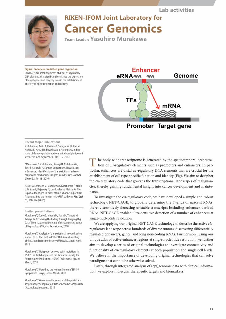
TFsmRNA
Target gene
EnhancerGenome
Promoter
eRNA
RIKEN-IFOM Joint Laboratory for
Cancer GenomicsTeam Leader: Yasuhiro Murakawa
Figure: Enhancer-mediated gene regulationEnhancers are small segments of distal cis-regulatory DNA elements that significantly enhance the expression of target genes and play key roles in the establishment of cell type-specific function and identity.
T he body-wide transcriptome is generated by the spatiotemporal orchestra-tion of cis-regulatory elements such as promoters and enhancers. In par-
ticular, enhancers are distal cis-regulatory DNA elements that are crucial for the establishment of cell type-specific function and identity (Fig). We aim to decipher the cis-regulatory code that governs the transcriptional landscapes of malignan-cies, thereby gaining fundamental insight into cancer development and mainte-nance.
To investigate the cis-regulatory code, we have developed a simple and robust technology, NET-CAGE, to globally determine the 5’-ends of nascent RNAs, thereby sensitively detecting unstable transcripts including enhancer-derived RNAs. NET-CAGE enabled ultra-sensitive detection of a number of enhancers at single-nucleotide resolution.
We are applying our original NET-CAGE technology to describe the active cis-regulatory landscape across hundreds of diverse tumors, discovering differentially regulated enhancers, genes, and long non-coding RNAs. Furthermore, using our unique atlas of active enhancer regions at single-nucleotide resolution, we further aim to develop a series of original technologies to investigate connectivity and functionality of cis-regulatory elements at both population and single-cell levels. We believe in the importance of developing original technologies that can solve paradigms that cannot be otherwise solved.
Lastly, through integrated analysis of (epi)genomic data with clinical informa-tion, we explore molecular therapeutic targets and biomarkers.
Recent Major PublicationsYoshihara M, Araki A, Kasama Y, Sunayama M, Abe M, Nishida K, Kawaji H, Hayashizaki Y, *Murakawa Y. Hot-spots of de novo point mutations in induced pluripotent stem cells. Cell Reports 21, 308-315 (2017)
*Murakawa Y. Yoshihara M, Kawaji H, Nishikawa M, Zayed H, Suzuki H, Fantom Consortium, Hayashizaki Y. Enhanced identification of transcriptional enhanc-ers provide mechanistic insights into diseases. Trends Genet 32, 76-88 (2016)
Hasler D, Lehmann G, Murakawa Y, Klironomos F, Jakob L, Grässer F, Rajewsky N, Landthaler M, Meister G. The Lupus autoantigen La prevents mis-channeling of tRNA fragments into the human microRNA pathway. Mol Cell 63, 110-124 (2016)
Invited presentationsMurakawa Y, Kume S, Maeda M, Suga M, Tamura M, Kobayashi N. “Seeing the Kidney through Imaging Big Data” The 61st Annual Meeting of the Japanese Society of Nephrology (Niigata, Japan) June, 2018
Murakawa Y. “Analysis of transcriptional network using a novel NET-CAGE method” The 91st Annual Meeting of the Japan Endocrine Society (Miyazaki, Japan) April, 2018
Murakawa Y. “Hotspot of de novo point mutations in iPSCs” The 17th Congress of the Japanese Society for Regenerative Medicine (17JSRM) (Yokohama, Japan) March, 2018
Murakawa Y. “Decoding the Human Genome” LINK-J Symposium (Tokyo, Japan) March, 2017
Murakawa Y. “Genome-wide analysis of the post-tran-scriptional gene regulation” Life of Genome Symposium (Kazan, Russia) August, 2016
21
Lab activities
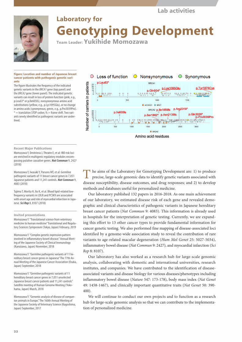
Num
ber o
f car
riers
in p
atie
nts
Amino acid residues
p.Leu63*
p.Gln934*p.Lys1095Glu p.Tyr1874Cys
p.Ile605fsp.Ile1859fs p.Arg2318*
p.Ser1882* p.Asn2135fs p.Gln3026*p.Pro3039Pro
Laboratory for
Genotyping DevelopmentTeam Leader: Yukihide Momozawa
Figure: Location and number of Japanese breast cancer patients with pathogenic genetic vari-antsThe figure illustrates the frequency of the indicated genetic variants in the BRCA1 gene (top panel) and the BRCA2 gene (lower panel). The indicated genetic variants can result in loss of protein function (pink, e.g., p.Leu63* or p.Ile605fs), nonsynonymous amino acid substitutions (yellow, e.g., p.Lys1095Glu), or no change in amino acids (synonymous, green, e.g., p.Pro3039Pro). * = translation STOP codon; fs = frame shift. Two vari-ants newly identified as pathogenic variants are under-lined.
T he aims of the Laboratory for Genotyping Development are: 1) to produce precise, large-scale genomic data to identify genetic variants associated with
disease susceptibility, disease outcomes, and drug responses; and 2) to develop methods and databases useful for personalized medicine.
Our laboratory published 132 papers in 2016-2018. As one main achievement of our laboratory, we estimated disease risk of each gene and revealed demo-graphic and clinical characteristics of pathogenic variants in Japanese hereditary breast cancer patients (Nat Commun 9: 4083). This information is already used in hospitals for the interpretation of genetic testing. Currently, we are expand-ing this effort to 13 other cancer types to provide fundamental information for cancer genetic testing. We also performed fine mapping of disease-associated loci identified by a genome-wide association study to reveal the contribution of rare variants to age-related macular degeneration (Hum Mol Genet 25: 5027-5034), inflammatory bowel disease (Nat Commun 9: 2427), and myocardial infarction (Sci Rep 8: 8107).
Our laboratory has also worked as a research hub for large-scale genomic analysis, collaborating with domestic and international universities, research institutes, and companies. We have contributed to the identification of disease-associated variants and disease biology for various diseases/phenotypes including inflammatory bowel disease (Nature 547: 173-178), body mass index (Nat Genet 49: 1458-1467), and clinically important quantitative traits (Nat Genet 50: 390-400).
We will continue to conduct our own projects and to function as a research hub for large-scale genomic analysis so that we can contribute to the implementa-tion of personalized medicine.
Recent Major PublicationsMomozawa Y, Dmitrieva J, Theatre E, et al: IBD risk loci are enriched in multigenic regulatory modules encom-passing putative causative genes. Nat Commun 9, 2427 (2018)
Momozawa Y, Iwasaki Y, Parsons MT, et al: Germline pathogenic variants of 11 breast cancer genes in 7,051 Japanese patients and 11,241 controls. Nat Commun 9, 4083 (2018)
Tajima T, Morita H, Ito K, et al. Blood lipid-related low-frequency variants in LDLR and PCSK9 are associated with onset age and risk of myocardial infarction in Japa-nese. Sci Rep 8, 8107 (2018)
Invited presentationsMomozawa Y. “Translational science from veterinary medicine to human medicine” Translational and Regula-tory Sciences Symposium (Tokyo, Japan) February, 2019
Momozawa Y. “Complex genetic expression pattern involved in inflammatory bowel disease.” Annual Meet-ing of the Japanese Society of Clinical Immunology (Karuizawa, Japan) November, 2018
Momozawa Y. “Germline pathogenic variants of 11 he-reditary breast cancer genes in Japanese” The 77th An-nual Meeting of the Japanese Cancer Association (Osaka, Japan) September, 2018
Momozawa Y. “Germline pathogenic variants of 11 hereditary breast cancer genes in 7,051 unselected Japanese breast cancer patients and 11,241 controls” Satellite meeting of Human Genome Meeting (Yoko-hama, Japan) March, 2018
Momozawa Y. “Genomic analysis of disease of compan-ion animals in Europe.” The 160th Annual Meeting of the Japanese Society of Veterinary Science (Kagoshima, Japan) September, 2017
22
Lab activities
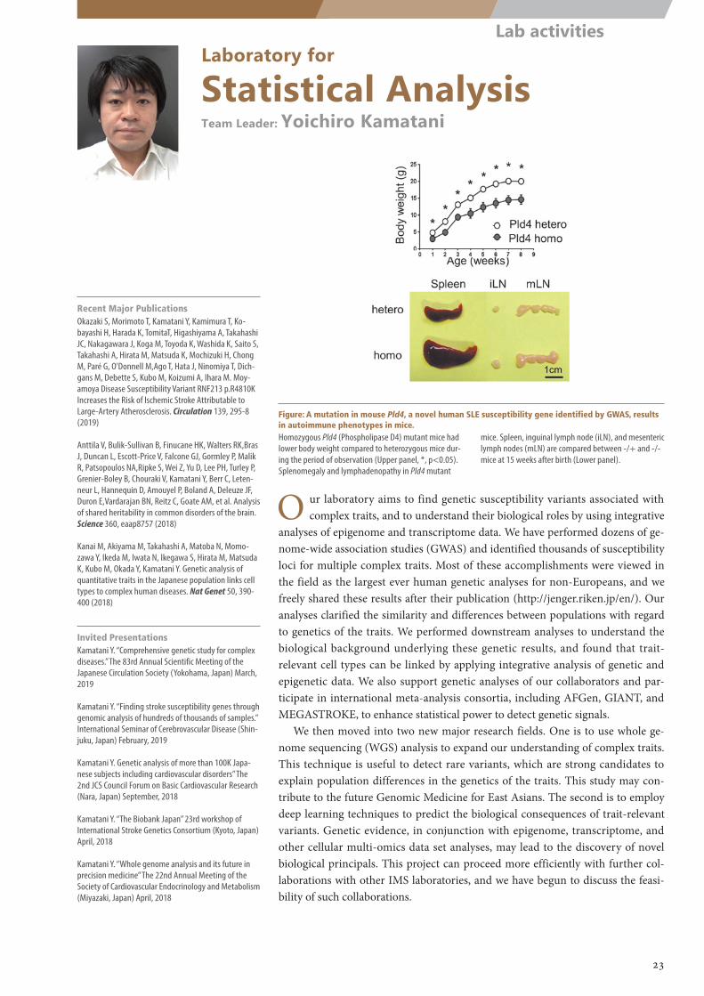
Laboratory for
Statistical AnalysisTeam Leader: Yoichiro Kamatani
O ur laboratory aims to find genetic susceptibility variants associated with complex traits, and to understand their biological roles by using integrative
analyses of epigenome and transcriptome data. We have performed dozens of ge-nome-wide association studies (GWAS) and identified thousands of susceptibility loci for multiple complex traits. Most of these accomplishments were viewed in the field as the largest ever human genetic analyses for non-Europeans, and we freely shared these results after their publication (http://jenger.riken.jp/en/). Our analyses clarified the similarity and differences between populations with regard to genetics of the traits. We performed downstream analyses to understand the biological background underlying these genetic results, and found that trait-relevant cell types can be linked by applying integrative analysis of genetic and epigenetic data. We also support genetic analyses of our collaborators and par-ticipate in international meta-analysis consortia, including AFGen, GIANT, and MEGASTROKE, to enhance statistical power to detect genetic signals.
We then moved into two new major research fields. One is to use whole ge-nome sequencing (WGS) analysis to expand our understanding of complex traits. This technique is useful to detect rare variants, which are strong candidates to explain population differences in the genetics of the traits. This study may con-tribute to the future Genomic Medicine for East Asians. The second is to employ deep learning techniques to predict the biological consequences of trait-relevant variants. Genetic evidence, in conjunction with epigenome, transcriptome, and other cellular multi-omics data set analyses, may lead to the discovery of novel biological principals. This project can proceed more efficiently with further col-laborations with other IMS laboratories, and we have begun to discuss the feasi-bility of such collaborations.
Recent Major PublicationsOkazaki S, Morimoto T, Kamatani Y, Kamimura T, Ko-bayashi H, Harada K, TomitaT, Higashiyama A, Takahashi JC, Nakagawara J, Koga M, Toyoda K, Washida K, Saito S, Takahashi A, Hirata M, Matsuda K, Mochizuki H, Chong M, Paré G, O'Donnell M,Ago T, Hata J, Ninomiya T, Dich-gans M, Debette S, Kubo M, Koizumi A, Ihara M. Moy-amoya Disease Susceptibility Variant RNF213 p.R4810K Increases the Risk of Ischemic Stroke Attributable to Large-Artery Atherosclerosis. Circulation 139, 295-8 (2019)
Anttila V, Bulik-Sullivan B, Finucane HK, Walters RK,Bras J, Duncan L, Escott-Price V, Falcone GJ, Gormley P, Malik R, Patsopoulos NA,Ripke S, Wei Z, Yu D, Lee PH, Turley P, Grenier-Boley B, Chouraki V, Kamatani Y, Berr C, Leten-neur L, Hannequin D, Amouyel P, Boland A, Deleuze JF, Duron E,Vardarajan BN, Reitz C, Goate AM, et al. Analysis of shared heritability in common disorders of the brain. Science 360, eaap8757 (2018)
Kanai M, Akiyama M, Takahashi A, Matoba N, Momo-zawa Y, Ikeda M, Iwata N, Ikegawa S, Hirata M, Matsuda K, Kubo M, Okada Y, Kamatani Y. Genetic analysis of quantitative traits in the Japanese population links cell types to complex human diseases. Nat Genet 50, 390-400 (2018)
Invited PresentationsKamatani Y. “Comprehensive genetic study for complex diseases.” The 83rd Annual Scientific Meeting of the Japanese Circulation Society (Yokohama, Japan) March, 2019
Kamatani Y. “Finding stroke susceptibility genes through genomic analysis of hundreds of thousands of samples.” International Seminar of Cerebrovascular Disease (Shin-juku, Japan) February, 2019
Kamatani Y. Genetic analysis of more than 100K Japa-nese subjects including cardiovascular disorders” The 2nd JCS Council Forum on Basic Cardiovascular Research (Nara, Japan) September, 2018
Kamatani Y. “The Biobank Japan” 23rd workshop of International Stroke Genetics Consortium (Kyoto, Japan) April, 2018
Kamatani Y. “Whole genome analysis and its future in precision medicine” The 22nd Annual Meeting of the Society of Cardiovascular Endocrinology and Metabolism (Miyazaki, Japan) April, 2018
Figure: A mutation in mouse Pld4, a novel human SLE susceptibility gene identified by GWAS, results in autoimmune phenotypes in mice.Homozygous Pld4 (Phospholipase D4) mutant mice had lower body weight compared to heterozygous mice dur-ing the period of observation (Upper panel, *, p<0.05). Splenomegaly and lymphadenopathy in Pld4 mutant
mice. Spleen, inguinal lymph node (iLN), and mesenteric lymph nodes (mLN) are compared between -/+ and -/- mice at 15 weeks after birth (Lower panel).
23
Lab activities
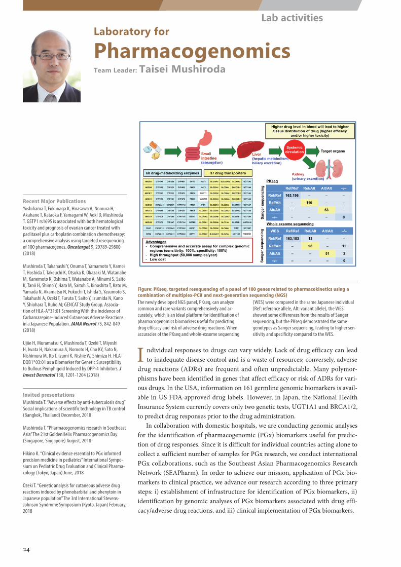
Laboratory for
PharmacogenomicsTeam Leader: Taisei Mushiroda
I ndividual responses to drugs can vary widely. Lack of drug efficacy can lead to inadequate disease control and is a waste of resources; conversely, adverse
drug reactions (ADRs) are frequent and often unpredictable. Many polymor-phisms have been identified in genes that affect efficacy or risk of ADRs for vari-ous drugs. In the USA, information on 161 germline genomic biomarkers is avail-able in US FDA-approved drug labels. However, in Japan, the National Health Insurance System currently covers only two genetic tests, UGT1A1 and BRCA1/2, to predict drug responses prior to the drug administration.
In collaboration with domestic hospitals, we are conducting genomic analyses for the identification of pharmacogenomic (PGx) biomarkers useful for predic-tion of drug responses. Since it is difficult for individual countries acting alone to collect a sufficient number of samples for PGx research, we conduct international PGx collaborations, such as the Southeast Asian Pharmacogenomics Research Network (SEAPharm). In order to achieve our mission, application of PGx bio-markers to clinical practice, we advance our research according to three primary steps: i) establishment of infrastructure for identification of PGx biomarkers, ii) identification by genomic analyses of PGx biomarkers associated with drug effi-cacy/adverse drug reactions, and iii) clinical implementation of PGx biomarkers.
Recent Major PublicationsYoshihama T, Fukunaga K, Hirasawa A, Nomura H, Akahane T, Kataoka F, Yamagami W, Aoki D, Mushiroda T. GSTP1 rs1695 is associated with both hematological toxicity and prognosis of ovarian cancer treated with paclitaxel plus carboplatin combination chemotherapy: a comprehensive analysis using targeted resequencing of 100 pharmacogenes. Oncotarget 9, 29789-29800 (2018)
Mushiroda T, Takahashi Y, Onuma T, Yamamoto Y, Kamei T, Hoshida T, Takeuchi K, Otsuka K, Okazaki M, Watanabe M, Kanemoto K, Oshima T, Watanabe A, Minami S, Saito K, Tanii H, Shimo Y, Hara M, Saitoh S, Kinoshita T, Kato M, Yamada N, Akamatsu N, Fukuchi T, Ishida S, Yasumoto S, Takahashi A, Ozeki T, Furuta T, Saito Y, Izumida N, Kano Y, Shiohara T, Kubo M; GENCAT Study Group. Associa-tion of HLA-A*31:01 Screening With the Incidence of Carbamazepine-Induced Cutaneous Adverse Reactions in a Japanese Population. JAMA Neurol 75, 842-849 (2018)
Ujiie H, Muramatsu K, Mushiroda T, Ozeki T, Miyoshi H, Iwata H, Nakamura A, Nomoto H, Cho KY, Sato N, Nishimura M, Ito T, Izumi K, Nishie W, Shimizu H. HLA-DQB1*03:01 as a Biomarker for Genetic Susceptibility to Bullous Pemphigoid Induced by DPP-4 Inhibitors. J Invest Dermatol 138, 1201-1204 (2018)
Invited presentationsMushiroda T. “Adverse effects by anti-tuberculosis drug” Social implications of scientific technology in TB control (Bangkok, Thailand) December, 2018
Mushiroda T. “Pharmacogenomics research in Southeast Asia” The 21st GoldenHelix Pharmacogenomics Day (Singapore, Singapore) August, 2018
Hikino K. “Clinical evidence essential to PGx informed precision medicine in pediatrics” International Sympo-sium on Pediatric Drug Evaluation and Clinical Pharma-cology (Tokyo, Japan) June, 2018
Ozeki T. “Genetic analysis for cutaneous adverse drug reactions induced by phenobarbital and phenytoin in Japanese population” The 3rd International Stevens-Johnson Syndrome Symposium (Kyoto, Japan) February, 2018
Figure: PKseq, targeted resequencing of a panel of 100 genes related to pharmacokinetics using a combination of multiplex-PCR and next-generation sequencing (NGS)The newly developed NGS panel, PKseq, can analyze common and rare variants comprehensively and ac-curately, which is an ideal platform for identification of pharmacogenomics biomarkers useful for predicting drug efficacy and risk of adverse drug reactions. When accuracies of the PKseq and whole-exsome sequencing
(WES) were compared in the same Japanese individual (Ref: reference allele, Alt: variant allele), the WES showed some differences from the results of Sanger sequencing, but the PKseq demonstrated the same genotypes as Sanger sequencing, leading to higher sen-sitivity and specificity compared to the WES.
24
Lab activities
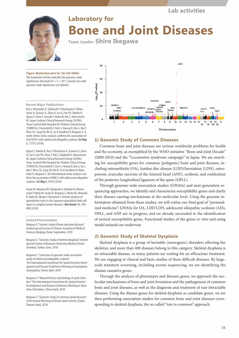
Laboratory for
Bone and Joint DiseasesTeam Leader: Shiro Ikegawa
Figure: Manhattan plot for the AIS GWASThe horizontal red line indicates the genome-wide significance threshold (P = 5 × 10⁻⁸). Genetic loci with genome-wide significance are labeled.
1) Genomic Study of Common DiseasesCommon bone and joint diseases are serious worldwide problems for health
and the economy, as exemplified by the WHO initiative “Bone and Joint Decade” (2000-2010) and the “Locomotive syndrome campaign” in Japan. We are search-ing for susceptibility genes for common (polygenic) bone and joint diseases, in-cluding osteoarthritis (OA), lumbar disc disease (LDD)/herniation (LDH), osteo-porosis, avascular necrosis of the femoral head (ANF), scoliosis, and ossification of the posterior longitudinal ligament of the spine (OPLL).
Through genome-wide association studies (GWASs) and next-generation se-quencing approaches, we identify and characterize susceptibility genes and clarify their disease-causing mechanisms at the molecular level. Using the genome in-formation obtained from these studies, we will realize our final goal of “personal-ized medicine”. GWASs for OA, LDD/LDH, adolescent idiopathic scoliosis (AIS), OPLL, and ANF are in progress, and we already succeeded in the identification of several susceptibility genes. Functional studies of the genes in vitro and using model animals are underway.
2) Genomic Study of Skeletal DysplasiaSkeletal dysplasia is a group of heritable (monogenic) disorders affecting the
skeleton, and more than 400 diseases belong to this category. Skeletal dysplasia is an intractable disease, so many patients are waiting for an efficacious treatment. We are engaging in clinical and basic studies of these difficult diseases. By large-scale mutation screening, including exome sequencing, we are identifying the disease causative genes.
Through the analyses of phenotypes and diseases genes, we approach the mo-lecular mechanisms of bone and joint formation and the pathogenesis of common bone and joint diseases, as well as the diagnosis and treatment of rare intractable diseases. Using the disease genes for skeletal dysplasia as candidate genes, we are then performing association studies for common bone and joint diseases corre-sponding to skeletal dysplasia, the so-called “rare to common” approach.
Recent Major PublicationsKou I, Watanabe K, Takahashi Y, Momozawa Y, Khan-shour A, Grauers A, Zhou H, Liu G, Fan YH, Takeda K, Ogura Y, Zhou T, Iwasaki Y, Kubo M, Wu Z, Matsumoto M; Japan Scoliosis Clinical Research Group (JSCRG); Texas Scottish Rite Hospital for Children Clinical Group (TSRHCCG), Einarsdottir E, Kere J, Huang D, Qiu G, Qiu Y, Wise CA, Song YQ, Wu N, Su P, Gerdhem P, Ikegawa S. A multi-ethnic meta-analysis confirms the association of rs6570507 with adolescent idiopathic scoliosis. Sci Rep 8, 11575 (2018)
Ogura Y, Takeda K, Kou I, Khanshour A, Grauers A, Zhou H, Liu G, Fan YH, Zhou T, Wu Z, Takahashi Y, Matsumoto M; Japan Scoliosis Clinical Research Group (JSCRG); Texas Scottish Rite Hospital for Children Clinical Group (TSRHCCG), Einarsdottir E, Kere J, Huang D, Qiu G, Xu L, Qiu Y, Wise CA, Song YQ, Wu N, Su P, Gerdhem P, Wata-nabe K, Ikegawa S. An international meta-analysis con-firms the association of BNC2 with adolescent idiopathic scoliosis. Sci Rep 8, 4730 (2018)
Kanai M, Akiyama M, Takahashi A, Matoba N, Momo-zawa Y, Ikeda M, Iwata N, Ikegawa S, Hirata M, Matsuda K, Kubo M, Okada Y, Kamatani Y. Genetic analysis of quantitative traits in the Japanese population links cell types to complex human diseases. Nat Genet 50, 390-400 (2018)
Invited PresentationsIkegawa S. “Genetic study of bone and joint diseases” Invited special lecture in Chinese Academy of Medical Sciences (Beijing, China) September, 2018
Ikegawa S. “Genomic study of skeletal dysplasia” Invited special lecture in Marmara University Medical School (Istanbul, Turkey) June, 2018
Ikegawa S. “Extension of genome-wide association study of adolescent idiopathic scoliosis” The International Consortium for Spinal Genetics Devel-opment and Disease Conference Meeting in Guangzhou (Guangzhou, China) April, 2018
Ikegawa S. “Natural history and etiology of spine disor-ders” The International Consortium for Spinal Genetics Development and Disease Conference Meeting in Shen-zhen (Shenzhen, China) April, 2018
Ikegawa S. “Genomic study of common spinal diseases” 2018 Annual Meeting of Taiwan Spine Society (Taipei, Taiwan) April, 2018
25
Lab activities
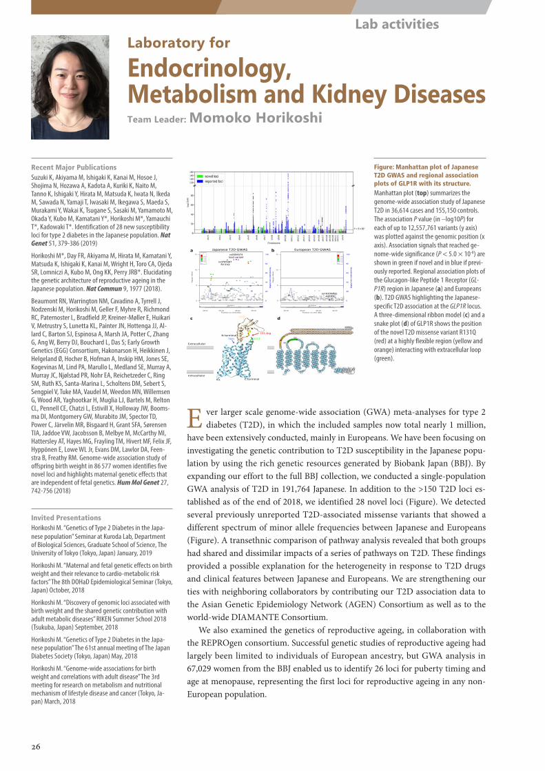
0
5
10
15
lo
g 10(
p−va
lue)
0
20
40
60
80
100
Recom
bination rate (cM/M
b)
0.20.40.60.8
r2
GLP1R
39.02 39.03 39.04 39.05Position on chr6 (Mb)
0
5
10
15
lo
g 10(
p−va
lue)
0
20
40
60
80
100
Recom
bination rate (cM/M
b)
0.20.40.60.8
r2
GLP1R
39.02 39.03 39.04 39.05Position on chr6 (Mb)
TM1 ECL1
131 Arg
ECD
Extracellular
IntracellularC terminal
N terminal
a b Japanese T2D GWAS European T2D GWAS
c d
ICL
rs9394574 lead variant
rs3765467 R131Q
rs10305492 A316T
Laboratory for
Endocrinology, Metabolism and Kidney DiseasesTeam Leader: Momoko Horikoshi
E ver larger scale genome-wide association (GWA) meta-analyses for type 2 diabetes (T2D), in which the included samples now total nearly 1 million,
have been extensively conducted, mainly in Europeans. We have been focusing on investigating the genetic contribution to T2D susceptibility in the Japanese popu-lation by using the rich genetic resources generated by Biobank Japan (BBJ). By expanding our effort to the full BBJ collection, we conducted a single-population GWA analysis of T2D in 191,764 Japanese. In addition to the >150 T2D loci es-tablished as of the end of 2018, we identified 28 novel loci (Figure). We detected several previously unreported T2D-associated missense variants that showed a different spectrum of minor allele frequencies between Japanese and Europeans (Figure). A transethnic comparison of pathway analysis revealed that both groups had shared and dissimilar impacts of a series of pathways on T2D. These findings provided a possible explanation for the heterogeneity in response to T2D drugs and clinical features between Japanese and Europeans. We are strengthening our ties with neighboring collaborators by contributing our T2D association data to the Asian Genetic Epidemiology Network (AGEN) Consortium as well as to the world-wide DIAMANTE Consortium.
We also examined the genetics of reproductive ageing, in collaboration with the REPROgen consortium. Successful genetic studies of reproductive ageing had largely been limited to individuals of European ancestry, but GWA analysis in 67,029 women from the BBJ enabled us to identify 26 loci for puberty timing and age at menopause, representing the first loci for reproductive ageing in any non-European population.
Recent Major PublicationsSuzuki K, Akiyama M, Ishigaki K, Kanai M, Hosoe J, Shojima N, Hozawa A, Kadota A, Kuriki K, Naito M, Tanno K, Ishigaki Y, Hirata M, Matsuda K, Iwata N, Ikeda M, Sawada N, Yamaji T, Iwasaki M, Ikegawa S, Maeda S, Murakami Y, Wakai K, Tsugane S, Sasaki M, Yamamoto M, Okada Y, Kubo M, Kamatani Y*, Horikoshi M*, Yamauchi T*, Kadowaki T*. Identification of 28 new susceptibility loci for type 2 diabetes in the Japanese population. Nat Genet 51, 379-386 (2019)
Horikoshi M*, Day FR, Akiyama M, Hirata M, Kamatani Y, Matsuda K, Ishigaki K, Kanai M, Wright H, Toro CA, Ojeda SR, Lomniczi A, Kubo M, Ong KK, Perry JRB*. Elucidating the genetic architecture of reproductive ageing in the Japanese population. Nat Commun 9, 1977 (2018).
Beaumont RN, Warrington NM, Cavadino A, Tyrrell J, Nodzenski M, Horikoshi M, Geller F, Myhre R, Richmond RC, Paternoster L, Bradfield JP, Kreiner-Møller E, Huikari V, Metrustry S, Lunetta KL, Painter JN, Hottenga JJ, Al-lard C, Barton SJ, Espinosa A, Marsh JA, Potter C, Zhang G, Ang W, Berry DJ, Bouchard L, Das S; Early Growth Genetics (EGG) Consortium, Hakonarson H, Heikkinen J, Helgeland Ø, Hocher B, Hofman A, Inskip HM, Jones SE, Kogevinas M, Lind PA, Marullo L, Medland SE, Murray A, Murray JC, Njølstad PR, Nohr EA, Reichetzeder C, Ring SM, Ruth KS, Santa-Marina L, Scholtens DM, Sebert S, Sengpiel V, Tuke MA, Vaudel M, Weedon MN, Willemsen G, Wood AR, Yaghootkar H, Muglia LJ, Bartels M, Relton CL, Pennell CE, Chatzi L, Estivill X, Holloway JW, Booms-ma DI, Montgomery GW, Murabito JM, Spector TD, Power C, Järvelin MR, Bisgaard H, Grant SFA, Sørensen TIA, Jaddoe VW, Jacobsson B, Melbye M, McCarthy MI, Hattersley AT, Hayes MG, Frayling TM, Hivert MF, Felix JF, Hyppönen E, Lowe WL Jr, Evans DM, Lawlor DA, Feen-stra B, Freathy RM. Genome-wide association study of offspring birth weight in 86 577 women identifies five novel loci and highlights maternal genetic effects that are independent of fetal genetics. Hum Mol Genet 27, 742-756 (2018)
Invited PresentationsHorikoshi M. “Genetics of Type 2 Diabetes in the Japa-nese population” Seminar at Kuroda Lab, Department of Biological Sciences, Graduate School of Science, The University of Tokyo (Tokyo, Japan) January, 2019
Horikoshi M. “Maternal and fetal genetic effects on birth weight and their relevance to cardio-metabolic risk factors” The 8th DOHaD Epidemiological Seminar (Tokyo, Japan) October, 2018
Horikoshi M. “Discovery of genomic loci associated with birth weight and the shared genetic contribution with adult metabolic diseases” RIKEN Summer School 2018 (Tsukuba, Japan) September, 2018
Horikoshi M. “Genetics of Type 2 Diabetes in the Japa-nese population” The 61st annual meeting of The Japan Diabetes Society (Tokyo, Japan) May, 2018
Horikoshi M. “Genome-wide associations for birth weight and correlations with adult disease” The 3rd meeting for research on metabolism and nutritional mechanism of lifestyle disease and cancer (Tokyo, Ja-pan) March, 2018
Figure: Manhattan plot of Japanese T2D GWAS and regional association plots of GLP1R with its structure.Manhattan plot (top) summarizes the genome-wide association study of Japanese T2D in 36,614 cases and 155,150 controls. The association P value (in –log10P) for each of up to 12,557,761 variants (y axis) was plotted against the genomic position (x axis). Association signals that reached ge-nome-wide significance (P < 5.0 × 10⁻⁸) are shown in green if novel and in blue if previ-ously reported. Regional association plots of the Glucagon-like Peptide 1 Receptor (GL-P1R) region in Japanese (a) and Europeans (b). T2D GWAS highlighting the Japanese-specific T2D association at the GLP1R locus. A three-dimensional ribbon model (c) and a snake plot (d) of GLP1R shows the position of the novel T2D missense variant R131Q (red) at a highly flexible region (yellow and orange) interacting with extracellular loop (green).
26
Lab activities
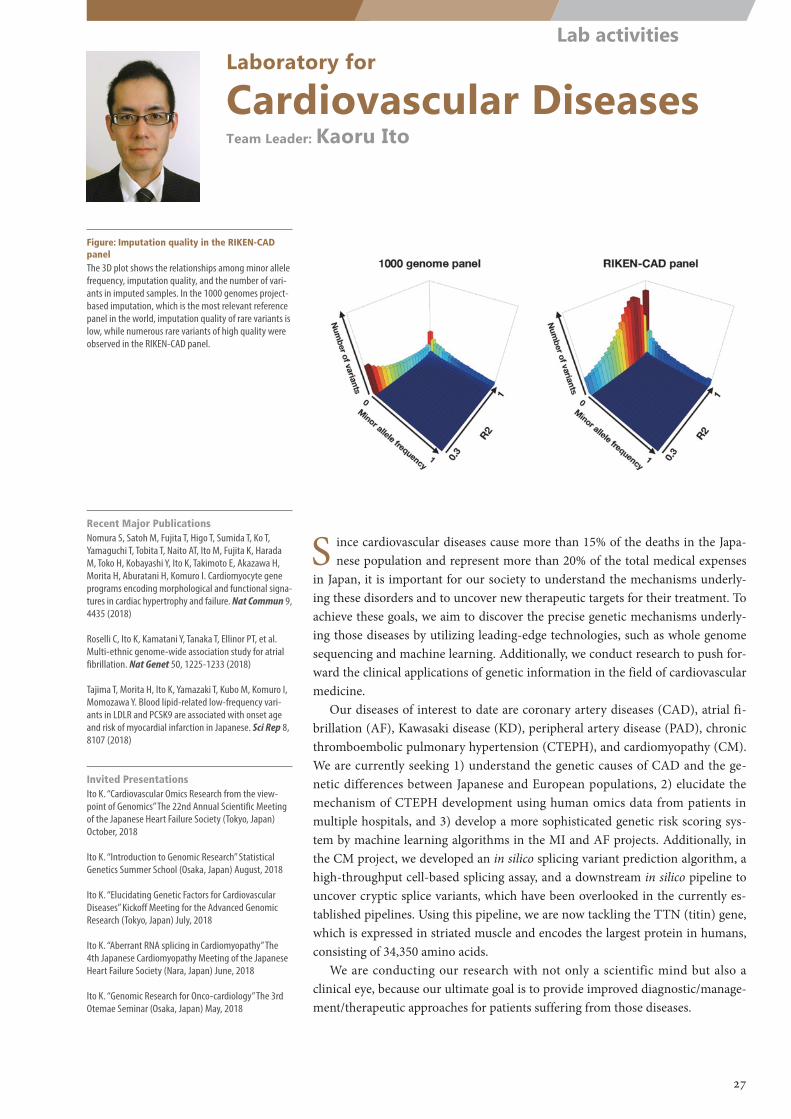
Laboratory for
Cardiovascular DiseasesTeam Leader: Kaoru Ito
Figure: Imputation quality in the RIKEN-CAD panelThe 3D plot shows the relationships among minor allele frequency, imputation quality, and the number of vari-ants in imputed samples. In the 1000 genomes project-based imputation, which is the most relevant reference panel in the world, imputation quality of rare variants is low, while numerous rare variants of high quality were observed in the RIKEN-CAD panel.
S ince cardiovascular diseases cause more than 15% of the deaths in the Japa-nese population and represent more than 20% of the total medical expenses
in Japan, it is important for our society to understand the mechanisms underly-ing these disorders and to uncover new therapeutic targets for their treatment. To achieve these goals, we aim to discover the precise genetic mechanisms underly-ing those diseases by utilizing leading-edge technologies, such as whole genome sequencing and machine learning. Additionally, we conduct research to push for-ward the clinical applications of genetic information in the field of cardiovascular medicine.
Our diseases of interest to date are coronary artery diseases (CAD), atrial fi-brillation (AF), Kawasaki disease (KD), peripheral artery disease (PAD), chronic thromboembolic pulmonary hypertension (CTEPH), and cardiomyopathy (CM). We are currently seeking 1) understand the genetic causes of CAD and the ge-netic differences between Japanese and European populations, 2) elucidate the mechanism of CTEPH development using human omics data from patients in multiple hospitals, and 3) develop a more sophisticated genetic risk scoring sys-tem by machine learning algorithms in the MI and AF projects. Additionally, in the CM project, we developed an in silico splicing variant prediction algorithm, a high-throughput cell-based splicing assay, and a downstream in silico pipeline to uncover cryptic splice variants, which have been overlooked in the currently es-tablished pipelines. Using this pipeline, we are now tackling the TTN (titin) gene, which is expressed in striated muscle and encodes the largest protein in humans, consisting of 34,350 amino acids.
We are conducting our research with not only a scientific mind but also a clinical eye, because our ultimate goal is to provide improved diagnostic/manage-ment/therapeutic approaches for patients suffering from those diseases.
Recent Major PublicationsNomura S, Satoh M, Fujita T, Higo T, Sumida T, Ko T, Yamaguchi T, Tobita T, Naito AT, Ito M, Fujita K, Harada M, Toko H, Kobayashi Y, Ito K, Takimoto E, Akazawa H, Morita H, Aburatani H, Komuro I. Cardiomyocyte gene programs encoding morphological and functional signa-tures in cardiac hypertrophy and failure. Nat Commun 9, 4435 (2018)
Roselli C, Ito K, Kamatani Y, Tanaka T, Ellinor PT, et al. Multi-ethnic genome-wide association study for atrial fibrillation. Nat Genet 50, 1225-1233 (2018)
Tajima T, Morita H, Ito K, Yamazaki T, Kubo M, Komuro I, Momozawa Y. Blood lipid-related low-frequency vari-ants in LDLR and PCSK9 are associated with onset age and risk of myocardial infarction in Japanese. Sci Rep 8, 8107 (2018)
Invited PresentationsIto K. “Cardiovascular Omics Research from the view-point of Genomics” The 22nd Annual Scientific Meeting of the Japanese Heart Failure Society (Tokyo, Japan) October, 2018
Ito K. “Introduction to Genomic Research” Statistical Genetics Summer School (Osaka, Japan) August, 2018
Ito K. “Elucidating Genetic Factors for Cardiovascular Diseases” Kickoff Meeting for the Advanced Genomic Research (Tokyo, Japan) July, 2018
Ito K. “Aberrant RNA splicing in Cardiomyopathy” The 4th Japanese Cardiomyopathy Meeting of the Japanese Heart Failure Society (Nara, Japan) June, 2018
Ito K. “Genomic Research for Onco-cardiology” The 3rd Otemae Seminar (Osaka, Japan) May, 2018
27
Lab activities
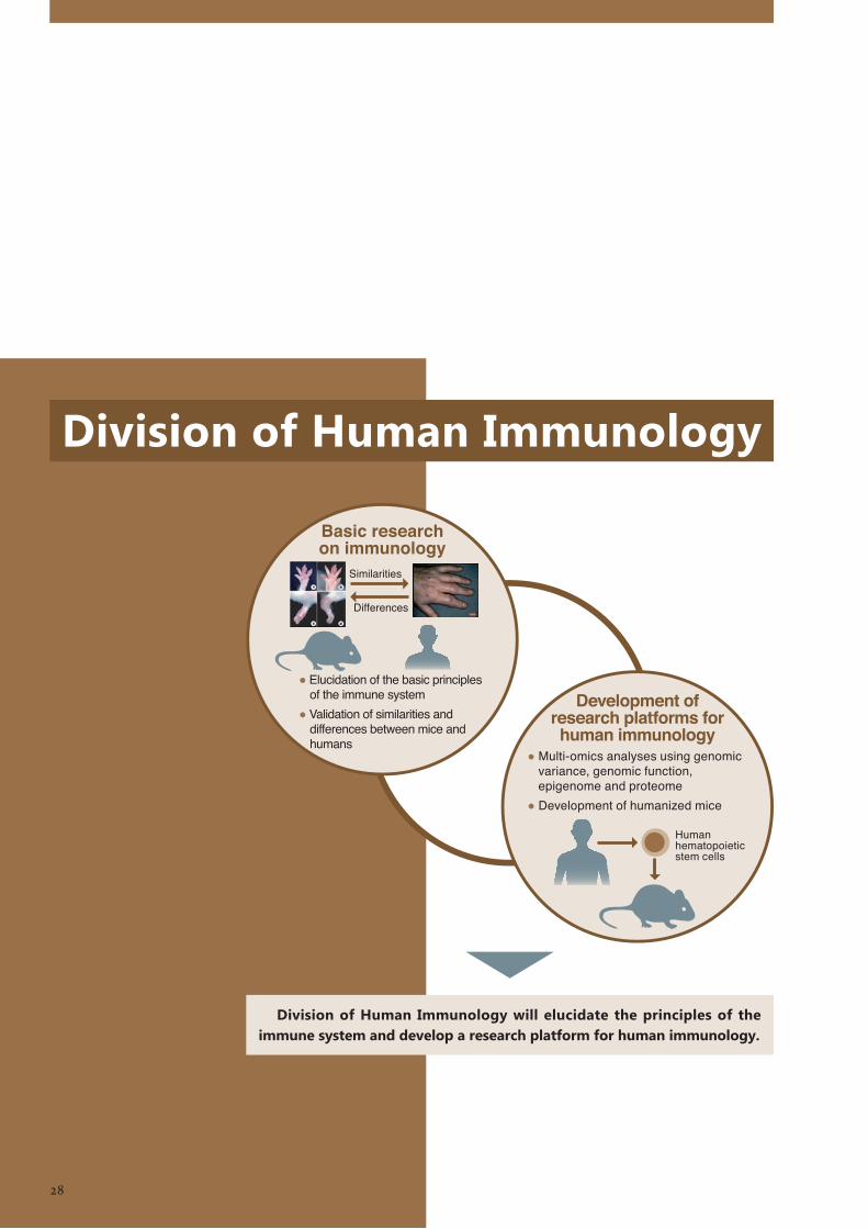
Division of Human Immunology
Division of Human Immunology will elucidate the principles of the immune system and develop a research platform for human immunology.
28
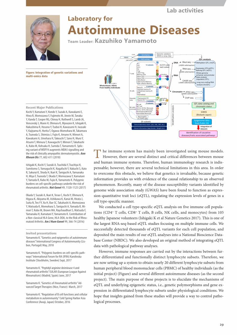
(>6 x10^7 cells)DNA
RNA
Methylation QTL (mQTL)
Expression QTL(eQTL) Splicing QTL (sQTL)
Genomic variations (WGS or array chip)
Chromatin
Whole genome enhancer and promoter state analysis (regulatory QTL)
Whole blood (~50ml) From healthy individuals
Th1Th2Th17TfhTregLAG3 TregMemory CD4Naïve CD4Naïve CD8CD8 effectorCD8+Central MemoryCD8+ effector Memory
Naïve BSwitched memory BUnSwitched memory BDNBPlasmablast
NK cellNKT cellCD14+ CD16+ MonocyteCD14+ CD16- MonocyteNonClassical MonocyteIntermidiate Monocyte
T cells
Plasmacytoid DCMyeloid DC
B cells
Dendritic cells
NK cells/ monocyte
2x10^4 cells/ each subset
12 subsets
5 subsets
2 subsets6 subsets
26 subsets
n=200~
PBMC
Neutrophils
Clinical laboratory exam. (LDL, HDL, CRP, etc.)
FACS sorting
Genotyping
activation
(4 x10^6 cells)
activation
RNA-seq
CAGE-seq
Bisul-seq
>1ml
(>5000 cells/reaction)
(>5000 cells/reaction)
(>5000 cells/reaction)
ATAC-seq(>5000 cells/reaction)
QTL analysis
Clinical information (Age,Gender, Weight etc.)
GWAS(Biobank Japan)
x
QTL data catalog and chromatin status
Identification of causative diseases/traits associated pathways
Epigenome data from Public DB
x
Stra
tific
atio
n an
alys
is
Integration of datasets
activation
Long non-coding RNA analysis (lncQTL)
activation
ChIP-seq
Laboratory for
Autoimmune DiseasesTeam Leader: Kazuhiko Yamamoto
Figure: Integration of genetic variations and multi-omics data
T he immune system has mainly been investigated using mouse models. However, there are several distinct and critical differences between mouse
and human immune systems. Therefore, human immunology research is indis-pensable; however, there are several technical limitations in this area. In order to overcome this obstacle, we believe that genetics is invaluable, because genetic information provides us with evidence of the causal relationship to an observed phenomenon. Recently, many of the disease susceptibility variants identified by genome wide association study (GWAS) have been found to function as expres-sion-quantitative trait loci (eQTL), regulating the expression levels of genes in a cell type-specific manner.
We conducted a cell type-specific eQTL analysis on five immune cell popula-tions (CD4+ T cells, CD8+ T cells, B cells, NK cells, and monocytes) from 105 healthy Japanese volunteers (Ishigaki K et al Nature Genetics 2017). This is one of the largest RNA-seq-based eQTL studies focusing on multiple immune cells. We successfully detected thousands of eQTL variants for each cell population, and deposited the main results of our eQTL analyses into a National Bioscience Data-base Center (NBDC). We also developed an original method of integrating eQTL data with pathological pathway analyses.
However, immune responses are carried out by the interactions between fur-ther differentiated and functionally distinct lymphocyte subsets. Therefore, we are now setting up a system to obtain nearly 20 different lymphocyte subsets from human peripheral blood mononuclear cells (PBMC) of healthy individuals (as the initial project) (Figure) and several different autoimmune diseases (as the second project). The main purpose of these projects is to elucidate the mechanisms of eQTL and underlying epigenetic status, i.e., genetic polymorphisms and gene ex-pression in differentiated lymphocyte subsets under physiological conditions. We hope that insights gained from these studies will provide a way to control patho-logical processes.
Recent Major PublicationsKochi Y, Kamatani Y, Kondo Y, Suzuki A, Kawakami E, Hiwa R, Momozawa Y, Fujimoto M, Jinnin M, Tanaka Y, Kanda T, Cooper RG, Chinoy H, Rothwell S, Lamb JA, Vencovský J, Mann H, Ohmura K, Myouzen K, Ishigaki K, Nakashima R, Hosono Y, Tsuboi H, Kawasumi H, Iwasaki Y, Kajiyama H, Horita T, Ogawa-Momohara M, Takamura A, Tsunoda S, Shimizu J, Fujio K, Amano H, Mimori A, Kawakami A, Umehara H, Takeuchi T, Sano H, Muro Y, Atsumi T, Mimura T, Kawaguchi Y, Mimori T, Takahashi A, Kubo M, Kohsaka H, Sumida T, Yamamoto K. Splic-ing variant of WDFY4 augments MDA5 signalling and the risk of clinically amyopathic dermatomyositis. Ann Rheum Dis 77, 602-611 (2018)
Ishigaki K, Kochi Y, Suzuki A, Tsuchida Y, Tsuchiya H, Sumitomo S, Yamaguchi K, Nagafuchi Y, Nakachi S, Kato R, Sakurai K, Shoda H, Ikari K, Taniguchi A, Yamanaka H, Miya F, Tsunoda T, Okada Y, Momozawa Y, Kamatani Y, Yamada R, Kubo M, Fujio K, Yamamoto K. Polygenic burdens on cell-specific pathways underlie the risk of rheumatoid arthritis. Nat Genet 49, 1120-1125 (2017)
Okada Y, Suzuki A, Ikari K, Terao C, Kochi Y, Ohmura K, Higasa K, Akiyama M, Ashikawa K, Kanai M, Hirata J, Suita N, Teo YY, Xu H, Bae SC, Takahashi A, Momozawa Y, Matsuda K, Momohara S, Taniguchi A, Yamada R, Mi-mori T, Kubo M, Brown MA, Raychaudhuri S, Matsuda F, Yamanaka H, Kamatani Y, Yamamoto K. Contribution of a Non-classical HLA Gene, HLA-DOA, to the Risk of Rheu-matoid Arthritis. Am J Hum Genet 99, 366-74 (2016)
Invited presentationsYamamoto K. “Genetics and epigenetics of autoimmune diseases” International Congress of Autoimmunity (Lis-bon, Portugal) May, 2018
Yamamoto K. “Polygenic burdens on cell-specific path-ways” International Forum for RA (IFRA) Karolinska Institute (Stockholm, Sweden) Sept, 2017
Yamamoto K. “Peptidyl arginine deiminase 4 and rheumatoid arthritis” EULAR (European League Against Rheumatism) (Madrid, Spain) June, 2017
Yamamoto K. “Genetics of rheumatoid arthritis” Ad-vanced Target Therapies (Nice, France) March, 2017
Yamamoto K. “Regulation of B cell functions and cellular metabolism in autoimmunity” Cold Spring Harbor Asia Conference (Awaji, Japan) October, 2016
29
Lab activities
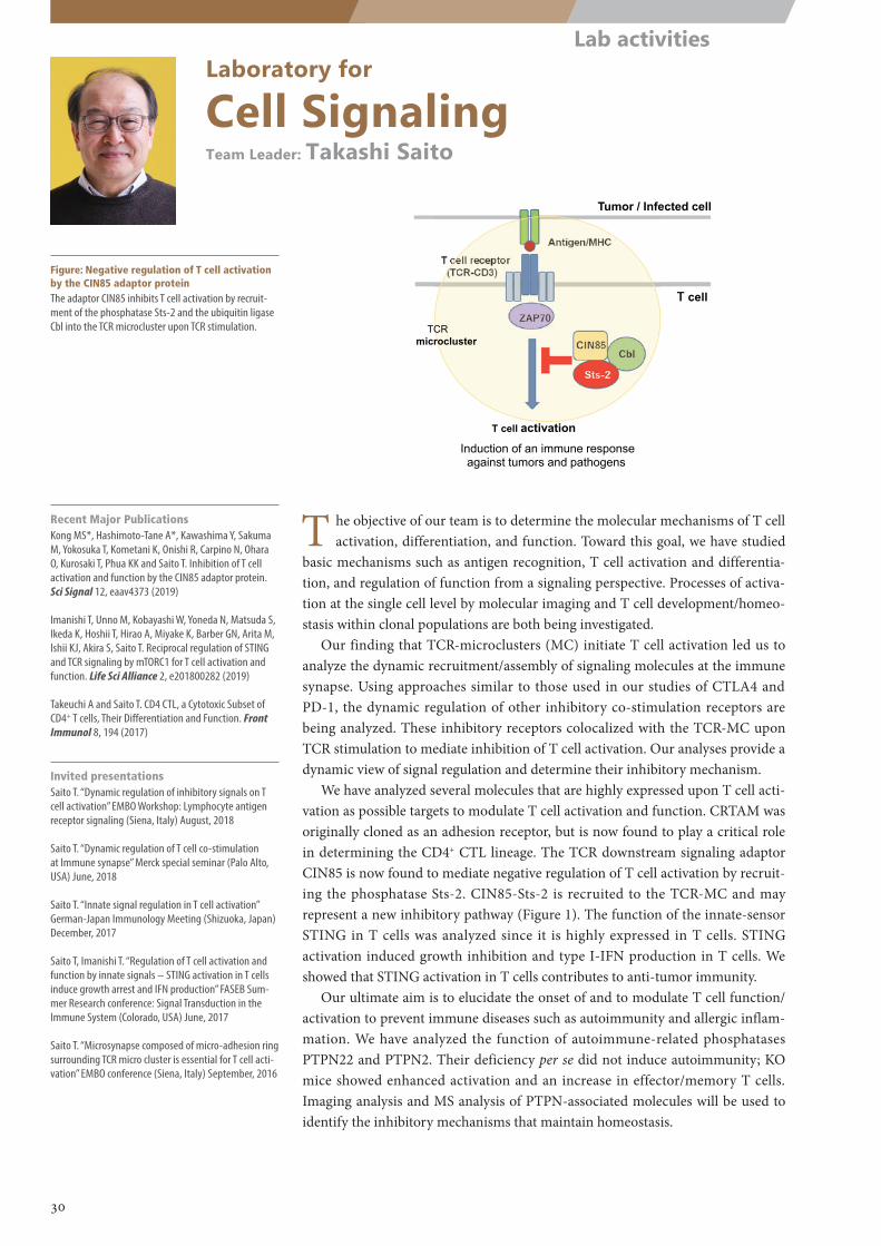
T cell activation
T cell receptor(TCR-CD3)
CIN85Cbl
Sts-2
ZAP70
Antigen/MHC
T cell
Tumor / Infected cell
Induction of an immune response against tumors and pathogens
TCR microcluster
Laboratory for
Cell SignalingTeam Leader: Takashi Saito
Figure: Negative regulation of T cell activation by the CIN85 adaptor proteinThe adaptor CIN85 inhibits T cell activation by recruit-ment of the phosphatase Sts-2 and the ubiquitin ligase Cbl into the TCR microcluster upon TCR stimulation.
T he objective of our team is to determine the molecular mechanisms of T cell activation, differentiation, and function. Toward this goal, we have studied
basic mechanisms such as antigen recognition, T cell activation and differentia-tion, and regulation of function from a signaling perspective. Processes of activa-tion at the single cell level by molecular imaging and T cell development/homeo-stasis within clonal populations are both being investigated.
Our finding that TCR-microclusters (MC) initiate T cell activation led us to analyze the dynamic recruitment/assembly of signaling molecules at the immune synapse. Using approaches similar to those used in our studies of CTLA4 and PD-1, the dynamic regulation of other inhibitory co-stimulation receptors are being analyzed. These inhibitory receptors colocalized with the TCR-MC upon TCR stimulation to mediate inhibition of T cell activation. Our analyses provide a dynamic view of signal regulation and determine their inhibitory mechanism.
We have analyzed several molecules that are highly expressed upon T cell acti-vation as possible targets to modulate T cell activation and function. CRTAM was originally cloned as an adhesion receptor, but is now found to play a critical role in determining the CD4+ CTL lineage. The TCR downstream signaling adaptor CIN85 is now found to mediate negative regulation of T cell activation by recruit-ing the phosphatase Sts-2. CIN85-Sts-2 is recruited to the TCR-MC and may represent a new inhibitory pathway (Figure 1). The function of the innate-sensor STING in T cells was analyzed since it is highly expressed in T cells. STING activation induced growth inhibition and type I-IFN production in T cells. We showed that STING activation in T cells contributes to anti-tumor immunity.
Our ultimate aim is to elucidate the onset of and to modulate T cell function/activation to prevent immune diseases such as autoimmunity and allergic inflam-mation. We have analyzed the function of autoimmune-related phosphatases PTPN22 and PTPN2. Their deficiency per se did not induce autoimmunity; KO mice showed enhanced activation and an increase in effector/memory T cells. Imaging analysis and MS analysis of PTPN-associated molecules will be used to identify the inhibitory mechanisms that maintain homeostasis.
Recent Major PublicationsKong MS*, Hashimoto-Tane A*, Kawashima Y, Sakuma M, Yokosuka T, Kometani K, Onishi R, Carpino N, Ohara O, Kurosaki T, Phua KK and Saito T. Inhibition of T cell activation and function by the CIN85 adaptor protein. Sci Signal 12, eaav4373 (2019)
Imanishi T, Unno M, Kobayashi W, Yoneda N, Matsuda S, Ikeda K, Hoshii T, Hirao A, Miyake K, Barber GN, Arita M, Ishii KJ, Akira S, Saito T. Reciprocal regulation of STING and TCR signaling by mTORC1 for T cell activation and function. Life Sci Alliance 2, e201800282 (2019)
Takeuchi A and Saito T. CD4 CTL, a Cytotoxic Subset of CD4+ T cells, Their Differentiation and Function. Front Immunol 8, 194 (2017)
Invited presentationsSaito T. “Dynamic regulation of inhibitory signals on T cell activation” EMBO Workshop: Lymphocyte antigen receptor signaling (Siena, Italy) August, 2018
Saito T. “Dynamic regulation of T cell co-stimulation at Immune synapse” Merck special seminar (Palo Alto, USA) June, 2018
Saito T. “Innate signal regulation in T cell activation” German-Japan Immunology Meeting (Shizuoka, Japan) December, 2017
Saito T, Imanishi T. “Regulation of T cell activation and function by innate signals – STING activation in T cells induce growth arrest and IFN production” FASEB Sum-mer Research conference: Signal Transduction in the Immune System (Colorado, USA) June, 2017
Saito T. “Microsynapse composed of micro-adhesion ring surrounding TCR micro cluster is essential for T cell acti-vation” EMBO conference (Siena, Italy) September, 2016
30
Lab activities
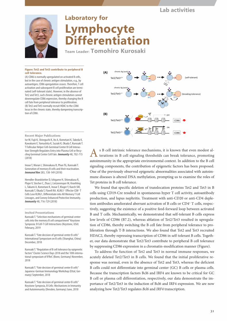
Tet2/Tet3+/+
chronic Ag (auto)
Tet2/Tet3−/−
chronic Ag (auto)
(self‐tolerant)
(breaking tolerance)
Tet2/Tet3−/−×
CD86
ON
Tet2/3HDAC Ac Ac Ac
Tet2/Tet3+/+ Tet2/3HDAC
CD86
OFF
(A)
(B)
Laboratory for
Lymphocyte DifferentiationTeam Leader: Tomohiro Kurosaki
Figure: Tet2 and Tet3 contribute to peripheral B cell tolerance.(A) CD86 is normally upregulated on activated B cells, but in the case of chronic antigen stimulation, e.g., by autoantigen, CD86 upregulation ceases. Therefore, T cell activation and subsequent B cell proliferation are termi-nated (self-tolerant state). However, in the absence of Tet2 and Tet3, such chronic antigen stimulation cannot downregulate CD86 expression, thereby changing the B cell fate from peripheral tolerance to proliferation.(B) Tet2 and Tet3 normally recruit HDAC to the CD86 locus in the chronic state, thereby dampening transcrip-tion of CD86.
A s B cell intrinsic tolerance mechanisms, it is known that even modest al-terations in B cell signaling thresholds can break tolerance, promoting
autoimmunity in the appropriate environmental context. In addition to the B cell signaling components, the contribution of epigenetic factors has been proposed. One of the previously observed epigenetic abnormalities associated with autoim-mune diseases is altered DNA methylation, prompting us to examine the roles of Tet proteins in B cell tolerance.
We found that specific deletion of translocation proteins Tet2 and Tet3 in B cells using CD19-Cre resulted in spontaneous hyper T cell activity, autoantibody production, and lupus nephritis. Treatment with anti-CD20 or anti-CD4 deple-tion antibodies ameliorated aberrant activation of B cells or CD4+ T cells, respec-tively, suggesting the existence of a positive feed-forward loop between activated B and T cells. Mechanistically, we demonstrated that self-tolerant B cells express low levels of CD86 (B7.2), whereas ablation of Tet2/Tet3 resulted in upregula-tion of CD86, thereby switching the B cell fate from peripheral tolerance to pro-liferation through T-B interactions. We also found that Tet2 and Tet3 recruited HDAC2, thereby repressing transcription of CD86 in self-tolerant B cells. Togeth-er, our data demonstrate that Tet2/Tet3 contribute to peripheral B cell tolerance by suppressing CD86 expression in a chromatin-modification manner (Figure).
To address the function of Tet2 and Tet3 in normal immune responses, we acutely deleted Tet2/Tet3 in B cells. We found that the initial proliferative re-sponse was normal, even in the absence of Tet2 and Tet3, whereas the deficient B cells could not differentiate into germinal center (GC) B cells or plasma cells. Because the transcription factors Bcl6 and IRF4 are known to be critical for GC B cell or plasma cell differentiation, respectively, our data demonstrate the im-portance of Tet2/Tet3 in the induction of Bcl6 and IRF4 expression. We are now analyzing how Tet2/Tet3 regulates Bcl6 and IRF4 transcription.
Recent Major PublicationsIse W, Fujii K, Shiroguchi K, Ito A, Kometani K, Takeda K, Kawakami E, Yamashita K, Suzuki K, Okada T, Kurosaki T. T Follicular Helper Cell-Germinal Center B Cell Interac-tion Strength Regulates Entry into Plasma Cell or Recy-cling Germinal Center Cell Fate. Immunity 48, 702-715 (2018)
Inoue T, Moran I, Shinnakasu R, Phan TG, Kurosaki T. Generation of memory B cells and their reactivation. Immunol Rev 283, 138-149 (2018)
Herndler-Brandstetter D, Ishigame H, Shinnakasu R, Plajer V, Stecher C, Zhao J, Lietzenmayer M, Kroehling L, Takumi A, Kometani K, Inoue T, Kluger Y, Kaech SM, Kurosaki T, Okada T, Flavell RA. KLRG1+ Effector CD8+ T Cells Lose KLRG1, Differentiate into All Memory T Cell Lineages, and Convey Enhanced Protective Immunity. Immunity 48, 716-729 (2018)
Invited PresentationsKurosaki T. “Selection mechanisms of germinal center cells into the memory B cell compartment” Keystone Symposia: B Cell-T Cell Interactions (Keystone, USA) February, 2019
Kurosaki T. “Fate decision of germinal center B cells” International Symposium on B cells (Shanghai, China) December, 2018
Kurosaki T. “Regulation of B cell tolerance by epigenetic factors” Cluster Science Days 2018 and the 10th interna-tional symposium of IFReC (Bonn, Germany) November, 2018
Kurosaki T. “Fate decision of germinal center B cells” Japanese-German Immunology Workshop (Ettal, Ger-many) September, 2018
Kurosaki T. “Fate decision of germinal center B cells” Keystone Symposia, B Cells: Mechanisms in Immunity and Autoimmunity (Dresden, Germany) June, 2018
31
Lab activities
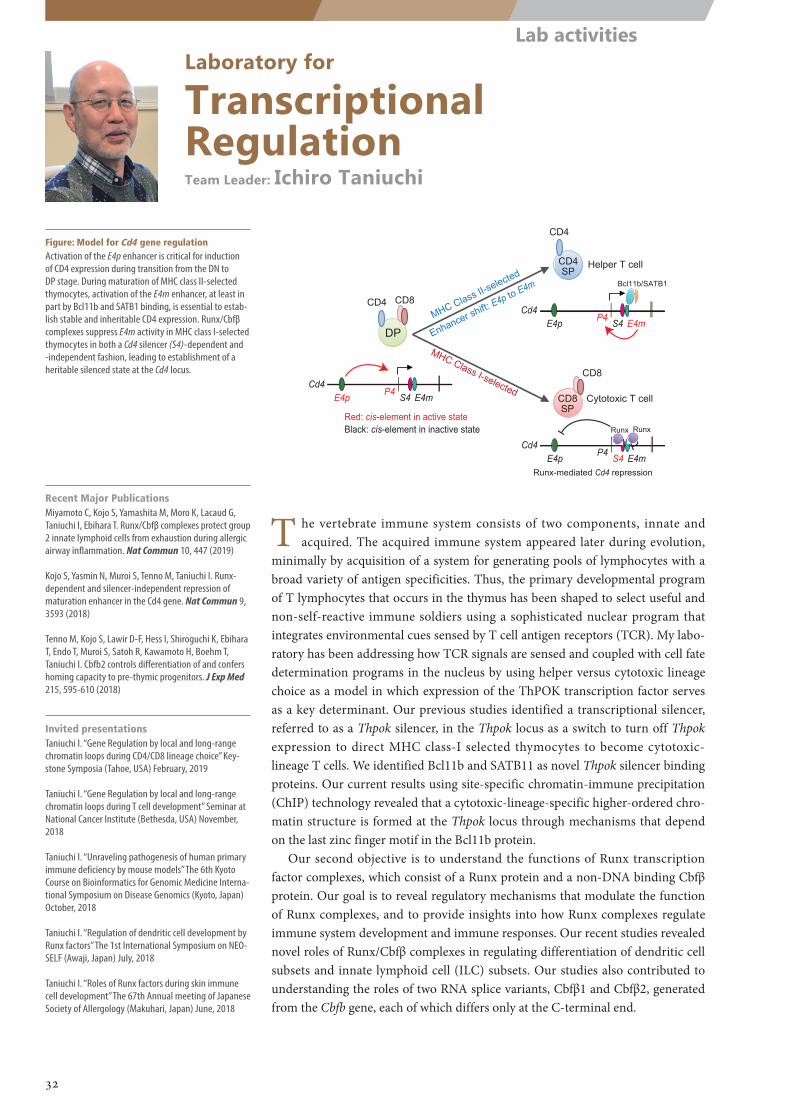
MHC Class II-selected
DP
CD4 CD8
CD4
CD8
CD4 SP
CD8 SP
Helper T cell
Cytotoxic T cell
Cd4E4p S4 E4m
Red: cis-element in active state
MHC Class I-selected
Black: cis-element in inactive state
P4
Cd4E4p S4 E4m
P4
Bcl11b/SATB1
Cd4E4p S4 E4m
P4
Enhancer shift: E4p to E4m
Runx Runx
Runx-mediated Cd4 repression
Laboratory for
Transcriptional RegulationTeam Leader: Ichiro Taniuchi
Figure: Model for Cd4 gene regulationActivation of the E4p enhancer is critical for induction of CD4 expression during transition from the DN to DP stage. During maturation of MHC class II-selected thymocytes, activation of the E4m enhancer, at least in part by Bcl11b and SATB1 binding, is essential to estab-lish stable and inheritable CD4 expression. Runx/Cbfβ complexes suppress E4m activity in MHC class I-selected thymocytes in both a Cd4 silencer (S4)-dependent and -independent fashion, leading to establishment of a heritable silenced state at the Cd4 locus.
T he vertebrate immune system consists of two components, innate and acquired. The acquired immune system appeared later during evolution,
minimally by acquisition of a system for generating pools of lymphocytes with a broad variety of antigen specificities. Thus, the primary developmental program of T lymphocytes that occurs in the thymus has been shaped to select useful and non-self-reactive immune soldiers using a sophisticated nuclear program that integrates environmental cues sensed by T cell antigen receptors (TCR). My labo-ratory has been addressing how TCR signals are sensed and coupled with cell fate determination programs in the nucleus by using helper versus cytotoxic lineage choice as a model in which expression of the ThPOK transcription factor serves as a key determinant. Our previous studies identified a transcriptional silencer, referred to as a Thpok silencer, in the Thpok locus as a switch to turn off Thpok expression to direct MHC class-I selected thymocytes to become cytotoxic-lineage T cells. We identified Bcl11b and SATB11 as novel Thpok silencer binding proteins. Our current results using site-specific chromatin-immune precipitation (ChIP) technology revealed that a cytotoxic-lineage-specific higher-ordered chro-matin structure is formed at the Thpok locus through mechanisms that depend on the last zinc finger motif in the Bcl11b protein.
Our second objective is to understand the functions of Runx transcription factor complexes, which consist of a Runx protein and a non-DNA binding Cbfβ protein. Our goal is to reveal regulatory mechanisms that modulate the function of Runx complexes, and to provide insights into how Runx complexes regulate immune system development and immune responses. Our recent studies revealed novel roles of Runx/Cbfβ complexes in regulating differentiation of dendritic cell subsets and innate lymphoid cell (ILC) subsets. Our studies also contributed to understanding the roles of two RNA splice variants, Cbfβ1 and Cbfβ2, generated from the Cbfb gene, each of which differs only at the C-terminal end.
Recent Major PublicationsMiyamoto C, Kojo S, Yamashita M, Moro K, Lacaud G, Taniuchi I, Ebihara T. Runx/Cbfβ complexes protect group 2 innate lymphoid cells from exhaustion during allergic airway inflammation. Nat Commun 10, 447 (2019)
Kojo S, Yasmin N, Muroi S, Tenno M, Taniuchi I. Runx-dependent and silencer-independent repression of maturation enhancer in the Cd4 gene. Nat Commun 9, 3593 (2018)
Tenno M, Kojo S, Lawir D-F, Hess I, Shiroguchi K, Ebihara T, Endo T, Muroi S, Satoh R, Kawamoto H, Boehm T, Taniuchi I. Cbfb2 controls differentiation of and confers homing capacity to pre-thymic progenitors. J Exp Med 215, 595-610 (2018)
Invited presentationsTaniuchi I. “Gene Regulation by local and long-range chromatin loops during CD4/CD8 lineage choice” Key-stone Symposia (Tahoe, USA) February, 2019
Taniuchi I. “Gene Regulation by local and long-range chromatin loops during T cell development” Seminar at National Cancer Institute (Bethesda, USA) November, 2018
Taniuchi I. “Unraveling pathogenesis of human primary immune deficiency by mouse models” The 6th Kyoto Course on Bioinformatics for Genomic Medicine Interna-tional Symposium on Disease Genomics (Kyoto, Japan) October, 2018
Taniuchi I. “Regulation of dendritic cell development by Runx factors” The 1st International Symposium on NEO-SELF (Awaji, Japan) July, 2018
Taniuchi I. “Roles of Runx factors during skin immune cell development” The 67th Annual meeting of Japanese Society of Allergology (Makuhari, Japan) June, 2018
32
Lab activities
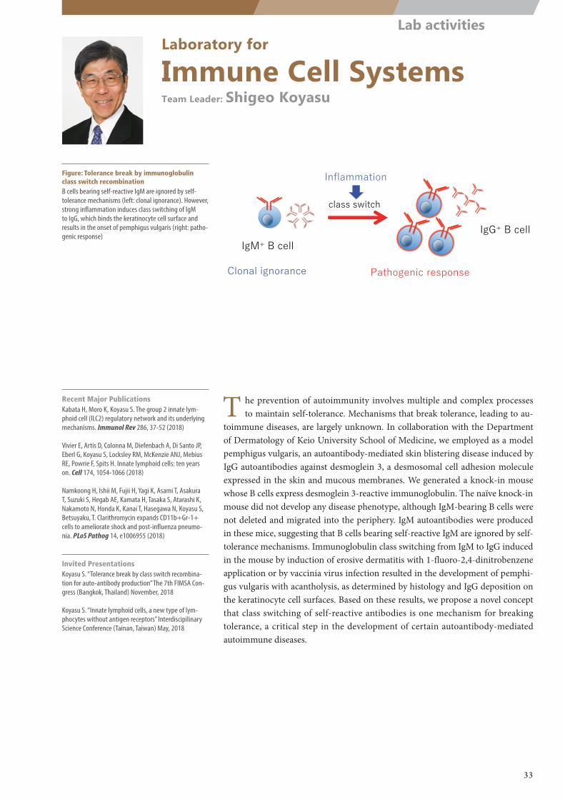
IgG+ B cellIgM+ B cell
Clonal ignorance Pathogenic response
class switch
Inflammation
Laboratory for
Immune Cell SystemsTeam Leader: Shigeo Koyasu
Figure: Tolerance break by immunoglobulin class switch recombinationB cells bearing self-reactive IgM are ignored by self-tolerance mechanisms (left: clonal ignorance). However, strong inflammation induces class switching of IgM to IgG, which binds the keratinocyte cell surface and results in the onset of pemphigus vulgaris (right: patho-genic response)
Recent Major PublicationsKabata H, Moro K, Koyasu S. The group 2 innate lym-phoid cell (ILC2) regulatory network and its underlying mechanisms. Immunol Rev 286, 37-52 (2018)
Vivier E, Artis D, Colonna M, Diefenbach A, Di Santo JP, Eberl G, Koyasu S, Locksley RM, McKenzie ANJ, Mebius RE, Powrie F, Spits H. Innate lymphoid cells: ten years on. Cell 174, 1054-1066 (2018)
Namkoong H, Ishii M, Fujii H, Yagi K, Asami T, Asakura T, Suzuki S, Hegab AE, Kamata H, Tasaka S, Atarashi K, Nakamoto N, Honda K, Kanai T, Hasegawa N, Koyasu S, Betsuyaku, T. Clarithromycin expands CD11b+Gr-1+ cells to ameliorate shock and post-influenza pneumo-nia. PLoS Pathog 14, e1006955 (2018)
Invited PresentationsKoyasu S. “Tolerance break by class switch recombina-tion for auto-antibody production” The 7th FIMSA Con-gress (Bangkok, Thailand) November, 2018
Koyasu S. “Innate lymphoid cells, a new type of lym-phocytes without antigen receptors” Interdiscipilinary Science Conference (Tainan, Taiwan) May, 2018
T he prevention of autoimmunity involves multiple and complex processes to maintain self-tolerance. Mechanisms that break tolerance, leading to au-
toimmune diseases, are largely unknown. In collaboration with the Department of Dermatology of Keio University School of Medicine, we employed as a model pemphigus vulgaris, an autoantibody-mediated skin blistering disease induced by IgG autoantibodies against desmoglein 3, a desmosomal cell adhesion molecule expressed in the skin and mucous membranes. We generated a knock-in mouse whose B cells express desmoglein 3-reactive immunoglobulin. The naïve knock-in mouse did not develop any disease phenotype, although IgM-bearing B cells were not deleted and migrated into the periphery. IgM autoantibodies were produced in these mice, suggesting that B cells bearing self-reactive IgM are ignored by self-tolerance mechanisms. Immunoglobulin class switching from IgM to IgG induced in the mouse by induction of erosive dermatitis with 1-fluoro-2,4-dinitrobenzene application or by vaccinia virus infection resulted in the development of pemphi-gus vulgaris with acantholysis, as determined by histology and IgG deposition on the keratinocyte cell surfaces. Based on these results, we propose a novel concept that class switching of self-reactive antibodies is one mechanism for breaking tolerance, a critical step in the development of certain autoantibody-mediated autoimmune diseases.
33
Lab activities
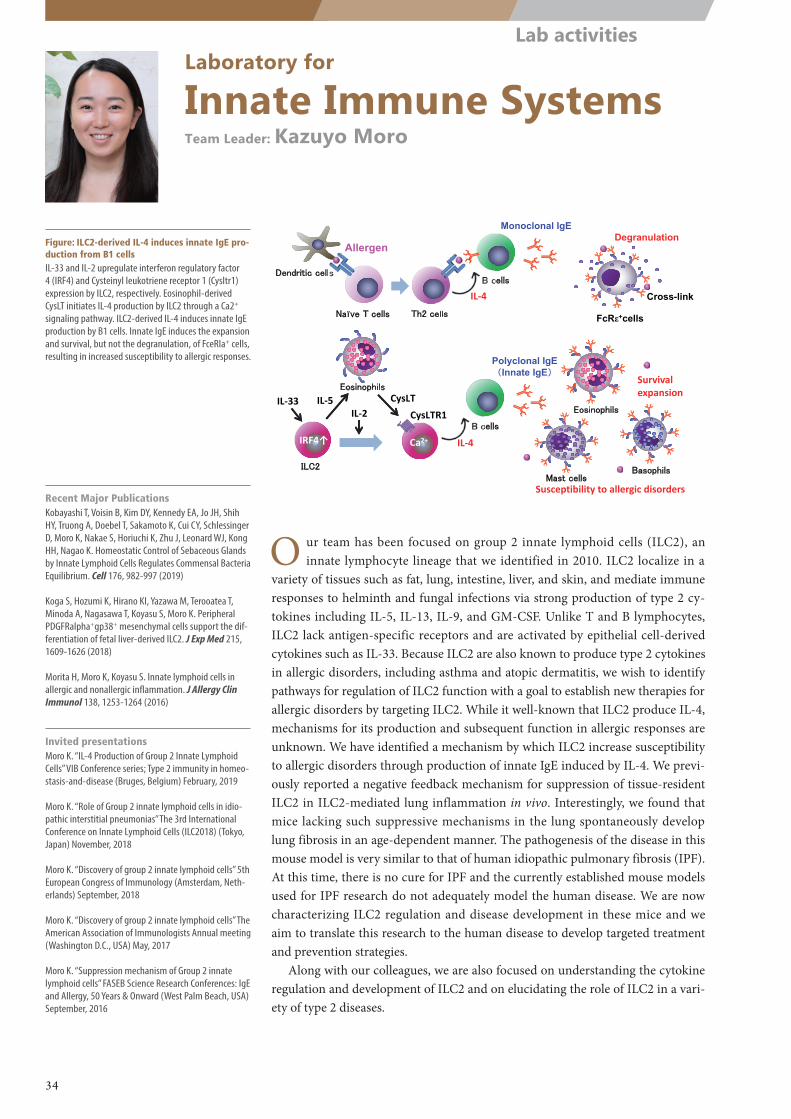
Th2 cellsNaïve T cells
Dendritic cellsB cells
IL‐4
Allergen
Monoclonal IgEDegranulation
Cross-link
ILC2
CysLTIL‐33IL‐2
Ca2+B cells
IL‐4
Polyclonal IgE(Innate IgE)
Eosinophils
BasophilsMast cells
Survivalexpansion
FcRε+cells
Susceptibility to allergic disorders
IRF4↑
Eosinophils
IL‐5CysLTR1
Laboratory for
Innate Immune SystemsTeam Leader: Kazuyo Moro
Figure: ILC2-derived IL-4 induces innate IgE pro-duction from B1 cellsIL-33 and IL-2 upregulate interferon regulatory factor 4 (IRF4) and Cysteinyl leukotriene receptor 1 (Cysltr1) expression by ILC2, respectively. Eosinophil-derived CysLT initiates IL-4 production by ILC2 through a Ca2+ signaling pathway. ILC2-derived IL-4 induces innate IgE production by B1 cells. Innate IgE induces the expansion and survival, but not the degranulation, of FceRIa+ cells, resulting in increased susceptibility to allergic responses.
O ur team has been focused on group 2 innate lymphoid cells (ILC2), an innate lymphocyte lineage that we identified in 2010. ILC2 localize in a
variety of tissues such as fat, lung, intestine, liver, and skin, and mediate immune responses to helminth and fungal infections via strong production of type 2 cy-tokines including IL-5, IL-13, IL-9, and GM-CSF. Unlike T and B lymphocytes, ILC2 lack antigen-specific receptors and are activated by epithelial cell-derived cytokines such as IL-33. Because ILC2 are also known to produce type 2 cytokines in allergic disorders, including asthma and atopic dermatitis, we wish to identify pathways for regulation of ILC2 function with a goal to establish new therapies for allergic disorders by targeting ILC2. While it well-known that ILC2 produce IL-4, mechanisms for its production and subsequent function in allergic responses are unknown. We have identified a mechanism by which ILC2 increase susceptibility to allergic disorders through production of innate IgE induced by IL-4. We previ-ously reported a negative feedback mechanism for suppression of tissue-resident ILC2 in ILC2-mediated lung inflammation in vivo. Interestingly, we found that mice lacking such suppressive mechanisms in the lung spontaneously develop lung fibrosis in an age-dependent manner. The pathogenesis of the disease in this mouse model is very similar to that of human idiopathic pulmonary fibrosis (IPF). At this time, there is no cure for IPF and the currently established mouse models used for IPF research do not adequately model the human disease. We are now characterizing ILC2 regulation and disease development in these mice and we aim to translate this research to the human disease to develop targeted treatment and prevention strategies.
Along with our colleagues, we are also focused on understanding the cytokine regulation and development of ILC2 and on elucidating the role of ILC2 in a vari-ety of type 2 diseases.
Recent Major PublicationsKobayashi T, Voisin B, Kim DY, Kennedy EA, Jo JH, Shih HY, Truong A, Doebel T, Sakamoto K, Cui CY, Schlessinger D, Moro K, Nakae S, Horiuchi K, Zhu J, Leonard WJ, Kong HH, Nagao K. Homeostatic Control of Sebaceous Glands by Innate Lymphoid Cells Regulates Commensal Bacteria Equilibrium. Cell 176, 982-997 (2019)
Koga S, Hozumi K, Hirano KI, Yazawa M, Terooatea T, Minoda A, Nagasawa T, Koyasu S, Moro K. Peripheral PDGFRalpha+gp38+ mesenchymal cells support the dif-ferentiation of fetal liver-derived ILC2. J Exp Med 215, 1609-1626 (2018)
Morita H, Moro K, Koyasu S. Innate lymphoid cells in allergic and nonallergic inflammation. J Allergy Clin Immunol 138, 1253-1264 (2016)
Invited presentationsMoro K. “IL-4 Production of Group 2 Innate Lymphoid Cells” VIB Conference series; Type 2 immunity in homeo-stasis-and-disease (Bruges, Belgium) February, 2019
Moro K. “Role of Group 2 innate lymphoid cells in idio-pathic interstitial pneumonias” The 3rd International Conference on Innate Lymphoid Cells (ILC2018) (Tokyo, Japan) November, 2018
Moro K. “Discovery of group 2 innate lymphoid cells” 5th European Congress of Immunology (Amsterdam, Neth-erlands) September, 2018
Moro K. “Discovery of group 2 innate lymphoid cells” The American Association of Immunologists Annual meeting (Washington D.C., USA) May, 2017
Moro K. “Suppression mechanism of Group 2 innate lymphoid cells” FASEB Science Research Conferences: IgE and Allergy, 50 Years & Onward (West Palm Beach, USA) September, 2016
34
Lab activities
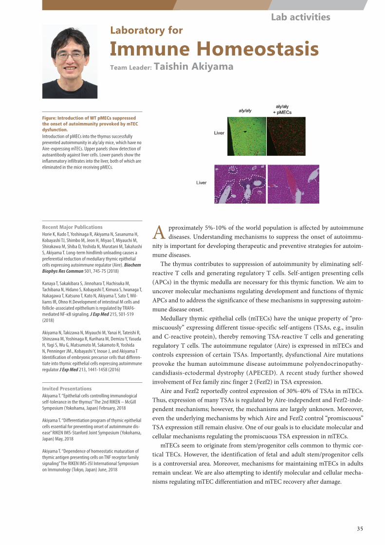
Laboratory for
Immune HomeostasisTeam Leader: Taishin Akiyama
Figure: Introduction of WT pMECs suppressed the onset of autoimmunity provoked by mTEC dysfunction.Introduction of pMECs into the thymus successfully prevented autoimmunity in aly/aly mice, which have no Aire-expressing mTECs. Upper panels show detection of autoantibody against liver cells. Lower panels show the inflammatory infiltrates into the liver, both of which are eliminated in the mice receiving pMECs.
A pproximately 5%-10% of the world population is affected by autoimmune diseases. Understanding mechanisms to suppress the onset of autoimmu-
nity is important for developing therapeutic and preventive strategies for autoim-mune diseases.
The thymus contributes to suppression of autoimmunity by eliminating self-reactive T cells and generating regulatory T cells. Self-antigen presenting cells (APCs) in the thymic medulla are necessary for this thymic function. We aim to uncover molecular mechanisms regulating development and functions of thymic APCs and to address the significance of these mechanisms in suppressing autoim-mune disease onset.
Medullary thymic epithelial cells (mTECs) have the unique property of “pro-miscuously” expressing different tissue-specific self-antigens (TSAs, e.g., insulin and C-reactive protein), thereby removing TSA-reactive T cells and generating regulatory T cells. The autoimmune regulator (Aire) is expressed in mTECs and controls expression of certain TSAs. Importantly, dysfunctional Aire mutations provoke the human autoimmune disease autoimmune polyendocrinopathy-candidiasis-ectodermal dystrophy (APECED). A recent study further showed involvement of Fez family zinc finger 2 (Fezf2) in TSA expression.
Aire and Fezf2 reportedly control expression of 30%-40% of TSAs in mTECs. Thus, expression of many TSAs is regulated by Aire-independent and Fezf2-inde-pendent mechanisms; however, the mechanisms are largely unknown. Moreover, even the underlying mechanisms by which Aire and Fezf2 control “promiscuous” TSA expression still remain elusive. One of our goals is to elucidate molecular and cellular mechanisms regulating the promiscuous TSA expression in mTECs.
mTECs seem to originate from stem/progenitor cells common to thymic cor-tical TECs. However, the identification of fetal and adult stem/progenitor cells is a controversial area. Moreover, mechanisms for maintaining mTECs in adults remain unclear. We are also attempting to identify molecular and cellular mecha-nisms regulating mTEC differentiation and mTEC recovery after damage.
Recent Major PublicationsHorie K, Kudo T, Yoshinaga R, Akiyama N, Sasanuma H, Kobayashi TJ, Shimbo M, Jeon H, Miyao T, Miyauchi M, Shirakawa M, Shiba D, Yoshida N, Muratani M, Takahashi S, Akiyama T. Long-term hindlimb unloading causes a preferential reduction of medullary thymic epithelial cells expressing autoimmune regulator (Aire). Biochem Biophys Res Commun 501, 745-75 (2018)
Kanaya T, Sakakibara S, Jinnohara T, Hachisuka M, Tachibana N, Hidano S, Kobayashi T, Kimura S, Iwanaga T, Nakagawa T, Katsuno T, Kato N, Akiyama T, Sato T, Wil-liams IR, Ohno H.Development of intestinal M cells and follicle-associated epithelium is regulated by TRAF6-mediated NF-κB signaling. J Exp Med 215, 501-519 (2018)
Akiyama N, Takizawa N, Miyauchi M, Yanai H, Tateishi R, Shinzawa M, Yoshinaga R, Kurihara M, Demizu Y, Yasuda H, Yagi S, Wu G, Matsumoto M, Sakamoto R, Yoshida N, Penninger JM., Kobayashi Y, Inoue J, and Akiyama T Identification of embryonic precursor cells that differen-tiate into thymic epithelial cells expressing autoimmune regulator J Exp Med 213, 1441-1458 (2016)
Invited PresentationsAkiyama T. “Epithelial cells controlling immunological self-tolerance in the thymus” The 2nd RIKEN – McGill Symposium (Yokohama, Japan) February, 2018
Akiyama T. “Differentiation program of thymic epithelial cells essential for preventing onset of autoimmune dis-ease” RIKEN IMS-Stanford Joint Symposium (Yokohama, Japan) May, 2018
Akiyama T. “Dependence of homeostatic maturation of thymic antigen presenting cells on TNF receptor family signaling” The RIKEN IMS-JSI International Symposium on Immunology (Tokyo, Japan) June, 2018
35
Lab activities
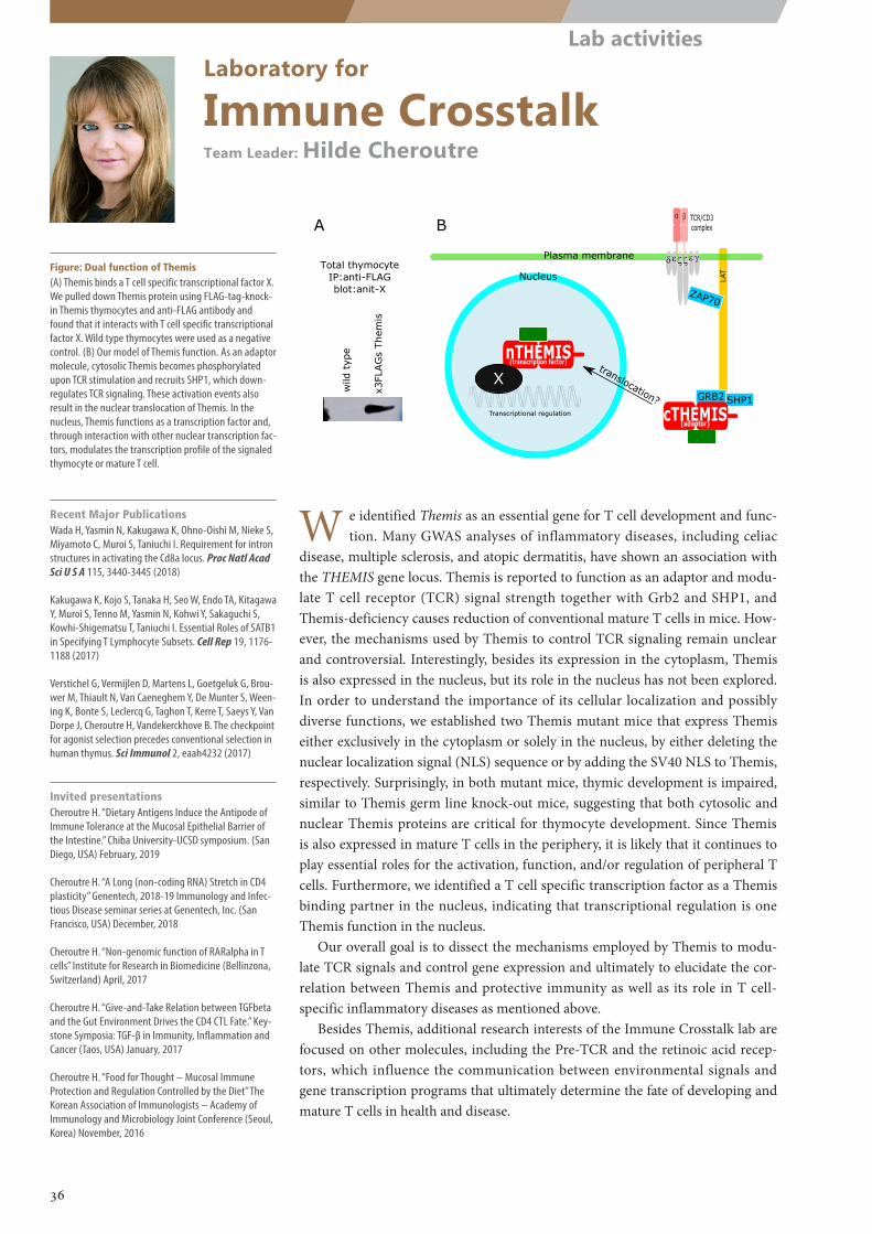
ZAP70
LAT
SHP1GRB2
cRAR�
���� ��
� �
Plasma membrane
Nucleus
cTHEMIS(adaptor)
nTHEMIS(transcription factor)
Transcriptional regulation
TCR/CD3 complex
translocation?
X
Total thymocyteIP:anti-FLAGblot:anit-X
wild
typ
e
x3FL
AG
s Th
emis
A B
Laboratory for
Immune CrosstalkTeam Leader: Hilde Cheroutre
Figure: Dual function of Themis(A) Themis binds a T cell specific transcriptional factor X. We pulled down Themis protein using FLAG-tag-knock-in Themis thymocytes and anti-FLAG antibody and found that it interacts with T cell specific transcriptional factor X. Wild type thymocytes were used as a negative control. (B) Our model of Themis function. As an adaptor molecule, cytosolic Themis becomes phosphorylated upon TCR stimulation and recruits SHP1, which down-regulates TCR signaling. These activation events also result in the nuclear translocation of Themis. In the nucleus, Themis functions as a transcription factor and, through interaction with other nuclear transcription fac-tors, modulates the transcription profile of the signaled thymocyte or mature T cell.
Recent Major PublicationsWada H, Yasmin N, Kakugawa K, Ohno-Oishi M, Nieke S, Miyamoto C, Muroi S, Taniuchi I. Requirement for intron structures in activating the Cd8a locus. Proc Natl Acad Sci U S A 115, 3440-3445 (2018)
Kakugawa K, Kojo S, Tanaka H, Seo W, Endo TA, Kitagawa Y, Muroi S, Tenno M, Yasmin N, Kohwi Y, Sakaguchi S, Kowhi-Shigematsu T, Taniuchi I. Essential Roles of SATB1 in Specifying T Lymphocyte Subsets. Cell Rep 19, 1176-1188 (2017)
Verstichel G, Vermijlen D, Martens L, Goetgeluk G, Brou-wer M, Thiault N, Van Caeneghem Y, De Munter S, Ween-ing K, Bonte S, Leclercq G, Taghon T, Kerre T, Saeys Y, Van Dorpe J, Cheroutre H, Vandekerckhove B. The checkpoint for agonist selection precedes conventional selection in human thymus. Sci Immunol 2, eaah4232 (2017)
Invited presentationsCheroutre H. “Dietary Antigens Induce the Antipode of Immune Tolerance at the Mucosal Epithelial Barrier of the Intestine.” Chiba University-UCSD symposium. (San Diego, USA) February, 2019
Cheroutre H. “A Long (non-coding RNA) Stretch in CD4 plasticity” Genentech, 2018-19 Immunology and Infec-tious Disease seminar series at Genentech, Inc. (San Francisco, USA) December, 2018
Cheroutre H. “Non-genomic function of RARalpha in T cells” Institute for Research in Biomedicine (Bellinzona, Switzerland) April, 2017
Cheroutre H. “Give-and-Take Relation between TGFbeta and the Gut Environment Drives the CD4 CTL Fate.” Key-stone Symposia: TGF-β in Immunity, Inflammation and Cancer (Taos, USA) January, 2017
Cheroutre H. “Food for Thought – Mucosal Immune Protection and Regulation Controlled by the Diet” The Korean Association of Immunologists – Academy of Immunology and Microbiology Joint Conference (Seoul, Korea) November, 2016
W e identified Themis as an essential gene for T cell development and func-tion. Many GWAS analyses of inflammatory diseases, including celiac
disease, multiple sclerosis, and atopic dermatitis, have shown an association with the THEMIS gene locus. Themis is reported to function as an adaptor and modu-late T cell receptor (TCR) signal strength together with Grb2 and SHP1, and Themis-deficiency causes reduction of conventional mature T cells in mice. How-ever, the mechanisms used by Themis to control TCR signaling remain unclear and controversial. Interestingly, besides its expression in the cytoplasm, Themis is also expressed in the nucleus, but its role in the nucleus has not been explored. In order to understand the importance of its cellular localization and possibly diverse functions, we established two Themis mutant mice that express Themis either exclusively in the cytoplasm or solely in the nucleus, by either deleting the nuclear localization signal (NLS) sequence or by adding the SV40 NLS to Themis, respectively. Surprisingly, in both mutant mice, thymic development is impaired, similar to Themis germ line knock-out mice, suggesting that both cytosolic and nuclear Themis proteins are critical for thymocyte development. Since Themis is also expressed in mature T cells in the periphery, it is likely that it continues to play essential roles for the activation, function, and/or regulation of peripheral T cells. Furthermore, we identified a T cell specific transcription factor as a Themis binding partner in the nucleus, indicating that transcriptional regulation is one Themis function in the nucleus.
Our overall goal is to dissect the mechanisms employed by Themis to modu-late TCR signals and control gene expression and ultimately to elucidate the cor-relation between Themis and protective immunity as well as its role in T cell-specific inflammatory diseases as mentioned above.
Besides Themis, additional research interests of the Immune Crosstalk lab are focused on other molecules, including the Pre-TCR and the retinoic acid recep-tors, which influence the communication between environmental signals and gene transcription programs that ultimately determine the fate of developing and mature T cells in health and disease.
36
Lab activities
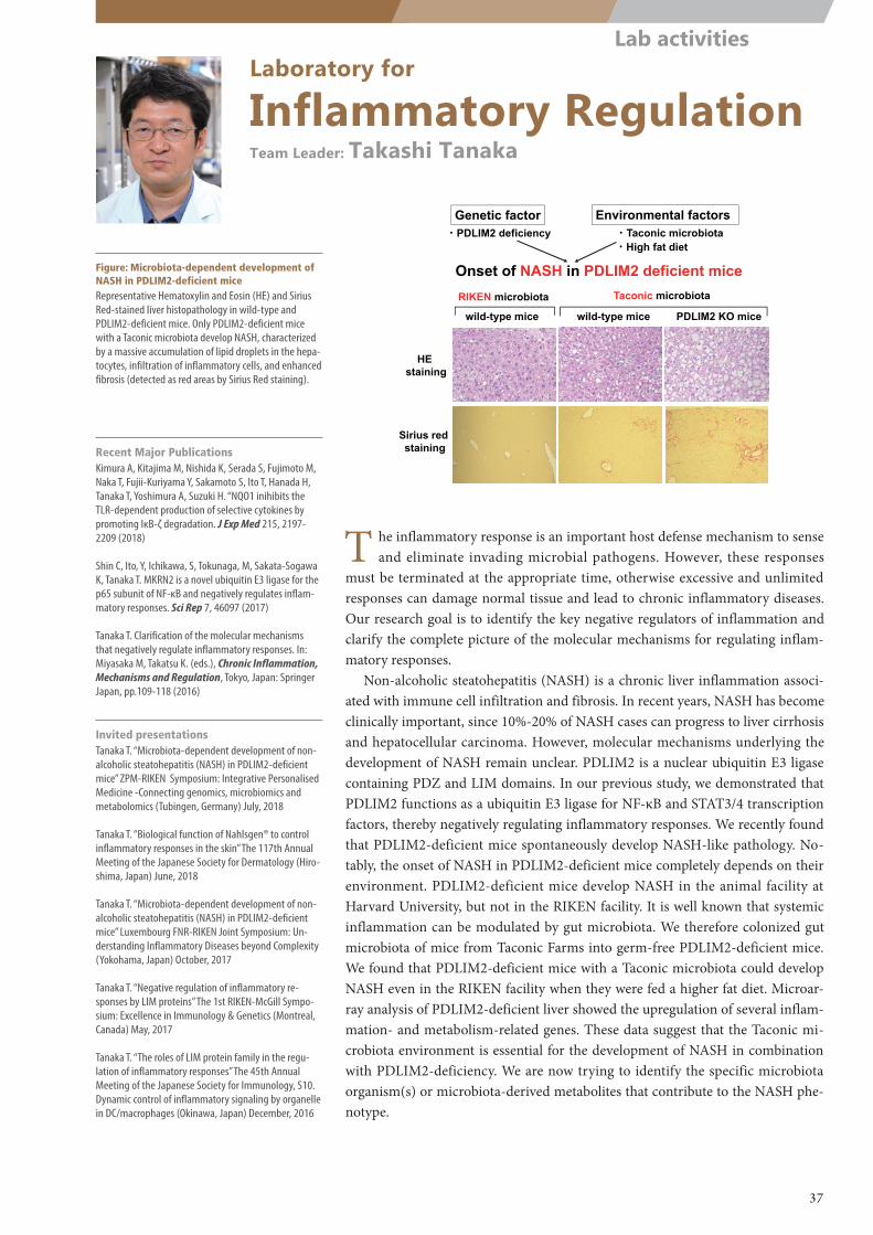
wild-type mice wild-type mice PDLIM2 KO mice
Taconic microbiotaRIKEN microbiota
Sirius red staining
HE staining
Genetic factor Environmental factors
・High fat diet・PDLIM2 deficiency
Onset of NASH in PDLIM2 deficient mice
・Taconic microbiota
Laboratory for
Inflammatory RegulationTeam Leader: Takashi Tanaka
Figure: Microbiota-dependent development of NASH in PDLIM2-deficient miceRepresentative Hematoxylin and Eosin (HE) and Sirius Red-stained liver histopathology in wild-type and PDLIM2-deficient mice. Only PDLIM2-deficient mice with a Taconic microbiota develop NASH, characterized by a massive accumulation of lipid droplets in the hepa-tocytes, infiltration of inflammatory cells, and enhanced fibrosis (detected as red areas by Sirius Red staining).
T he inflammatory response is an important host defense mechanism to sense and eliminate invading microbial pathogens. However, these responses
must be terminated at the appropriate time, otherwise excessive and unlimited responses can damage normal tissue and lead to chronic inflammatory diseases. Our research goal is to identify the key negative regulators of inflammation and clarify the complete picture of the molecular mechanisms for regulating inflam-matory responses.
Non-alcoholic steatohepatitis (NASH) is a chronic liver inflammation associ-ated with immune cell infiltration and fibrosis. In recent years, NASH has become clinically important, since 10%-20% of NASH cases can progress to liver cirrhosis and hepatocellular carcinoma. However, molecular mechanisms underlying the development of NASH remain unclear. PDLIM2 is a nuclear ubiquitin E3 ligase containing PDZ and LIM domains. In our previous study, we demonstrated that PDLIM2 functions as a ubiquitin E3 ligase for NF-κΒ and STAT3/4 transcription factors, thereby negatively regulating inflammatory responses. We recently found that PDLIM2-deficient mice spontaneously develop NASH-like pathology. No-tably, the onset of NASH in PDLIM2-deficient mice completely depends on their environment. PDLIM2-deficient mice develop NASH in the animal facility at Harvard University, but not in the RIKEN facility. It is well known that systemic inflammation can be modulated by gut microbiota. We therefore colonized gut microbiota of mice from Taconic Farms into germ-free PDLIM2-deficient mice. We found that PDLIM2-deficient mice with a Taconic microbiota could develop NASH even in the RIKEN facility when they were fed a higher fat diet. Microar-ray analysis of PDLIM2-deficient liver showed the upregulation of several inflam-mation- and metabolism-related genes. These data suggest that the Taconic mi-crobiota environment is essential for the development of NASH in combination with PDLIM2-deficiency. We are now trying to identify the specific microbiota organism(s) or microbiota-derived metabolites that contribute to the NASH phe-notype.
Recent Major PublicationsKimura A, Kitajima M, Nishida K, Serada S, Fujimoto M, Naka T, Fujii-Kuriyama Y, Sakamoto S, Ito T, Hanada H, Tanaka T, Yoshimura A, Suzuki H. “NQO1 inihibits the TLR-dependent production of selective cytokines by promoting IκB-ζ degradation. J Exp Med 215, 2197-2209 (2018)
Shin C, Ito, Y, Ichikawa, S, Tokunaga, M, Sakata-Sogawa K, Tanaka T. MKRN2 is a novel ubiquitin E3 ligase for the p65 subunit of NF-κB and negatively regulates inflam-matory responses. Sci Rep 7, 46097 (2017)
Tanaka T. Clarification of the molecular mechanisms that negatively regulate inflammatory responses. In: Miyasaka M, Takatsu K. (eds.), Chronic Inflammation, Mechanisms and Regulation, Tokyo, Japan: Springer Japan, pp.109-118 (2016)
Invited presentationsTanaka T. “Microbiota-dependent development of non-alcoholic steatohepatitis (NASH) in PDLIM2-deficient mice” ZPM-RIKEN Symposium: Integrative Personalised Medicine -Connecting genomics, microbiomics and metabolomics (Tubingen, Germany) July, 2018
Tanaka T. “Biological function of Nahlsgen® to control inflammatory responses in the skin” The 117th Annual Meeting of the Japanese Society for Dermatology (Hiro-shima, Japan) June, 2018
Tanaka T. “Microbiota-dependent development of non-alcoholic steatohepatitis (NASH) in PDLIM2-deficient mice” Luxembourg FNR-RIKEN Joint Symposium: Un-derstanding Inflammatory Diseases beyond Complexity (Yokohama, Japan) October, 2017
Tanaka T. “Negative regulation of inflammatory re-sponses by LIM proteins” The 1st RIKEN-McGill Sympo-sium: Excellence in Immunology & Genetics (Montreal, Canada) May, 2017
Tanaka T. “The roles of LIM protein family in the regu-lation of inflammatory responses” The 45th Annual Meeting of the Japanese Society for Immunology, S10. Dynamic control of inflammatory signaling by organelle in DC/macrophages (Okinawa, Japan) December, 2016
37
Lab activities
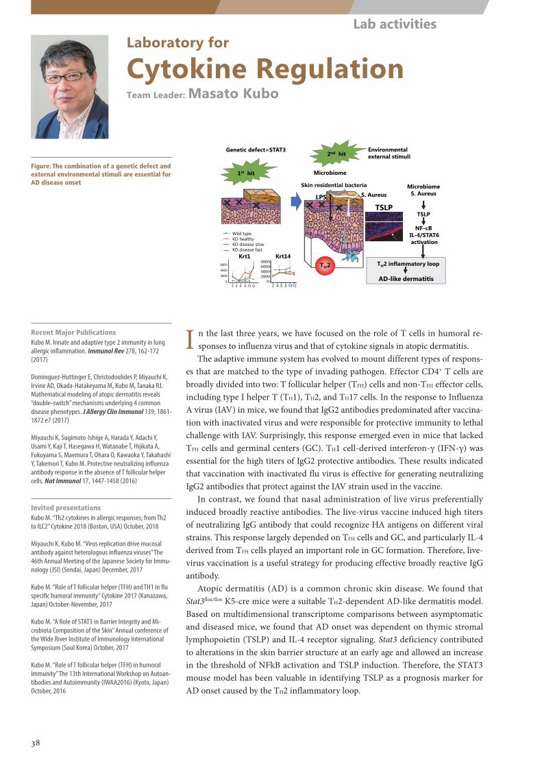
Krt1
6000
4000
2000
0
Krt10
12000
8000
4000
0
Ivl300
200
100
02 4 6 8 10 12 2 4 6 8 10 12 2 4 6 8 10 12
stratum granulosum
Krt1 Krt14
Microbiome S. Aureus
TSLP
NF-B IL-4/STAT6activation
2nd hit
AD-like dermatitis
TH2 inflammatory loop
1st hit
2
Microbiome
TH2
TSLP LPS
Genetic defect=STAT3
Skin residential bacteria
Environmental external stimuli
ControlKO-H
KO-D E arlyKO-D Slow
Wild typeKO healthyKO disease slowKO disease fast
S. Aureus
Laboratory for
Cytokine RegulationTeam Leader: Masato Kubo
Figure: The combination of a genetic defect and external environmental stimuli are essential for AD disease onset
Recent Major PublicationsKubo M. Innate and adaptive type 2 immunity in lung allergic inflammation. Immunol Rev 278, 162-172 (2017)
Dominguez-Huttinger E, Christodoulides P, Miyauchi K, Irvine AD, Okada-Hatakeyama M, Kubo M, Tanaka RJ. Mathematical modeling of atopic dermatitis reveals “double-switch” mechanisms underlying 4 common disease phenotypes. J Allergy Clin Immunol 139, 1861-1872 e7 (2017)
Miyauchi K, Sugimoto-Ishige A, Harada Y, Adachi Y, Usami Y, Kaji T, Hasegawa H, Watanabe T, Hijikata A, Fukuyama S, Maemura T, Ohara O, Kawaoka Y, Takahashi Y, Takemori T, Kubo M. Protective neutralizing influenza antibody response in the absence of T follicular helper cells. Nat Immunol 17, 1447-1458 (2016)
Invited presentationsKubo M. “Th2 cytokines in allergic responses; from Th2 to ILC2” Cytokine 2018 (Boston, USA) October, 2018
Miyauchi K, Kubo M. “Virus replication drive mucosal antibody against heterologous influenza viruses” The 46th Annual Meeting of the Japanese Society for Immu-nology (JSI) (Sendai, Japan) December, 2017
Kubo M. “Role of T follicular helper (TFH) and TH1 in flu specific humoral immunity” Cytokine 2017 (Kanazawa, Japan) October-November, 2017
Kubo M. “A Role of STAT3 in Barrier Integrity and Mi-crobiota Composition of the Skin” Annual conference of the Wide River Institute of Immunology International Symposium (Soul Korea) October, 2017
Kubo M. “Role of T follicular helper (TFH) in humoral immunity” The 13th International Workshop on Autoan-tibodies and Autoimmunity (IWAA2016) (Kyoto, Japan) October, 2016
I n the last three years, we have focused on the role of T cells in humoral re-sponses to influenza virus and that of cytokine signals in atopic dermatitis.The adaptive immune system has evolved to mount different types of respons-
es that are matched to the type of invading pathogen. Effector CD4+ T cells are broadly divided into two: T follicular helper (TFH) cells and non-TFH effector cells, including type I helper T (TH1), TH2, and TH17 cells. In the response to Influenza A virus (IAV) in mice, we found that IgG2 antibodies predominated after vaccina-tion with inactivated virus and were responsible for protective immunity to lethal challenge with IAV. Surprisingly, this response emerged even in mice that lacked TFH cells and germinal centers (GC). TH1 cell-derived interferon-γ (IFN-γ) was essential for the high titers of IgG2 protective antibodies. These results indicated that vaccination with inactivated flu virus is effective for generating neutralizing IgG2 antibodies that protect against the IAV strain used in the vaccine.
In contrast, we found that nasal administration of live virus preferentially induced broadly reactive antibodies. The live-virus vaccine induced high titers of neutralizing IgG antibody that could recognize HA antigens on different viral strains. This response largely depended on TFH cells and GC, and particularly IL-4 derived from TFH cells played an important role in GC formation. Therefore, live-virus vaccination is a useful strategy for producing effective broadly reactive IgG antibody.
Atopic dermatitis (AD) is a common chronic skin disease. We found that Stat3flox/flox K5-cre mice were a suitable TH2-dependent AD-like dermatitis model. Based on multidimensional transcriptome comparisons between asymptomatic and diseased mice, we found that AD onset was dependent on thymic stromal lymphopoietin (TSLP) and IL-4 receptor signaling. Stat3 deficiency contributed to alterations in the skin barrier structure at an early age and allowed an increase in the threshold of NFkB activation and TSLP induction. Therefore, the STAT3 mouse model has been valuable in identifying TSLP as a prognosis marker for AD onset caused by the TH2 inflammatory loop.
38
Lab activities
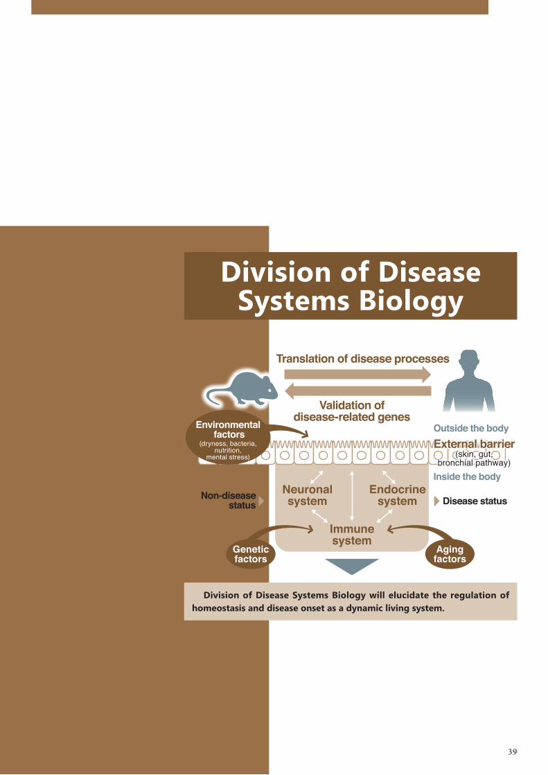
Division of Disease Systems Biology
Division of Disease Systems Biology will elucidate the regulation of homeostasis and disease onset as a dynamic living system.
39
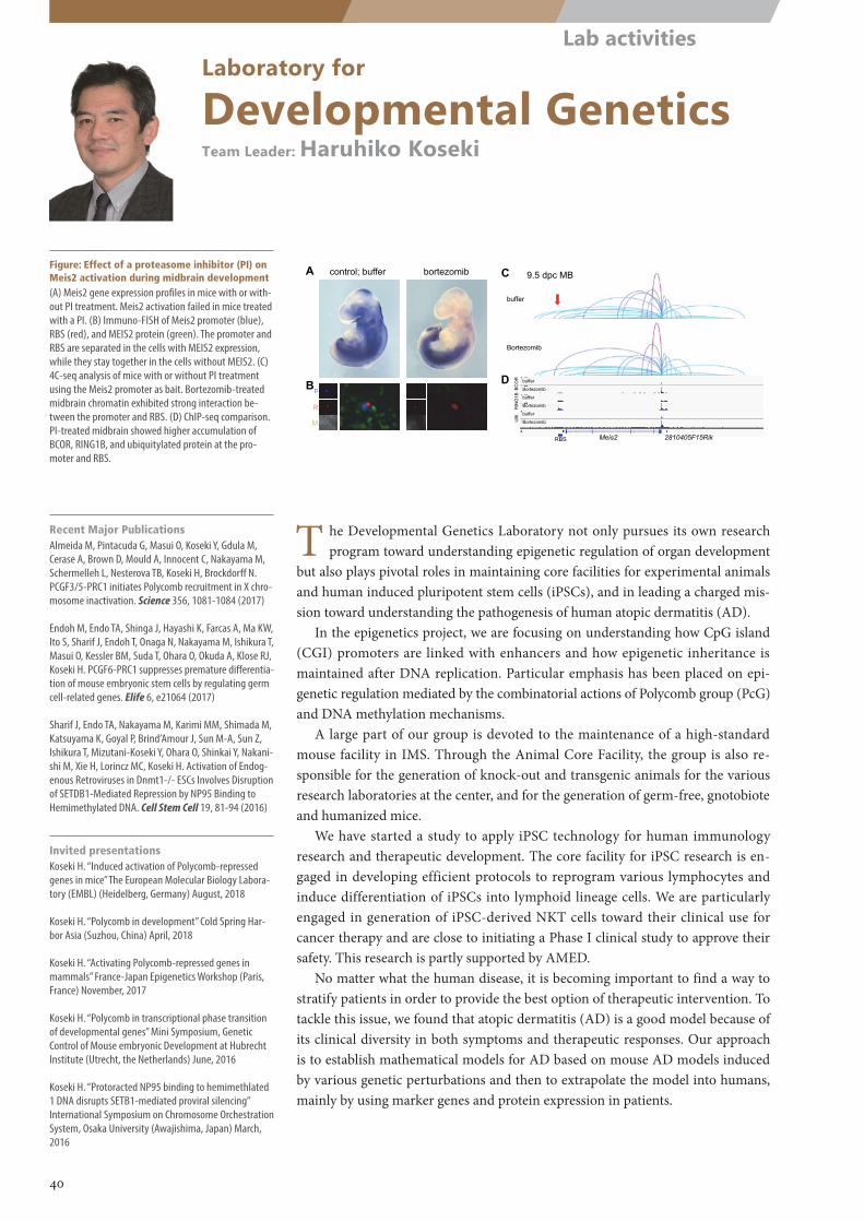
control; buffer bortezomib
Meis2 2810405F15RikRBS
9.5 dpc MB
buffer
Bortezomib
buffer
Bortezomib
buffer
Bortezomib
buffer
Bortezomib
BC
OR
RIN
G1B
UBI
A
B
C
Dp
M
R
Laboratory for
Developmental GeneticsTeam Leader: Haruhiko Koseki
Figure: Effect of a proteasome inhibitor (PI) on Meis2 activation during midbrain development(A) Meis2 gene expression profiles in mice with or with-out PI treatment. Meis2 activation failed in mice treated with a PI. (B) Immuno-FISH of Meis2 promoter (blue), RBS (red), and MEIS2 protein (green). The promoter and RBS are separated in the cells with MEIS2 expression, while they stay together in the cells without MEIS2. (C) 4C-seq analysis of mice with or without PI treatment using the Meis2 promoter as bait. Bortezomib-treated midbrain chromatin exhibited strong interaction be-tween the promoter and RBS. (D) ChIP-seq comparison. PI-treated midbrain showed higher accumulation of BCOR, RING1B, and ubiquitylated protein at the pro-moter and RBS.
Recent Major PublicationsAlmeida M, Pintacuda G, Masui O, Koseki Y, Gdula M, Cerase A, Brown D, Mould A, Innocent C, Nakayama M, Schermelleh L, Nesterova TB, Koseki H, Brockdorff N. PCGF3/5-PRC1 initiates Polycomb recruitment in X chro-mosome inactivation. Science 356, 1081-1084 (2017)
Endoh M, Endo TA, Shinga J, Hayashi K, Farcas A, Ma KW, Ito S, Sharif J, Endoh T, Onaga N, Nakayama M, Ishikura T, Masui O, Kessler BM, Suda T, Ohara O, Okuda A, Klose RJ, Koseki H. PCGF6-PRC1 suppresses premature differentia-tion of mouse embryonic stem cells by regulating germ cell-related genes. Elife 6, e21064 (2017)
Sharif J, Endo TA, Nakayama M, Karimi MM, Shimada M, Katsuyama K, Goyal P, Brind’Amour J, Sun M-A, Sun Z, Ishikura T, Mizutani-Koseki Y, Ohara O, Shinkai Y, Nakani-shi M, Xie H, Lorincz MC, Koseki H. Activation of Endog-enous Retroviruses in Dnmt1-/- ESCs Involves Disruption of SETDB1-Mediated Repression by NP95 Binding to Hemimethylated DNA. Cell Stem Cell 19, 81-94 (2016)
Invited presentationsKoseki H. “Induced activation of Polycomb-repressed genes in mice” The European Molecular Biology Labora-tory (EMBL) (Heidelberg, Germany) August, 2018
Koseki H. “Polycomb in development” Cold Spring Har-bor Asia (Suzhou, China) April, 2018
Koseki H. “Activating Polycomb-repressed genes in mammals” France-Japan Epigenetics Workshop (Paris, France) November, 2017
Koseki H. “Polycomb in transcriptional phase transition of developmental genes” Mini Symposium, Genetic Control of Mouse embryonic Development at Hubrecht Institute (Utrecht, the Netherlands) June, 2016
Koseki H. “Protoracted NP95 binding to hemimethlated 1 DNA disrupts SETB1-mediated proviral silencing” International Symposium on Chromosome Orchestration System, Osaka University (Awajishima, Japan) March, 2016
T he Developmental Genetics Laboratory not only pursues its own research program toward understanding epigenetic regulation of organ development
but also plays pivotal roles in maintaining core facilities for experimental animals and human induced pluripotent stem cells (iPSCs), and in leading a charged mis-sion toward understanding the pathogenesis of human atopic dermatitis (AD).
In the epigenetics project, we are focusing on understanding how CpG island (CGI) promoters are linked with enhancers and how epigenetic inheritance is maintained after DNA replication. Particular emphasis has been placed on epi-genetic regulation mediated by the combinatorial actions of Polycomb group (PcG) and DNA methylation mechanisms.
A large part of our group is devoted to the maintenance of a high-standard mouse facility in IMS. Through the Animal Core Facility, the group is also re-sponsible for the generation of knock-out and transgenic animals for the various research laboratories at the center, and for the generation of germ-free, gnotobiote and humanized mice.
We have started a study to apply iPSC technology for human immunology research and therapeutic development. The core facility for iPSC research is en-gaged in developing efficient protocols to reprogram various lymphocytes and induce differentiation of iPSCs into lymphoid lineage cells. We are particularly engaged in generation of iPSC-derived NKT cells toward their clinical use for cancer therapy and are close to initiating a Phase I clinical study to approve their safety. This research is partly supported by AMED.
No matter what the human disease, it is becoming important to find a way to stratify patients in order to provide the best option of therapeutic intervention. To tackle this issue, we found that atopic dermatitis (AD) is a good model because of its clinical diversity in both symptoms and therapeutic responses. Our approach is to establish mathematical models for AD based on mouse AD models induced by various genetic perturbations and then to extrapolate the model into humans, mainly by using marker genes and protein expression in patients.
40
Lab activities
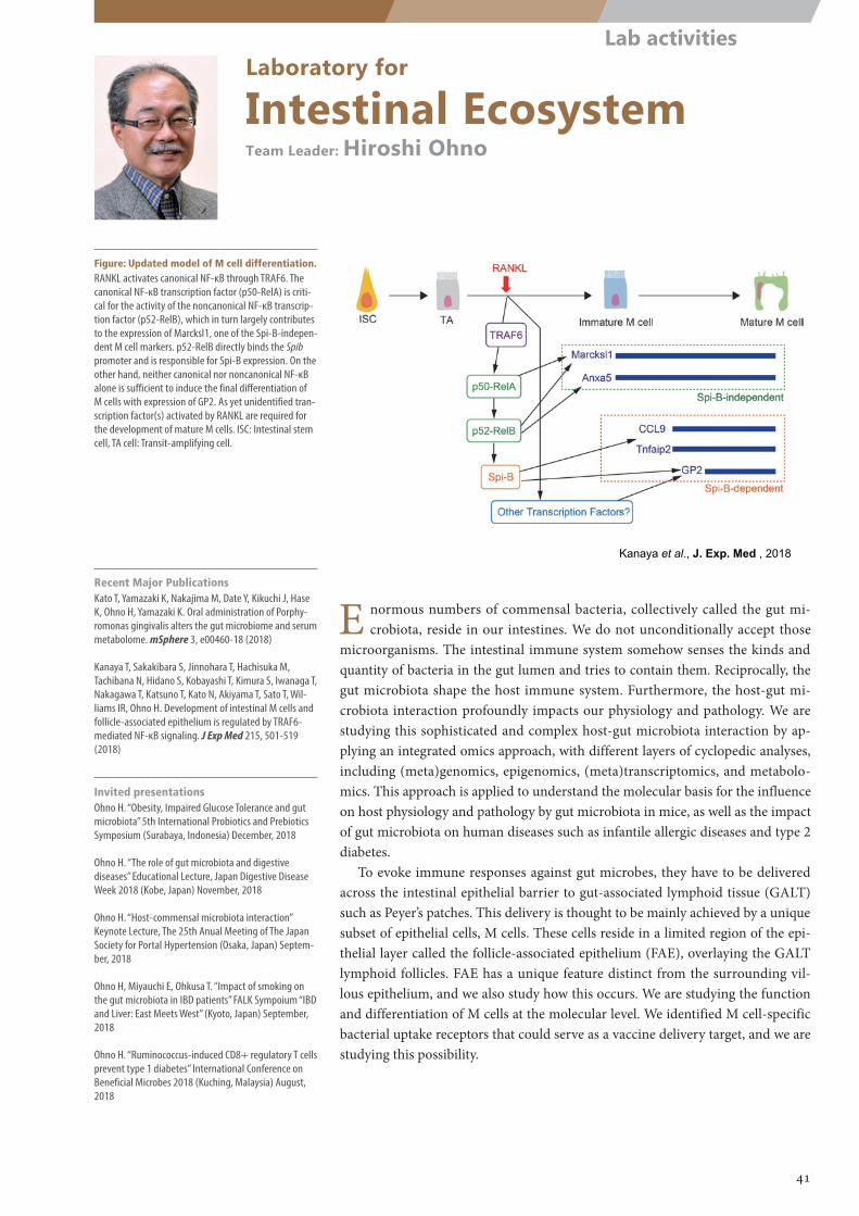
Kanaya et al., J. Exp. Med , 2018
Laboratory for
Intestinal EcosystemTeam Leader: Hiroshi Ohno
Figure: Updated model of M cell differentiation.RANKL activates canonical NF-κB through TRAF6. The canonical NF-κB transcription factor (p50-RelA) is criti-cal for the activity of the noncanonical NF-κB transcrip-tion factor (p52-RelB), which in turn largely contributes to the expression of Marcksl1, one of the Spi-B-indepen-dent M cell markers. p52-RelB directly binds the Spib promoter and is responsible for Spi-B expression. On the other hand, neither canonical nor noncanonical NF-κB alone is sufficient to induce the final differentiation of M cells with expression of GP2. As yet unidentified tran-scription factor(s) activated by RANKL are required for the development of mature M cells. ISC: Intestinal stem cell, TA cell: Transit-amplifying cell.
E normous numbers of commensal bacteria, collectively called the gut mi-crobiota, reside in our intestines. We do not unconditionally accept those
microorganisms. The intestinal immune system somehow senses the kinds and quantity of bacteria in the gut lumen and tries to contain them. Reciprocally, the gut microbiota shape the host immune system. Furthermore, the host-gut mi-crobiota interaction profoundly impacts our physiology and pathology. We are studying this sophisticated and complex host-gut microbiota interaction by ap-plying an integrated omics approach, with different layers of cyclopedic analyses, including (meta)genomics, epigenomics, (meta)transcriptomics, and metabolo-mics. This approach is applied to understand the molecular basis for the influence on host physiology and pathology by gut microbiota in mice, as well as the impact of gut microbiota on human diseases such as infantile allergic diseases and type 2 diabetes.
To evoke immune responses against gut microbes, they have to be delivered across the intestinal epithelial barrier to gut-associated lymphoid tissue (GALT) such as Peyer’s patches. This delivery is thought to be mainly achieved by a unique subset of epithelial cells, M cells. These cells reside in a limited region of the epi-thelial layer called the follicle-associated epithelium (FAE), overlaying the GALT lymphoid follicles. FAE has a unique feature distinct from the surrounding vil-lous epithelium, and we also study how this occurs. We are studying the function and differentiation of M cells at the molecular level. We identified M cell-specific bacterial uptake receptors that could serve as a vaccine delivery target, and we are studying this possibility.
Recent Major PublicationsKato T, Yamazaki K, Nakajima M, Date Y, Kikuchi J, Hase K, Ohno H, Yamazaki K. Oral administration of Porphy-romonas gingivalis alters the gut microbiome and serum metabolome. mSphere 3, e00460-18 (2018)
Kanaya T, Sakakibara S, Jinnohara T, Hachisuka M, Tachibana N, Hidano S, Kobayashi T, Kimura S, Iwanaga T, Nakagawa T, Katsuno T, Kato N, Akiyama T, Sato T, Wil-liams IR, Ohno H. Development of intestinal M cells and follicle-associated epithelium is regulated by TRAF6-mediated NF-κB signaling. J Exp Med 215, 501-519 (2018)
Invited presentationsOhno H. “Obesity, Impaired Glucose Tolerance and gut microbiota” 5th International Probiotics and Prebiotics Symposium (Surabaya, Indonesia) December, 2018
Ohno H. “The role of gut microbiota and digestive diseases” Educational Lecture, Japan Digestive Disease Week 2018 (Kobe, Japan) November, 2018
Ohno H. “Host-commensal microbiota interaction” Keynote Lecture, The 25th Anual Meeting of The Japan Society for Portal Hypertension (Osaka, Japan) Septem-ber, 2018
Ohno H, Miyauchi E, Ohkusa T. “Impact of smoking on the gut microbiota in IBD patients” FALK Sympoium “IBD and Liver: East Meets West” (Kyoto, Japan) September, 2018
Ohno H. “Ruminococcus-induced CD8+ regulatory T cells prevent type 1 diabetes” International Conference on Beneficial Microbes 2018 (Kuching, Malaysia) August, 2018
41
Lab activities
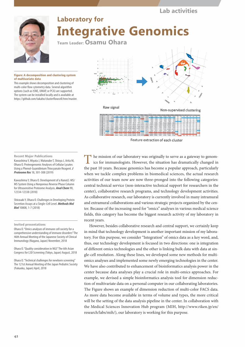
Laboratory for
Integrative GenomicsTeam Leader: Osamu Ohara
Figure: A decomposition and clustering system of multivariate dataThis example shows decomposition and clustering of multi-color flow cytometry data. Several algorithm options (such as tSNE, UMAP, or PCA) are supported. The system can be installed locally and is available at https://github.com/takaho/clusterflowcell/tree/master.
Recent Major PublicationsKawashima Y, Miyata J, Watanabe T, Shioya J, Arita M, Ohara O. Proteogenomic Analyses of Cellular Lysates Using a Phenol-Guanidinium Thiocyanate Reagent. J Proteome Res 18, 301-308 (2019)
Kawashima Y, Ohara O. Development of a NanoLC-MS/MS System Using a Nonporous Reverse Phase Column for Ultrasensitive Proteome Analysis. Anal Chem 90, 12334-12338 (2018)
Shirasaki Y, Ohara O. Challenges in Developing Protein Secretion Assays at a Single-Cell Level. Methods Mol Biol 10808, 1-7 (2018)
Invited presentationsOhara O. “Omics analyses of immune cell society for a comprehensive understanding of immune disorders” The 46th Annual Meeting of the Japanese Society of Clinical Immunology (Nagano, Japan) November, 2018
Ohara O. “Quality consideration in NGS” The 6th Asian Congress for LSD Screening (Tokyo, Japan) August, 2018
Ohara O. “Technical challenges for newborn screening” The 121st Annual Meeting of the Japan Pediatric Society (Fukuoka, Japan) April, 2018
T he mission of our laboratory was originally to serve as a gateway to genom-ics for immunologists. However, the situation has dramatically changed in
the past 10 years. Because genomics has become a popular approach, particularly when we tackle complex problems in biomedical sciences, the actual research activities of our team now are now three-pronged into the following categories: central technical service (non-interactive technical support for researchers in the center), collaborative research programs, and technology development activities. As collaborative research, our laboratory is currently involved in many intramural and extramural collaborations and various strategic projects organized by the cen-ter. Because of the increasing need for “omics” analyses in various medical science fields, this category has become the biggest research activity of my laboratory in recent years.
However, besides collaborative research and central support, we certainly keep in mind that technology development is another important mission of my labora-tory. For this purpose, we consider “Integration” of omics data as a key word, and, thus, our technology development is focused in two directions: one is integration of different omics technologies and the other is linking bulk data with data at sin-gle-cell resolution. Along these lines, we developed some new methods for multi-omics analyses and implemented some newly emerging technologies in the center. We have also contributed to enhancement of bioinformatics analysis power in the center because data analyses play a crucial role in multi-omics approaches. For example, we devised a simple bioinformatics analysis tool for dimension reduc-tion of multivariate data on a personal computer in our collaborating laboratories. The Figure shows an example of dimension reduction of multi-color FACS data. As more data become available in terms of volume and types, the more critical will be the setting of the data analysis pipeline in the center. In collaboration with the Medical Sciences Innovation Hub program (MIH, http://www.riken.jp/en/research/labs/mih/), our laboratory is working for this purpose.
42
Lab activities
(1) polysaccharide association (mucus/diet)
(2) MAFF↑mucosal associatedfunctional factor
(3)
(4) epithelial health
Bacteroides spp. (B.theta / B.vulgatus)
Clostridiales↑metabolic activity↑
diverse microbiota
IgA
inner mucus
outer mucus
intestinal content
metabolic activity↑bacteria-bacteriainteraction
SCFA
(-)IgA
-1 1expression
level
MAFF
(+)
B. theta genes
(maffC/D)
WT
∆MAF
F
Colon 10 days after DSS
B.th
eta
B.th
eta
69
SSC
IgA
B.theta
IgA Binding IgA-dependent transcriptional modulation Protection from colitis
Laboratory for
Mucosal ImmunityTeam Leader: Sidonia Fagarasan
Figure: A schematic representation of how IgA binding to Bacteroides species impacts its local-ization and metabolic reprogramming contrib-uting to symbiosis with Firmicutes species and fitness of the epithelial barrier
Understanding the role of IgA in shaping gut microbiotaOur group has obtained over the years robust evidence that the adaptive im-
mune system controls the growth and distribution of commensals within the intestinal lumen. We demonstrated that mucosal IgA is required for maintaining diverse and balanced communities of commensal bacteria, and that such IgA pro-duction is highly controlled by the expression of T cell signaling receptors (PD-1) and transcription factors (Foxp3, Bcl6) of T cells. However, how exactly IgA functions in symbiotic process is not well understood. Our recent work showed a novel mechanism by which heavily glycosylated IgA efficiently coats bacterial sur-faces, especially Bacteroidetes sp., thereby modulating bacterial genes within the mucus environment. We provisionally named the set of genes upregulated in the presence of IgA as Mucus-Associated Functional Factors (MAFF). Importantly, glycosylated IgA and MAFF upregulation by Bacteroidetes sp. promoted symbi-otic interactions between different bacterial phyla, like Firmicutes, and regulated the composition and metabolic function of the overall microbial community (Fig-ure). This work was featured by the JEM among the ten best papers published in 2018.
Understanding how activation of the immune system shifts metabolites, leading to neuro-endocrine disorders in mouse
We revealed novel and unexpected effects of immune system activation on systemic metabolic homeostasis with far-reaching effects on major physiological systems of the body. We demonstrated that persistent T cell activation resulted in drastic systemic metabolic changes, including depletion of serum amino acids. Furthermore, we linked the depletion of tyrosine and tryptophan in the periphery to reduced synthesis of brain monoamine neurotransmitters such as dopamine and serotonin. We showed that such biochemical alterations caused abnormal be-havior in mice, dominated by anxiety and enhanced fear responses.
Recent Major Publications1. Nakajima A, Vogelzang A, Maruya M, Miyajima M,
Murata M, Son A, Kuwahara T, Tsuruyama T, Yamada S, Matsuura M, Nakase H, Peterson DA, Fagarasan S, Su-zuki K. IgA regulates the composition and metabolic function of gut microbiota by promoting symbiosis between bacteria. J Exp Med 215, 2019-2034 (2018)
2. Miyajima M, Zhang B, Sugiura Y, Sonomura K, Guer-rini MM, Tsutsui Y, Maruya M, Vogelzang A, Chamoto K, Honda K, Hikida T, Qin H, Sanuki R, Suzuki K, Furu-kawa T, Ishihama Y, Matsuda F, Suematsu M, Honjo T, Fagarasan S. Metabolic shift induced by systemic activation of T cells in PD-1 deficient mice perturbs brain monoamines and emotional behavior. Nat Immunol 18, 1342-1352 (2017)
3. Chamoto K, Chowdhury PS, Kumar A, Sonomura K, Matsuda F, Fagarasan S, Honjo T. Mitochondrial activation chemicals synergize with surface receptor PD-1 blockade for T cell-dependent antitumor activ-ity. Proc Natl Acad Sci U S A 114, E761-E770 (2017)
Invited presentationsFagarasan S. “Excessive T cell activation in the absence of PD-1 affects behavior”. Cold Spring Asia meeting on inflammation: Basic Mechanisms & Relevant Diseases (Suzhou, China) December, 2017
Fagarasan S. “Involvement of PD-1 in antibody diversifi-cation and immune homeostasis” The 19th International Conference on Lymphatic Tissues and Germinal Centres in Immune Reactions (Venice, Italy) September, 2017. Organizer
Fagarasan S. “Involvement of PD-1 in antibody diversifi-cation and body homeostasis” NHRI/IBMS International conference on inflammation and disease (Taipei, Tai-wan) February, 2017
Fagarasan S. “Involvement of T cells in antibody diver-sification and shaping of the microbial landscape” Im-mune Profiling in Health and Disease 2016 (The Nature Conference) (Seattle, USA) October, 2016
Fagarasan S. “On germinal centers, IgA synthesis and the intestinal ecosystem” International Congress of Im-munology ICI 2016 (Melbourne, Australia) August, 2016
43
Lab activities
(gated on CD3ε+ TCRβ+ cells in colon LP)
Germ-freeGerm-free+11 strains
CD
8α
IFNγ
IFNγ+CD8T
Laboratory for
Gut HomeostasisTeam Leader: Kenya Honda
Figure: Germ-free mice inoculated with human-derived 11 commensal bacterial strains show a large accumulation of IFNγ-producing CD8 T cells in the colonic lamina propria.
T here are multiple immune cell subsets residing in the intestine, many of which may be correlated with the diversity of the microbiota. The Labora-
tory for Gut Homeostasis has been aiming to identify microbial consortia respon-sible for regulation of specific branches of the host immune system and to under-stand the underlying mechanisms. Our long-term goal is to establish strategies to treat/prevent pathological conditions such as inflammatory bowel disease (IBD), allergy, cancer, and obesity by manipulation of the microbiota.
Previously, we succeeded in isolating 17 human gut-derived clostridial strains that together are able to induce intestinal CD4+Foxp3+ regulatory T (Treg) cells, in part through production of short chain fatty acids. Oral introduction of these 17 strains in mouse models reduced the disease severity of allergy, colitis, and graft versus host disease (GVHD). In addition to Treg-inducing strains, we iso-lated and identified Klebsiella strains from saliva samples of IBD patients. These Klebsiella strains were innocuous in the oral cavity but potently induced TH1 cells when they colonized the intestine. The isolated Klebsiella strains were resistant to multiple antibiotics, tended to colonize in dysbiotic microbiota, and elicited a se-vere gut inflammation in mice genetically prone to colitis, indicating that the oral cavity may serve as a reservoir for potential intestinal pathobionts and exacerbate intestinal disease. In our latest project, we have succeeded in identifying 11 bacte-rial strains of healthy human donor origin that are capable of robust induction of IFNγ-producing CD8+ T cells in the intestine. Colonization with the 11-strain consortium protected mice from Listeria monocytogenes infection and enhanced the therapeutic efficacy of immune checkpoint inhibitors in syngeneic tumour models. We have also succeeded in identifying trypsin-degrading gut bacteria and dermatitis-suppressing skin commensal bacteria. Our findings should ultimately permit the design of bacterial consortia that durably activate or suppress specific adaptive immune programs, resulting in development of state-of-the-art thera-peutics for numerous human diseases.
Recent Major PublicationsTanoue T, Morita S, Plichta DR, Skelly AN, Suda W, Sugiura Y, Narushima S, Vlamakis H, Motoo I, Sugita K, Shiota A, Takeshita K, Yasuma K, Riethmacher D, Kaisho T, Norman JM, Mucida D, Suematsu M, Yaguchi T, Bucci V, Inoue T, Kawakami Y, Olle B, Roberts B, Hattori M, Xavier RJ, Atarashi K, Honda K. A defined commensal con-sortium elicites CD8 T cells and anti-cancer immunity. Nature 565, 600-605 (2019)
Atarashi K, Suda W, Luo C, Kawaguchi T, Motoo I, Na-rushima S, Kiguchi Y, Yasuma K, Watanabe E, Tanoue T, Thaiss CA, Sato M, Toyooka K, Said HS, Yamagami H, Rice SA, Gevers D, Johnson RC, Segre JA, Chen K, Kolls JK, Elinav E, Morita H, Xavier RJ, Hattori M, Honda K. Ectopic colonization of oral bacteria in the intestine drives TH1 cell induction and inflammation. Science 358, 359-365 (2017)
Honda K, Littman DR. The microbiota in adaptive im-mune homeostasis and disease. Nature 535, 75-84 (2016)
Invited presentationsHonda K. “Microbial Regulation of Host Immune Re-sponses” KEYSTONE SYMPOSIA Microbiome: Chemical Mechanisms and Biological Consequences (Montreal, Canada) March, 2019
Honda K. “Immune modulation by the gut microbiota” NYC Inflammatory Bowel Disease Research Day (New York, USA) September, 2018
Honda K. “Modulation of the immune system by the gut microbiota” Cell-Weizmann Institute of Science Sympo-sium: Next Gen Immunology (Rehovot, Israel) February, 2018
Honda K. “Modulation of the immune system by the gut microbiota” CSHL Fundamental Immunology & Its Therapeutic Potential (Cold Spring Harbor, USA) April, 2017
Honda K. “Regulation of T Cells by the Gut Microbiota” Gordon Research Conference (GRC) in Immunochemistry and Immunobiology (Lucca, Italy) June, 2016
44
Lab activities
Laboratory for
Skin HomeostasisTeam Leader: Masayuki Amagai
Figure: Comprehensive analysis of skin barrier homeostasisOur team is trying to clarify the mechanisms of skin bar-rier homeostasis by focusing on the stratum corneum (SC), tight junction (TJ), and SG1 cells. We established a live imaging system, especially focusing on keratin and pH, to study the cornification process in mice. We also study host-microbe interactions on skin.
Recent Major PublicationsSomeya T, Amagai M: Toward a new generation of smart skins. Nat Biotechnol 37, 382-388 (2019)
Yokouchi M, Atsugi T, Logtestijn MV, Tanaka RJ, Kajimura M, Suematsu M, Furuse M, Amagai M, Kubo A. Epider-mal cell turnover across tight junctions based on Kelvin’s tetrakaidecahedron cell shape. eLife 5 e19593 (2016)
Invited presentationsAmagai M. “Peripheral tolerance to autoimmune target in pemphigus” The 3rd Inflammatory Skin Disease Sum-mit (Vienna, Austria) December, 2018
Amagai M. “Peripheral tolerance to desmoglein3, pem-phigus vulgaris antigen” “Frontiers in Skin Immunity” International Symposium of the CRC156 (Heidelberg, Germany) June, 2018
Amagai M. “3D in Vivo Imaging of Skin Barrier” Interna-tional Investigative Dermatology 2018 (Orlando, USA) May, 2018
Amagai M. “Cracking the Codes of Autoimmune and Allergic Skin Diseases” Rudi Cormane Lecture at the 47th Annual Meeting of the European Society of Dermato-logical Research (Salzburg, Austria) September, 2017
Amagai M. “Skin barrier homeostasis and its failure in atopic dermatitis” Plenary lecture at 25th European Academy of Dermatology and Venereology Congress (Vienna, Austria) September, 2016
S kin is where immunity meets external antigens. Cutaneous sensitization is now considered to be the initial key step in many allergic disorders, not
only atopic dermatitis (AD), but also asthma, food allergy, and anaphylaxis. Skin harbors several barriers to prevent easy penetration of external antigens into the body. However, the exact molecular mechanisms by which the skin barriers form and are maintained are largely unknown.
Epidermis, the outermost component of the skin, is composed of keratinized stratified squamous epithelia and consists of the stratum basale, stratum spino-sum, stratum granulosum (SG), and stratum corneum (SC) from bottom to top. Our group has been focusing on the SC as an air-liquid barrier and the tight junc-tion (TJ) as a liquid-liquid barrier formed between SG2 cells among many other skin barriers. A fundamental biophysical paradox regarding the function of the epidermis is how it can maintain the barrier, yet still constantly replace and shed cells.
Our group is trying clarify how epidermal barrier homeostasis is maintained under normal conditions and how impaired barrier function occurs and affects microenvironments of the skin in various disease conditions. Our experimental approaches are comprehensive, combining molecular biology, biochemistry, ultra-structural anatomy, live imaging, microbiology, and systems biology. For example, we have recently succeeded in characterizing isolated SG1 cells and performing intravital imaging of Ca2+/organelles/pH during cornification.
Another of our strengths is to be able to go back and forth between our basic science findings in mice and those in clinical science in humans with various skin diseases. Our goal is to understand skin barrier homeostasis in health and disease and to provide more targeted therapeutic approaches with fewer side effects to patients suffering from severe allergic diseases.
45
Lab activities
Laboratory for
Tissue DynamicsTeam Leader: Takaharu Okada
Figure: Differentiation pathways of effector CD8+ T cells to memory CD8+ T cells
T he goal of the laboratory is to mechanistically understand the in vivo cellular dynamics that underlie tissue homeostasis and their breakdown during dis-
ease development. As a most recent focus, we study how barrier tissue function is maintained by interactions between different cell types such as peripheral nerves, epithelial cells, and immune cells. As a strategy for tackling this problem, we use multi-dimensional fluorescent imaging, in particular two-photon microscopy, to analyze cellular activities in the tissues. For example, by intravital imaging, we have found that sensory nerve endings in the skin epidermis are constitutively remodeled through interactions with the skin barrier structure, and that impair-ment of the barrier in mouse models of itchy dermatitis disrupts the nerve re-modeling and leads to aberrant activation of the sensory nerves. In addition, in order to address molecular mechanisms of nerve activities observed by the imag-ing study, we have started internal collaborations at RIKEN to analyze the gene expression of individual sensory neurons with different functional characteristics.
We have also been continuing our studies to understand how the fate of acti-vated immune cells is determined during longitudinal, adaptive immune respons-es. For this purpose, we develop and utilize fate-mapping tools that enable us to irreversibly label lymphocytes at particular phases of their activation and/or dif-ferentiation. By utilizing this technique, in an international collaboration we have recently found that effector cytotoxic T cells receiving intermediate amounts of activating and inflammatory signals become all known types of CD8+ memory T cells while retaining high cytotoxic and proliferative capacity. In other collabora-tions, our fate-mapping technique contributed to elucidation of the mechanisms by which germinal center B cells choose their differentiation pathway to memory B cells or plasma cells.
Recent Major PublicationsHerndler-Brandstetter D*, Ishigame H*†, Shinnakasu R, Plajer V, Stecher C, Zhao J, Lietzenmayer M, Kroehling L, Takumi A, Kometani K, Inoue T, Kluger Y, Kaech SM, Kurosaki T, Okada T†, Flavell RA†. KLRG1+ effector CD8+ T cells lose KLRG1, differentiate into all memory T cell lineages, and convey enhanced protective immunity. Immunity 48, 716-729.e8 (2018) *equal contribution, †co-corresponding authors.
Ise W, Fujii K, Shiroguchi K, Ito A, Kometani K, Takeda K, Kawakami E, Yamashita K, Suzuki K, Okada T, Kurosaki T. T Follicular Helper Cell-Germinal Center B Cell Interac-tion Strength Regulates Entry into Plasma Cell or Recy-cling Germinal Center Cell Fate. Immunity 48, 702-715.e4 (2018)
Shinnakasu R, Inoue T, Kometani K, Moriyama S, Adachi Y, Nakayama M, Takahashi Y, Fukuyama H, Okada T, Kurosaki T. Regulated selection of germinal-center cells into the memory B cell compartment. Nat Immunol 17, 861-869 (2016)
Invited presentationsOkada T. “Imaging of epidermal nerve dynamics and activity in normal and pruritic dermatitis conditions” The 47th Annual Meeting of the Japanese Society for Im-munology, Joint Symposium 2 “Crosstalk between im-mune and nervous system at surface” (Fukuoka, Japan) December, 2018
Okada T. “Imaging of skin nerves involved in chronic inflammation” The 39th Annual Meeting of the Japanese Society of Inflammation and Regeneration, Symposium 6 (Tokyo, Japan) July, 2018
Okada T. “Intravital imaging of dendritic cells” The 117th Annual Meeting of the Japanese Dermatological Asso-ciation, Educational Lecture 32 (Hiroshima, Japan) June, 2018
Okada T. “Dynamic interactions of sensory nerves, keratinocytes, and immune cells in the skin” The Banff International Research Station for Mathematical Innova-tion and Discovery (BIRS) of Casa Matemática Oaxaca (CMO). “Quantitative Analysis of Immune Cell Migration and Spatial Processes in Health and Disease’’ (Oaxaca, Mexico) June, 2018
Okada T. “Imaging of epidermal nerve dynamics and activity in normal and pruritic dermatitis conditions” IMS-JSI International Symposium on Immunology 2018 “Checkpoint in Medical Science and Its Technology” (Tokyo, Japan) June, 2018
46
Lab activities
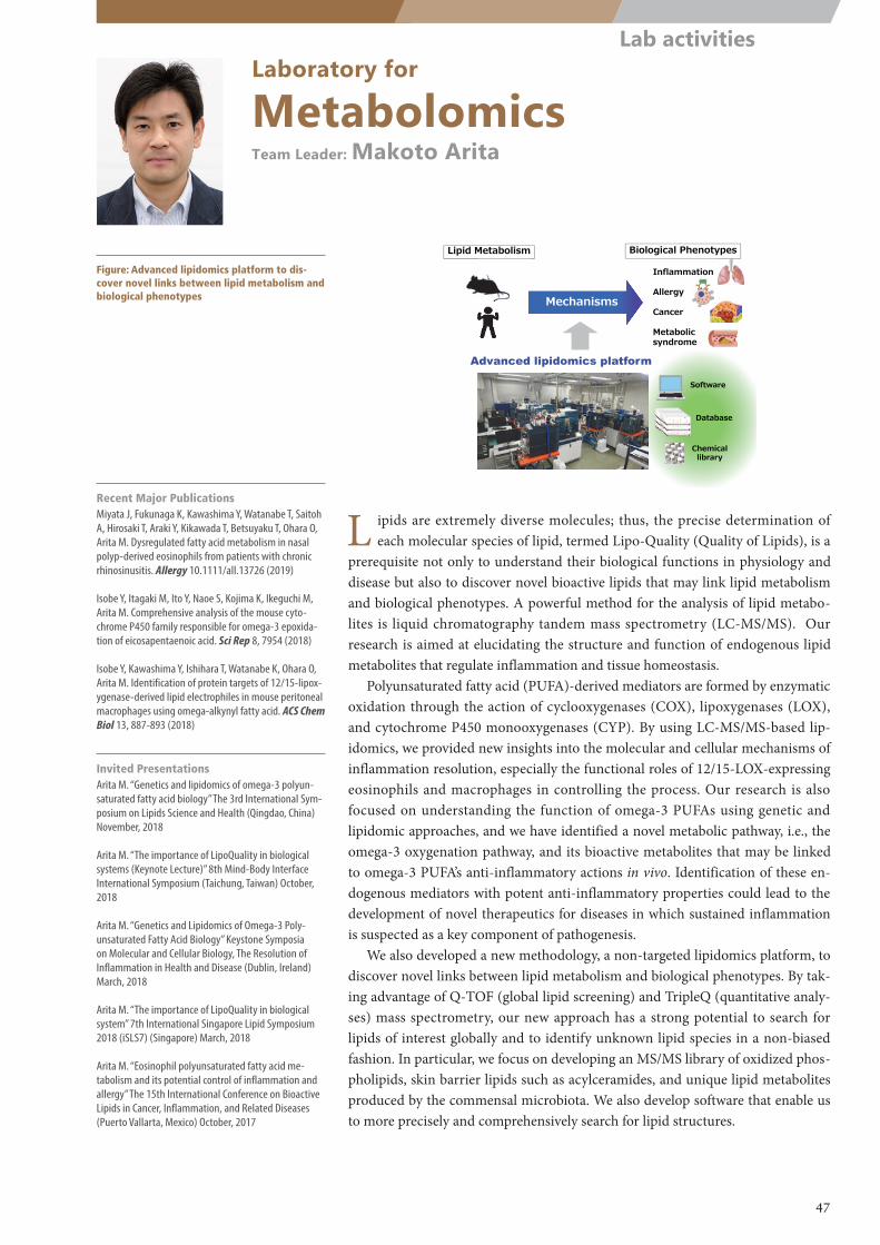
Lipid Metabolism Biological Phenotypes
Inflammation
Allergy
Cancer
Metabolicsyndrome
Database
Software
Chemicallibrary
Advanced lipidomics platform
MechanismsMechanisms
m/z
m/z
m/z
m/z
m/z
m/z
Laboratory for
MetabolomicsTeam Leader: Makoto Arita
Figure: Advanced lipidomics platform to dis-cover novel links between lipid metabolism and biological phenotypes
L ipids are extremely diverse molecules; thus, the precise determination of each molecular species of lipid, termed Lipo-Quality (Quality of Lipids), is a
prerequisite not only to understand their biological functions in physiology and disease but also to discover novel bioactive lipids that may link lipid metabolism and biological phenotypes. A powerful method for the analysis of lipid metabo-lites is liquid chromatography tandem mass spectrometry (LC-MS/MS). Our research is aimed at elucidating the structure and function of endogenous lipid metabolites that regulate inflammation and tissue homeostasis.
Polyunsaturated fatty acid (PUFA)-derived mediators are formed by enzymatic oxidation through the action of cyclooxygenases (COX), lipoxygenases (LOX), and cytochrome P450 monooxygenases (CYP). By using LC-MS/MS-based lip-idomics, we provided new insights into the molecular and cellular mechanisms of inflammation resolution, especially the functional roles of 12/15-LOX-expressing eosinophils and macrophages in controlling the process. Our research is also focused on understanding the function of omega-3 PUFAs using genetic and lipidomic approaches, and we have identified a novel metabolic pathway, i.e., the omega-3 oxygenation pathway, and its bioactive metabolites that may be linked to omega-3 PUFA’s anti-inflammatory actions in vivo. Identification of these en-dogenous mediators with potent anti-inflammatory properties could lead to the development of novel therapeutics for diseases in which sustained inflammation is suspected as a key component of pathogenesis.
We also developed a new methodology, a non-targeted lipidomics platform, to discover novel links between lipid metabolism and biological phenotypes. By tak-ing advantage of Q-TOF (global lipid screening) and TripleQ (quantitative analy-ses) mass spectrometry, our new approach has a strong potential to search for lipids of interest globally and to identify unknown lipid species in a non-biased fashion. In particular, we focus on developing an MS/MS library of oxidized phos-pholipids, skin barrier lipids such as acylceramides, and unique lipid metabolites produced by the commensal microbiota. We also develop software that enable us to more precisely and comprehensively search for lipid structures.
Recent Major PublicationsMiyata J, Fukunaga K, Kawashima Y, Watanabe T, Saitoh A, Hirosaki T, Araki Y, Kikawada T, Betsuyaku T, Ohara O, Arita M. Dysregulated fatty acid metabolism in nasal polyp-derived eosinophils from patients with chronic rhinosinusitis. Allergy 10.1111/all.13726 (2019)
Isobe Y, Itagaki M, Ito Y, Naoe S, Kojima K, Ikeguchi M, Arita M. Comprehensive analysis of the mouse cyto-chrome P450 family responsible for omega-3 epoxida-tion of eicosapentaenoic acid. Sci Rep 8, 7954 (2018)
Isobe Y, Kawashima Y, Ishihara T, Watanabe K, Ohara O, Arita M. Identification of protein targets of 12/15-lipox-ygenase-derived lipid electrophiles in mouse peritoneal macrophages using omega-alkynyl fatty acid. ACS Chem Biol 13, 887-893 (2018)
Invited PresentationsArita M. “Genetics and lipidomics of omega-3 polyun-saturated fatty acid biology” The 3rd International Sym-posium on Lipids Science and Health (Qingdao, China) November, 2018
Arita M. “The importance of LipoQuality in biological systems (Keynote Lecture)” 8th Mind-Body Interface International Symposium (Taichung, Taiwan) October, 2018
Arita M. “Genetics and Lipidomics of Omega-3 Poly-unsaturated Fatty Acid Biology” Keystone Symposia on Molecular and Cellular Biology, The Resolution of Inflammation in Health and Disease (Dublin, Ireland) March, 2018
Arita M. “The importance of LipoQuality in biological system” 7th International Singapore Lipid Symposium 2018 (iSLS7) (Singapore) March, 2018
Arita M. “Eosinophil polyunsaturated fatty acid me-tabolism and its potential control of inflammation and allergy” The 15th International Conference on Bioactive Lipids in Cancer, Inflammation, and Related Diseases (Puerto Vallarta, Mexico) October, 2017
47
Lab activities
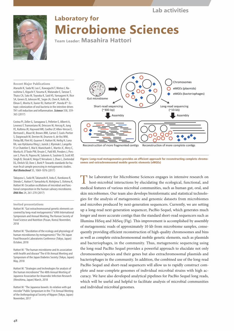
Laboratory for
Microbiome SciencesTeam Leader: Masahira Hattori
Figure: Long-read metagenomics provides an efficient approach for reconstructing complete chromo-somes and extrachromosomal mobile genetic elements (eMGEs)
T he Laboratory for Microbiome Sciences engages in intensive research on host-microbial interactions by elucidating the ecological, functional, and
medical features of various microbial communities, such as human gut, oral, and skin microbiomes. Our team also develops bioinformatic and statistical technolo-gies for the analysis of metagenomic and genomic datasets from microbiomes and microbes produced by next-generation sequencers. Currently, we are setting up a long-read next-generation sequencer, PacBio Sequel, which generates much longer and more accurate contigs than the standard short-read sequencers such as Illumina HiSeq and MiSeq (Fig). This improvement is accomplished by assembly of metagenomic reads of approximately 10 kb from microbiome samples, conse-quently providing efficient reconstruction of high-quality chromosomes and bins as well as complete extrachromosomal mobile genetic elements, such as plasmids and bacteriophages, in the community. Thus, metagenomic sequencing using the long-read PacBio Sequel provides a powerful approach to elucidate not only chromosomes/species and their genes but also extrachromosomal plasmids and bacteriophages in the community. In addition, the combined use of the long-read PacBio Sequel and short-read sequencers will allow us to rapidly construct com-plete and near-complete genomes of individual microbial strains with high ac-curacy. We have also developed analytical pipelines for PacBio Sequel long reads, which will be useful and helpful to facilitate analysis of microbial communities and individual microbial genomes.
Recent Major PublicationsAtarashi K, Suda W, Luo C, Kawaguchi T, Motoo I, Na-rushima S, Kiguchi Y, Yasuma K, Watanabe E, Tanoue T, Thaiss CA, Sato M, Toyooka K, Said HS, Yamagami H, Rice SA, Gevers D, Johnson RC, Segre JA, Chen K, Kolls JK, Elinav E, Morita H, Xavier RJ, Hattori M*, Honda K*: Ec-topic colonization of oral bacteria in the intestine drives TH1 cell induction and inflammation. Science 358, 359-365 (2017)
Costea PI, Zeller G, Sunagawa S, Pelletier E, Alberti A, Levenez F, Tramontano M, Driessen M, Hercog R, Jung FE, Kultima JR, Hayward MR, Coelho LP, Allen-Vercoe E, Bertrand L, Blaut M, Brown JRM, Carton T, Cools-Portier S, Daigneault M, Derrien M, Druesne A, de Vos WM, Finlay BB, Flint HJ, Guarner F, Hattori M, Heilig H, Luna RA, van Hylckama Vlieg J, Junick J, Klymiuk I, Langella P, Le Chatelier E, Mai V, Manichanh C, Martin JC, Mery C, Morita H, O’Toole PW, Orvain C, Patil KR, Penders J, Pers-son S, Pons N, Popova M, Salonen A, Saulnier D, Scott KP, Singh B, Slezak K, Veiga P, Versalovic J, Zhao L, Zoetendal EG, Ehrlich SD, Dore J, Bork P: Towards standards for hu-man fecal sample processing in metagenomic studies. Nat Biotechnol 35, 1069-1076 (2017)
Takayasu L, Suda W, Takanashi K, Iioka E, Kurokawa R, Shindo C, Hattori Y, Yamashita N, Nishijima S, Oshima K, Hattori M: Circadian oscillations of microbial and func-tional composition in the human salivary microbiome. DNA Res 24, 261-270 (2017)
Invited presentationsHattori M. “Gut extrachromosomal genetic elements un-covered by long-read metagenomics” 69th International Symposium and Annual Meeting, The Korean Society of Food Science and Nutrition (Pusan, Korea) November, 2018
Hattori M. “Elucidation of the ecology and physiology of human microbiomes by metagenomics” The 7th Japan Food Research Laboratories Conference (Tokyo, Japan) October, 2018
Hattori M. “The human microbiome and its association with health and disease” The 61th Annual Meeting and Symposium of the Japan Diabetes Society (Tokyo, Japan) May, 2018
Hattori M. “Strategies and technologies for analysis of the human microbiome” The 48th Annual Meeting of Japanese Association for Anaerobic Infection Research (Hiroshima, Japan) March, 2018
Hattori M. “The Japanese bowels: its relation with gut microbe” Public Symposium in the 71st Annual Meeting of the Anthropological Society of Nippon (Tokyo, Japan) November, 2017
48
Lab activities
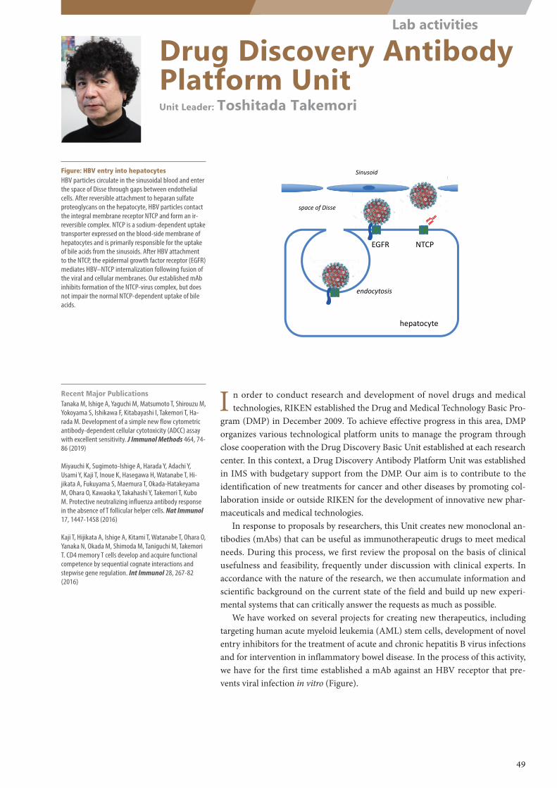
NTCPEGFR
hepatocyte
Sinusoid
space of Disse
endocytosis
Drug Discovery Antibody Platform UnitUnit Leader: Toshitada Takemori
Figure: HBV entry into hepatocytesHBV particles circulate in the sinusoidal blood and enter the space of Disse through gaps between endothelial cells. After reversible attachment to heparan sulfate proteoglycans on the hepatocyte, HBV particles contact the integral membrane receptor NTCP and form an ir-reversible complex. NTCP is a sodium-dependent uptake transporter expressed on the blood-side membrane of hepatocytes and is primarily responsible for the uptake of bile acids from the sinusoids. After HBV attachment to the NTCP, the epidermal growth factor receptor (EGFR) mediates HBV–NTCP internalization following fusion of the viral and cellular membranes. Our established mAb inhibits formation of the NTCP-virus complex, but does not impair the normal NTCP-dependent uptake of bile acids.
Recent Major PublicationsTanaka M, Ishige A, Yaguchi M, Matsumoto T, Shirouzu M, Yokoyama S, Ishikawa F, Kitabayashi I, Takemori T, Ha-rada M. Development of a simple new flow cytometric antibody-dependent cellular cytotoxicity (ADCC) assay with excellent sensitivity. J Immunol Methods 464, 74-86 (2019)
Miyauchi K, Sugimoto-Ishige A, Harada Y, Adachi Y, Usami Y, Kaji T, Inoue K, Hasegawa H, Watanabe T, Hi-jikata A, Fukuyama S, Maemura T, Okada-Hatakeyama M, Ohara O, Kawaoka Y, Takahashi Y, Takemori T, Kubo M. Protective neutralizing influenza antibody response in the absence of T follicular helper cells. Nat Immunol 17, 1447-1458 (2016)
Kaji T, Hijikata A, Ishige A, Kitami T, Watanabe T, Ohara O, Yanaka N, Okada M, Shimoda M, Taniguchi M, Takemori T. CD4 memory T cells develop and acquire functional competence by sequential cognate interactions and stepwise gene regulation. Int Immunol 28, 267-82 (2016)
I n order to conduct research and development of novel drugs and medical technologies, RIKEN established the Drug and Medical Technology Basic Pro-
gram (DMP) in December 2009. To achieve effective progress in this area, DMP organizes various technological platform units to manage the program through close cooperation with the Drug Discovery Basic Unit established at each research center. In this context, a Drug Discovery Antibody Platform Unit was established in IMS with budgetary support from the DMP. Our aim is to contribute to the identification of new treatments for cancer and other diseases by promoting col-laboration inside or outside RIKEN for the development of innovative new phar-maceuticals and medical technologies.
In response to proposals by researchers, this Unit creates new monoclonal an-tibodies (mAbs) that can be useful as immunotherapeutic drugs to meet medical needs. During this process, we first review the proposal on the basis of clinical usefulness and feasibility, frequently under discussion with clinical experts. In accordance with the nature of the research, we then accumulate information and scientific background on the current state of the field and build up new experi-mental systems that can critically answer the requests as much as possible.
We have worked on several projects for creating new therapeutics, including targeting human acute myeloid leukemia (AML) stem cells, development of novel entry inhibitors for the treatment of acute and chronic hepatitis B virus infections and for intervention in inflammatory bowel disease. In the process of this activity, we have for the first time established a mAb against an HBV receptor that pre-vents viral infection in vitro (Figure).
49
Lab activities
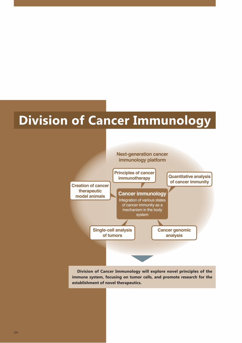
Division of Cancer Immunology
Division of Cancer Immunology will explore novel principles of the immune system, focusing on tumor cells, and promote research for the establishment of novel therapeutics.
50
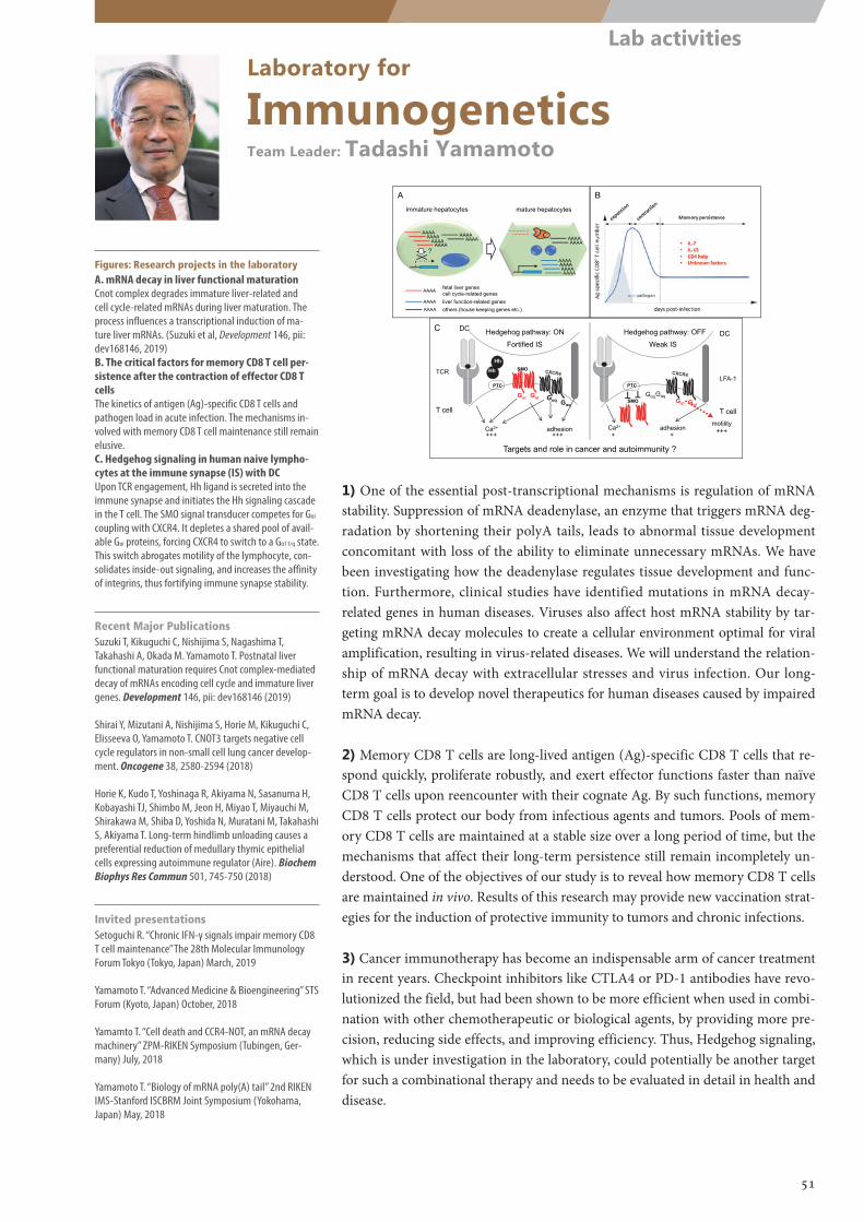
immature hepatocytes mature hepatocytes
AAAAAAAA
AAAAAAAA
AAAAAAAA
AA
AAAAAAAAAAAAAAAA
AAAAAAAA
fetal liver genescell cycle-related genesliver function-related genesothers (house keeping genes etc.)
AAAA
AAAAAAAA
?
A B
C
Ca2+ adhesion
Hedgehog pathway: ON
Ca2+ adhesion
Hedgehog pathway: OFF
PTC
GαqGαi Gαi
Gαi
TCR
+++ +++ + +
DC
T cell T cell
LFA-1PTC
SMO
SMOGαq
GαqGαi
Gαq
Weak IS Fortified ISDC
Hh
T T
Targets and role in cancer and autoimmunity ?
Hh
motility+++
Laboratory for
ImmunogeneticsTeam Leader: Tadashi Yamamoto
Figures: Research projects in the laboratoryA. mRNA decay in liver functional maturationCnot complex degrades immature liver-related and cell cycle-related mRNAs during liver maturation. The process influences a transcriptional induction of ma-ture liver mRNAs. (Suzuki et al, Development 146, pii: dev168146, 2019)B. The critical factors for memory CD8 T cell per-sistence after the contraction of effector CD8 T cellsThe kinetics of antigen (Ag)-specific CD8 T cells and pathogen load in acute infection. The mechanisms in-volved with memory CD8 T cell maintenance still remain elusive.C. Hedgehog signaling in human naive lympho-cytes at the immune synapse (IS) with DCUpon TCR engagement, Hh ligand is secreted into the immune synapse and initiates the Hh signaling cascade in the T cell. The SMO signal transducer competes for Gαi coupling with CXCR4. It depletes a shared pool of avail-able Gαi proteins, forcing CXCR4 to switch to a Gα11/q state. This switch abrogates motility of the lymphocyte, con-solidates inside-out signaling, and increases the affinity of integrins, thus fortifying immune synapse stability.
Recent Major PublicationsSuzuki T, Kikuguchi C, Nishijima S, Nagashima T, Takahashi A, Okada M. Yamamoto T. Postnatal liver functional maturation requires Cnot complex-mediated decay of mRNAs encoding cell cycle and immature liver genes. Development 146, pii: dev168146 (2019)
Shirai Y, Mizutani A, Nishijima S, Horie M, Kikuguchi C, Elisseeva O, Yamamoto T. CNOT3 targets negative cell cycle regulators in non-small cell lung cancer develop-ment. Oncogene 38, 2580-2594 (2018)
Horie K, Kudo T, Yoshinaga R, Akiyama N, Sasanuma H, Kobayashi TJ, Shimbo M, Jeon H, Miyao T, Miyauchi M, Shirakawa M, Shiba D, Yoshida N, Muratani M, Takahashi S, Akiyama T. Long-term hindlimb unloading causes a preferential reduction of medullary thymic epithelial cells expressing autoimmune regulator (Aire). Biochem Biophys Res Commun 501, 745-750 (2018)
Invited presentationsSetoguchi R. “Chronic IFN-γ signals impair memory CD8 T cell maintenance” The 28th Molecular Immunology Forum Tokyo (Tokyo, Japan) March, 2019
Yamamoto T. “Advanced Medicine & Bioengineering” STS Forum (Kyoto, Japan) October, 2018
Yamamto T. “Cell death and CCR4-NOT, an mRNA decay machinery” ZPM-RIKEN Symposium (Tubingen, Ger-many) July, 2018
Yamamoto T. “Biology of mRNA poly(A) tail” 2nd RIKEN IMS-Stanford ISCBRM Joint Symposium (Yokohama, Japan) May, 2018
1) One of the essential post-transcriptional mechanisms is regulation of mRNA stability. Suppression of mRNA deadenylase, an enzyme that triggers mRNA deg-radation by shortening their polyA tails, leads to abnormal tissue development concomitant with loss of the ability to eliminate unnecessary mRNAs. We have been investigating how the deadenylase regulates tissue development and func-tion. Furthermore, clinical studies have identified mutations in mRNA decay-related genes in human diseases. Viruses also affect host mRNA stability by tar-geting mRNA decay molecules to create a cellular environment optimal for viral amplification, resulting in virus-related diseases. We will understand the relation-ship of mRNA decay with extracellular stresses and virus infection. Our long-term goal is to develop novel therapeutics for human diseases caused by impaired mRNA decay.
2) Memory CD8 T cells are long-lived antigen (Ag)-specific CD8 T cells that re-spond quickly, proliferate robustly, and exert effector functions faster than naïve CD8 T cells upon reencounter with their cognate Ag. By such functions, memory CD8 T cells protect our body from infectious agents and tumors. Pools of mem-ory CD8 T cells are maintained at a stable size over a long period of time, but the mechanisms that affect their long-term persistence still remain incompletely un-derstood. One of the objectives of our study is to reveal how memory CD8 T cells are maintained in vivo. Results of this research may provide new vaccination strat-egies for the induction of protective immunity to tumors and chronic infections.
3) Cancer immunotherapy has become an indispensable arm of cancer treatment in recent years. Checkpoint inhibitors like CTLA4 or PD-1 antibodies have revo-lutionized the field, but had been shown to be more efficient when used in combi-nation with other chemotherapeutic or biological agents, by providing more pre-cision, reducing side effects, and improving efficiency. Thus, Hedgehog signaling, which is under investigation in the laboratory, could potentially be another target for such a combinational therapy and needs to be evaluated in detail in health and disease.
51
Lab activities
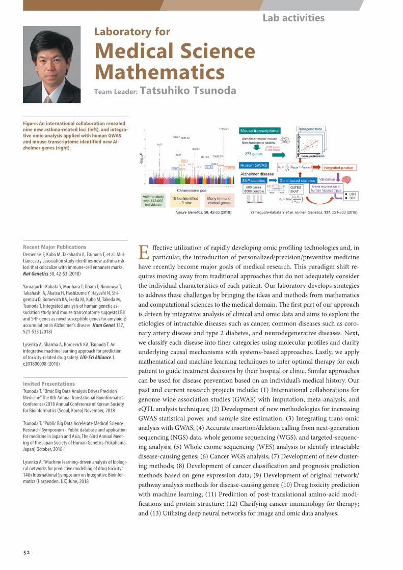
Laboratory for
Medical Science MathematicsTeam Leader: Tatsuhiko Tsunoda
Figure: An international collaboration revealed nine new asthma-related loci (left), and integra-tive omic-analysis applied with human GWAS and mouse transcriptome identified new Al-zheimer genes (right).
Recent Major PublicationsDemenais F, Kubo M, Takahashi A, Tsunoda T, et al. Mul-tiancestry association study identifies new asthma risk loci that colocalize with immune-cell enhancer marks. Nat Genetics 50, 42-53 (2018)
Yamaguchi-Kabata Y, Morihara T, Ohara T, Ninomiya T, Takahashi A, Akatsu H, Hashizume Y, Hayashi N, Shi-gemizu D, Boroevich KA, Ikeda M, Kubo M, Takeda M, Tsunoda T. Integrated analysis of human genetic as-sociation study and mouse transcriptome suggests LBH and SHF genes as novel susceptible genes for amyloid-β accumulation in Alzheimer's disease. Hum Genet 137, 521-533 (2018)
Lysenko A, Sharma A, Boroevich KA, Tsunoda T. An integrative machine learning approach for prediction of toxicity-related drug safety. Life Sci Alliance 1, e201800098 (2018)
Invited PresentationsTsunoda T. “Omic Big Data Analysis Drives Precision Medicine” The 8th Annual Translational Bioinformatics Conference/2018 Annual Conference of Korean Society for Bioinformatics (Seoul, Korea) November, 2018
Tsunoda T. “Public Big Data Accelerate Medical Science Research” Symposium - Public database and application for medicine in Japan and Asia, The 63rd Annual Meet-ing of the Japan Society of Human Genetics (Yokohama, Japan) October, 2018
Lysenko A. “Machine learning-driven analysis of biologi-cal networks for predictive modelling of drug toxicity” 14th International Symposium on Integrative Bioinfor-matics (Harpenden, UK) June, 2018
E ffective utilization of rapidly developing omic profiling technologies and, in particular, the introduction of personalized/precision/preventive medicine
have recently become major goals of medical research. This paradigm shift re-quires moving away from traditional approaches that do not adequately consider the individual characteristics of each patient. Our laboratory develops strategies to address these challenges by bringing the ideas and methods from mathematics and computational sciences to the medical domain. The first part of our approach is driven by integrative analysis of clinical and omic data and aims to explore the etiologies of intractable diseases such as cancer, common diseases such as coro-nary artery disease and type 2 diabetes, and neurodegenerative diseases. Next, we classify each disease into finer categories using molecular profiles and clarify underlying causal mechanisms with systems-based approaches. Lastly, we apply mathematical and machine learning techniques to infer optimal therapy for each patient to guide treatment decisions by their hospital or clinic. Similar approaches can be used for disease prevention based on an individual’s medical history. Our past and current research projects include: (1) International collaborations for genome-wide association studies (GWAS) with imputation, meta-analysis, and eQTL analysis techniques; (2) Development of new methodologies for increasing GWAS statistical power and sample size estimation; (3) Integrating trans-omic analysis with GWAS; (4) Accurate insertion/deletion calling from next-generation sequencing (NGS) data, whole genome sequencing (WGS), and targeted-sequenc-ing analysis; (5) Whole exome sequencing (WES) analysis to identify intractable disease-causing genes; (6) Cancer WGS analysis; (7) Development of new cluster-ing methods; (8) Development of cancer classification and prognosis prediction methods based on gene expression data; (9) Development of original network/pathway analysis methods for disease-causing genes; (10) Drug toxicity prediction with machine learning; (11) Prediction of post-translational amino-acid modi-fications and protein structure; (12) Clarifying cancer immunology for therapy; and (13) Utilizing deep neural networks for image and omic data analyses.
52
Lab activities
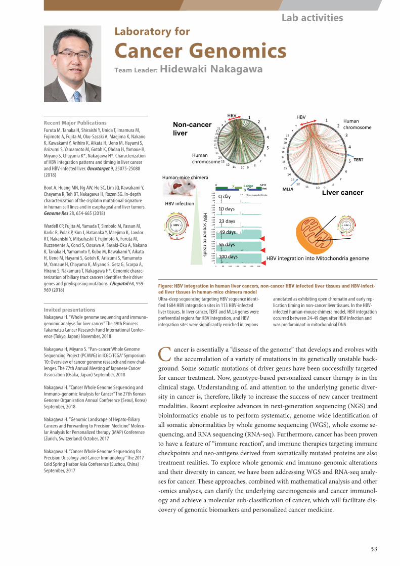
HBV integration into Mitochondria genome
HBV infection2,000
4,000
6,000
8,000
2,000
4,000
6,000
8,000
2,000
4,000
6,000
8,000
2,000
4,000
6,000
8,000
2,000
4,000
6,000
8,000
2,000
4,000
6,000
8,000
0 500 1,000 1,500 2,000 2,500 3,000
0
0
0
0
0
0
0 3,215XP
Large S (bp)
Rea
d de
pth
*Cross-mapped with miceDay 0_#1
Day 10_#1
Day 23_#1
Day 49_#1
Day 56_#1
Day 100_ #1
O day
10 days
23 days
49 days
56 days
100 days
HBV sequence reads
Human‐mice chimera
Non-cancer liver
Liver cancer
HBV 12
3
4
56
789101112
1314
15
16171819202122
XY
Human chromosome TERT
HBV 12
3
4
5
6
7
891011
1213
14
15
16
17
1819
202122
XY
MLL4
Human chromosome
Laboratory for
Cancer GenomicsTeam Leader: Hidewaki Nakagawa
Figure: HBV integration in human liver cancers, non-cancer HBV infected liver tissues and HBV-infect-ed liver tissues in human-mice chimera modelUltra-deep sequencing targeting HBV sequence identi-fied 1684 HBV integration sites in 113 HBV-infected liver tissues. In liver cancer, TERT and MLL4 genes were preferential regions for HBV integration, and HBV integration sites were significantly enriched in regions
annotated as exhibiting open chromatin and early rep-lication timing in non-cancer liver tissues. In the HBV-infected human-mouse chimera model, HBV integration occurred between 24-49 days after HBV infection and was predominant in mitochondrial DNA.
C ancer is essentially a “disease of the genome” that develops and evolves with the accumulation of a variety of mutations in its genetically unstable back-
ground. Some somatic mutations of driver genes have been successfully targeted for cancer treatment. Now, genotype-based personalized cancer therapy is in the clinical stage. Understanding of, and attention to the underlying genetic diver-sity in cancer is, therefore, likely to increase the success of new cancer treatment modalities. Recent explosive advances in next-generation sequencing (NGS) and bioinformatics enable us to perform systematic, genome-wide identification of all somatic abnormalities by whole genome sequencing (WGS), whole exome se-quencing, and RNA sequencing (RNA-seq). Furthermore, cancer has been proven to have a feature of “immune reaction”, and immune therapies targeting immune checkpoints and neo-antigens derived from somatically mutated proteins are also treatment realities. To explore whole genomic and immuno-genomic alterations and their diversity in cancer, we have been addressing WGS and RNA-seq analy-ses for cancer. These approaches, combined with mathematical analysis and other -omics analyses, can clarify the underlying carcinogenesis and cancer immunol-ogy and achieve a molecular sub-classification of cancer, which will facilitate dis-covery of genomic biomarkers and personalized cancer medicine.
Recent Major PublicationsFuruta M, Tanaka H, Shiraishi Y, Unida T, Imamura M, Fujimoto A, Fujita M, Oku-Sasaki A, Maejima K, Nakano K, Kawakami Y, Arihiro K, Aikata H, Ueno M, Hayami S, Ariizumi S, Yamamoto M, Gotoh K, Ohdan H, Yamaue H, Miyano S, Chayama K*, Nakagawa H*. Characterization of HBV integration patterns and timing in liver cancer and HBV-infected liver. Oncotarget 9, 25075-25088 (2018)
Boot A, Huang MN, Ng AW, Ho SC, Lim JQ, Kawakami Y, Chayama K, Teh BT, Nakagawa H, Rozen SG. In-depth characterization of the cisplatin mutational signature in human cell lines and in esophageal and liver tumors. Genome Res 28, 654-665 (2018)
Wardell CP, Fujita M, Yamada T, Simbolo M, Fassan M, Karlic R, Polak P, Kim J, Hatanaka Y, Maejima K, Lawlor RT, Nakanishi Y, Mitsuhashi T, Fujimoto A, Furuta M, Ruzzenente A, Conci S, Oosawa A, Sasaki-Oku A, Nakano K, Tanaka H, Yamamoto Y, Kubo M, Kawakami Y, Aikata H, Ueno M, Hayami S, Gotoh K, Ariizumi S, Yamamoto M, Yamaue H, Chayama K, Miyano S, Getz G, Scarpa A, Hirano S, Nakamura T, Nakagawa H*. Genomic charac-terization of biliary tract cancers identifies their driver genes and predisposing mutations. J Hepatol 68, 959-969 (2018)
Invited presentationsNakagawa H. “Whole genome sequencing and immuno-genomic analysis for liver cancer” The 49th Princess Takamatsu Cancer Research Fund International Confer-ence (Tokyo, Japan) November, 2018
Nakagawa H, Miyano S. “Pan-cancer Whole Genome Sequencing Project (PCAWG) in ICGC/TCGA” Symposium 10: Overview of cancer genome research and new chal-lenges. The 77th Annual Meeting of Japanese Cancer Association (Osaka, Japan) September, 2018
Nakagawa H. “Cancer Whole Genome Sequencing and Immuno-genomic Analysis for Cancer” The 27th Korean Genome Organization Annual Conference (Seoul, Korea) September, 2018
Nakagawa H. “Genomic Landscape of Hepato-Biliary Cancers and Forwarding to Precision Medicine” Molecu-lar Analysis for Personalized therapy (MAP) Conference (Zurich, Switzerland) October, 2017
Nakagawa H. “Cancer Whole Genome Sequencing for Precision Oncology and Cancer Immunology” The 2017 Cold Spring Harbor Asia Conference (Suzhou, China) September, 2017
53
Lab activities
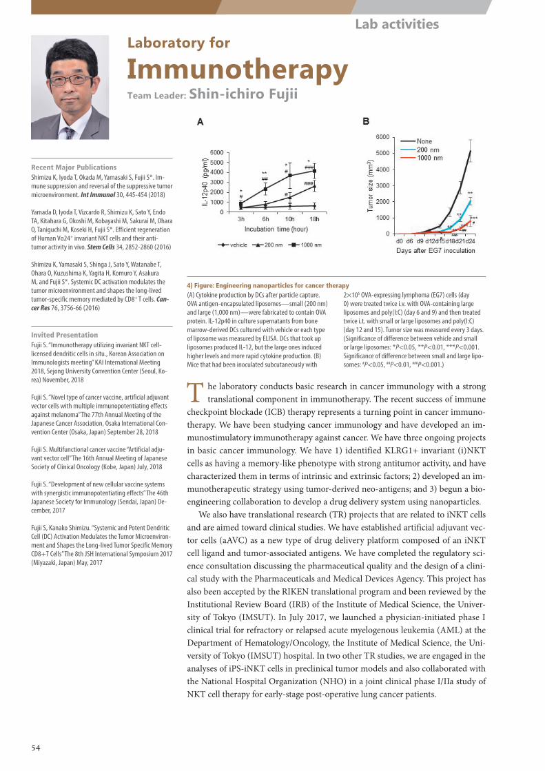
Laboratory for
ImmunotherapyTeam Leader: Shin-ichiro Fujii
4) Figure: Engineering nanoparticles for cancer therapy(A) Cytokine production by DCs after particle capture. OVA antigen-encapsulated liposomes—small (200 nm) and large (1,000 nm)—were fabricated to contain OVA protein. IL-12p40 in culture supernatants from bone marrow-derived DCs cultured with vehicle or each type of liposome was measured by ELISA. DCs that took up liposomes produced IL-12, but the large ones induced higher levels and more rapid cytokine production. (B) Mice that had been inoculated subcutaneously with
2×10⁵ OVA-expressing lymphoma (EG7) cells (day 0) were treated twice i.v. with OVA-containing large liposomes and poly(I:C) (day 6 and 9) and then treated twice i.t. with small or large liposomes and poly(I:C) (day 12 and 15). Tumor size was measured every 3 days. (Significance of difference between vehicle and small or large liposomes: *P<0.05, **P<0.01, ***P<0.001. Significance of difference between small and large lipo-somes: #P<0.05, ##P<0.01, ###P<0.001.)
T he laboratory conducts basic research in cancer immunology with a strong translational component in immunotherapy. The recent success of immune
checkpoint blockade (ICB) therapy represents a turning point in cancer immuno-therapy. We have been studying cancer immunology and have developed an im-munostimulatory immunotherapy against cancer. We have three ongoing projects in basic cancer immunology. We have 1) identified KLRG1+ invariant (i)NKT cells as having a memory-like phenotype with strong antitumor activity, and have characterized them in terms of intrinsic and extrinsic factors; 2) developed an im-munotherapeutic strategy using tumor-derived neo-antigens; and 3) begun a bio-engineering collaboration to develop a drug delivery system using nanoparticles.
We also have translational research (TR) projects that are related to iNKT cells and are aimed toward clinical studies. We have established artificial adjuvant vec-tor cells (aAVC) as a new type of drug delivery platform composed of an iNKT cell ligand and tumor-associated antigens. We have completed the regulatory sci-ence consultation discussing the pharmaceutical quality and the design of a clini-cal study with the Pharmaceuticals and Medical Devices Agency. This project has also been accepted by the RIKEN translational program and been reviewed by the Institutional Review Board (IRB) of the Institute of Medical Science, the Univer-sity of Tokyo (IMSUT). In July 2017, we launched a physician-initiated phase I clinical trial for refractory or relapsed acute myelogenous leukemia (AML) at the Department of Hematology/Oncology, the Institute of Medical Science, the Uni-versity of Tokyo (IMSUT) hospital. In two other TR studies, we are engaged in the analyses of iPS-iNKT cells in preclinical tumor models and also collaborated with the National Hospital Organization (NHO) in a joint clinical phase I/IIa study of NKT cell therapy for early-stage post-operative lung cancer patients.
Recent Major PublicationsShimizu K, Iyoda T, Okada M, Yamasaki S, Fujii S*. Im-mune suppression and reversal of the suppressive tumor microenvironment. Int Immunol 30, 445-454 (2018)
Yamada D, Iyoda T, Vizcardo R, Shimizu K, Sato Y, Endo TA, Kitahara G, Okoshi M, Kobayashi M, Sakurai M, Ohara O, Taniguchi M, Koseki H, Fujii S*. Efficient regeneration of Human Vα24+ invariant NKT cells and their anti-tumor activity in vivo. Stem Cells 34, 2852-2860 (2016)
Shimizu K, Yamasaki S, Shinga J, Sato Y, Watanabe T, Ohara O, Kuzushima K, Yagita H, Komuro Y, Asakura M, and Fujii S*. Systemic DC activation modulates the tumor microenvironment and shapes the long-lived tumor-specific memory mediated by CD8+ T cells. Can-cer Res 76, 3756-66 (2016)
Invited PresentationFujii S. “Immunotherapy utilizing invariant NKT cell-licensed dendritic cells in situ., Korean Association on Immunologists meeting” KAI International Meeting 2018, Sejong University Convention Center (Seoul, Ko-rea) November, 2018
Fujii S. “Novel type of cancer vaccine, artificial adjuvant vector cells with multiple immunopotentiating effects against melanoma” The 77th Annual Meeting of the Japanese Cancer Association, Osaka International Con-vention Center (Osaka, Japan) September 28, 2018
Fujii S. Multifunctional cancer vaccine “Artificial adju-vant vector cell” The 16th Annual Meeting of Japanese Society of Clinical Oncology (Kobe, Japan) July, 2018
Fujii S. “Development of new cellular vaccine systems with synergistic immunopotentiating effects” The 46th Japanese Society for Immunology (Sendai, Japan) De-cember, 2017
Fujii S, Kanako Shimizu. “Systemic and Potent Dendritic Cell (DC) Activation Modulates the Tumor Microenviron-ment and Shapes the Long-lived Tumor Specific Memory CD8+T Cells” The 8th JSH International Symposium 2017 (Miyazaki, Japan) May, 2017
54
Lab activities
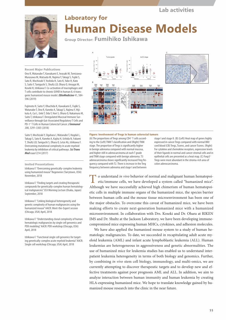
NormalAdenoma
T3T1 T2
20
40
60
80
%Tr
egs
in C
D4+
Tce
lls
CancerNormal I II III
20
40
60
80
%Tr
egs
in C
D4+
Tce
lls
Tumorgrade AJCC stage
IV LiverT4Cancer Adenoma
A
B
C
Laboratory for
Human Disease ModelsGroup Director: Fumihiko Ishikawa
T o understand in vivo behavior of normal and malignant human hematopoi-etic/immune cells, we have developed a system called “humanized mice”.
Although we have successfully achieved high chimerism of human hematopoi-etic cells in multiple immune organs of the humanized mice, the species barrier between human cells and the mouse tissue microenvironment has been one of the major obstacles. To overcome this caveat of humanized mice, we have been making efforts to create next-generation humanized mice with a humanized microenvironment. In collaboration with Drs. Koseki and Dr. Ohara at RIKEN IMS and Dr. Shultz at the Jackson Laboratory, we have been developing immune-compromised mice expressing human MHCs, cytokines, and adhesion molecules.
We have also applied the humanized mouse system to a study of human he-matologic malignancies. To date, we succeeded in recapitulating adult acute my-eloid leukemia (AML) and infant acute lymphoblastic leukemia (ALL). Human leukemias are heterogeneous in aggressiveness and genetic abnormalities. The use of humanized mice for leukemia studies has enabled us to understand inter-patient leukemia heterogeneity in terms of both biology and genomics. Further, by combining in vivo stem cell biology, immunology, and multi-omics, we are currently attempting to discover therapeutic targets and to develop new and ef-fective treatments against poor prognosis AML and ALL. In addition, we aim to analyze interaction between human immunity and human leukemia by creating HLA-expressing humanized mice. We hope to translate knowledge gained by hu-manized mouse research into the clinic in the near future.
Recent Major PublicationsOno R, Watanabe T, Kawakami E, Iwasaki M, Tomizawa-Murasawa M, Matsuda M, Najima Y, Takagi S, Fujiki S, Sato R, Mochizuki Y, Yoshida H, Sato K, Yabe H, Kato S, Saito Y, Taniguchi S, Shultz LD, Ohara O, Amagai M, Koseki H, Ishikawa F. Co-activation of macrophages and T cells contribute to chronic GVHD in human IL-6 trans-genic humanised mouse model. EBioMedicine 41, 584-596 (2019)
Fujimoto H, Saito Y, Ohuchida K, Kawakami E, Fujiki S, Watanabe T, Ono R, Kaneko A, Takagi S, Najima Y, Hiji-kata A, Cui L, Ueki T, Oda Y, Hori S, Ohara O, Nakamura M, Saito T, Ishikawa F. Deregulated Mucosal Immune Sur-veillance through Gut-Associated Regulatory T Cells and PD-1+ T Cells in Human Colorectal Cancer. J Immunol 200, 3291-3303 (2018)
Saito Y, Mochizuki Y, Ogahara I, Watanabe T, Hogdal L, Takagi S, Sato K, Kaneko A, Kajita H, Uchida N, Fukami T, Shultz LD, Taniguchi S, Ohara O, Letai AG, Ishikawa F. Overcoming mutational complexity in acute myeloid leukemia by inhibition of critical pathways. Sci Trans Med eaao1214 (2017)
Invited PresentationsIshikawa F. “Overcoming genetically-complex leukemia using humanized mouse” Regeneron (Tarrytown, USA) November, 2018
Ishikawa F. “Finding targets and creating therapeutic compounds for genetically-complex human hematolog-ical malignancies” JCA Morning Lecture (Osaka, Japan) September, 2018
Ishikawa F. “Linking biological heterogeneity and genetic complexity of human malignancies using the humanized mouse” AACR: Meet-the-Expert session (Chicago, USA) April, 2018
Ishikawa F. “Understanding clonal complexity of human hematologic malignancies by single cell genomics and PDX modeling” AACR: PDX workshop (Chicago, USA) April, 2018
Ishikawa F. “Functional single cell genomics for target-ing genetically-complex acute myeloid leukemia” AACR: Single cell workshop (Chicago, USA) April, 2018
Figure: Involvement of Tregs in human colorectal tumors(A) The proportions of Tregs among CD4+ T cells accord-ing to the (Left) TNM T classification and (Right) TNM stage. The proportion of Tregs is significantly higher in benign adenoma compared with normal mucosa, and higher still in adenocarcinoma at each T grade and TNM stage compared with benign adenoma. T3 adenocarcinoma shows significantly increased Treg fre-quency compared with T2. There is increase in the Treg frequency between adenoma and stage I and between
stage I and stage II. (B) (Left) Heat map of genes highly expressed in cancer Tregs compared with normal BM/cord blood (CB) Tregs, Tconvs, and cancer Tconvs. (Right) For cytokine and chemokine receptors, expression levels of their ligands in normal and cancer stromal cells and in epithelial cells are presented as a heat map. (C) Foxp3+ Tregs were most abundant in the stroma-rich area of colon adenocarcinoma.
55
Lab activities
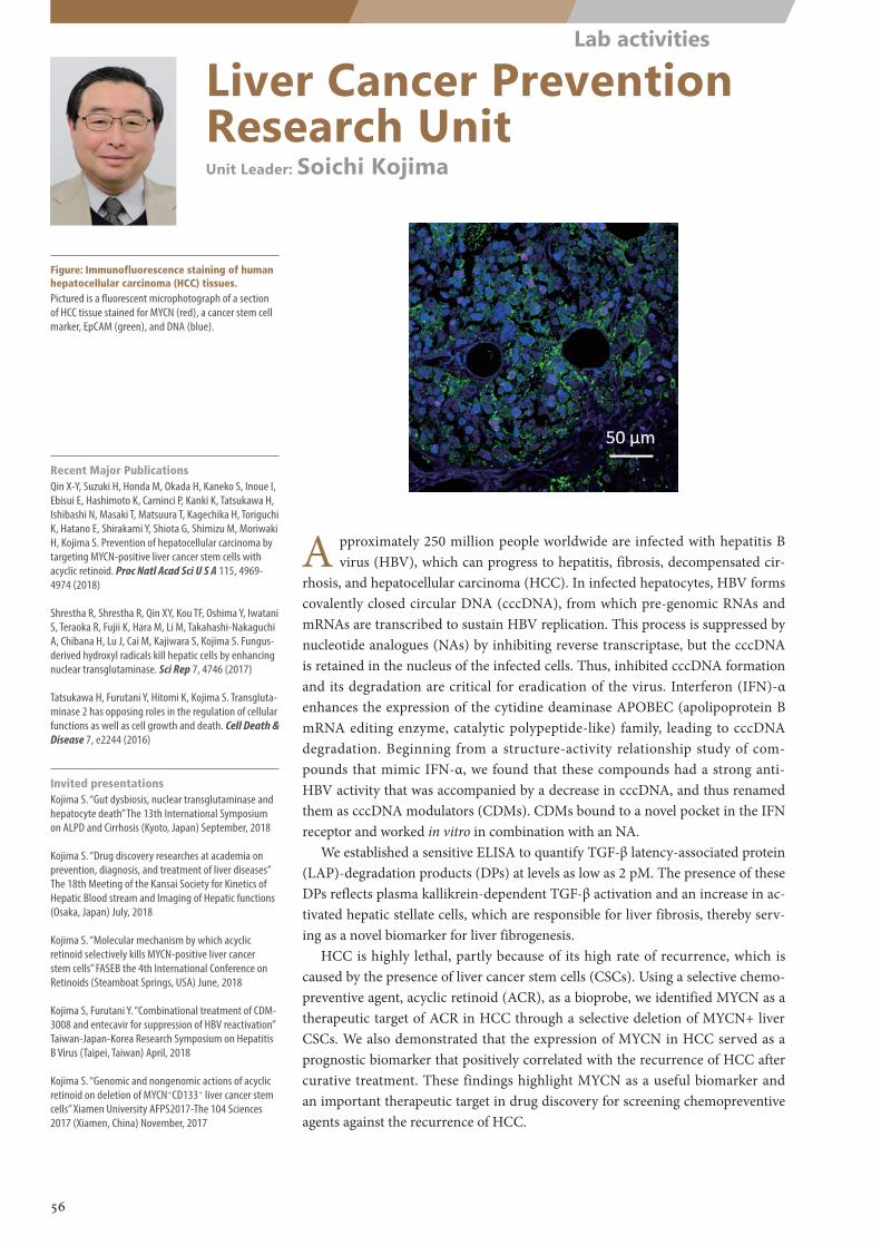
Liver Cancer Prevention Research UnitUnit Leader: Soichi Kojima
Figure: Immunofluorescence staining of human hepatocellular carcinoma (HCC) tissues.Pictured is a fluorescent microphotograph of a section of HCC tissue stained for MYCN (red), a cancer stem cell marker, EpCAM (green), and DNA (blue).
A pproximately 250 million people worldwide are infected with hepatitis B virus (HBV), which can progress to hepatitis, fibrosis, decompensated cir-
rhosis, and hepatocellular carcinoma (HCC). In infected hepatocytes, HBV forms covalently closed circular DNA (cccDNA), from which pre-genomic RNAs and mRNAs are transcribed to sustain HBV replication. This process is suppressed by nucleotide analogues (NAs) by inhibiting reverse transcriptase, but the cccDNA is retained in the nucleus of the infected cells. Thus, inhibited cccDNA formation and its degradation are critical for eradication of the virus. Interferon (IFN)-α enhances the expression of the cytidine deaminase APOBEC (apolipoprotein B mRNA editing enzyme, catalytic polypeptide-like) family, leading to cccDNA degradation. Beginning from a structure-activity relationship study of com-pounds that mimic IFN-α, we found that these compounds had a strong anti-HBV activity that was accompanied by a decrease in cccDNA, and thus renamed them as cccDNA modulators (CDMs). CDMs bound to a novel pocket in the IFN receptor and worked in vitro in combination with an NA.
We established a sensitive ELISA to quantify TGF-β latency-associated protein (LAP)-degradation products (DPs) at levels as low as 2 pM. The presence of these DPs reflects plasma kallikrein-dependent TGF-β activation and an increase in ac-tivated hepatic stellate cells, which are responsible for liver fibrosis, thereby serv-ing as a novel biomarker for liver fibrogenesis.
HCC is highly lethal, partly because of its high rate of recurrence, which is caused by the presence of liver cancer stem cells (CSCs). Using a selective chemo-preventive agent, acyclic retinoid (ACR), as a bioprobe, we identified MYCN as a therapeutic target of ACR in HCC through a selective deletion of MYCN+ liver CSCs. We also demonstrated that the expression of MYCN in HCC served as a prognostic biomarker that positively correlated with the recurrence of HCC after curative treatment. These findings highlight MYCN as a useful biomarker and an important therapeutic target in drug discovery for screening chemopreventive agents against the recurrence of HCC.
Recent Major PublicationsQin X-Y, Suzuki H, Honda M, Okada H, Kaneko S, Inoue I, Ebisui E, Hashimoto K, Carninci P, Kanki K, Tatsukawa H, Ishibashi N, Masaki T, Matsuura T, Kagechika H, Toriguchi K, Hatano E, Shirakami Y, Shiota G, Shimizu M, Moriwaki H, Kojima S. Prevention of hepatocellular carcinoma by targeting MYCN-positive liver cancer stem cells with acyclic retinoid. Proc Natl Acad Sci U S A 115, 4969-4974 (2018)
Shrestha R, Shrestha R, Qin XY, Kou TF, Oshima Y, Iwatani S, Teraoka R, Fujii K, Hara M, Li M, Takahashi-Nakaguchi A, Chibana H, Lu J, Cai M, Kajiwara S, Kojima S. Fungus-derived hydroxyl radicals kill hepatic cells by enhancing nuclear transglutaminase. Sci Rep 7, 4746 (2017)
Tatsukawa H, Furutani Y, Hitomi K, Kojima S. Transgluta-minase 2 has opposing roles in the regulation of cellular functions as well as cell growth and death. Cell Death & Disease 7, e2244 (2016)
Invited presentationsKojima S. “Gut dysbiosis, nuclear transglutaminase and hepatocyte death” The 13th International Symposium on ALPD and Cirrhosis (Kyoto, Japan) September, 2018
Kojima S. “Drug discovery researches at academia on prevention, diagnosis, and treatment of liver diseases” The 18th Meeting of the Kansai Society for Kinetics of Hepatic Blood stream and Imaging of Hepatic functions (Osaka, Japan) July, 2018
Kojima S. “Molecular mechanism by which acyclic retinoid selectively kills MYCN-positive liver cancer stem cells” FASEB the 4th International Conference on Retinoids (Steamboat Springs, USA) June, 2018
Kojima S, Furutani Y. “Combinational treatment of CDM-3008 and entecavir for suppression of HBV reactivation” Taiwan-Japan-Korea Research Symposium on Hepatitis B Virus (Taipei, Taiwan) April, 2018
Kojima S. “Genomic and nongenomic actions of acyclic retinoid on deletion of MYCN+CD133+ liver cancer stem cells” Xiamen University AFPS2017-The 104 Sciences 2017 (Xiamen, China) November, 2017
56
Lab activities
Special Programs for Young Leaders
RIKEN Hakubi Fellows ProgramRIKEN offers junior Principal Investigator (PI) positions, the RIKEN
Hakubi Fellows, for exceptionally talented researchers for a maximum of 7 years. RIKEN Hakubi Fellows are expected to independently engage in creative and ambitious research in natural and mathematical sciences, including research areas bordering the humanities and social sciences. An important goal of the RIKEN Hakubi Program is to foster stimulating interactions among the Fellows with diverse backgrounds and to create an intellectual hub of scientists with different disciplines within and be-yond RIKEN.
“Hakubi” is a phrase derived from a classical Chinese story about five siblings in ancient China, all gifted, but the most brilliant one had white (haku) eyebrows (bi).
Young Chief Investigator ProgramThe Young Chief Investigator Program (YCI) aims to provide a career
path for young investigators who conduct multidisciplinary research that will bridge immunology with other research fields. In this program, the se-lected Young Chief Investigator (age below 40) will head an independent research laboratory but will have access to mentoring by multiple senior specialists in related research fields. Mentors provide guidance for ex-perimental design, preparation of papers and presentations, promotion of international visibility, and obtaining research funding. The YCI laboratory will also share space, equipment, and facilities with a host laboratory in IMS.
57
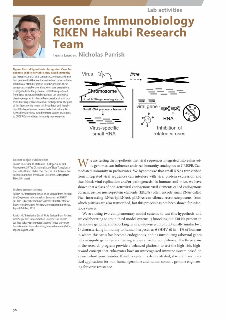
Genome Immunobiology RIKEN Hakubi Research TeamTeam Leader: Nicholas Parrish
Figure: Central Hypothesis - Integrated Virus Se-quences Enable Heritable RNA-based ImmunityWe hypothesize that viral sequences are integrated into host genome loci that are transcribed and processed into small RNAs. After integration into the genome, these sequences are stable over time, even over generations if integrated into the germline. Small RNAs produced from these integrated viral sequences can guide RNA-cleaving enzymes to silence the expression of viral pro-teins, blocking replication and/or pathogenesis. The goal of this laboratory is to test this hypothesis and thereby reject the hypothesis or demonstrate that eukaryotes have a heritable RNA-based immune system analogous to CRISPR/Cas-mediated immunity in prokaryotes.
Recent Major PublicationsParrish NF, Feurer ID, Matsuoka LK, Rega SA, Perri R, Alexopoulos SP. The Changing Face of Liver Transplanta-tion in the United States: The Effect of HCV Antiviral Eras on Transplantation Trends and Outcomes. Transplant Direct (in press)
Invited presentationsParrish NF. “Interfering Small RNAs Derived from Ancient Viral Sequences in Mammalian Genomes: a CRISPR/Cas-like Eukaryotic Immune System?” RIKEN Center for Biosystems Dynamics Research, internal seminar (Kobe, Japan) October, 2018
Parrish NF. “Interfering Small RNAs Derived from Ancient Viral Sequences in Mammalian Genomes: a CRISPR/Cas-like Eukaryotic Immune System?” Tokyo University Department of Neurochemistry, internal seminar (Tokyo, Japan) August, 2018
W e are testing the hypothesis that viral sequences integrated into eukaryot-ic genomes can influence antiviral immunity, analogous to CRISPR/Cas-
mediated immunity in prokaryotes. We hypothesize that small RNAs transcribed from integrated viral sequences can interfere with viral protein expression and thus block viral replication and/or pathogenesis. In humans and mice, we have shown that a class of non-retroviral endogenous viral elements called endogenous bornavirus-like nucleoprotein elements (EBLNs) often encode small RNAs called Piwi-interacting RNAs (piRNAs). piRNAs can silence retrotransposons, from which piRNAs are also transcribed, but this process has not been shown for infec-tious viruses.
We are using two complementary model systems to test this hypothesis and are collaborating to test a third model system: 1) knocking out EBLNs present in the mouse genome, and knocking in viral sequences into functionally similar loci, 2) characterizing immunity to human herpesvirus 6 (HHV-6) in ~1% of humans in whom this virus has become endogenous, and 3) introducing arboviral genes into mosquito genomes and testing arboviral vector competence. The three arms of the research program provide a balanced platform to test the high-risk, high-reward concept that eukaryotes have an unrecognized immune system based on virus-to-host gene transfer. If such a system is demonstrated, it would have prac-tical applications for non-human germline and human somatic genome engineer-ing for virus resistance.
58
Lab activities
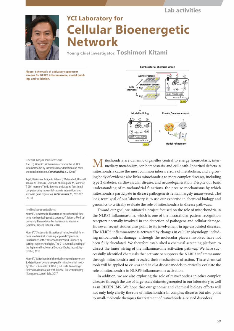
1280 chemicals
+
1
2
+
Activator screen
Suppressor screens K+
NLRP3‐PYCARD‐CASP1
ROS
pro‐IL‐1β IL‐1β
1 2 3
IL‐1β
Lysosome
? ??
X
K+
NLRP3‐PYCARD‐CASP1
ROS
pro‐IL‐1β IL‐1β
1 2 3
IL‐1β
Lysosome
? pHATP
X
Combinatorial chemical screen
Model building Ex vivo / in vivo analysis
K+
NLRP3‐PYCARD‐CASP1
pro‐IL‐1β IL‐1β
IL‐1β
Lyso
K+
O2
ATP pH
Model refinement
YCI Laboratory for
Cellular Bioenergetic NetworkYoung Chief Investigator: Toshimori Kitami
Figure: Schematic of activator-suppressor screens for NLRP3 inflammasome, model build-ing, and validation.
Recent Major PublicationsTran UT, Kitami T. Niclosamide activates the NLRP3 inflammasome by intracellular acidification and mito-chondrial inhibition. Commun Biol 2, 2 (2019)
Kaji T, Hijikata A, Ishige A, Kitami T, Watanabe T, Ohara O, Yanaka N, Okada M, Shimoda M, Taniguchi M, Takemori T. CD4 memory T cells develop and acquire functional competence by sequential cognate interactions and stepwise gene regulation. Int Immunol 28, 267-282 (2016)
Invited presentationsKitami T. “Systematic dissection of mitochondrial func-tions via chemical genetics approach” Saitama Medical University Research Center for Genomic Medicine (Saitama, Japan) October, 2018
Kitami T. “Systematic dissection of mitochondrial func-tions via chemical screening approach” Symposia: Renaissance of the ‘Mitochondrial World’ unveiled by cutting-edge technologies. The 91st Annual Meeting of the Japanese Biochemical Society (Kyoto, Japan) Sep-tember, 2018
Kitami T. “Mitochondrial chemical compendium version 2: detection of genotype-specific mitochondrial toxic-ity” The 1st Annual COCKPI-T (Co-Create Knowledge for Pharma Innovation with Takeda) Presentation Day (Kanagawa, Japan) July, 2017
M itochondria are dynamic organelles central to energy homeostasis, inter-mediary metabolism, ion homeostasis, and cell death. Inherited defects in
mitochondria cause the most common inborn errors of metabolism, and a grow-ing body of evidence also links mitochondria to more complex diseases, including type 2 diabetes, cardiovascular disease, and neurodegeneration. Despite our basic understanding of mitochondrial functions, the precise mechanisms by which mitochondria participate in disease pathogenesis remain largely unanswered. The long-term goal of our laboratory is to use our expertise in chemical biology and genomics to critically evaluate the role of mitochondria in disease pathways.
Toward our goal, we initiated a project focused on the role of mitochondria in the NLRP3 inflammasome, which is one of the intracellular pattern recognition receptors normally involved in the detection of pathogens and cellular damage. However, recent studies also point to its involvement in age-associated diseases. The NLRP3 inflammasome is activated by changes in cellular physiology, includ-ing mitochondrial damage, although the molecular players involved have not been fully elucidated. We therefore established a chemical screening platform to dissect the inner wiring of the inflammasome activation pathway. We have suc-cessfully identified chemicals that activate or suppress the NLRP3 inflammasome through mitochondria and revealed their mechanisms of action. These chemical tools will be applied to ex vivo and in vivo disease models to critically evaluate the role of mitochondria in NLRP3 inflammasome activation.
In addition, we are also exploring the role of mitochondria in other complex diseases through the use of large-scale datasets generated in our laboratory as well as in RIKEN IMS. We hope that our genomic and chemical biology efforts will not only help clarify the role of mitochondria in complex diseases but also point to small-molecule therapies for treatment of mitochondria-related disorders.
59
Lab activities
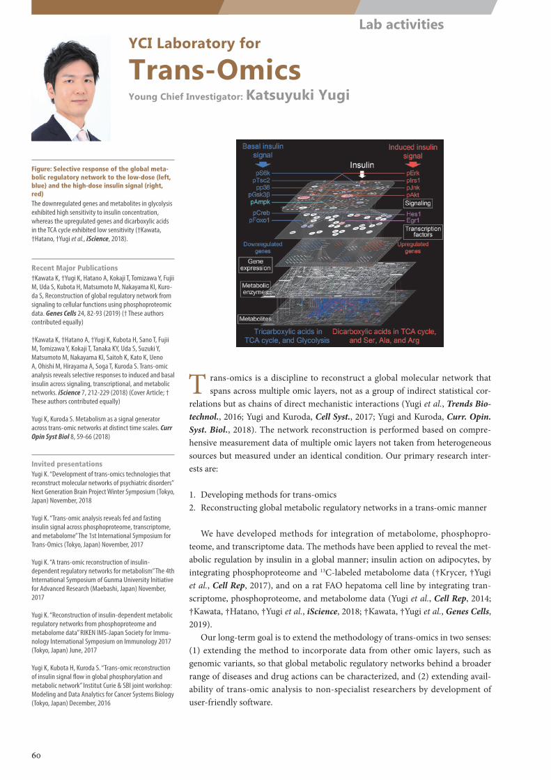
YCI Laboratory for
Trans-OmicsYoung Chief Investigator: Katsuyuki Yugi
Figure: Selective response of the global meta-bolic regulatory network to the low-dose (left, blue) and the high-dose insulin signal (right, red)The downregulated genes and metabolites in glycolysis exhibited high sensitivity to insulin concentration, whereas the upregulated genes and dicarboxylic acids in the TCA cycle exhibited low sensitivity (†Kawata, †Hatano, †Yugi et al., iScience, 2018).
T rans-omics is a discipline to reconstruct a global molecular network that spans across multiple omic layers, not as a group of indirect statistical cor-
relations but as chains of direct mechanistic interactions (Yugi et al., Trends Bio-technol., 2016; Yugi and Kuroda, Cell Syst., 2017; Yugi and Kuroda, Curr. Opin. Syst. Biol., 2018). The network reconstruction is performed based on compre-hensive measurement data of multiple omic layers not taken from heterogeneous sources but measured under an identical condition. Our primary research inter-ests are:
1. Developing methods for trans-omics2. Reconstructing global metabolic regulatory networks in a trans-omic manner
We have developed methods for integration of metabolome, phosphopro-teome, and transcriptome data. The methods have been applied to reveal the met-abolic regulation by insulin in a global manner; insulin action on adipocytes, by integrating phosphoproteome and 13C-labeled metabolome data (†Krycer, †Yugi et al., Cell Rep, 2017), and on a rat FAO hepatoma cell line by integrating tran-scriptome, phosphoproteome, and metabolome data (Yugi et al., Cell Rep, 2014; †Kawata, †Hatano, †Yugi et al., iScience, 2018; †Kawata, †Yugi et al., Genes Cells, 2019).
Our long-term goal is to extend the methodology of trans-omics in two senses: (1) extending the method to incorporate data from other omic layers, such as genomic variants, so that global metabolic regulatory networks behind a broader range of diseases and drug actions can be characterized, and (2) extending avail-ability of trans-omic analysis to non-specialist researchers by development of user-friendly software.
Recent Major Publications†Kawata K, †Yugi K, Hatano A, Kokaji T, Tomizawa Y, Fujii M, Uda S, Kubota H, Matsumoto M, Nakayama KI, Kuro-da S, Reconstruction of global regulatory network from signaling to cellular functions using phosphoproteomic data. Genes Cells 24, 82-93 (2019) († These authors contributed equally)
†Kawata K, †Hatano A, †Yugi K, Kubota H, Sano T, Fujii M, Tomizawa Y, Kokaji T, Tanaka KY, Uda S, Suzuki Y, Matsumoto M, Nakayama KI, Saitoh K, Kato K, Ueno A, Ohishi M, Hirayama A, Soga T, Kuroda S. Trans-omic analysis reveals selective responses to induced and basal insulin across signaling, transcriptional, and metabolic networks. iScience 7, 212-229 (2018) (Cover Article; † These authors contributed equally)
Yugi K, Kuroda S. Metabolism as a signal generator across trans-omic networks at distinct time scales. Curr Opin Syst Biol 8, 59-66 (2018)
Invited presentationsYugi K. “Development of trans-omics technologies that reconstruct molecular networks of psychiatric disorders” Next Generation Brain Project Winter Symposium (Tokyo, Japan) November, 2018
Yugi K. “Trans-omic analysis reveals fed and fasting insulin signal across phosphoproteome, transcriptome, and metabolome” The 1st International Symposium for Trans-Omics (Tokyo, Japan) November, 2017
Yugi K. “A trans-omic reconstruction of insulin-dependent regulatory networks for metabolism” The 4th International Symposium of Gunma University Initiative for Advanced Research (Maebashi, Japan) November, 2017
Yugi K. “Reconstruction of insulin-dependent metabolic regulatory networks from phosphoproteome and metabolome data” RIKEN IMS-Japan Society for Immu-nology International Symposium on Immunology 2017 (Tokyo, Japan) June, 2017
Yugi K, Kubota H, Kuroda S. “Trans-omic reconstruction of insulin signal flow in global phosphorylation and metabolic network” Institut Curie & SBI joint workshop: Modeling and Data Analytics for Cancer Systems Biology (Tokyo, Japan) December, 2016
60
Lab activities
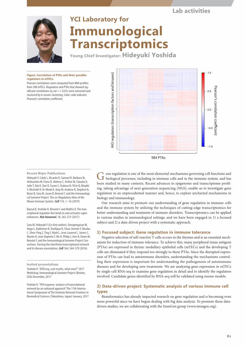
−1.0
−0.5
0.0
0.5
1.0
Pearson’s correlation coefficient
984 PTAs
443 regulators (transcription factors and kinases)
Fig. correlations of PTAs and their possible regulators in TECs
YCI Laboratory for
Immunological TranscriptomicsYoung Chief Investigator: Hideyuki Yoshida
Figure: Correlation of PTAs and their possible regulators in mTECs.Pearson correlations were computed from RNA profiles from 308 mTECs. Regulators and PTAs that showed sig-nificant correlations (q-val <= 0.05) were extracted and clustered by k-means clustering. Color-code indicates Pearson’s correlation coefficient.
Recent Major PublicationsHideyuki Y, Caleb L, Ricardo R, Samuel R, Barbara M, Aleksandra W, Fiona D, Aleksey C, Arthur M, Claudia D, Julie T, Edy K, Dan D, Susan S, Tsukasa N, YiLin Q, Bingfei Y, Michelle R, Ki-Wook K, Amy W, Andrew R, Stephen N, Brian B, Sara M, Jason B, Benoist C and the Immunologi-cal Genome Project. The cis-Regulatory Atlas of the Mouse Immune System. Cell 176, 1–16 (2019)
Bansal K, Yoshida H, Benoist C and Mathis D. The tran-scriptional regulator Aire binds to and activates super-enhancers. Nat Immunol. 18, 263-273 (2017)
Sara M, Hideyuki Y (Co-first author), Devapregasan M, Hugo L, Katherine R, Towfique R, Chun-Jimmie Y, Nicolas C, Shen-Ying Z, Ting F, Mark L, Jean-Laurent C, James C, Martin H, Jean-Baptiste T, Nir H, Philip J, Aviv R, Diane M, Benoist C and the Immunological Genome Project Con-sortium. Parsing the interferon transcriptional network and its disease associations. Cell 164, 564-578 (2016)
Invited presentationsYoshida H. “ATACseq, cool results, what next?” 2017 Workshop, Immunological Genome Project (Boston, USA) December, 2017
Yoshida H. “IFN response: analysis of transcriptional network by an unbiased approach” The 11th Interna-tional Symposium of The Institute Network Frontiers in Biomedical Sciences (Tokushima, Japan) January, 2017
G ene regulation is one of the most elemental mechanisms governing cell functions and biological processes, including in immune cells and in the immune system, and has
been studied in many contexts. Recent advances in epigenome and transcriptome profil-ing, taking advantage of next-generation sequencing (NGS), enable us to investigate gene regulation in an unprecedented manner and, hence, to explore uncharted mechanisms in biology and immunology.
Our research aims to promote our understanding of gene regulation in immune cells and the immune system by utilizing the techniques of cutting-edge transcriptomics for better understanding and treatment of immune disorders. Transcriptomics can be applied to various studies in immunological settings, and we have been engaged in 1) a focused subject and 2) a data-driven project with a systematic approach.
1) Focused subject: Gene regulation in immune toleranceNegative selection of self-reactive T cells occurs in the thymus and is an essential mech-
anism for induction of immune tolerance. To achieve this, many peripheral tissue antigens (PTAs) are expressed in thymic medullary epithelial cells (mTECs) and the developing T cells are eliminated if they respond too strongly to these PTAs. Since the disrupted expres-sion of PTAs can lead to autoimmune disorders, understanding the mechanisms control-ling their expression is important for understanding the pathogenesis of autoimmune diseases and for developing new treatments. We are analyzing gene expression in mTECs by single-cell RNA-seq to examine gene regulation in detail and to identify the regulators involved. Candidate genes identified by RNA-seq will be validated using mouse models.
2) Data-driven project: Systematic analysis of various immune cell types
Bioinformatics has already impacted research on gene regulation and is becoming even more powerful since we have begun dealing with big data analysis. To promote these data-driven studies, we are collaborating with the ImmGen group (www.immgen.org).
61
Lab activities
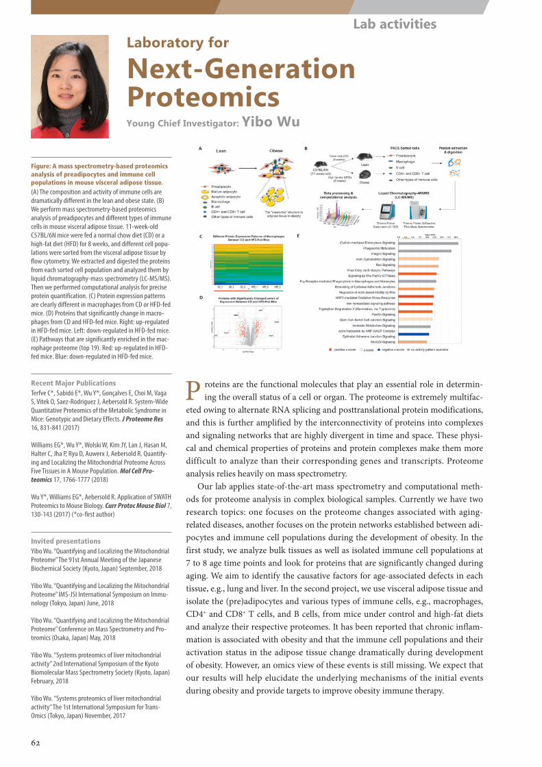
Laboratory for
Next-Generation ProteomicsYoung Chief Investigator: Yibo Wu
Figure: A mass spectrometry-based proteomics analysis of preadipocytes and immune cell populations in mouse visceral adipose tissue.(A) The composition and activity of immune cells are dramatically different in the lean and obese state. (B) We perform mass spectrometry-based proteomics analysis of preadipocytes and different types of immune cells in mouse visceral adipose tissue. 11-week-old C57BL/6N mice were fed a normal chow diet (CD) or a high-fat diet (HFD) for 8 weeks, and different cell popu-lations were sorted from the visceral adipose tissue by flow cytometry. We extracted and digested the proteins from each sorted cell population and analyzed them by liquid chromatography-mass spectrometry (LC-MS/MS). Then we performed computational analysis for precise protein quantification. (C) Protein expression patterns are clearly different in macrophages from CD or HFD-fed mice. (D) Proteins that significantly change in macro-phages from CD and HFD-fed mice. Right: up-regulated in HFD-fed mice. Left: down-regulated in HFD-fed mice. (E) Pathways that are significantly enriched in the mac-rophage proteome (top 19). Red: up-regulated in HFD-fed mice. Blue: down-regulated in HFD-fed mice.
Recent Major PublicationsTerfve C*, Sabidó E*, Wu Y*, Gonçalves E, Choi M, Vaga S, Vitek O, Saez-Rodriguez J, Aebersold R. System-Wide Quantitative Proteomics of the Metabolic Syndrome in Mice: Genotypic and Dietary Effects. J Proteome Res 16, 831-841 (2017)
Williams EG*, Wu Y*, Wolski W, Kim JY, Lan J, Hasan M, Halter C, Jha P, Ryu D, Auwerx J, Aebersold R. Quantify-ing and Localizing the Mitochondrial Proteome Across Five Tissues in A Mouse Population. Mol Cell Pro-teomics 17, 1766-1777 (2018)
Wu Y*, Williams EG*, Aebersold R. Application of SWATH Proteomics to Mouse Biology. Curr Protoc Mouse Biol 7, 130-143 (2017) (*co-first author)
Invited presentationsYibo Wu. “Quantifying and Localizing the Mitochondrial Proteome” The 91st Annual Meeting of the Japanese Biochemical Society (Kyoto, Japan) September, 2018
Yibo Wu. “Quantifying and Localizing the Mitochondrial Proteome” IMS-JSI International Symposium on Immu-nology (Tokyo, Japan) June, 2018
Yibo Wu. “Quantifying and Localizing the Mitochondrial Proteome” Conference on Mass Spectrometry and Pro-teomics (Osaka, Japan) May, 2018
Yibo Wu. “Systems proteomics of liver mitochondrial activity” 2nd International Symposium of the Kyoto Biomolecular Mass Spectrometry Society (Kyoto, Japan) February, 2018
Yibo Wu. “Systems proteomics of liver mitochondrial activity” The 1st International Symposium for Trans-Omics (Tokyo, Japan) November, 2017
P roteins are the functional molecules that play an essential role in determin-ing the overall status of a cell or organ. The proteome is extremely multifac-
eted owing to alternate RNA splicing and posttranslational protein modifications, and this is further amplified by the interconnectivity of proteins into complexes and signaling networks that are highly divergent in time and space. These physi-cal and chemical properties of proteins and protein complexes make them more difficult to analyze than their corresponding genes and transcripts. Proteome analysis relies heavily on mass spectrometry.
Our lab applies state-of-the-art mass spectrometry and computational meth-ods for proteome analysis in complex biological samples. Currently we have two research topics: one focuses on the proteome changes associated with aging-related diseases, another focuses on the protein networks established between adi-pocytes and immune cell populations during the development of obesity. In the first study, we analyze bulk tissues as well as isolated immune cell populations at 7 to 8 age time points and look for proteins that are significantly changed during aging. We aim to identify the causative factors for age-associated defects in each tissue, e.g., lung and liver. In the second project, we use visceral adipose tissue and isolate the (pre)adipocytes and various types of immune cells, e.g., macrophages, CD4+ and CD8+ T cells, and B cells, from mice under control and high-fat diets and analyze their respective proteomes. It has been reported that chronic inflam-mation is associated with obesity and that the immune cell populations and their activation status in the adipose tissue change dramatically during development of obesity. However, an omics view of these events is still missing. We expect that our results will help elucidate the underlying mechanisms of the initial events during obesity and provide targets to improve obesity immune therapy.
62
Lab activities
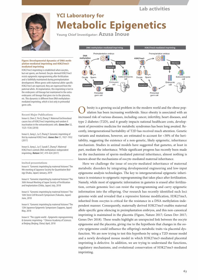
YCI Laboratory for
Metabolic EpigeneticsYoung Chief Investigator: Azusa Inoue
Figure: Developmental dynamics of DNA meth-ylation-mediated imprinting and H3K27me3-mediated imprintingH3K27me3 imprinting is established when oocytes, but not sperm, are formed. Oocyte-derived H3K27me3 resists epigenetic reprogramming after fertilization and is faithfully maintained during preimplantation development. When genes with maternal allele-specific H3K27me3 are expressed, they are expressed from the paternal allele. At implantation, this imprinting is lost in the embryonic cell lineage but maintained in the extra-embryonic cell lineage that gives rise to the placenta, etc. This dynamics is different from DNA methylation-mediated imprinting, which is lost only in primordial germ cells.
Recent Major PublicationsInoue A, Chen Z, Yin Q, Zhang Y. Maternal Eed knockout causes loss of H3K27me3 imprinting and random X inactivation in the extraembryonic cells. Genes Dev 32, 1525-1536 (2018)
Inoue A, Jiang L, Lu F, Zhang Y. Genomic imprinting of Xist by maternal H3K27me3. Genes Dev 31, 1927-1932 (2017)
Inoue A, Jiang L, Lu F, Suzuki T, Zhang Y. Maternal H3K27me3 controls DNA methylation-independent imprinting. Nature 547, 419-424 (2017)
Invited presentationsInoue A. “Genomic imprinting by maternal histones” The 9th meeting of Japanese Society for Quantitative Biol-ogy (Osaka, Japan) January, 2019
Inoue A. “Genomic imprinting by maternal histones” The 36th Annual Meeting of Japan Society of Fertilization and Implantation (Chiba, Japan) July, 2018
Inoue A. “Genomic imprinting by maternal histones” The 16th Stem Cell Research Symposium (Fukuoka, Japan) June, 2018
Inoue A. “Genomic imprinting by maternal histones” The 12th Japanese Epigenetic Symposium (Sapporo, Japan) May, 2018
Inoue A. “The zygote world –Epigenetic reprogramming & Genomic imprinting–” Chinese Academy of Sciences at Beijing (Beijing, China) April, 2018
O besity is a growing social problem in the modern world and the obese pop-ulation has been increasing worldwide. Since obesity is associated with an
increased risk of various diseases, including cancer, infertility, heart diseases, and type 2 diabetes (T2D), and it greatly impacts national healthcare costs, develop-ment of preventive medicine for metabolic syndromes has been long awaited. Re-cently, intergenerational heritability of T2D has received much attention. Genetic variants and mutations, however, are estimated to account for <30% of the heri-tability, suggesting the existence of a non-genetic, likely epigenetic, inheritance mechanism. Studies in animal models have suggested that gametes, at least in part, mediate the inheritance. While significant progress has recently been made on the mechanisms of sperm-mediated paternal inheritance, almost nothing is known about the mechanisms of oocyte-mediated maternal inheritance.
Here we challenge the issue of oocyte-mediated inheritance of maternal metabolic disorders by integrating developmental engineering and low-input epigenome analysis technologies. The key to intergenerational epigenetic inheri-tance is resistance to epigenetic reprogramming that takes place after fertilization. Namely, while most of epigenetic information in gametes is erased after fertiliza-tion, certain genomic loci can resist the reprogramming and carry epigenetic information into the offspring. Our research has recently identified such loci genome-wide and revealed that a repressive histone modification, H3K27me3, inherited from oocytes is critical for the resistance in a DNA methylation-inde-pendent manner. Consequently, maternally derived H3K27me3 enables maternal allele-specific gene silencing in preimplantation embryos, and this non-canonical imprinting is maintained in the placenta (Figure, Nature 2017; Genes Dev 2017; Genes Dev 2018). These results highlight an unexpected link between the oocyte epigenome and the placenta, giving rise to the hypothesis that changes in the oo-cyte epigenome could influence the offspring’s metabolic traits via placental dys-function. We are now trying to test this hypothesis by using a T2D mouse model and a newly developed mouse model in which H3K27me3-mediated placental imprinting is defective. In addition, we are trying to understand the functions, regulatory mechanisms, and evolutional conservation of H3K27me3-mediated imprinting.
63
Lab activities
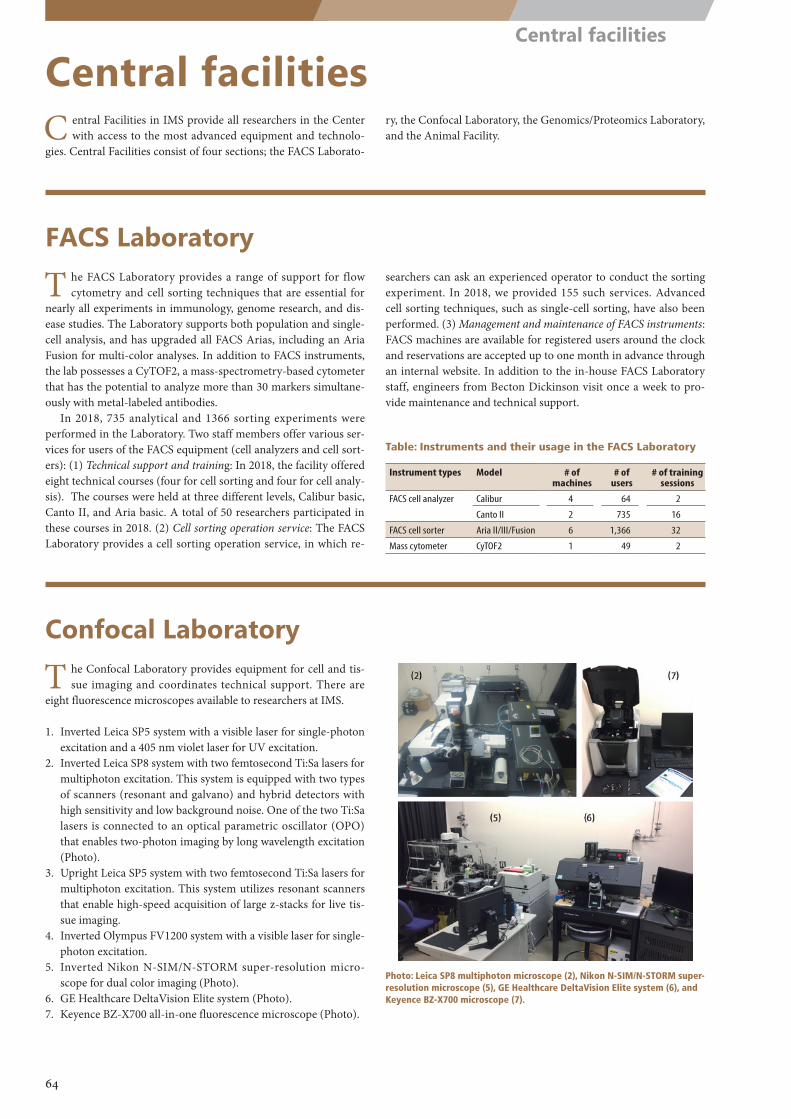
Table: Instruments and their usage in the FACS Laboratory
Instrument types Model # of machines
# of users
# of training sessions
FACS cell analyzer Calibur 4 64 2
Canto II 2 735 16
FACS cell sorter Aria II/III/Fusion 6 1,366 32
Mass cytometer CyTOF2 1 49 2
Photo: Leica SP8 multiphoton microscope (2), Nikon N-SIM/N-STORM super-resolution microscope (5), GE Healthcare DeltaVision Elite system (6), and Keyence BZ-X700 microscope (7).
Central facilitiesC entral Facilities in IMS provide all researchers in the Center
with access to the most advanced equipment and technolo-gies. Central Facilities consist of four sections; the FACS Laborato-
ry, the Confocal Laboratory, the Genomics/Proteomics Laboratory, and the Animal Facility.
T he FACS Laboratory provides a range of support for flow cytometry and cell sorting techniques that are essential for
nearly all experiments in immunology, genome research, and dis-ease studies. The Laboratory supports both population and single-cell analysis, and has upgraded all FACS Arias, including an Aria Fusion for multi-color analyses. In addition to FACS instruments, the lab possesses a CyTOF2, a mass-spectrometry-based cytometer that has the potential to analyze more than 30 markers simultane-ously with metal-labeled antibodies.
In 2018, 735 analytical and 1366 sorting experiments were performed in the Laboratory. Two staff members offer various ser-vices for users of the FACS equipment (cell analyzers and cell sort-ers): (1) Technical support and training: In 2018, the facility offered eight technical courses (four for cell sorting and four for cell analy-sis). The courses were held at three different levels, Calibur basic, Canto II, and Aria basic. A total of 50 researchers participated in these courses in 2018. (2) Cell sorting operation service: The FACS Laboratory provides a cell sorting operation service, in which re-
searchers can ask an experienced operator to conduct the sorting experiment. In 2018, we provided 155 such services. Advanced cell sorting techniques, such as single-cell sorting, have also been performed. (3) Management and maintenance of FACS instruments: FACS machines are available for registered users around the clock and reservations are accepted up to one month in advance through an internal website. In addition to the in-house FACS Laboratory staff, engineers from Becton Dickinson visit once a week to pro-vide maintenance and technical support.
T he Confocal Laboratory provides equipment for cell and tis-sue imaging and coordinates technical support. There are
eight fluorescence microscopes available to researchers at IMS.
1. Inverted Leica SP5 system with a visible laser for single-photon excitation and a 405 nm violet laser for UV excitation.
2. Inverted Leica SP8 system with two femtosecond Ti:Sa lasers for multiphoton excitation. This system is equipped with two types of scanners (resonant and galvano) and hybrid detectors with high sensitivity and low background noise. One of the two Ti:Sa lasers is connected to an optical parametric oscillator (OPO) that enables two-photon imaging by long wavelength excitation (Photo).
3. Upright Leica SP5 system with two femtosecond Ti:Sa lasers for multiphoton excitation. This system utilizes resonant scanners that enable high-speed acquisition of large z-stacks for live tis-sue imaging.
4. Inverted Olympus FV1200 system with a visible laser for single-photon excitation.
5. Inverted Nikon N-SIM/N-STORM super-resolution micro-scope for dual color imaging (Photo).
6. GE Healthcare DeltaVision Elite system (Photo).7. Keyence BZ-X700 all-in-one fluorescence microscope (Photo).
FACS Laboratory
Confocal Laboratory
64
Central facilities
Table: Central services provided by the Genomics Lab in 2018
Next-generation DNA sequencing # of samples # of teams
RNA sequencing 2,438 21ChIP sequencing 326 4Others (Exome etc) 1,311 5
Proteomics # of samples # of teams
Mass spectrometry analysis 41 2Multiplex suspension array 1,304 7
Sanger DNA sequencing # of samples # of teams
36 cm capillary 10,312 1350 cm capillary 2,304 17
cDNA clone delivery # of samples # of teams
37 2
Primer/labeled probe delivery # of samples # of teams
21 1
Figure: Application of the CRISPR/Cas system by electroporation of fertil-ized eggs
+1 mm
10 mm
O ne of the missions of the Laboratory for Integrative Genom-ics is to serve as a technical support service lab that provides
state-of-the-art genome- and proteome-wide analyses for research groups in IMS. We thus offer a variety of services to suit the needs of different labs. These include DNA sequencing, proteomics analysis, multiplex suspension array, cDNA/genomic clone dis-tribution, and primer/labeled probe distribution for qRT-PCR analysis of immune cells (Table). In practice, next-generation DNA sequencing on an Illumina HiSeq2500 (ver4) is supported both by our laboratory and the Laboratory for Genotyping Development. We also perform mass spectrometry-based quantitative proteome analysis using an AB SCIEX TripleTOF 5600. Utilizing this sup-port system, even researchers who are not familiar with such in-strumental analyses can easily undertake genomics and proteomics approaches when necessary.
Besides these support activities for conventional genomics and related technologies, it should be noted that IMS strongly encour-ages collaborations between experts and non-experts in genom-ics. Because the Division of Genomic Medicine at IMS has many unique technologies and know-how in transcriptomics and genet-ics (e.g., CAGE technologies, GWAS, and whole genome sequenc-ing), we expect that the intramural interactions among the Divi-
sions of Human Immunology, Disease Systems Biology, Cancer Immunology, and Genome Medicine fostered by these technolo-gies will greatly enhance research activities in IMS. In this respect, the genomics support activity may serve as a gateway to initiate efficient collaboration among the divisions in IMS.
W e continue to maintain over 50,000 mice in the SPF area and 1,500 mice in an isolated area. The SPF area also con-
tains 550 germ-free or gnotobiotic mice in vinyl isolator rooms and in vinyl isolation bio-bubble rooms. The former are used by several IMS research groups, in particular the mucosal immunologists, and the latter are for “humanized mice”. We introduce mouse lines into the SPF area via a combination of in vitro fertilization (IVF) and embryo transfer methods, and have also generated cryostocks of genetic resources (frozen embryos and sperms) for 737 lines. We also maintain relatively large colonies of several commonly used strains, such as Rag1 KO and Cre deleters, and provide them to users on demand. We have also provided technical assistance to generate knockout and transgenic mice (54 lines). In addition, we have made KO and KI mice (53 lines) using the CRISPR/Cas sys-tem and have created 21 lines of germ-free mice. We provide space for new experiments in the animal facility, e.g., the CRISPR/Cas system using fertilized egg electroporation (Figure), and behavioral testing for germ-free mice.
We have generated genetically modified mice to improve the efficacy of transplantation of human hematopoietic stem cells into NOD.Cg-Prkdcscid Il2rgtm1Wjl/SzJ (NSG) mice by better “human-izing” the host strain. For this purpose, we have introduced large
genomic fragments containing human genes encoding MHC, cy-tokines, adhesion molecules, virus receptors, and others into the NSG mice. We maintain such transgenic and knock-in mice with confirmed expression of human genes on a C57BL/6 background and have begun backcrossing them onto the NSG mouse back-ground using the speed-congenic method.
Genomics and related activities
Animal Facility
65
Central facilities
RIKEN International Program Associate (IPA)
I MS accepted eight international students as RIKEN Interna-tional Program Associates (IPA). Under this IPA program, IMS
lab heads host international students from collaborating graduate schools and supervise their Ph.D. program as Joint Supervisors. The students receive a daily living allowance and housing costs for up to a maximum of three years.
The IPA students who studied at IMS in 2018 wereJiahui Ma (Peking University, China) in the Laboratory for Ge-nome Information AnalysisYan Jun Lan (ETH Zurich, Switzerland) in the Laboratory for Ad-vanced Genomics Circuit
Shruti Bhagat (Karolinska Institute, Sweden) in the Preventive Medicine and Applied Genomics UnitHsing-Fang Lu (Taipei Medical University, Taiwan) in the Labo-ratory for Bone and Joint DiseasesJack Thomas Flanagan (The University of Liverpool, UK) in the Laboratory for Endocrinology, Metabolism and Kidney DiseasesMichael Boettcher (Bielefeld University, Germany) in the Labo-ratory for Transcriptome TechnologyMengqian Li (Tokyo Medical and Dental University, from China) in the Liver Cancer Prevention Research UnitDaniela Kajihara (University of Sao Paulo, Brazil) in the Labora-tory for Transcriptome Technology
RIKEN Special Postdoctoral Researcher (SPDR) Program
R IKEN’s Special Postdoctoral Researcher Program was insti-tuted to provide young and creative scientists the opportunity
to be involved in autonomous and independent research in line with RIKEN objectives and research fields. The positions are com-petitive, but if selected, researchers receive salaries and research budgets (1 million yen) from RIKEN and they are able to conduct their research at one of its laboratories.
This year, eight postdocs conducted their research at IMS through the SPDR program.
Eiji Miyauchi (Laboratory for Intestinal Ecosystem)Alexis Vogelzang (Laboratory for Mucosal Immunity)Keiichiro Shiraga (Laboratory for Skin Homeostasis)Xiaoxi Liu (Laboratory for Genotyping Development)Callum Parr (Laboratory for Advanced Genomics Circuit)Rei Nakano (Laboratory for Cellular Function Conversion Tech-nology)Tsuyoshi Kiniwa (Laboratory for Innate Immune Systems)Divya Mundackal Sivaraman (Laboratory for Advanced Ge-nomics Circuit)
RIKEN Junior Research Associate (JRA) Program
T he Junior Research Associate Program was launched in 1996 to encourage young scientists with fresh ideas and youthful
enthusiasm to collaborate with, and learn from, senior scientists with years of experience. This program provides part-time posi-tions at RIKEN for young researchers enrolled in university Ph.D. programs. The JRA program serves the dual purpose of fostering the development of these young scientists while also energizing RIKEN with their innovative thinking.
This year, 24 JRA students studied in IMS.
Yosuke Ito (Preventive Medicine and Applied Genomics Unit)Junichiro Takano (Laboratory for Developmental Genetics)Yoshihiro Ito (Laboratory for Skin Homeostasis)Eiichiro Watanabe (Laboratory for Gut Homeostasis)Takaaki Kawaguchi (Laboratory for Gut Homeostasis)Keiko Usui (Laboratory for Skin Homeostasis)Mamoru Ogawa (Laboratory for Metabolomics)Hiroki Sugishita (Laboratory for Developmental Genetics)
Hiroe Tetsu (Laboratory for Innate Immune Systems)Manabu Nagayama (Laboratory for Gut Homeostasis)Ari Morimoto (Laboratory for Skin Homeostasis)Yuki Ariyasu (Laboratory for Metabolomics)Shohei Egami (Laboratory for Skin Homeostasis)Shintaro Ono (Laboratory for Integrative Genomics)Ryota Sato (Laboratory for Lymphocyte Differentiation)Kyosuke Shishikura (Laboratory for Metabolomics)Iori Motoo (Laboratory for Gut Homeostasis)Tsuyoshi Yamane (Laboratory for Metabolomics)Naoko Toki (Laboratory for Transcriptome Technology)Makoto Iwasaki (Laboratory for Human Disease Models)Nao Otomo (Laboratory for Bone and Joint Diseases)Akiko Oguchi (Laboratory for Transcriptome Technology)Tomoaki Takahashi (Preventive Medicine and Applied Genom-ics Unit)Takahiro Matsunaga (Laboratory for Gut Homeostasis)
66
Other programs
Part 3
Research Projects
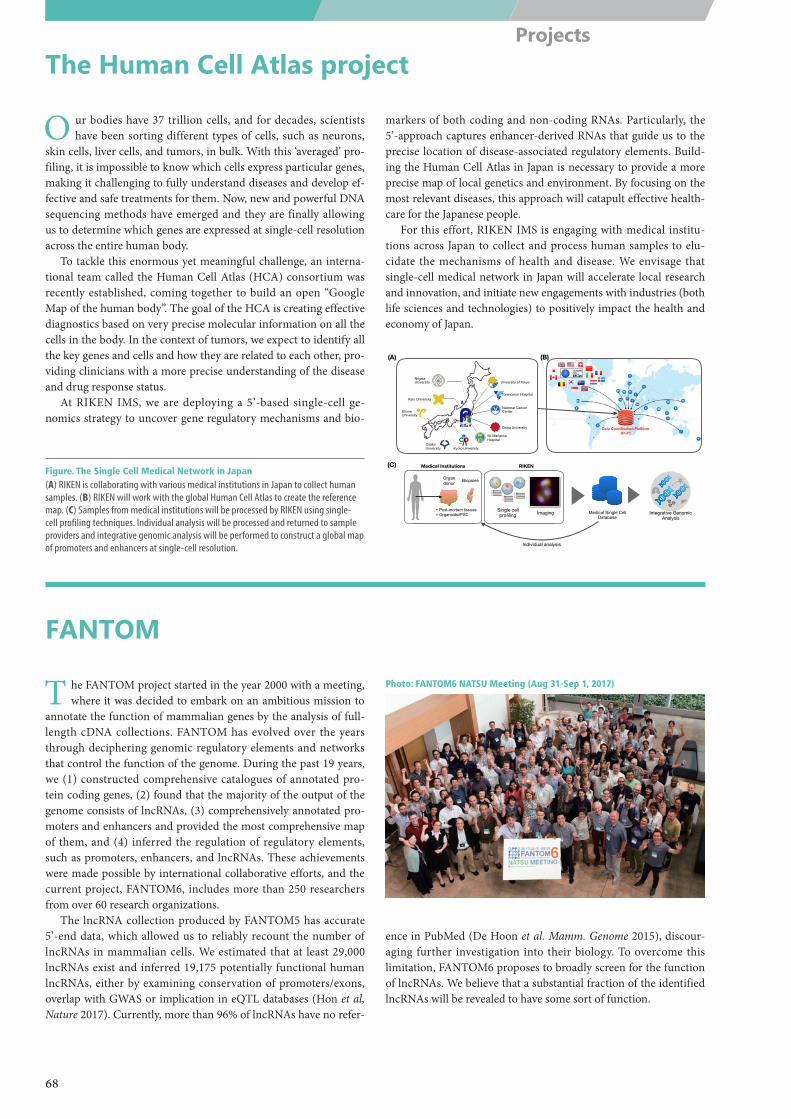
N
5’-5’-5’-5’-
5’-5’-5’-5’-
5’-5’-5’-5’-
RIKEN
Single cell profiling
University of Tokyo
Toranomon Hospital
National Cancer Center
Chiba University
St. Marianna Hospital
Kyoto UniversityOsaka University
Ehime University
Keio University
Niigata University
Medical Single Cell Database
Imaging
Medical Institutions
Integrative Genomic Analysis
Individual analysis
(A) (B)
(C)
Organ donor
+ Post-mortem tissues+ Organoids/iPSC
Biopsies
Photo: FANTOM6 NATSU Meeting (Aug 31-Sep 1, 2017)
The Human Cell Atlas project
FANTOM
O ur bodies have 37 trillion cells, and for decades, scientists have been sorting different types of cells, such as neurons,
skin cells, liver cells, and tumors, in bulk. With this ‘averaged’ pro-filing, it is impossible to know which cells express particular genes, making it challenging to fully understand diseases and develop ef-fective and safe treatments for them. Now, new and powerful DNA sequencing methods have emerged and they are finally allowing us to determine which genes are expressed at single-cell resolution across the entire human body.
To tackle this enormous yet meaningful challenge, an interna-tional team called the Human Cell Atlas (HCA) consortium was recently established, coming together to build an open “Google Map of the human body”. The goal of the HCA is creating effective diagnostics based on very precise molecular information on all the cells in the body. In the context of tumors, we expect to identify all the key genes and cells and how they are related to each other, pro-viding clinicians with a more precise understanding of the disease and drug response status.
At RIKEN IMS, we are deploying a 5’-based single-cell ge-nomics strategy to uncover gene regulatory mechanisms and bio-
markers of both coding and non-coding RNAs. Particularly, the 5’-approach captures enhancer-derived RNAs that guide us to the precise location of disease-associated regulatory elements. Build-ing the Human Cell Atlas in Japan is necessary to provide a more precise map of local genetics and environment. By focusing on the most relevant diseases, this approach will catapult effective health-care for the Japanese people.
For this effort, RIKEN IMS is engaging with medical institu-tions across Japan to collect and process human samples to elu-cidate the mechanisms of health and disease. We envisage that single-cell medical network in Japan will accelerate local research and innovation, and initiate new engagements with industries (both life sciences and technologies) to positively impact the health and economy of Japan.
T he FANTOM project started in the year 2000 with a meeting, where it was decided to embark on an ambitious mission to
annotate the function of mammalian genes by the analysis of full-length cDNA collections. FANTOM has evolved over the years through deciphering genomic regulatory elements and networks that control the function of the genome. During the past 19 years, we (1) constructed comprehensive catalogues of annotated pro-tein coding genes, (2) found that the majority of the output of the genome consists of lncRNAs, (3) comprehensively annotated pro-moters and enhancers and provided the most comprehensive map of them, and (4) inferred the regulation of regulatory elements, such as promoters, enhancers, and lncRNAs. These achievements were made possible by international collaborative efforts, and the current project, FANTOM6, includes more than 250 researchers from over 60 research organizations.
The lncRNA collection produced by FANTOM5 has accurate 5’-end data, which allowed us to reliably recount the number of lncRNAs in mammalian cells. We estimated that at least 29,000 lncRNAs exist and inferred 19,175 potentially functional human lncRNAs, either by examining conservation of promoters/exons, overlap with GWAS or implication in eQTL databases (Hon et al, Nature 2017). Currently, more than 96% of lncRNAs have no refer-
ence in PubMed (De Hoon et al. Mamm. Genome 2015), discour-aging further investigation into their biology. To overcome this limitation, FANTOM6 proposes to broadly screen for the function of lncRNAs. We believe that a substantial fraction of the identified lncRNAs will be revealed to have some sort of function.
Figure. The Single Cell Medical Network in Japan(A) RIKEN is collaborating with various medical institutions in Japan to collect human samples. (B) RIKEN will work with the global Human Cell Atlas to create the reference map. (C) Samples from medical institutions will be processed by RIKEN using single-cell profiling techniques. Individual analysis will be processed and returned to sample providers and integrative genomic analysis will be performed to construct a global map of promoters and enhancers at single-cell resolution.
68
Projects
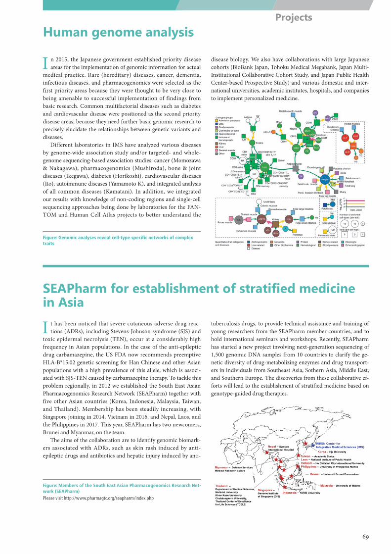
RIKEN Center forIntegrative Medical Sciences (IMS)Korea – Inje University
Taiwan – Academia Sinica
Thailand –Department of Medical Sciences, Mahidol University,Khon Kaen University,Chulalongkorn University,Thailand Center of Excellencefor Life Sciences (TCELS)
Malaysia – University of Malaya
Indonesia – YARSI University Singapore –Genome Instituteof Singapore (GIS)
Vietnam – Ho Chi Minh City International University
Nepal – SwaconInternational Hospital
Laos – National Institute of Public Health
Philippines – University of Philippines ManilaMyanmar – Defence ServicesMedical Research Centre
Brunei – Universiti Brunei Darussalam
Human genome analysis
SEAPharm for establishment of stratified medicine in Asia
I n 2015, the Japanese government established priority disease areas for the implementation of genomic information for actual
medical practice. Rare (hereditary) diseases, cancer, dementia, infectious diseases, and pharmacogenomics were selected as the first priority areas because they were thought to be very close to being amenable to successful implementation of findings from basic research. Common multifactorial diseases such as diabetes and cardiovascular disease were positioned as the second priority disease areas, because they need further basic genomic research to precisely elucidate the relationships between genetic variants and diseases.
Different laboratories in IMS have analyzed various diseases by genome-wide association study and/or targeted- and whole-genome sequencing-based association studies: cancer (Momozawa & Nakagawa), pharmacogenomics (Mushiroda), bone & joint diseases (Ikegawa), diabetes (Horikoshi), cardiovascular diseases (Ito), autoimmune diseases (Yamamoto K), and integrated analysis of all common diseases (Kamatani). In addition, we integrated our results with knowledge of non-coding regions and single-cell sequencing approaches being done by laboratories for the FAN-TOM and Human Cell Atlas projects to better understand the
disease biology. We also have collaborations with large Japanese cohorts (BioBank Japan, Tohoku Medical Megabank, Japan Multi-Institutional Collaborative Cohort Study, and Japan Public Health Center-based Prospective Study) and various domestic and inter-national universities, academic institutes, hospitals, and companies to implement personalized medicine.
I t has been noticed that severe cutaneous adverse drug reac-tions (ADRs), including Stevens-Johnson syndrome (SJS) and
toxic epidermal necrolysis (TEN), occur at a considerably high frequency in Asian populations. In the case of the anti-epileptic drug carbamazepine, the US FDA now recommends preemptive HLA-B*15:02 genetic screening for Han Chinese and other Asian populations with a high prevalence of this allele, which is associ-ated with SJS-TEN caused by carbamazepine therapy. To tackle this problem regionally, in 2012 we established the South East Asian Pharmacogenomics Research Network (SEAPharm) together with five other Asian countries (Korea, Indonesia, Malaysia, Taiwan, and Thailand). Membership has been steadily increasing, with Singapore joining in 2014, Vietnam in 2016, and Nepal, Laos, and the Philippines in 2017. This year, SEAPharm has two newcomers, Brunei and Myanmar, on the team.
The aims of the collaboration are to identify genomic biomark-ers associated with ADRs, such as skin rash induced by anti-epileptic drugs and antibiotics and hepatic injury induced by anti-
tuberculosis drugs, to provide technical assistance and training of young researchers from the SEAPharm member countries, and to hold international seminars and workshops. Recently, SEAPharm has started a new project involving next-generation sequencing of 1,500 genomic DNA samples from 10 countries to clarify the ge-netic diversity of drug-metabolizing enzymes and drug transport-ers in individuals from Southeast Asia, Sothern Asia, Middle East, and Southern Europe. The discoveries from these collaborative ef-forts will lead to the establishment of stratified medicine based on genotype-guided drug therapies.
Figure: Genomic analyses reveal cell-type specific networks of complex traits
Figure: Members of the South East Asian Pharmacogenomics Research Net-work (SEAPharm)Please visit http://www.pharmagtc.org/seapharm/index.php
69
Projects
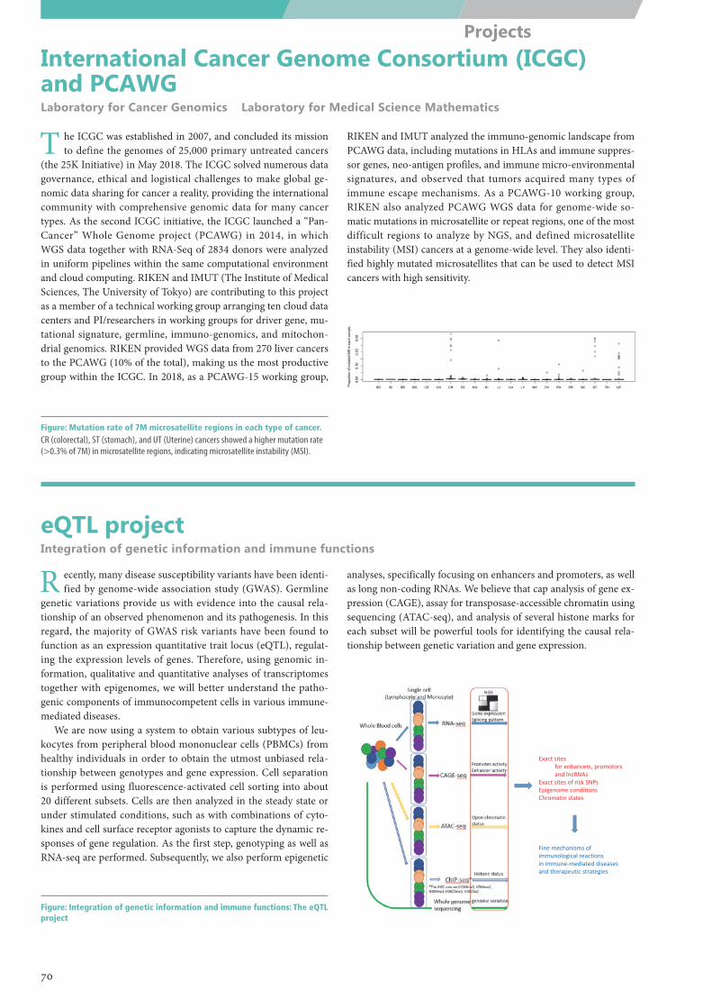
Exact sites for enhancers, promotorsand lncRNAs
Exact sites of risk SNPsEpigenome conditionsChromatin states
Fine mechanisms of immunological reactions in immune‐mediated diseasesand therapeutic strategies
International Cancer Genome Consortium (ICGC) and PCAWGLaboratory for Cancer Genomics Laboratory for Medical Science Mathematics
eQTL projectIntegration of genetic information and immune functions
T he ICGC was established in 2007, and concluded its mission to define the genomes of 25,000 primary untreated cancers
(the 25K Initiative) in May 2018. The ICGC solved numerous data governance, ethical and logistical challenges to make global ge-nomic data sharing for cancer a reality, providing the international community with comprehensive genomic data for many cancer types. As the second ICGC initiative, the ICGC launched a “Pan-Cancer” Whole Genome project (PCAWG) in 2014, in which WGS data together with RNA-Seq of 2834 donors were analyzed in uniform pipelines within the same computational environment and cloud computing. RIKEN and IMUT (The Institute of Medical Sciences, The University of Tokyo) are contributing to this project as a member of a technical working group arranging ten cloud data centers and PI/researchers in working groups for driver gene, mu-tational signature, germline, immuno-genomics, and mitochon-drial genomics. RIKEN provided WGS data from 270 liver cancers to the PCAWG (10% of the total), making us the most productive group within the ICGC. In 2018, as a PCAWG-15 working group,
RIKEN and IMUT analyzed the immuno-genomic landscape from PCAWG data, including mutations in HLAs and immune suppres-sor genes, neo-antigen profiles, and immune micro-environmental signatures, and observed that tumors acquired many types of immune escape mechanisms. As a PCAWG-10 working group, RIKEN also analyzed PCAWG WGS data for genome-wide so-matic mutations in microsatellite or repeat regions, one of the most difficult regions to analyze by NGS, and defined microsatellite instability (MSI) cancers at a genome-wide level. They also identi-fied highly mutated microsatellites that can be used to detect MSI cancers with high sensitivity.
R ecently, many disease susceptibility variants have been identi-fied by genome-wide association study (GWAS). Germline
genetic variations provide us with evidence into the causal rela-tionship of an observed phenomenon and its pathogenesis. In this regard, the majority of GWAS risk variants have been found to function as an expression quantitative trait locus (eQTL), regulat-ing the expression levels of genes. Therefore, using genomic in-formation, qualitative and quantitative analyses of transcriptomes together with epigenomes, we will better understand the patho-genic components of immunocompetent cells in various immune-mediated diseases.
We are now using a system to obtain various subtypes of leu-kocytes from peripheral blood mononuclear cells (PBMCs) from healthy individuals in order to obtain the utmost unbiased rela-tionship between genotypes and gene expression. Cell separation is performed using fluorescence-activated cell sorting into about 20 different subsets. Cells are then analyzed in the steady state or under stimulated conditions, such as with combinations of cyto-kines and cell surface receptor agonists to capture the dynamic re-sponses of gene regulation. As the first step, genotyping as well as RNA-seq are performed. Subsequently, we also perform epigenetic
analyses, specifically focusing on enhancers and promoters, as well as long non-coding RNAs. We believe that cap analysis of gene ex-pression (CAGE), assay for transposase-accessible chromatin using sequencing (ATAC-seq), and analysis of several histone marks for each subset will be powerful tools for identifying the causal rela-tionship between genetic variation and gene expression.
Figure: Mutation rate of 7M microsatellite regions in each type of cancer.CR (colorectal), ST (stomach), and UT (Uterine) cancers showed a higher mutation rate (>0.3% of 7M) in microsatellite regions, indicating microsatellite instability (MSI).
Figure: Integration of genetic information and immune functions: The eQTL project
70
Projects
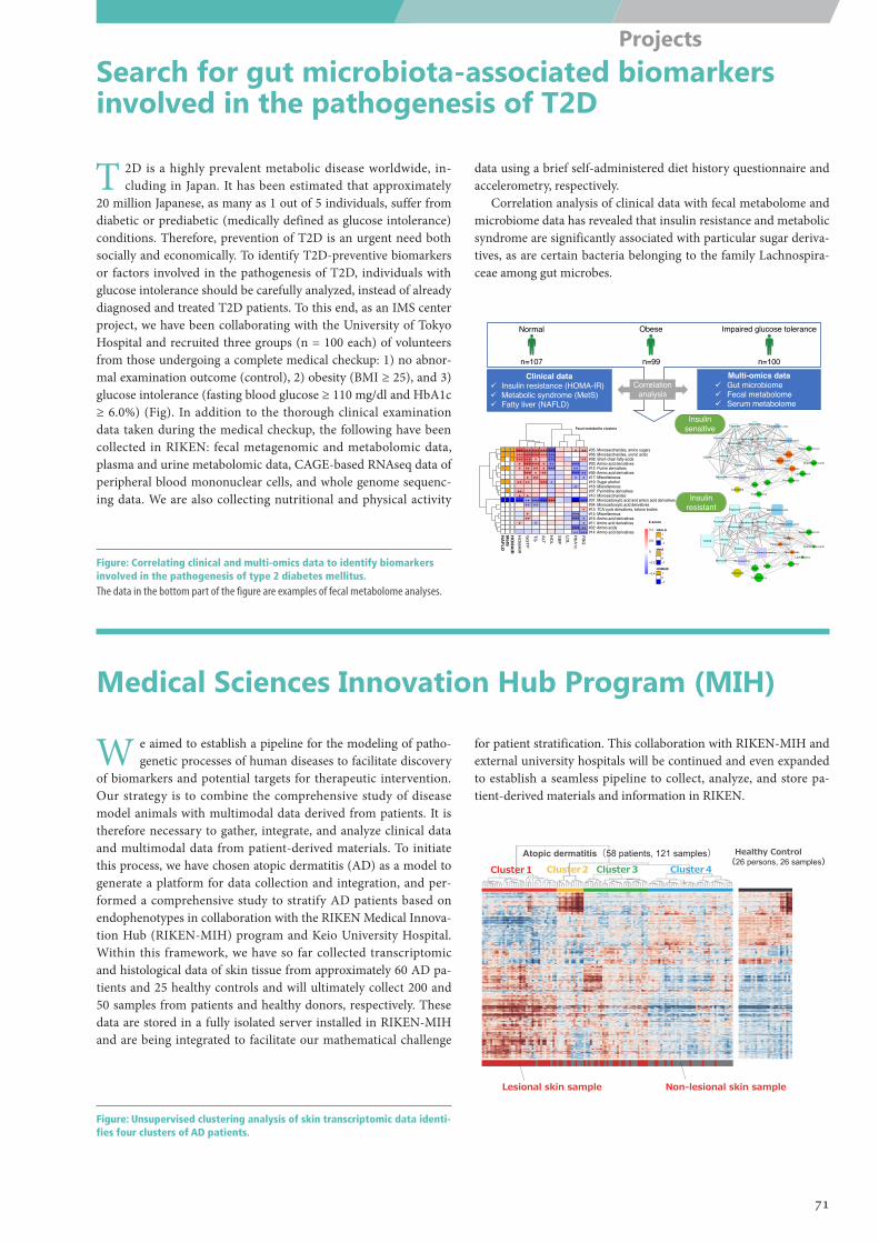
#05: Monosaccharides, amino sugars#06: Monosaccharides, uronic acids#08: Short-chain fatty acids#03: Amino acid derivatives#12: Purine derivatives#09: Amino acid derivatives#17: Miscellaneous#19: Sugar alcohol#18: Miscellaneous#07: Pyrimidine derivatives#10: Monosaccharides#01: Monocarboxylic acid and amino acid derivatives#04: Monocarboxylic acid derivatives#13: TCA cycle derivatives, ketone bodies#15: Miscellaneous#16: Amino acid derivatives#11: Amino acid derivatives#02: Amino acids#14: Amino acid derivatives
+++++++++
++
++
+++
+++ +
++++++++++++++
++++
++ +
++++ +
++
++++++
+++++++
++
+++++ +
++++++
++
++
+++
+++
+++++++++++
+++ +
+++
+
+++++
++++ +
+++
++++++
+++++
++
++
+++
+++ + ++
+++++H
OM
AIR
GG
TP
TG ALT
HD
L
SB
P
LDL
HbA
1c
FBG
ME05ME06ME08ME03ME12ME09ME17ME19ME18ME07ME10ME01ME04ME13ME15ME16ME11ME02ME14H
OMAIR
MetS
NAFLD
NAFLD10−1
MetS10−1
HOMAIR10−1
−0.4
−0.2
0
0.2
0.4
Normal Obese Impaired glucose tolerance
Clinical dataü Insulin resistance (HOMA-IR)ü Metabolic syndrome (MetS)ü Fatty liver (NAFLD)
Multi-omics dataü Gut microbiomeü Fecal metabolomeü Serum metabolome
n=107 n=99 n=100
CoprococcusCollinsella
Glucosamine
Dorea
Mannose
Blautia
Parabacteroides
AlloseXylose
N-Acetylmannosamine
Fucose
Sorbose
Rhamnose
FructoseGlucuronic acid
Glucose
TagatoseGalactose
Arabinose
Lyxose
Galacturonic acid
Bacteroides
Lachnospira
Faecalibacterium
Alistipes
Subdoligranulum
Flavonifractor
CoprococcusCollinsella
Glucosamine
Blautia
Mannose
Dorea
XyloseFucose
Allose
N-AcetylmannosamineSorbose
Parabacteroides
Lyxose
Glucose
Tagatose
Arabinose
Galacturonic acid
Fructose
Galactose
Rhamnose
Subdoligranulum
Glucuronic acid
Bacteroides
Lachnospira
Alistipes
Faecalibacterium
Flavonifractor
Insulin sensitive
Insulin resistant
+++++++++
++
++
+++
+++ +
++++++++++++++
++++
++ +
++++ +
++
++++++
+++++++
++
+++++ +
++++++
++
++
+++
+++
+++++++++++
+++ +
+++
+
+++++
++++ +
+++
++++++
+++++
++
++
+++
+++ + ++
+++++HO
MAIR
GG
TP
TG ALT
HDL
SBP
LDL
HbA1c
FBG
ME05ME06ME08ME03ME12ME09ME17ME19ME18ME07ME10ME01ME04ME13ME15ME16ME11ME02ME14HO
MAIR
MetS
NAFLD
NAFLD10−1
MetS10−1
HOMAIR10−1
−0.4
−0.2
0
0.2
0.4
Z-score
Correlation analysis
Fecal metabolite clusters
Cluster1 Cluster2 Cluster3 Cluster4
Healthy Control(26 persons, 26 samples)
Atopic dermatitis(58 patients, 121 samples)
Lesional skin sample Non-lesional skin sample
Search for gut microbiota-associated biomarkers involved in the pathogenesis of T2D
Medical Sciences Innovation Hub Program (MIH)
T 2D is a highly prevalent metabolic disease worldwide, in-cluding in Japan. It has been estimated that approximately
20 million Japanese, as many as 1 out of 5 individuals, suffer from diabetic or prediabetic (medically defined as glucose intolerance) conditions. Therefore, prevention of T2D is an urgent need both socially and economically. To identify T2D-preventive biomarkers or factors involved in the pathogenesis of T2D, individuals with glucose intolerance should be carefully analyzed, instead of already diagnosed and treated T2D patients. To this end, as an IMS center project, we have been collaborating with the University of Tokyo Hospital and recruited three groups (n = 100 each) of volunteers from those undergoing a complete medical checkup: 1) no abnor-mal examination outcome (control), 2) obesity (BMI ≥ 25), and 3) glucose intolerance (fasting blood glucose ≥ 110 mg/dl and HbA1c ≥ 6.0%) (Fig). In addition to the thorough clinical examination data taken during the medical checkup, the following have been collected in RIKEN: fecal metagenomic and metabolomic data, plasma and urine metabolomic data, CAGE-based RNAseq data of peripheral blood mononuclear cells, and whole genome sequenc-ing data. We are also collecting nutritional and physical activity
data using a brief self-administered diet history questionnaire and accelerometry, respectively.
Correlation analysis of clinical data with fecal metabolome and microbiome data has revealed that insulin resistance and metabolic syndrome are significantly associated with particular sugar deriva-tives, as are certain bacteria belonging to the family Lachnospira-ceae among gut microbes.
W e aimed to establish a pipeline for the modeling of patho-genetic processes of human diseases to facilitate discovery
of biomarkers and potential targets for therapeutic intervention. Our strategy is to combine the comprehensive study of disease model animals with multimodal data derived from patients. It is therefore necessary to gather, integrate, and analyze clinical data and multimodal data from patient-derived materials. To initiate this process, we have chosen atopic dermatitis (AD) as a model to generate a platform for data collection and integration, and per-formed a comprehensive study to stratify AD patients based on endophenotypes in collaboration with the RIKEN Medical Innova-tion Hub (RIKEN-MIH) program and Keio University Hospital. Within this framework, we have so far collected transcriptomic and histological data of skin tissue from approximately 60 AD pa-tients and 25 healthy controls and will ultimately collect 200 and 50 samples from patients and healthy donors, respectively. These data are stored in a fully isolated server installed in RIKEN-MIH and are being integrated to facilitate our mathematical challenge
for patient stratification. This collaboration with RIKEN-MIH and external university hospitals will be continued and even expanded to establish a seamless pipeline to collect, analyze, and store pa-tient-derived materials and information in RIKEN.
Figure: Correlating clinical and multi-omics data to identify biomarkers involved in the pathogenesis of type 2 diabetes mellitus. The data in the bottom part of the figure are examples of fecal metabolome analyses.
Figure: Unsupervised clustering analysis of skin transcriptomic data identi-fies four clusters of AD patients.
71
Projects
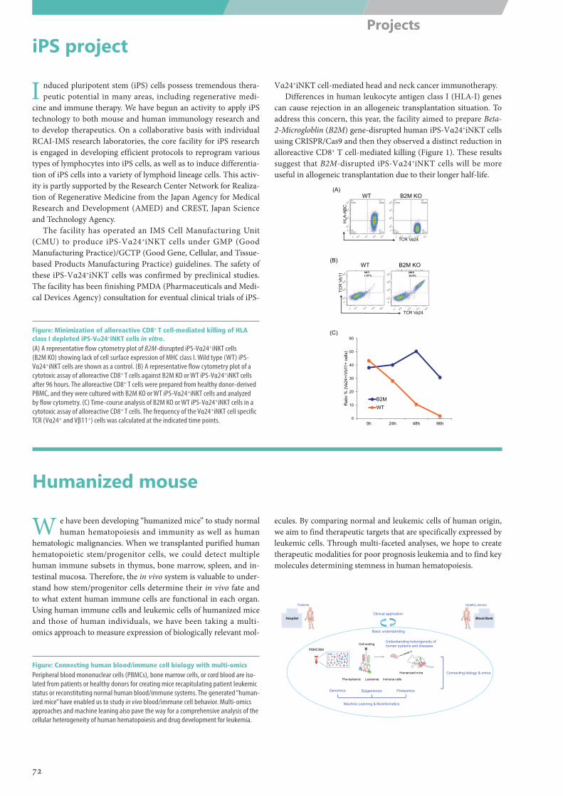
TCR Va24
HLA
-AB
C
WT B2M KO(A)
B2M KOWT
TCR Va24
TCR
Vb1
1
(B)
0
10
20
30
40
50
60
0h 24h 48h 96h
Rat
io %
(Va2
4+/V
b11+
cel
ls)
B2MWT
(C)
Patients
Hospital
Genomics Epigenomics Proteomics
Machine Learning & Bioinformatics
Connecting biology & omics
Healthy donors
Blood BankClinical application
Basic understanding
Understanding heterogeneity of human systems and diseases
Humanized mice
PBMC/BM
Cell sorting
Pre-leukemic Leukemia Immune cells
iPS project
Humanized mouse
I nduced pluripotent stem (iPS) cells possess tremendous thera-peutic potential in many areas, including regenerative medi-
cine and immune therapy. We have begun an activity to apply iPS technology to both mouse and human immunology research and to develop therapeutics. On a collaborative basis with individual RCAI-IMS research laboratories, the core facility for iPS research is engaged in developing efficient protocols to reprogram various types of lymphocytes into iPS cells, as well as to induce differentia-tion of iPS cells into a variety of lymphoid lineage cells. This activ-ity is partly supported by the Research Center Network for Realiza-tion of Regenerative Medicine from the Japan Agency for Medical Research and Development (AMED) and CREST, Japan Science and Technology Agency.
The facility has operated an IMS Cell Manufacturing Unit (CMU) to produce iPS-Vα24+iNKT cells under GMP (Good Manufacturing Practice)/GCTP (Good Gene, Cellular, and Tissue-based Products Manufacturing Practice) guidelines. The safety of these iPS-Vα24+iNKT cells was confirmed by preclinical studies. The facility has been finishing PMDA (Pharmaceuticals and Medi-cal Devices Agency) consultation for eventual clinical trials of iPS-
Vα24+iNKT cell-mediated head and neck cancer immunotherapy.Differences in human leukocyte antigen class I (HLA-I) genes
can cause rejection in an allogeneic transplantation situation. To address this concern, this year, the facility aimed to prepare Beta-2-Microgloblin (B2M) gene-disrupted human iPS-Vα24+iNKT cells using CRISPR/Cas9 and then they observed a distinct reduction in alloreactive CD8+ T cell-mediated killing (Figure 1). These results suggest that B2M-disrupted iPS-Vα24+iNKT cells will be more useful in allogeneic transplantation due to their longer half-life.
W e have been developing “humanized mice” to study normal human hematopoiesis and immunity as well as human
hematologic malignancies. When we transplanted purified human hematopoietic stem/progenitor cells, we could detect multiple human immune subsets in thymus, bone marrow, spleen, and in-testinal mucosa. Therefore, the in vivo system is valuable to under-stand how stem/progenitor cells determine their in vivo fate and to what extent human immune cells are functional in each organ. Using human immune cells and leukemic cells of humanized mice and those of human individuals, we have been taking a multi-omics approach to measure expression of biologically relevant mol-
ecules. By comparing normal and leukemic cells of human origin, we aim to find therapeutic targets that are specifically expressed by leukemic cells. Through multi-faceted analyses, we hope to create therapeutic modalities for poor prognosis leukemia and to find key molecules determining stemness in human hematopoiesis.
Figure: Minimization of alloreactive CD8+ T cell-mediated killing of HLA class I depleted iPS-Vα24+iNKT cells in vitro.(A) A representative flow cytometry plot of B2M-disrupted iPS-Vα24+iNKT cells (B2M KO) showing lack of cell surface expression of MHC class I. Wild type (WT) iPS-Vα24+iNKT cells are shown as a control. (B) A representative flow cytometry plot of a cytotoxic assay of alloreactive CD8+ T cells against B2M KO or WT iPS-Vα24+iNKT cells after 96 hours. The alloreactive CD8+ T cells were prepared from healthy donor-derived PBMC, and they were cultured with B2M KO or WT iPS-Vα24+iNKT cells and analyzed by flow cytometry. (C) Time-course analysis of B2M KO or WT iPS-Vα24+iNKT cells in a cytotoxic assay of alloreactive CD8+ T cells. The frequency of the Vα24+iNKT cell specific TCR (Vα24+ and Vβ11+) cells was calculated at the indicated time points.
Figure: Connecting human blood/immune cell biology with multi-omicsPeripheral blood mononuclear cells (PBMCs), bone marrow cells, or cord blood are iso-lated from patients or healthy donors for creating mice recapitulating patient leukemic status or reconstituting normal human blood/immune systems. The generated “human-ized mice” have enabled us to study in vivo blood/immune cell behavior. Multi-omics approaches and machine leaning also pave the way for a comprehensive analysis of the cellular heterogeneity of human hematopoiesis and drug development for leukemia.
72
Projects
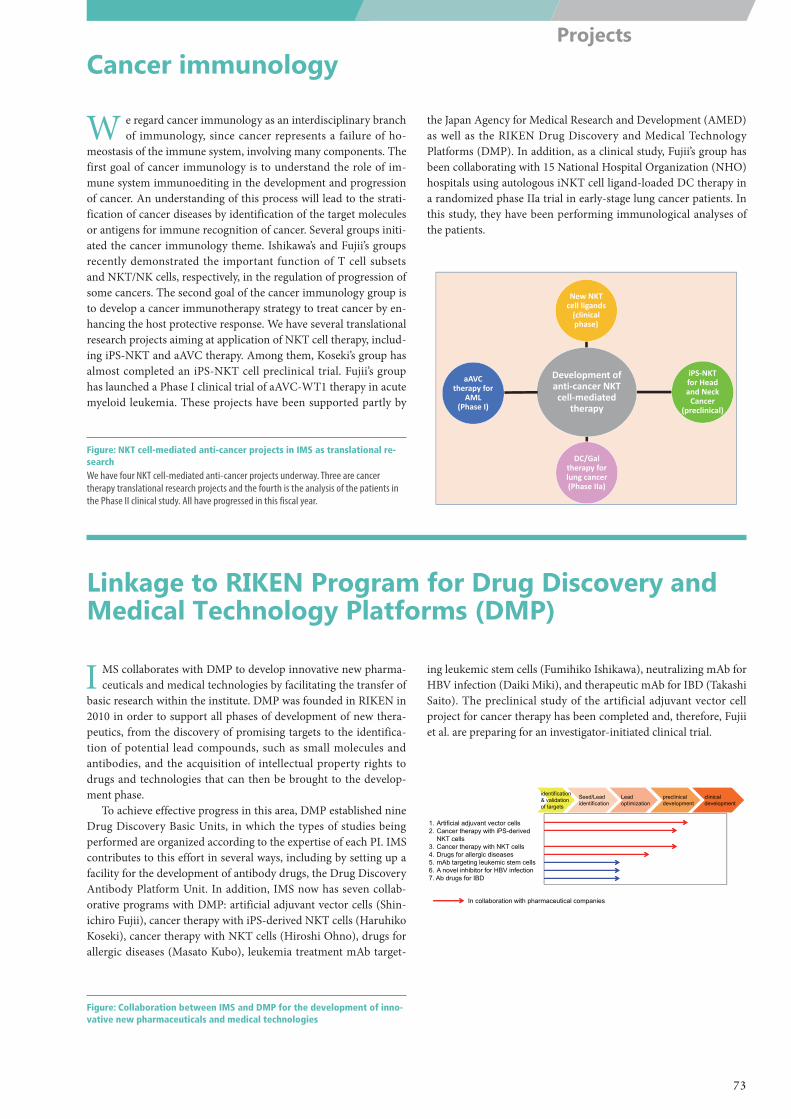
Development of anti‐cancer NKT cell‐mediated
therapy
New NKT cell ligands(clinical phase)
iPS‐NKTfor Head and Neck Cancer
(preclinical)
DC/Gal therapy for lung cancer (Phase IIa)
aAVCtherapy for
AML(Phase I)
1. Artificial adjuvant vector cells 2. Cancer therapy with iPS-derived
NKT cells 3. Cancer therapy with NKT cells 4. Drugs for allergic diseases 5. mAb targeting leukemic stem cells6. A novel inhibitor for HBV infection7. Ab drugs for IBD
identification& validation of targets
Seed/Leadidentification
Leadoptimization
preclinicaldevelopmentpreclinicaldevelopment
clinical development
In collaboration with pharmaceutical companies
Cancer immunology
Linkage to RIKEN Program for Drug Discovery and Medical Technology Platforms (DMP)
W e regard cancer immunology as an interdisciplinary branch of immunology, since cancer represents a failure of ho-
meostasis of the immune system, involving many components. The first goal of cancer immunology is to understand the role of im-mune system immunoediting in the development and progression of cancer. An understanding of this process will lead to the strati-fication of cancer diseases by identification of the target molecules or antigens for immune recognition of cancer. Several groups initi-ated the cancer immunology theme. Ishikawa’s and Fujii’s groups recently demonstrated the important function of T cell subsets and NKT/NK cells, respectively, in the regulation of progression of some cancers. The second goal of the cancer immunology group is to develop a cancer immunotherapy strategy to treat cancer by en-hancing the host protective response. We have several translational research projects aiming at application of NKT cell therapy, includ-ing iPS-NKT and aAVC therapy. Among them, Koseki’s group has almost completed an iPS-NKT cell preclinical trial. Fujii’s group has launched a Phase I clinical trial of aAVC-WT1 therapy in acute myeloid leukemia. These projects have been supported partly by
the Japan Agency for Medical Research and Development (AMED) as well as the RIKEN Drug Discovery and Medical Technology Platforms (DMP). In addition, as a clinical study, Fujii’s group has been collaborating with 15 National Hospital Organization (NHO) hospitals using autologous iNKT cell ligand-loaded DC therapy in a randomized phase IIa trial in early-stage lung cancer patients. In this study, they have been performing immunological analyses of the patients.
I MS collaborates with DMP to develop innovative new pharma-ceuticals and medical technologies by facilitating the transfer of
basic research within the institute. DMP was founded in RIKEN in 2010 in order to support all phases of development of new thera-peutics, from the discovery of promising targets to the identifica-tion of potential lead compounds, such as small molecules and antibodies, and the acquisition of intellectual property rights to drugs and technologies that can then be brought to the develop-ment phase.
To achieve effective progress in this area, DMP established nine Drug Discovery Basic Units, in which the types of studies being performed are organized according to the expertise of each PI. IMS contributes to this effort in several ways, including by setting up a facility for the development of antibody drugs, the Drug Discovery Antibody Platform Unit. In addition, IMS now has seven collab-orative programs with DMP: artificial adjuvant vector cells (Shin-ichiro Fujii), cancer therapy with iPS-derived NKT cells (Haruhiko Koseki), cancer therapy with NKT cells (Hiroshi Ohno), drugs for allergic diseases (Masato Kubo), leukemia treatment mAb target-
ing leukemic stem cells (Fumihiko Ishikawa), neutralizing mAb for HBV infection (Daiki Miki), and therapeutic mAb for IBD (Takashi Saito). The preclinical study of the artificial adjuvant vector cell project for cancer therapy has been completed and, therefore, Fujii et al. are preparing for an investigator-initiated clinical trial.
Figure: NKT cell-mediated anti-cancer projects in IMS as translational re-searchWe have four NKT cell-mediated anti-cancer projects underway. Three are cancer therapy translational research projects and the fourth is the analysis of the patients in the Phase II clinical study. All have progressed in this fiscal year.
Figure: Collaboration between IMS and DMP for the development of inno-vative new pharmaceuticals and medical technologies
73
Projects
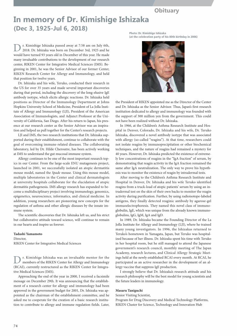
Photo: Dr. Kimishige Ishizaka (at the celebration party of his 80th birthday in 2006)
In memory of Dr. Kimishige Ishizaka (Dec 3, 1925-Jul 6, 2018)
D r. Kimishige Ishizaka passed away at 7:58 am on July 6th, 2018. Dr. Ishizaka was born on December 3rd, 1925 and he
would have turned 93 years old in December of this year. He made many invaluable contributions to the development of our research center, RIKEN Center for Integrative Medical Sciences (IMS). Be-ginning in 2001, he was the Senior Advisor of our former Center, RIKEN Research Center for Allergy and Immunology, and held that position for twelve years.
Dr. Ishizaka and his wife, Teruko, conducted their research in the US for over 35 years and made several important discoveries during that period, including the discovery of the long-elusive IgE antibody isotype, which elicits allergic reactions. Dr. Ishizaka held positions as Director of the Immunology Department at Johns Hopkins University School of Medicine, President of La Jolla Insti-tute of Allergy and Immunology (LJI), President of the American Association of Immunologists, and Adjunct Professor at the Uni-versity of California, San Diego. After his return to Japan, his pres-ence at our research center as the Senior Advisor was an inspira-tion and helped us pull together for the Center’s research projects.
LJI and IMS, the two research institutions that Dr. Ishizaka sup-ported during their establishment, continue to collaborate with the goal of overcoming immune-related diseases. The collaborating laboratory, led by Dr. Hilde Cheroutre, has been actively working at IMS to understand the gut mucosal immune system.
Allergy continues to be one of the most important research top-ics in our Center. From the large-scale ENU mutagenesis project, launched in 2001, we successfully isolated an atopic dermatitis mouse model, named the Spade mouse. Using this mouse model, multiple laboratories in the Center and clinical dermatologists at university hospitals collaborate for the elucidation of atopic dermatitis pathogenesis. IMS allergy research has expanded to be-come a multidisciplinary project involving immunology, genomics, epigenetics, neuroscience, informatics, and clinical medicine. In addition, young researchers are pioneering new concepts for the regulation of asthma and other allergic diseases by the innate im-mune system.
The scientific discoveries that Dr. Ishizaka left us, and his strict but collaborative attitude toward science, will continue to remain in our hearts and inspire us forever.
Tadashi YamamotoDirector,RIKEN Center for Integrative Medical Sciences
D r. Kimishige Ishizaka was an invaluable mentor for the members of the RIKEN Center for Allergy and Immunology
(RCAI), currently restructured as the RIKEN Center for Integra-tive Medical Sciences (IMS).
Approaching the end of the year in 2000, I received a facsimile message on December 29th. It was announcing that the establish-ment of a research center for allergy and immunology had been approved in the government budget for 2001, Dr. Ishizaka was ap-pointed as the chairman of the establishment committee, and he asked me to cooperate for the creation of a basic research institu-tion to contribute to allergy and immune regulation fields. Later,
the President of RIKEN appointed me as the Director of the Center and Dr. Ishizaka as the Senior Advisor. Thus, Japan’s first research institution dedicated to allergy and immunology was founded with the support of 300 million yen from the government. This could not have been realized without Dr. Ishizaka.
In 1966, at the Children’s Asthma Research Institute and Hos-pital in Denver, Colorado, Dr. Ishizaka and his wife, Dr. Teruko Ishizaka, discovered a novel antibody isotype that was associated with allergy (so-called “reagins”). At that time, researchers could not isolate reagins by immunoprecipitation or other biochemical techniques, and the nature of reagins had remained a mystery for 40 years. However, Dr. Ishizaka predicted the existence of extreme-ly low concentrations of reagins in the “IgA fraction” of serum, by demonstrating that reagin activity in the IgA fraction remained the same after IgA neutralization. The only way to prove his hypoth-esis was to monitor the existence of reagin by intradermal tests.
After moving to the Children’s Asthma Research Institute and Hospital in Denver, Dr. Ishizaka and his wife Teruko identified reagins from a truck-load of atopic patients’ serum by using an in-tradermal test on the skin of their own backs to monitor the reagin activity during purification. Further, by using radioisotope-labeled antigens, they finally detected reaginic antibody by agarose-gel immuoelectrophoresis. They named this novel class of immuno-globulin, IgE, which was unique from the already known immuno-globulins, IgG, IgM, IgA and IgD.
In 1989, Dr. Ishizaka became the Founding Director of the La Jolla Institute for Allergy and Immunology (LJI), where he trained many young investigators. In 1996, the Ishizakas returned to Teruko’s hometown in Yamagata, Japan, but Teruko was hospital-ized because of her illness. Dr. Ishizaka spent his time with Teruko in her hospital room, but he still managed to attend the Japanese government’s research council, monthly meeting of The Japan Academy, research lectures, and Clinical Allergy Strategic Meet-ings held at the newly established RCAI every month. At RCAI, he participated as an active researcher in the development of an al-lergy vaccine that suppress IgE production.
I strongly believe that Dr. Ishizaka’s research attitude and his research philosophy will be the best model for young scientists and the future leaders in immunology.
Masaru TaniguchiSenior Visiting Scientist, Program for Drug Discovery and Medical Technology Platforms, RIKEN Cluster for Science, Technology and Innovation Hub
74
Obituary

Part 4
Events
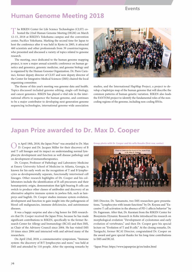
Group photo with Dr. Cooper and his wife Rosalie (first row, center)
Human Genome Meeting 2018
T he RIKEN Center for Life Science Technologies (CLST) co-hosted the 22nd Human Genome Meeting (HGM) on March
12-15, 2018 at RIKEN’s Yokohama campus and the convention center, Pacifico Yokohama. Marking the second time for Japan to host the conference after it was held in Kyoto in 2005, it attracted 460 scientists and other professionals from 39 countries/regions, who presented and discussed a variety of topics related to genome research.
The meeting, once dedicated to the human genome mapping project, is now a major annual scientific conference on human ge-netics and genomics, genomic medicine, and genome biology and is organized by the Human Genome Organization. Dr. Piero Carn-inci, former deputy director of CLST and now deputy director of the Center for Integrative Medical Sciences (IMS) chaired the local organizing committee.
The theme of this year’s meeting was genome data and health. Topics discussed included genome editing, single-cell biology, and cancer genomics. RIKEN has played a vital role in the inter-national efforts to sequence the human genome, and continues to be a major contributor to developing next-generation genome sequencing technologies, international genome-wide association
studies, and the International HapMap Project, a project to de-velop a haplotype map of the human genome that will describe the common patterns of human genetic variation. RIKEN also leads the FANTOM project to identify the fundamental roles of the non-coding regions of the genome, including non-coding RNAs.
Japan Prize awarded to Dr. Max D. Cooper
O n April 18th, 2018, the Japan Prize* was awarded to Dr. Max D. Cooper and Dr. Jacques Miller for their discovery of B
and T cell lineages and its impact on understanding normal lym-phocyte development and function as well disease pathology and on development of immunotherapeutics.
Dr. Cooper, Professor of Pathology and Laboratory Medicine at Emory University School of Medicine in Atlanta, Georgia, is known for his early work on the recognition of T and B lympho-cytes as developmentally separate, functionally intertwined cell lineages. Other research highlights of Dr. Cooper and his col-laborators include the identification of B cell precursors and their hematopoietic origin, demonstration that IgM-bearing B cells can switch to produce other classes of antibodies and discovery of an alternative adaptive immune system in jawless fish, such as lam-preys and hagfish. Dr. Cooper studies immune system evolution, development and function to gain insight into the pathogenesis of blood cell malignancies, immune deficiencies, and autoimmune diseases.
It was a happy surprise and also a big honor for IMS research-ers that Dr. Cooper received the Japan Prize, because he has made significant contributions to RIKEN, specifically to the former Re-search Center for Allergy and Immunology (RCAI) and then IMS, as Chair of the Advisory Council since 2004. He has visited IMS 10 times since 2004 and interacted with and advised many of the researchers.
On April 23rd, 2018, a commemorative symposium, “Immune system: the discovery of B/T lymphocytes and more,” was held at IMS and attended by 110 people. After the opening remarks by
IMS Director, Dr. Yamamoto, two IMS researchers gave presenta-tions, “Lymphocytes with innate functions” by Dr. Koyasu and “Ex-cessive T cell activation in the absence of PD-1 affects behavior” by Dr. Fagarasan. After that, Dr. Kuratani from the RIKEN Center for Biosystems Dynamic Research in Kobe introduced his research on morphological evolution “Development of cyclostomes and early evolution of vertebrates,” and then Dr. Cooper gave his special lecture on “Evolution of T and B cells.” At the closing remarks, Dr. Taniguchi, former RCAI Director, congratulated Dr. Cooper on the award and gave special thanks for his long-time contributions to IMS and RCAI.
*Japan Prize: https://www.japanprize.jp/en/index.html
76
Events
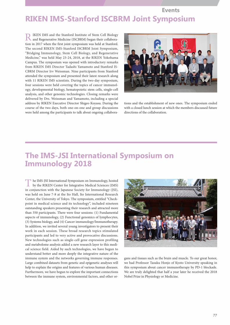
RIKEN IMS-Stanford ISCBRM Joint Symposium
The IMS-JSI International Symposium on Immunology 2018
R IKEN IMS and the Stanford Institute of Stem Cell Biology and Regenerative Medicine (ISCBRM) began their collabora-
tion in 2017 when the first joint symposium was held at Stanford. The second RIKEN IMS-Stanford ISCBRM Joint Symposium, “Bridging Immunology, Stem Cell Biology, and Regenerative Medicine,” was held May 23-24, 2018, at the RIKEN Yokohama Campus. The symposium was opened with introductory remarks from RIKEN IMS Director Tadashi Yamamoto and Stanford IS-CBRM Director Irv Weissman. Nine participants from Stanford attended the symposium and presented their latest research along with 11 RIKEN IMS scientists. During the two-day symposium, four sessions were held covering the topics of cancer immunol-ogy, developmental biology, hematopoietic stem cells, single-cell analysis, and other genomic technologies. Closing remarks were delivered by Drs. Weissman and Yamamoto, including a special address by RIKEN Executive Director Shigeo Koyasu. During the course of the two days, both one-on-one and group discussions were held among the participants to talk about ongoing collabora-
tions and the establishment of new ones. The symposium ended with a closed lunch session at which the members discussed future directions of the collaboration.
T he IMS-JSI International Symposium on Immunology, hosted by the RIKEN Center for Integrative Medical Sciences (IMS)
in conjunction with the Japanese Society for Immunology (JSI), was held on June 7-8 at the Ito Hall, Ito International Research Center, the University of Tokyo. The symposium, entitled “Check-point in medical science and its technology”, included nineteen outstanding speakers presenting their research and attracted more than 350 participants. There were four sessions: (1) Fundamental aspects of immunology, (2) Functional genomics of lymphocytes, (3) Systems biology, and (4) Cancer immunology/Immunotherapy. In addition, we invited several young investigators to present their work in each session. These broad research topics stimulated participants and led to very active and provocative discussions. New technologies such as single-cell gene expression profiling and metabolome analysis added a new research layer to this medi-cal science field. Aided by such technologies, we have begun to understand better and more deeply the integrative nature of the immune system and the networks governing immune responses. Large combined datasets from genetic and epigenetic analyses will help to explain the origins and features of various human diseases. Furthermore, we have begun to explore the important connections between the immune system, environmental factors, and other or-
gans and tissues such as the brain and muscle. To our great honor, we had Professor Tasuku Honjo of Kyoto University speaking in this symposium about cancer immunotherapy by PD-1 blockade. We are truly delighted that half a year later he received the 2018 Nobel Prize in Physiology or Medicine.
77
Events
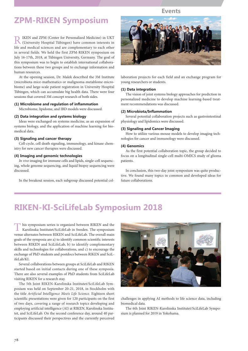
ZPM-RIKEN Symposium
RIKEN-KI-SciLifeLab Symposium 2018
R IKEN and ZPM (Center for Personalized Medicine) in UKT (University Hospital Tübingen) have common interests in
life and medical sciences and are complementary to each other in several fields. We held the first ZPM-RIKEN symposium on July 16-17th, 2018, at Tübingen University, Germany. The goal of this symposium was to begin to establish international collabora-tions between these two groups and to exchange information and human resources.
At the opening session, Dr. Malek described the 3M Institute (microbiota-mice-mathematics or malignoma-metablome-micro-biome) and large-scale patient registration in University Hospital Tübingen, which can accumulate big health data. There were four sessions that covered 3M concept research of both sides.
(1) Microbiome and regulation of inflammationMicrobiome, lipidome, and IBD models were discussed.
(2) Data integration and systems biologyIdeas were exchanged on systems medicine, as an expansion of
systems biology, and the application of machine learning for bio-medical data.
(3) Signaling and cancer therapyCell-cycle, cell death signaling, immunology, and kinase chem-
istry for new cancer therapies were discussed.
(4) Imaging and genomic technologiesIn vivo imaging for immune cells and lipids, single-cell sequenc-
ing, whole genome sequencing, and liquid biopsy sequencing were discussed.
In the breakout session, each subgroup discussed potential col-
laboration projects for each field and an exchange program for young researchers or students.
(1) Data integrationThe vision of joint systems biology approaches for prediction in
personalized medicine to develop machine learning-based treat-ment recommendations was discussed.
(2) Microbiota/InflammationSeveral potential collaboration projects such as gastrointestinal
physiology and lipidomics were discussed.
(3) Signaling and Cancer ImagingHow to utilize various mouse models to develop imaging tech-
nologies for cancer and immunology were discussed.
(4) GenomicsAs the first potential collaboration topic, the group decided to
focus on a longitudinal single-cell multi-OMICS study of glioma patients.
In conclusion, this two-day joint symposium was quite produc-tive. We found many topics in common and developed ideas for future collaborations.
T his symposium series is organized between RIKEN and the Karolinska Institutet/SciLifeLab in Sweden. The symposium
venue alternates between RIKEN and SciLifeLab. The overall main goals of the symposia are a) to identify common scientific interests between RIKEN and SciLifeLab, b) to identify complementary skills and technologies for collaborations, and c) to encourage the exchange of PhD students and postdocs between RIKEN and SciL-ifeLab/KI.
Several collaborations between groups at SciLifeLab and RIKEN started based on initial contacts during one of these symposia. There are also several examples of PhD students from SciLifeLab visiting RIKEN for a research stay.
The 5th Joint RIKEN-Karolinska Institutet/SciLifeLab Sym-posium was held on September 20-21, 2018, in Stockholm with the title Artificial Intelligence Meets Life Science. Eighteen short scientific presentations were given for 120 participants on the first of two days, covering a range of research topics developing and employing artificial intelligence (AI) at RIKEN, Karolinska Institu-tet, and SciLifeLab. On the second conference day, around 40 par-ticipants discussed their perspectives and the currently perceived
challenges in applying AI methods to life science data, including biomedical data.
The 6th Joint RIKEN-Karolinska Institutet/SciLifeLab Sympo-sium is planned for 2019 in Yokohama.
78
Events
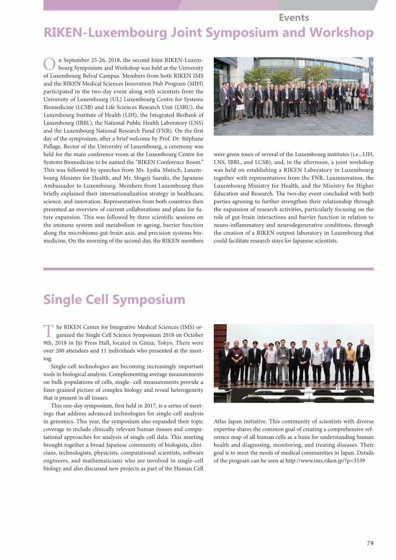
RIKEN-Luxembourg Joint Symposium and Workshop
Single Cell Symposium
O n September 25-26, 2018, the second Joint RIKEN-Luxem-bourg Symposium and Workshop was held at the University
of Luxembourg Belval Campus. Members from both RIKEN IMS and the RIKEN Medical Sciences Innovation Hub Program (MIH) participated in the two-day event along with scientists from the University of Luxembourg (UL) Luxembourg Centre for Systems Biomedicine (LCSB) and Life Sciences Research Unit (LSRU), the Luxembourg Institute of Health (LIH), the Integrated BioBank of Luxembourg (IBBL), the National Public Health Laboratory (LNS) and the Luxembourg National Research Fund (FNR). On the first day of the symposium, after a brief welcome by Prof. Dr. Stéphane Pallage, Rector of the University of Luxembourg, a ceremony was held for the main conference room at the Luxembourg Centre for Systems Biomedicine to be named the “RIKEN Conference Room.” This was followed by speeches from Ms. Lydia Mutsch, Luxem-bourg Minister for Health, and Mr. Shigeji Suzuki, the Japanese Ambassador to Luxembourg. Members from Luxembourg then briefly explained their internationalization strategy in healthcare, science, and innovation. Representatives from both countries then presented an overview of current collaborations and plans for fu-ture expansion. This was followed by three scientific sessions on the immune system and metabolism in ageing, barrier function along the microbiome-gut-brain axis, and precision systems bio-medicine. On the morning of the second day, the RIKEN members
were given tours of several of the Luxembourg institutes (i.e., LIH, LNS, IBBL, and LCSB), and, in the afternoon, a joint workshop was held on establishing a RIKEN Laboratory in Luxembourg together with representatives from the FNR, Luxinnovation, the Luxembourg Ministry for Health, and the Ministry for Higher Education and Research. The two-day event concluded with both parties agreeing to further strengthen their relationship through the expansion of research activities, particularly focusing on the role of gut-brain interactions and barrier function in relation to neuro-inflammatory and neurodegenerative conditions, through the creation of a RIKEN outpost laboratory in Luxembourg that could facilitate research stays for Japanese scientists.
T he RIKEN Center for Integrative Medical Sciences (IMS) or-ganized the Single Cell Science Symposium 2018 on October
9th, 2018 in Jiji Press Hall, located in Ginza, Tokyo. There were over 200 attendees and 11 individuals who presented at the meet-ing.
Single-cell technologies are becoming increasingly important tools in biological analysis. Complementing average measurements on bulk populations of cells, single- cell measurements provide a finer-grained picture of complex biology and reveal heterogeneity that is present in all tissues.
This one-day symposium, first held in 2017, is a series of meet-ings that address advanced technologies for single-cell analysis in genomics. This year, the symposium also expanded their topic coverage to include clinically relevant human tissues and compu-tational approaches for analysis of single-cell data. This meeting brought together a broad Japanese community of biologists, clini-cians, technologists, physicists, computational scientists, software engineers, and mathematicians who are involved in single-cell biology and also discussed new projects as part of the Human Cell
Atlas Japan initiative. This community of scientists with diverse expertise shares the common goal of creating a comprehensive ref-erence map of all human cells as a basis for understanding human health and diagnosing, monitoring, and treating diseases. Their goal is to meet the needs of medical communities in Japan. Details of the program can be seen at http://www.ims.riken.jp/?p=3539
79
Events
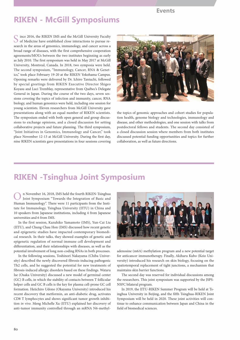
RIKEN - McGill Symposiums
RIKEN -Tsinghua Joint Symposium
S ince 2016, the RIKEN IMS and the McGill University Faculty of Medicine have established close interactions to pursue re-
search in the areas of genomics, immunology, and cancer across a broad range of diseases, with the first comprehensive cooperation agreements/MOUs between the two institutes beginning as early as July 2010. The first symposium was held in May 2017 at McGill University, Montreal, Canada. In 2018, two symposia were held. The second symposium, “Immunology, Cancer, RNA & Genet-ics,” took place February 19-20 at the RIKEN Yokohama Campus. Opening remarks were delivered by Dr. Ichiro Taniuchi, followed by special greetings from RIKEN Executive Director Shigeo Koyasu and Luci Tremblay, representative from Québec’s Delegate General in Japan. During the course of the two days, seven ses-sions covering the topics of infection and immunity, cancer, RNA biology, and human genomics were held, including one session for young scientists. Eleven researchers from McGill University gave presentations along with an equal number of RIKEN scientists. The symposium ended with both open general and group discus-sions to exchange opinions, and a closed discussion for setting collaborative projects and future planning. The third symposium, “Joint Initiatives in Genomics, Immunology and Cancer,” took place November 12-13 at McGill University. During the first day, nine RIKEN scientists gave presentations in four sessions covering
the topics of genomic approaches and cohort studies for popula-tion health, genome biology and technologies, immunology and disease, and other methodologies, and one session with talks from postdoctoral fellows and students. The second day consisted of a closed discussion session where members from both institutes discussed potential funding opportunities and topics for further collaboration, as well as future directions.
O n November 16, 2018, IMS held the fourth RIKEN-Tsinghua Joint Symposium “Towards the Integration of Basic and
Human Immunology”. There were 11 participants from the Insti-tute for Immunology, Tsinghua University (IITU) in China and 10 speakers from Japanese institutions, including 4 from Japanese universities and 6 from IMS.
In the first session, Kazuhiko Yamamoto (IMS), Yun-Cai Liu (IITU), and Chung Chau Hon (IMS) discussed how recent genetic and epigenetic studies have impacted contemporary biomedi-cal research. In their talks, they showed examples of genetic and epigenetic regulation of normal immune cell development and differentiation, and their relationships with diseases, as well as the potential involvement of long non-coding RNAs in both processes.
In the following sessions, Toshinori Nakayama (Chiba Univer-sity) described the newly discovered fibrosis-inducing pathogenic Th2 cells, and he suggested the potential for new treatments of fibrosis-induced allergic disorders based on these findings. Wataru Ise (Osaka University) discussed a new model of germinal center (GC) B cells, in which the stability of contacts between T follicular helper cells and GC B cells is the key for plasma cell-prone GC cell formation. Heiichiro Udono (Okayama University) introduced his recent discovery that metformin, an anti-diabetic drug, activates CD8 T lymphocytes and shows significant tumor growth inhibi-tion in vivo. Meng Michelle Xu (IITU) explained her discovery of anti-tumor immunity controlled through an mRNA N6-methyl-
adenosine (m6A) methylation program and a new potential target for anticancer immunotherapy. Finally, Akiharu Kubo (Keio Uni-versity) introduced his research on skin biology, focusing on the spatiotemporal replacement of tight junctions, a mechanism that maintains skin barrier functions.
The second day was reserved for individual discussions among the researchers. This joint symposium was supported by the JSPS-NSFC bilateral program.
In 2019, the IITU-RIKEN Summer Program will be held at Ts-inghua University in Beijing, and the fifth Tsinghua-RIKEN Joint Symposium will be held in 2020. These joint activities will con-tinue to enhance communication between Japan and China in the field of biomedical sciences.
80
Events
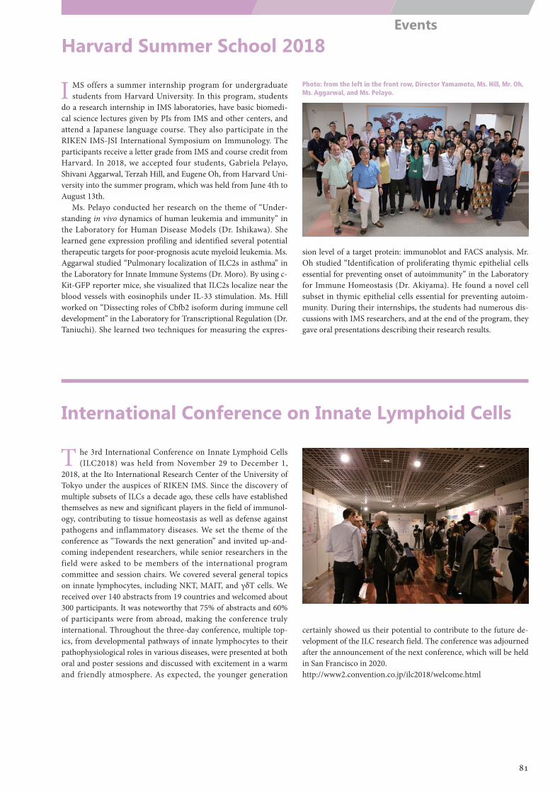
Harvard Summer School 2018
International Conference on Innate Lymphoid Cells
I MS offers a summer internship program for undergraduate students from Harvard University. In this program, students
do a research internship in IMS laboratories, have basic biomedi-cal science lectures given by PIs from IMS and other centers, and attend a Japanese language course. They also participate in the RIKEN IMS-JSI International Symposium on Immunology. The participants receive a letter grade from IMS and course credit from Harvard. In 2018, we accepted four students, Gabriela Pelayo, Shivani Aggarwal, Terzah Hill, and Eugene Oh, from Harvard Uni-versity into the summer program, which was held from June 4th to August 13th.
Ms. Pelayo conducted her research on the theme of “Under-standing in vivo dynamics of human leukemia and immunity” in the Laboratory for Human Disease Models (Dr. Ishikawa). She learned gene expression profiling and identified several potential therapeutic targets for poor-prognosis acute myeloid leukemia. Ms. Aggarwal studied “Pulmonary localization of ILC2s in asthma” in the Laboratory for Innate Immune Systems (Dr. Moro). By using c-Kit-GFP reporter mice, she visualized that ILC2s localize near the blood vessels with eosinophils under IL-33 stimulation. Ms. Hill worked on “Dissecting roles of Cbfb2 isoform during immune cell development” in the Laboratory for Transcriptional Regulation (Dr. Taniuchi). She learned two techniques for measuring the expres-
sion level of a target protein: immunoblot and FACS analysis. Mr. Oh studied “Identification of proliferating thymic epithelial cells essential for preventing onset of autoimmunity” in the Laboratory for Immune Homeostasis (Dr. Akiyama). He found a novel cell subset in thymic epithelial cells essential for preventing autoim-munity. During their internships, the students had numerous dis-cussions with IMS researchers, and at the end of the program, they gave oral presentations describing their research results.
T he 3rd International Conference on Innate Lymphoid Cells (ILC2018) was held from November 29 to December 1,
2018, at the Ito International Research Center of the University of Tokyo under the auspices of RIKEN IMS. Since the discovery of multiple subsets of ILCs a decade ago, these cells have established themselves as new and significant players in the field of immunol-ogy, contributing to tissue homeostasis as well as defense against pathogens and inflammatory diseases. We set the theme of the conference as “Towards the next generation” and invited up-and-coming independent researchers, while senior researchers in the field were asked to be members of the international program committee and session chairs. We covered several general topics on innate lymphocytes, including NKT, MAIT, and γδT cells. We received over 140 abstracts from 19 countries and welcomed about 300 participants. It was noteworthy that 75% of abstracts and 60% of participants were from abroad, making the conference truly international. Throughout the three-day conference, multiple top-ics, from developmental pathways of innate lymphocytes to their pathophysiological roles in various diseases, were presented at both oral and poster sessions and discussed with excitement in a warm and friendly atmosphere. As expected, the younger generation
certainly showed us their potential to contribute to the future de-velopment of the ILC research field. The conference was adjourned after the announcement of the next conference, which will be held in San Francisco in 2020.http://www2.convention.co.jp/ilc2018/welcome.html
Photo: from the left in the front row, Director Yamamoto, Ms. Hill, Mr. Oh, Ms. Aggarwal, and Ms. Pelayo.
81
Events
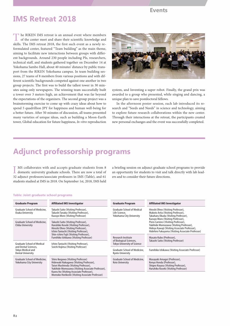
IMS Retreat 2018
Adjunct professorship programs
T he RIKEN IMS retreat is an annual event where members of the center meet and share their scientific knowledge and
skills. The IMS retreat 2018, the first such event as a newly re-formulated center, featured “Team building” as the main theme, aiming to facilitate new interactions between groups with differ-ent backgrounds. Around 230 people including PIs, researchers, technical staff, and students gathered together on December 14 at Yokohama Sambo Hall, about 40 minutes’ distance by public trans-port from the RIKEN Yokohama campus. In team building ses-sions, 27 teams of 8 members from various positions and with dif-ferent scientific backgrounds competed against one another in two group projects. The first was to build the tallest tower in 30 min-utes using only newspapers. The winning team successfully built a tower over 3 meters high, an achievement that was far beyond the expectations of the organizers. The second group project was a brainstorming exercise to come up with crazy ideas about how to spend 5 quadrillion JPY for happiness and human well-being for a better future. After 50 minutes of discussion, all teams presented many varieties of unique ideas, such as building a Moon-Earth tower, Global education for future happiness, In vitro reproduction
system, and Inventing a super robot. Finally, the grand prix was awarded to a group who presented, while singing and dancing, a unique plan to save postdoctoral fellows.
In the afternoon poster session, each lab introduced its re-search and “Seeds and Needs” in science and technology, aiming to explore future research collaborations within the new center. Through their interactions at the retreat, the participants created new personal exchanges and the event was successfully completed.
I MS collaborates with and accepts graduate students from 8 domestic university graduate schools. There are now a total of
32 adjunct professors/associate professors in IMS (Table), and 61 students studied at IMS in 2018. On September 1st, 2018, IMS held
a briefing session on adjunct graduate school programs to provide an opportunity for students to visit and talk directly with lab lead-ers and to consider their future directions.
Table: Joint graduate school programs
Graduate Program Affiliated IMS Investigator
Graduate School of Medicine, Osaka University
Takashi Saito (Visiting Professor), Takashi Tanaka (Visiting Professor), Kazuyo Moro (Visiting Professor)
Graduate School of Medicine, Chiba University
Takashi Saito (Visiting Professor), Haruhiko Koseki (Visiting Professor), Hiroshi Ohno (Visiting Professor), Ichiro Taniuchi (Visiting Professor), Shin-ichiro Fujii (Visiting Professor), Fumihiko Ishikawa (Visiting Professor)
Graduate School of Medical and Dental Sciences, Tokyo Medical and Dental University
Ichiro Taniuchi (Visiting Professor), Soichi Kojima (Visiting Professor)
Graduate School of Medicine, Yokohama City University
Shiro Ikegawa (Visiting Professor) Hidewaki Nakagawa (Visiting Professor), Taisei Mushiroda (Visiting Professor), Yukihide Momozawa (Visiting Associate Professor), Kaoru Ito (Visiting Associate Professor), Momoko Horikoshi (Visiting Associate Professor)
Graduate Program Affiliated IMS Investigator
Graduate School of Medical Life Science, Yokohama City University
Hiroshi Ohno (Visiting Professor), Makoto Arita (Visiting Professor), Takaharu Okada (Visiting Professor), Kazuyo Moro (Visiting Professor), Piero Carninci (Visiting Professor), Yukihide Momozawa (Visiting Professor), Hideya Kawaji (Visiting Associate Professor), Hidehiro Fukuyama (Visiting Associate Professor)
Research Institute of Biological Sciences, Tokyo University of Science
Masato Kubo (Professor), Takashi Saito (Visiting Professor)
Graduate School of Medicine, Kyoto University
Fumihiko Ishikawa (Visiting Associate Professor)
Graduate School of Medicine, Keio University
Masayuki Amagai (Professor), Kenya Honda (Professor), Shigeo Koyasu (Visiting Professor), Haruhiko Koseki (Visiting Professor)
82
Events

Part 5
Data and Statistics
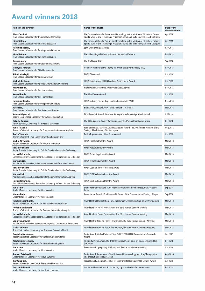
Award winners 2018Name of the awardee Name of the award Date of the
announcement
Piero Carninci, Team Leader, Laboratory for Transcriptome Technology
The Commendation for Science and Technology by the Minister of Education, Culture, Sports, Science and Technology, Prizes for Science and Technology, Research Category
Apr 2018
Hiroshi Ohno, Team Leader, Laboratory for Intestinal Ecosystem
The Commendation for Science and Technology by the Minister of Education, Culture, Sports, Science and Technology, Prizes for Science and Technology, Research Category
Apr 2018
Haruhiko Koseki, Team Leader, Laboratory for Developmental Genetics
55th ERWIN von BALZ PRIZE Nov 2018
Hiroshi Ohno, Team Leader, Laboratory for Intestinal Ecosystem
The Hideyo Noguchi Memorial Award for Medical Science Nov 2018
Kazuyo Moro, Team Leader, Laboratory for Innate Immune Systems
The 8th Nagase Prize Sep 2018
Masayuki Amagai, Team Leader, Laboratory for Skin Homeostasis
Honorary Member of the Society for Investigative Dermatology (SID) Nov 2018
Shin-ichiro Fujii, Team Leader, Laboratory for Immunotherapy
RIKEN Eiho Award Jun 2018
Michiel de Hoon, Team Leader, Laboratory for Applied Computational Genomics
RIKEN Baiho Award (RIKEN Excellent Achievement Award) Jun 2018
Kenya Honda, Team Leader, Laboratory for Gut Homeostasis
Highly Cited Researchers 2018 by Clarivate Analytics Nov 2018
Kenya Honda, Team Leader, Laboratory for Gut Homeostasis
The 87th Kitasato Award Jun 2018
Haruhiko Koseki, Team Leader, Laboratory for Developmental Genetics
RIKEN Industry Partnerships Contribution Award FY2018 Nov 2018
Kaoru Ito, Team Leader, Laboratory for Cardiovascular Diseases
Best Reviewer Award 2017, International Heart Journal Mar 2018
Kosuke Miyauchi, Deputy Team Leader, Laboratory for Cytokine Regulation
2018 Academic Award, Japanese Society of Interferon & Cytokine Research Jul 2018
Takashi Kanaya, Senior Scientist, Laboratory for Intestinal Ecosystem
The 13th Japanese Society for Immunology (JSI) Young Investigator Award Dec 2018
Yuuri Yasuoka, Research Scientist, Laboratory for Comprehensive Genomic Analysis
Young Scientists' Excellent Oral Presentation Award, The 20th Annual Meeting of the Society of Evolutionary Studies, Japan
Aug 2018
Senko Tsukuda, Research Scientist, Liver Cancer Prevention Research Unit
Taisho Toyama Award, Liver Forum Award Jun 2018
Michio Miyajima, Research Scientist, Laboratory for Mucosal Immunity
RIKEN Research Incentive Award Mar 2018
Takahiro Suzuki, Senior Scientist, Laboratory for Cellular Function Conversion Technology
RIKEN Research Incentive Award Mar 2018
Hazuki Takahashi, Special Fixed Term Contract Researcher, Laboratory for Transcriptome Technology
RIKEN Technology Incentive Award Mar 2018
Marina Lizio, Postdoctoral Researcher, Laboratory for Genome Information Analysis
RIKEN Technology Incentive Award Mar 2018
Takahiro Suzuki, Senior Scientist, Laboratory for Cellular Function Conversion Technology
RIKEN CLST Researcher Incentive Award Mar 2018
Marina Lizio, Postdoctoral Researcher, Laboratory for Genome Information Analysis
RIKEN CLST Technician Incentive Award Mar 2018
Hazuki Takahashi, Special Fixed Term Contract Researcher, Laboratory for Transcriptome Technology
RIKEN CLST Technician Incentive Award Mar 2018
Yudai Iino, Student Trainee, Laboratory for Metabolomics
Best Presentation Award, 17th Pharma-Bioforum of the Pharmaceutical Society of Japan
Sep 2018
Mio Yoshida, Student Trainee, Laboratory for Metabolomics
Presentation Award, 17th Pharma-Bioforum of the Pharmaceutical Society of Japan Sep 2018
Joachim Luginbuehl, Research Scientist, Laboratory for Advanced Genomics Circuit
Award for Oral Presentation, The 22nd Human Genome Meeting Trainee Symposium Mar 2018
Jordan Ramilowski, Research Scientist, Laboratory for Genome Information Analysis
Award for Best Poster Presentation, The 22nd Human Genome Meeting Mar 2018
Hazuki Takahashi, Special Fixed Term Contract Researcher, Laboratory for Transcriptome Technology
Award for Best Poster Presentation, The 22nd Human Genome Meeting Mar 2018
Saumya Agrawal, Postdoctoral Researcher, Laboratory for Applied Computational Genomics
Award for Outstanding Poster Presentation, The 22nd Human Genome Meeting Mar 2018
Tsukasa Kouno, Research Associate, Laboratory for Advanced Genomics Circuit
Award for Outstanding Poster Presentation, The 22nd Human Genome Meeting Mar 2018
Yasutaka Motomura, Research Scientist, Laboratory for Innate Immune Systems
Poster Award, Medical Science Prize, FY2017 SPDR&FPR Presentation of research results
Jan 2018
Yasutaka Motomura, Research Scientist, Laboratory for Innate Immune Systems
Immunity Poster Award, The 3rd International Conference on Innate Lymphoid Cells (ILC2018)
Dec 2018
Yudai Iino, Student Trainee, Laboratory for Metabolomics
Poster Award, Lipoquality, JSPS Scientific Research on Innovative Area Jun 2018
Sonoko Takahashi, Student Trainee, Laboratory for Tissue Dynamics
Poster Award, Symposium of the Division of Pharmacology and Drug Therapeutics, Pharmaceutical Society of Japan
Aug 2018
XianYang Qin, Research Scientist, Liver Cancer Prevention Research Unit
Federation of American Societies for Experimental Biology (FASEB), Travel Award Jun 2018
Tadashi Takeuchi, Student Trainee, Laboratory for Intestinal Ecosystem
Ursula and Fritz Melchers Travel Award, Japanese Society for Immunology Dec 2018
84
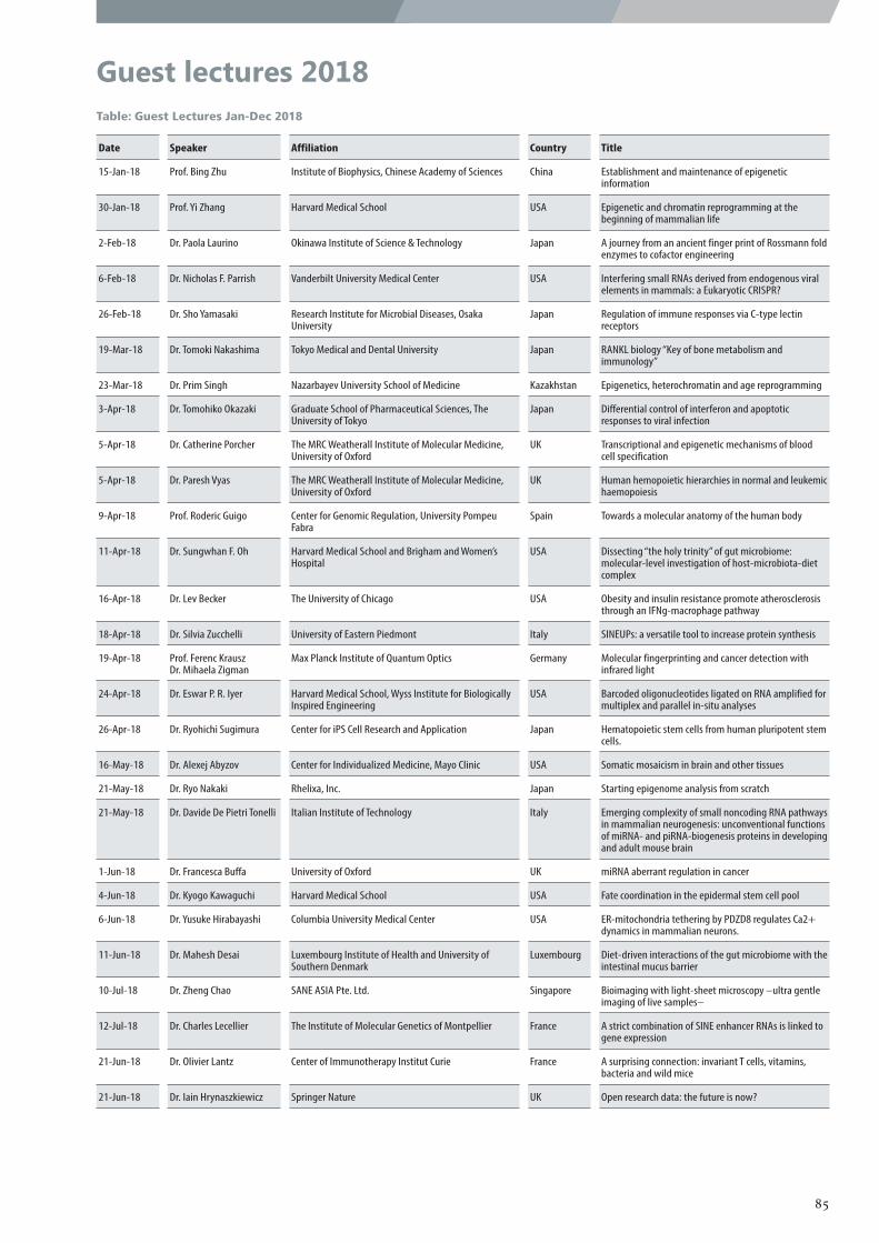
Guest lectures 2018Table: Guest Lectures Jan-Dec 2018
Date Speaker Affiliation Country Title
15-Jan-18 Prof. Bing Zhu Institute of Biophysics, Chinese Academy of Sciences China Establishment and maintenance of epigenetic information
30-Jan-18 Prof. Yi Zhang Harvard Medical School USA Epigenetic and chromatin reprogramming at the beginning of mammalian life
2-Feb-18 Dr. Paola Laurino Okinawa Institute of Science & Technology Japan A journey from an ancient finger print of Rossmann fold enzymes to cofactor engineering
6-Feb-18 Dr. Nicholas F. Parrish Vanderbilt University Medical Center USA Interfering small RNAs derived from endogenous viral elements in mammals: a Eukaryotic CRISPR?
26-Feb-18 Dr. Sho Yamasaki Research Institute for Microbial Diseases, Osaka University
Japan Regulation of immune responses via C-type lectin receptors
19-Mar-18 Dr. Tomoki Nakashima Tokyo Medical and Dental University Japan RANKL biology “Key of bone metabolism and immunology”
23-Mar-18 Dr. Prim Singh Nazarbayev University School of Medicine Kazakhstan Epigenetics, heterochromatin and age reprogramming
3-Apr-18 Dr. Tomohiko Okazaki Graduate School of Pharmaceutical Sciences, The University of Tokyo
Japan Differential control of interferon and apoptotic responses to viral infection
5-Apr-18 Dr. Catherine Porcher The MRC Weatherall Institute of Molecular Medicine, University of Oxford
UK Transcriptional and epigenetic mechanisms of blood cell specification
5-Apr-18 Dr. Paresh Vyas The MRC Weatherall Institute of Molecular Medicine, University of Oxford
UK Human hemopoietic hierarchies in normal and leukemic haemopoiesis
9-Apr-18 Prof. Roderic Guigo Center for Genomic Regulation, University Pompeu Fabra
Spain Towards a molecular anatomy of the human body
11-Apr-18 Dr. Sungwhan F. Oh Harvard Medical School and Brigham and Women’s Hospital
USA Dissecting “the holy trinity” of gut microbiome: molecular-level investigation of host-microbiota-diet complex
16-Apr-18 Dr. Lev Becker The University of Chicago USA Obesity and insulin resistance promote atherosclerosis through an IFNg-macrophage pathway
18-Apr-18 Dr. Silvia Zucchelli University of Eastern Piedmont Italy SINEUPs: a versatile tool to increase protein synthesis
19-Apr-18 Prof. Ferenc Krausz Dr. Mihaela Zigman
Max Planck Institute of Quantum Optics Germany Molecular fingerprinting and cancer detection with infrared light
24-Apr-18 Dr. Eswar P. R. Iyer Harvard Medical School, Wyss Institute for Biologically Inspired Engineering
USA Barcoded oligonucleotides ligated on RNA amplified for multiplex and parallel in-situ analyses
26-Apr-18 Dr. Ryohichi Sugimura Center for iPS Cell Research and Application Japan Hematopoietic stem cells from human pluripotent stem cells.
16-May-18 Dr. Alexej Abyzov Center for Individualized Medicine, Mayo Clinic USA Somatic mosaicism in brain and other tissues
21-May-18 Dr. Ryo Nakaki Rhelixa, Inc. Japan Starting epigenome analysis from scratch
21-May-18 Dr. Davide De Pietri Tonelli Italian Institute of Technology Italy Emerging complexity of small noncoding RNA pathways in mammalian neurogenesis: unconventional functions of miRNA- and piRNA-biogenesis proteins in developing and adult mouse brain
1-Jun-18 Dr. Francesca Buffa University of Oxford UK miRNA aberrant regulation in cancer
4-Jun-18 Dr. Kyogo Kawaguchi Harvard Medical School USA Fate coordination in the epidermal stem cell pool
6-Jun-18 Dr. Yusuke Hirabayashi Columbia University Medical Center USA ER-mitochondria tethering by PDZD8 regulates Ca2+ dynamics in mammalian neurons.
11-Jun-18 Dr. Mahesh Desai Luxembourg Institute of Health and University of Southern Denmark
Luxembourg Diet-driven interactions of the gut microbiome with the intestinal mucus barrier
10-Jul-18 Dr. Zheng Chao SANE ASIA Pte. Ltd. Singapore Bioimaging with light-sheet microscopy –ultra gentle imaging of live samples–
12-Jul-18 Dr. Charles Lecellier The Institute of Molecular Genetics of Montpellier France A strict combination of SINE enhancer RNAs is linked to gene expression
21-Jun-18 Dr. Olivier Lantz Center of Immunotherapy Institut Curie France A surprising connection: invariant T cells, vitamins, bacteria and wild mice
21-Jun-18 Dr. Iain Hrynaszkiewicz Springer Nature UK Open research data: the future is now?
85
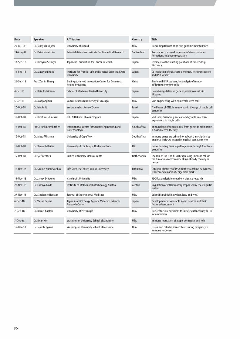
Date Speaker Affiliation Country Title
25-Jul-18 Dr. Takayuki Nojima University of Oxford USA Noncoding transcription and genome maintenance
21-Aug-18 Dr. Patrick Matthias Friedrich Miescher Institute for Biomedical Research Switzerland Acetylation is a novel regulator of stress granules formation and phase separation
13-Sep-18 Dr. Hiroyuki Seimiya Japanese Foundation for Cancer Research Japan Telomere as the starting point of anticancer drug discovery
14-Sep-18 Dr. Masayuki Horie Institute for Frontier Life and Medical Sciences, Kyoto University
Japan Co-evolution of eukaryote genomes, retrotransposons and RNA viruses
26-Sep-18 Prof. Zemin Zhang Beijing Advanced Innovation Center for Genomics, Peking University
China Single cell RNA sequencing analysis of tumor-infiltrating immune cells
4-Oct-18 Dr. Keisuke Nimura School of Medicine, Osaka University Japan How dysregulation of gene expression results in diseases
5-Oct-18 Dr. Xiaoyang Wu Cancer Research University of Chicago USA Skin engineering with epidermal stem cells.
10-Oct-18 Dr. Ido Amit Weizmann Institute of Scienc Israel The Power of ONE: immunology in the age of single cell genomics
12-Oct-18 Dr. Hirofumi Shintaku RIKEN Hakubi Fellows Program Japan SINC-seq: dissecting nuclear and cytoplasmic RNA expressions in single cells
16-Oct-18 Prof. Frank Brombacher International Centre for Genetic Engineering and Biotechnology
South Africa Immunology of tuberculosis: from genes to biomarkers & host directed therapy
16-Oct-18 Dr. Musa Mhlanga University of Cape Town South Africa Immune genes are primed for robust transcription by proximal lncRNAs located in nuclear compartments
17-Oct-18 Dr. Kenneth Baillie University of Edinburgh, Roslin Institute UK Understanding disease pathogenesis through functional genomics
19-Oct-18 Dr. Sjef Verbeek Leiden University Medical Cente Netherlands The role of FxCR and FxCR expressing immune cells in the tumor microenvironment in antibody therapy in cancer
12-Nov-18 Dr. Saulius Klimašauskas Life Sciences Center, Vilnius University Lithuania Catalytic plasticity of DNA methyltransferases: writers, readers and erasers of epigenetic marks.
13-Nov-18 Dr. Jamey D. Young Vanderbilt University USA 13C flux analysis in metabolic disease research
27-Nov-18 Dr. Fumiyo Ikeda Institute of Molecular Biotechnology Austria Austria Regulation of inflammatory responses by the ubiquitin system
27-Nov-18 Dr. Stephanie Houston Journal of Experimental Medicine USA Scientific publishing: what, how and why?
6-Dec-18 Dr. Yurina Sekine Japan Atomic Energy Agency, Materials Sciences Research Center
Japan Development of wearable sweat devices and their future advancement
7-Dec-18 Dr. Daniel Kaplan University of Pittsburgh USA Nociceptors are sufficient to initiate cutaneous type-17 inflammation
7-Dec-18 Dr. Brian Kim Washington University School of Medicine USA Immune regulation of atopic dermatitis and itch
19-Dec-18 Dr. Takeshi Egawa Washington University School of Medicine USA Tissue and cellular homeostasis during lymphocyte immune responses
86
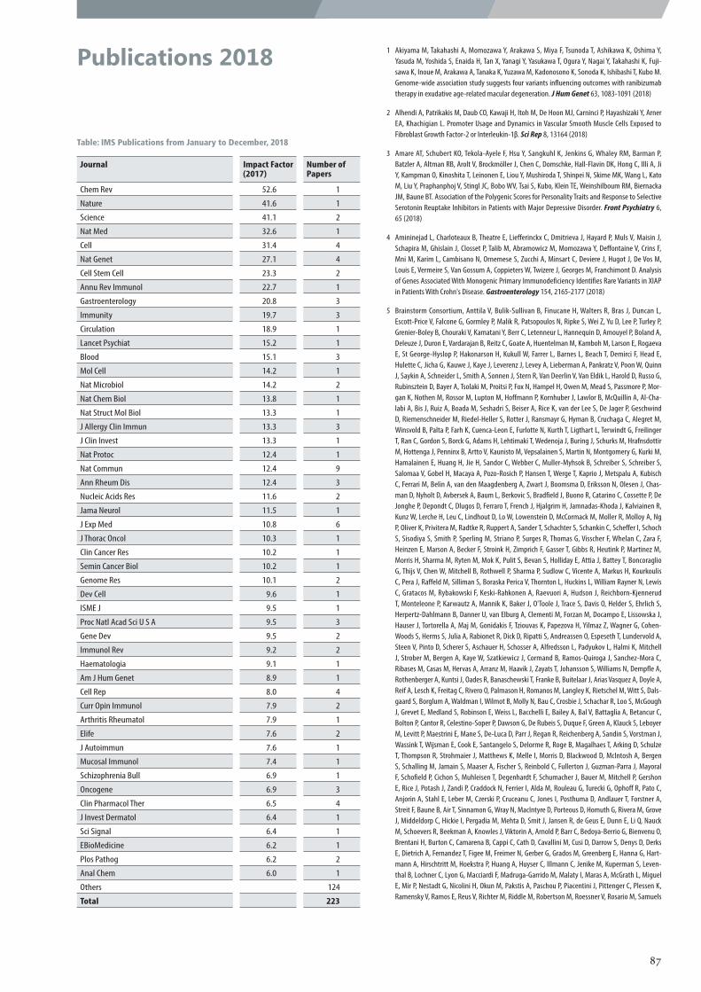
Publications 2018 1 Akiyama M, Takahashi A, Momozawa Y, Arakawa S, Miya F, Tsunoda T, Ashikawa K, Oshima Y, Yasuda M, Yoshida S, Enaida H, Tan X, Yanagi Y, Yasukawa T, Ogura Y, Nagai Y, Takahashi K, Fuji-sawa K, Inoue M, Arakawa A, Tanaka K, Yuzawa M, Kadonosono K, Sonoda K, Ishibashi T, Kubo M. Genome-wide association study suggests four variants influencing outcomes with ranibizumab therapy in exudative age-related macular degeneration. J Hum Genet 63, 1083-1091 (2018)
2 Alhendi A, Patrikakis M, Daub CO, Kawaji H, Itoh M, De Hoon MJ, Carninci P, Hayashizaki Y, Arner EA, Khachigian L. Promoter Usage and Dynamics in Vascular Smooth Muscle Cells Exposed to Fibroblast Growth Factor-2 or Interleukin-1β. Sci Rep 8, 13164 (2018)
3 Amare AT, Schubert KO, Tekola-Ayele F, Hsu Y, Sangkuhl K, Jenkins G, Whaley RM, Barman P, Batzler A, Altman RB, Arolt V, Brockmöller J, Chen C, Domschke, Hall-Flavin DK, Hong C, Illi A, Ji Y, Kampman O, Kinoshita T, Leinonen E, Liou Y, Mushiroda T, Shinpei N, Skime MK, Wang L, Kato M, Liu Y, Praphanphoj V, Stingl JC, Bobo WV, Tsai S, Kubo, Klein TE, Weinshilboum RM, Biernacka JM, Baune BT. Association of the Polygenic Scores for Personality Traits and Response to Selective Serotonin Reuptake Inhibitors in Patients with Major Depressive Disorder. Front Psychiatry 6, 65 (2018)
4 Amininejad L, Charloteaux B, Theatre E, Liefferinckx C, Dmitrieva J, Hayard P, Muls V, Maisin J, Schapira M, Ghislain J, Closset P, Talib M, Abramowicz M, Momozawa Y, Deffontaine V, Crins F, Mni M, Karim L, Cambisano N, Ornemese S, Zucchi A, Minsart C, Deviere J, Hugot J, De Vos M, Louis E, Vermeire S, Van Gossum A, Coppieters W, Twizere J, Georges M, Franchimont D. Analysis of Genes Associated With Monogenic Primary Immunodeficiency Identifies Rare Variants in XIAP in Patients With Crohn's Disease. Gastroenterology 154, 2165-2177 (2018)
5 Brainstorm Consortium, Anttila V, Bulik-Sullivan B, Finucane H, Walters R, Bras J, Duncan L, Escott-Price V, Falcone G, Gormley P, Malik R, Patsopoulos N, Ripke S, Wei Z, Yu D, Lee P, Turley P, Grenier-Boley B, Chouraki V, Kamatani Y, Berr C, Letenneur L, Hannequin D, Amouyel P, Boland A, Deleuze J, Duron E, Vardarajan B, Reitz C, Goate A, Huentelman M, Kamboh M, Larson E, Rogaeva E, St George-Hyslop P, Hakonarson H, Kukull W, Farrer L, Barnes L, Beach T, Demirci F, Head E, Hulette C, Jicha G, Kauwe J, Kaye J, Leverenz J, Levey A, Lieberman A, Pankratz V, Poon W, Quinn J, Saykin A, Schneider L, Smith A, Sonnen J, Stern R, Van Deerlin V, Van Eldik L, Harold D, Russo G, Rubinsztein D, Bayer A, Tsolaki M, Proitsi P, Fox N, Hampel H, Owen M, Mead S, Passmore P, Mor-gan K, Nothen M, Rossor M, Lupton M, Hoffmann P, Kornhuber J, Lawlor B, McQuillin A, Al-Cha-labi A, Bis J, Ruiz A, Boada M, Seshadri S, Beiser A, Rice K, van der Lee S, De Jager P, Geschwind D, Riemenschneider M, Riedel-Heller S, Rotter J, Ransmayr G, Hyman B, Cruchaga C, Alegret M, Winsvold B, Palta P, Farh K, Cuenca-Leon E, Furlotte N, Kurth T, Ligthart L, Terwindt G, Freilinger T, Ran C, Gordon S, Borck G, Adams H, Lehtimaki T, Wedenoja J, Buring J, Schurks M, Hrafnsdottir M, Hottenga J, Penninx B, Artto V, Kaunisto M, Vepsalainen S, Martin N, Montgomery G, Kurki M, Hamalainen E, Huang H, Jie H, Sandor C, Webber C, Muller-Myhsok B, Schreiber S, Schreiber S, Salomaa V, Gobel H, Macaya A, Pozo-Rosich P, Hansen T, Werge T, Kaprio J, Metspalu A, Kubisch C, Ferrari M, Belin A, van den Maagdenberg A, Zwart J, Boomsma D, Eriksson N, Olesen J, Chas-man D, Nyholt D, Avbersek A, Baum L, Berkovic S, Bradfield J, Buono R, Catarino C, Cossette P, De Jonghe P, Depondt C, Dlugos D, Ferraro T, French J, Hjalgrim H, Jamnadas-Khoda J, Kalviainen R, Kunz W, Lerche H, Leu C, Lindhout D, Lo W, Lowenstein D, McCormack M, Moller R, Molloy A, Ng P, Oliver K, Privitera M, Radtke R, Ruppert A, Sander T, Schachter S, Schankin C, Scheffer I, Schoch S, Sisodiya S, Smith P, Sperling M, Striano P, Surges R, Thomas G, Visscher F, Whelan C, Zara F, Heinzen E, Marson A, Becker F, Stroink H, Zimprich F, Gasser T, Gibbs R, Heutink P, Martinez M, Morris H, Sharma M, Ryten M, Mok K, Pulit S, Bevan S, Holliday E, Attia J, Battey T, Boncoraglio G, Thijs V, Chen W, Mitchell B, Rothwell P, Sharma P, Sudlow C, Vicente A, Markus H, Kourkoulis C, Pera J, Raffeld M, Silliman S, Boraska Perica V, Thornton L, Huckins L, William Rayner N, Lewis C, Gratacos M, Rybakowski F, Keski-Rahkonen A, Raevuori A, Hudson J, Reichborn-Kjennerud T, Monteleone P, Karwautz A, Mannik K, Baker J, O'Toole J, Trace S, Davis O, Helder S, Ehrlich S, Herpertz-Dahlmann B, Danner U, van Elburg A, Clementi M, Forzan M, Docampo E, Lissowska J, Hauser J, Tortorella A, Maj M, Gonidakis F, Tziouvas K, Papezova H, Yilmaz Z, Wagner G, Cohen-Woods S, Herms S, Julia A, Rabionet R, Dick D, Ripatti S, Andreassen O, Espeseth T, Lundervold A, Steen V, Pinto D, Scherer S, Aschauer H, Schosser A, Alfredsson L, Padyukov L, Halmi K, Mitchell J, Strober M, Bergen A, Kaye W, Szatkiewicz J, Cormand B, Ramos-Quiroga J, Sanchez-Mora C, Ribases M, Casas M, Hervas A, Arranz M, Haavik J, Zayats T, Johansson S, Williams N, Dempfle A, Rothenberger A, Kuntsi J, Oades R, Banaschewski T, Franke B, Buitelaar J, Arias Vasquez A, Doyle A, Reif A, Lesch K, Freitag C, Rivero O, Palmason H, Romanos M, Langley K, Rietschel M, Witt S, Dals-gaard S, Borglum A, Waldman I, Wilmot B, Molly N, Bau C, Crosbie J, Schachar R, Loo S, McGough J, Grevet E, Medland S, Robinson E, Weiss L, Bacchelli E, Bailey A, Bal V, Battaglia A, Betancur C, Bolton P, Cantor R, Celestino-Soper P, Dawson G, De Rubeis S, Duque F, Green A, Klauck S, Leboyer M, Levitt P, Maestrini E, Mane S, De-Luca D, Parr J, Regan R, Reichenberg A, Sandin S, Vorstman J, Wassink T, Wijsman E, Cook E, Santangelo S, Delorme R, Roge B, Magalhaes T, Arking D, Schulze T, Thompson R, Strohmaier J, Matthews K, Melle I, Morris D, Blackwood D, McIntosh A, Bergen S, Schalling M, Jamain S, Maaser A, Fischer S, Reinbold C, Fullerton J, Guzman-Parra J, Mayoral F, Schofield P, Cichon S, Muhleisen T, Degenhardt F, Schumacher J, Bauer M, Mitchell P, Gershon E, Rice J, Potash J, Zandi P, Craddock N, Ferrier I, Alda M, Rouleau G, Turecki G, Ophoff R, Pato C, Anjorin A, Stahl E, Leber M, Czerski P, Cruceanu C, Jones I, Posthuma D, Andlauer T, Forstner A, Streit F, Baune B, Air T, Sinnamon G, Wray N, MacIntyre D, Porteous D, Homuth G, Rivera M, Grove J, Middeldorp C, Hickie I, Pergadia M, Mehta D, Smit J, Jansen R, de Geus E, Dunn E, Li Q, Nauck M, Schoevers R, Beekman A, Knowles J, Viktorin A, Arnold P, Barr C, Bedoya-Berrio G, Bienvenu O, Brentani H, Burton C, Camarena B, Cappi C, Cath D, Cavallini M, Cusi D, Darrow S, Denys D, Derks E, Dietrich A, Fernandez T, Figee M, Freimer N, Gerber G, Grados M, Greenberg E, Hanna G, Hart-mann A, Hirschtritt M, Hoekstra P, Huang A, Huyser C, Illmann C, Jenike M, Kuperman S, Leven-thal B, Lochner C, Lyon G, Macciardi F, Madruga-Garrido M, Malaty I, Maras A, McGrath L, Miguel E, Mir P, Nestadt G, Nicolini H, Okun M, Pakstis A, Paschou P, Piacentini J, Pittenger C, Plessen K, Ramensky V, Ramos E, Reus V, Richter M, Riddle M, Robertson M, Roessner V, Rosario M, Samuels
Table: IMS Publications from January to December, 2018
Journal Impact Factor (2017)
Number of Papers
Chem Rev 52.6 1
Nature 41.6 1
Science 41.1 2
Nat Med 32.6 1
Cell 31.4 4
Nat Genet 27.1 4
Cell Stem Cell 23.3 2
Annu Rev Immunol 22.7 1
Gastroenterology 20.8 3
Immunity 19.7 3
Circulation 18.9 1
Lancet Psychiat 15.2 1
Blood 15.1 3
Mol Cell 14.2 1
Nat Microbiol 14.2 2
Nat Chem Biol 13.8 1
Nat Struct Mol Biol 13.3 1
J Allergy Clin Immun 13.3 3
J Clin Invest 13.3 1
Nat Protoc 12.4 1
Nat Commun 12.4 9
Ann Rheum Dis 12.4 3
Nucleic Acids Res 11.6 2
Jama Neurol 11.5 1
J Exp Med 10.8 6
J Thorac Oncol 10.3 1
Clin Cancer Res 10.2 1
Semin Cancer Biol 10.2 1
Genome Res 10.1 2
Dev Cell 9.6 1
ISME J 9.5 1
Proc Natl Acad Sci U S A 9.5 3
Gene Dev 9.5 2
Immunol Rev 9.2 2
Haematologia 9.1 1
Am J Hum Genet 8.9 1
Cell Rep 8.0 4
Curr Opin Immunol 7.9 2
Arthritis Rheumatol 7.9 1
Elife 7.6 2
J Autoimmun 7.6 1
Mucosal Immunol 7.4 1
Schizophrenia Bull 6.9 1
Oncogene 6.9 3
Clin Pharmacol Ther 6.5 4
J Invest Dermatol 6.4 1
Sci Signal 6.4 1
EBioMedicine 6.2 1
Plos Pathog 6.2 2
Anal Chem 6.0 1
Others 124
Total 223
87
J, Sandor P, Stein D, Tsetsos F, Van Nieuwerburgh F, Weatherall S, Wendland J, Wolanczyk T, Worbe Y, Zai G, Goes F, McLaughlin N, Nestadt P, Grabe H, Depienne C, Konkashbaev A, Lanzagorta N, Valencia-Duarte A, Bramon E, Buccola N, Cahn W, Cairns M, Chong S, Cohen D, Crespo-Facorro B, Crowley J, Davidson M, DeLisi L, Dinan T, Donohoe G, Drapeau E, Duan J, Haan L, Hougaard D, Karachanak-Yankova S, Khrunin A, Klovins J, Kucinskas V, Lee Chee Keong J, Limborska S, Lough-land C, Lonnqvist J, Maher B, Mattheisen M, McDonald C, Murphy K, Nenadic I, van Os J, Pantelis C, Pato M, Petryshen T, Quested D, Roussos P, Sanders A, Schall U, Schwab S, Sim K, So H, Stogmann E, Subramaniam M, Toncheva D, Waddington J, Walters J, Weiser M, Cheng W, Cloninger R, Curtis D, Gejman P, Henskens F, Mattingsdal M, Oh S, Scott R, Webb B, Breen G, Churchhouse C, Bulik C, Daly M, Dichgans M, Faraone S, Guerreiro R, Holmans P, Kendler K, Koeleman B, Mathews C, Price A, Scharf J, Sklar P, Williams J, Wood N, Cotsapas C, Palotie A, Smoller J, Sullivan P, Rosand J, Cor-vin A, Neale B, Schott J, Anney R, Elia J, Grigoroiu-Serbanescu M, Edenberg H, Murray R. Analysis of shared heritability in common disorders of the brain. Science 360, eaap8757(2018)
6 Baillie JK, Haley C, Gray A, Barrett J, Stahl E, Tenesa A, Brown B, Andersson R, Shaefer U, Zeggini E, Asimit J, Faulkner GJ, Lizio M, Daub CO, Itoh M, Kondo N, Lassmann T, Kawai J, Bajic V, Heutink P, Michael R, Kawaji H, Albin S, Suzuki H, Plenge R, Satsangi J, Wells C, Freeman TC, Hayashizaki Y, Carninci P, Forrest AR, Hume DA. Shared activity patterns arising at genetic susceptibility loci reveal underlying genomic and cellular architecture of human disease. PLoS Comput Biol (2018)
7 Bardales J, Wieser E, Kawaji H, Murakawa Y, Darzacq X. Selective Activation of Alternative MYC Core Promoters by Wnt-Responsive Enhancers. Genes (Basel) 9, E270 (2018)
8 Boisson B, Honda Y, Ajiro M, Bustamante J, Bendavid M, Gennery AR, Kawasaki Y, Ichishima J, Osawa M, Nihira H, Shiba T, Tanaka T, Chrabieh M, Bigio B, Hur H, Yuval I, Liang Y, Okada S, Izawa K, Nishikomori R, Ohara O, Heike T, Abel L, Puel A, Saito MK, Casanova J, Hagiwara M, Yasumi T. Rescue of recurrent deep intronic mutation underlying cell type–dependent quantitative NEMO deficiency. J Clin Invest 129, 583-597 (2018)
9 Boot A, Huang M, Ng AW, Ho S, Lim J, Kawakami Y, Chayama K, Teh B, Nakagawa H, Rozen SG. In-depth characterization of the cisplatin mutational signature in human cell lines and in esopha-geal and liver tumors. Genome Res 28, 654-665 (2018)
10 Burla B, Arita M, Arita M, Bendt AK, Cazenave-Gassiot A, Dennis E, Ekroos K, Han X, Ikeda K, Li-ebisch, Lin K, Loh, Meikle J, Orešič, Quehenberger, Shevchenko A, Torta, Wakelam MJ, Wheelock CE, Wenk MR. MS-based lipidomics of human blood plasma - a community-initiated position paper to develop accepted guidelines. J Lipid Res 59, 2001-2017 (2018)
11 Castiglioni I, Caccia R, Garcia-Manteiga J, Ferri G, Caretti G, Molineris I, Nishioka K, Gabellini D. The Trithorax protein Ash1L promotes myoblast fusion by activating Cdon expression. Nat Com-mun 9, 5026 (2018)
12 Chiou T, Suda W, Oshima K, Hattori M, Matsuzaki C, Yamamoto K, Takahashi T. Draft Genome Sequence of Lactobacillus kosoi NBRC 113063, Isolated from Kôso, a Japanese Sugar-Vegetable Fermented Beverage. Microbiol Resour Announc 7, e01173-18 (2018)
13 Chiou T, Suda W, Oshima K, Hattori M, Matsuzaki C, Yamamoto K, Takahashi T. Lactobacillus kosoi sp. nov., a fructophilic species isolated from kôso, a Japanese sugar-vegetable fermented bever-age. Antonie Van Leeuwenhoek 111, 1149-1156 (2018)
14 Cohen I, Zhao D, Bar C, Valdes VJ, Dauber-Decker KL, Nguyen MB, Nakayama M, Rendl M, Bick-more WA, Koseki H, Zheng D, Ezhkova E. PRC1 Fine-tunes Gene Repression and Activation to Safeguard Skin Development and Stem Cell Specification. Cell Stem Cell 22, 726-739 e7 (2018)
15 Cole J, Xu H, Ryan K, Jaworek T, Dueker N, McArdle P, Gaynor B, Cheng C, O'Connell J, Steve B, Ma-lik R, Ahmed NU, Amouyel P, Anjum S, Bis J, Crosslin D, Danesh J, Engelter S, Fornage M, Frossard P, Gieger C, Giese A, Grond-Ginsbach C, Ho WK, Holliday E, Hopewell J, Hussain M, Iqbal W, Jabeen S, Jannes J, Kamal A, Kamatani Y, Kanse S, Kloss M, Lathrop M, Leys D, Lindgren A, Longstreth, Jr W, Mahmood K, Meisinger C, Metso. T, Mosley, Jr T, Mosley, Jr T, Müller-Nurasyid M, Norrving B, Parati E, Peters A, Pezzini A, Quereshi I, Rasheed A, Rauf A, Salam T, Shen J, Slowik A, Stanne T, Strauch K, Tatlisumak T, Thijs V, Tiedt S, Traylor M, Waldenberger M, Walters M, Zhao W, Boncora-glio G, Debette S, Jern C, Levi C, Markus H, Meschia J, Rolfs A, Rothwell P, Saleheen D, Seshadri S, Sharma P, Sudlow C, Worrall B, Stine O, Kittner S, Mitchell B. Genetics of the thrombomodulin-endothelial cell protein C receptor system and the risk of early-onset ischemic stroke. PLoS One 13, e0206554 (2018)
16 Conway E, Jerman E, Healy E, Ito S, Holoch D, Oliviero G, Deevy O, Glancy E, Fitzpatrick DJ, Mucha M, Watson A, Rice AM, Chammas P, Huang C, Pratt-Kelly I, Koseki Y, Nakayama M, Ishikura T, Streubel G, Wynne K, Hokamp K, McLysaght A, Ciferri C, Croce LD, Cagney G, Margueron R, Koseki H, Bracken AP. A Family of Vertebrate-Specific Polycombs Encoded by the LCOR/LCORL Genes Bal-ance PRC2 Subtype Activities. Mol Cell 70, 408-421 e8 (2018)
17 Costea PI, Hildebrand F, Hattori M, Raes J, Knight R, Bork P. Enterotypes in the landscape of gut microbial community composition. Nat Microbiol 3, 8-16 (2018)
18 Cvetesic N, Leitch HG, Borkowska M, Mueller F, Carninci P, Hajkova P, Lenhard B. SLIC-CAGE: high-resolution transcription start site mapping using nanogram-levels of total RNA. Genome Res 24, 1943-1956 (2018)
19 Danese E, Raimondi S, Montagnana M, Tagetti, Langaee T, Borgiani P, Ciccacci C, Carcas A, Borobia A, Tong H, Dávila‐Fajardo C, Botton M, Bourgeois S, Deloukas P, Caldwell M, Burmester J, Ber R, Cavallari L, Drozda, Huang M, Zhao, Cen H, Gonzalez‐Conejero R, Roldan V, Nakamura Y, Mushiroda T, Gong I, Kim R, Hirai K, Itoh, Isaza C, Beltrán L, Jiménez‐Varo E, Cañadas‐Garre
M, Giontella A, Kringen, Haug K, Gwak H, Lee K, Minuz P, Lee M, Lubitz S, Scott S, Mazzaccara C, Sacchetti L, Genç E, Özer M, Pathare A, Krishnamoorthy R, Paldi A, Siguret V, Loriot M, Kutala V, Suarez‐Kurtz G, Perini J, Denny J, Ramirez A, Mittal B, Rathore S, Sagreiya H, Altman R, Shahin M, Khalifa S, Limdi, Rivers C, Shendre A, Dillon C, Suriapranata I, Zhou H, Tan, Tatarunas V, Lesau-skaite V, Zhang Y, Maitland‐van der Zee A, Verhoef T, de Boer A, Taljaard M, Zambon C, Pengo V, Zhang J, Pirmohamed M, Johnson J, Fava C. The effect of CYP4F2, VKORC1 and CYP2C9 in influ-encing coumarin dose. A single patient data meta‐analysis in more than 15,000 individuals. Clin Pharmacol Ther 10.1002/cpt.1323 (Epub 2018)
20 Dehzangi A, López, Taherzadeh, Sharma A, Tsunoda T. SumSec: Accurate Prediction of Su-moylation Sites Using Predicted Secondary Structure. Molecules 23, E3260 (2018)
21 Demenais F, Margaritte-Jeannin P, Barnes K, Cookson W, Altmüller J, Ang W, Barr R, Beaty T, Becker A, Beilby J, Bisgaard H, Bjornsdottir U, Bleecker E, Bønnelykke K, Boomsma D, Bouzigon E, Brightling C, Brossard M, Brusselle G, Burchard E, Burkart K, Bush A, Chan-Yeung M, Chung K, Alves A, Curtin J, Custovic A, Daley D, Jongste J, Del-Rio-Navarro B, Donohue K, Duijts L, Eng C, Eriksson J, Farrall M, Fedorova Y, Feenstra B, Ferreira M, Freidin M, Gajdos Z, Gauderman J, Gehring U, Geller F, Genuneit J, Gharib S, Gilliland F, Granell R, Graves P, Gudbjartsson D, Haahtela T, Heckbert S, Heederik D, Heinrich J, Heliövaara M, Henderson J, Himes B, Hirose H, Hirschhorn J, Hofman A, Holt P, Hottenga J, Hudson T, Hui J, Imboden M, Ivanov V, Jaddoe V, James A, Janson C, Jarvelin M, Jarvis D, Jones G, Jonsdottir I, Jousilahti P, Kabesch M, Kähönen M, Kantor D, Karunas A, Khusnutdinova E, Koppelman G, Kozyrskyj A, Kreiner E, Kubo, Kumar R, Kumar A, Kuokkanen M, Lahousse L, Laitinen T, Laprise C, Lathrop M, Lau S, Lee Y, Lehtimäki T, Letort S, Levin A, Li G, Liang L, Loehr L, London S, Loth D, Manichaikul A, Marenholz I, Martinez F, Matheson M, Mathias R, Matsumoto K, Mbarek H, McArdle W, Melbye M, Melén E, Meyers D, Michel S, Mohamdi H, Musk A, Myers R, Nieuwenhuis M, Noguchi E, O’Connor G, Ogorodova L, Palmer C, Palotie A, Park J, Pennell C, Pershagen G, Polonikov A, Postma D, Probst-Hensch N, Puzyrev V, Raby B, Raitakari O, Ramasamy A, Rich S, Robertson C, Romieu I, Salam M, Salomaa V, Schlünssen V, Scott R, Seliva-nova P, Sigsgaard T, Simpson A, Siroux V, Smith L, Solodilova M, Standl M, Stefansson K, Strachan D, Stricker B, Takahashi A, Thompson P, Thorleifsson G, Thorsteinsdottir U, Tiesler C, Torgerson D, Tsunoda T, Uitterlinden A, van der Valk R, Vaysse A, Vedantam S, von Berg A, von Mutius E, Vonk J, Waage J, Wareham N, Weiss S, White W, Wickman M, Widén E, Willemsen G, Williams L, Wouters I, Yang J, Zhao J, Moffatt M, Ober C, Nicolae D. Multiancestry association study identifies new asthma risk loci that colocalize with immune-cell enhancer marks. Nat Genet 50, 42-53 (2018)
22 Dimitrova EE, Kondo T, Feldmann A, Nakayama M, Koseki Y, Konietzny R, Kessler BM, Koseki H, Klose RJ. FBXL19 recruits CDK-Mediator to CpG islands of developmental genes priming them for activation during lineage commitment. Elife 7, e37084 (2018)
23 Djekidel MN, Inoue A, Matoba S, Suzuki T, Zhang C, Lu F, Jiang L, Zhang Y. Reprogramming of Chromatin Accessibility in Somatic Cell Nuclear Transfer Is DNA Replication Independent. Cell Rep 23, 1939–1947 (2018)
24 Eric V, David A, Colonna M, Diefenbach A, Santo JD, Eberi G, Koyasu S, Locksley RM, McKenzie A, Mebius R, Powrie F, Spits H. Innate Lymphoid Cells: 10 Years On. Cell 174, 1054-1066 (2018)
25 Feitosa M, Kraja A, Chasman D, Sung Y, Winkler T, Ntalla I, Guo X, Franceschini N, Cheng C, Sim X, Vojinovic D, Marten J, Musani S, Li C, Bentley A, Brown M, Schwander K, Richard M, Noordam R, Aschard H, Bartz T, Bielak L, Dorajoo R, Fisher V, Hartwig F, Horimoto A, Lohman K, Manning A, Rankinen T, Smith A, Tajuddin S, Wojczynski M, Alver M, Boissel M, Cai Q, Campbell A, Chai J, Chen X, Divers J, Gao C, Goel A, Hagemeijer Y, Harris S, Meian H, Hsu F, Jackson A, Kähönen M, Kasturiratne A, Komulainen P, Kühnel B, Laguzzi F, Luan J, Matoba N, Nolte I, Padmanabhan S, Riaz M, Rueedi R, Robino A, Said M, Scott R, Sofer T, Stančáková A, Takeuchi F, Tayo B, van der Most P, Varga T, Vitart V, Wang Y, Ware E, Warren H, Weiss S, Wen W, Yanek L, Zhang W, Zhao J, Afaq S, Amin N, Amini M, Arking D, Aung T, Boerwinkle E, Borecki I, Brown M, Brumat M, Burke G, Canouil M, Chakravarti A, Charumathi S, Chen Y, Connell J, Correa A, Fuentes L, Mutsert R, de Silva H, Deng X, Ding J, Duan Q, Eaton C, Ehret G, Eppinga R, Evangelou E, Faul J, Felix S, Forouhi N, Forrester T, Franco O, Friedlander Y, Gandin I, Gao H, Ghanbari M, Gigante B, Gu C, Gu D, Hagenaars S, Hallmans G, Harris T, He J, Heikkinen S, Heng C, Hirata M, Howard B, Ikram M, Consortium I, John U, Katsuya T, Khor C, Kilpeläinen T, Koh W, Krieger J, Kritchevsky S, Kubo M, Kuusisto J, Lakka T, Langefeld C, Langenberg C, Launer L, Lehne B, Lewis C, Li Y, Lin S, Liu J, Liu J, Loh M, Louie T, Mägi R, McKenzie C, Meitinger T, Metspalu A, Milaneschi Y, Milani L, Mohlke K, Momozawa Y, Nalls M, Nelson C, Sotoodehnia N, Norris J, O'Connell J, Palmer N, Perls T, Pedersen N, Peters A, Peyser P, Poulter N, Raffel L, Raitakari O, Roll K, Rose L, Rosendaal F, Rotter J, Schmidt C, Schreiner P, Schupf N, Scott W, Sever P, Shi Y, Sidney S, Sims M, Sitlani C, Smith J, Snieder H, Starr J, Strauch K, Stringham H, Tan N, Tang H, Taylor K, Teo Y, Tham Y, Turner S, Uitterlinden A, Vollenweider P, Waldenberger M, Wang L, Wang Y, Wei W, Williams C, Yao J, Caizheng Y, Yuan J, Zhao W, Zonderman A, Becker D, Boehnke M, Bowden D, Chambers J, Deary I, Esko T, Farrall M, Franks P, Freedman B, Froguel P, Gasparini P, Gieger C, Jonas J, Kamatani Y, Kato N, Kooner J, Kutalik Z, Laakso M, Laurie C, Leander K, Lehtimäki T, Cohort Study L, Magnusson P, Oldehinkel A, Polasek O, Porteous D, Rauramaa R, Samani N, Scott J, Shu X, Harst P, Wagenknecht L, Wareham N, Watkins H, Weir D, Wickremasinghe A, Wu T, Zheng W, Bouchard C, Christensen K, Evans M, Gudnason V, Horta B, Kardia S, Liu Y, Pereira A, Psaty B, Ridker P, van Dam R, Gauderman W, Zhu X, Mook-Kanamori D, Fornage M, Rotimi C, Cupples L, Kelly T, Fox E, Hayward C, van Duijn C, Tai E, Wong T, Kooperberg C, Palmas W, Rice K, Morrison A, Elliott P, Caulfield M, Munroe P, Rao D, Province M, Levy D. Novel genetic associations for blood pressure identified via gene-alcohol interaction in up to 570K individuals across multiple ancestries. PLoS One 13, e0198166 (2018)
26 Fujii R, Hishida A, Nakatochi M, Furusyo N, Murata M, Tanaka K, Shimanoe C, Suzuki S, Watanabe M, Kuriyama N, Koyama T, Takezaki T, Shimoshikiryo I, Arisawa K, Katsuura‐Kamano S, Takashima N, Turin T, Kuriki K, Endoh K, Mikami H, Nakamura Y, Oze I, Ito H, Kubo M, Momozawa Y, Kondo
88
T, Naito M, Wakai K. Association of genetic risk score and chronic kidney disease in a Japanese population. Nephrology (Carlton) 10.1111/nep.13479 (Epub 2018)
27 Fujii S, Yamasaki S, Sato Y, Shimizu K. Vaccine Designs Utilizing Invariant NKT-Licensed Antigen-Presenting Cells Provide NKT or T Cell Help for B Cell Responses. Front Immunol 9, 1267 (2018)
28 Fujimori S, Fukunaga K, Takahashi A, Mushiroda T, Kubo, Hanada R, Hayashida M, Sakurai T, Iwakiri K, Sakamoto C. Bactericidal/Permeability-Increasing Fold-Containing Family B Member 4 May Be Associated with NSAID-Induced Enteropathy. Dig Dis Sci 64, 401-408 (2018)
29 Fujimoto H, Saito Y, Ohuchida K, Kawakami E, FUjiki S, Watanabe T, Ono R, Kaneko A, Takagi S, Najima Y, Hijikata A, Lin C, Ueki T, Oda Y, Hori S, Ohara O, Nakamura M, Saito T, Ishikawa F. De-regulated Mucosal Immune Surveillance through Gut-Associated Regulatory T Cells and PD-1+ T Cells in Human Colorectal Cancer. J Immunol 200, 3291-3303 (2018)
30 Fujiwara S, Hoshizaki M, Ichida Y, Lex D, Kuroda E, Ishii KJ, Magi S, Okada M, Takao H, Gandou M, Imai H, Hara R, Herzog H, Yoshimura A, Okamura H, Penninger JM, Slutsky AS, Uhlig S, Kuba K, Imai Y. Pulmonary phagocyte-derived NPY controls the pathology of severe influenza virus infec-tion. Nat Microbiol 10.1038/s41564-018-0289-1 (Epub 2018)
31 Fujita T, Kitamura F, Oji A, Tanigawa N, Yuno M, Ikawa M, Taniuchi I, Fujii H. Transgenic mouse lines expressing the 3xFLAG‐dCas9 protein for enChIP analysis. Genes Cells 23, 318-325 (2018)
32 Fukaya T, Fukui T, Uto T, Takagi H, Nasu J, Miyanaga N, Arimura K, Nakamura T, Koseki H, Choi-jookhuu N, Hishikawa Y, Sato K. Pivotal Role of IL-22 Binding Protein in the Epithelial Autoregu-lation of Interleukin-22 Signaling in the Control of Skin Inflammation. Front Immunol 2018, 1418 (2018)
33 Furuta M, Tanaka H, Shiraishi Y, Uchida T, Imamura M, Fujimoto A, Fujita M, Oku A, Maejima K, Kawakami Y, Arihiro K, Aikata H, Ueno, Hayami S, Ariizumi S, Yamamoto M, Gotoh K, Ohdan H, Yamaue H, Miyano S, Chayama K, Nakagawa H. Characterization of HBV integration patterns and timing in liver cancer and HBV-infected livers. Oncotarget 9, 25075-25088 (2018)
34 Furutani Y, Yoshihara Y. Proteomic Analysis of Dendritic Filopodia-rich Fraction Isolated by Telen-cephalin and Vitronectin Interaction. Front Synaptic Neurosci 10, 27 (2018)
35 Giampietro PF, Pourquie O, Raggio C, Ikegawa S, Turnpenny PD, Gray R, Dunwoodie SL, Gurnett CA, Alman B, Cheung K, Kusumi K, Hadley-Miller N, Wise CA. Summary of the first inaugural joint meeting of the International Consortium for scoliosis genetics and the International Consortium for vertebral anomalies and scoliosis, March 16-18, 2017, Dallas, Texas. Am J Med Genet A 176, 253-256 (2018)
36 Gonsalves SG, Dirksen RT, Sangkuhl K, Pulk R, Alvarellos M, Vo T, Hikino K, Roden D, Klein T, Poler M, Patel S, Caudle KE, Gordon R, Brandom B, Biesecker LG. Clinical Pharmacogenetics Implemen-tation Consortium (CPIC) guideline for the use of potent volatile anesthetic agents and succinyl-choline in the context of RYR1 or CACNA1S genotypes. Clin Pharmacol Ther 10.1002/cpt.1319 (Epub 2018)
37 Grohmann M, Wiede F, Dodd GT, Gurzov EN, Ooi GJ, Butt T, Rasmiena AA, Kaur S, Gulati T, Goh PK, Treloar AE, Archer S, Brown WA, Muller M, Watt MJ, Ohara O, McLean CA, Tiganis T. Obesity Drives STAT-1-Dependent NASH and STAT-3-Dependent HCC. Cell 175, 1289-1306 e20 (2018)
38 Guerrini MM, Vogelzang A, Fagarasan S. A Hen in the Wolf Den: A Pathobiont Tale. Immunity 48, 628-631 (2018)
39 Guigó Serra R, De Hoon M. Recent Advances in Genome Analysis. F1000Res 7, F1000 Faculty Rev-1968 (2018)
40 Guler R, Mpotje T, Ozturk M, Nono JK, Parihar S, Chia JE, Aziz NA, Hlaka L, Kumar S, Roy S, Penn-Nicholson A, Hanekom WA, Zak DE, Scriba TJ, Suzuki H, Brombacher F. Batf2 differentially regu-lates tissue immunopathology in type 1 and type 2 diseases. Mucosal Immunol 12, 390-402 (2018)
41 Guo L, Elcioglu NH, Karalar OK, Topkar MO, Wang Z, Sakamoto Y, Matsumoto N, Miyake N, Nishimura G, Ikegawa S. Dysosteosclerosis is also caused by TNFRSF11A mutation. J Hum Genet 63, 769-774 (2018)
42 Gusev O, Hayashizaki Y, Usui K. Nucleic acid amplification-based diagnostics for pulmonary diseases: What is the current state and perspectives of nucleic acid amplification technologies used in diagnostics associated with pulmonary diseases? In: Kaneko T. (ed), Clinical Relevance of Genetic Factors in Pulmonary Diseases, Singapore: Springer, 333-344 (2018)
43 Han B, Akiyama M, Kim K, Oh H, Choi H, Lee C, Jung S, Lee H, Kim E, Cook S, Haritunians T, Yamazaki K, Park S, Ye B, McGovern D, Esaki M, Kawaguchi T, Khor S, Taylor K, Rotter J, Suzuki Y, Matsui T, Motoya S, Bang S, Kim T, Momozawa Y, Kamatani Y, Tokunaga K, Kubo, Okada Y, Yang S, Song K. Amino acid position 37 of HLA-DRβ1 affects susceptibility to Crohn's disease in Asians. Hum Mol Genet 27, 3901-3910 (2018)
44 Han S, Qi X, Sugihara K, Dang JH, Endo TA, Miller KL, Kim E, Miura T, Torii KU. MUTE Directly Orchestrates Cell-State Switch and the Single Symmetric Division to Create Stomata. Dev Cell 45, 303-315 e5 (2018)
45 Handoko L, Kaczkowski B, Hon C, Lizio M, Wakamori M, Matsuda T, Ito T, Jeyamohan P, Sato Y, Sakamoto K, Yokoyama S, Kimura H, Minoda A, Umehara T. JQ1 affects BRD2-dependent and
independent transcriptional regulation without disrupting H4-hyperacetylated chromatin. Epi-genetics 13, 410-431 (2018)
46 Hara M, Hachiya T, Sutoh Y, Matsuo K, Nishida Y, Shimanoe C, Tanaka K, Shimizu A, Ohnaka K, Kawaguchi T, Oze I, Matsuda F, Ito H, Kawai S, Hishida A, Okada R, Sasakabe T, Hirata A, Ibusuki R, Nindita Y, Furusyo N, Ikezaki H, Kuriyama N, Ozaki E, Mikami H, Nakamura Y, Suzuki S, Hosono A, Katsuura-Kamano S, Arisawa K, Kuriki K, Endoh K, Takashima N, Kadota A, Nakatochi M, Momozawa Y, Kubo, Naito M, Wakai K. Genomewide Association Study of Leisure-Time Exercise Behavior in Japanese Adults. Med Sci Sports Exerc 50, 2433-2441 (2018)
47 Hara S, Kawasaki S, Tsujikawa M, Yoshihara M, Busch C, Winegarner A, Nishida K. Transcription factor AP-2β regulates human corneal endothelial–specific genes. J Biol Chem 294, 2460-2469 (2018)
48 Herndler-Brandstetter D, Ishigame H, Shinnakasu R, Plajer V, Stecher C, Zhao J, Lietzenmayer M, Kroehling L, Takumi A, Kometani K, Inoue T, Kluger Y, Kaech SM, Kurosaki T, Okada T, Flavell RA. KLRG1+ Effector CD8+ T Cells Lose KLRG1, Differentiate into All Memory T Cell Lineages, and Convey Enhanced Protective Immunity. Immunity 48, 716-729 e8 (2018)
49 Hiejima E, Shibata H, Yasumi T, Shimodera S, Hori M, Izawa K, Kawai T, Matsuoka M, Ohara A, Nishikomori R, Ohara O, Heike T. Characterization of a large UNC13D gene duplication in a patient with familial hemophagocytic lymphohistiocytosis type 3. Clin Immunol 191, 63-66 (2018)
50 Hikino K, Fukunaga K, Mushiroda T. Gap between the US and Japan in coverage of pharma-cogenomic biomarkers by health insurance programs: More coverage is needed in Japan. Drug Metab Pharmacokinet 33, 243-249 (2018)
51 Hirasaki M, Ueda A, Asaka MN, Uranishi K, Suzuki A, Kohda M, Mizuno Y, Okazaki Y, Nishimoto M, Sharif J, Koseki H, Okuda A. Identification of the Coiled-Coil Domain as an Essential Methyl-CpG-Binding Domain Protein 3 Element for Preserving Lineage Commitment Potential of Embryonic Stem Cells. Stem Cells 36, 1355-1367 (2018)
52 Hishida A, Nakatochi M, Akiyama M, Kamatani Y, Nishiyama T, Ito H, Oze I, Nishida Y, Hara M, Takashima N, Turin T, Watanabe M, Suzuki S, Ibusuki R, Shimoshikiryo I, Nakamura Y, Mikami H, Ikezaki H, Furusyo N, Kuriki K, Endoh K, Koyama T, Matsui D, Uemura H, Arisawa K, Sasakabe T, Okada R, Kawai S, Naito M, Momozawa Y, Kubo, Wakai K. Genome-Wide Association Study of Renal Function Traits: Results from the Japan Multi-Institutional Collaborative Cohort Study. Am J Nephrol 47, 304-316 (2018)
53 Hitomi K, Tahara-Hanaoka S, Miki H, Iwata K, Shiro S, Kubo M, Shibuya A. Allergin-1 on mast cells suppresses house dust mite-induced airway hyperresponsiveness in mice. Int Immunol 30, 429-434 (2018)
54 Hosoi A, Takeda K, Nagaoka K, Iino T, Matsushita H, Satoshi U, Aoki S, Matsushima K, Kubo M, Morikawa T, Kitaura K, Suzuki R, Kakimi K. Increased diversity with reduced evenness of tumor infiltrating T-cells for the successful cancer immunotherapy. Sci Rep 8, 1058 (2018)
55 Igarashi M, Watanabe K, Tsuduki T, Kimura I, Kubota N. NAPE-PLD controls OEA synthesis and fat absorption by regulating lipoprotein synthesis in an in vitro model of intestinal epithelial cells. FASEB J 33, 3167-3179 (2018)
56 Iida A, Takeshita E, Kosugi S, Kamatani Y, Momozawa Y, Kubo M, Nakagawa E, Kurosawa K, Inoue K, Goto Y. A novel intragenic deletion in OPHN1 in a Japanese patient with Dandy-Walker mal-formation. Hum Genome Var 6, 1 (2018)
57 Ikeda M, Mushiroda T. The genetics of antipsychotic response in schizophrenia. Lancet Psychia-try 5, 291-292 (2018)
58 Ikeda M, Takahashi A, Kamatani Y, Momozawa Y, Saito T, Kondo K, Shimasaki A, Kawase K, Sakusabe T, Yoshimi I, Toyota T, Wakuda T, Kikuchi M, Kanahara N, Yamamori H, Yuka Y, Watanabe Y, Hoya S, Aleksic B, Kushima I, Arai H, Takaki M, Hattori K, Kunugi H, Okahisa Y, Ohnuma T, Ozaki N, Someya T, Ryota H, Yoshikawa T, Kubo, Iwata N. Genome-Wide Association Study Detected Novel Susceptibility Genes for Schizophrenia and Shared Trans-Populations/Diseases Genetic Effect. Schizophr Bull 10.1093/schbul/sby140 (Epub 2018)
59 Ikeda M, Tanaka S, Saito T, Ozaki N, Kamatani Y, Iwata N. Re-evaluating classical body type theo-ries: genetic correlation between psychiatric disorders and body mass index. Psychol Med 48, 1745-1748 (2018)
60 Ikeda N, Asono K, Kikuchi K, Uchida Y, ikegami H, Takagi R, Yotsumoto, S, Shibuya T, Makino-Okamura C, Fukuyama H, Watanabe T, Ohmuraya, Araki K, Nishitai G, Tanaka M. Emergence of immunoregulatory Ym1+Ly6Chi monocytes during recovery phase of tissue injury. Sci Immunol 3, eaat0207 (2018)
61 Ingle JN, Kalari KR, Wickerham L, von Minckwitz G, Fasching PA, Furukawa Y, Mushiroda T, Goetz MP, Barman P, Carlson EE, Rastogi P, Costantino JP, Cairns J, Paik S, Bear HD, Kubo, Wang L, Wolmark N, Weinshilboum RM. Germline genome-wide association studies in women receiving neoadjuvant chemotherapy with or without bevacizumab. Pharmacogenet Genomics 28, 147-152 (2018)
62 Inoue A, Chen Z, Yin Q, Zhang Y. Maternal Eed knockout causes loss of H3K27me3 imprinting and random X-inactivation in the extra-embryonic cells. Genes Dev 32, 1525-1536 (2018)
63 Inoue T, Moran I, Shinnakasu R, Poulain SG, Kurosaki T. Generation of memory B cells and their
89
reactivation. Immunol Rev 283, 138-149 (2018)
64 Ise W, Fujii K, Shiroguchi K, Ito A, Kometani K, Takeda K, Kawakami E, Yamashita K, Suzuki K, Okada T, Kurosaki T. T Follicular Helper Cell-Germinal Center B Cell Interaction Strength Regulates Entry into Plasma Cell or Recycling Germinal Center Cell Fate. Immunity 48, 702-715 e4 (2018)
65 Isobe Y, Itagaki M, Ito Y, Naoe S, Kojima K, Ikeguchi M, Arita M. Comprehensive analysis of the mouse cytochrome P450 family responsible for omega-3 epoxidation of eicosapentaenoic acid. Sci Rep 8, 7954 (2018)
66 Isobe Y, Kawashima Y, Ishihara T, Watanabe K, Ohara O, Arita M. Identification of Protein Targets of 12/15-Lipoxygenase-Derived Lipid Electrophiles in Mouse Peritoneal Macrophages Using Omega-Alkynyl Fatty Acid. ACS Chem Biol 13, 887-893 (2018)
67 Ito T, Hirose K, Nakajima H. Bidirectional roles of IL-22 in the pathogenesis of allergic airway inflammation. Allergol Int 68, 4-8 (2018)
68 Iwasawa K, Suda W, Tsunoda T, Oikawa-Kawamoto M, Umetsu S, Takayasu L, Inui A, Fujisawa T, Morita H, Sogo T, Hattori M. Dysbiosis of the salivary microbiota in pediatric-onset primary sclerosing cholangitis and its potential as a biomarker. Sci Rep 8, 5480 (2018)
69 Jansz N, Nesterova T, Keniry A, Iminitoff M, Hickey PF, Pintacuda G, Masui O, Kobelke S, Geoghe-gan N, Breslin KA, Willson TA, Rogers K, Kay GF, Fox AH, Koseki H, Brockdorff N, Murphy JM, Blewitt ME. Smchd1 Targeting to the Inactive X Is Dependent on the Xist-HnrnpK-PRC1 Pathway. Cell Rep 25, 1912-1923 (2018)
70 Joh K, Matsuhisa F, Kitajima S, Nishioka K, Higashimoto K, Yatsuki H, Kono T, Koseki H, Soejima H. Growing oocyte-specific transcription-dependent de novo DNA methylation at the imprinted Zrsr1-DMR. Epigenetics Chromatin 11, 28 (2018)
71 Kabata H, Moro K, Koyasu S. The group 2 innate lymphoid cell (ILC2) regulatory network and its underlying mechanisms. Immunol Rev 286, 37-52 (2018)
72 Kaito S, Toya T, Yoshifuji K, Kurosawa S, Inamoto K, Takeshita K, Suda W, Kakihana K, Honda K, Hattori M, Ohashi K. Fecal microbiota transplantation with frozen capsules for a patient with refractory acute gut graft-versus-host disease. Blood Advances 2, 3097-3101 (2018)
73 Kamatani Y, Kondo Y, Suzuki A, Kawakami E, Hiwa R, Momozawa Y, Fujimoto M, Jinnin M, Tanaka Y, Takashi K, Cooper RG, Chinoy H, Rothwell S, Lamb JA, Vencovský J, Mann H, Ohmura K, Myouzen K, Ishigaki K, Nakashima R, Hosono Y, Tsuboi H, Kawasumi H, Iwasaki Y, Kajiyama H, Horita T, Ogawa-Momohara M, Takamura A, Tsunoda S, Shimizu J, Fujio K, Amano H, Mimori A, Kawakami A, Umehara H, Takeuchi T, Sano H, Muro Y, Atsumi T, Mimura T, Kawaguchi Y, Mimori T, Takahashi A, Kubo, Kohsaka H, Sumida T, Yamamoto K, Yamamoto K. Splicing variant of WDFY4 augments MDA5 signalling and the risk of clinically amyopathic dermatomyositis. Ann Rheum Dis 77, 602-611 (2018)
74 Kanai M, Akiyama M, Takahashi A, Matoba N, Momozawa Y, Ikeda M, Iwata N, Ikegawa S, Hirata M, Matsuda K, Kubo M, Okada Y, Kamatani Y. Genetic analysis of quantitative traits in the Japanese population links cell types to complex human diseases. Nat Genet 50, 390-400 (2018)
75 Kanaya T, Sakakibara S, Jinnohara T, Hachisuka M, Tachibana N, Hidano S, Kobayashi T, Kimura S, Iwanaga T, Nakagawa T, Katsuno T, Kato N, Akiyama T, Sato T, Williams IR, Ohno H. Development of intestinal M cells and follicle-associated epithelium is regulated by TRAF6-mediated NF-κB signaling. J Exp Med 215, 501-519 (2018)
76 Kato M, Yokoyama C, Kawasaki A, Takeda C, Koike T, Onoe H, Iriki A. Individual identity and affec-tive valence in marmoset calls: in vivo brain imaging with vocal sound playback. Anim Cogn 21, 331-343 (2018)
77 Kawashima Y, Miyata J, Watanabe T, Shioya J, Arita M, Ohara O. Proteogenomic analyses of cellu-lar lysates using a phenol-guanidinium thiocyanate reagent. J Proteome Res 18, 301-308 (2018)
78 Kawashima Y, Ohara O. Development of a NanoLC-MS/MS System Using a Nonporous Reverse Phase Column for Ultrasensitive Proteome Analysis. Anal Chem 90, 12334-12338 (2018)
79 Khanshour AM, Kou I, Fan Y, Einarsdottir E, Makki N, Kidane YH, Kere J, Grauers A, Johnson TA, Paria N, Patel C, Singhania R, Kamiya N, Takeda K, Otomo N, Watanabe K, Luk KDK, Cheung KMC, Herring JA, Rios JJ, Ahituv N, Gerdhem P, Gurnett CA, Song YQ, Ikegawa S, Wise CA. Genome-wide meta-analysis and replication studies in multiple ethnicities identify novel adolescent idiopathic scoliosis susceptibility loci. Hum Mol Genet 27, 3986-3998 (2018)
80 Kimura A, Kitajima M, Nishida K, Serada S, Fujimoto M, Naka T, Fujii-Kuriyama Y, Sakamoto S, Ito T, Hanada H, Tanaka T, Yoshimura A, Suzuki H. NQO1 inhibits the TLR-dependent production of selective cytokines by promoting IκB-ζ degradation. J Exp Med 215, 2197-2209 (2018)
81 Kinoshita Y, Takashi Y, Ito N, Ikegawa S, Mano H, Ushiku T, Fukayama M, Nangaku M, Fukumoto S. Ectopic expression of Klotho in fibroblast growth factor 23 (FGF23)-producing tumors that cause tumor-induced rickets/osteomalacia (TIO). Bone Rep 10, 100192 (2018)
82 Kitazume S, Imamaki R, Ogawa K, Kizuka Y, Komi Y, Kojima S, Kotani N, Honke K, Honda T, Taniguchi N. Glycosylation controls cooperative PECAM-VEGFR2-β3 integrin functions at the endothelial surface for tumor angiogenesis. Oncogene 37, 4287–4299 (2018)
83 Kochi Y, Kamatani Y, Kondo Y, Suzuki A, Kawakami E, Hiwa R, Momozawa Y, Fujimoto M, Jinnin
M, Tanaka Y, Takashi K, Cooper RG, Chinoy H, Rothwell S, Lamb JA, Vencovský J, Mann H, Ohmura K, Myouzen K, Ishigaki K, Nakashima R, Hosono Y, Tsuboi H, Kawasumi H, Iwasaki Y, Kajiyama H, Horita T, Ogawa-Momohara M, Takamura A, Tsunoda S, Shimizu J, Fujio K, Amano H, Mimori A, Kawakami A, Umehara H, Takeuchi T, Sano H, Muro Y, Atsumi T, Mimura T, Kawaguchi Y, Mimori T, Takahashi A, Kubo, Kohsaka H, Sumida T, Yamamoto K, Yamamoto K. Splicing variant of WDFY4 augments MDA5 signalling and the risk of clinically amyopathic dermatomyositis. Ann Rheum Dis 77, 602–611 (2018)
84 Koga S, Hozumi K, Hirano K, Yazawa M, Terooatea TW, Minoda A, Nagasawa T, Koyasu S, Moro K. Peripheral PDGFRα+gp38+ mesenchymal cells support the differentiation of fetal liver-derived ILC2. J Exp Med 215, 1609-1626 (2018)
85 Koizumi T, Hattori M, Nara K. Ectomycorrhizal fungal communities in alpine relict forests of Pinus pumila on Mt. Norikura, Japan. Mycorrhiza 28, 129-145 (2018)
86 Kojo S, Yasmin N, Muroi S, Tenno M, Taniuchi I. Runx-dependent and silencer-independent repression of a maturation enhancer in the Cd4 gene. Nat Commun 9, 3593 (2018)
87 Kon A, Yamazaki S, Nannya Y, Kataoka K, Ota Y, Nakagawa MM, Yoshida K, Shiozawa Y, Morita M, Yoshizato T, Sanada M, Nakayama M, Koseki H, Nakauchi H, Ogawa S. Physiological Srsf2 P95H expression causes impaired hematopoietic stem cell functions and aberrant RNA splicing in mice. Blood 131, 621-635 (2018)
88 Kou I, Ikegawa S, Momozawa Y. A multi-ethnic meta-analysis confirms the association of rs6570507 with adolescent idiopathic scoliosis. Sci Rep 8, 11575 (2018)
89 Kubo M. Mast cells and basophils in allergic inflammation. Curr Opin Immunol 54, 74-79 (2018)
90 Kumagai Y, Yoshizawa S, Nakajima Y, Watanabe M, Fukunaga T, Ogura Y, Hayashi T, Oshima K, Hattori M, Ikeuchi M, Kogure K, DeLong EF, Iwasaki W. Solar-panel and parasol strategies shape the proteorhodopsin distribution pattern in marine Flavobacteriia. ISME J 12, 1329-1343 (2018)
91 Kurokawa S, Kishimoto T, Mizuno S, Masaoka T, Naganuma M, Liang K, Kitazawa M, Nakashima M, Shindo C, Suda W, Hattori M, Kanai T, Mimura M. The effect of fecal microbiota transplantation on psychiatric symptoms among patients with irritable bowel syndrome, functional diarrhea and functional constipation: An open-label observational study. J Affect Disord 235, 506-512 (2018)
92 Lereau Bernier M, Poulain SG, Segard B, Tauran Y, Danoy M, Shinohara M, Kimura K, Kato S, Kido T, Miyajima A, Sakai Y, Plessy CG, Leclerc E. Profiling of derived-hepatocyte progenitors from induced pluripotent stem cells using NanoCAGE promoter analysis. Biochem Eng J 142, 7-17 (2018)
93 Li J, Kang J, Arita M, Kim K, Li X, Zhang H. An omega-3 polyunsaturated fatty acid derivative, 18-HEPE, protects against CXCR4-associated melanoma metastasis. Carcinogenesis 39, 1380-1388 (2018)
94 Li M, Mulkey F, Jiang C, O'Neill B, Schneider B, Shen F, Friedman P, Momozawa Y, Kubo, Niedz-wiecki D, Hochster H, Lenz H, Atkins J, Rugo H, Halabi S, Kelly W, McLeod H, Innocenti F, Ratain MJ, Venook A, Owzar K, Kroetz D. Identification of a Genomic Region Between SLC29A1 and HSP90AB1 Associated with Risk of Bevacizumab-Induced Hypertension: CALGB 80405 (Alliance). Clin Cancer Res 24, 4734-4744 (2018)
95 Li M, Qin X, Furutani Y, Inoue I, Sekihara S, Kagechika H, Kojima S. Prevention of acute liver injury by suppressing plasma kallikrein-dependent activation of latent TGF-β. Biochem Biophys Res Commun 504, 857-864 (2018)
96 Lin Q, Ohtsuji M, Amano H, Tsurui H, Tada N, Sato R, Fukuyama H, Nishimura H, Verbeek SJ, Hirose S. FcγRIIb on B Cells and Myeloid Cells Modulates B Cell Activation and Autoantibody Responses via Different but Synergistic Pathways in Lupus-Prone Yaa Mice. J Immunol 201, 3199-3210 (2018)
97 Liu D, Ray B, Neavin DR, Zhang J, Athreya AP, Biernacka JM, Bobo WV, Hall-Flavin DK, Skime MK, Zhu H, Jenkins GD, Batzler A, Kalari KR, Boakye-Agyeman F, Matson WR, Bhasin SS, Mushiroda T, Nakamura Y, Kubo, Iyer RK, Wang L, Frye MA, Kaddurah-Daouk R, Weinshilboum RM. Beta-defensin 1, aryl hydrocarbon receptor and plasma kynurenine in major depressive disorder: metabolomics-informed genomics. Transl Psychiatry 8, 10 (2018)
98 Liu J, Zhou Y, Liu S, Song X, Yang XZ, Fan Y, Chen W, Akdemir ZC, Yan Z, Zuo Y, Du R, Liu Z, Yuan B, Zhao S, Liu G, Chen Y, Zhao Y, Lin M, Zhu Q, Niu Y, Liu P, Ikegawa S, Song YQ, Posey JE, Qiu G; DISCO (Deciphering disorders Involving Scoliosis and COmorbidities) Study, Zhang F, Wu Z, Lupski JR, Wu N. The coexistence of copy number variations (CNVs) and single nucleotide polymorphisms (SNPs) at a locus can result in distorted calculations of the significance in associating SNPs to disease. Hum Genet 137, 553-567 (2018)
99 Liu Y, Chang J, Hon C, Fukui N, Tanaka N, Zhang Z, Lee MM, Minoda A. Chromatin accessibility landscape of articular knee cartilage reveals aberrant enhancer regulation in osteoarthritis. Sci Rep 8, 15499 (2018)
100 Lizio M, Abugessaisa I, Noguchi S, Kondo A, Hasegawa A, Hon C, De Hoon M, Oki S, Hayashizaki Y, Carninci P, Kasukawa T, Kawaji H. Update of the FANTOM web resource: expansion to provide additional transcriptome atlases. Nucleic Acids Res 47, D752-D758 (2018)
101 Lu HF, Wong HS, Chen BK, Liao HT, Hsu YW, Ikegawa S, Cho EC, Hung KS, Chang WC. Integra-
90
tive genomic analysis for the functional roles of ITPKC in bone mineral density. Biosci Rep 38, BSR20181481 (2018)
102 Lu Y, Kweon S, Tanikawa C, Jia W, Xiang Y, Cai Q, Zeng C, Schmit S, Shin A, Matsuo K, Jee S, Kim D, Kim J, Wen W, Shi J, Guo X, Li B, Wang N, Zhang B, Li X, Shin M, Li H, Ren Z, Oh J, Oze I, Ahn Y, Jung K, Conti D, Schumacher F, Rennert G, Jenkins M, Campbell P, Hoffmeister M, Casey G, Gru-ber S, Gao J, Gao Y, Pan Z, Kamatani Y, Zeng Y, Shu X, Long J, Matsuda K, Zheng W. Large-Scale Genome-Wide Association Study of East Asians Identifies Loci Associated With Risk for Colorectal Cancer. Gastroenterology 156, 1455-1466 (2018)
103 Lysenko A, Sharma A, Boroevich KA, Tsunoda T. An integrative machine learning approach for prediction of toxicity-related drug safety. Life Sci Alliance 1, e201800098 (2018)
104 Marletaz F, Firbas PN, Maeso I, Tena JJ, Bogdanovic O, Perry M, Wyatt CD, de la Calle-Mustienes E, Bertrand S, Burguera D, Acemel RD, van Heeringen S, Naranjo S, Herrera-Ubeda C, Skvortsova K, Jimenez-Gancedo S, Aldea D, Marquez Y, Buono L, Kozmikova I, Permanyer J, Louis A, Albuixech-Crespo B, Le Petillon Y, Florian AL, Subriana L, Duckett PE, Farahani E, Aury JM, Mangenot S, Wincker P, Albalat R, Benito-Gutierrez E, Canestro C, Castro F, D'Aniello S, Ferrier DE, Huang S, Laudet V, Marais GA, Pontarotti P, Schubert M, Seitz H, Somorjai I, Takahashi T, Mirabeau O, Xu A, Yu J, Carninci P, Martinez-Morales JR, Crollius HR, Kozmik Z, Weirauch M, Garcia-Fernandez J, Lister R, Lenhard B, Holland PW, Escriva H, Gomez-Skarmeta JL, Irimia M. Amphioxus functional genomics and the origins of vertebrate gene regulation. Nature 564, 64-70 (2018)
105 Mendoza P, Martinez-Martin N, Bovolenta ER, Reyes-Garau D, Hernansanz-Agustin P, Delgado P, Diaz-Munoz MD, Oeste CL, Fernandez-Pisonero I, Castellano E, Martinez-Ruiz A, Alonso-Lopez D, Santos E, Bustelo X, Kurosaki T, Alarcon B. R-Ras2 is required for germinal center formation to aid B cells during energetically demanding processes. Sci Signal 11, eaal1506 (2018)
106 Michelini F, Jalihal A, Francia S, Meers C, Neeb ZT, Rossiello F, Gioia U, Aguado J, Luke B, Biamonti G, Mariusz N, Storici F, Carninci P, Walter NG, d’Adda di Fagagna F. From “cellular” RNA to “smart” RNA: multiple roles of RNA in genome stability and Beyond. Chem Rev 118, 4365-4403 (2018)
107 Miyai T, Takano J, Endo T, Kawakami E, Agata Y, Motomura Y, Kubo M, Kashima Y, Suzuki Y, Kawa-moto H, Ikawa T. Three-step transcriptional priming that drives the commitment of multipotent progenitors toward B cells. Genes Dev 32, 112-126 (2018)
108 Miyashita N, Horie M, Suzuki HI, Yoshihara M, Djureinovic D, Persson J, Brunstrom H, Lindskog C, Elfving H, Micke P, Saito A, Nagase T. An integrative analysis of transcriptome and epigenome features of ASCL1-positive lung adenocarcinomas. J Thorac Oncol 13, 1676-1691 (2018)
109 Momozawa Y, Dmitrieva J, Theatre E, Deffontaine V, Rahmouni S, Charloteaux B, Crins F, Docam-po E, Elansary M, Gori A, Lecut C, Mariman R, Mni M, Oury C, Altukhov I, Alexeev D, Aulchenko Y, Amininejad L, Bouma G, Hoentjen F, Löwenberg M, Oldenburg B, Pierik M, Vander Meulen-de Jong A, Janneke van der Woude C, Visschedijk M, Lathrop M, Hugot J, Weersma R, De Vos M, Franchimont D, Vermeire S, Kubo M, Louis E, Georges M. IBD risk loci are enriched in multigenic regulatory modules encompassing putative causative genes. Nat Commun 9, 2427 (2018)
110 Momozawa Y, Iwasaki Y, Parsons M, Kamatani Y, Takahashi A, Tamura C, Katagiri T, Yoshida T, Na-kamura S, Sugano K, Miki Y, Hirata M, Matsuda K, Spurdle A, Kubo M. Germline pathogenic vari-ants of 11 breast cancer genes in 7,051 Japanese patients and 11,241 controls. Nat Commun 9, 4083 (2018)
111 Moriyasu T, Nakamura R, Deloer S, Senba M, Kubo M, Inoue M, Culleton R, Hamano S. Schisto-soma mansoni infection suppresses the growth of Plasmodium yoelii parasites in the liver and reduces gametocyte infectivity to mosquitoes. PLoS Negl Trop Dis 12, e0006197 (2018)
112 Murao N, Matsubara S, Matsuda T, Noguchi H, Mutoh T, Mutoh M, Koseki H, Namihira M, Nakashima K. Np95/Uhrf1 regulates tumor suppressor gene expression of neural stem/precursor cells, contributing to neurogenesis in the adult mouse brain. Neurosci Res S0168-0102(18)30276-1 (2018)
113 Mushiroda T, Takahashi Y, Onuma T, Yamamoto Y, Kamei T, Hoshida T, Takeuchi K, Otsuka K, Okazaki M, Watanabe M, Kanemoto K, Oshima T, Watanabe A, Minami S, Saito K, Tanii H, Shimo Y, Hara M, Saitoh S, Kinoshita T, Kato M, Yamada N, Akamatsu N, Fukuchi T, Ishida S, Yasumoto S, Takahashi A, Ozeki T, Furuta T, Saito Y, Izumida N, Kano Y, Shiohara T, Kubo . Association of HLA-A*31:01 Screening With the Incidence of Carbamazepine-Induced Cutaneous Adverse Reactions in a Japanese Population. JAMA Neurol 75, 842-849 (2018)
114 Mutai H, Miya F, Shibata H, Yasutomi Y, Tsunoda T, Matsunaga T. Gene expression dataset for whole cochlea of Macaca fascicularis. Sci Rep 8, 15554 (2018)
115 Naganuma M, Sugimoto S, Mitsuyama K, Kobayashi T, Yoshimura N, Ohi H, Tanaka S, Andoh A, Ohmiya N, Saigusa K, Yamamoto T, Morohoshi Y, Ichikawa H, Matsuoka K, Hisamatsu T, Watanabe K, Mizuno S, Suda W, Hattori M, Fukuda S, Hirayama A, Abe T, Watanabe M, Hibi T, Suzuki Y, Kanai T, INDIGO Study Group . INDIGO Study Group: Efficacy of Indigo naturalis in a Multicenter Random-ized Controlled Trial of Patients with Ulcerative Colitis. Gastroenterology 154, 935-947 (2018)
116 Nagashima H, Okuyama Y, Fujita T, Takeda T, Motomura Y, Moro K, Hidaka T, Omori K, Sakurai T, Machiyama T, Ndhlovu LC, Riccardi C, So T, Ishii N. GITR cosignal in ILC2s controls allergic lung inflammation. J Allergy Clin Immunol 141, 1939-1943 e8 (2018)
117 Nagatake T, Shiogama Y, Inoue A, Kikuta J, Honda T, Tiwari P, Kishi T, Yanagisawa A, Isobe Y, Matsumoto N, Shimojou M, Morimoto S, Suzuki H, Hirata S, Steneberg P, Edlund H, Aoki J,
Arita M, Kunisawa J. The 17,18-epoxyeicosatetraenoic acid-G protein-coupled receptor 40 axis ameliorates contact hypersensitivity by inhibiting neutrophil mobility in mice and cynomolgus macaques. J Allergy Clin Immunol 142, 470-484 e12 (2018)
118 Naito T, Muroi S, Taniuchi I, Kondo M. Data on the TGFβ response of CD4+ T cells in the absence of Eed. Data Brief 17, 1180-1183 (2018)
119 Naito T, Muroi S, Taniuchi I, Kondo M. Loss of Eed leads to lineage instability and increased CD8 expression of mouse CD4+ T cells upon TGFβ signaling. Mol Immunol 94, 140-152 (2018)
120 Nakade Y, Iwata Y, Furuichi K, Mita M, Hamase K, Konno R, Miyake T, Sakai N, Kitajima S, Toyama T, Shinozaki Y, Sagara A, Miyagawa T, Hara A, Shimizu M, Kamikawa Y, Sato K, Oshima M, Yoneda-Nakagawa, Yamamura Y, Kaneko S, Miyamoto T, Katane, Homma, Morita H, Suda W, Hattori M, Wada T. Gut microbiota-derived D-serine protects against acute kidney injury. JCI Insight 3, 97957 (2018)
121 Nakagawa H, Fujita M, Fujimoto A. Genome sequencing analysis of liver cancer for precision medicine. Semin Cancer Biol 55, 120-127 (2018)
122 Nakagawa-Senda H, Hachiya T, Shimizu A, Hosono S, Oze I, Watanabe M, Matsuo K, Ito H, Hara M, Nishida Y, Endoh K, Kuriki K, Katsuura-Kamano S, Arisawa K, Nindita Y, Ibusuki R, Suzuki S, Hosono A, Mikami H, Nakamura Y, Takashima N, Nakamura Y, Kuriyama N, Ozaki E, Furusyo N, Ikezaki H, Nakatochi M, Sasakabe T, Kawai S, Okada R, Hishida A, Naito M, Wakai K, Momozawa Y, Kubo M, Tanaka H. A genome-wide association study in the Japanese population identifies the 12q24 locus for habitual coffee consumption: The J-MICC Study. Sci Rep 8, 1493 (2018)
123 Nakajima A, Vogelzang A, Maruya M, Miyajima M, Murata M, Son A, Kuwahara T, Tsuruyama T, Yamada S, Matsuura M, Nakase H, Peterson DA, Fagarasan S, Suzuki K. IgA regulates the compo-sition and metabolic function of gut microbiota by promoting symbiosis between bacteria. J Exp Med 215, 2019-2034 (2018)
124 Nakano R, Kitanaka T, Namba S, Kitanaka N, Sugiya H. Protein kinase Cε regulates nuclear translocation of extracellular signal-regulated kinase, which contributes to bradykinin-induced cyclooxygenase-2 expression. Sci Rep 8, 8535 (2018)
125 Namkoong H, Ishii M, Fujii H, Yagi K, Asami T, Asakura T, Suzuki S, Hegab AE, Kamata H, Tasaka S, Atarashi K, Nakamoto N, Iwata S, Honda K, Kanai T, Hasegawa N, Koyasu S, Betsuyaku T. Clar-ithromycin expands CD11b+Gr-1+ cells to ameliorate shock and post-influenza pneumonia. PLoS Pathog 14, e1006955 (2018)
126 Nanki K, Mizuno S, Matsuoka K, Ono K, Sugimoto S, Kiyohara H, Arai M, Nakashima M, Takeshita K, Saigusa K, Senoh M, Fukuda T, Naganuma M, Kato H, Suda W, Hattori M, Kanai T. Fecal mi-crobiota transplantation for recurrent Clostridium difficile infection in a patient with ulcerative colitis. Intest Res 16, 142-146 (2018)
127 Nguyen Q, Aguado J, Iannelli F, Suzuki AM, Rossiello F, d'Adda di Fagagna F, Carninci P. Target-enrichment sequencing for detailed characterization of small RNAs. Nat Protoc 13, 768-786 (2018)
128 Nishioka K, Miyazaki H, Soejima H. Unbiased shRNA screening, using a combination of FACS and high-throughput sequencing, enables identification of novel modifiers of Polycomb silencing. Sci Rep 8, 12128 (2018)
129 Nomura S, Satoh M, Fujita T, Higo T, Tomokazu S, Ko T, Yamaguchi T, Tobita T, Naito AT, Ito M, Fujita K, Harada M, Toko H, Kobayashi Y, Ito K, Takimoto E, Akazawa H, Morita H, Aburatani H, Komuro I. Cardiomyocyte gene programs encoding morphological and functional signatures in cardiac hypertrophy and failure. Nat Commun 9, 4435 (2018)
130 Ogura Y, Matsumoto M, Ikegawa S, Watanabe K. Epigenetics for curve progression of adolescent idiopathic scoliosis. EBioMedicine 37, 36-37 (2018)
131 Ogura Y, Takeda K, Kou I, Khanshour A, Grauers A, Zhou H, Liu G, Fan YH, Zhou T, Wu Z, Takahashi Y, Matsumoto M; Japan Scoliosis Clinical Research Group (JSCRG); Texas Scottish Rite Hospital for Children Clinical Group (TSRHCCG), Einarsdottir E, Kere J, Huang D, Qiu G, Xu L, Qiu Y, Wise CA, Song YQ, Wu N, Su P, Gerdhem P, Watanabe K, Ikegawa S. An international meta-analysis confirms the association of BNC2 with adolescent idiopathic scoliosis. Sci Rep 8, 4730 (2018)
132 Ogura Y, Yabuki S, Fujibayashi S, Okada E, Iwanami A, Watanabe K, Nakamura M, Matsumoto M, Ishii K, Ikegawa S. A screening method to distinguish syndromic from sporadic spinal extradural arachnoid cyst. J Orthop Sci 23, 455-458 (2018)
133 Ohki K, Kiyokawa N, Saito Y, Hirabayashi S, Nakabayashi K, Ichikawa H, Momozawa Y, Okamura K, Yoshimi A, Ogata-Kawata H, Sakamoto H, Kato M, Fukushima K, Hasegawa D, Fukushima H, Imai M, Kajiwara R, Koike T, Komori I, Matsui A, Mori M, Moriwaki K, Noguchi Y, Park M, Ueda T, Yama-moto S, Matsuda K, Yoshida T, Matsumoto K, Hata K, Kubo, Matsubara Y, Takahashi H, Fukushima T, Hayashi Y, Koh K, Manabe A, Ohara A. Clinical and molecular characteristics of MEF2D fusion-positive precursor B-cell acute lymphoblastic leukemia in childhood, including a novel transloca-tion resulting in MEF2D-HNRNPH1 gene fusion. Haematologica 10.3324/haematol.2017 (Epub 2018)
134 Okada Y, Eyre S, Suzuki A, Kochi Y, Yamamoto K. Genetics of rheumatoid arthritis: 2018 status. Ann Rheum Dis 10.1136/annrheumdis-2018-213678 (Epub 2018)
135 Okada Y, Momozawa Y, Sakaue S, Kanai M, Ishigaki K, Akiyama M, Kishikawa T, Arai Y, Sasaki
91
T, Kosaki K, Suematsu M, Matsuda K, Yamamoto K, Kubo, Hirose N, Kamatani Y. Deep whole-genome sequencing reveals recent selection signatures linked to evolution and disease risk of Japanese. Nat Commun 9, 1631 (2018)
136 Okubo K, Kurosawa M, Kamiya M, Urano Y, Suzuki A, Yamamoto K, Hase K, Homma K, Sasaki J, Miyauchi H, Hoshino T, Hayashi M, Mayadas TN, Hirahashi J. Macrophage extracellular trap formation promoted by platelet activation is a key mediator of rhabdomyolysis-induced acute kidney injury. Nat Med 24, 232-238 (2018)
137 Okunoa M, Ayabea T, Yokotae I, Musyaf I, Shiga K, Kikuchi T, Kikuchi N, Ohtake A, Nakamura A, Nakabayashi K, Okamura K, Momozawa Y, Kubo M, Suzuki J, Urakami T, Kawamura T, Amemiya S, Ogata T, Sugihara S, Fukami M. Protein‐altering variants of PTPN2 in childhood‐onset Type 1A diabetes. Diabet Med 35, 376-380 (2018)
138 Onishi H, Udagawa C, Kubo, Nakamura S, Akashi-Tanaka S, Kuwayama T, Watanabe C, Takamaru T, Takei H, Ishikawa T, Miyahara K, Matsumoto H, Hasegawa Y, Momozawa Y, Low S, Kutomi G, Shima H, Satomi F, Okazaki M, Zaha H, Onomura M, Matsukata A, Sagara Y, Baba S, Yamada A, Shimada K, Shimizu D, Tsugawa K, Shimo A, Hartman M, Chan C, Lee S, Itaru E, Zembutsu H. A genome-wide association study identifies three novel genetic markers for response to tamoxi-fen: A prospective multicenter study. PLoS One 13, e0201606 (2018)
139 Ono S, Nakayama M, Kanegane H, Hoshino A, Shimodera S, Shibata H, Fujino H, Fujino T, Yuno-mae Y, Okano T, Yamashita M, Yasumi T, Izawa K, Takagi M, Imai K, Zhang K, Marsh R, Picard C, Latour S, Ohara O, Morio T. Comprehensive molecular diagnosis of Epstein–Barr virus-associated lymphoproliferative diseases using next-generation sequencing. Int J Hematol 108, 319-328 (2018)
140 Ota MS, Kondo K, Li Y, Iseki S, Yamashita A, Gibson CW, Kondo T. Amelogenin X impacts age-dependent increase of frequency and number in labial incisor grooves in C57BL/6. Biochem Biophys Res Commun 496, 324-327 (2018)
141 Otani T, Noma H, Sugasawa S, Kuchiba A, Goto A, Yamaji T, Kochi Y, Iwasaki M, Matsui S, Tsunoda T. Exploring predictive biomarkers from clinical genome-wide association studies via multidimen-sional hierarchical mixture models. Eur J Hum Genet 10.1038/s41431-018-0251-y (Epub 2018)
142 Otomo N, Mizumoto S, Lu HF, Takeda K, Campos-Xavier B, Mittaz-Crettol L, Guo L, Takikawa K, Nakamura M, Yamada S, Matsumoto M, Watanabe K, Ikegawa S. Identification of novel LFNG mutations in spondylocostal dysostosis. J Hum Genet 10.1038/s10038-018-0548-2 (Epub 2018)
143 Patel PN, Gorham JM, Ito K, Seidman CE. In Vivo and In Vitro Methods to Identify DNA Sequence Variants that Alter RNA Splicing. Curr Protoc Hum Genet 97, e60 (2018)
144 Phillips EJ, Sukasem C, Whirl‐Carrillo M, Müller DJ, Dunnenberger HM, Chantratita W, Goldspiel B, Chen Y, Carleton BC, George AL, Mushiroda T, Klein T, Gammal RS, Pirmohamed M. Clinical Pharmacogenetics Implementation Consortium Guideline for HLA Genotype and Use of Carbam-azepine and Oxcarbazepine: 2017 Update. Clin Pharmacol Ther 103, 574-581 (2018)
145 Podbevsek P, Fasolo F, Bon C, Cimatti L, Reisser S, Carninci P, Bussi G, Zucchelli S, Place J, Gustin-cich S. Structural determinants of the SINE B2 element embedded in the long non-coding RNA activator of translation AS Uchl1. Sci Rep 16, 3189 (2018)
146 Pradier L, Blanchard-Brégeon V, Bohme A, Debeir T, Menager J, Benoit P, Barneoud P, Taupin V, Bertrand P, Dugay P, Cameron B, Yi S, Naimi S, Duchesne M, Gagnaire M, Weeden T, Travaline T, Reczek D, Khiroug L, Slaoui M, Brunel P, Fukuyama H, Ravetch J, Canton T, Cohen C. SAR228810: an antibody for protofibrillar amyloid beta peptide designed to reduce the risk of amyloid-related imaging abnormalities (ARIA). Alzheimers Res Ther 10, 117 (2018)
147 Qin X, Dohmae N, Kojima S. Reply to Yoshida: Liver cancer stem cells: Identification and lipid metabolic reprogramming. Proc Natl Acad Sci U S A 115, E6390-E6391 (2018)
148 Qin X, Lu J, Cai M, Kojima S. Arachidonic acid suppresses hepatic cell growth through ROS-mediated activation of nuclear transglutaminase. FEBS Open Bio 8, 1703-1710 (2018)
149 Qin X, Suzuki H, honda m, Okada H, Kaneko S, Inoue I, Ebisui E, Hashimoto K, Carninci P, Kanki K, Tatsukawa H, Ishibashi N, Masaki T, Matsuura T, Kagechika H, Toriguchi K, Hatano E, Shirakami Y, shiota g, Shimizu M, Moriwaki H, Kojima S. Prevention of hepatocellular carcinoma by targeting MYCN-positive liver cancer stem cells with acyclic retinoid. Proc Natl Acad Sci U S A 115, 4969-4974 (2018)
150 Raju S, Kometani K, Kurosaki T, Shaw AS, Egawa T. The adaptor molecule CD2AP in CD4 T cells modulates differentiation of follicular helper T cells during chronic LCMV infection. PLoS Pathog 14, e1007053 (2018)
151 Rashkin S, Chua K, Ho C, Mulkey F, Jiang C, Mushiroda T, Kubo, Friedman P, Rugo H, McLeod H, Ratain M, Castikkos F, Naughton M, Overmoyer B, Toppmeyer D, Witte, Owzar K, Kroetz C. A Pharmacogenetic Prediction Model of Progression-Free Survival in Breast Cancer using Genome-Wide Genotyping Data from CALGB 40502 (Alliance). Clin Pharmacol Ther 10.1002/cpt.1241 (Epub 2018)
152 Roselli C, Chaffin M, Weng L, Aeschbacher S, Ahlberg G, Albert C, Almgren P, Alonso A, Anderson C, Aragam K, Arking D, Barnard J, Bartz T, Benjamin E, Bihlmeyer N, Bis J, Bloom H, Boerwinkle E, Bottinger E, Brody J, Calkins H, Campbell A, Cappola T, Carlquist J, Chasman D, Chen L, Chen Y, Choi E, Choi S, Christophersen I, Chung M, Cole J, Conen D, Cook J, Crijns H, Cutler M, Damrauer
S, Daniels B, Darbar D, Delgado G, Denny J, Dichgans M, Dörr M, Dudink E, Dudley S, Esa N, Esko T, Eskola M, Fatkin D, Felix S, Ford I, Franco O, Geelhoed B, Grewal R, Gudnason V, Guo X, Gupta N, Gustafsson S, Gutmann R, Hamsten A, Harris T, Hayward C, Heckbert S, Hernesniemi J, Hock-ing L, Hofman A, Horimoto A, Huang J, Huang P, Huffman J, Ingelsson E, Ipek E, Ito K, Jimenez-Conde J, Johnson R, Jukema J, Kääb S, Kähönen M, Kamatani Y, Kane J, Kastrati A, Kathiresan S, Katschnig-Winter P, Kavousi M, Kessler T, Kietselaer B, Kirchhof P, Kleber M, Knight S, Krieger J, Kubo, Launer L, Laurikka J, Lehtimäki T, Leineweber K, Lemaitre R, Li M, Lim H, Lin H, Lin H, Lind L, Lindgren C, Lokki M, London B, Loos R, Low S, Lu Y, Lyytikäinen L, Macfarlane P, Magnusson P, Mahajan A, Malik R, Mansur A, Marcus G, Margolin L, Margulies K, März W, McManus D, Me-lander O, Mohanty S, Montgomery J, Morley M, Morris A, Müller-Nurasyid M, Natale A, Nazarian S, Neumann B, Newton-Cheh C, Niemeijer M, Nikus K, Nilsson P, Noordam R, Oellers H, Olesen M, Orho-Melander M, Padmanabhan S, Pak H, Paré G, Pedersen N, Pera J, Pereira A, Porteous D, Psaty B, Pulit S, Pullinger C, Rader D, Refsgaard L, Ribasés M, Ridker P, Rienstra M, Risch L, Roden D, Rosand J, Rosenberg M, Rost N, Rotter J, Saba S, Sandhu R, Schnabel R, Schramm K, Schunkert H, Schurman C, Scott S, Seppälä I, Shaffer C, Shah S, Shalaby A, Shim J, Shoemaker M, Siland J, Sinisalo J, Sinner M, Slowik A, Smith A, Smith B, Smith J, Smith J, Smith N, Soliman E, Sotoodehnia N, Stricker B, Sun A, Sun H, Svendsen J, Tanaka T, Tanriverdi K, Taylor K, Teder-Laving M, Teumer A, Thériault S, Trompet S, Tucker N, Tveit A, Uitterlinden A, Harst P, Gelder I, Wagoner D, Verweij N, Vlachopoulou E, Völker U, Wang B, Weeke P, Weijs B, Weiss R, Weiss S, Wells Q, Wiggins K, Wong J, Woo D, Worrall B, Yang P, Yao J, Yoneda Z, Zeller T, Zeng L, Lubitz S, Lunetta K, Ellinor P. Multi-ethnic genome-wide association study for atrial fibrillation. Nat Genet 50, 1225-1233 (2018)
153 Roudnicky F, Poghosyan S, Poyet C, Vella G, Yoon S, Schwager S, Karaman S, Shin JW, Otto VI, Detmar M. Alternative transcription of a shorter, non-antiangiogenic thrombospondin-2 variant in cancer-associated blood vessels. Oncogene 37, 2573-2585 (2018)
154 Roy S, Schmeier S, Kaczkowski B, Arner E, Alam T, Ozturk M, Tamgue O, Parihar S, Kawaji H, Itoh M, Lassmann T, Carninci P, Hayashizaki Y, Forrest AR, Guler R, Bajic V, Brombacher F, Suzuki H. Transcriptional landscape of Mycobacterium tuberculosis infection in macrophages. Sci Rep 8, 6758 (2018)
155 Safiye SE, Inubushi T, Kurosaka H, Minagi HO, Kuremoto K, Sakai T, Taniuchi I, Yamashiro T. Runx1-Stat3 signaling regulates the epithelial stem cells in continuously growing incisors. Sci Rep 8, 10906 (2018)
156 Safiye SE, Kurosaka H, Inubushi T, Minagi HO, Kuremoto K, Sakai T, Taniuchi I, Yamashiro T. Runx1-Stat3-Tgfb3 signaling network regulating the anterior palatal development. Sci Rep 8, 11208 (2018)
157 Sakaue S, Hirata J, Maeda Y, Kawakami E, Nii T, Kishikawa T, Ishigaki K, Terao C, Suzuki K, Akiyama M, Suita N, Masuda T, Ogawa K, Yamamoto K, Saeki Y, Matsushita M, Yoshimura M, Matsuoka H, Ikari K, Taniguchi A, Yamanaka H, Kawaji H, Lassmann T, Itoh M, Yoshitomi H, Ito H, Ohmura K, Forrest A, Hayashizaki Y, Carninci P, Kumanogoh A, Kamatani Y, De Hoon M, Yamamoto K, Okada Y. Integration of genetics and miRNA–target gene network identified disease biology implicated in tissue specificity. Nucleic Acids Res 46, 11898-11909 (2018)
158 Sharif J, Koseki H. Hemimethylation: DNA's lasting odd couple. Science 359, 1102-1103 (2018)
159 Sharif J, Koseki H. Rewriting the past: de novo activity of PRC2 restores global H3K27 methyla-tion patterns. Nat Struct Mol Biol 25, 197-199 (2018)
160 Sharma R, Sharma A, Raicar G, Tsunoda T, Patil A. OPAL+: Length‐Specific MoRF Prediction in Intrinsically Disordered Protein Sequences. Proteomics 10.1002/pmic.201800058 (Epub 2018)
161 Shen X, Opulente D, Kominek J, Zhou X, Steenwyk J, Buh K, Haase M, Wisecaver JH, Wang M, Boudouris JT, Schneider RM, Langdon QK, Ohkuma M, Endoh R, Takashima M, Manabe R, Čadež N, Libkind D, Rosa CA, DeVirgilio J, Hulfachor AB, Groenewald M, Kurtzman CP, Hittinger CT, Rokas A. Tempo and mode of genome evolution in the budding yeast subphylum. Cell 175, 1533-1545 e20 (2018)
162 Shepshelovich D, Townsend A, Espin‐Garcia O, Latifovic L, O’Callaghan C, Jonker D, Tu D, Chen E, Morgen E, Price T, Shapiro J, Siu L, Kubo, Dobrovic A, Ratain M, Xu, Mushiroda T, Liu G. Fc-gamma receptor polymorphisms, cetuximab therapy, and overall survival in the CCTG CO.20 trial of metastatic colorectal cancer. Cancer Med 7, 5478-5487 (2018)
163 Shibayama Y, Fanucchi S, Fok E, Dalla E, Börner K, Chang EY, Stoychev S, Imakaev M, Grimm D, Wang KC, Li G, Sung W, Mhlanga MM. Immune genes are primed for robust transcription by proximal long noncoding RNAs located in nuclear compartments. Nat Genet 51, 138-150 (2018)
164 Shiga Y, Akiyama M, Nishiguchi KM, Sato K, Shimozawa N, Takahashi A, Momozawa Y, Hirata M, Matsuda K, Yamaji T, Iwasaki M, Tsugane S, Oze I, Mikami H, Naito M, Wakai K, Yoshikawa M, Miyake M, Yamashiro K, Japan Glaucoma Society Omics Group, Kashiwagi K, Iwata T, Mabuchi F, Takamoto M, Ozaki M, Kawase K, Aihara M, Araie M, Yamamoto T, Kiuchi Y, Nakamura M, Ikeda Y, Sonoda K, Ishibashi T, Nitta K, Iwase A, Shirato S, Oka Y, Satoh M, Sasaki M, Fuse N, Suzuki Y, Cheng C, Khor C, Baskaran M, Perera S, Aung T, Vithana EN, Bailey JN, Kang JH, Pasquale LR, Haines JL, NEIGHBORHOOD Consortium, Wiggs JL, Burdon KP, Gharahkhani P, Hewitt AW, Mackey DA, MacGregor S, Craig JE, Allingham RR, Hauser M, Ashaye A, Budenz DL, Akafo S, Williams SE, Kamatani Y, Nakazawa T, Kubo M. Genome-wide association study identifies seven novel suscep-tibility loci for primary open-angle glaucoma. Hum Mol Genet 27, 1486-1496 (2018)
165 Shigemizu D, Miya F, Akiyama S, Okuda S, Boroevich KA, Fujimoto A, Nakagawa H, Ozaki K,
92
Niida S, Kanemura Y, Okamoto N, Saitoh S, Kato M, Yamasaki M, Matsunaga T, Mutai H, Kosaki K, Tatsuhiko T. IMSindel: An accurate intermediate-size indel detection tool incorporating de novo assembly and gapped global-local alignment with split read analysis. Sci Rep 8, 5608 (2018)
166 Shima H, Takamatsu-Ichihara E, Shino M, Yamagata K, Katsumoto T, Aikawa Y, Fujita S, Koseki H, Kitabayashi I. Ring1A and Ring1B inhibit expression of Glis2 to maintain murine MOZ-TIF2 AML stem cells. Blood 131, 1833-1845 (2018)
167 Shimanoe C, Hachiya T, Hara M, Nishida Y, Tanaka K, Sutoh Y, Shimizu A, Hishida A, Kawai S, Okada R, Tamura T, Matsuo K, Ito H, Ozaki E, Matsui D, Ibusuki R, Shimoshikiryo I, Takashima N, Kadota A, Arisawa K, Uemura H, Suzuki S, Watanabe M, Kuriki K, Endoh K, Mikami H, Nakamura Y, Momozawa Y, Kubo, Nakatochi M, Naito M, Wakai K. A genome‐wide association study of coping behaviors suggests FBXO45 is associated with emotional expression. Genes Brain Behav 10.1111/gbb.12481 (Epub 2018)
168 Shimizu K, Iyoda T, Okada M, Yamasaki S, Fujii S. Immune suppression and reversal of the sup-pressive tumor microenvironment. Int Immunol 30, 445-454 (2018)
169 Shiraga K, Tanaka K, Arikawa T, Saito S, Ogawa Y. Reconsideration of the relaxational and vibra-tional line shapes of liquid water based on ultrabroadband dielectric spectroscopy. Phys Chem Chem Phys 20, 26200-26209 (2018)
170 Shirai Y, Mizutani A, Nishijima S, Horie M, Kikuguchi C, Elisseeva O, Yamamoto T. CNOT3 targets negative cell cycle regulators in non-small cell lung cancer development. Oncogene 10.1038/s41388-018-0603-7 (Epub 2018)
171 Stein RR, Tanoue T, Szabady RL, Bhattarai SK, Olle B, Norman JM, Suda W, Oshima K, Hattori M, Gerber GK, Sander C, Honda K, Bucci V. Computer-guided design of optimal microbial consortia for immune system modulation. Elife 7, e30916 (2018)
172 Sumitomo S, Nagafuchi Y, Tsuchida Y, Tsuchiya H, Ota M, Ishigaki K, Nakachi S, Kato R, Sakurai K, Hanata N, Tateishi S, Kanda H, Suzuki A, Kochi Y, Fujio K, Yamamoto K. A gene module associ-ated with dysregulated TCR signaling pathways in CD4+ T cell subsets in rheumatoid arthritis. J Autoimmun 89, 21-29 (2018)
173 Sung Y, Winkler T, de las Fuentes L, Bentley A, Brown M, Kraja A, Schwander K, Ntalla I, Guo X, Franceschini N, Lu Y, Cheng C, Sim X, Vojinovic D, Marten J, Musani S, Li C, Feitosa M, Kilpeläinen T, Richard M, Noordam R, Aslibekyan S, Aschard H, Bartz T, Dorajoo R, Liu Y, Manning A, Rankinen T, Smith A, Tajuddin S, Tayo B, Warren H, Zhao W, Zhou Y, Matoba N, Sofer T, Alver M, Amini M, Bois-sel M, Chai J, Chen X, Divers J, Gandin I, Gao C, Giulianini F, Goel A, Harris S, Hartwig F, Horimoto A, Hsu F, Jackson A, Kähönen M, Kasturiratne A, Kühnel B, Leander K, Lee W, Lin K, ’an Luan J, McKenzie C, Meian H, Nelson C, Rauramaa R, Schupf N, Scott R, Sheu W, Stančáková A, Takeuchi F, van der Most P, Varga T, Wang H, Wang Y, Ware E, Weiss S, Wen W, Yanek L, Zhang W, Zhao J, Afaq S, Alfred T, Amin N, Arking D, Aung T, Barr R, Bielak L, Boerwinkle E, Bottinger E, Braund P, Brody J, Broeckel U, Cabrera C, Cade B, Caizheng Y, Campbell A, Canouil M, Chakravarti A, Chauhan G, Christensen K, Cocca M, Collins F, Connell J, de Mutsert R, de Silva H, Debette S, Dörr M, Duan Q, Eaton C, Ehret G, Evangelou E, Faul J, Fisher V, Forouhi N, Franco O, Friedlander Y, Gao H, Gigante B, Graff M, Gu C, Gu D, Gupta P, Hagenaars S, Harris T, He J, Heikkinen S, Heng C, Hirata M, Hofman A, Howard B, Hunt S, Irvin M, Jia Y, Joehanes R, Justice A, Katsuya T, Kaufman J, Kerrison N, Khor C, Koh W, Koistinen H, Komulainen P, Kooperberg C, Krieger J, Kubo M, Kuusisto J, Langefeld C, Langenberg C, Launer L, Lehne B, Lewis C, Li Y, Lim S, Lin S, Liu C, Liu J, Liu J, Liu K, Liu Y, Loh M, Lohman K, Long J, Louie T, Mägi R, Mahajan A, Meitinger T, Metspalu A, Milani L, Momozawa Y, Morris A, Mosley T, Munson P, Murray A, Nalls M, Nasri U, Norris J, North K, Ogunniyi A, Padma-nabhan S, Palmas W, Palmer N, Pankow J, Pedersen N, Peters A, Peyser P, Polasek O, Raitakari O, Renström F, Rice T, Ridker P, Robino A, Robinson J, Rose L, Rudan I, Sabanayagam C, Salako B, Sandow K, Schmidt C, Schreiner P, Scott W, Seshadri S, Sever P, Sitlani C, Smith J, Snieder H, Starr J, Strauch K, Tang H, Taylor K, Teo Y, Tham Y, Uitterlinden A, Waldenberger M, Wang L, Wang Y, Wei W, Williams C, Wilson G, Wojczynski M, Yao J, Yuan J, Zonderman A, Becker D, Boehnke M, Bowden D, Chambers J, Chen Y, de Faire U, Deary I, Esko T, Farrall M, Forrester T, Franks P, Freedman B, Froguel P, Gasparini P, Gieger C, Horta B, Hung Y, Jonas J, Kato N, Kooner J, Laakso M, Lehtimäki T, Liang K, Magnusson P, Newman A, Oldehinkel A, Pereira A, Redline S, Rettig R, Samani N, Scott J, Shu X, van der Harst P, Wagenknecht L, Wareham N, Watkins H, Weir D, Wickremasinghe A, Wu T, Zheng W, Kamatani Y, Laurie C, Bouchard C, Cooper R, Evans M, Gudnason V, Kardia S, Kritchevsky S, Levy D, O’Connell J, Psaty B, van Dam R, Sims M, Arnett D, Mook-Kanamori D, Kelly T, Fox E, Hayward C, Fornage M, Rotimi C, Province M, van Duijn C, Tai E, Wong T, Loos R, Reiner A, Rotter J, Zhu X, Bierut L, Gauderman W, Caulfield M, Elliott P, Rice K, Munroe P, Morrison A, Cupples L, Rao D, Chasman D. A Large-Scale Multi-ancestry Genome-wide Study Accounting for Smoking Behavior Identifies Multiple Significant Loci for Blood Pressure. Am J Hum Genet 102, 375-400 (2018)
174 Suvichapanich S, Fukunaga K, Zahroh H, Mushiroda T, Mahasirimongkol S, Toyo-oka L, Chaikled-kaew U, Jittikoon J, Yuliwulandari R, Yanai H, Tokunaga K. NAT2 ultra-slow acetylator and risk of anti-tuberculosis drug-induced liver injury: a genotype-based meta-analysis. Pharmacogenet Genomics 28, 167-176 (2018)
175 Suzuki S, Endoh R, Manabe R, Ohkuma M, Hirakawa Y. Multiple Losses of photosynthesis and convergent reductive genome evolution in the colourless green algae Prototheca. Sci Rep 8, 940 (2018)
176 Tajima T, Morita H, Ito K, Yamazaki T, Kubo M, Komuro I, Momozawa Y. Blood lipid-related low-frequency variants in LDLR and PCSK9 are associated with onset age and risk of myocardial infarction in Japanese. Sci Rep 8, 8107 (2018)
177 Takahashi H, Kozhuharova A, Sharma H, Hirose M, Ohyama T, Fasolo F, Yamazaki T, Cotella D, San-toro C, Zucchelli S, Gustincich S, Carninci P. Identification of functional features of synthetic SINE-UPs, antisense lncRNAs that specifically enhance protein translation. PLoS One 13, e0183229 (2018)
178 Takahashi Y, Kou I, Ogura Y, Miyake A, Takeda K, Nakajima M, Minami S, Kawakami N, Uno K, Ito M, Yonezawa I, Kaito T, Yanagida H, Watanabe K, Taneichi H, Harimaya K, Taniguchi Y, Kotani T, Tsuji T, Suzuki T, Sudo H, Fujita N, Yagi M, Chiba K, Kono K, Sakuma T, Akazawa T, Nishida K, Kakutani K, Shigematsu H, Iida T, Demura S, Hosogane N, Okada E, Nakamura M, Matsumoto M, Watanabe K, Ikegawa S. A Replication Study for the Association of rs11190870 With Curve Severity in Adoles-cent Idiopathic Scoliosis in Japanese. Spine (Phila Pa 1976) 43, 688-692 (2018)
179 Takeda K, Kou I, Mizumoto S, Yamada S, Kawakami N, Nakajima M, Otomo N, Ogura Y, Miyake N, Matsumoto N, Kotani T, Sudo H, Yonezawa I, Uno K, Taneichi H, Watanabe K, Shigematsu H, Sugawara R, Taniguchi Y, Minami S, Nakamura M, Matsumoto M; Japan Early Onset Scoliosis Re-search Group, Watanabe K, Ikegawa S. Screening of known disease genes in congenital scoliosis. Mol Genet Genomic Med 6, 966-974 (2018)
180 Takeuchi F, Akiyama M, Matoba N, Katsuya T, Nakatochi M, Tabara Y, Narita A, Saw W, Moon S, Spracklen C, Chai J, Kim Y, Zhang C, Wang C, Li H, Wu J, Dorajoo R, Nierenberg J, Wang Y, He J, Bennett D, Takahashi A, Momozawa Y, Hirata M, Matsuda K, Rakugi H, Nakashima E, Isono M, Shirota M, Hozawa A, Ichihara S, Matsubara T, Yamamoto K, Kohara K, Igase M, Han S, Gordon-Larsen P, Huang W, Lee N, Adair L, Hwang M, Lee J, Chee M, Charumathi S, Zhao W, Liu J, Reilly D, Sun L, Huo S, Edwards T, Long J, Chang L, Chen C, Yuan J, Koh W, Friedlander Y, Kelly T, Wei W, Xu L, Cai H, Xiang Y, Lin K, Clarke R, Walters R, Millwood I, Li L, Chambers J, Kooner J, Elliott P, Harst P, iGEN-BP i, Chen Z, Sasaki M, Shu X, Jonas J, He J, Heng C, Chen Y, Zheng W, Lin X, Teo Y, Tai E, Cheng C, Wong T, Sim X, Mohlke K, Yamamoto M, Kim B, Miki T, Yokota M, Kamatani Y, Kubo, Kato N. Interethnic analyses of blood pressure loci in populations of East Asian and European descent. Nat Commun 9, 5052 (2018)
181 Takeuchi F, Kukimoto I, Li Z, Li S, Li N, Hu Z, Takahashi A, Inoue S, Yokoi S, Chen J, Hang D, Kuroda M, Matsuda F, Mizuno M, Mori S, Wu P, Tanaka N, Matsuo K, Kamatani Y, Kubo, Ma D, Shi Y. Genome-wide association study of cervical cancer suggests a role for ARRDC3 gene in human papilloma-virus infection. Hum Mol Genet 28, 341-348 (2018)
182 Tanaka J, Ogawa M, Hojo H, Kawashima Y, Mabuchi Y, Ohata K, Nakamura S, Yasuhara R, Taka-matsu K, Irié T, Fukada T, Sakai T, Inoue T, Nishimura R, Ohara O, Saito I, Ohba S, Tsuji T, Mishima K. Generation of orthotopically functional salivary gland from embryonic stem cells. Nat Commun (2018)
183 Tanaka M, Ishige A, Yaguchi M, Matsumoto T, Shirouzu M, Yokoyama S, Ishikawa F, Kitabayashi I, Takemori T, Harada M. Development of a simple new flow cytometric antibody-dependent cellular cytotoxicity (ADCC) assay with excellent sensitivity. J Immunol Methods 10.1016/j.jim.2018.10.014 (Epub 2018)
184 Tanikawa C, Kamatani Y, Takahashi A, Momozawa Y, Leveque K, Nagayama S, Mimori K, Masaki M, Ishii H, Inazawa J, Yasuda J, Tsuboi A, Shimizu A, Sasaki M, Yamaji T, Sawada N, Iwasaki M, Tsug-ane S, Naito M, Wakai K, Koyama T, Takezaki T, Yuji K, Murakami Y, Nakamura Y, Kubo, Matsuda K. GWAS identifies two novel colorectal cancer loci at 16q24.1 and 20q13.12. Carcinogenesis 39, 652-660 (2018)
185 Tanikawa C, Kamatani Y, Toyoshima O, Sakamoto H, Ito H, Takahashi A, Momozawa Y, Hirata M, Fuse N, Takagi-Igarashi T, Shimizu A, Sasaki M, Yamaji T, Sawada N, Iwasaki M, Tsugane S, Naito M, Hishida A, Wakai K, Furusyo N, Murakami Y, Nakamura Y, Imoto I, Inazawa J, Inazawa J, Oze I, sato N, Tonioka F, Sugimura H, Hirose H, Yoshida T, Matsuo K, Kubo, Matsuda K. Genome‐wide as-sociation study identifies gastric cancer susceptibility loci at 12q24.11‐12 and 20q11.21. Cancer Sci 109, 4015-4024 (2018)
186 Tanikawa C, Ueda K, Suzuki A, Iida A, Nakamura R, Atsuta N, Tohnai G, Sobue G, Saichi N, Momo-zawa Y, Kamatani Y, Kubo M, Yamamoto K, Nakamura Y, Matsuda K. Citrullination of RGG motifs in FET proteins by PAD4 regulates protein aggregation and ALS susceptibility. Cell Rep 22, 1473-1483 (2018)
187 Taniuchi I. CD4 Helper and CD8 Cytotoxic T Cell Differentiation. Annu Rev Immunol 36, 579-601 (2018)
188 Tara S, Isshiki Y, Nakajima-Takagi Y, Oshima M, Aoyama K, Tanaka T, Shinoda D, Koide S, Saraya A, Miyagi S, Manabe I, Matsui H, Koseki H, Bardwell VJ, Iwama A. Bcor insufficiency promotes initiation and progression of myelodysplastic syndrome. Blood 132, 2470-2483 (2018)
189 Tashiro Y, Murakami A, Hara Y, Shimizu T, Kubo M, Goitsuka R, Kishimoto H, Azuma T. High-affinity IgM+ memory B cells are defective in differentiation into IgM antibody-secreting cells by re-stimulation with a T cell-dependent antigen. Sci Rep 8, 14559 (2018)
190 Tatsumi D, Hayashi Y, Endo M, Kobayashi H, Yoshioka T, Kiso K, Kanno S, Nakai Y, Maeda I, Mo-chizuki K, Tachibana M, Koseki H, Okuda A, Yasui A, Kono T, Matsui Y. DNMTs and SETDB1 function as co-repressors in MAX-mediated repression of germ cell-related genes in mouse embryonic stem cells. PLoS One 13, e0205969 (2018)
191 Taylor J, Bongartz T, Massey J, Mifsud B, Spiliopoulou A, Scott I, Wang J, Morgan M, Plant D, Co-lombo M, Orchard P, Twigg S, McInnes I, Porter D, Freeston J, Nam J, Heather C, Isaacs J, Strathdee J, Arnett D, de Hair, Tak P, Aslibekyan S, van Vollenhoven R, Padyukov L, Bridges L, Pitzalis C, Cope A, Verstappen S, Emery P, Barnes M, Agakov F, McKeigue P, Mushiroda T, Kubo, Weinshilboum R,
93
Barton A, Morgan A, Barrett J. Genome-wide association study of response to methotrexate in early rheumatoid arthritis patients. Pharmacogenomics J 18, 528-53 (2018)
192 Tenno M, Kojo S, Lawir D, Hess I, Shiroguchi K, Ebihara T, Endo TA, Muroi S, Satoh R, Kawamoto H, Boehm T, Taniuchi I. Cbfβ2 controls differentiation of and confers homing capacity to prethymic progenitors. J Exp Med 215, 595-610 (2018)
193 Teumer A, Chaker L, Groeneweg S, Li Y, Munno C, Barbieri C, Schultheiss U, Traglia M, Ahluwalia T, Akiyama M, Appel E, Arking D, Arnold A, Astrup A, Beekman M, Beilby J, Bekaert S, Boerwinkle E, Brown S, Buyzere M, Campbell P, Ceresini G, Cerqueira C, Cucca F, Deary I, Deelen J, Eckardt K, Ekici A, Eriksson J, Ferrrucci L, Fiers T, Fiorillo E, Ford I, Fox C, Fuchsberger C, Galesloot T, Gieger C, Gögele M, Grandi A, Grarup N, Greiser K, Haljas K, Hansen T, Harris S, Heemst D, Heijer M, Hicks A, Hollander W, Homuth G, Hui J, Ikram M, Ittermann T, Jensen R, Jing J, Jukema J, Kajantie E, Kamatani Y, Kasbohm E, Kaufman J, Kiemeney L, Kloppenburg M, Kronenberg F, Kubo, Lahti J, Lapauw B, Li S, Liewald D, Cohort Study L, Lim E, Linneberg A, Marina M, Mascalzoni D, Matsuda K, Medenwald D, Meisinger C, Meulenbelt I, Meyer T, Meyer zu Schwabedissen H, Mikolajczyk R, Moed M, Netea-Maier R, Nolte I, Okada Y, Pala M, Pattaro C, Pedersen O, Petersmann A, Porcu E, Postmus I, Pramstaller P, Psaty B, Ramos Y, Rawal R, Redmond P, Richards J, Rietzschel E, Rivadeneira F, Roef G, Rotter J, Sala C, Schlessinger D, Selvin E, Slagboom P, Soranzo N, Sørensen T, Spector T, Starr J, Stott D, Taes Y, Taliun D, Tanaka T, Thuesen B, Tiller D, Toniolo D, Uitterlinden A, Visser W, Walsh J, Wilson S, Wolffenbuttel B, Yang Q, Zheng H, Cappola A, Peeters R, Naitza S, Völ-zke H, Sanna S, Köttgen A, Visser T, Medici M. Genome-wide analyses identify a role for SLC17A4 and AADAT in thyroid hormone regulation. Nat Commun 9, 4455 (2018)
194 Tsuchida Y, Sumitomo S, ota m, tsuchiya h, nagafuchi y, Shoda H, Fujio K, ishigaki k, Suzuki A, Kochi Y. Reduction of CD83 Expression on B Cells and the Genetic Basis for Rheumatoid Arthritis: Comment on the Article by Thalayasingam et al. Arthritis Rheumatol 70, 1695-1696 (2018)
195 Tsuchiya K, Tabe Y, Ohkawa T, Usui K, Yuri M, Ai T, Yang H, Matsushita H, Hanami T, Yamanaka Y, Okuzawa A, Morishita S, Takaku T, Misawa S, Horii T, Hayashizaki Y, Ohsaka A. Eprobe mediated RT-qPCR for the detection of leukemia-associated fusion genes. PLoS One 13, e0202429 (2018)
196 Tsukiji N, Osada M, Sasaki T, Shirai T, Satoh K, Inoue O, Umetani N, Mochizuki C, Saito T, Kojima S, Shinmori H, Ozaki Y, Inoue K. Cobalt-hematoporphyrin inhibits CLEC-2-podoplanin interaction, tumor metastasis, and arterial/venous thrombosis in mice. Blood Advances 2, 2214-2225 (2018)
197 Uchida N, Takahashi K, Iwasaki R, Yamada R, Yoshimura M, Endo TA, Kimura S, Zhang H, Nomoto M, Tada Y, Kinoshita T, Itami K, Hagihara S, Torii KU. Chemical hijacking of auxin signaling with an engineered auxin-TIR1 pair. Nat Chem Biol 14, 299-305 (2018)
198 Ueda T, Tarui H, Kido N, Imaizumi K, Hikosaka K, Abe T, Minegishi D, Tada Y, Nakagawa M, Tanaka S, Omiya T, Morikaku K, Kawahara M, Kikuchi-Ueda T, Akuta T, Ono Y. The complete mitochondrial genome of Sarcoptes scabiei var. nyctereutis from the Japanese raccoon dog: prediction and detection of two transfer RNAs (tRNA-A and tRNA-Y). Genomics S0888-7543(18)30414-2 (2018)
199 Ueharaguchi Y, Honda T, Kusuba N, Hanakawa S, Adachi A, Sawada Y, Otsuka A, Kitoh A, Dainichi T, Egawa G, Nakashima C, Nakajima S, Murata T, Ono S, Arita M, Narumiya S, Miyachi Y, Kabashima K. Thromboxane A2 facilitates IL-17A production from Vγ4+ γδ T cells and promotes psoriatic dermatitis in mice. J Allergy Clin Immunol 142, 680-683 e2 (2018)
200 Ujiie H, Muramatsu K, Mushiroda T, Ozeki T, Miyoshi H, Iwata H, Nakamura A, Nomoto H, Cho K, Sato N, Nishimura M, Ito T, Izumi K, Nishie W, Shimizu H. HLA-DQB1*03:01 as a Biomarker for Genetic Susceptibility to Bullous Pemphigoid Induced by DPP-4 Inhibitors. J Invest Dermatol 138, 1201-1204 (2018)
201 Umeyama T, Ito T. DMS-Seq for In Vivo Genome-wide Mapping of Protein-DNA Interactions and Nucleosome Centers. Curr Protoc Mol Biol 123, e60 (2018)
202 Ürel-Demir G, Simsek-Kiper PO, Akgün-Doğan Ö, Göçmen R, Wang Z, Matsumoto N, Miyake N, Utine GE, Nishimura G, Ikegawa S, Boduroglu K. Further expansion of the mutational spectrum of spondylo-meta-epiphyseal dysplasia with abnormal calcification. J Hum Genet 63, 1003-1007 (2018)
203 Ushijima M, Uruno T, Nishikimi A, Sanematsu F, Kamikaseda Y, Kunimura K, Sakata D, Okada T. The Rac Activator DOCK2 Mediates Plasma Cell Differentiation and IgG Antibody Production. Front Immunol 9, 243 (2018)
204 Ushijima T, Kawaguchi K, Matsumoto T, Takagi M, Kondoh T, Nishimura G, Iida A, Ikegawa S, Haga N, Kato G. Double non-contiguous fractures in a patient with spondylo-epiphyseal dysplasia with spinal ankylosis treated with open and percutaneous spinal fixation technique: a case report. BMC Res Notes 11, 106 (2018)
205 Vacca A, Ito M, Kawaji H, Arner E, Lassmann T, Daub CO, Carninci P, Forrest A, Hayashizaki Y, Aitken S, Semple C. Conserved temporal ordering of promoter activation implicates common mechanisms governing the immediate early response across cell types and stimuli. Open Biol 8, 180011 (2018)
206 VanderWeele DJ, Finney R, Katayama K, Gillard M, Paner G, Imoto S, Yamaguchi R, Wheeler D, Justin L, Maggie C, Pontier A, Nguyen Y, Maejima K, Sasaki-Oku A, Nakano K, Tanaka H, Griend D, Ratain MJ, Miyano S, Nakagawa H. Genomic heterogeneity within individual prostate cancer foci impacts predictive biomarkers of targeted therapy. Eur Urol Focus S2405-4569(18)30007-5 (2018)
207 Vizcardo R, Klemen ND, Islam S, Gurusamy D, Tamaoki N, Yamada D, Koseki H, Kidder BL, Yu Z, Jia L, Henning AN, Good ML, Bosch-Marce M, Maeda T, Liu C, Abdullaev Z, Pack S, Palmer DC, Stron-cek DF, Ito F, Flomerfelt FA, Kruhlak MJ, Restifo NP. Generation of Tumor Antigen-Specific iPSC-Derived Thymic Emigrants Using a 3D Thymic Culture System. Cell Rep 22, 3175-3190 (2018)
208 Vogelzang A, Guerrini MM, Minato N, Fagarasan S. Microbiota - an amplifier of autoimmunity. Curr Opin Immunol 55, 15-21 (2018)
209 Wada H, Yasmin N, Kakugawa K, Ohno-Oishi M, Nieke S, Miyamoto C, Muroi S, Taniuchi I. Re-quirement for intron structures in activating the Cd8a locus. Proc Natl Acad Sci U S A 115, 3440-3445 (2018)
210 Wakabayashi T, Naito H, Suehiro J, Lin Y, Kawaji H, Iba T, Kouno T, Ishikawa-Kato S, Furuno M, Takara K, Muramatsu F, Weizhen J, Kidoya H, Ishihara K, Hayashizaki Y, Nishida K, Yoder MC, Takakura N. CD157 Marks Tissue-Resident Endothelial Stem Cells with Homeostatic and Regen-erative Properties. Cell Stem Cell 22, 384-397 e6 (2018)
211 Wei W, Li X, Feng Q, Kubo, Kullo I, Peissig P, Karlson E, Jarvik G, Lee MM, Shang N, Larson E, Edwards T, Shaffer C, Mosley J, Maeda S, Horikoshi M, Ritchie M, Williams M, Larson E, Crosslin D, Bland S, Pacheco J, Rasmussen-Torvik L, Cronkite D, Hripcsak G, Cox N, Wilke R, Stein C, Rotter J, Momozawa Y, Roden D, Krauss R, Denny J. LPA Variants are Associated with Residual Cardiovas-cular Risk in Patients Receiving Statins. Circulation 138, 1839-1849 (2018)
212 Xing S, Shao P, Li F, Zhao X, Seo W, Wheat J, Ramasamy S, Wang J, Li X, Peng W, Yu S, Liu C, Taniuchi I, Sweetser D, Xue H. Tle corepressors are differentially partitioned to instruct CD8+ T cell lineage choice and identity. J Exp Med 215, 2211-2226 (2018)
213 Yakushiji-Kaminatsui N, Kondo T, Hironaka K, Sharif J, Endo TA, Nakayama M, Masui O, Koseki Y, Kondo K, Ohara O, Vidal M, Morishita Y, Koseki H. Variant PRC1 competes with retinoic acid-re-lated signals to repress Meis2 in the mouse distal forelimb bud. Development 145, dev166348 (2018)
214 Yamada M, Takahashi N, Matsuda Y, Sato K, Yokoji M, Sulijaya B, Maekawa T, Ushiki T, Mikami Y, Hayatsu M, Mizutani Y, Kishino S, Ogawa J, Arita M, Tabeta K, Maeda T, Yamazaki K. A bacterial metabolite ameliorates periodontal pathogen-induced gingival epithelial barrier disruption via GPR40 signaling. Sci Rep 8, 9008 (2018)
215 Yamaguchi-Kabata Y, Morihara T, Ohara T, Ninomiya T, Takahashi A, Akatsu H, Hashizume Y, Hayashi N, Shigemizu D, Boroevich KA, Ikeda M, Kubo, Takeda M, Tatsuhiko T. Integrated analysis of human genetic association study and mouse transcriptome suggests LBH and SHF genes as novel susceptible genes for amyloid-β accumulation in Alzheimer’s disease. Hum Genet 137, 521-533 (2018)
216 Yamashita A, Kondo K, Kunishima Y, Iseki S, Kondo T, Ota MS. Postnatal development of bitter taste avoidance behavior in mice is associated with ACTIN-dependent localization of bitter taste receptors to the microvilli of taste cells. Biochem Biophys Res Commun 495, 2579-2583 (2018)
217 Yazaki J, Galli M, Kim Y, Ecker JR. Profiling Interactome Networks with the HaloTag‐NAPPA In Situ Protein Array. Curr Protoc Plant Biol 3, e20071 (2018)
218 Yoko-o T, Uemura M, Komatsuzaka A, Ikeda K, Ichikawa D, Takase K, Kanazawa N, Saito K, Kinoshita T, Taguchi R, Jigami Y. Lipid moiety of glycosylphosphatidylinositol-anchored proteins contributes to the determination of their final destination in yeast. Genes Cells 23, 880-892 (2018)
219 Yoshihama T, Fukunaga K, Hirasawa A, Nomura H, Akahane T, Kataoka F, Yamagami W, Aoki D, Mushiroda T. GSTP1 rs1695 is associated with both hematological toxicity and prognosis of ovar-ian cancer treated with paclitaxel plus carboplatin combination chemotherapy: a comprehensive analysis using targeted resequencing of 100 pharmacogenes. Oncotarget 9, 29789-29800 (2018)
220 Yugi K, Kuroda S. Metabolism as a signal generator across trans-omic networks at distinct time scales. Curr Opin Syst Biol 8, 59-66 (2018)
221 Zhang PG, Yeung J, Gupta I, Ramirez M, Ha T, Swanson DJ, Nagao-Sato S, Itoh M, Kawaji H, Lassmann T, Daub CO, Arner E, de Hoon MJ, The FANTOM consortium, Carninci P, Forrest AR, Hayashizaki Y, Goldowitz D. Discovery of Transcription Factors Novel to Mouse Cerebellar Granule Cell Development through Laser-Capture Microdissection. Cerebellum 17, 308-325 (2018)
222 Zhang S, Clark NE, Freije CA, Pauwels E, Taggart AJ, Okada S, Mandel H, Garcia P, Ciancanelli MJ, Biran A, Lafaille FG, Tsumura M, Cobat A, Luo J, Volpi S, Zimmer B, Sakata S, Dinis A, Ohara O, Reino EJ, Dobbs K, Hasek M, Holloway SP, McCammon K, Hussong SA, DeRosa N, Van Skike CE, Katolik A, Lorenzo L, Hyodo M, Faria E, Halwani R, Fukuhara R, Smith GA, Galvan V, Damha MJ, Al-Muhsen S, Yuval I, Boeke JD, Notarangelo LD, Studer L, Kobayashi M, Diogo L, Fairbrother WG, Abel L, Rosenberg BR, Hart PJ, Etzioni A, Casanova J. Inborn Errors of RNA Lariat Metabolism in Humans with Brainstem Viral Infection. Cell 172, 952-965 (2018)
223 Zhou X, Cheung CL, Karasugi T, Karppinen J, Samartzis D, Hsu YH, Mak TS, Song YQ, Chiba K, Kawaguchi Y, Li Y, Chan D, Cheung KM, Ikegawa S, Cheah KS, Sham PC. Trans-Ethnic Polygenic Analysis Supports Genetic Overlaps of Lumbar Disc Degeneration With Height, Body Mass Index, and Bone Mineral Density. Front Genet 9, 267 (2018)
94
Budget, personnel and patent
IMS Budget FY2018
IMS Budget FY2018 JPY Million
Government funding for operations 3,064
External competitive funding 2,046
Total 5,110
Personnel FY2018
Category Number
Director 1
Deputy Director 3
Senior Advisor 1
Team Leader 34
Unit Leader 5
Coordinator 5
Deputy Team Leader 9
Senior Scientist 28
Senior Research Scientist 3
Research Scientist 54
Postdoctoral Researcher 24
Special Postdoctoral Researcher 8
Research Fellow 2
Research Associate 15
Senior Technical Scientist 5
Technical Scientist 22
Technical Staff I 74
Technical Staff II 49
International Program Associate 6
Junior Research Associate 24
Intern 2
Student Trainee 110
Research Administrator 4
Research Administrative Support Staff 3
Assistant 31
Part-time Staff 46
Senior Visiting Scientist 23
Visiting Scientist 170
Visiting Technical Scientist 23
Visiting Researcher 3
Temporary Staffing 15
Research Consultant 3
Consultant 1
Temporary Employee 1
Total 807
PatentsThere were 24 patents filed from January to December, 2018.
Patents Total International patents (PCT)
Domestic patents (Japan)
2018 24 21 3
95
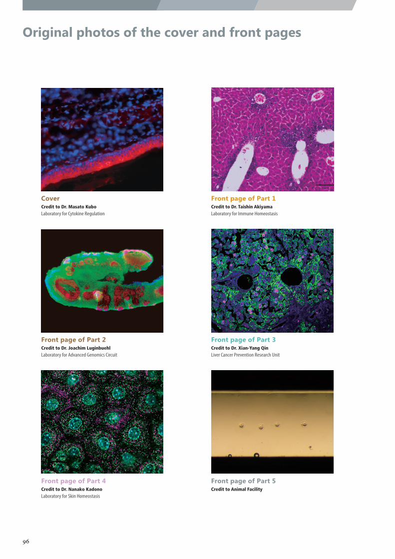
Original photos of the cover and front pages
CoverCredit to Dr. Masato KuboLaboratory for Cytokine Regulation
Front page of Part 1Credit to Dr. Taishin AkiyamaLaboratory for Immune Homeostasis
Front page of Part 2Credit to Dr. Joachim LuginbuehlLaboratory for Advanced Genomics Circuit
Front page of Part 3Credit to Dr. Xian-Yang QinLiver Cancer Prevention Research Unit
Front page of Part 4Credit to Dr. Nanako KadonoLaboratory for Skin Homeostasis
Front page of Part 5Credit to Animal Facility
96
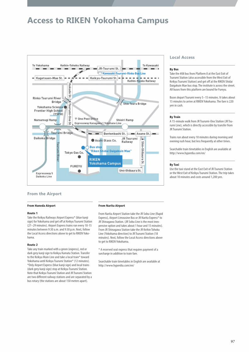
Access to RIKEN Yokohama Campus
By BusTake the #08 bus from Platform 8 at the East Exit of Tsurumi Station (also accessible from the West Exit of Keikyu Tsurumi Station) and get off at the RIKEN Shidai Daigakuin Mae bus stop. The institute is across the street. All buses from this platform are bound for Fureyu.
Buses depart Tsurumi every 5–15 minutes. It takes about 15 minutes to arrive at RIKEN Yokohama. The fare is 220 yen in cash.
From Haneda Airport
Route 1Take the Keikyu Railways Airport Express* (blue kanji sign) for Yokohama and get off at Keikyu Tsurumi Station (27–29 minutes). Airport Express trains run every 10-15 minutes between 9:30 a.m. and 9:30 p.m. Next, follow the Local Access directions above to get to RIKEN Yoko-hama.
Route 2Take any train marked with a green (express), red or dark grey kanji sign to Keikyu Kamata Station. Transfer to the Keikyu Main Line and take a local train* toward Yokohama until Keikyu Tsurumi Station* (12 minutes).*Only Airport Express (blue kanji sign) and local trains (dark grey kanji sign) stop at Keikyu Tsurumi Station. Note that Keikyu Tsurumi Station and JR Tsurumi Station are two different railway stations and are separated by a bus rotary (the stations are about 150 meters apart).
From Narita Airport
From Narita Airport Station take the JR Sobu Line (Rapid Express), Airport Limousine Bus or JR Narita Express* to JR Shinagawa Station. (JR Sobu Line is the most inex-pensive option and takes about 1 hour and 15 minutes). From JR Shinagawa Station take the JR Keihin Tohoku Line (Yokohama direction) to JR Tsurumi Station (18 minutes). Next, follow the Local Access directions above to get to RIKEN Yokohama.
* A reserved seat express that requires payment of a surcharge in addition to train fare.
Searchable train timetables in English are available at http://www.hyperdia.com/en/
By TrainA 15-minute walk from JR Tsurumi-Ono Station (JR Tsu-rumi Line), which is directly accessible by transfer from JR Tsurumi Station.
Trains run about every 10 minutes during morning and evening rush hour, but less frequently at other times.
Searchable train timetables in English are available at http://www.hyperdia.com/en/
By TaxiUse the taxi stand at the East Exit of JR Tsurumi Station or the West Exit of Keikyu Tsurumi Station. The trip takes about 10 minutes and costs around 1,200 yen.
Local Access
From the Airport
97
RIKEN Center for Integrative Medical Scienceshttp://www.ims.riken.jp/english/
RIKEN Yokohama Campus
1-7-22 Suehiro-cho, Tsurumi-ku, Yokohama City,
Kanagawa, 230-0045, Japan
Tel :045-503-9121
Fax :045-503-9113
Email : [email protected]
RIKEN IMSAnnual Report 2018
RIKEN 2019-038
RIK
EN IM
S An
nu
al Rep
ort 2018 R
IKEN
Cen
ter for In
tegrative M
edical Scien
ces