restraint stress and repeated corticotrophin-releasing factor receptor activation in the amygdala...
TRANSCRIPT

Restraint stress and repeated CRF receptor activation in theamygdala both increase amyloid β precursor protein (APP) andamyloid-β (Aβ) peptide but have divergent effects on BDNF andpre-synaptic proteins in the prefrontal cortex of rats
Balmiki Ray, Denise L. Gaskins, Tammy J. Sajdyk, John P. Spence, Stephanie D. Fitz,Anantha Shekhar, and Debomoy K. Lahiri*Department of Psychiatry, Institute of Psychiatric Research, Indiana University School ofMedicine, 791 Union Drive, Indianapolis, IN 46202, USA
AbstractBoth environmental stress and anxiety may represent important risk factors for Alzheimer'sdisease (AD) pathogenesis. Previous studies demonstrate that restraint stress is associated withincreased amyloid beta (Aβ) and decreased brain-derived neurotrophic factor (BDNF) levels in thebrain. Aβ deposition, synaptic loss, and neurodegeneration define major hallmarks of AD, andBDNF is responsible for the maintenance of neurons. In contrast to restraint stress, repeatedinjections of sub-anxiogenic doses of the corticotrophin releasing factor receptor agonisturocortin1 (Ucn1) administered in the basolateral amygdala (BLA) of rats elicits persistentanxiety-like responses. We hypothesized that both restraint stress and Ucn1-induced anxietywould contribute to a neurobiological abnormality that would change the levels of Aβ precursorprotein (APP) and Aβ as well as BDNF and pre-synaptic markers. In the first experiment, adultmale Wister rats (N=5) were subjected to three-hour restraint, as compared to unstressed controls.In the second experiment, adult male Wistar rats (N=6) were subjected to sub-anxiogenic doses ofUcn1 (6 fmol/100 nl) administered in the BLA, as compared to controls. Following eachrespective treatment, the social interaction (SI) test was performed to measure anxiety-likebehavior. Protein studies were then conducted to quantify levels of APP, Aβ, BDNF andpresynaptic proteins in the prefrontal cortex (PFC). In both experiments, we detected differencesin either corticosterone levels or the SI test associated with a stress response. Our findings indicatethat both restraint stress and Ucn1 administration in the BLA lead to increased APP and Aβdeposition. However, restraint-induced stress leads to reductions in the levels of BDNF andpresynaptic markers, while Ucn1-induced anxiety is associated with increases in the levels of eachrespective protein. This demonstrates a convergent role for stress response and Ucn1-inducedanxiety in the regulation of APP and Aβ, but opposing roles for each respective treatment in theregulation of BDNF and presynaptic markers.
© 2011 IBRO. Published by Elsevier Ltd. All rights reserved.*Correspondence: Professor Debomoy K. Lahiri, Ph.D. Indiana University School of Medicine Institute of Psychiatric Research 791Union Drive, Indianapolis, IN 46202 Tel: (317) 274-2706; Fax: (317) 274-1365 [email protected].
Publisher's Disclaimer: This is a PDF file of an unedited manuscript that has been accepted for publication. As a service to ourcustomers we are providing this early version of the manuscript. The manuscript will undergo copyediting, typesetting, and review ofthe resulting proof before it is published in its final citable form. Please note that during the production process errors may bediscovered which could affect the content, and all legal disclaimers that apply to the journal pertain.
NIH Public AccessAuthor ManuscriptNeuroscience. Author manuscript; available in PMC 2012 July 08.
Published in final edited form as:Neuroscience. 2011 June 16; 184: 139–150. doi:10.1016/j.neuroscience.2011.03.067.
NIH
-PA Author Manuscript
NIH
-PA Author Manuscript
NIH
-PA Author Manuscript

KeywordsAging; Alzheimer; Anxiety; APP; BDNF; Brain; CNS; Dementia; Mental illness; Restraint stress;Social interaction; Synaptic proteins; Ucn1
INTRODUCTIONAlzheimer's disease (AD) is a complex neurodegenerative disorder that is influenced bymultiple factors including genetics, the environment, and gene × environment interactions(Lahiri et al., 2009). To date, a growing body of evidence has implicated psychologicalstress and anxiety as potential contributing factors to the development of AD (Wilson et al.,2005, Csernansky et al., 2006, Palmer et al., 2007). A major hallmark feature of AD is thedeposition of the amyloid-β (Aβ) peptide. In patients with AD, Aβ peptide is deposited asplaques in the central nervous system (CNS), and Aβ deposition is associated withneurodegeneration in AD (Selkoe, 2008). Previous studies in rodents demonstrate that anacute stressor leads to increases in the formation of Aβ peptide, and these increases can bedetected in the levels of both amyloid-β precursor protein (APP) messenger RNA (mRNA)and Aβ peptides (Rosa et al., 2005, Kang et al., 2007). Due to its effects on the levels ofAPP and Aβ in the CNS, these findings provide evidence that stress may be a potentialcontributing factor for the development of AD. Likewise, the downstream effects of stresson neurotrophic factors and presynaptic proteins also represent important molecular targetsassociated with AD pathophysiology (Tapia-Arancibia et al., 2008).
APP is a transmembrane protein that is cleaved by β and γ secretase to generate Aβ, and Aβdeposition forms plaques observed in AD patients (Sambamurti et al., 2002). APP can becleaved in neuronal and non-neuronal cells by two different proteolytic pathways. Forinstance, the α-secretase protein cleaves APP within its Aβ domain to produce sAPPα. This‘non-amyloidogenic pathway’ precludes the production of the Aβ peptide (Ray et al.,2009b). On the contrary, β-secretase cleaves the N-terminus of the Aβ peptide sequence ofAPP, and then γ-secretase further cleaves the protein to produce Aβ peptide, a mechanismdefined as the ‘amyloidogenic’ pathway (Sambamurti et al., 2002). This mechanism leads tothe production of Aβ with 42 amino acids residue (Aβ 1-42) and Aβ with 40 amino acidsresidue (Aβ 1-40). The larger form of Aβ (i.e. Aβ 1-42) leads to more aggregates than theshorter form (i.e. Aβ 1-40) in AD patients. Deposited Aβ peptide, especially Aβ (1-42), canlead to severe neuro-inflammation and neurodegeneration due to the production of reactiveoxygen species (ROS). In AD patients, significant decreases in the levels of brain derivedneurotrophic factor (BDNF) have been documented in hippocampal and cortical regions(Hock et al., 2000). In addition, previous studies demonstrate that a single or repeatedrestraint-induced stress in rats leads to decreases in BDNF mRNA levels in the hippocampus(Smith et al., 1995). BDNF and other neurotrophins regulate multiple cellular functions bysupporting the development, the differentiation and the maintenance of neurons (Cohen-Cory et al., 1996). Therefore, neurotrophins are essential for normal brain functionthroughout life.
Corticotrophin releasing factor (CRF) plays a critical role in activating the behavioral andphysiological responses to stress. Its biological function is carried out through activation oftwo receptor subtypes, corticotropin-releasing factor receptor 1 (CRFR1) and CRF receptor2 (CRFR2). CRFR1 and CRFR2 receptors are 70% homologous at the protein level andcontain a putative signal peptide, an extra cellular N-terminal domain (ECD1) and seventransmembrane domains. CRFR1 receptors are distributed throughout the brain, whereas thelocation of the CRFR2 receptors is more restricted to specific brain regions (Chalmers et al.,1995, Van Pett et al., 2000). The mammalian family of ligands for the CRFR1 and CRFR2
Ray et al. Page 2
Neuroscience. Author manuscript; available in PMC 2012 July 08.
NIH
-PA Author Manuscript
NIH
-PA Author Manuscript
NIH
-PA Author Manuscript

receptors includes CRF, urocortin I (UCN1), UCN II, and UCN III. These ligands differ intheir tissue distribution and receptor pharmacology. For example, CRF binds to CRFR1 withhigh affinity, whereas UCN1 binds with high affinity to both CRFR1 and CRFR2(Lovenberg et al., 1995). UCN II and UCN III appear to be selective for CRFR2 (Lewis etal., 2001, Inoue et al., 2003).
CRF is the principal neuroregulator of the hypothalamic-pituitary-adrenal (HPA) axis, and isthe major mediator of the stress response (Bale and Vale, 2004). Following a stressor, CRFis released from the paraventricular nucleus (PVN) of the hypothalamus activating the HPAaxis. CRF then binds to CRFR1 in the anterior pituitary resulting in the secretion of adrenalcorticotrophic hormone (ACTH). ACTH then stimulates the release of glucocorticoids (i.e.corticosterone) from the adrenal cortex that act via a negative-feedback system to inhibitfurther CRF release from the hypothalamus. Corticosterone binds primarily to two receptortypes including mineralocorticoid receptors (MR) and glucocorticoid receptors (GR). Inresponse to stress, CRFR2 may function as an inhibitory or modulatory receptor to dampenHPA activation (Bale and Vale, 2004).
In humans, chronic stress is associated with the development of psychiatric disorders insusceptible individuals including anxiety and depression (Arborelius et al., 1999,Rosenkranz et al., 2010). Additionally, chronic stress leads to changes in the amygdala inrodents, a brain region implicated in both anxiety and fear-based learning (Bale and Vale,2004). For instance, both electrical and pharmacological stimulation of the amygdalainduces an enhanced cardiovascular response and behavioral arousal consistent with a fight-or-flight response (Kapp et al., 1982, al Maskati and Zbrozyna, 1989). By selectivelytargeting the basolateral amygdala (BLA) using pharmacological manipulation, previousstudies demonstrate that the amygdala also regulates social aspects of anxiety and fear-basedlearning (Sanders and Shekhar, 1995, Sajdyk and Shekhar, 2000, Sajdyk et al., 2008). Forexample, mimicking repeated episodes of the stress response, repeated sub-anxiogenic dosesof the CRF receptor agonist urocortin1 (Ucn1) microinjected into the basolateral amygdala(BLA) of rats once a day for 5 consecutive days (termed ‘priming’) leads to the developmentof pathological anxiety in that long-lasting behavioral changes are observed in socialinteraction (SI) and elevated plus maze (EPM) tests of anxiety (Rainnie et al., 2004).Likewise, rats primed with Ucn1 in the BLA demonstrated both increased anxiety-likebehaviors as well as physiological sensitivity to intravenous sodium lactate infusions(Sajdyk and Shekhar, 2000). This physiological response to lactate infusion has beendocumented in subjects with panic or posttraumatic stress disorders, but not social orgeneralized anxiety disorders.
Given the previously documented involvement of stress and anxiety in the regulation of ADbiomarkers, we hypothesized that restraint stress and repeated stimulation of CRF receptorswithin the BLA would lead to dysregulation in biomarkers associated with AD. Weobserved significant increases in total intracellular APP and Aβ peptide (x-40) with eachrespective condition, but only detected an increase in the level of Aβ (x-42) following three-hour (hr) restraint-induced stress. Interestingly, three-hr restraint stress negatively regulatesBDNF and pre-synaptic proteins, while Ucn1 administration into the BLA positivelyregulates these proteins. Together, these findings reveal an important role for stress in theregulation of APP and Aβ in rats, and define BDNF as a potential marker of interestassociated with synaptic integrity and the pathophysiology of AD.
Ray et al. Page 3
Neuroscience. Author manuscript; available in PMC 2012 July 08.
NIH
-PA Author Manuscript
NIH
-PA Author Manuscript
NIH
-PA Author Manuscript

MATERIALS AND METHODSAnimals
Male Wistar rats (275–300 g); Harlan Laboratories, Indianapolis, IN, USA) were used inthese experiments. Upon arrival, the animals were housed individually in a temperature-controlled room (22°C) and had access to food and water ad libitum. The room wasmaintained at 12–12 hr light/dark cycle with light on at 0700 hr. Animal care procedureswere conducted in accordance with the National Institutes of Health Guidelines for the Careand Use of Laboratory Animals and the Guidelines of the Indiana University–PurdueUniversity Indianapolis Institutional Animal Care and Use Committee.
Experimental ProtocolIn experiment 1, the rats assigned to the ‘restraint stress’ group (N=5) were placed in adecapicone for three hrs (Brain TreeScientific, Braintree, MA, USA). The unstressed controlrats (N=5) were kept in their home cage during this time period. Social interaction wasmeasured immediately after the restraint stress. In experiment 2, the rats received a dailybilateral i.c. microinjection into the BLA with either vehicle [Veh,1% bovine serumalbumin/100 nl/side] (N=6) or a sub-anxiogenic dose of Urocortin 1 [ (Ucn1), 6 fmoles/100nl/side] (n=6) for five consecutive days. Urocortin1 (Sigma-Aldrich, St Louis, MO, USA)was dissolved in a vehicle of 1% bovine albumin in distilled water. The rats were tested fordifferences in social interaction, as compared to controls 30 minutes following the primingmicroinjection on D5. The brains were harvested for protein studies immediately aftercompletion of the SI test of each experiment.
Surgical ProceduresFor experiment 2, BLA cannulation surgeries were conducted on the rats at least 1 weekfollowing arrival from the supplier. Rats were anesthetized with isoflurane (2.5%) andplaced in a stereotaxic apparatus. Bilateral injection cannulas (26 gauge; Plastics One,Roanoke, VA, USA) were implanted into the BLA [anteroposterior (AP): –2.1; mediolateral(ML): +5.0; dorsoventral (DV): –8.0; incisor bar: –3.3 mm] according to a standardstereotaxic atlas of the rat brain (Paxinos and Watson, 1986). The cannulas were secured tothe skull with three stainless steel screws (2.8 mm; Plastics One) and locktite adhesive(Applied Industrial Technologies, Indianapolis, IN, USA). After completion of surgery, allanimals received buprenex (Sigma, St. Louis, MO; 1 mg/kg, s.c.) and were placed on awarming pad until they had fully recovered from the anesthetic. The rats were allowed torecover in their home cages for at least 5 days prior to any behavioral testing.
Social interaction testFollowing three-hr restraint or Ucn1- priming injections into the BLA, the social interaction(SI) test was utilized to characterize the anxiety-like response to each respective condition.A day before the SI test, the rats were exposed to the test room for at least 30 minutes andthen placed into the SI apparatus alone for a 5 minute habituation session. A previouslystandardized version of the SI test was used to measure social interaction, in which theexperimental rats were placed in an open field (0.9 m long × 0.9 m wide with walls 0.3 mhigh) with a novel male Wistar rat. During the five minute test, the total amount of time thetreated rat initiated interaction with the partner rat was recorded (sniffing, grooming, etc.), asdescribed previously (Shekhar and Keim, 1997). All tests were videotaped andindependently scored at a later time by two individuals who were unaware of the animals’treatment using cumulative stopwatches. Between each session, the apparatus were cleanedwith 70% ethanol.
Ray et al. Page 4
Neuroscience. Author manuscript; available in PMC 2012 July 08.
NIH
-PA Author Manuscript
NIH
-PA Author Manuscript
NIH
-PA Author Manuscript

Dissection and preparation of brain lysateAfter the SI test all animals were anaesthetized and promptly decapitated. The brains werecarefully removed, and the frontal cortices were dissected and stored in -80°C freezer forfurther analysis. Brain dissections were carried out as described earlier (Sajdyk et al., 2008).To prepare protein lysates, the frozen tissues were homogenized in Tris-HCl buffered saline(TBS) [pH 7.6] containing 140 mM NaCl, 3 mM KCl, 25 mM Tris-HCl (pH 7.6), 5 mMEDTA, 2mM Phenanthroline, 0.5% SDS, EDTA-free protease inhibitor cocktail that wassupplemented with protease inhibitor (Roche, Indianapolis, IN, USA). The resulting sampleswere then homogenized using ‘polytron’ homogenizer, and were centrifuged at 4°C(12,000g) for 10 minutes to obtain TBS soluble fraction of the tissue. This fraction wassubjected to protein estimation using the Bradford assay, as previously described (Ray et al.,2009a). The resulting volume of supernatant containing a fixed amount of protein wasanalyzed for Western immunoblotting.
Corticosterone assay in the plasma in rats following restraint stress verses controlsAfter the SI test, trunk blood was collected from the rats following restraint stress and incontrols. Whole blood was centrifuged at 10,000×g to collect plasma. Plasma samples werealiquoted and frozen at -80°C freezer. To measure corticosterone, a competitive EIA assaywas used (Enzo Life Sciences, Plymouth Meeting, PA, USA). Briefly, 97.5 μl of plasmawas added with 2.5 μl of corticosterone displacement reagent to displace boundcorticosterone present in plasma. The plasma samples were diluted 1:20, and the assay wascarried out as per manufacturer's protocol.
Western immunoblottingEqual amounts of protein from the denatured lysates from each respective experimental andcontrol groups were loaded in 10% Bis-Tris ‘Criterion’ polyacrylamide gels (BioRad,Hercules, CA, USA) and separated at 180 V for 2 hrs. Proteins were then electrophoreticallytransferred onto a PVDF membrane (BioRad) using the chilled transfer buffer (25 mM Trisbase, 200mM Glycine and 20% Methanol) at 30V for 5 hrs at 4°C. After the protein transfer,the membrane was blocked in 5% nonfat milk in a Tris buffered saline, (pH 7.4) containing0.05% Tween-20 (TBST) at room temperature (RT) for 1 hr. Different parts of themembrane were then probed with 22C11 (Amyloid Precursor protein, Millipore, Billerica,MA, USA) and β-actin (Sigma-Aldrich, USA) antibody. Brain homogenate (15μg ofprotein) was treated with 1(N) HCL to bring the pH down to 2.5 and incubated at roomtemperature for 15 minutes. The acid treated lysates were subsequently treated with 1(N)NaOH to bring back to normal neutral pH. Acid treatment increases detectable amount ofneurotrophic factors in the sample (Okragly and Haak-Frendscho, 1997). Acid treatedsamples were mixed with Laemmli buffer and ran in a 10% SDS-PAGE, as previouslydescribed. The membrane was then probed with BDNF (1:1000 dil) (Abcam, Cambridge,MA, USA) and β-actin antibody (1: 100,000 dil). Levels of pre-synaptic proteins (SNAP25;1:10,000dil and syntaxin6; 1: 3000 dil) were also measured in a similar fashion usingspecific antibodies (Millipore and BD Bioscience, Rockville, MD, USA, respectively).Detection of protein band signals was achieved by adding chemoluminescent buffer (GE,Buckinghamshire, UK) to the blot, which was immediately photographed using GEchemoluminescent detection film. A specific strip of the membrane was also probed withmonoclonal anti-β-actin antibody (Sigma) for normalization purposes.
ELISAELISA was utilized to detect BDNF in an acid treated brain homogenate according to themanufacturer's protocol (Promega, Madison, WI, USA). The resulting BDNF levels (in pg/ml) were normalized by protein content of the lysates (μg/ml) and the unit was converted to
Ray et al. Page 5
Neuroscience. Author manuscript; available in PMC 2012 July 08.
NIH
-PA Author Manuscript
NIH
-PA Author Manuscript
NIH
-PA Author Manuscript

pg/μg of lysate. Additionally, sensitive chemiluminescence Aβ assays were utilized tomeasure Aβ (x-40) and Aβ (x-42) in the rat cortical homogenate (Covance, Princeton, NJ,USA). The individual kit measures Aβ (x-40) and Aβ (x-42), respectively. (x-40) ELISA kitdetects full length sequence of Aβ (1-40) and also peptides generated by N-terminalcleavages, such as Aβ (3-40), Aβ (11-40) etc. Similarly (x-42) ELISA kit detects full lengthsequence of Aβ (1-42) and also peptides generated by N-terminal cleavages, such as Aβ(3-42), Aβ (11-42) etc. Chemiluminescence signals obtained in Aβ ELISAs werenormalized by the protein content of the lysates and plotted as ‘% control’.
Statistical analysesStatistical analyses were performed by SPSS using a student's t-test, and the results wereplotted using GraphPad Prism 4.0 software (GraphPad Software, La Jolla, CA, USA). Alldata were presented as mean ± SEM, and p-value < 0.05 were considered significant for allanalyses. For SI, a one-tailed t-test was performed, which was consistent with our a priorihypothesis.
RESULTSBoth three-hr restraint and repeated Ucn1 injections into the BLA lead to decreases insocial interaction in rats
The social interaction test was performed to measure the effects of three-hr restraint andrepeated Ucn1 administration on anxiety-like behavior in rats. Both three-hr restraint stress,and Ucn1 administration resulted in decreases in social interaction in rats (p=0.06 and0.0008 respectively) (Figure 1A and 1B). To confirm that three-hr restraint stress leads tothe activation of the HPA-axis, plasma corticosterone levels were determined followingrestraint stress. Plasma corticosterone was significantly increased in the plasma of ratsfollowing restraint stress versus controls (Figure 1C).
Both three-hr restraint and repeated Ucn1 injections into the BLA increases intracellularlevels of APP and amyloid-β (x-40)
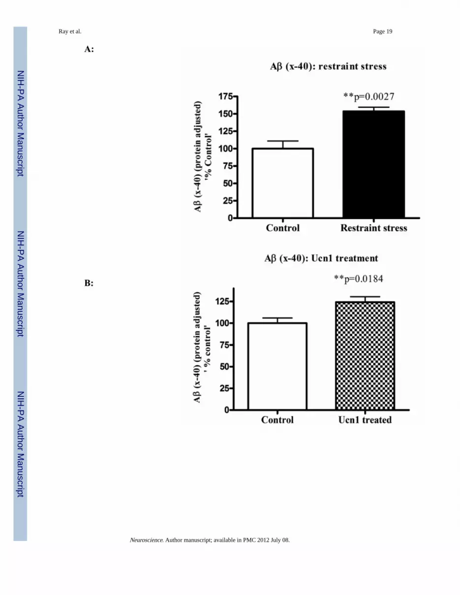
Following the social interaction test, the rats were decapitated, and brain lysates wereisolated from cortical tissue. Western immunoblotting revealed a significant increase(p=0.008 and p=0.0002 respectively)) in the levels of total intracellular APP following boththree-hr restraint and repeated Ucn1 injections into the CNS (Figure 2A and 2B). The totalAPP bands were normalized with β-actin bands. The levels of Aβ (x-40) were significantlyincreased (p=0.0027) in the cortex following both three-hr restraint stress and repeated Ucn1injections verses controls (Figure 3A and 3B). While we observed a significant increase(p=0.0048) in the level of Aβ (x-42) in the frontal cortex following restraint stress (Figure3C), repeated Ucn1 injections into the CNS did not affect cortical levels of Aβ (x-42)(p=0.6) (Figure 3D).
Three-hr restraint decreases and repeated Ucn1 injections the BLA increases intracellularlevels of BDNF, respectively
Following three-hr restraint stress, we observed a significant decrease (p=0.025) in the brainlevels of BDNF by Western immunoblotting in the stressed rats versus controls (Figure 4A).On the contrary, the repeated injection of Ucn1 resulted in increased levels of BDNF(p=0.02) in the frontal cortex of Ucn1 primed animals verses controls (Figure 5A). Theseresults were further confirmed utilizing an ELISA that is sensitive to BDNF detection(Figure 4B and 5B). For ELISA, individual BDNF values (pg/ml) were converted to pg/μgby normalizing protein content of the corresponding brain lysate as measured by theBradford assay.
Ray et al. Page 6
Neuroscience. Author manuscript; available in PMC 2012 July 08.
NIH
-PA Author Manuscript
NIH
-PA Author Manuscript
NIH
-PA Author Manuscript

Three-hr restraint decreases whereas repeated Ucn1 injections into the BLA increasesintracellular levels of pre-synaptic proteins, respectively
Pre-synaptic markers provide an important biological measure of neuronal integrity as wellas synaptic plasticity. Western immunoblot analyses of pre-synaptic proteins syntaxin6revealed a significant decrease (p=0.008) in the cortex following three hr restraint versuscontrols (Figure 6A), and a decreasing trend for SNAP25 levels (p=0.08; Figure 6B).Following repeated Ucn1 injections, significant increases in the levels of syntaxin6 (p=0.02)and SNAP25 (p=0.0076) were detected in the cortical lysate versus controls (Figure 7A and7B).
DiscussionIn the present study, we observed reductions in social interaction associated with both three-hr restraint stress and Ucn1 repeated injections into the BLA. Additionally, we foundsignificant increases in total intracellular APP and Aβ peptide (x-40) associated with eachrespective condition. However, a significant increase was only observed in the level of Aβ(x-42) following three-hr restraint-induced stress. While we observed significant decreasesin the level of BDNF in the cortical lysate from rats after three-hr restraint stress, Ucn1administration resulted in significant increases in the level of BDNF in the cortex. Westernimmunoblotting and ELISA studies revealed decreases in the levels of pre-synaptic proteins(syntaxin6 and SNAP25) in the cortical lysate following three-hr restraint stress; however,the levels of SNAP25, a pre-synaptic protein of SNARE complex, was significantlyincreased in the cortical lysate following Ucn1-induced anxiety.
Late onset AD is the most common cause of dementia in the elderly population, and oftendisplays a sporadic mode of transmission (Lahiri et al., 2003). As opposed to a purelygenetic approach to evaluate the risk factors, more emphasis in recent years has focused onthe contribution of environmental factors associated with AD (Migliore and Coppede, 2009,Zawia et al., 2009). For example, an epidemiological study in Nigeria revealed noassociation between the well known risk gene APOE ε 4 and AD in a local population,which further emphasizes the role of environmental factors in the pathogenesis of AD(Gureje et al., 2006). Consistent with other environmental factors that underlie AD, recentstudies indicate that stress may also represent an important risk factor (Smith et al., 2005,Wilson et al., 2005).
A previous study demonstrated that acute restraint stress leads to increases in the levels ofAβ in brain interstitial fluid (ISF), and the effect was mediated by a corticotropin-releasingfactor (CRF)-dependent mechanism (Kang et al., 2007). In this study, it was postulated thatthe increase in the levels of ISF Aβ is likely due to the increase in neuronal activityassociated with CRF, which was increased in response to stress. In a cell culture basedstudy, APP processing leads to a shift towards an intracellular route following stimulationby heat shock protein in human astrocyte (Shepherd et al., 2000). Additionally, a recentstudy using gene expression profiling demonstrates a 1.64-fold increase in APP (App)expression in DBA/2J mice following the force swim test, a behavioral test that activates astress response (Tsolakidou et al., 2010). However, the effect of acute restraint stress onAPP has not yet been addressed. In the present study, we observed a significant increase(~55%) in the levels of APP in frontal cortex following restraint stressed versus unstressedrats. Because APP was significantly increased in the frontal cortical lysate followingrestraint stress, we further assayed the levels of Aβ peptide (both x-40 and x-42). Wedetected significant increases in Aβ peptide (both x-40 and x-42). This increase in Aβ caneither result from increased intracellular processing of APP, the up-regulation of BACE-1 ora direct effect that can be attributed to the action of CRF on neurons (Tamagno et al., 2005,Kang et al., 2007). While the exact mechanism that underlies this increase in Aβ peptides
Ray et al. Page 7
Neuroscience. Author manuscript; available in PMC 2012 July 08.
NIH
-PA Author Manuscript
NIH
-PA Author Manuscript
NIH
-PA Author Manuscript

following restraint stress is not fully understood, a shift in APP processing towards theintracellular compartment lead to an “amyloidogenic state” in the neuron (LaFerla et al.,2007). In the present study, we observed a significant increase in corticosterone levelsfollowing restraint stress. Therefore, increases in corticosterone and other hormones inresponse to the activation of the HPA axis following a stressful condition may independentlyup-regulate the levels APP and BACE-1, which may ultimately contribute to the formationof Aβ peptides (Green et al., 2006). A previous study has also shown that CRF itself canindependently stimulate neurons that can lead to the release of more Aβ peptides (Kang etal., 2007).
In addition to the effects on APP and Aβ, we also detected significant decreases in the levelsof BDNF in the frontal cortex following restraint stress versus unstressed rats. In a previousstudy, a significant decrease in BDNF positive cells was detected following chronic isolationin frontal cortex and hippocampus that is consistent with our findings (Chen et al., 2008).Additionally, a previous study found that 6 hrs of restraint stress in rats is also associatedwith significant decreases in BDNF mRNA levels in the hippocampus (Murakami et al.,2005). Because BDNF plays a critical role in the regulation of synaptic plasticity, wehypothesized that acute restraint stress would lead to decreases in the levels of pre-synapticproteins. We found significant decreases in the levels of the pre-synaptic protein synataxin-6in the prefrontal cortical lysate following restraint stressed versus unstressed rats. Inaddition, the levels of the pre-synaptic protein SNAP25 were also lower in the stressedgroup; however, the difference did not reach significance. Consistent with these findings,previous findings indicate that BDNF treatment in organotypic hippocampal slice cultureresults in an increase in both the number of synapses as well as docked synaptic vesicles(Tyler and Pozzo-Miller, 2001). Interestingly, the lack of BDNF can also play importantroles in trafficking of APP. For example, the exogenous administration of BDNF in primaryneuron culture leads to decreases in Aβ production mediated through up-regulation ofSORLA (Rohe et al., 2009). Hence, decreased levels of BDNF following restraint stress mayalso contribute to the increases in Aβ production following restraint stress.
Stress resulting from physical restraint leads to a complex physiological response, andinvolves multiple structures in the CNS including the amygdala and the hypothalamus. Tospecifically target the effects of the amygdaloid nuclei on APP and Aβ, site directedinjections of Ucn1 were conducted into the BLA, a brain region that is known to mediate theeffects of CRF on anxiety. Interestingly, different molecular sequelae were observedfollowing repeated activation of CRF receptors with Ucn1 treatment, as compared torestraint stress. Ucn1 is a peptide that shows sequence homology with both urotensin 1 andCRF (Vaughan et al., 1995), and produces anxiety-like behavior in rodents (Moreau et al.,1997). In fact, the site directed injection of Ucn1 into the BLA of rats acts as a potentanxiogenic peptide, and leads to a more robust effect on anxiety-like behavior than that ofCRF (Sajdyk et al., 1999). Therefore, we hypothesized that the levels of APP and Aβpeptides would also be increased in Ucn1-treated rat in the frontal cortex. In the presentstudy, we observed a robust increase in the levels of APP in Ucn1 injected rats consistentwith our findings in rats following restraint stress. Additionally, we observed a significantincrease in the level of Aβ (x-40) in the frontal cortical lysate of Ucn1-treated rats versusuntreated controls. However, the levels of Aβ (x-42) were left unchanged. These findingssuggest that the increases APP may underlie the increases seen in Aβ peptides that wereobserved following both Ucn1 treatment and restraint stress.
In contrary to the decreases observed following restraint stress, we observed significantincreases in the levels of BDNF in the frontal cortex of Ucn1 injected rats. Although chronicstressors increase APP and other markers of AD in adult rodents (Dong et al., 2008), asimilar increase in cortical BDNF levels are seen in very early adolescent rats following
Ray et al. Page 8
Neuroscience. Author manuscript; available in PMC 2012 July 08.
NIH
-PA Author Manuscript
NIH
-PA Author Manuscript
NIH
-PA Author Manuscript

short-term social isolation stress where significant synaptic reorganization is thought tooccur (Meng et al., 2011). Additionally, a previous study demonstrates that CRFR1 receptorsignaling in cerebellar granular cells leads to increases in BDNF mRNA levels (Bayatti etal., 2005). Likewise, CRFR1 receptor signaling in locus coeruleus also increases BDNFsignaling via ERK-MAPK cascade (Bayatti et al., 2005). Since Ucn1 also has primarystimulatory effects on CRFR2 receptor, the increase in the levels of BDNF may potentiallybe due to CRFR2 mediated effects on neurons projecting from the amygdala to theprefrontal cortex.
Consistent with the increase of BDNF in the frontal cortex following Ucn1 injections intothe BLA, we also observed significant increases in the levels of pre-synaptic proteinsSNAP25 and syntaxin6 in Ucn1 injected rats versus controls. Hence, repeated Ucn1injections into the BLA nucleus results in a complex cascade of signal transduction events.Our findings suggest that the increases in APP and Aβ peptide and BDNF may result fromthe effects on CRFR1 receptors. Likewise, the increases in BDNF may underlie theincreases in the levels of pre-synaptic proteins SNAP25 and syntaxin6. A previous study hasrevealed that decreases in BDNF levels are mediated by Aβ (1-42) (Hjorth et al., 2010).Interestingly, the BDNF level is associated with phagocytosis of Aβ (1-42) by macrophases(Asami et al., 2006). In a cell culture model, BDNF was found to protect neurons from Aβ(1-42)-mediated damage. Therefore, increases in the levels of BDNF may be responsible forthe lack of increase in Aβ (x-42) levels in the frontal cortex in Ucn1 injected rats.Ultimately, the increases seen in BDNF and pre-synaptic proteins could be due tocompensatory mechanism in response to chronic Ucn1 injections into the BLA associatedwith increases in APP and Aβ generation.
Mechanistically, whether the aforementioned restraint-induced stress or Ucn1-inducedanxiety triggers cellular oxidative stress remains unclear. It is known that aging andneurodegenerative disorders are associated with increased cellular oxidative stress; however,we have not directly assayed oxidative stress markers in the present work because of theexperimental design. Nevertheless, we do not rule out the possibility that the induced stresstested in this work may cause oxidative stress resulting from different reactive oxygenspecies such as superoxide, hydrogen peroxide and hydroxyl radicals. The effect ofoxidative stress and growth factors in the regulation of neuronal gene expression, such asAPP and BACE, has been studied (Lahiri and Nall, 1995, Ge et al., 2004, Lahiri et al.,2006). Further, transcriptional activation of APP gene by stress was previously reported(Dewji et al., 1995). The interaction of various transcription factors with the BACE1promoter can modulate synaptic plasticity, neuronal apoptosis and oxidative stress, whichare relevant to the pathogenesis of AD (Lahiri et al., 2006). It would be interesting tospeculate the underlying mechanism. Nuclear factor erythroid 2-related factor 2 (NrF2),which protects against Aβ (Kanninen et al., 2008), is known to play a role in antioxidantdefense in the cell and has been shown to be neuroprotective in an animal model making aninteresting target for studies into preventing or reversing neurodegeneration in dementias (deVries et al., 2008, Ohtsuji et al., 2008). Analysis of the transcription factor binding sites onboth APP and BACE, using the TESS program (Transcriptional Element Search System)reveals sites for heat shock element and the transcription factor Nrf2 in the APP and BACE1promoter (Lahiri and Robakis, 1991, Sambamurti et al., 2004). Another study suggests thatp-hydroxybenzyl alcohol (HBA) protects against brain damage by modulatingcytoprotective genes, such as NrF2, and neurotrophic factors, including BDNF (Kam et al.,2011). The above results taken with our present data suggest that restraint-induced stressmay cause cellular oxidative stress, which results in down regulation of cytoprotective genessuch as BDNF and in upregulation of APP gene expression leading to the amyloidogenicpathway. Thus, restraint-induced stress triggers APP and Aβ peptide expression at the costof cytoprotective BDNF and synaptic proteins. Our present study would have great
Ray et al. Page 9
Neuroscience. Author manuscript; available in PMC 2012 July 08.
NIH
-PA Author Manuscript
NIH
-PA Author Manuscript
NIH
-PA Author Manuscript

translational implication in understanding the neurobiology and therapeutic targets for thestress-related psychiatric disorders, such as AD, anxiety, depression and schizophrenia.
In conclusion, our findings identified significant increases in APP and Aβ levels followingboth restraint stress and sub-anxiogenic doses of Ucn1 administered into the BLA. Becausethe regulation of APP and Aβ deposition represent biological markers that are associatedwith AD pathogenesis, environmental stressors and persistent anxiety may representpredisposing factors that may contribute to AD pathogenesis. Furthermore, these findingsdemonstrate a negative role for restraint stress and a positive role for Ucn1-induced anxietyin the regulation of BDNF and presynaptic markers. These results indicate that the levels ofAPP and Aβ are likely regulated by distinct mechanisms from BNDF and pre-synapticmarkers following restraint stress and repeated Ucn1 injections into the BLA. Ultimately,the effects of stress and persistent anxiety likely define important factors that can contributeto AD. Overall, the present molecular-driven study in vivo may contribute a greattranslational impact for AD and anxiety, and pave the way for identifying biomarkers andnovel therapeutic strategies for these devastating disorders.
AcknowledgmentsWe thank Justin Long and Jason bailey for technical assistance. We are also thankful to Philip Johnson for fruitfuldiscussion. This work was supported by grants from the National Institute of Health (AG 18379 and AG 18884) toDKL and (RO1 MH065702) to AS.
REFRERENCEal Maskati HA, Zbrozyna AW. Stimulation in prefrontal cortex area inhibits cardiovascular and motor
components of the defence reaction in rats. J Auton Nerv Syst. 1989; 28:117–125. [PubMed:2625500]
Arborelius L, Owens MJ, Plotsky PM, Nemeroff CB. The role of corticotropin-releasing factor indepression and anxiety disorders. J Endocrinol. 1999; 160:1–12. [PubMed: 9854171]
Asami T, Ito T, Fukumitsu H, Nomoto H, Furukawa Y, Furukawa S. Autocrine activation of culturedmacrophages by brain-derived neurotrophic factor. Biochem Biophys Res Commun. 2006;344:941–947. [PubMed: 16631618]
Bale TL, Vale WW. CRF and CRF receptors: role in stress responsivity and other behaviors. AnnuRev Pharmacol Toxicol. 2004; 44:525–557. [PubMed: 14744257]
Bayatti N, Hermann H, Lutz B, Behl C. Corticotropin-releasing hormone-mediated induction ofintracellular signaling pathways and brain-derived neurotrophic factor expression is inhibited by theactivation of the endocannabinoid system. Endocrinology. 2005; 146:1205–1213. [PubMed:15591144]
Chalmers DT, Lovenberg TW, De Souza EB. Localization of novel corticotropin-releasing factorreceptor (CRF2) mRNA expression to specific subcortical nuclei in rat brain: comparison withCRF1 receptor mRNA expression. J Neurosci. 1995; 15:6340–6350. [PubMed: 7472399]
Chen JX, Li W, Zhao X, Yang JX. Effects of the Chinese traditional prescription Xiaoyaosandecoction on chronic immobilization stress-induced changes in behavior and brain BDNF, TrkB,and NT-3 in rats. Cell Mol Neurobiol. 2008; 28:745–755. [PubMed: 17647101]
Cohen-Cory S, Escandon E, Fraser SE. The cellular patterns of BDNF and trkB expression suggestmultiple roles for BDNF during Xenopus visual system development. Dev Biol. 1996; 179:102–115. [PubMed: 8873757]
Csernansky JG, Dong H, Fagan AM, Wang L, Xiong C, Holtzman DM, Morris JC. Plasma cortisoland progression of dementia in subjects with Alzheimer-type dementia. Am J Psychiatry. 2006;163:2164–2169. [PubMed: 17151169]
de Vries HE, Witte M, Hondius D, Rozemuller AJ, Drukarch B, Hoozemans J, van Horssen J. Nrf2-induced antioxidant protection: a promising target to counteract ROS-mediated damage inneurodegenerative disease? Free Radic Biol Med. 2008; 45:1375–1383. [PubMed: 18824091]
Ray et al. Page 10
Neuroscience. Author manuscript; available in PMC 2012 July 08.
NIH
-PA Author Manuscript
NIH
-PA Author Manuscript
NIH
-PA Author Manuscript

Dewji NN, Do C, Bayney RM. Transcriptional activation of Alzheimer's beta-amyloid precursorprotein gene by stress. Brain Res Mol Brain Res. 1995; 33:245–253. [PubMed: 8750883]
Dong H, Yuede CM, Yoo HS, Martin MV, Deal C, Mace AG, Csernansky JG. Corticosterone andrelated receptor expression are associated with increased beta-amyloid plaques in isolated Tg2576mice. Neuroscience. 2008; 155:154–163. [PubMed: 18571864]
Ge YW, Maloney B, Sambamurti K, Lahiri DK. Functional characterization of the 5' flanking regionof the BACE gene: identification of a 91 bp fragment involved in basal level of BACE promoterexpression. FASEB J. 2004; 18:1037–1039. [PubMed: 15059977]
Green KN, Billings LM, Roozendaal B, McGaugh JL, LaFerla FM. Glucocorticoids increase amyloid-beta and tau pathology in a mouse model of Alzheimer's disease. J Neurosci. 2006; 26:9047–9056.[PubMed: 16943563]
Gureje O, Ogunniyi A, Baiyewu O, Price B, Unverzagt FW, Evans RM, Smith-Gamble V, Lane KA,Gao S, Hall KS, Hendrie HC, Murrell JR. APOE epsilon4 is not associated with Alzheimer'sdisease in elderly Nigerians. Ann Neurol. 2006; 59:182–185. [PubMed: 16278853]
Hjorth E, Frenkel D, Weiner H, Schultzberg M. Effects of immunomodulatory substances onphagocytosis of abeta(1-42) by human microglia. Int J Alzheimers Dis. 2010 2010.
Hock C, Heese K, Hulette C, Rosenberg C, Otten U. Region-specific neurotrophin imbalances inAlzheimer disease: decreased levels of brain-derived neurotrophic factor and increased levels ofnerve growth factor in hippocampus and cortical areas. Arch Neurol. 2000; 57:846–851. [PubMed:10867782]
Inoue K, Valdez GR, Reyes TM, Reinhardt LE, Tabarin A, Rivier J, Vale WW, Sawchenko PE, KoobGF, Zorrilla EP. Human urocortin II, a selective agonist for the type 2 corticotropin-releasingfactor receptor, decreases feeding and drinking in the rat. J Pharmacol Exp Ther. 2003; 305:385–393. [PubMed: 12649393]
Kam KY, Yu SJ, Jeong N, Hong JH, Jalin AM, Lee S, Choi YW, Lee CK, Kang SG. p-hydroxybenzylalcohol prevents brain injury and behavioral impairment by activating Nrf2, PDI, and neurotrophicfactor genes in a rat model of brain ischemia. Mol Cells. 2011
Kang JE, Cirrito JR, Dong H, Csernansky JG, Holtzman DM. Acute stress increases interstitial fluidamyloid-beta via corticotropin-releasing factor and neuronal activity. Proc Natl Acad Sci U S A.2007; 104:10673–10678. [PubMed: 17551018]
Kanninen K, Malm TM, Jyrkkanen HK, Goldsteins G, Keksa-Goldsteine V, Tanila H, Yamamoto M,Yla-Herttuala S, Levonen AL, Koistinaho J. Nuclear factor erythroid 2-related factor 2 protectsagainst beta amyloid. Mol Cell Neurosci. 2008; 39:302–313. [PubMed: 18706502]
Kapp BS, Gallagher M, Underwood MD, McNall CL, Whitehorn D. Cardiovascular responses elicitedby electrical stimulation of the amygdala central nucleus in the rabbit. Brain Res. 1982; 234:251–262. [PubMed: 7059829]
LaFerla FM, Green KN, Oddo S. Intracellular amyloid-beta in Alzheimer's disease. Nat Rev Neurosci.2007; 8:499–509. [PubMed: 17551515]
Lahiri DK, Farlow MR, Sambamurti K, Greig NH, Giacobini E, Schneider LS. A critical analysis ofnew molecular targets and strategies for drug developments in Alzheimer's disease. Curr DrugTargets. 2003; 4:97–112. [PubMed: 12558063]
Lahiri DK, Ge YW, Rogers JT, Sambamurti K, Greig NH, Maloney B. Taking down the unindictedco-conspirators of amyloid beta-peptide-mediated neuronal death: shared gene regulation ofBACE1 and APP genes interacting with CREB, Fe65 and YY1 transcription factors. CurrAlzheimer Res. 2006; 3:475–483. [PubMed: 17168646]
Lahiri DK, Maloney B, Zawia NH. The LEARn model: an epigenetic explanation for idiopathicneurobiological diseases. Mol Psychiatry. 2009; 14:992–1003. [PubMed: 19851280]
Lahiri DK, Nall C. Promoter activity of the gene encoding the beta-amyloid precursor protein is up-regulated by growth factors, phorbol ester, retinoic acid and interleukin-1. Brain Res Mol BrainRes. 1995; 32:233–240. [PubMed: 7500834]
Lahiri DK, Robakis NK. The promoter activity of the gene encoding Alzheimer beta-amyloidprecursor protein (APP) is regulated by two blocks of upstream sequences. Brain Res Mol BrainRes. 1991; 9:253–257. [PubMed: 1851527]
Ray et al. Page 11
Neuroscience. Author manuscript; available in PMC 2012 July 08.
NIH
-PA Author Manuscript
NIH
-PA Author Manuscript
NIH
-PA Author Manuscript
Lewis K, Li C, Perrin MH, Blount A, Kunitake K, Donaldson C, Vaughan J, Reyes TM, Gulyas J,Fischer W, Bilezikjian L, Rivier J, Sawchenko PE, Vale WW. Identification of urocortin III, anadditional member of the corticotropin-releasing factor (CRF) family with high affinity for theCRF2 receptor. Proc Natl Acad Sci U S A. 2001; 98:7570–7575. [PubMed: 11416224]
Lovenberg TW, Liaw CW, Grigoriadis DE, Clevenger W, Chalmers DT, De Souza EB, Oltersdorf T.Cloning and characterization of a functionally distinct corticotropin-releasing factor receptorsubtype from rat brain. Proc Natl Acad Sci U S A. 1995; 92:836–840. [PubMed: 7846062]
Meng Q, Li N, Han X, Shao F, Wang W. Effects of adolescent social isolation on the expression ofbrain-derived neurotrophic factors in the forebrain. Eur J Pharmacol. 2011; 650:229–232.[PubMed: 20946893]
Migliore L, Coppede F. Environmental-induced oxidative stress in neurodegenerative disorders andaging. Mutat Res. 2009; 674:73–84. [PubMed: 18952194]
Moreau JL, Kilpatrick G, Jenck F. Urocortin, a novel neuropeptide with anxiogenic-like properties.Neuroreport. 1997; 8:1697–1701. [PubMed: 9189917]
Murakami S, Imbe H, Morikawa Y, Kubo C, Senba E. Chronic stress, as well as acute stress, reducesBDNF mRNA expression in the rat hippocampus but less robustly. Neurosci Res. 2005; 53:129–139. [PubMed: 16024125]
Ohtsuji M, Katsuoka F, Kobayashi A, Aburatani H, Hayes JD, Yamamoto M. Nrf1 and Nrf2 playdistinct roles in activation of antioxidant response element-dependent genes. J Biol Chem. 2008;283:33554–33562. [PubMed: 18826952]
Okragly AJ, Haak-Frendscho M. An acid-treatment method for the enhanced detection of GDNF inbiological samples. Exp Neurol. 1997; 145:592–596. [PubMed: 9217096]
Palmer K, Berger AK, Monastero R, Winblad B, Backman L, Fratiglioni L. Predictors of progressionfrom mild cognitive impairment to Alzheimer disease. Neurology. 2007; 68:1596–1602. [PubMed:17485646]
Paxinos, G.; Watson, C. The rat brain in stereotaxic coordinates. Ed 2.. Academic; New York:
Rainnie DG, Bergeron R, Sajdyk TJ, Patil M, Gehlert DR, Shekhar A. Corticotrophin releasing factor-induced synaptic plasticity in the amygdala translates stress into emotional disorders. J Neurosci.2004; 24:3471–3479. [PubMed: 15071094]
Ray B, Bailey JA, Sarkar S, Lahiri DK. Molecular and immunocytochemical characterization ofprimary neuronal cultures from adult rat brain: Differential expression of neuronal and glialprotein markers. J Neurosci Methods. 2009a; 184:294–302. [PubMed: 19720084]
Ray B, Banerjee PK, Greig NH, Lahiri DK. Memantine treatment decreases levels of secretedAlzheimer's amyloid precursor protein (APP) and amyloid beta (Abeta) peptide in the humanneuroblastoma cells. Neurosci Lett. 2009b
Rohe M, Synowitz M, Glass R, Paul SM, Nykjaer A, Willnow TE. brain-derived neurotrophic factorreduces amyloidogenic processing through control of SORLA gene expression. J Neurosci. 2009;29:15472–15478. [PubMed: 20007471]
Rosa ML, Guimaraes FS, de Oliveira RM, Padovan CM, Pearson RC, Del Bel EA. Restraint stressinduces beta-amyloid precursor protein mRNA expression in the rat basolateral amygdala. BrainRes Bull. 2005; 65:69–75. [PubMed: 15680546]
Rosenkranz JA, Venheim ER, Padival M. Chronic stress causes amygdala hyperexcitability in rodents.Biol Psychiatry. 2010; 67:1128–1136. [PubMed: 20378100]
Sajdyk TJ, Johnson PL, Leitermann RJ, Fitz SD, Dietrich A, Morin M, Gehlert DR, Urban JH,Shekhar A. Neuropeptide Y in the amygdala induces long-term resilience to stress-inducedreductions in social responses but not hypothalamic-adrenal-pituitary axis activity orhyperthermia. J Neurosci. 2008; 28:893–903. [PubMed: 18216197]
Sajdyk TJ, Schober DA, Gehlert DR, Shekhar A. Role of corticotropin-releasing factor and urocortinwithin the basolateral amygdala of rats in anxiety and panic responses. Behav Brain Res. 1999;100:207–215. [PubMed: 10212068]
Sajdyk TJ, Shekhar A. Sodium lactate elicits anxiety in rats after repeated GABA receptor blockade inthe basolateral amygdala. Eur J Pharmacol. 2000; 394:265–273. [PubMed: 10771292]
Sambamurti K, Greig NH, Lahiri DK. Advances in the cellular and molecular biology of the beta-amyloid protein in Alzheimer's disease. Neuromolecular Med. 2002; 1:1–31. [PubMed: 12025813]
Ray et al. Page 12
Neuroscience. Author manuscript; available in PMC 2012 July 08.
NIH
-PA Author Manuscript
NIH
-PA Author Manuscript
NIH
-PA Author Manuscript

Sambamurti K, Kinsey R, Maloney B, Ge YW, Lahiri DK. Gene structure and organization of thehuman beta-secretase (BACE) promoter. FASEB J. 2004; 18:1034–1036. [PubMed: 15059975]
Sanders SK, Shekhar A. Regulation of anxiety by GABAA receptors in the rat amygdala. PharmacolBiochem Behav. 1995; 52:701–706. [PubMed: 8587908]
Selkoe DJ. Soluble oligomers of the amyloid beta-protein impair synaptic plasticity and behavior.Behav Brain Res. 2008; 192:106–113. [PubMed: 18359102]
Shekhar A, Keim SR. The circumventricular organs form a potential neural pathway for lactatesensitivity: implications for panic disorder. J Neurosci. 1997; 17:9726–9735. [PubMed: 9391025]
Shepherd CE, Bowes S, Parkinson D, Cambray-Deakin M, Pearson RC. Expression of amyloidprecursor protein in human astrocytes in vitro: isoform-specific increases following heat shock.Neuroscience. 2000; 99:317–325. [PubMed: 10938437]
Smith MA, Makino S, Kvetnansky R, Post RM. Stress and glucocorticoids affect the expression ofbrain-derived neurotrophic factor and neurotrophin-3 mRNAs in the hippocampus. J Neurosci.1995; 15:1768–1777. [PubMed: 7891134]
Smith RC, Rosen KM, Pola R, Magrane J. Stress proteins in Alzheimer's disease. Int J Hyperthermia.2005; 21:421–431. [PubMed: 16048839]
Tamagno E, Parola M, Bardini P, Piccini A, Borghi R, Guglielmotto M, Santoro G, Davit A, Danni O,Smith MA, Perry G, Tabaton M. Beta-site APP cleaving enzyme up-regulation induced by 4-hydroxynonenal is mediated by stress-activated protein kinases pathways. J Neurochem. 2005;92:628–636. [PubMed: 15659232]
Tapia-Arancibia L, Aliaga E, Silhol M, Arancibia S. New insights into brain BDNF function in normalaging and Alzheimer disease. Brain Res Rev. 2008; 59:201–220. [PubMed: 18708092]
Tsolakidou A, Czibere L, Putz B, Trumbach D, Panhuysen M, Deussing JM, Wurst W, Sillaber I,Landgraf R, Holsboer F, Rein T. Gene expression profiling in the stress control brain regionhypothalamic paraventricular nucleus reveals a novel gene network including amyloid betaprecursor protein. BMC Genomics. 2010; 11:546. [PubMed: 20932279]
Tyler WJ, Pozzo-Miller LD. BDNF enhances quantal neurotransmitter release and increases thenumber of docked vesicles at the active zones of hippocampal excitatory synapses. J Neurosci.2001; 21:4249–4258. [PubMed: 11404410]
Van Pett K, Viau V, Bittencourt JC, Chan RK, Li HY, Arias C, Prins GS, Perrin M, Vale W,Sawchenko PE. Distribution of mRNAs encoding CRF receptors in brain and pituitary of rat andmouse. J Comp Neurol. 2000; 428:191–212. [PubMed: 11064361]
Vaughan J, Donaldson C, Bittencourt J, Perrin MH, Lewis K, Sutton S, Chan R, Turnbull AV, LovejoyD, Rivier C, et al. Urocortin, a mammalian neuropeptide related to fish urotensin I and tocorticotropin-releasing factor. Nature. 1995; 378:287–292. [PubMed: 7477349]
Wilson RS, Barnes LL, Bennett DA, Li Y, Bienias JL, Mendes de Leon CF, Evans DA. Proneness topsychological distress and risk of Alzheimer disease in a biracial community. Neurology. 2005;64:380–382. [PubMed: 15668449]
Zawia NH, Lahiri DK, Cardozo-Pelaez F. Epigenetics, oxidative stress, and Alzheimer disease. FreeRadic Biol Med. 2009; 46:1241–1249. [PubMed: 19245828]
Ray et al. Page 13
Neuroscience. Author manuscript; available in PMC 2012 July 08.
NIH
-PA Author Manuscript
NIH
-PA Author Manuscript
NIH
-PA Author Manuscript
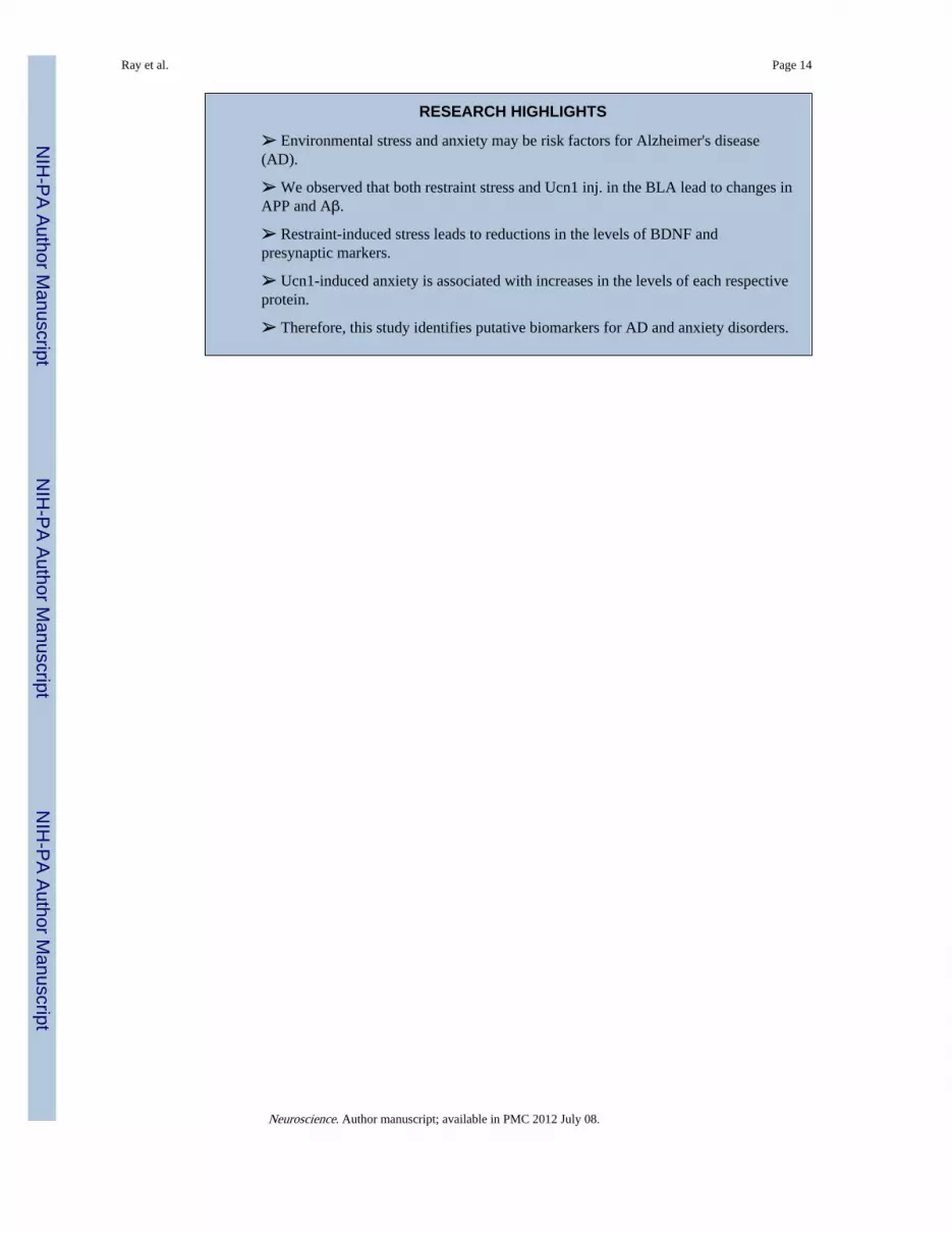
RESEARCH HIGHLIGHTS
➢ Environmental stress and anxiety may be risk factors for Alzheimer's disease(AD).
➢ We observed that both restraint stress and Ucn1 inj. in the BLA lead to changes inAPP and Aβ.
➢ Restraint-induced stress leads to reductions in the levels of BDNF andpresynaptic markers.
➢ Ucn1-induced anxiety is associated with increases in the levels of each respectiveprotein.
➢ Therefore, this study identifies putative biomarkers for AD and anxiety disorders.
Ray et al. Page 14
Neuroscience. Author manuscript; available in PMC 2012 July 08.
NIH
-PA Author Manuscript
NIH
-PA Author Manuscript
NIH
-PA Author Manuscript

Ray et al. Page 15
Neuroscience. Author manuscript; available in PMC 2012 July 08.
NIH
-PA Author Manuscript
NIH
-PA Author Manuscript
NIH
-PA Author Manuscript

Figure 1A. Social interaction of control and stressed animals after three hrs of restraint stressThe social interaction test was conducted to measure anxiety-like behavior in rats. Followingthree hrs of restraint stress, we have observed a trend in decreased social interaction instressed rats (P=0.06 by one tailed t-test).Figure 1B: Social interaction of vehicle and Ucn1-treated animals: Following repeatedUcn1 injections into the BLA, the rats showed significant decreases in social interactionsconsistent with increased anxiety-like behavior.Figure 1C: Plasma corticosterone levels in control and restraint stressed animals:Plasma corticosterone was measured after three hrs restraint stress from both stressed andcontrol animals. A competitive EIA assay was utilized to measure corticosterone and thelevels were significantly higher in the stressed animals versus controls.
Ray et al. Page 16
Neuroscience. Author manuscript; available in PMC 2012 July 08.
NIH
-PA Author Manuscript
NIH
-PA Author Manuscript
NIH
-PA Author Manuscript

Ray et al. Page 17
Neuroscience. Author manuscript; available in PMC 2012 July 08.
NIH
-PA Author Manuscript
NIH
-PA Author Manuscript
NIH
-PA Author Manuscript
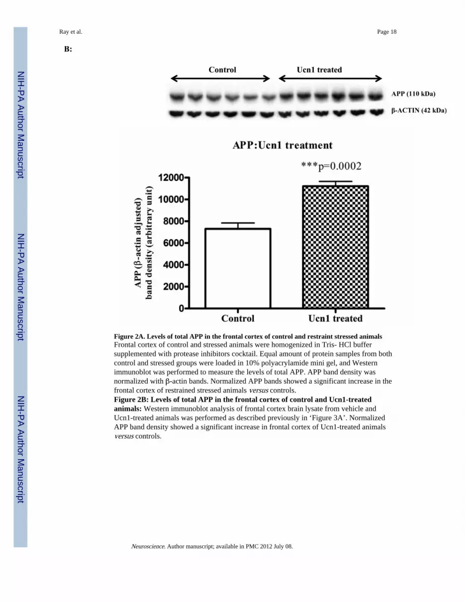
Figure 2A. Levels of total APP in the frontal cortex of control and restraint stressed animalsFrontal cortex of control and stressed animals were homogenized in Tris- HCl buffersupplemented with protease inhibitors cocktail. Equal amount of protein samples from bothcontrol and stressed groups were loaded in 10% polyacrylamide mini gel, and Westernimmunoblot was performed to measure the levels of total APP. APP band density wasnormalized with β-actin bands. Normalized APP bands showed a significant increase in thefrontal cortex of restrained stressed animals versus controls.Figure 2B: Levels of total APP in the frontal cortex of control and Ucn1-treatedanimals: Western immunoblot analysis of frontal cortex brain lysate from vehicle andUcn1-treated animals was performed as described previously in ‘Figure 3A’. NormalizedAPP band density showed a significant increase in frontal cortex of Ucn1-treated animalsversus controls.
Ray et al. Page 18
Neuroscience. Author manuscript; available in PMC 2012 July 08.
NIH
-PA Author Manuscript
NIH
-PA Author Manuscript
NIH
-PA Author Manuscript
Ray et al. Page 19
Neuroscience. Author manuscript; available in PMC 2012 July 08.
NIH
-PA Author Manuscript
NIH
-PA Author Manuscript
NIH
-PA Author Manuscript
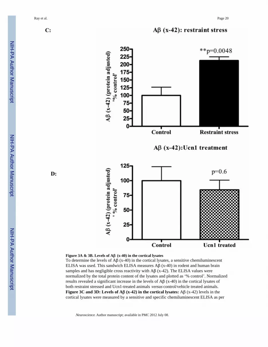
Figure 3A & 3B. Levels of Aβ (x-40) in the cortical lysatesTo determine the levels of Aβ (x-40) in the cortical lysates, a sensitive chemiluminescentELISA was used. This sandwich ELISA measures Aβ (x-40) in rodent and human brainsamples and has negligible cross reactivity with Aβ (x-42). The ELISA values werenormalized by the total protein content of the lysates and plotted as ‘% control’. Normalizedresults revealed a significant increase in the levels of Aβ (x-40) in the cortical lysates ofboth restraint stressed and Ucn1-treated animals versus control/vehicle treated animals.Figure 3C and 3D: Levels of Aβ (x-42) in the cortical lysates: Aβ (x-42) levels in thecortical lysates were measured by a sensitive and specific chemiluminescent ELISA as per
Ray et al. Page 20
Neuroscience. Author manuscript; available in PMC 2012 July 08.
NIH
-PA Author Manuscript
NIH
-PA Author Manuscript
NIH
-PA Author Manuscript

manufacturer's protocol. Aβ (x-42) signals were normalized and plotted in the same way asdescribed in ‘Figure 3A and 3B’. Normalized Aβ (1-42) showed a significant increase in thecortex of restraint stressed animals versus controls. However, Aβ (x-42) levels did not differbetween the cortical lysates of vehicle and Ucn1-treated animals.
Ray et al. Page 21
Neuroscience. Author manuscript; available in PMC 2012 July 08.
NIH
-PA Author Manuscript
NIH
-PA Author Manuscript
NIH
-PA Author Manuscript

Figure 4A. Levels of BDNF in the frontal cortex of control and restraint stressed animals:Western immunoblot analysisAcid treated frontal cortex lysates were analyzed by western immunoblotting to detect thelevels of BDNF (for details see ‘Materials and Methods’). BDNF band densities (primarilyBDNF dimer) were normalized by β-actin signals. Normalized BDNF showed a significantdecrease in the cortex of animals underwent 3 hrs restraint stress versus controls.Figure 4A: Levels of BDNF in the frontal cortex of control and restraint stressedanimals: ELISA analysis. To confirm the BDNF Western immunoblot results, BDNFlevels were measured in acid treated cortical lysates by a sensitive colorimetric ELISA.BDNF values (pg/ml) were converted to pg/μg, normalized by protein contents of thelysates and plotted. BDNF ELISA also revealed a significant decrease in the cortex ofrestraint stressed animals versus controls.
Ray et al. Page 22
Neuroscience. Author manuscript; available in PMC 2012 July 08.
NIH
-PA Author Manuscript
NIH
-PA Author Manuscript
NIH
-PA Author Manuscript

Figure 5A. Levels of BDNF in the frontal cortex of vehicle and Ucn1-treated animals: Westernimmunoblot analysisBDNF Westernblotting was performed with the cortical lysates of vehicle and Ucn1-treatedanimals in a similar way described in ‘Figure 4A’. β-actin adjusted BDNF band densities(BDNF dimer) showed a significant increase in the cortical lysates of Ucn1-treated animalsversus vehicle. The lower band observed in this figure is due to secondary ‘artifact’ andexcluded from the analyses.Figure 5B: Levels of BDNF in the frontal cortex of vehicle and Ucn1-treated animals:ELISA analysis. To confirm the findings of ‘Figure 3A’, a sensitive ELISA was used todetermine the levels of BDNF in the acid treated cortical lysates of vehicle and Ucn1-treatedanimals. BDNF values (pg/ml) were converted to pg/μg, normalized by protein contents ofthe lysates and plotted. Results showed a significant increase in the cortical lysates of Ucn1-treated animals versus vehicle.
Ray et al. Page 23
Neuroscience. Author manuscript; available in PMC 2012 July 08.
NIH
-PA Author Manuscript
NIH
-PA Author Manuscript
NIH
-PA Author Manuscript

Figure 6A. Levels of syntaxin6 in the cortical lysates of control and restraint stressed animalsWestern immunoblot analyses of cortical lysates of control and restrained stressed animalswere carried out with specific monoclonal syntaxin6 antibody, which recognizes one of theimportant pre-synaptic proteins syntaxin6. The bands were normalized by β-actin signals.Normalized syntaxin6 bands showed a significant decrease in the lysates of restraint stressedanimals versus controls.Figure 6B: Levels of SNAP25 in the cortical lysates of control and restraint stressedanimals: Western immunoblot analyses of the same samples mentioned in ‘Figure 6A’ foranother pre-synaptic protein SNAP25 showed a decreasing trend (p=0.08) in the corticallysates of restraint stressed animals versus controls.
Ray et al. Page 24
Neuroscience. Author manuscript; available in PMC 2012 July 08.
NIH
-PA Author Manuscript
NIH
-PA Author Manuscript
NIH
-PA Author Manuscript

Figure 7A. Levels of SNAP25 in the cortical lysates of vehicle and Ucn1-treated animalsWestern immunoblot analyses of cortical lysates of vehicle and Ucn1-treated animals werecarried out to detect the levels of cortical SNAP25. SNAP25 bands were normalized by β-actin signals and plotted. Normalized SNAP25 showed a significant increase in the cortex ofUcn1-treated animals versus vehicle treated animals.Figure 7B: Levels of syntaxin6 in the cortical lysates of vehicle and Ucn1-treatedanimals: Western immunoblot analyses revealed a significant increase in the levels ofsyntaxin6 in the cortical lysates of Ucn1-treated animals versus vehicle treated animals.
Ray et al. Page 25
Neuroscience. Author manuscript; available in PMC 2012 July 08.
NIH
-PA Author Manuscript
NIH
-PA Author Manuscript
NIH
-PA Author Manuscript