remodeling of acl allografts is inhibited by peracetic acid sterilization
TRANSCRIPT

SYMPOSIUM: NEW APPROACHES TO ALLOGRAFT TRANSPLANTATION
Remodeling of ACL Allografts is Inhibited by Peracetic Acid
Sterilization
Sven U. Scheffler MD, Johannes Gonnermann CMD,
Julia Kamp CMD, Dorothea Przybilla,
Axel Pruss MD
Published online: 20 May 2008
� The Association of Bone and Joint Surgeons 2008
Abstract Sterilization of allografts for anterior cruciate
ligament (ACL) reconstruction has become an important
prerequisite to prevent disease transmission. However,
current sterilization techniques impair the biological or
mechanical properties of such treated grafts. Peracetic acid
(PAA) has been successfully used to sterilize bone allo-
grafts without these disadvantages and does not impair the
mechanical properties of soft tissue grafts in vitro. We
asked whether PAA sterilization would influence recellu-
larization, restoration of crimp length and pattern, and
revascularization of ACL grafts during early healing. We
used an in vivo sheep model for open ACL reconstruction.
We also correlated the histologic findings with the
restoration of anteroposterior stability and structural
properties during load-to-failure testing. PAA slowed
remodeling activity at 6 and 12 weeks compared to
nonsterilized allografts and autografts. The mechanical
properties of PAA grafts were also reduced compared to
these control groups at both time points. We conclude PAA
sterilization currently should not be used to sterilize soft
tissue grafts typically used in ACL reconstruction.
Introduction
Allografts have recently gained popularity in orthopaedic
sports medicine, especially in primary and revision
reconstruction of the anterior cruciate ligament (ACL) [2].
Allografts eliminate harvest morbidity and reportedly
allow faster rehabilitation, earlier return to full activity, and
possible cost reduction [4, 10, 25]. Disadvantages of using
allografts, however, include potential disease transmission
and prolonged graft healing [7, 11, 22, 26, 31, 33]. Even
though the risk of infection following allograft transplan-
tation is very small, fatal incidences have been reported
[31]. As a consequence, different sterilization techniques
have been developed to eliminate infectious pathogens
from the graft before transplantation. These include irra-
diation [32], chemical processing [15], and antibiotic soaks
[29]. However, most current sterilization procedures have
inherent disadvantages affecting biological properties and
mechanical function of the graft. Gamma irradiation
reduces the mechanical strength of ACL allografts when
used at levels required to eliminate bacterial as well as viral
pathogens [6, 18]. Antibiotic soaks reportedly provide only
surface sterilization and chemical processing techniques,
such as with ethylene oxide, have provoked synovial
inflammation and have been abandoned [32].
Each author certifies that he or she has no commercial associations
(eg, consultancies, stock ownership, equity interest, patent/licensing
arrangements, etc) that might pose a conflict of interest in connection
with the submitted article. This study was funded by research grants
of the Charite Research Funds and the Musculoskeletal Transplant
Foundation.
Each author certifies that his or her institution has approved the
animal protocol for this investigation and that all investigations were
conducted in conformity with ethical principles of research.
S. U. Scheffler (&), J. Gonnermann, J. Kamp
Sports Medicine & Arthroscopy Service, Department for
Orthopaedic Surgery and Traumatology, Center for
Musculoskeletal Surgery, Charite University Medicine Berlin,
Charite Platz 1, 10117 Berlin, Germany
e-mail: [email protected]
D. Przybilla
Institute for Laboratory Medicine and Pathobiochemistry,
Charite University Medicine Berlin, Berlin, Germany
A. Pruss
Tissue Bank Institute of Transfusion Medicine, Charite
University Medicine Berlin, Berlin, Germany
123
Clin Orthop Relat Res (2008) 466:1810–1818
DOI 10.1007/s11999-008-0288-2

Peracetic acid (PAA) has been used since the early
1980s [28] mainly to sterilize bone allografts. Several
preliminary in vitro studies suggest the feasibility of this
technique with no adverse effects on the structural and
mechanical properties as well as the possibility of full
biological incorporation of such treated bone grafts [16,
17]. This technique has also been applied to soft tissue
grafts. For example, full sterilization of Achilles tendon
was achieved with 0.2% PAA solution [28]. Analyses of
the mechanical function of bone-patellar tendon-bone
grafts in vitro revealed no adverse effects of PAA sterili-
zation compared to nonsterilized grafts [21]. However,
cellularity and vascularity are substantially upregulated
during the first 3 months of healing [23]. However, cellu-
larity and vascularity are substantially upregulated during
the first 3 months of healing in ACL autografts. The extent
of remodeling activity is seen in reorganization and/or
dissolution of the extracellular matrix [3, 14, 23], including
the crimp length and crimp pattern. The remodeling
behavior of PAA sterilized allografts in-vivo is unknown.
We first hypothesized there would be no time-dependent
differences in cellularity, vascularity, and crimp length and
pattern of PAA-sterilized ACL allografts compared to
nonsterilized allografts or autografts during early healing
in an animal model. We also hypothesized PAA-sterilized
and nonsterilized ACL allografts would exhibit similar
anteroposterior knee stability and structural properties.
Materials and Methods
We used 16 mature female merino-breed sheep (2–3 years
old) to ascertain the effects of PAA sterilization at 6
(n = 8) and 12 weeks (n = 8) of healing. In our previous
study [22], we used the identical ACL reconstruction
model. We compared cellularity, vascularity, crimp length/
pattern, and mechanical properties between eight nonster-
ilized fresh-frozen allografts, eight autografts, and eight
intact ACLs at 6 and 12 weeks postoperatively. The data
from the previous study were used as controls for the PAA-
sterilized allografts investigated in this current study. The
number of specimens used in this study was determined
by performing a power analysis on preliminary data to
estimate the required sample size to ensure 80% power
(a = 0.05; b = 0.2). All animal procedures were con-
ducted according to the guidelines of the National Institute
of Health (Germany) for the use of laboratory animals. All
animals were checked for bony maturity by dental status
and preoperative radiograph of the knee. Normal health
status was confirmed by a veterinarian.
The first two animals received PAA-sterilized long
flexor tendon grafts previously harvested from sheep not
included in this study. In the other animals, the long flexor
tendon of the superficial flexor digital muscle was har-
vested as a free soft tissue graft from each left hind limb
prior to ipsilateral ACL reconstruction (Fig. 1). It was
placed in sterile bags and immediately transported on ice to
the local tissue bank where PAA sterilization was con-
ducted no later than 6 hours after harvest. Afterwards,
grafts were stored for at least 6 days at -18�C. Steriliza-
tion was carried out under highest safety and sterility
standards in ‘‘class A in B’’ laboratories as outlined in the
European guideline for Good Manufacturing Practice of
Human Tissue (Annex 1 ECGMP). All procedures occur-
red under aseptic conditions with constant laminar airflow.
Grafts were consecutively rinsed under high pressure to
completely remove blood remnants from the graft tissue
using sterile water at 37�C for 30 minutes. Any remaining
fat was removed by placing the tissue into a mixture of
chloroform (extra pure, 99.4%) and methanol (for analysis,
99.8%) (v/v, 2/1) under constant agitation (laboratory
shaker THYS 2, MLW, Leipzig, Germany) for 2 hours
while the delipidating solution was changed every
30 minutes. Tissues were sonicated eight times with
methanol in an ultrasonic bath (Sonorex RK 510 H,
Bandelin Electronic, Berlin, Germany) for 15 minutes to
completely remove any residual chloroform. Methanol was
removed by flushing the tissues twice with sterile deionized
water. The sterilization procedure was carried out under
constant agitation (laboratory shaker THYS 2, MLW,
Leipzig, Germany) under low pressure (200 mbar) at room
temperature in a desiccator for 4 hours (Fig. 1). Lipid-free
transplants were covered with PAA solution (v/v, 1/7.5).
PAA solution consisted of 2% peracetic acid (Kesla-
Chemie, Wolfen), 96% ethanol (Merck, Darmstadt) and
aqua ad iniectabila (Ampuwa; Fresenius, Bad Homburg,
Germany) (ratio v/v/v 2/1/1), providing a final sterilization
solution of 1% PAA. PAA was removed by washing the
Fig. 1 The superficial long flexor tendon was used as an allograft for
ACL reconstruction.
Volume 466, Number 8, August 2008 PAA Sterilization of ACL Allografts 1811
123

grafts six times for 20 minutes with Soerensen buffer. This
was followed by rinsing the grafts with aqua ad injectabila
twice. At the end of the procedure, the absence of peracetic
acid was confirmed by a Reflectoquant1 PAA test (Merck
Eurolab GmbH, Darmstadt) with a sensitivity of 5 ppm.
Grafts were dried in sterile air and stored in sterile bags at
-18�C until time of transplantation. No antibiotic solution
was added.
On the day of surgery, the PAA-sterilized graft was
transported to the surgical animal facilities, thawed at room
temperature, and then augmented with two #2 Ethibond
Excel polyester sutures (Ethicon, Inc., Piscataway, NJ,
USA) at each end. The graft length was between 60 and
70 mm.
An open ACL reconstruction was performed on each left
hind limb as previously described (Fig. 2) [22, 34]. All
surgery was performed by one individual (SS). The joint
was opened by a medial arthrotomy, the ACL excised, and
the knee was brought into deep flexion for placement of a
guide pin in the femoral anatomical footprint of the ACL.
The guide pin was overdrilled to a depth of 20 mm
matching the diameter of the prepared graft. Graft fixation
was achieved at the femoral cortex with a fixation button
(Flipptack1, Fa. Karl Storz GmbH, Tuttlingen, Germany).
The tibial tunnel was placed in an identical fashion into the
tibial footprint of the ACL and drilled through the tibial
cortex. The graft was fixed by placing multiple knots of the
augmented graft sutures onto a bone bridge that was cre-
ated 1 cm distally to the exit of the tibial tunnel. Before
final fixation, the knee was moved through several cycles
of full flexion and extension to eliminate slack of the graft
sutures.
All animals were immediately allowed full weight
bearing with no limitation of range of motion. Antiin-
flammatory drugs (Finadyne1 [1%, Essex Pharma GmbH,
Munich, Germany] 1 mg/kg s.c.) were given during the
first 3 postoperative days. Wound status and gait pattern
were recorded daily. The animals’ gaits were subjectively
assessed by the same person and graded from 1 (no weight
bearing) to 5 (full weight bearing with no signs of limping).
All animals were released to an outside farm without any
restriction of motion 2 weeks postoperatively.
The animals were sacrificed at 6 or 12 weeks and the
left knee was removed, leaving the skin and all soft tissue
structures intact. The knee was inspected intraarticularly
for inflammation, effusion, the synovial coverage of the
ACL graft, the status of the cartilage, and any degenerative
changes.
To address our first hypothesis of whether biological
remodeling was delayed in PAA grafts, we histologically
examined cellularity, vascularity, and crimp length and
pattern. Two researchers (JG, JK) performed all histolog-
ical analyses independently, blinded to time of sacrifice
and specimen numbers. Tissue samples were immediately
fixed in 4% formalin and embedded in paraffin. Serial cuts
4-lm thick were prepared and mounted on slides with 3%
silane (Sigma Chemical, St. Louis, MO, USA). A high-
resolution microscope (Leica DMRB, Leica GmbH,
Bensheim, Germany) linked to a digital image analysis
system (KS 400 Imaging System, Release 3.0, Carl Zeiss
Vision, Eching, Germany) was used for histological anal-
ysis. Hematoxylin and eosin and Masson-Goldner
trichrome stains were used for assessment of cellularity,
cell morphology, crimp length and pattern, and appearance
of foreign-body, giant, and inflammatory cells in the PAA
grafts. Cellularity (as mean cell density per mm2) was
quantified in 10 regions of interest (ROI) (0.06 mm2 each)
randomly chosen in each of two longitudinal sections per
specimen in regular intervals along the complete section
length.
During tendon remodeling dissolution and reorganiza-
tion of collagen bundles occur, which can be quantified by
the changes of their typical wave-like structure observed in
soft tissue grafts. The wave-like structure of collagen
bundles can be visualized due to its anisotropy with
polarized light microscopy. Collagen crimp length was
defined as the wave frequency (lm) of collagen bundles
measured in the same ROIs (0.06 mm2) using a calibrated
scale with the digital image analysis system. Descriptive
documentation of fiber alignment and orientation was also
reported to provide further insights into the remodeling
behavior.
One of us (JK) performed immunohistochemical anal-
ysis to quantify vascular density as previously described by
Unterhauser et al. [30]. Two transverse sections of intact
midsubstance graft tissue per specimen were used for
quantitative analysis of vascularity. Transverse sections
were subdivided into a subsynovial (Sub), an intermediate
(Mid), and a central (Cnt) region. In each subregion, five
representative regions of interest (0.06 mm2) were identi-
fied and the number of vessel cross-sections was countedFig. 2 A schematic of open ACL reconstruction procedure is shown.
1812 Scheffler et al. Clinical Orthopaedics and Related Research
123

with a clear positive signal after immunostaining. The
endothelial surface cells of blood vessels were immuno-
stained with rabbit antihuman von Willebrand factor
(DAKO, Glostrup, Denmark) in transverse sections. The
tissue samples were hydrated and pretreated with 0.1%
protease (type XIV, bacterial, Sigma Aldrich Chemie
GmbH, Steinheim, Germany) for 10 minutes at 37�C. Ten-
percent normal horse serum (Vector Laboratories Inc.,
Burlington, CA, USA) was used for 20 minutes to block
nonspecific binding sites at room temperature. The anti-
body was diluted 1:200 and added to the tissue samples
overnight in a humidity chamber at 4�C. The samples were
then rinsed in tris-buffered saline and incubated with bio-
tinylated horse antimouse immunoglobulin G secondary
antibody (Vector Laboratories) for 30 minutes. This was
followed by incubation with an avidin-biotin complex
(ABC kit; Vector Laboratories) linked with alkaline
phosphatase as a reporter enzyme. Staining was achieved
with Neufuchsin as a chromogen. Tissue samples were
counterstained with methylene-green for a few seconds,
dehydrated, and mounted in a xylol-soluble mount (Vitro-
Clud, R Langenbrinck, Emmendingen, Germany).
To test our second hypothesis and to evaluate the
mechanical function of PAA grafts, two loading conditions
were simulated on a material testing machine (model 1455,
Zwick GmbH, Ulm, Germany): an anteroposterior drawer
test and a load-to-failure test of the femur-ACL graft-tibia
complex. AP drawer testing of the ACL reconstructed knee
was performed (1) with all soft-tissue structures left intact,
(2) with only the ACL allograft and PCL intact, and (3) as
an anterior drawer test with only the ACL allograft. All
tests were performed at 60� of flexion with all motions
restrained except the AP translation.
After preloading the knee with 5 N, an AP load of
± 50 Nwas applied perpendicular to the longitudinal axis of
the tibia 10 times at a speed of 120 mm/min. AP laxity was
recorded from the tenth cycle for each specimen (Table 1).
The knees were then removed from the mechanical testing
machine and the diameter of the ACL allografts were cal-
culated according to a technique described by Ellis [8].
The joints were remounted at 30� of flexion and the
longitudinal axis of the ACL allograft aligned parallel to
the loading direction of the testing apparatus. After
applying a preload of 5 N to the femur-ACL graft-tibia
complex, a load-to-failure test was carried out at a speed of
120 mm/min (Table 2). The failure mode and a load-dis-
placement curve were recorded and the structural
properties, such as failure load and stiffness (in the linear
region between 30% and 90% of the maximum load), as
well as stress at failure as a mechanical property of the
graft tissue were analyzed using in-house software.
A Shapiro-Wilk W test showed the experimental data
were not distributed normally. Therefore, a nonparametric
Mann-Whitney U test was used to compare cellularity,
vascularity, and crimp length between PAA allografts and
the previously published data for nonsterilized allografts,
autografts, and the intact ACL. Anterior-posterior laxity
and structural properties were also analyzed for differ-
ences among the aforementioned groups using the Mann-
Whitney U test.
Table 1. Cellularity and crimp length at 6 and 12 weeks of healing
Variable PAA allografts ns-allografts [6, 21] Autografts [6, 21] Intact ACL [21]
Cells (/mm2) 6 weeks 109 ± 111* 134 ± 72�* 226 ± 61� 569 ± 92§
12 weeks 240 ± 238*� 656 ± 464 547 ± 264 —
Crimp length (lm) 6 weeks 278 ± 123*� 177 ± 57� 203 ± 98� 60 ± 41
12 weeks 309 ± 75*� 122 ± 24� 199 ± 106� —
* Different from autografts (p\ 0.05); �different from ns allografts (p\ 0.05); �different from the intact ACL (p\ 0.05); (Stain, Masson
Goldner’s trichrome; original magnification 9200); §different from ns-allografts and autografts (p\ 0.05).
Table 2. Anteroposterior laxity following drawer testing at ± 50 N
Variable PAA allografts Fresh-frozen allografts§ Autografts§ Intact ACL§
Time of healing (wks) 6 12 6 12 6 12 —
Complete knee joint (mm) 5.1 ± 3.1 4.8 ± 1.5 5.7 ± 1.6 5.4 ± 1.2 6.0 ± 2.2 6.1 ± 0.6 2.7 ± 1.2�
ACL graft/PCL (mm) 9.2 ± 5.7 11.6 ± 5� 7.4 ± 3.7 8.2 ± 2.6 8.7 ± 4.9 7.7 ± 1.4 3.9 ± 3.2�
ACL graft (mm) 6.6 ± 2.6� 8.3 ± 4.7*� 4.1 ± 3.5 1.3 ± 0.3 1.4 ± 0.4 1.1 ± 0.2 1.6 ± 1.7�
* p\ 0.05, significant larger than fresh-frozen allografts; �p\ 0.05, significantly larger than autografts; �p\ 0.05, significantly smaller than all
study groups; §All data presented for these groups have been previously published [21], using identical material and methods as for the PAA
allografts.
Volume 466, Number 8, August 2008 PAA Sterilization of ACL Allografts 1813
123

Results
All animals in all experimental groups could fully weight
bear and had normal gait pattern at 2 weeks postoperatively.
At time of sacrifice, all animals showed free range of motion
with no detectable effusion and no inflammatory reaction
was present in either the PAA or control groups. We
observed fibrotic ganglion cyst-like tissue at the tibial tunnel
entrance in 10 of 16 PAA specimens, which was not
observed in any of the control groups. These animals had no
loss of motion compared to animals without such changes.
At 6 weeks postoperatively, cellularity of PAA allo-
grafts was similar to that in nonsterilized allografts
(p = 0.234), but was lower than in the autograft group
(p = 0.01) and the intact ACL (p\ 0.001). PAA-treated
allografts showed little influx of cells into the periphery,
with vast areas of the grafts being acellular (Fig. 3). At
12 weeks, slightly increased (p = 0.139) cellularity was
observed in the PAA group, which was lower than in
nonsterilized allografts (p = 0.04), autografts (p = 0.023),
and in the intact ACL (p = 0.036) (Table 1) (Fig. 3).
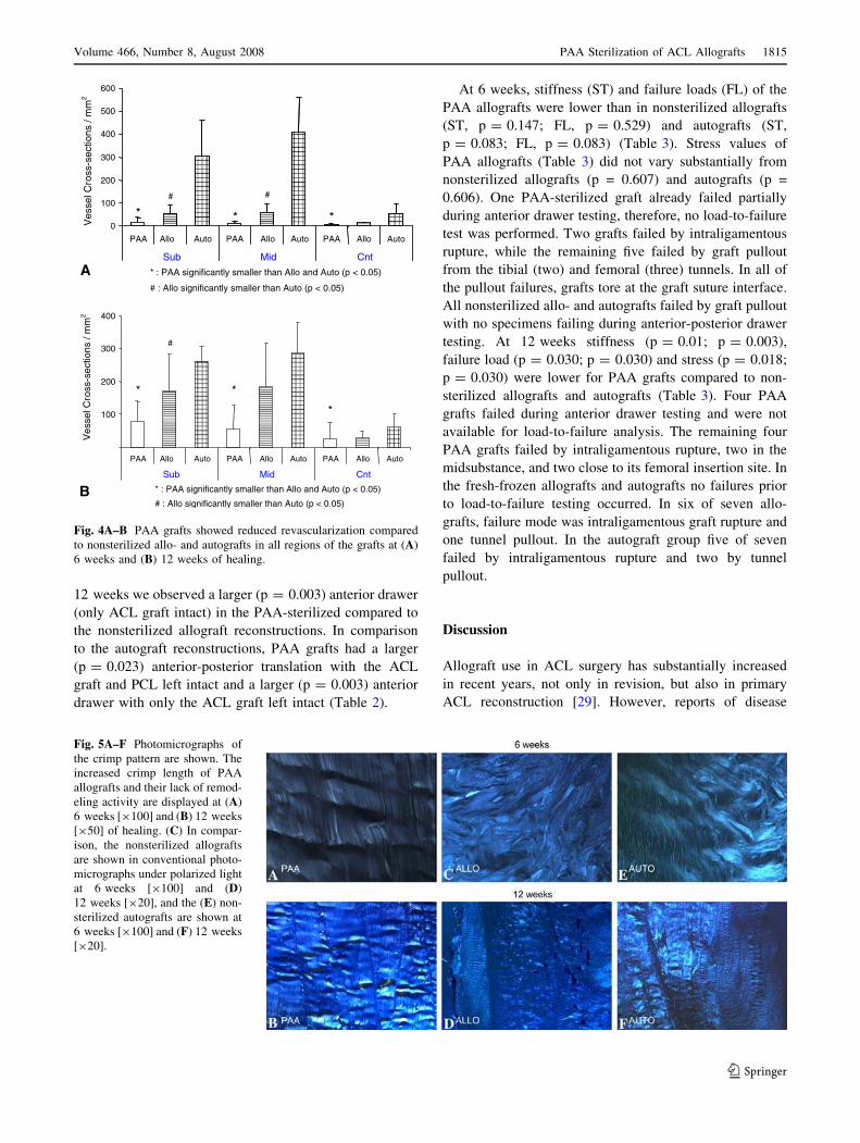
We observed lower vascularity of PAA allografts than in
nonsterilized allografts (sub p = 0.031, mid p = 0.008, cnt
p = 0.01), autografts (sub p = 0.008, med p = 0.008, cnt p =
0.001) and the intact ACL (Sub, 61.9 ± 31.7/mm2; Mid,
53.6 ± 23.4/mm2; Cnt, 27 ± 13/mm2) (sub p = 0.002, mid
p = 0.001, cnt p = 0.001) in all graft regions at 6 weeks
(Fig. 4A). Sparse vascularity was confined to the periphery
of the PAA-sterilized graft tissue. At 12 weeks, vascularity
remained lower compared to nonsterilized allografts (mid
p = 0.008, cnt p = 0.024) and autografts (sub p = 0.000, mid
p = 0.000, cnt p = 0.015) in all graft regions with the
exception of the subsynovial region in the PAA group that
did not differ from nonsterilized allografts (Fig. 4B)
(p = 0.113). We observed no differences between the PAA
group and the intact ACLs Sub (p = 0.470) and Mid
(p = 0.210) region, but lower values were seen in the Cnt
(p = 0.031) region. Qualitatively, nonsterilized allo- and
autografts showed overall hypervascularity compared to
PAA allografts, which in turn looked similar to the intact
ACL.
Crimp length of PAA allografts at 6 weeks was similar to
that in nonsterilized allografts (p = 0.093) and autografts
(p = 0.074). We observed longer PAA crimp length than in
the intact ACL (p = 0.001) at this time point (Table 1)
(Fig. 5). Qualitatively, both nonsterilized allo- and auto-
grafts had lost their typical crimp pattern in large areas of
the graft tissue with reorganization of crimp pattern at the
periphery and small areas of intact crimp pattern remaining
in the graft center. PAA-sterilized grafts, in contrast, dis-
played only small areas of dissolving crimp, while in large
parts of the grafts crimp pattern remained unchanged. At
12 weeks, crimp length was longer than in nonsterilized
allografts (p = 0.001) and autografts (p = 0.055) as well as
the intact ACL (p = 0.001) (Table 1, Fig. 5). Qualitatively,
both nonsterilized allografts and autografts showed
continuing high crimp turnover at 12 weeks, while crimp
pattern of PAA grafts remained more homogeneous,
although more irregular than at 6 weeks.
At 6 weeks, we observed similar anterior-posterior
stability in PAA and nonsterilized allograft ACL recon-
structions for each of the three testing conditions (all soft
tissue structures intact, ACL graft/PCL intact, ACL graft
only) (Table 2). When comparing PAA allografts with
autograft ACL reconstructions, a larger (p = 0.010) ante-
rior drawer was found for the PAA group with only the
ACL graft left intact, while no differences were found
for the other two test settings (Table 2) (p[ 0.05). At
Fig. 3A–F Histologic samples
demonstrate the reduced cellularity
of (A) PAA grafts at 6 weeks and
(B) 12 weeks, compared to (C)
nonsterilized allografts at 6 weeks
(stain, hematoxylin and eosin; ori-
ginal magnification9100) and (D)
12 weeks (stain, MassonGoldner’s
trichrome; original magnification
9200) of healing, and (E) nonster-
ilized autografts at 6 weeks (stain,
hematoxylin and eosin; original
magnification 9100) and (F)
12 weeks (stain, MassonGoldner’s
trichrome; original magnification
9200) of healing.
1814 Scheffler et al. Clinical Orthopaedics and Related Research
123
12 weeks we observed a larger (p = 0.003) anterior drawer
(only ACL graft intact) in the PAA-sterilized compared to
the nonsterilized allograft reconstructions. In comparison
to the autograft reconstructions, PAA grafts had a larger
(p = 0.023) anterior-posterior translation with the ACL
graft and PCL left intact and a larger (p = 0.003) anterior
drawer with only the ACL graft left intact (Table 2).
At 6 weeks, stiffness (ST) and failure loads (FL) of the
PAA allografts were lower than in nonsterilized allografts
(ST, p = 0.147; FL, p = 0.529) and autografts (ST,
p = 0.083; FL, p = 0.083) (Table 3). Stress values of
PAA allografts (Table 3) did not vary substantially from
nonsterilized allografts (p = 0.607) and autografts (p =
0.606). One PAA-sterilized graft already failed partially
during anterior drawer testing, therefore, no load-to-failure
test was performed. Two grafts failed by intraligamentous
rupture, while the remaining five failed by graft pullout
from the tibial (two) and femoral (three) tunnels. In all of
the pullout failures, grafts tore at the graft suture interface.
All nonsterilized allo- and autografts failed by graft pullout
with no specimens failing during anterior-posterior drawer
testing. At 12 weeks stiffness (p = 0.01; p = 0.003),
failure load (p = 0.030; p = 0.030) and stress (p = 0.018;
p = 0.030) were lower for PAA grafts compared to non-
sterilized allografts and autografts (Table 3). Four PAA
grafts failed during anterior drawer testing and were not
available for load-to-failure analysis. The remaining four
PAA grafts failed by intraligamentous rupture, two in the
midsubstance, and two close to its femoral insertion site. In
the fresh-frozen allografts and autografts no failures prior
to load-to-failure testing occurred. In six of seven allo-
grafts, failure mode was intraligamentous graft rupture and
one tunnel pullout. In the autograft group five of seven
failed by intraligamentous rupture and two by tunnel
pullout.
Discussion
Allograft use in ACL surgery has substantially increased
in recent years, not only in revision, but also in primary
ACL reconstruction [29]. However, reports of disease
600
******
***
CntMidSub
AutoAlloPAAAutoAlloPAAAutoAlloPAA
-
2
-
2
Ve
sse
l C
ross-s
ectio
ns / m
m2
500
400
300
200
100
0
* : PAA significantly smaller than Allo and Auto (p < 0.05)
# : Allo significantly smaller than Auto (p < 0.05)
Ve
sse
l C
r oss-s
ections / m
m2
# : Allo significantly smaller than Auto (p < 0.05)
* : PAA significantly smaller than Allo and Auto (p < 0.05)
*
**
#
# #
CntMidSub
AutoAlloPAAAutoAlloPAAAutoPAA Allo
400
300
200
100
A
B
Fig. 4A–B PAA grafts showed reduced revascularization compared
to nonsterilized allo- and autografts in all regions of the grafts at (A)
6 weeks and (B) 12 weeks of healing.
Fig. 5A–F Photomicrographs of
the crimp pattern are shown. The
increased crimp length of PAA
allografts and their lack of remod-
eling activity are displayed at (A)
6 weeks [9100] and (B) 12 weeks
[950] of healing. (C) In compar-
ison, the nonsterilized allografts
are shown in conventional photo-
micrographs under polarized light
at 6 weeks [9100] and (D)
12 weeks [920], and the (E) non-
sterilized autografts are shown at
6 weeks [9100] and (F) 12 weeks
[920].
Volume 466, Number 8, August 2008 PAA Sterilization of ACL Allografts 1815
123

transmission following ACL reconstruction with nonster-
ilized allografts, even though very limited in number,
underline the importance of graft sterilization [31]. Fur-
thermore, current sterilization techniques have been
associated with certain disadvantages, such as interference
with biological healing or reduction of mechanical prop-
erties of such treated grafts [5, 6, 13, 18, 19, 24, 27].
Peracetic acid has been successfully used to sterilized bone
allografts [16, 17]. and PAA sterilization does not alter the
material, structural, and viscoelastic properties of human
bone-patellar tendon-bone graft in vitro [21]. We therefore
hypothesized PAA allografts would exhibit similar bio-
logical changes in terms of cellularity, vascularity, and
crimp structure, and provide equivalent mechanical func-
tion compared to nonsterilized allografts and autologous
ACL reconstructions during early healing.
It is important to mention the limitations of this study
before considering use in humans. First, as in any animal
study, it is almost impossible to fully control the postop-
erative weight bearing or apply specific rehabilitation
protocols as it is the case in human patients, which could
have affected our results. Second, we only evaluated early
healing of the PAA-treated ACL grafts. Maybe a certain
recovery of biological and mechanical function would have
been possible, when longer healing times were considered.
However, an increasing number of primary ACL recon-
structions use allografts in combination with accelerated
rehabilitation protocol, since this procedure avoids graft
harvest morbidity. Therefore, the increased early weight
bearing during the first 3 months of accelerated rehabili-
tation could be detrimental for the clinical outcome of
patients with PAA-sterilized ACL allografts.
Based on our data the first hypothesis must be rejected:
PAA sterilization delayed or even partial inhibited the
biological remodeling of PAA grafts. This led to impaired
functional knee stability and reduced structural properties
of PAA grafts during subsequent healing up to 3 months.
Our finding of inhibited graft remodeling due to PAA
sterilization confirms findings of several other authors’ [1,
12, 35] and our own report [22] of nonsterilized allo- and
autograft ACL reconstructions in vivo. Both graft types
undergo a maturation process, which is characterized by
overall hypocellularity and hypervascularity as early as
6 weeks postoperatively, and a significant recellularization
and revascularization that occurs from the periphery
towards the center of the grafts [22]. By 12 weeks, the
cellularity of the intact ACL was restored in autografts [1,
12, 35] and allografts [1, 12, 35] and that hypervascularity
remained. This graft maturation has been defined as the
ligamentization process [1, 12, 35]. In contrast, PAA graft
cellularity only slightly increased during early healing and
did not recover to values of the intact ACL, while graft
vascularity never exceeded the values for the intact ACL
and substantially lagged behind nonsterilized ACL allo-
and autografts [1, 12, 35]. The longer crimp of ACL auto-
and allografts than in the intact ACL, appears characteristic
for the time of simultaneous degradation and reorganiza-
tion of the extracellular matrix [7]. Restoration of the intact
ACL crimp length occurs with continuing graft maturity by
1 year postoperatively [7]. However, our quantitative
analysis of crimp length of PAA grafts remains inconclu-
sive. It is unclear whether the substantial crimp elongation
of the PAA grafts was a result of inhibited healing or just a
delay of remodeling, since no later time points were
evaluated. Nonetheless, our qualitative analysis of the
crimp pattern suggests the extent of crimp degradation and
reorganization was substantially less in PAA grafts than
what had been previously shown for nonsterilized allo- and
autografts [7], providing further evidence of impaired
remodeling activity.
Mechanical testing of PAA grafts also revealed a neg-
ative effect of PAA sterilization on mechanical function.
Several authors have reported recovery of anterior knee
laxity [22, 34] and structural properties [9, 20, 22, 34] from
6 to 12 weeks in various animal models for autologous and
allogeneic ACL reconstruction, but anterior knee laxity in
PAA reconstructed knees became even greater from 6 to
12 weeks, while structural properties deteriorated during
this time period.
We are uncertain as to what caused the compromised
remodeling activity in PAA-sterilized allografts. All grafts
were extensively washed with water directly after PAA
treatment for removal of the agent and were analyzed for
any remains of peracetic acid at the end of the sterilization
Table 3. Structural properties after load-to-failure testing
Variable PAA allografts Fresh-frozen Allografts [21] Autografts [21] Intact ACL [21]
Time (weeks) 6 12 6 12 6 12 —
Stiffness (N/mm) 34.1 ± 14* 43.1 ± 16.5* 62.5 ± 36.9 67.8 ± 15.6 61.2 ± 27.5 72.6 ± 15.9 173 ± 19.6�
Failure load (N) 161.1 ± 77.3� 107.9 ± 40.8* 199.4 ± 129.7 280.5 ± 116.3 232.4 ± 82.5 391.5 ± 160.1 1670.5 ± 375.6�
Stress (MPa) 9.8 ± 7.5 5.0 ± 1.5* 7.6 ± 5.9 11.45 ± 6.02 7.2 ± 3.5 10 ± 3.4 87.9 ± 26.0�
* p\ 0.05, significant lower than fresh-frozen allografts and autografts at respective time points; �p\ 0.05, significantly lower than autografts;�p\ 0.05, significantly higher than all study groups.
1816 Scheffler et al. Clinical Orthopaedics and Related Research
123

procedure. No peracetic acid was noted in detectable
amounts in any of the allografts before transplantation.
PAA is a very unstable substance that dissolves continu-
ously into acetic acid while releasing oxygen and heat.
Therefore, it is unlikely PAA itself was causing the chan-
ges in biological remodeling activity at the respective time
points. However, it is possible ultrastructural graft changes
occurred after PAA sterilization and, consequently,
impaired the healing process.
We suggest caution be exercised when considering
using PAA-sterilized allografts for ACL reconstruction.
Careful clinical followup examinations of patients who
already have had ACL reconstruction with PAA-sterilized
grafts is warranted to substantiate our findings. Only then
final recommendations can be given, whether PAA steril-
ization is a safe and appropriate technique for soft tissue
grafts in ACL reconstruction.
Acknowledgments We thank Frank Schweiger and Sven Schurig
for their skillful technical assistance.
References
1. Amiel D, Kleiner JB, Roux RD, Harwood FL, Akeson WH. The
phenomenon of ‘‘ligamentization’’: anterior cruciate ligament
reconstruction with autogenous patellar tendon. J Orthop Res.
1986;4:162–172.
2. Baer GS, Harner CD. Clinical outcomes of allograft versus
autograft in anterior cruciate ligament reconstruction. Clin Sports
Med. 2007;26:661–681.
3. Butler DL. Kappa Delta Award paper. Anterior cruciate ligament:
its normal response and replacement. J Orthop Res. 1989;7:910–
921.
4. Cole DW, Ginn TA, Chen GJ, Smith BP, Curl WW, Martin DF,
Poehling GG. Cost comparison of anterior cruciate ligament
reconstruction: autograft versus allograft. Arthroscopy. 2005;21:
786–790.
5. Cooper DE, Arnoczky SP, Warren RF. Contaminated patellar
tendon grafts: incidence of positive cultures and efficacy of an
antibiotic solution soak–an in vitro study. Arthroscopy. 1991;7:
272–274.
6. Curran AR, Adams DJ, Gill JL, Steiner ME, Scheller AD. The
biomechanical effects of low-dose irradiation on bone-patellar
tendon-bone allografts. Am J Sports Med. 2004;32:1131–1135.
7. Dustmann M, Schmidt T, Gangey I, Unterhauser FN, Weiler A,
Scheffler SU. The extracellular remodeling of free-soft-tissue
autografts and allografts for reconstruction of the anterior cruci-
ate ligament: a comparison study in a sheep model. Knee Surg
Sports Traumatol Arthrosc. 2008;16:360–369.
8. Ellis DG. Cross-sectional area measurements for tendon speci-
mens: a comparison of several methods. J Biomech. 1969;2:
175–186.
9. Goradia VK, Rochat MC, Grana WA, Rohrer MD, Prasad HS.
Tendon-to-bone healing of a semitendinosus tendon autograft
used for ACL reconstruction in a sheep model. Am J Knee Surg.
2000;13:143–151.
10. Harner CD, Olson E, Irrgang JJ, Silverstein S, Fu FH, Silbey M.
Allograft versus autograft anterior cruciate ligament reconstruc-
tion: 3- to 5-year outcome. Clin Orthop Relat Res. 1996;324:
134–144.
11. Hepatitis C virus transmission from an antibody-negative organ
and tissue donor–United States. MMWR Morb Mortal Wkly Rep.
2003;52:273–274, 276.
12. Jackson DW, Grood ES, Goldstein JD, Rosen MA, Kurzweil PR,
Cummings JF, Simon TM. A comparison of patellar tendon
autograft and allograft used for anterior cruciate ligament
reconstruction in the goat model. Am J Sports Med. 1993;21:
176–185.
13. Molina ME, Nonweiller DE, Evans JA, Delee JC. Contaminated
anterior cruciate ligament grafts: the efficacy of 3 sterilization
agents. Arthroscopy. 2000;16:373–378.
14. Murray MM, Spector M. Fibroblast distribution in the antero-
medial bundle of the human anterior cruciate ligament: the
presence of alpha-smooth muscle actin-positive cells. J Orthop
Res. 1999;17:18–27.
15. Prolo DJ, Pedrotti PW, White DH. Ethylene oxide sterilization of
bone, dura mater, and fascia lata for human transplantation.
Neurosurgery. 1980;6:529–539.
16. Pruss A, Baumann B, Seibold M, Kao M, Tintelnot K, von
Versen R, Radtke H, Dorner T, Pauli G, Gobel UB. Validation of
the sterilization procedure of allogeneic avital bone transplants
using peracetic acid-ethanol. Biologicals. 2001;29:59–66.
17. Pruss A, Gobel UB, Pauli G, Kao M, Seibold M, Monig HJ,
Hansen A, von Versen R. Peracetic acid-ethanol treatment of
allogeneic avital bone tissue transplants–a reliable sterilization
method. Ann Transplant. 2003;8:34–42.
18. Rappe M, Horodyski M, Meister K, Indelicato PA. Nonirradiated
versus irradiated Achilles allograft: in vivo failure comparison.
Am J Sports Med. 2007;35:1653–1658.
19. Roberts TS, Drez D Jr, McCarthy W, Paine R. Anterior cruciate
ligament reconstruction using freeze-dried, ethylene oxide-
sterilized, bone-patellar tendon-bone allografts. Two year results
in thirty-six patients. Am J Sports Med. 1991;19:35–41.
20. Rodeo SA, Arnoczky SP, Torzilli PA, Hidaka C, Warren RF.
Tendon-healing in a bone tunnel. A biomechanical and
histological study in the dog. J Bone Joint Surg Am. 1993;75:
1795–1803.
21. Scheffler SU, Scherler J, Pruss A, von Versen R, Weiler A.
Biomechanical comparison of human bone-patellar tendon-bone
grafts after sterilization with peracetic acid ethanol. Cell Tissue
Bank. 2005;6:109–115.
22. Scheffler SU, Schmidt T, Gangey I, Dustmann M, Unterhauser F,
Weiler A. Fresh-frozen free-tendon allografts versus autografts in
anterior cruciate ligament reconstruction: delayed remodeling
and inferior mechanical function during long-term healing in
sheep. Arthroscopy. 2008;24:448–458.
23. Scheffler SU, Unterhauser FN, Weiler A. Graft remodeling and
ligamentization in ACL reconstruction. In: Prodromos CC, et al.
eds. The Anterior Cruciate Ligament: Reconstruction and Basic
Science. Philadelphia, PA: Saunders/Elsevier; 2007, pp. 407–416,
Part K, Chapter 55.
24. Schwartz HE, Matava MJ, Proch FS, Butler CA, Ratcliffe A,
Levy M, Butler DL. The effect of gamma irradiation on anterior
cruciate ligament allograft biomechanical and biochemical
properties in the caprine model at time zero and at 6 months after
surgery. Am J Sports Med. 2006;34:1747–1755.
25. Shelton WR, Papendick L, Dukes AD. Autograft versus allograft
anterior cruciate ligament reconstruction. Arthroscopy. 1997;13:
446–449.
26. Siebold R, Buelow JU, Bos L, Ellermann A. Primary ACL
reconstruction with fresh-frozen patellar versus Achilles tendon
allografts. Arch Orthop Trauma Surg. 2003;123:180–185.
27. Silvaggio VJ, Fu FH, Georgescu HI, Evans CH. The induction of
IL-1 by freeze-dried ethylene oxide-treated bone-patellar tendon-
bone allograft wear particles: an in vitro study. Arthroscopy.
1993;9:82–86.
Volume 466, Number 8, August 2008 PAA Sterilization of ACL Allografts 1817
123

28. Starke R, Hackensellner HA, von Versen R. Experimental studies
of the sterilization of transplantation material with peracetic acid
[in German]. Z Exp Chir Transplant Kunstliche Organe. 1984;17:
254–258.
29. Tom JA, Rodeo SA. Soft tissue allografts for knee reconstruction
in sports medicine. Clin Orthop Relat Res. 2002;402:135–156.
30. Unterhauser FN, Bail HJ, Hoher J, Haas NP, Weiler A. Endo-
ligamentous revascularization of an anterior cruciate ligament
graft. Clin Orthop Relat Res. 2003;414:276–288.
31. Update: allograft-associated bacterial infections–United States.
MMWR Morb Mortal Wkly Rep. 2002;51:207–210.
32. Vangsness CT Jr, Garcia IA, Mills CR, Kainer MA, Roberts MR,
Moore TM. Allograft transplantation in the knee: tissue regula-
tion, procurement, processing, and sterilization. Am J Sports Med.
2003;31:474–481.
33. Victor J, Bellemans J, Witvrouw E, Govaers K, Fabry G. Graft
selection in anterior cruciate ligament reconstruction–prospective
analysis of patellar tendon autografts compared with allografts.
Int Orthop. 1997;21:93–97.
34. Weiler A, Peine R, Pashmineh-Azar A, Abel C, Sudkamp NP,
Hoffmann RF. Tendon healing in a bone tunnel. Part I: Biome-
chanical results after biodegradable interference fit fixation in a
model of anterior cruciate ligament reconstruction in sheep.
Arthroscopy. 2002;18:113–123.
35. Weiler A, Peters G, Maurer J, Unterhauser FN, Sudkamp NP.
Biomechanical properties and vascularity of an anterior cruciate
ligament graft can be predicted by contrast-enhanced magnetic
resonance imaging. A two-year study in sheep. Am J Sports Med.
2001;29:751–761.
1818 Scheffler et al. Clinical Orthopaedics and Related Research
123