release of fgf1 and p40 synaptotagmin 1 correlates with their membrane destabilizing ability
TRANSCRIPT
Release of FGF1 and p40 synaptotagmin 1 correlates with theirmembrane destabilizing ability.
Irene Graziani‡, Cinzia Bagalá‡, Maria Duarte‡, Raffaella Soldi‡, Vihren Kolev‡, FrancescaTarantini⊥, Thallapuranam Krishnaswamy Suresh Kumarλ, Andrew Doyleπ, DavidNeivandtπ, Chin Yuζ, Thomas Maciag‡, and Igor Prudovsky‡,*‡ Maine Medical Center Research Institute, Scarborough, Maine 04074, USA
⊥ Department of Critical Care Medicine and Surgery, Gerontology and Geriatrics Unit, Universityof Florence, Florence 50139, Italy
λ Department of Chemistry and Biochemistry, University of Arkansas, Fayetteville, AZ 72701, USA
π Department of Chemical and Biological Engineering, and Functional Genomics Program,University of Maine, Orono, ME 04469 USA
ζ Department of Chemistry, National Tsing Hua University, Hsinchu 30043, Taiwan
AbstractFibroblast growth factor (FGF)1 is released from cells as a constituent of a complex that containsthe small calcium binding protein S100A13, and the p40 kDa form of synaptotagmin (Syt)1, throughan ER-Golgi-independent stress-induced pathway. FGF1 and the other components of its secretorycomplex are signal peptide-less proteins. We examined their capability to interact with lipid bilayersby studying protein-induced carboxyfluorescein release from liposomes of different phospholipid(pL) compositions. FGF1, p40 Syt1, and S100A13 induced destabilization of liposomes composedof acidic but not of zwitterionic pL. We produced mutants of FGF1 and p40 Syt1, in which specificbasic amino acid residues in the regions that bind acidic pL were substituted. The ability of thesemutants to induce liposomes destabilization was strongly attenuated, and they exhibited drasticallydiminished spontaneous and stress-induced release. Apparently, the non-classical release of FGF1and p40 Syt1 involves destabilization of membranes containing acidic pL.
KeywordsFGF1; synaptotagmin 1; S100A13; non-classical release; membrane; phospholipid
Abbreviations5,6 carboxyfluorescein (CF); fibroblast growth factor (FGF); molten globule (MG); phospholipid(pL); phosphatidylinositol (pI); phosphatidylserine (pS); phosphatidylglycerol (pG);phosphatidylcholine (pC); synaptotagmin (Syt); wild type (WT)
* To whom correspondence should be addressed. Center for Molecular Medicine, Maine Medical Center Research Institute, 81 ResearchDrive, Scarborough ME 04074. Telephone: 207-885-8146; Fax 201-885-8179; Email: [email protected] article is dedicated to the memory of Tom Maciag, friend, scientist, and mentor.
NIH Public AccessAuthor ManuscriptBiochem Biophys Res Commun. Author manuscript; available in PMC 2007 October 13.
Published in final edited form as:Biochem Biophys Res Commun. 2006 October 13; 349(1): 192–199.
NIH
-PA Author Manuscript
NIH
-PA Author Manuscript
NIH
-PA Author Manuscript

INTRODUCTIONFibroblast growth factor (FGF)1 regulates embryonic development of vertebrates [1] and playsimportant roles in angiogenesis, inflammation, wound healing, and as a neurotrophic factor[2,3]. Similar to another ubiquitous and biologically important prototype member of the FGFfamily, FGF2, FGF1 belongs to a large group of proteins that lack a conventional signalsequence and gain access to the extracellular compartment independently of the endoplasmicreticulum (ER)-Golgi apparatus [4–13]. Indeed, FGF1 release is insensitive to Brefeldin A[14], which blocks ER-to-Golgi vesicular transport [15], and FGF1 does not appear to bepresent in the cytoplasmic vesicles [16]. Thus, FGF1 export through exocytotic fusion ofsecretory vesicles with the cell membrane is unlikely. FGF1 is secreted from cells upon stressstimulation such as heat shock [14], hypoxia [17], serum starvation [18], and treatment withoxidized LDL [19]. The availability of free intracellular copper ions is necessary for FGF1release, and in vitro data suggest the formation of a copper- and stress-dependent multiproteinexport complex [20], which contains the calcium-binding proteins, S100A13 and p40 Syt1[21,22]. p40 Syt1 is a non-transmembrane isoform of the integral component of secretoryvesicles, synaptotagmin (Syt)1, that is involved in the fusion of exocytotic vesicles with theplasma membrane [23]. Our laboratory demonstrated that p40 Syt1 is produced by alternativeinitiation of translation of p65 Syt1 mRNA [24].
Both p40 Syt1 and S100A13 display a constitutive and stress-induced release from transfectedNIH 3T3 cells. Their constitutive release is blocked when they are cotransfected into the cellsalong with FGF1; however upon stress, they are released in a complex with FGF1 [21,22].
Although significant progress has been achieved in the study of non-classical protein release,the final step that allows these polypeptides to translocate across the cell membrane remainselusive. Confocal immunofluorescence microscopy studies demonstrated that in response tostress, cytosolic FGF1, S100A13, and p40 Syt1 traffic to the inner surface of the plasmamembrane of the cells which co-express them [16]. It was suggested that the assembly of theFGF1 release complex may occur in this locale through the interaction of the individualpolypeptide components with membrane phospholipids (pL). Indeed, it is known that: (i) FGF1is able to bind acidic pL in a solid phase pL assay [25]; (ii) Syt1 can bind phosphatidylserine(pS) through its C2A domain and phosphorylated forms of phosphatidylinositol (pI) throughits C2B domain [26]; and (iii) some members of the S100 family, such as S100A6 andS100A10, bind pL and they are important regulators of the functions of pL-binding proteins[27,28]. However, the ability of S100A13 to interact with pL has not been evaluated.
FGF1 disrupts acidic pL-containing liposome integrity [29], is able to deform lipid bilayers[30], and it exhibits molten globule (MG) character at temperatures above 30°C [31,32], atacidic pH and in the presence of acidic pL [29]. MG is a partially unfolded conformation thatbestows more hydrophobic features on the protein, and therefore increases the capability tointeract with lipids [33]. Given the pL binding capability and pL-induced MG state of FGF1[25,29], we investigated whether specific membrane pL play a role in FGF1 non-classicalrelease. To this end, we studied the interaction of FGF1, S100A13, and p40 Syt1 with liposomesof various pL compositions. Our results demonstrated that the three members of the FGF1release complex altered the integrity of membranes composed of acidic pL. Based on thesedata, we found that the mutations of specific basic amino acid residues in the C2B domain ofp40 Syt1 and in the C-terminal part of FGF1 attenuated their ability to destabilize liposomesand blocked or drastically decreased their release from the cells.
Graziani et al. Page 2
Biochem Biophys Res Commun. Author manuscript; available in PMC 2007 October 13.
NIH
-PA Author Manuscript
NIH
-PA Author Manuscript
NIH
-PA Author Manuscript
MATERIALS AND METHODSMaterials
Dioleoylphosphatidylinositol (pI), dioleoylphosphatidylglycerol (pG),dioleoylphosphatidylserine (pS), and dioleoylphosphatidylcholine (pC) were purchased fromAvanti Polar Lipid (Alabaster, AL). The fluorescent dye, 5,6-carboxyfluorescein (CF), waspurchased from Molecular Probes, Inc (Eugene, OR).
Plasmids and recombinant proteinsThe plasmids for eukaryotic expression of FGF1 (pXZ38) and p40 Syt1 (p40-Syt1:Myc inpMEX hygro) were prepared as previously described [14,21,22]. The K326,327,331Q mutantof p40 Syt1 was produced by mutagenesis from the p40 Syt1:Myc pMEX Hygro and GST-p40Syt1-pGEX-KG original plasmids [22,34] for eukaryotic and prokayotic expression,respectively. FGF1 K126,127A and FGF1 K114,115A mutants were produced by mutagenesisof the FGF1:HA pCR3.1 construct (gift of Andrew Baird, Human BioMolecular ResearchInstitute, San Diego, CA) for eukaryotic expression, and the FGF1 pET3C construct [35] forprokaryotic expression. Recombinant FGF1, S100A13, and p40 Syt1 were produced andpurified by HPLC as previously described [21,34,35].
Liposome preparation and fluorescence measurementCF-loaded unilammelar liposomes were prepared as previously described [36]. Thefluorescence of liposomes resuspended in 10 mM HEPES, 150 mM NaCl (pH 7.0) wasmonitored for 10 minutes by a Fluorolog-3 spectrofluorimeter (Jobin Yvon Horiba, Edison,NJ) at an excitation wavelength 470 nm and an emission wavelength 520 nm at 50°C, aspreviously described [29,36]. Different concentrations of FGF1, S100A13, and p40 Syt1recombinant proteins were added to the cuvette at the second minute of the experiment.
Cell culture, heat shock, conditioned media processing, and immunoblot analysisNIH 3T3 cells were grown to 70% confluence in Dulbecco’s-modified Eagle’s medium(DMEM; HyClone) supplemented with 10% (v/v) bovine calf serum (BCS, HyClone) and 1Xantibiotic/antimycotic mixture (Gibco ), on human fibronectin-coated (10 μg/cm2) 10 cmdishes (Corning). For transient transfections, p40 Syt1 WT, p40 K326,327,331Q Syt1, FGF1-HA WT, FGF1:HA K114,115A or FGF1:HA K126,127A DNA were used in combination withthe JetPEI transfectant reagent according to the manufacturer’s instructions (Qbiogene Inc.).
Transiently transfected NIH 3T3 cells grown to 70–80% confluency were washed with DMEMcontaining 5 units/ml heparin (Sigma), and heat shock was performed as previously described[14] in DMEM containing 5 units/ml heparin for 110 minutes at 42°C; control cultures wereincubated at 37°C in the same medium. Released p40 Syt1 was isolated using heparinSepharose chromatography as described [22]. Released FGF1:HA was immunoprecipitatedusing anti-HA monoclonal antibodies (Covance) as described [21]. FGF1 and p40 Syt1 wereresolved by SDS PAGE and detected by immunoblotting as described [14,22].
Confocal microscopy analysisNIH 3T3 cells were plated on fibronectin-coated glass coverslips in 6-well plates, grown to70% confluence, and then transiently transfected with 1 μg per well of either p40 Syt1:Myc,p40 Syt1:Myc K326,327,331Q, FGF1:HA, FGF1:HA K114,115A, or FGF1:HA K126,127ADNA, as described above. After 24 hours, the culture medium was substituted with DMEMcontaining 5 units/ml heparin, and heat shock was performed as described above. The cellswere fixed with 4% formaldehyde and immunostained as previously described [16] using amonoclonal anti-Myc antibody (Oncogene) or a monoclonal anti-HA antibody (Covance)
Graziani et al. Page 3
Biochem Biophys Res Commun. Author manuscript; available in PMC 2007 October 13.
NIH
-PA Author Manuscript
NIH
-PA Author Manuscript
NIH
-PA Author Manuscript
followed by a fluorescein-conjugated anti–mouse IgG antibody and Hoechst 34580 (both fromMolecular Probes). The cells were examined using a 100x objective of a LTCS-SP confocalsystem (Leica) at a 237 μM confocal pinhole.
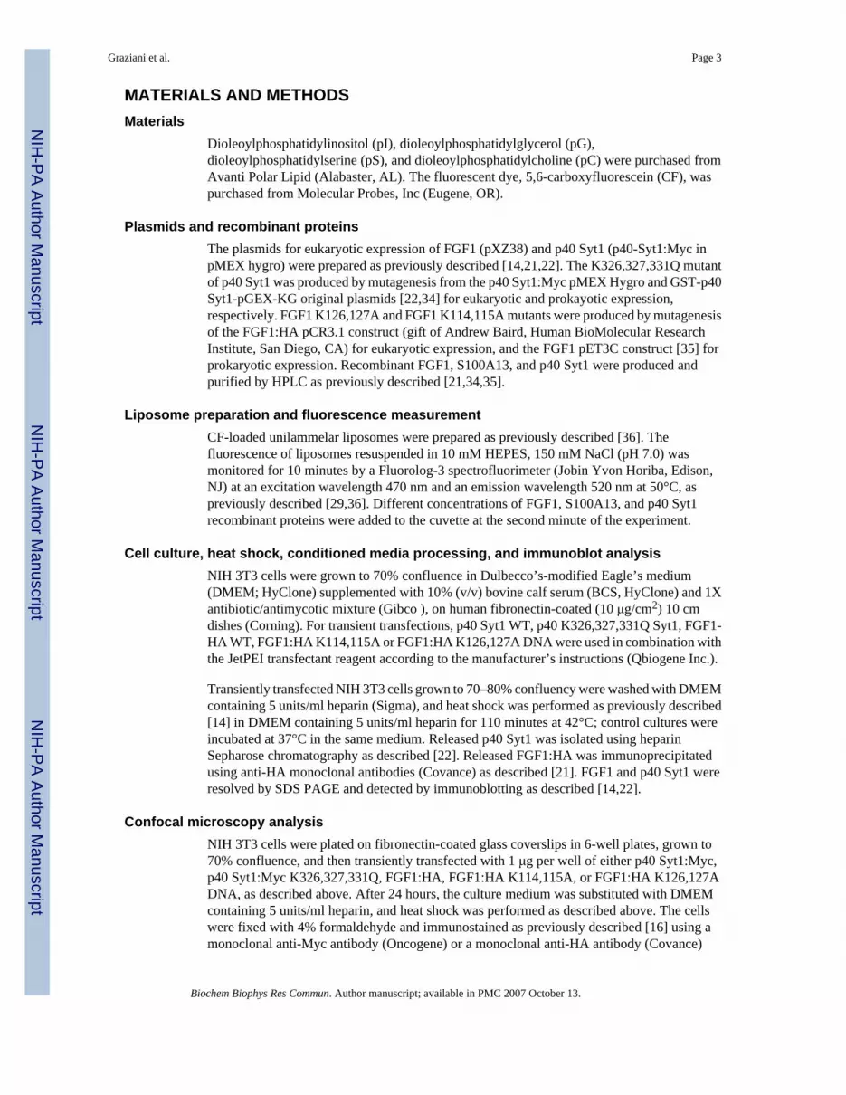
RESULTSProteins of the FGF1 release complex induce liposome destabilization that is dependent onliposome composition and protein concentration
Liposomes loaded with fluorescent molecules represent an established model for studying themembrane destabilizing activity of proteins [37]. FGF1 destabilized mixed pG/pC liposomes[29]. To investigate the interaction of FGF1 with various plasma membrane pL, we comparedthe destabilizing effect of FGF1 on liposomes consisting of several acidic (pS, pI, pG) and azwitterionic (pC) pL. We also evaluated whether other components of the FGF1 releasepathway, i.e. S100A13 and p40 Syt1, induce liposome destabilization (Figure 1A, B, C). Therecombinant forms of FGF1, S100A13, and p40 Syt1 were added individually to the pLliposome suspension in the cuvette, and the CF release was detected fluorimetrically. Therelease of CF from liposomes results in a dequenching of fluorescence due to a sharp decreaseof CF concentration [29]. The final protein concentrations in the cuvette were 1 μM, 0.5 μM,0.25 μM, 0.125 μM. The pL concentration was 2 μM. We used α-chymotrypsin (Sigma) at themaximal concentration employed for studied proteins as a standard negative control becauseit is unable to induce liposome leakage [29], and 0.1% Triton X-100 served as a positive controlfor complete liposome destabilization (maximal CF release).
We found that FGF1 induced destabilization of pI, pG, and pS liposomes, but not the pCliposomes. The dependence of the ability of FGF1 to induce liposome destabilization upontheir pL composition may be represented by the following relationship: pI > pG > pS > pC(Figure 1A).
S100A13 exhibited destabilizing effects on liposomes with a similar relationship as FGF1: pG> pI > pS > pC (Figure 1B). Conversely, p40 Syt1 was not able to induce any CF release frompC, pS, and pG liposomes, but very efficiently destabilized pI liposomes (Figure 1C). It isimportant to stress that none of the proteins were able to induce CF release from zwitterionic(pC) liposomes (Figure 1A, B, C). The extent of CF release was proportional to proteinconcentration. Thus, S100A13 at 1 μM concentration induced CF release from pG liposomesalmost equivalent to complete liposome destabilization induced by 0.1% Triton X-100 (Figure1B).
We concluded that the proteins of the FGF1 release complex were able to induce destabilizationof liposomes composed of acidic pL, but not zwitterionic pL, in a protein concentration-dependent manner. In addition, for p40 Syt1, this ability exhibited selectivity towards a specificacidic pL (pI).
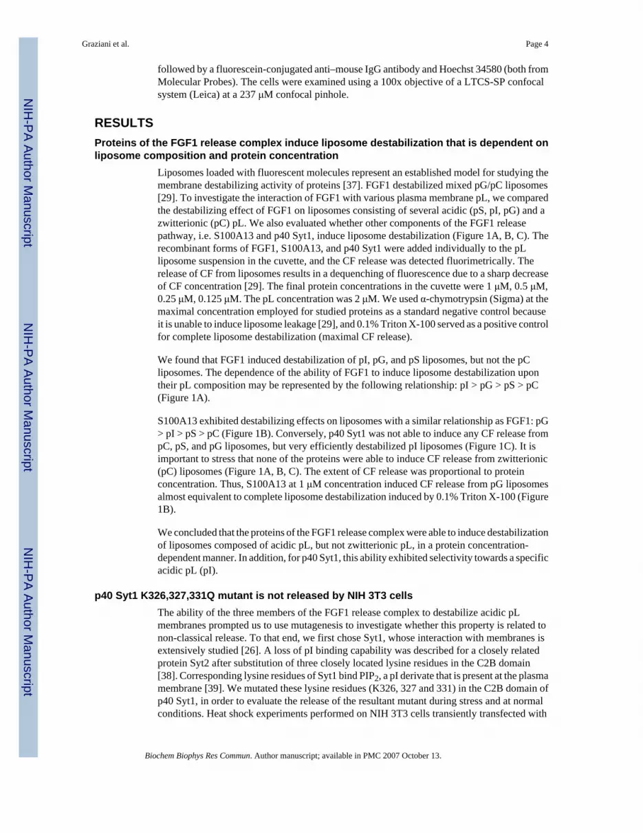
p40 Syt1 K326,327,331Q mutant is not released by NIH 3T3 cellsThe ability of the three members of the FGF1 release complex to destabilize acidic pLmembranes prompted us to use mutagenesis to investigate whether this property is related tonon-classical release. To that end, we first chose Syt1, whose interaction with membranes isextensively studied [26]. A loss of pI binding capability was described for a closely relatedprotein Syt2 after substitution of three closely located lysine residues in the C2B domain[38]. Corresponding lysine residues of Syt1 bind PIP2, a pI derivate that is present at the plasmamembrane [39]. We mutated these lysine residues (K326, 327 and 331) in the C2B domain ofp40 Syt1, in order to evaluate the release of the resultant mutant during stress and at normalconditions. Heat shock experiments performed on NIH 3T3 cells transiently transfected with
Graziani et al. Page 4
Biochem Biophys Res Commun. Author manuscript; available in PMC 2007 October 13.
NIH
-PA Author Manuscript
NIH
-PA Author Manuscript
NIH
-PA Author Manuscript
either the mutant or the WT form of p40 Syt1 demonstrated that, unlike the WT form, the p40Syt1 K326,327,331Q mutant was not released either at 37º or 42ºC (Figure 2A). This suggeststhat the lysine residues at position 326, 327, 331 are required for the non-classical release ofp40 Syt1 from cells.
p40 Syt1 K326,327,331Q does not display enhanced perimembrane localization intransfected cell
Previously, we demonstrated that when the WT form of p40 Syt1 is co-expressed with FGF1,it migrates to the plasma membrane under stress conditions like other members of the FGF1release complex [16]. Since p40 Syt1 K326,327,331Q is not released by NIH 3T3 cells, weevaluated its localization by confocal fluorescence microscopy during heat shock and at normalconditions in transiently transfected NIH 3T3 cells. Our study demonstrated that p40 Syt1K326,327,331Q presented a diffuse cytosolic distribution pattern both at 37º and 42ºC (Figure2B), which was unlike the WT form that displayed an enhanced localization in the vicinity ofthe cell membrane under both conditions (Figure 2B).
K326,327,331Q mutations in the p40 Syt1 C2B domain lead to a sharp decrease of pI liposomedestabilization activity
The inability of p40 Syt1 K326,327,331Q to be released by NIH 3T3 cells and to exhibitpreferential localization in the vicinity of the plasma membrane, prompted us to explore itsliposome permeabilizing activity. The p40 Syt1 K326,327,331Q-induced CF release from pIliposomes was strongly reduced compared to WT p40 Syt1 (Figure 2C). These data suggestthat the lysine residues at positions 326, 327, and 331 play a crucial role in the ability of p40Syt1 to bind the plasma membrane, destabilize it, and exit to the extracellular compartment.
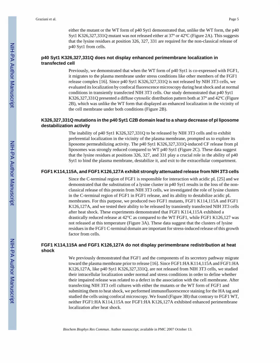
FGF1 K114,115A, and FGF1 K126,127A exhibit strongly attenuated release from NIH 3T3 cellsSince the C-terminal region of FGF1 is responsible for interaction with acidic pL [25] and wedemonstrated that the substitution of a lysine cluster in p40 Syt1 results in the loss of the non-classical release of this protein from NIH 3T3 cells, we investigated the role of lysine clustersin the C-terminal region of FGF1 in FGF1 release, and its ability to destabilize acidic pLmembranes. For this purpose, we produced two FGF1 mutants, FGF1 K114,115A and FGF1K126,127A, and we tested their ability to be released by transiently transfected NIH 3T3 cellsafter heat shock. These experiments demonstrated that FGF1 K114,115A exhibited adrastically reduced release at 42°C as compared to the WT FGF1, while FGF1 K126,127 wasnot released at this temperature (Figure 3A). These data suggest that the clusters of lysineresidues in the FGF1 C-terminal domain are important for stress-induced release of this growthfactor from cells.
FGF1 K114,115A and FGF1 K126,127A do not display perimembrane redistribution at heatshock
We previously demonstrated that FGF1 and the components of its secretory pathway migratetoward the plasma membrane prior to release [16]. Since FGF1:HA K114,115A and FGF1:HAK126,127A, like p40 Syt1 K326,327,331Q, are not released from NIH 3T3 cells, we studiedtheir intracellular localization under normal and stress conditions in order to define whethertheir impaired release was related to a defect in the association with the cell membrane. Aftertransfecting NIH 3T3 cell cultures with either the mutants or the WT form of FGF1 andsubmitting them to heat shock, we performed immunofluorescence staining for the HA tag andstudied the cells using confocal microscopy. We found (Figure 3B) that contrary to FGF1 WT,neither FGF1:HA K114,115A nor FGF1:HA K126,127A exhibited enhanced perimembranelocalization after heat shock.
Graziani et al. Page 5
Biochem Biophys Res Commun. Author manuscript; available in PMC 2007 October 13.
NIH
-PA Author Manuscript
NIH
-PA Author Manuscript
NIH
-PA Author Manuscript
K114,115A or K126,127A mutations in the C-terminal region of FGF1 lead to the decrease ofits ability to destabilize liposomes
To determine whether the attenuated secretion of FGF1:HA K114,115A and FGF1:HAK126,127A correlates with the decreased ability to destabilize acidic pL liposomes, weproduced recombinant forms of FGF1 K114,115A and FGF1 K126,127A mutants and testedtheir effects on CF-loaded acidic pL liposomes. Recombinant FGF1 K114,115A and FGF1K126,127A (Figure 3C) demonstrated a strong decrease of destabilizing activity towards pGliposomes as compared to destabilizing activity of the recombinant WT FGF1, suggesting thatlysine clusters at the C-terminal domain of FGF1 are critical for both membrane destabilizationand non-classical protein release.
DISCUSSIONDespite the absence of a classical signal sequence in its structure, FGF1 exhibits stress-inducedrelease [14]. However, the mechanism used by FGF1 to cross the cell membrane is not known.Previously it was demonstrated that FGF1 binds acidic pL [25], and is able to deform lipidbilayers [30]. Moreover, FGF1 destabilizes liposomes containing acidic pL and assumes apartially unfolded MG-like conformation in the presence of acidic pL [29]. In the MG-likestate conformation, FGF1 retains most of its secondary structure but its tertiary structure isalmost completely disrupted. The solvent-exposed non-polar surface(s) in the MG-like stateprobably facilitates the interaction of FGF1 with the lipid membrane. Subsequently, the boundFGF1 appears to disorganize the lipid membrane, due to acquired hydrophobic characteristicsthat may facilitate its solubility in membranes and perturb their integrity [29,33]. However,since FGF1 is released in association with S100A13 and p40 Syt1 [21,22], the question arisedwhether the two latter proteins could also display membrane destabilizing properties. Similarlyto FGF1 [25], Syt1 is able to associate with acidic pL, particularly pS and pI [26]. In addition,some S100 family members bind pL or pL-binding proteins [27,28]. These previousobservations prompted us to undertake a comparative study of the interaction of FGF1, p40Syt1, and S100A13 with artificial membranes composed of different pL, to determine whetherindividual members of the FGF1 release complex exhibit preferential membrane destabilizingabilities dependent upon specific pL membrane composition. We chose pC, a zwitterionic pL,and three acidic pL - pI , pG, and pS. All of these pL are present in the plasma membrane[40–42], and acidic pL are preferentially localized in the plasma membrane inner leaflet [40].We observed that the three proteins destabilized lipid membranes composed of acidic pL butnot of pC, which is a zwitterionic pL. While FGF1 and S100A13 exhibited destabilizing activitytowards pG, pS, and pI membranes, p40 Syt1 induced selective destabilization of only the pIliposomes.
Since we previously demonstrated that when the components of the FGF1 release complex areco-expressed, they localize at the plasma membrane in response to stress [16], the observationthat FGF1 release complex members destabilize acidic pL liposomes suggests a critical rolefor plasma membrane pL in the assembly and export of the FGF1 release complex [12].Particularly, the preference exhibited by p40 Syt1 in permeabilizing pI liposomes indicates theexistence of sites of specific pL compositions that recruit different constituents of the FGF1release complex at the inner leaflet of the plasma membrane. Therefore, it is likely that specificpL function as anchors for individual proteins, promoting the formation of the multiproteinFGF1 release complex at the inner leaflet of the cell membrane. Additionally, we shouldconsider the interaction between acidic pL and the components of the FGF1 secretion pathwayin terms of reciprocal structural changes: acidic pL might be responsible for inducing tertiarystructure modifications that allow the proteins to become more hydrophobic [29]; on the otherhand, the proteins might be responsible of membrane deformation [30]. Thus, both of thesemechanisms might ultimately contribute to protein release. This might be the case of p40 Syt1,
Graziani et al. Page 6
Biochem Biophys Res Commun. Author manuscript; available in PMC 2007 October 13.
NIH
-PA Author Manuscript
NIH
-PA Author Manuscript
NIH
-PA Author Manuscript

which binds pS and pI respectively through the C2A and C2B domains [26], but exhibitsdestabilizing activity only towards pI liposomes. It is also important to mention that the abilityof some pL, i.e. pS, to flip from the inner to the outer face of the plasma membrane in responseto stress [43,44] might also be involved in the protein translocation process.
The calcium-binding protein, S1001A13, which is released from cells in association with FGF1[21], and similarly to FGF1 exhibits destabilizing activity upon acidic pL liposomes (Figure2), is a member of the S100 proteins family. S100 proteins regulate the functions of their proteinpartners through induced conformational changes. In particular, S100A10 regulates the processof insertion of annexin II into the plasma membrane [45]. Similarly to S100A10, S100A13combined with acidic pL might promote FGF1 and p40 Syt1 conformational changes, andtranslocation of the FGF1 release complex across the plasma membrane.
The establishment of membrane destabilizing properties of the FGF1 release complexcomponents prompted us to use mutational analysis to identify the membrane destabilizingdomains of p40 Syt1 and FGF1. To this purpose, we used available information regarding theacidic pL-binding domains of synaptotagmins [38,39] and FGF1 [25]. We found that mutationof lysines 326, 327, and 331 to glutamines resulted in the inability of p40 Syt1 to be releasedfrom NIH 3T3 cells both at 37º and at 42ºC, as well as a strong decrease of pI liposomedestabilization. Additionally, whereas p40 Syt1 WT exhibited perimembrane localization, p40Syt1 K326,327,331Q was distributed diffusely in the cytoplasm of transfected cells. Similarly,substitution of lysine residues in the C-terminal domain of FGF1 abolished its capability to bereleased from NIH 3T3 cells, to localize at the plasma membrane under stress conditions, andto perturb acidic pL bilayer integrity. Thus, clusters of lysine residues (i) are responsible forthe interaction of p40 Syt1 and FGF1 with the plasma membrane, (ii) are required for theirnon-classical release, (iii) are at least partially responsible for the destabilization of acidic pLmembranes.
In conclusion, this work demonstrated that the members of non-classical FGF1 release complexexhibited the ability to destabilize artificial membranes composed of acidic pL, which arepreferentially localized in the inner leaflet of the cell membrane [46, Micheva, 2001 #3641].Moreover, the membrane destabilizing ability of p40 Syt1 and FGF1 correlated with their non-classical release from NIH 3T3 cells. Additionally, we previously reported that interleukin1α, a protein exported through a non-classical release pathway similar to FGF1, also efficientlypermeabilizes pG liposomes [36]. Further identification of the membrane destabilizingdomains of S100A13 and structural characterization of the interaction of the FGF1 releasecomplex components with lipid membranes is important for the understanding of themechanism of non-classical protein release, which may be applied to the rational design oftherapeutic compounds that interfere with this process.
Acknowledgements
This work was supported in part by NIH grants, HL 32348, HL 35627, and P20 RR 15555 (Project # 4) to IP and bythe NSF grant 0132384 to DN. It was also partially supported by the grants to TKSK: NIH NCRR COBRE (P20RR15569), Department of Energy (DE-FG02-01ER15161), and to CY: The Arkansas Bioscience Institute and theNational Science Council, Taiwan (NSC 94-2320-B007-005 and NSC-94-2113-M007-036).
The authors wish to thank the laboratory of R. Middaugh at the University of Kansas for their help in the design ofthe liposome studies, and Andrew Baird from the Human BioMolecular Research Institute, San Diego, CA for theFGF1:HA construct.
The work was performed by IG and MD in partial fulfillment of the requirements for Ph.D. degree from the Departmentof Critical Care Medicine and Surgery, Gerontology and Geriatrics Unit, University of Florence, Florence, Italy, andfrom the Life and Health Science University of Minho in Braga, Portugal, respectively.
Graziani et al. Page 7
Biochem Biophys Res Commun. Author manuscript; available in PMC 2007 October 13.
NIH
-PA Author Manuscript
NIH
-PA Author Manuscript
NIH
-PA Author Manuscript
References1. Bottcher RT, Niehrs C. Fibroblast growth factor signaling during early vertebrate development. Endocr
Rev 2005;26:63–77. [PubMed: 15689573]2. Friesel R, Maciag T. Fibroblast growth factor prototype release and fibroblast growth factor receptor
signaling. Thromb Haemost 1999;82:748–754. [PubMed: 10605778]3. Dailey L, Ambrosetti D, Mansukhani A, Basilico C. Mechanisms underlying differential responses to
FGF signaling. Cytokine Growth Factor Rev 2005;16:233–247. [PubMed: 15863038]4. Nickel W. Unconventional secretory routes: direct protein export across the plasma membrane of
mammalian cells. Traffic 2005;6:607–614. [PubMed: 15998317]5. Florkiewicz RZ, Majack RA, Buechler RD, Florkiewicz E. Quantitative export of FGF-2 occurs
through an alternative, energy- dependent, non-ER/Golgi pathway. J Cell Physiol 1995;162:388–399.[PubMed: 7860646]
6. Mignatti P, Morimoto T, Rifkin DB. Basic fibroblast growth factor, a protein devoid of secretory signalsequence, is released by cells via a pathway independent of the endoplasmic reticulum-Golgi complex.J Cell Physiol 1992;151:81–93. [PubMed: 1560052]
7. Andrei C, Dazzi C, Lotti L, Torrisi MR, Chimini G, Rubartelli A. The secretory route of the leaderlessprotein interleukin 1beta involves exocytosis of endolysosome-related vesicles. Mol Biol Cell1999;10:1463–1475. [PubMed: 10233156]
8. Andrei C, Margiocco P, Poggi A, Lotti LV, Torrisi MR, Rubartelli A. Phospholipases C and A2 controllysosome-mediated IL-1 beta secretion: Implications for inflammatory processes. Proc Natl Acad SciU S A 2004;101:9745–9750. [PubMed: 15192144]
9. Hunter-Lavin C, Davies EL, Bacelar MM, Marshall MJ, Andrew SM, Williams JH. Hsp70 releasefrom peripheral blood mononuclear cells. Biochem Biophys Res Commun 2004;324:511–517.[PubMed: 15474457]
10. Jeske NA, Glucksman MJ, Roberts JL. Metalloendopeptidase EC3.4.24.15 is constitutively releasedfrom the exofacial leaflet of lipid rafts in GT1-7 cells. J Neurochem 2004;90:819–828. [PubMed:15287887]
11. Lee SJ, Saravanan RS, Damasceno CM, Yamane H, Kim BD, Rose JK. Digging deeper into the plantcell wall proteome. Plant Physiol Biochem 2004;42:979–988. [PubMed: 15707835]
12. Prudovsky I, Mandinova A, Soldi R, Bagala C, Graziani I, Landriscina M, Tarantini F, Duarte M,Bellum S, Doherty H, Maciag T. The non-classical export routes: FGF1 and IL-1alpha point the way.J Cell Sci 2003;116:4871–4881. [PubMed: 14625381]
13. Jaye M, Howk R, Burgess W, Ricca GA, Chiu IM, Ravera MW, O’Brien SJ, Modi WS, Maciag T,Drohan WN. Human endothelial cell growth factor: cloning, nucleotide sequence, and chromosomelocalization. Science 1986;233:541–545. [PubMed: 3523756]
14. Jackson A, Friedman S, Zhan X, Engleka KA, Forough R, Maciag T. Heat shock induces the releaseof fibroblast growth factor 1 from NIH 3T3 cells. Proc Natl Acad Sci U S A 1992;89:10691–10695.[PubMed: 1279690]
15. Misumi Y, Miki K, Takatsuki A, Tamura G, Ikehara Y. Novel blockade by brefeldin A of intracellulartransport of secretory proteins in cultured rat hepatocytes. J Biol Chem 1986;261:11398–11403.[PubMed: 2426273]
16. Prudovsky I, Bagala C, Tarantini F, Mandinova A, Soldi R, Bellum S, Maciag T. The intracellulartranslocation of the components of the fibroblast growth factor 1 release complex precedes theirassembly prior to export. J Cell Biol 2002;158:201–208. [PubMed: 12135982]
17. Carreira, C Mouta; Landriscina, M.; Bellum, S.; Prudovsky, I.; Maciag, T. The comparative releaseof FGF1 by hypoxia and temperature stress. Growth Factors 2001;18:277–285. [PubMed: 11519826]
18. Shin JT, Opalenik SR, Wehby JN, Mahesh VK, Jackson A, Tarantini F, Maciag T, Thompson JA.Serum-starvation induces the extracellular appearance of FGF-1. Biochim Biophys Acta1996;1312:27–38. [PubMed: 8679713]
19. Ananyeva NM, Tijurmin AV, Berliner JA, Chisolm GM, Liau G, Winkles JA, Haudenschild CC.Oxidized LDL mediates the release of fibroblast growth factor-1. Arterioscler Thromb Vasc Biol1997;17:445–453. [PubMed: 9102162]
Graziani et al. Page 8
Biochem Biophys Res Commun. Author manuscript; available in PMC 2007 October 13.
NIH
-PA Author Manuscript
NIH
-PA Author Manuscript
NIH
-PA Author Manuscript
20. Landriscina M, Bagala C, Mandinova A, Soldi R, Micucci I, Bellum S, Prudovsky I, Maciag T. Copperinduces the assembly of a multiprotein aggregate implicated in the release of fibroblast growth factor1 in response to stress. J Biol Chem 2001;276:25549–25557. [PubMed: 11432880]
21. Landriscina M, Soldi R, Bagala C, Micucci I, Bellum S, Tarantini F, Prudovsky I, Maciag T. S100A13participates in the release of fibroblast growth factor 1 in response to heat shock in vitro. J Biol Chem2001;276:22544–22552. [PubMed: 11410600]
22. LaVallee TM, Tarantini F, Gamble S, Carreira CM, Jackson A, Maciag T. Synaptotagmin-1 is requiredfor fibroblast growth factor-1 release. J Biol Chem 1998;273:22217–22223. [PubMed: 9712835]
23. Yoshihara M, Montana ES. The synaptotagmins: calcium sensors for vesicular trafficking.Neuroscientist 2004;10:566–574. [PubMed: 15534041]
24. Bagala C, Kolev V, Mandinova A, Soldi R, Mouta C, Graziani I, Prudovsky I, Maciag T. Thealternative translation of synaptotagmin 1 mediates the non-classical release of FGF1. BiochemBiophys Res Commun 2003;310:1041–1047. [PubMed: 14559220]
25. Tarantini F, Gamble S, Jackson A, Maciag T. The cysteine residue responsible for the release offibroblast growth factor-1 residues in a domain independent of the domain for phosphatidylserinebinding. J Biol Chem 1995;270:29039–29042. [PubMed: 7493920]
26. Bai J, Chapman ER. The C2 domains of synaptotagmin--partners in exocytosis. Trends Biochem Sci2004;29:143–151. [PubMed: 15003272]
27. Rescher U, Gerke V. Annexins--unique membrane binding proteins with diverse functions. J Cell Sci2004;117:2631–2639. [PubMed: 15169834]
28. Nacken W, Sorg C, Kerkhoff C. The myeloid expressed EF-hand proteins display a diverse patternof lipid raft association. FEBS Lett 2004;572:289–293. [PubMed: 15304364]
29. Mach H, Middaugh CR. Interaction of partially structured states of acidic fibroblast growth factorwith phospholipid membranes. Biochemistry 1995;34:9913–9920. [PubMed: 7543282]
30. Doyle AW, Fick J, Himmelhaus M, Eck W, Graziani I, Prudovsky I, Grunze M, Maciag T, NeivandtD. Protein deformation of lipid hybrid bilayer membranes studied by Sum Frequency GenerationVibrational Spectroscopy (SFS). Langmuir 2004;20:8961–8965. [PubMed: 15461473]
31. Copeland RA, Ji H, Halfpenny AJ, Williams RW, Thompson KC, Herber WK, Thomas KA, BrunerMW, Ryan JA, Marquis-Omer D, et al. The structure of human acidic fibroblast growth factor andits interaction with heparin. Arch Biochem Biophys 1991;289:53–61. [PubMed: 1716876]
32. Dabora JM, Sanyal G, Middaugh CR. Effect of polyanions on the refolding of human acidic fibroblastgrowth factor. J Biol Chem 1991;266:23637–23640. [PubMed: 1721051]
33. Bychkova VE, Pain RH, Ptitsyn OB. The ‘molten globule’ state is involved in the translocation ofproteins across membranes? FEBS Lett 1988;238:231–234. [PubMed: 3049159]
34. Tarantini F, LaVallee T, Jackson A, Gamble S, Carreira C Mouta, Garfinkel S, Burgess WH, MaciagT. The extravesicular domain of synaptotagmin-1 is released with the latent fibroblast growth factor-1homodimer in response to heat shock. J Biol Chem 1998;273:22209–22216. [PubMed: 9712834]
35. Engleka KA, Maciag T. Inactivation of human fibroblast growth factor-1 (FGF-1) activity byinteraction with copper ions involves FGF-1 dimer formation induced by copper-catalyzed oxidation.J Biol Chem 1992;267:11307–11315. [PubMed: 1375939]
36. Mandinova A, Soldi R, Graziani I, Bagala C, Bellum S, Landriscina M, Tarantini F, Prudovsky I,Maciag T. S100A13 mediates the copper-dependent stress-induced release of IL-1{alpha} from bothhuman U937 and murine NIH 3T3 cells. J Cell Sci 2003;116:2687–2696. [PubMed: 12746488]
37. Matsuzaki K, Murase O, Tokuda H, Funakoshi S, Fujii N, Miyajima K. Orientational andaggregational states of magainin 2 in phospholipid bilayers. Biochemistry 1994;33:3342–3349.[PubMed: 8136371]
38. Kida Y, Sakaguchi M, Fukuda M, Mikoshiba K, Mihara K. Amino acid residues before thehydrophobic region which are critical for membrane translocation of the N-terminal domain ofsynaptotagmin II. FEBS Lett 2001;507:341–345. [PubMed: 11696368]
39. Bai J, Tucker WC, Chapman ER. PIP2 increases the speed of response of synaptotagmin and steersits membrane-penetration activity toward the plasma membrane. Nat Struct Mol Biol 2004;11:36–44. [PubMed: 14718921]
Graziani et al. Page 9
Biochem Biophys Res Commun. Author manuscript; available in PMC 2007 October 13.
NIH
-PA Author Manuscript
NIH
-PA Author Manuscript
NIH
-PA Author Manuscript

40. McLaughlin S, Hangyas-Mihalyne G, Zaitseva I, Golebiewska U. Reversible - through calmodulin -electrostatic interactions between basic residues on proteins and acidic lipids in the plasmamembrane. Biochem Soc Symp 2005:189–198. [PubMed: 15649142]
41. Momchilova-Pankova AB, Markovska TT, Koumanov KS. Acyl-CoA synthetase activity dependson the phospholipid composition of rat liver plasma membranes. J Lipid Mediat Cell Signal1995;11:13–23. [PubMed: 7728415]
42. Schroeder F, Fontaine RN, Feller DJ, Weston KG. Drug-induced surface membrane phospholipidcomposition in murine fibroblasts. Biochim Biophys Acta 1981;643:76–88. [PubMed: 7236693]
43. Kuypers FA, de Jong K. The role of phosphatidylserine in recognition and removal of erythrocytes.Cell Mol Biol (Noisy-le-grand) 2004;50:147–158. [PubMed: 15095785]
44. Kagan VE, Borisenko GG, Serinkan BF, Tyurina YY, Tyurin VA, Jiang J, Liu SX, Shvedova AA,Fabisiak JP, Uthaisang W, Fadeel B. Appetizing rancidity of apoptotic cells for macrophages:oxidation, externalization, and recognition of phosphatidylserine. Am J Physiol Lung Cell MolPhysiol 2003;285:L1–17. [PubMed: 12788785]
45. Donato R. Intracellular and extracellular roles of S100 proteins. Microsc Res Tech 2003;60:540–551.[PubMed: 12645002]
46. Holz RW, Hlubek MD, Sorensen SD, Fisher SK, Balla T, Ozaki S, Prestwich GD, Stuenkel EL,Bittner MA. A pleckstrin homology domain specific for phosphatidylinositol 4, 5-bisphosphate(PtdIns-4,5-P2) and fused to green fluorescent protein identifies plasma membrane PtdIns-4,5-P2 asbeing important in exocytosis. J Biol Chem 2000;275:17878–17885. [PubMed: 10747966]
Graziani et al. Page 10
Biochem Biophys Res Commun. Author manuscript; available in PMC 2007 October 13.
NIH
-PA Author Manuscript
NIH
-PA Author Manuscript
NIH
-PA Author Manuscript
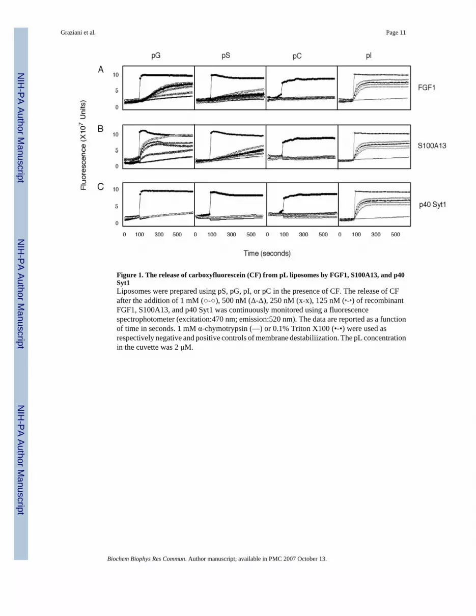
Figure 1. The release of carboxyfluorescein (CF) from pL liposomes by FGF1, S100A13, and p40Syt1Liposomes were prepared using pS, pG, pI, or pC in the presence of CF. The release of CFafter the addition of 1 mM (○-○), 500 nM (Δ-Δ), 250 nM (x-x), 125 nM (⋄-⋄) of recombinantFGF1, S100A13, and p40 Syt1 was continuously monitored using a fluorescencespectrophotometer (excitation:470 nm; emission:520 nm). The data are reported as a functionof time in seconds. 1 mM α-chymotrypsin (—) or 0.1% Triton X100 (•-•) were used asrespectively negative and positive controls of membrane destabiliization. The pL concentrationin the cuvette was 2 μM.
Graziani et al. Page 11
Biochem Biophys Res Commun. Author manuscript; available in PMC 2007 October 13.
NIH
-PA Author Manuscript
NIH
-PA Author Manuscript
NIH
-PA Author Manuscript

Figure 2. A. The release of p40 Syt1 WT and p40 Syt1 K326,327,331Q from NIH 3T3 cellsNIH 3T3 cells transfected with p40 Syt1 WT or p40 Syt1 K326,327,321Q were incubated for2 hours at 37°C or 42°C. p40 Syt1 from conditioned medium was adsorbed to heparin-Sepharose and analyzed by immunoblotting as described [14]. CL- cell lysate; CM-conditioned medium. B. Confocal fluorescence microscopy analysis of p40 Syt1:Myc WTand p40 Syt1:Myc K326,327,331Q cellular localization at normal and stress conditions.NIH 3T3 cells transfected with either p40 Syt1:Myc WT or p40 Syt1:Myc K326,327,331Qwere incubated for 2 hours at 37°C or 42°C. The cells were fixed with formaldehyde andimmunofluorescently stained for Myc tag (green) as described in the “Materials and Methods”.The nuclei were stained with Hoechst 34580 (blue). The cells were studied using a confocal
Graziani et al. Page 12
Biochem Biophys Res Commun. Author manuscript; available in PMC 2007 October 13.
NIH
-PA Author Manuscript
NIH
-PA Author Manuscript
NIH
-PA Author Manuscript
microscope with an X100 objective. Median horizontal confocal cell sections are presented.Bar- 20 μm. C. Analysis of the CF release from pI liposomes induced by p40 Syt1 WT andp40 Syt1 K326,327,331Q. The experiments on CF release from pI liposomes induced by 500nM p40 Syt1 WT or p40 Syt1 K326,327,331Q were performed as described in Figure 1.
Graziani et al. Page 13
Biochem Biophys Res Commun. Author manuscript; available in PMC 2007 October 13.
NIH
-PA Author Manuscript
NIH
-PA Author Manuscript
NIH
-PA Author Manuscript
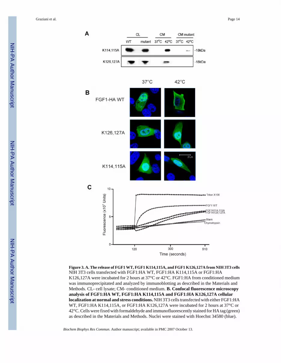
Figure 3. A. The release of FGF1 WT, FGF1 K114,115A, and FGF1 K126,127A from NIH 3T3 cellsNIH 3T3 cells transfected with FGF1:HA WT, FGF1:HA K114,115A or FGF1:HAK126,127A were incubated for 2 hours at 37°C or 42°C. FGF1:HA from conditioned mediumwas immunoprecipitated and analyzed by immunoblotting as described in the Materials andMethods. CL- cell lysate; CM- conditioned medium. B. Confocal fluorescence microscopyanalysis of FGF1:HA WT, FGF1:HA K114,115A and FGF1:HA K126,127A cellularlocalization at normal and stress conditions. NIH 3T3 cells transfected with either FGF1:HAWT, FGF1:HA K114,115A, or FGF1:HA K126,127A were incubated for 2 hours at 37°C or42°C. Cells were fixed with formaldehyde and immunofluorescently stained for HA tag (green)as described in the Materials and Methods. Nuclei were stained with Hoechst 34580 (blue).
Graziani et al. Page 14
Biochem Biophys Res Commun. Author manuscript; available in PMC 2007 October 13.
NIH
-PA Author Manuscript
NIH
-PA Author Manuscript
NIH
-PA Author Manuscript
The cells were studied using a confocal microscope with a X100 objective. Median horizontalconfocal cell sections are presented. Bar- 20 μm. C. Analysis of the CF release from pGliposomes induced by FGF1 WT, FGF1 K114,115A, or FGF1 K126,127A. The experimentson CF release from pG liposomes induced by 500 nM FGF1 WT, FGF1 K114,115A, or FGF1K126,127A were performed as described in Figure 1.
Graziani et al. Page 15
Biochem Biophys Res Commun. Author manuscript; available in PMC 2007 October 13.
NIH
-PA Author Manuscript
NIH
-PA Author Manuscript
NIH
-PA Author Manuscript