regulation of endoplasmic reticulum turnover by selective autophagy
TRANSCRIPT

LETTERdoi:10.1038/nature14498
Regulation of endoplasmic reticulum turnover byselective autophagyAliaksandr Khaminets1*, Theresa Heinrich2*, Muriel Mari3,4, Paolo Grumati1, Antje K. Huebner2, Masato Akutsu5,Lutz Liebmann2, Alexandra Stolz1, Sandor Nietzsche6, Nicole Koch7, Mario Mauthe3,4, Istvan Katona8, Britta Qualmann7,Joachim Weis8, Fulvio Reggiori3,4, Ingo Kurth21, Christian A. Hubner21 & Ivan Dikic1,5,91
The endoplasmic reticulum (ER) is the largest intracellular endo-membrane system, enabling protein and lipid synthesis, ion home-ostasis, quality control of newly synthesized proteins and organellecommunication1. Constant ER turnover and modulation is neededto meet different cellular requirements and autophagy has animportant role in this process2–8. However, its underlying regula-tory mechanisms remain unexplained. Here we show that membersof the FAM134 reticulon protein family are ER-resident receptorsthat bind to autophagy modifiers LC3 and GABARAP, and facil-itate ER degradation by autophagy (‘ER-phagy’). Downregulationof FAM134B protein in human cells causes an expansion of the ER,while FAM134B overexpression results in ER fragmentation andlysosomal degradation. Mutant FAM134B proteins that causesensory neuropathy in humans9 are unable to act as ER-phagyreceptors. Consistently, disruption of Fam134b in mice causesexpansion of the ER, inhibits ER turnover, sensitizes cells to
stress-induced apoptotic cell death and leads to degeneration ofsensory neurons. Therefore, selective ER-phagy via FAM134 pro-teins is indispensable for mammalian cell homeostasis and con-trols ER morphology and turnover in mice and humans.
Selective autophagy is a degradative pathway that controls the qual-ity and abundance of proteins and cellular organelles and is mediatedby autophagy receptors that simultaneously bind the designated targetand LC3/GABARAP proteins on autophagosomal membranes10–12. Toidentify proteins involved in selective autophagy we performed yeasttwo-hybrid screens using LC3B (microtubule associated protein lightchain 3B; MAP1LC3B) and GATE16/GABARAPL2 (c-aminobutyricacid receptor-associated protein-like 2) as baits13. Members of thereticulon-homology-domain-containing FAM134 protein family werefound to be interaction partners of LC3B and GABARAPL2 and thisinteraction was confirmed using FAM134B as the bait (SupplementaryTable 1). Also, overexpressed and endogenous FAM134B directly
a db
HA
2% input
GST
GST–LC3A
GST–LC3B
GST–G
ABARAPL1
GST–G
ABARAPL2
FAM134B–HA
Lc3b
Fam134bIgG
+/+ –/– IgG +/+ –/–
0.2% input IP
*
RTN4–MycFAM134B Merge Inset
CLIMP-63 Merge Inset
GFP–SEC61BFAM134B Merge Inset
FAM134B
c
Reticulon
domain
LIR
FAM134B CC
Q145X S309X
Po
nceau
e
Co
-lo
caliz
atio
n w
ith F
AM
134B
Co
-localiz
atio
n w
ith F
AM
134B
***1.0
0.8
0.6
0.4
0.2
0.0
1.0
0.8
0.6
0.4
0.2
0.0
75
kDa
25
35
kDa
1117
48
63
FAM134A_HUMAN
FAM134B_HUMAN
FAM134C_HUMAN
Fam134b_MOUSE
FAM134B_BOVIN
FAM134B_CHICK
fam134b_DANRE
RTN
4–M
yc
CLI
MP-6
3
GFP
–SEC
61B
Figure 1 | FAM134B binds LC3-like modifiers and co-localizes with ERmarker proteins. a, Overexpressed FAM134B–HA (haemagglutinin) bindsGST–LC3-like modifiers. b, Co-immunoprecipitation (IP) of Fam134b inFam134b1/1 and Fam134b–/– MEFs with Lc3b. c, Domain architecture ofFAM134B and alignment of the LIR motif. Blue, reticulon domain; red,LC3-interacting region (LIR); green, coiled-coil (CC) domain; Q145X and
S309X, disease-associated stop mutations. Residues with full (*), strong (:)and weak (.) conservation are indicated. d, Endogenous FAM134B co-stainswith ER-markers in A549 cells. Representatives of ten images are shown.Scale bar, 10 mm. e, Quantification of FAM134B co-localization presented asPearson’s correlation coefficient (r); ***P , 0.001, one-way ANOVA,n 5 50 cells.
*These authors contributed equally to this work.1These authors jointly supervised this work.
1Institute of Biochemistry II, Goethe University School of Medicine, Theodor-Stern-Kai 7, 60590 Frankfurt am Main, Germany. 2Institute of Human Genetics, Jena University Hospital, Friedrich-Schiller-University Jena, Kollegiengasse 10, 07743 Jena, Germany. 3Department of Cell Biology, Center for Molecular Medicine, University Medical Center Utrecht, Heidelberglaan 100, 3584 CX Utrecht, TheNetherlands. 4Department of Cell Biology, University Medical Center Utrecht, University of Groningen, Antonious Deusinglaan 1, 3713 AV Groningen, The Netherlands. 5Buchmann Institute for MolecularLife Sciences, Goethe University Frankfurt, Riedberg Campus, Max-von-Laue-Straße 15, 60438 Frankfurt am Main, Germany. 6Electron Microscopy Center, Jena University Hospital, Friedrich-Schiller-University Jena, Ziegelmuhlenweg 1, 07743 Jena, Germany. 7Institute for Biochemistry I, Jena University Hospital, Friedrich-Schiller-University Jena, 07743 Jena, Germany. 8Institute of Neuropathology,RWTH Aachen University Hospital, Pauwelsstr. 30, 52074 Aachen, Germany. 9Institute of Immunology, School of Medicine University of Split, Mestrovicevo setaliste bb, 21 000 Split, Croatia.
0 0 M O N T H 2 0 1 5 | V O L 0 0 0 | N A T U R E | 1
G2015 Macmillan Publishers Limited. All rights reserved
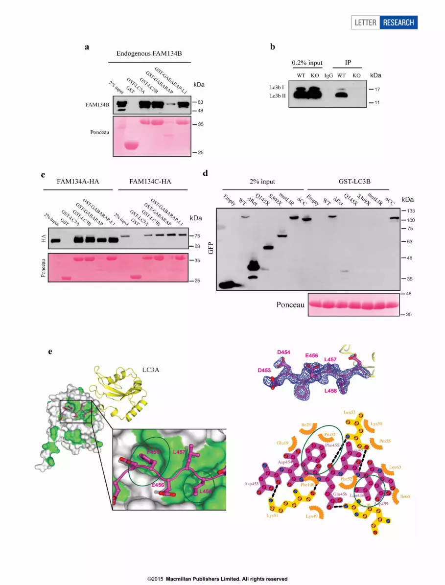
interacts with LC3/GABARAP in mammalian cells (Fig. 1a, b andExtended Data Fig. 1a, b). Similar results were obtained forFAM134A and FAM134C proteins (Extended Data Fig. 1c).FAM134 proteins contain a conserved putative LC3-interacting region(LIR motif) (Fig. 1c). Substitution of amino acids of this LIR motif withalanines (DDFELL/AAAAAA; mutLIR) completely abolished theinteraction of FAM134B with LC3-like modifiers, whereas deletionof the C-terminal coiled-coil domain (DCC) did not affect LC3 binding(Extended Data Fig. 1d). Truncated FAM134B proteins (Q145X,S309X) causing sensory neuropathy in humans9 lack the C-terminalLIR motif and consequently did not bind LC3-like proteins (ExtendedData Fig. 1d). Moreover, the crystal structure of a LIR-motif-containing FAM134B peptide bound to LC3A further demonstrated
that amino acids F455 and L458 of FAM134B mediate the interactionwith the hydrophobic pockets of LC3A (Extended Data Fig. 1e)14,15.These data thus show a direct interaction of FAM134 proteins with theautophagy machinery and identify a potential novel family of auto-phagy receptors.
Endogenous and overexpressed FAM134B co-localized withCLIMP-63 and SEC61B, marker proteins of sheet-like cisternal ERand to a lesser extent with RTN4-positive tubular ER (Fig. 1d, e andExtended Data Fig. 2a–c). We found that both the N-terminus and theLIR-motif-containing C-terminus, which flank the reticulon homo-logy domain of FAM134B, face the cytoplasm, suggesting a hairpintopology of the protein16,17 (Extended Data Fig. 2d, e). Liposome-bind-ing assays confirmed that FAM134B binds to membranes (Extended
HA LC3BKDEL–RFP Merge
c
MER
ERER
PM
Ctrl
ER ER
ER ERM
M
Ctrl + Baf
ER
MM
M
M
MN
PM
eGFP–FAM134B
ER
M M
M
N
PM
eGFP–FAM134B + Baf
ALN
KDEL
CD63
AL
GFP
CD63
AL
AL
ER
ER
ER
M
N
**
*
eGFP–FAM134B + Baf
AL
AL
ER
M
M
M
*
*
eGFP–FAM134B + Baf
HA
eGFP–FAM134B eGFP–FAM134B
b
**
Cells
with m
Cherr
y+/e
GF
P– p
uncta
e (%
)
Cells
with m
Cherr
y+/e
GF
P– p
uncta
e (%
)h ig
0
20
40
60
100
80
**
0
20
40
60
100
80
MEFsU2OS
a
FA
M1
34
B–H
AF
AM
13
4B
(mu
tLIR
)–H
A
e
d
f
eGFP mCherry Merge
mCherry–eGFP–FAM134B
FA
M134B
FA
M134B
(mutL
IR)
Cells
with L
C3B
/KD
EL–R
FP
-p
ositiv
e p
uncta
e (%
)
0
10
20
50
40
30
60
70
NS NS
****
GFP
FAM
134B
–HA
FAM
134B
(mut
LIR)–HA
FAM
134A
–HA
RTN
4A–M
yc
FAM
134B
FAM
134B
(mut
LIR)
Atg5+/+
Atg5–/
–
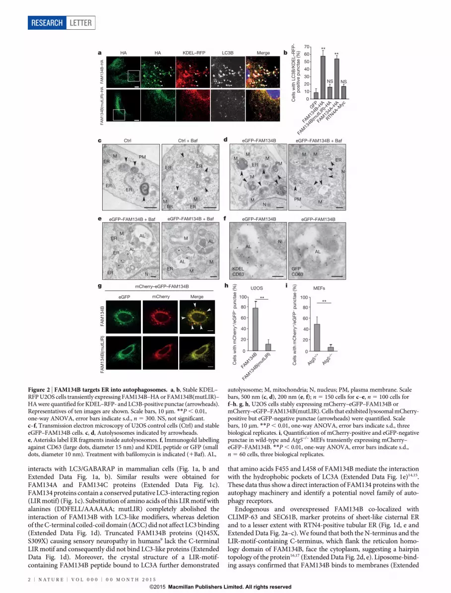
Figure 2 | FAM134B targets ER into autophagosomes. a, b, Stable KDEL–RFP U2OS cells transiently expressing FAM134B–HA or FAM134B(mutLIR)–HA were quantified for KDEL–RFP- and LC3B-positive punctae (arrowheads).Representatives of ten images are shown. Scale bars, 10 mm. **P , 0.01,one-way ANOVA, error bars indicate s.d., n 5 300. NS, not significant.c–f, Transmission electron microscopy of U2OS control cells (Ctrl) and stableeGFP–FAM134B cells. c, d, Autolysosomes indicated by arrowheads.e, Asterisks label ER fragments inside autolysosomes. f, Immunogold labellingagainst CD63 (large dots, diameter 15 nm) and KDEL peptide or GFP (smalldots, diameter 10 nm). Treatment with bafilomycin is indicated (1Baf). AL,
autolysosome; M, mitochondria; N, nucleus; PM, plasma membrane. Scalebars, 500 nm (c, d), 200 nm (e, f); n 5 150 cells for c–e, n 5 100 cells forf–h. g, h, U2OS cells stably expressing mCherry–eGFP–FAM134B ormCherry–eGFP–FAM134B(mutLIR). Cells that exhibited lysosomal mCherry-positive but eGFP-negative punctae (arrowheads) were quantified. Scalebars, 10 mm. **P , 0.01, one-way ANOVA, error bars indicate s.d., threebiological replicates. i, Quantification of mCherry-positive and eGFP-negativepunctae in wild-type and Atg5–/– MEFs transiently expressing mCherry–eGFP–FAM134B. **P , 0.01, one-way ANOVA, error bars indicate s.d.,n 5 60 cells, three biological replicates.
2 | N A T U R E | V O L 0 0 0 | 0 0 M O N T H 2 0 1 5
RESEARCH LETTER
G2015 Macmillan Publishers Limited. All rights reserved
Data Fig. 3a) and its incubation with liposomes resulted in a reducedliposome size (Extended Data Fig. 3b, c). Neuropathy-associated pro-teins lacking both the coiled-coil domain and the LIR-motif (S309X)showed weaker binding to membranes but retained the ability to shapethem, whereas deletion of a part of the reticulon domain (Q145X)abolished the formation of small-diameter liposomes (ExtendedData Fig. 3c). These data indicate that FAM134B is capable of remod-elling membranes in vitro via its reticulon homology domain.
Transient overexpression of FAM134B in U2OS cells stably expres-sing the ER marker protein KDEL–RFP led to pronounced morpho-logical changes and fragmentation of the ER. FAM134B-positive ERfragments co-localized with LC3B and CD63-positive lysosomes(Fig. 2a–f). In contrast, the ability of FAM134B to fragment andsequester ER into LC3-positive autophagosomes was abolishedupon expression of a LIR-motif-deficient FAM134B (Fig. 2a, b).Overexpression of RTN4A, another reticulon-domain-containingprotein16, induced morphological ER changes, but did not lead to aformation of LC3B/KDEL–RFP-positive punctae (Fig. 2b). Theseobservations suggest that FAM134B, through its LIR motif, acts asan ER-selective autophagy receptor. To dissect the fate of FAM134B-induced ER fragments, we performed transmission electron micro-scopy (Fig. 2c–f). Lysosomes and autolysosomes were more abundantin U2OS cells stably overexpressing eGFP–FAM134B comparedto controls, an effect which was even more prominent upon inhibi-tion of lysosomal degradation by bafilomycin A1 (Fig. 2c–e).Autolysosomes in bafilomycin A1-treated and eGFP–FAM134B-expressing cells showed an accumulation of ribosome-decorated ERfragments in their interior (Fig. 2e). The presence of ER-derived struc-tures in autolysosomes was confirmed by double immunogold labellingusing antibodies against the lysosomal marker CD63 and the KDELpeptide (Fig. 2f). Double labelling with CD63 and GFP antibodiesproved that eGFP–FAM134B was delivered into autolysosomes(Fig. 2f). ER turnover was further investigated by monitoring the degra-dation of the ER-resident protein SEC61B. Increased degradation ofSEC61B was observed in FAM134B-overexpressing cells but not incells expressing FAM134(mutLIR) (Extended Data Fig. 4a). Deliveryof ER fragments into lysosomes was further confirmed in U2OS cellsstably expressing FAM134B with a tandem fusion of mCherry andeGFP (mCherry–eGFP–FAM134B), as indicated by mCherry-positivebut eGFP-negative punctae, owing to the higher stability of mCherryin acidic milieux13 (Fig. 2g, h). In contrast, only few mCherry-positiveand eGFP-negative punctae were observed in cells stably expressingmCherry–eGFP–FAM134B(mutLIR) (Fig. 2g, h) or in autophagy-deficient Atg5–/– mouse embryonic fibroblasts (MEFs) expressingwild-type mCherry–eGFP–FAM134B (Fig. 2i and Extended DataFig. 4b). Moreover, ER fragmentation and degradation was abolishedafter FAM134B overexpression in Atg5–/– MEFs (Extended DataFig. 4c–f). These data demonstrate that FAM134B is an ER-anchoredautophagy receptor that mediates ER delivery into lysosomes throughsequestration into autophagosomes (ER-phagy). Notably, this mech-anism is different from a recently described ER-phagy pathway in yeastthat operates independently from the core autophagy machinery18.
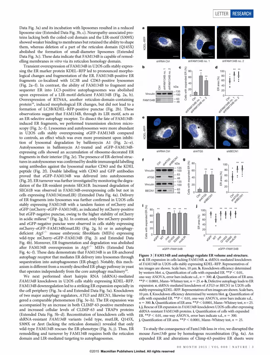
We next performed short hairpin RNA (shRNA)-mediatedFAM134B knockdown in U2OS cells stably expressing KDEL–RFP.FAM134B downregulation led to a striking ER expansion especially inthe cell periphery (Fig. 3a–d and Extended Data Fig. 5a). Knockdownof two major autophagy regulators, ATG5 and BECN1, likewise trig-gered a comparable phenomenon (Fig. 3e–h). The ER expansion wasaccompanied by an increase in both CLIMP-63-positive membranesand increased cellular levels of CLIMP-63 and TRAPa proteins(Extended Data Fig. 5b–d). Reconstitution of knockdown cells withshRNA-resistant FAM134B variants (wild type, mutLIR, Q145X,S309X or Dret (lacking the reticulon domain)) revealed that onlywild-type FAM134B rescues the ER-phenotype (Fig. 3i, j). Thus, ERremodelling and turnover by FAM134B requires both the reticulondomain and LIR-mediated targeting to autophagosomes.
To study the consequence of Fam134b loss in vivo, we disrupted themouse Fam134b gene by homologous recombination (Fig. 4a). Anexpanded ER and alterations of Climp-63-positive ER sheets were
Cells
with e
xp
and
ed
ER
(%
)
shFAM134B no. 1 shFAM134B no. 2shRNA Ctrl
**80
shATG5 shBECN1
** **
**** ** ** **
d
eGFP–FAM134B
KD
EL–R
FP
i j
shRNA Ctrl
b c
f
Vinculin
Vinculin
ATG5
BECN1C
ells
with e
xp
and
ed
ER
(%
)20
0
40
60
80
100
ER
are
a (%
)
g h
ER
are
a (%
)
Cells
with e
xp
and
ed
ER
(%
)
**
20
0
40
60
ER
are
a (%
)
20
0
40
60
80
100
Vinculin
FAM134B
0
40
60
20
20
40
0
60
80
100
20
40
0
60
80
80
a
e
KD
EL–R
FP
eGFP–FAM134B
**** ** ** **
****
****
100135
48
100
48
100
63
kDa
kDa
63
63
48
135
135
shBEC
N1
shATG
5
Ctrl
eGFP W
TΔr
et
Q14
5X
S309X
mut
LIR
eGFP W
TΔr
et
Q14
5X
S309X
mut
LIR
Ctrl
shATG
5
shBEC
N1
Ctrl
shATG
5
shBEC
N1
Ctrl
shFA
M13
4B n
o. 1
shFA
M13
4B n
o. 2
Ctr
l
shFA
M134B
no
. 1
shFA
M134B
no
. 2
Ctrl
shFA
M13
4B n
o. 1
shFA
M13
4B n
o. 2
Figure 3 | FAM134B and autophagy regulate ER volume and structure.a–d, ER expansion in cells lacking FAM134B. a, shRNA-mediated knockdownof FAM134B in U2OS cells stably expressing KDEL–RFP. Representatives often images are shown. Scale bars, 10 mm. b, Knockdown efficiency determinedby western blot. c, Quantification of cells with expanded ER. **P , 0.01,one-way ANOVA, error bars indicate s.d., n 5 300. d, Quantification of ER area.**P , 0.0001, Mann–Whitney test, n 5 25. e–h, Defective autophagy leads to ERexpansion. e, shRNA-mediated knockdown of ATG5 or BECN1 in U2OS cellsstably expressing KDEL–RFP. Representatives of ten images are shown. Scale bars,10 mm. f, Knockdown efficiency determined by western blot. g, Quantification ofcells with expanded ER. **P , 0.01, one-way ANOVA, error bars indicate s.d.,n 5 300. h, Quantification of ER area. **P , 0.0001, Mann–Whitney test, n = 25.i, j, Rescue of ER expansion in FAM134B-knockdown U2OS cells after expressingshRNA-resistant FAM134B proteins. i, Quantification of cells with expandedER. **P , 0.01, one-way ANOVA, error bars indicate s.d., n 5 300.j, Quantification of ER area. **P , 0.0001, Mann–Whitney test, n 5 25.
0 0 M O N T H 2 0 1 5 | V O L 0 0 0 | N A T U R E | 3
LETTER RESEARCH
G2015 Macmillan Publishers Limited. All rights reserved

observed in MEFs isolated from Fam134b–/– mice (Fig. 4b). However,autolysosome number, turnover of long-lived proteins, Lc3b proces-sing and p62 degradation were unaltered in Fam134b–/– MEFs, sug-gesting that although Fam134b specifically affects ER morphology andturnover via autophagy proteins, it is dispensable for basal or inducedbulk autophagy and other types of selective autophagy (Extended DataFig. 6). Of note, cytoplasmic protein aggregates that accumulated after2 h of treatment with puromycin were removed during a 3 h recoveryin both control and Fam134b–/– MEFs (Extended Data Fig. 6m, n).Nutrient starvation has previously been shown to induce ER degrada-tion5,19, accordingly, starvation triggered rapid Fam134b degradationin MEFs (Fig. 4c and Extended Fig. 4f). To a lesser extent we observedstarvation-induced degradation of other structural ER proteins includ-
ing Climp-63 and Rtn4 (Fig. 4c–e). Importantly, turnover of Climp-63and Rtn4 was significantly impaired in Fam134b–/– MEFs upon nutri-ent starvation (Fig. 4c–e), revealing that Fam134b acts as a generalautophagy receptor for ER-phagy and may contribute to the dynamicsof both Climp-63-positive sheets and Rtn4-enriched tubules/edges ofsheets. Impaired ER turnover in Fam134b-deficient MEFs and A549cells affected cell viability under various stress conditions and led to celldeath by involving the mitochondrial apoptotic pathway (ExtendedData Fig. 7). This, together with other reports20–22, suggests that acompromised ER homeostasis undermines long-term cell survival.
Phenotypic analyses of aged Fam134b–/– mice revealed a reducedsensitivity towards noxious heat, which indicates a loss of small noci-ceptive neurons (Fig. 4f). Sensory nerve conduction velocities were
6–8 22–24A
xo
n c
ount
(×10
3)
f
Stimulus intensity (V)
Senso
ry a
mp
litud
e (m
V)
NS
Tail-
flic
k late
ncy (s)
0
10
20
30
40
50
Age (months)
*
6–8 22–24
Age (months)
3.6
3.4
3.2
3.0
NS *
g h i
Age (months)
NS *
15
20
25
30
35
40
Senso
ry N
CV
(m
s–1)
6 12
Fam134b+/+ Fam134b–/–
MM
M
M
M
Fam134b+/+
Fam134b–/–Fam134b+/+
Fam134b–/–
2 4 6 8 10
0.1
0.2
0.3**
0
Fam134b+/+
Fam134b–/–Fam134b+/+
Fam134b–/–
Fam134b+/+ Fam134b–/–j k
9
Fam134b–/–
5
5 876 9
a
CLIMP-63Calregulin
Cells
with e
xp
and
ed
ER
sheets
(%
)
CLIMP-63Calregulin
**
0
20
40
60
80Fam134b–/–Fam134b+/+
–/–
Fam134b+/+
b
c d e
0.2
0.4
0.6
0.8
1.0
1.2
0 2 4 6
+/+
–/–
** *
Mean C
limp
-63 b
and
inte
nsity
no
rmaliz
ed
to
vin
culin
0.0
EBSS (h)
0.0
0.2
0.4
0.6
0.8
1.0
1.2
1.4
*
Mean R
tn4 b
and
inte
nsity
no
rmaliz
ed
to
vin
culin
0 2 4 6
EBSS (h)
EBSS (h)
Fam134b–/–Fam134b+/+
0 2 4 6 0 2 4
Vinculin
Climp-63
Vinculin
Rtn4
Vinculin
Fam134b
6
+/+–/–
100
63
100
63
100
48
kDa
Figure 4 | Impaired selective ER turnover and sensory neuropathy inFam134b–/– mice. a, Cre–lox-mediated deletion of exons 6–8 of Fam134b(loxP site, black triangle). b, Expanded ER in Fam134b–/– MEFs. **P , 0.01,one-way ANOVA, error bars indicate s.d., n 5 300. Representative of tenimages is shown. Scale bars, 10 mm. c, Protein turnover in MEFs after Earle’sBalanced Salt Solution (EBSS) starvation. d, e, Densitometry of c normalizedto vinculin in three (d) or four (e) independent experiments. *P , 0.05, **P ,
0.01, one-way ANOVA. f, Tail withdrawal latency from noxious heat stimulus(tail-flick assay, 45 uC). NS, not significant, *P , 0.05, Mann–Whitney U-test,error bars indicate s.e.m. (n 5 13 for Fam134b1/1, n 5 11 for Fam134b–/–).g, Sensory nerve conductance velocities (NCVs) recorded from the mouse tail.*P , 0.05, one-way ANOVA (n 5 6 for both Fam134b1/1 and Fam134b–/– at
6–8 months; n 5 10 for Fam134b1/1 and n 5 12 for Fam134b–/– at 22–24months). h, Input–output curves of sensory nerve amplitudes at 12–15 months(n 5 13 for Fam134b1/1 and n 5 11 for Fam134b–/–). **P , 0.005, two-wayANOVA, error bars indicate s.e.m. i, Afferent sensory fibres cut distally to thedorsal root ganglia were quantified. (n 5 7 for Fam134b1/1 and n 5 7 forFam134b–/–). NS, not significant, *P , 0.05, Student’s t-test, error bars indicates.e.m. j, k, Transmission electron miscroscopy images showing enlarged ERtubules and distorted Golgi cisternae in dorsal root ganglia tissue sections(45-week-old Fam134b–/– mice). Arrowheads indicate representative examplesfor ER (j) and Golgi compartment (k). Scale bars: 1 mm (j); 0.5 mm (k). n 5 3animals for each genotype. M, mitochondria.
4 | N A T U R E | V O L 0 0 0 | 0 0 M O N T H 2 0 1 5
RESEARCH LETTER
G2015 Macmillan Publishers Limited. All rights reserved
reduced, and together with a severe reduction of the sensory amplitudesuggested additional loss of myelinated axons (Fig. 4g, h). In line withthis, sensory axon numbers of Fam134b–/– mice were significantlyreduced in aged animals (Fig. 4i). No changes of lower motor neuronswere found, suggesting that neurodegeneration is restricted to peri-pheral sensory nerves (Extended Data Fig. 8a–f). Ultrastructural ana-lyses of the somata of sensory neurons in tissue sections revealed ERexpansions in Fam134b–/– mice at 10 months of age, which were absentin wild-type littermates (Fig. 4j). Notably, Golgi cisternae were alsodilated in 10-month-old Fam134b–/– animals (Fig. 4k). No obviouschanges in other subcellular compartments were observed. The ultra-structure of sensory neurons before phenotypic disease onset was fur-ther examined by a transmission electron microscopy approachallowing the maintenance of the neuronal architecture23. While neur-ites in cultured sensory neurons from 3-month-old Fam134b1/1 andFam134b–/– mice were indistinguishable, the ER in the cell body wasswollen in approximately 60% of Fam134b–/– neurons (Extended DataFig. 9). The in vivo data support the role of Fam134b as an ER-phagyreceptor and demonstrate its relevance for sensory axon maintenance.
In summary, we show thatFAM134Bis essential forER homeostasis invitro and in vivo, and that its reticulon domain and LIR motif are bothrequired for ER-phagy (Extended Data Fig. 10). We propose thatFAM134B promotes membrane remodelling and ER scission via themembrane-bending capacity of its reticulon domain, whereas the LIRmotif is necessary to target the ER fragments to autophagosomesfor lysosomal degradation. A similar coordination between organellescission and autophagosomal sequestration has been proposed formitophagy24.
Maintaining ER homeostasis via FAM134B-mediated autophagy isessential for cell survival, especially under stress conditions. This isparticularly relevant in vivo, where FAM134B inhibition, coupled withage-associated attenuation of autophagy, could contribute to an accu-mulation of misfolded or aggregated proteins within the ER leading toimpaired proteostasis, compromised neuronal survival, and progress-ive neurodegeneration. An accompanying paper identified two novelreceptors for selective ER-phagy in yeast termed Atg39 and Atg40,with the latter harbouring a reticulon domain25, and showed thatER-phagy is required for ER turnover and yeast cell fitness under stressconditions. Thus, our studies unveil a novel evolutionarily conservedmolecular pathway that regulates the turnover and structure of the ER,and provide insight into selective autophagy in physiological organelledynamics and pathological conditions.
Online Content Methods, along with any additional Extended Data display itemsandSourceData, are available in the online version of the paper; references uniqueto these sections appear only in the online paper.
Received 17 November 2014; accepted 24 April 2015.
Published online 3 June 2015.
1. Borgese, N., Francolini, M. & Snapp, E. Endoplasmic reticulum architecture:structures in flux. Curr. Opin. Cell Biol. 18, 358–364 (2006).
2. Walter, P. & Ron, D. The unfolded protein response: from stress pathway tohomeostatic regulation. Science 334, 1081–1086 (2011).
3. Schuck, S., Prinz, W. A., Thorn, K. S., Voss, C. & Walter, P. Membrane expansionalleviates endoplasmic reticulum stress independently of the unfolded proteinresponse. J. Cell Biol. 187, 525–536 (2009).
4. Maiuolo, J., Bulotta, S., Verderio, C., Benfante, R. & Borgese, N. Selective activationof the transcription factor ATF6 mediates endoplasmic reticulum proliferationtriggered by a membrane protein. Proc. Natl Acad. Sci. USA 108, 7832–7837(2011).
5. Hamasaki,M.,Noda, T.,Baba,M.& Ohsumi, Y. Starvation triggers the delivery of theendoplasmic reticulum to the vacuole via autophagy in yeast. Traffic 6, 56–65(2005).
6. Bernales, S.,McDonald, K. L.&Walter,P. Autophagy counterbalancesendoplasmicreticulum expansion during the unfolded protein response. PLoS Biol. 4, e423(2006).
7. Tasdemir, E. et al. Cell cycle-dependent induction of autophagy, mitophagy andreticulophagy. Cell Cycle 6, 2263–2267 (2007).
8. Yorimitsu, T. & Klionsky, D. J. Eating the endoplasmic reticulum: quality control byautophagy. Trends Cell Biol. 17, 279–285 (2007).
9. Kurth, I. et al. Mutations in FAM134B, encoding a newly identified Golgi protein,cause severe sensory and autonomic neuropathy. Nature Genet. 41, 1179–1181(2009).
10. Stolz, A., Ernst, A. & Dikic, I. Cargo recognition and trafficking in selectiveautophagy. Nature Cell Biol. 16, 495–501 (2014).
11. Weidberg, H., Shvets, E. & Elazar, Z. Biogenesis and cargo selectivity ofautophagosomes. Annu. Rev. Biochem. 80, 125–156 (2011).
12. Rogov, V., Dotsch, V., Johansen, T. & Kirkin, V. Interactions between autophagyreceptors and ubiquitin-like proteins form the molecular basis for selectiveautophagy. Mol. Cell 53, 167–178 (2014).
13. Kirkin, V. et al. A role for NBR1 in autophagosomal degradation of ubiquitinatedsubstrates. Mol. Cell 33, 505–516 (2009).
14. Wild, P. et al. Phosphorylation of the autophagy receptor optineurin restrictsSalmonella growth. Science 333, 228–233 (2011).
15. Rogov, V. V. et al. Structural basis for phosphorylation-triggered autophagicclearance of Salmonella. Biochem. J. 454, 459–466 (2013).
16. Voeltz, G. K., Prinz, W. A., Shibata, Y., Rist, J. M. & Rapoport, T. A. A class ofmembrane proteins shaping the tubular endoplasmic reticulum. Cell 124,573–586 (2006).
17. Shibata, Y. et al. Mechanisms determining the morphology of the peripheral ER.Cell 143, 774–788 (2010).
18. Schuck, S., Gallagher, C. M. & Walter, P. ER-phagy mediates selective degradationof endoplasmic reticulum independently of the core autophagy machinery. J. CellSci. 127, 4078–4088 (2014).
19. Cebollero, E., Reggiori, F. & Kraft, C. Reticulophagy and ribophagy: regulateddegradation of protein production factories. Int. J. Cell Biol. 2012, 182834 (2012).
20. Beetz, C. et al. A spastic paraplegia mouse model reveals REEP1-dependent ERshaping. J. Clin. Invest. 123, 4273–4282 (2013).
21. Hubner, C.A.&Kurth, I.Membrane-shapingdisorders: a commonpathway inaxondegeneration. Brain 137, 3109–3121 (2014).
22. Renvoise, B. & Blackstone, C. Emerging themes of ER organization in thedevelopment and maintenance of axons. Curr. Opin. Neurobiol. 20, 531–537(2010).
23. Spoerri, P. E., Dresp, W. & Heyder, E. A simple embedding technique for monolayerneuronal cultures grown in plastic flasks. Acta Anat. 107, 221–223 (1980).
24. Mao, K., Wang, K., Liu, X. & Klionsky, D. J. The scaffold protein Atg11 recruits fissionmachinery todrive selective mitochondria degradation byautophagy.Dev. Cell26,9–18 (2013).
25. Mochida, K. et al. Receptor-mediated selective autophagy degrades theendoplasmic reticulum and the nucleus. Nature http://dx.doi.org/10.1038/nature14506 (2015).
Supplementary Information is available in the online version of the paper.
Acknowledgements We would like to thank S. Horwitz, K. Rajalingam, C. Behrends andJ. Lippincott-Schwartz for cell lines and vectors, N. Mizushima for Atg5–/– and controlimmortalized MEFs, H.-P. Hauri and H. Farhan for vectors, and S. Gießelmann andK. Schorr for excellent technical assistance. We acknowledge D. McEwan, D. Hoeller,D. Popovic and K. Koch for critical reading of the manuscript and valuable insights. Wealso thank M. M. Kessels for support. This work was supported by grants from theDeutsche Forschungsgemeinschaft to I.D. (DI 931/3-1), I.K. (KU 1587/2-1, KU 1587/3-1, KU 1587/4-1), C.A.H. (HU 800/5-1, RTG 1715, HU 800/6-1, HU 800/7-1), B.Q.(QU116/6-2, RTG1715), J.W. (WE1406/13-1), the Cluster of Excellence‘Macromolecular Complexes’ of the Goethe University Frankfurt (EXC115), LOEWEgrant Ub-Net and LOEWE Centrum for Gene and Cell therapy Frankfurt and theEuropean Research Council/ERC grant agreement number (250241-LineUb) to I.D.F.R. is supported by ECHO (700.59.003), ALW Open Program (821.02.017 and822.02.014), DFG-NWO cooperation (DN82-303) and ZonMW VICI (016.130.606)grants. P.G. is supported by the 7.FP, COFUND, Goethe International PostdocProgramme GO-IN, No. 291776.
Author Contributions A.K. performed biochemical analyses, immunofluorescence andcellular localization, functional analysis and contributed to interpretation of data andmanuscript writing and preparation. T.H. characterized Fam134b–/– mice, carried outFAM134B topology analysis and contributed to manuscript preparation. M.Mar.performed transmission electron microscopy of cells and neurons in culture. P.G.performed apoptosis and autophagy analysis, and contributed to manuscriptpreparation and writing. A.K.H. generated the Fam134b–/– mouse model and wasinvolved in mouse phenotyping. M.A. performed crystal structure assay. L.L. performedthe electrophysiological analysis of Fam134b–/– mice. S.N., I. Ka. and J.W. performedtransmission electron microscopy on murine tissues. A.S. performed fractionation andautophagy flux experiments. M.Mau. carried out the assay for the turnover of long-livedproteins. N.K. performed liposome assays, B.Q. supervised liposome assays. F.R., I.Ku.,C.A.H. and I.D. designed the study, analysed data andwrote the manuscript. I.Ku., C.A.H.and I.D. contributed equally to the study. All the authors discussed the results and themanuscript.
Author Information Atomiccoordinates of the crystal structure of FAM134B-LIR–LC3Ahave been deposited in the Protein DataBank under accession number4ZDV. Reprintsand permissions information is available at www.nature.com/reprints. The authorsdeclare no competing financial interests. Readers are welcome to comment on theonline version of the paper. Correspondence and requests for materials shouldbe addressed to I.D. ([email protected]), C.A.H. ([email protected]) or I.Ku. ([email protected])
0 0 M O N T H 2 0 1 5 | V O L 0 0 0 | N A T U R E | 5
LETTER RESEARCH
G2015 Macmillan Publishers Limited. All rights reserved

METHODSPlasmids, antibodies and reagents. pcDNA3.1(1)-FAM134A-HA, pcDNA3.1(1)-FAM134B and pcDNA 3.1(1)-FAM134C were generated by subcloningFAM134A, FAM134B and FAM134C ORFs into pcDNA 3.1(1) (Invitrogen)from pOTB7-FAM134A (MHS1011-59267), pOTB7-FAM134B (MHS1011-9199640) and pBluescript-FAM134C (MHS1010-9204031), respectively (ThermoScientific), using HindIII and XhoI cloning sites. pcDNA3.1(1)-FAM134A-HAconstruct contained a 50-nucleotide N-terminal deletion that did not affect theORF. pcDNA3.1(1)-FAM134A/B(mutLIR) (DDFELLD LIR substituted by sevenalanine residues) and pcDNA3.1(1)-FAM134B DH1/2 (lacking amino acids83–140 and 196–248) were generated by site-directed mutagenesis. pGEX4T1-hGABARAP(DG), pGEX4T1-hGABARAPL1(DG), pGEX4T1-hGABARAPL2(GATE-16)(DG), pGEX4T1-hMAP1LC3A(DG), pGEX4T1-hMAP1LC3B(DG)and pGEX4T1-hMAP1LC3C(DG) have been described previously13. FAM134Bwas cloned into Gateway entry vector pDONR223 (Invitrogen) and then recom-bined further into pHage-eGFP (Invitrogen) by the gateway LR reaction(Invitrogen) to generate pHage-eGFP-FAM134B as described26. pHage-eGFP-FAM134B Dret (lacking amino acids 80–250), Q145X (stop codon at position145), S309X (stop codon at position 309), mutLIR (DDFELLD LIR motifsubstituted by alanine residues) and DCC (stop codon at position 464) were gen-erated by site-directed mutagenesis. FAM134B and the Dret mutant were deliv-ered into pcDNA3.1/N mCherry-eGFP and pET-60-DEST destination vectorsby the Gateway LR reaction to generate mCherry-eGFP-FAM134B. pcDNA3.1/N mCherry-eGFP was generated as follows: eGFP was added to pcDNA3.1/NmCherry destination vector (Invitrogen) through PCR amplification and liga-tion into pcDNA3.1/N mCherry using an ApaI restriction site. To generate theFAM134B(mutLIR) double tag vector, DDFELLD LIR has been substitutedwith seven alanine residues. Other plasmids used in the study were peGFP-C1 (Clontech), KDEL–RFP (pmRFP-C1-KDEL-RFP, a gift from J. Lippincott-Schwartz27), pcDNA3.1(1)-RTN4A (NogoA)-mycHis (a gift from S.Strittmatter28), and GFP–SEC61 fusion (a gift from H.-P. Hauri and H. Farhan29).Mouse monoclonal antibodies. Anti-b-Actin (Abcam), anti-HA (Santa Cruz, sc-7392), anti-c-Myc (Santa Cruz, sc-40), anti-GFP (Santa-Cruz, sc-9996), anti-LC3B(MBL, M152-3), anti-mono- and -polyubiquitinylated conjugates (Enzo, BML-PW8810), anti-p62 (MBL, M162-3), anti-CLIMP-63 (Enzo, ALX-804-604), anti-LAMP1 (DSHB University of Iowa, H4A3), anti-LC3 (NanoTools, 0260-100/LC3-2G6), anti-GM130 (BD Transduction Laboratories, 610823), anti-caspase-8(MBL, M058-3), anti-G-adaptin (BD Transduction), anti-vinculin (Sigma).Rabbit polyclonal antibodies. Anti-LC3B (MBL, PM063), anti-p62 (Enzo,PW9860) anti-dsRed (Clontech, 632496), anti-ATG5 (Cell Signaling, 2630),anti-BECN1 (Cell Signaling, 3495), anti-CKAP4 (ProteinTech, 16686-1-AP),anti-NogoA1B (Abcam, ab47085), anti-TRAPa (Abcam, ab133238), anti-PARP (Cell Signaling, 9542), anti-caspase-9 (Cell Signaling, 9502).Goat polyclonal antibody. Anti-Calregulin (Santa Cruz, sc-6467).
For immunoelectron microscopy analyses, sections were labelled with a mono-clonal antibody anti-KDEL (Calbiochem) and a biotin-conjugated monoclonalantibody recognizing CD63 (Calbiochem).Secondary antibodies. HRP-conjugated secondary antibodies were purchasedfrom Santa Cruz Biotechnology; Cy3- and Cy5-conjugated antibodies were fromJackson ImmunoResearch and Alexa488-conjugated antibodies were fromInvitrogen. Secondary gold-labelled rabbit anti-goat, -mouse and -biotin antibod-ies were purchased from Nordic MUbio, Dako and Rockland ImmunochemicalsInc., respectively.Chemicals. Bafilomycin A, MG132, CCCP and thapsigargin were purchased fromSigma. Tunicamycin was obtained from Santa Cruz while Ku-0063794 was fromAxon Medchem. b-NGF was obtained from Preprotec.Antibody generation. Antisera were raised against synthetic peptide sequenceschosen on the basis of predicted antigenicity and uniqueness within the family ofFAM134 proteins. Peptides were synthesized with N- or C-terminal cysteine(indicated in brackets in the amino-acid sequences given below) and coupled tothe keyhole limpet hemocyanin (KLH) carrier protein. For FAM134A, rabbits wereimmunized against the peptide GLGPEMPPKPPDVLP(C) (amino acids 503–517;NP_739561.2), for FAM134B against (C)GYTPQTDTSDDLDRP (amino acids307–321; NP_001030023.1) and (C)LPTELKRKKQQLDSAHR (amino acids352–368; NP_001030023.1), for FAM134C against (C)AFRGRRAMSGSWERDQ (amino acids 18–33; NP_080777.1). Resulting antisera were purified on anaffinity matrix made by conjugation with the same peptide.shRNA knockdown. Annealed oligonucleotides were cloned into pLKO.1puro30
using AgeI and EcoRI cloning sites. pLKO.1 puro shRNA control was provided byD. Popovic (see ref. 31). The following pairs of oligonucleotides were used to targetspecified genes: FAM134B no. 1, CCGGGAGGTATCCTGGACTGATAATCTCGAGATTATCAGTCCAGGATACCTCTTTTTG and AATTCAAAAAGAGGTATCCTGGACTGATAATCTCGAGATTATCAGTCCAGGATACCTC; FAM
134B no. 2, CCGGCACAAGGATGACAGTGAATTACTCGAGTAATTCACTGTCATCCTTGTGTTTTTG and AATTCAAAAACACAAGGATGACAGTGAATTACTCGAGTAATTCACTGTCATCCTTGTG; ATG5, CCGGCATCTGAGCTACCCGGATAATCTCGAGATTATCCGGGTAGCTCAGATGTTTTTGand AATTCAAAAACATCTGAGCTACCCGGATAATCTCGAGATTATCCGGGTAGCTCAGATG; BECN1, CCGGAAGATTGAAGACACAGGAGGCCTCGAGGCCTCCTGTGTCTTCAATCTTTTTTTG and AATTCAAAAAAAGATTGAAGACACAGGAGGCCTCGAGGCCTCCTGTGTCTTCAATCTT.Liposome-shaping assay. The liposome-shaping assay was performed asdescribed previously20. For freeze-fracture experiments, 1 mg of liposomes wasincubated with 5 mM protein in liposome buffer containing 0.3 M sucrose for 15min at 37 uC. Small aliquots (1 to 2 ml) of vesicle suspension were freeze-fractured.The samples were examined using an EM 902 A electron microscope (Zeiss).Images were acquired using a 1k FastScan CCD camera (TVIPS). Diameters ofapproximately 1,600 to 2,600 liposome structures per incubation condition weredetermined using ImageJ software.Liposome co-floatation assay. The liposome co-floatation assay was performedas described in ref. 20. Liposomes (150 mg) were incubated with 0.5 mM protein for15 min at 37 uC in 0.3 M sucrose in liposome buffer (25 mM HEPES-KOH, pH 7.2;25 mM KCl; 2.5 mM Mg-acetate; 100 mM K-glutamate) and mixed with 150 ml75% sucrose in liposome buffer. The mixture was overlaid with 200ml 35% sucroseand 200 ml liposome buffer and then centrifuged at 200,000g for 30 min at 28 uC.Six fractions, each containing 100 ml of eluate, were collected from top to bottomand analysed by SDS–PAGE and immunoblotting.Cell culture. Human Embryonic Kidney 293 cells ((HEK293T) (ATCC), U2OS,A549, WT and Atg5–/– MEFs32)) were maintained at 37 uC with 5% CO2 in DMEMmedium (Gibco) supplemented with 10% fetal calf serum (Gibco) and 100 U ml21
penicillin and streptomycin (Invitrogen). Starvation was conducted by incubatingcells in EBSS medium (Invitrogen). Cells were treated with 10 mM KU-0063794,200 ng ml21 bafilomycin A1, 1 mM thapsigargin and 2 mg ml21 tunicamycin forthe indicated time periods. Primary MEFs were isolated from embryos andimmortalized with the SV40 large T antigen. All cell lines were checked for myco-plasma presence using Venor GeM Classic mycoplasma detection kit for conven-tional PCR (MB Minerva biolabs). Primary sensory neurons from mice were nottested for mycoplasma due to the short duration in culture.Generation of stable cell lines. To generate U2OS cells stably expressing KDEL–RFP, mCherry–eGFP–FAM134B and mCherry–eGFP–FAM134B(mutLIR) cellswere transfected with GeneJuice (Merck), followed by selection in DMEM med-ium containing 600 mg ml21 geneticin (G418, Invitrogen) for 15–21 days.
U2OS cells stably expressing shRNA oligonucleotides were generated by lenti-viral infection as described previously31. A shRNA-resistant pHage–eGFP–FAM134B variant (against shRNA FAM134B #1) was generated by site-directedmutagenesis and was stably introduced in U2OS cells via lentiviral transductionand selection in medium containing 10 mg ml21 blasticidin (Invitrogen).Apoptosis analysis by FACS. Apoptosis assays on wild type and Fam134b–/–
MEFs were performed using Annexin V-FITC Apoptosis detection Kit (EnzoLife Sciences) according to the manufacturer’s protocol. Numbers of annexin-V- and propidium-iodide-positive cells were detected using BDFACS Canto IIand data were processed with FlowJo program.Protein aggregate clearance assay. Puromycin treatment (5 mg ml21) was carriedout for 2 h in wild-type and Fam134b–/– MEFs. After treatment, cells were eitherfixed in 4% PFA for 10 min or washed three times with fresh medium, withoutpuromycin, and incubated for a further 3 or 6 h prior to fixation and immuno-fluorescence analysis.Subcellular fractionation. Fractionation was performed similar to ref. 33.Samples were loaded on a linear 0–20% iodaxinol gradient. Centrifugation tookplace in a SW28 swing-out rotor for 115 min at maximum speed (28,000 r.p.m.).After centrifugation 600-ml fractions were taken (top to bottom) and subjected towestern blot analyses.Protein expression and purification. FAM134B (450–459) and LC3A (2–120)chimaera was cloned into pGEX6P1 and expressed as glutathione-S-transferase(GST) fusion proteins in Escherichia coli BL21 DE3. Cells were lysed by sonicationin lysis buffer (25 mM Tris, 100 mM NaCl, pH 8.5), and the lysate was cleared bycentrifugation. The expressed protein was purified by glutathione–Sepharose 4B,cleaved by PreScission Protease, and further purified by size-exclusion chromato-graphy (HiLoad 16/600 Superdex 75 column, GE Healthcare Life Sciences) in 25mM Tris, 100 mM NaCl, pH 8.5.Crystallization, X-ray data collection and structure determination. FAM134-LIR–LC3A crystals were obtained using 8% Tacsimate (Hampton Research) pH8.0, 20% Poly ethylene glycol 3350 as a reservoir solution by the sitting-dropvapour diffusion method at 293 K. Data were collected at Diamond LightSource beam line I03 and processed with iMOSFILM. The crystal structure ofFAM134B-LIR–LC3A was determined by molecular replacement with Molrep
RESEARCH LETTER
G2015 Macmillan Publishers Limited. All rights reserved

using the LC3A structure (Protein Data Bank ID: 3ECI) as a search model. Manualmodel building and refinement were done with Coot and Phenix. The final stat-istics of refined models are shown in Supplementary Table 2 and the atomiccoordinates have been deposited in the Protein Data Bank under accession num-ber 4ZDV.GST pull-down. HEK293T cells were transfected with indicated constructs usingGeneJuice (Merck) for 24 h. Cells were lysed in lysis buffer (50 mM HEPES, pH 7.5,150 mM NaCI, 1 mM EDTA, 1 mM EGTA, 1% Triton X-100, 10% glycerol, 25mM NaF supplemented with PMSF (Sigma), aprotinin (Sigma), leupeptin(Biomol), and sodium vanadate (Sigma)). Lysates were cleared by centrifugationat 12000g for 10 min, and incubated with GST-fusion-protein-loaded beads for2 h. Beads were then washed three times in lysis buffer, resuspended in Laemmlibuffer and boiled. Supernatants were loaded on SDS–PAGE.Endogenous co-immunoprecipitation between FAM134B and LC3B. Threeconfluent 15-cm diameter Petri dishes of wild-type and Fam134b–/– MEFs wereharvested in 50 mM Tris/HCl (pH 8), 120 mM NaCl, 1% (v/v) NP40, completeprotease inhibitor cocktail (Roche), and 1 mM PMSF. Lysates were cleared bycentrifugation at 12000g for 10 min, and incubated over night at 4 uC withFAM134B primary antibody. Lysates and FAM134B antibody solutions wereincubated with Protein A-Agarose beads (Roche) for 4 h at 4 uC. Beads were thenwashed three times in lysis buffer, resuspended in Laemmli buffer and boiled.Supernatants were loaded on SDS–PAGE.Long-lived protein degradation assay. The turnover of long-lived proteins uponautophagy induction in wild-type and Fam134b-knockout MEFs was measured aspreviously described34.Immunofluorescence microscopy. Cells were grown on coverslips and after eachexperiment fixed with 4% PFA for 20 min. Cells were permeabilized with 0.1%saponin in PBS for 30 min and blocked with 5% bovin serum albumin (BSA) inPBS for 1 h. Subsequently the coverslips were incubated in a wet chamber withprimary antibody solution for 2 h, washed in the permeabilizing buffer and thenincubated with secondary antibody solution for 1 h. Coverslips were finally washedthree times and mounted on microscope slides using the mounting medium(Molecular Probes). Fluorescence signals were captured using a LSM510 META(Zeiss) or a Leica TCS SP8 confocal laser-scanning microscope. Images wereprocessed using ImageJ software. Co-localization was analysed using VolocityDemo software (Perkin Elmer).Fluorescence protein protection (FPP) assay. The FPP assay was carried outwith COS-7 cells transiently transfected with FAM134B–eGFP- (C-terminal eGFPtag), eGFP–FAM134B- (N-terminal eGFP tag), CD3–RFP-, or RFP–KDEL-expressing plasmids (Lipofectamine2000, Invitrogen). Twenty four hours aftertransfection cells were transferred to a perfusion chamber (37 uC) and kept inlive-cell buffer (50 mM HEPES, 23 mM NaCl, 3 mM MgCl2, 100 nM CaCl2,1 mM EGTA, 107 mM glutamate, 1 mM ATP, 2 mM DTT). Images were recordedwith a 203 objective every 20 s for 10 min at an inverted microscope (AxioObserver Z.1; AxioCam MRm, Zeiss). After plasma membrane permeabilizationwith digitonin (18 mM, 80 s) cells were washed for 80 s with live-cell buffer.Subsequently, trypsin (1 mM) was added to the cells. ImageJ was used for fluor-escence quantification measurements. Mean fluorescent values of single cells weresubtracted from background fluorescence and normalized to fluorescence at thestarting time point.Protein sequence alignment. The multisequence FAM134 protein sequencealignment was generated with ClustalW using the default settings.ER area measurements. Cells with expanded ER were visually defined if KDEL–RFP-labelled ER occupied over approximately 80% of cell area. Additionally, theER area measurements were conducted using ImageJ as follows. Backgroundthreshold was manually defined and set for all images. Borders of each cell weredrawn and the ER area was then calculated and presented as a fraction of the totalcell area.Ultrastructural analyses. For conventional TEM, U2OS cells transfected witheither control or FAM134B shRNA, or overexpressing eGFP–FAM134B treatedor untreated with 200 nM bafilomycinA1, were scrapped from culture flask andpelleted by centrifugation. Pellets were resuspended in 2% glutaraldehyde in 0.1 Mcacodylate buffer (pH 7.4) for at least 2 h at room temperature. Fixative wasreplaced by cacodylate buffer and fixation was prolonged overnight. Cells werethen embedded in Epon resin as previously described35. Subsequently, 55-nmsections were cut and stained with uranyl acetate and lead citrate using the EMAC-20 contrasting instrument (Leica Microsystems). For the immunoelctronmicroscopy analyses, U2OS cells stably expressing eGFP–FAM134B and grownto 80% confluence were treated with 200 nM bafilomycin A1 for 6 h. Cells werefixed in an equal volume as the culture medium by adding a freshly prepared mixof 4% PFA and 0.4% glutaraldehyde in 0.1 M phosphate buffer (pH 7.4) for 3 h,followed by a post-fixation in 2% PFA/0.2% glutaraldehyde for 2 h at roomtemperature and an overnight incubation at 4 uC. Cells were then embedded for
the Tokuyasu procedure before cutting ultrathin cryo-sections and immuno-goldlabelling as previously described36. Cell sections were examined using an 80 kVtransmission electron microscope (Jeol 1200-EX).
To preserve the neuron native morphology, a monolayer culture of dorsal rootganglia (DRG) neurons was embedded in Epon resin in their original position in25-cm2 plastic culture flasks following the flat embedding procedure23. After Eponresin polymerization at high temperature, the embedded material was stripped offthe plastic flask using needle-nose pliers and forceps. Small circular specimens(#0.5 cm in diameter) of Epon-embedded material were cut using a drill bit(mounted with a rotating cutting tool) and glue onto supports for ultra-micro-tome. Ultrathin serial sections of 50 nm were cut parallel to the neuron monolayerusing an ultra-microtome (Leica Microsystems) and transferred onto Formvarcarbon-coated copper grids for staining using the EM AC-20 contrasting instru-ments (Leica Microsystems).Generation of Fam134b-knockout mice. A 10.4-kb fragment of the 129/SvJmouse genomic lambda phage library (Stratagene) containing exons 5 to 9 ofthe mouse Fam134b gene was subcloned into the pKO-V901 vector containinga diphtheria toxin A (DTA) cassette (Lexicon Genetics). A pgk-promoter-drivenneomycin resistance cassette (neo) flanked by FRT-sites and a 59 loxP site wasinserted into an EcoRV site in intron 5 of Fam134b. A second loxP site togetherwith an additional 39 EcoRI site was inserted into the ClaI site of exon 8. Thetargeting construct was linearized and electroporated into R1 mouse embryonicstem (ES) cells. Homologous recombination was verified by Southern blot analysisof neomycin-resistant clones using EcoRI and an external 498-bp Southern probe.Correctly targeted embryonic stem cell clones were injected into C57BL/6 blas-tocysts to generate chimaeras. Resulting chimaeric mice were crossed with Cre-deleter mice to remove exons 6–8 of Fam134b. For PCR genotyping, DNA wasisolated from tail biopsies by alkaline lysis and used in a single PCR mix containingthe primers: F1, 59-ACCCCATAGTTCATACTAGGC-39; R1, 59-CGTAACAGAGGTTGGTGAGG-39; and F2, 59-CATGGCAATGACATTTCTCC -39. Aproduct size of 280 bp was obtained for the wild-type allele and a 420-bp ampliconfor the knockout allele. Studies were performed in a mixed 129SvJ/C57BL/6 back-ground in the F4 and F5 generations. Animals were chosen from the animal housedatabase without prior inspection/pre-selection of animals. Both male and femalemice were used, except for tail-flick experiments, whihc included only male mice.Age of the mice and sample size is given for each experiment. The experimenterwas blinded to the genotype of the mice in all behavioural tests and for axoncounting. Animal care and experimental procedures were performed in accord-ance with the guidelines established by the animal welfare committee of the Stateof Thuringia.FAM134B protein expression analysis. MEFs were prepared from embryonic-day-13.5 mouse pups as described previously37. Primary MEFs were harvested in50 mM Tris/HCl (pH 8), 120 mM NaCl, 0.5% (v/v) NP40, complete proteaseinhibitor cocktail (Roche), and 1 mM PMSF. Homogenates were incubated onice for 15 min and centrifuged for 30 min at 16,900g. Thrity micrograms ofsupernatant protein was subjected to SDS–PAGE and western blot analysis usingan affinity-purified antibody against the Fam134b-specific peptide LPTELKRKKQQLDSAHR.Recording of tail sensory nerve conductance velocities. Mice were anaesthetizedand body temperature was maintained with a warming pad. Tail sensory nerveconduction velocities (TSNCVs) were recorded 30 mm proximal to the stimu-lation electrodes. Sensory action potentials were evoked with increasing intensity(0–10 V, increment 1 V, 50 ms duration). Electrodes with a tip diameter of 1–2 mmand an impedance of 0.1 MV were used (WE30030.1H10, Science Products).Signals were processed with an extracellular amplifier (EXT 02F, NPI; high-passfilter: 3 Hz, low-pass filter: 2 kHz). The sample frequency was 20 kHz. For analysisSignal 3 software (CED) was used. TSNCVs were calculated by dividing thedistance by the take-off latency of five averaged sensory potentials. Amplitudeswere determined from peak to peak.Pain-related behavioural assays/tail-flick assay. Thermal stimulation (at 45 uC)was applied to the tail of adult male mice after their habituation to the testenvironment using the water immersion method38. The time from onset of stimu-lation to rapid movement of the tail was recorded. Two separate determinations oftail-flick latency per mouse were recorded and averaged. Statistical comparisons oftwo groups of data were made using the Mann–Whitney U-test. The experimenterwas blinded to the genotype of the mice.Histological analysis of spinal cord and afferent sensory fibres. Murine tissuewas fixed by in vivo perfusion of fixative solution containing 4% paraformaldehydeand 1% glutaraldehyde in PBS. The fourth lumbal DRG and spinal cord wasisolated, post-fixed overnight at 4 uC in fixative and further processed for section-ing. Afferent sensory fibre bundles distal to the DRG were counted using ImageJsoftware. Cross-sections of the spinal cord with a thickness of 10 mm underwent
LETTER RESEARCH
G2015 Macmillan Publishers Limited. All rights reserved

the cresyl violet staining protocol for quantification of Nissl-stained motor neuroncell bodies in the ventral horn.DRG cultures. Primary DRG neurons were prepared from adult mice killed viacervical dislocation. DRGs were extracted from the spinal cord and rinsedthree times with Hank’s buffered salt solution (HBSS) followed by an incuba-tion in collagenase solution (3 mg ml21 solved in HBSS) for one hour at37 uC. Activated trypsin was added to a final concentration of 0.1% andDRGs were incubated for further 10 min at 37 uC. DRGs were rinsed threetimes with HBSS and then dissociated by using BSA-blocked, fire-polishedglass Pasteur pipettes of decreasing diameters. The single cell suspensionwas centrifuged for 5 min at 160g and supernatant was removed from thecell pellet. DMEM containing 10% horse serum was added to the cell pelletand again centrifuged for 5 min at 160g. The supernatant was carefullyremoved and cells were resuspended in Neurobasal-A medium supplementedwith 2 mM L-glutamine and 2% B27. Cells were plated on poly-L-lysine coatedcover slips or T25 cell culture flasks and placed at 37 uC in a 5% CO2 atmo-sphere. Thirty minutes after plating, medium was replaced by fresh culturemedium containing b-NGF (50 ng ml21).Ultrastructural analysis of DRG sensory neurons and motoneurons of thespinal cord in vivo. Tissue of DRG and spinal cord was fixed in 4% glutaralde-hyde. The tissues were post-fixed with osmium tetroxyde. Ultrathin sections weretreated with uranyl acetate and lead citrate and viewed with a Philips CM10transmission electron microscope.Statistical analysis. Data are presented as mean 6 s.e.m. or s.d. Statistical analysisof two experimental groups was performed using one-way ANOVA or parametrictwo-tailed Student’s t-test after confirming a parametric distribution by NormalQ-Q plots. For non-parametric distributed data, the Mann–Whitney U-test wasapplied. If more than two groups were compared, one-way ANOVA was per-formed, and differences between subjects were analysed by a subsequentBonferroni’s or Tukey honest significant difference test. If necessary, two-way
ANOVA was applied. Results were considered significant at P , 0.05. No statist-ical methods were used to predetermine sample size.
26. Behrends, C., Sowa, M. E., Gygi, S. P. & Harper, J. W. Network organization of thehuman autophagy system. Nature 466, 68–76 (2010).
27. Altan-Bonnet, N. et al. Golgi inheritance in mammalian cells is mediated throughendoplasmic reticulum export activities. Mol. Biol. Cell 17, 990–1005 (2006).
28. GrandPre, T., Nakamura, F., Vartanian, T. & Strittmatter, S. M. Identification of theNogo inhibitor of axon regeneration as a Reticulon protein. Nature 403, 439–444(2000).
29. Klopfenstein, D. R. et al. Subdomain-specific localization of CLIMP-63 (p63) in theendoplasmic reticulum is mediated by its luminal alpha-helical segment. J. CellBiol. 153, 1287–1300 (2001).
30. Stewart, S. A. et al. Lentivirus-delivered stable gene silencing by RNAi in primarycells. RNA 9, 493–501 (2003).
31. Popovic, D. et al. Rab GTPase-activating proteins in autophagy: regulation ofendocytic and autophagy pathways by direct binding to human ATG8 modifiers.Mol. Cell. Biol. 32, 1733–1744 (2012).
32. Kuma, A. et al. The role of autophagy during the early neonatal starvation period.Nature 432, 1032–1036 (2004).
33. Yang, M., Ellenberg, J., Bonifacino, J. S. & Weissman, A. M. The transmembranedomain of a carboxyl-terminal anchored protein determines localization to theendoplasmic reticulum. J. Biol. Chem. 272, 1970–1975 (1997).
34. Chan, E. Y., Kir, S. & Tooze, S. A. siRNA screening of the kinome identifies ULK1as a multidomain modulator of autophagy. J. Biol. Chem. 282, 25464–25474(2007).
35. Verheije, M. H. et al. Mouse hepatitis coronavirus RNA replication depends onGBF1-mediated ARF1 activation. PLoS Pathog. 4, e1000088 (2008).
36. Slot, J. W. & Geuze, H. J. Cryosectioning and immunolabeling. Nature Protocols 2,2480–2491 (2007).
37. Khundadze, M. et al. A hereditary spastic paraplegia mouse model supports a roleof ZFYVE26/SPASTIZIN for the endolysosomal system. PLoS Genet. 9, e1003988(2013).
38. Le Bars, D., Gozariu, M. & Cadden, S. W. Animal models of nociception. Pharmacol.Rev. 53, 597–652 (2001).
RESEARCH LETTER
G2015 Macmillan Publishers Limited. All rights reserved
LETTER RESEARCH
G2015 Macmillan Publishers Limited. All rights reserved

Extended Data Figure 1 | FAM134 proteins bind to GST-LC3-likemodifiers. a, A549 cell lysates were added to beads with various immobilizedGST fusion proteins (GST, GST–LC3A, GST–LC3B, GST–GABARAP, GST–GABARAP–L1), followed by western blotting using an antibody againstFAM134B. b, Co-immunoprecipitation between Fam134b and Lc3b in wild-type (WT) mouse embryonic fibroblasts (MEFs). MEFs isolated fromFam134b-knockout (KO) mice served as control. c, Interaction of FAM134Aand FAM134C with LC3-like modifiers. d, FAM134B proteins lacking thereticulon domain (Dret), disease-associated truncating FAM134B variants
Q145X and S309X, and FAM134B with a mutated LIR-motif (mutLIR) fail tobind GST–LC3-like modifiers (LC3B) in contrast to wild-type FAM134Band FAM134B lacking the coiled-coil domain (DCC). e, Crystal structure ofa FAM134B-LIR peptide interacting with LC3A. Ribbon and surface represen-tation model of the FAM134B-LIR–LC3A interaction and crystallographicsymmetry related molecule. (LIR, magenta; LC3A, yellow). 2Fo 2 Fc electrondensity (blue mesh) of FAM134B-LIR (amino acids 453–459), contoured at 1sand with ball-and-stick model.
RESEARCH LETTER
G2015 Macmillan Publishers Limited. All rights reserved
LETTER RESEARCH
G2015 Macmillan Publishers Limited. All rights reserved

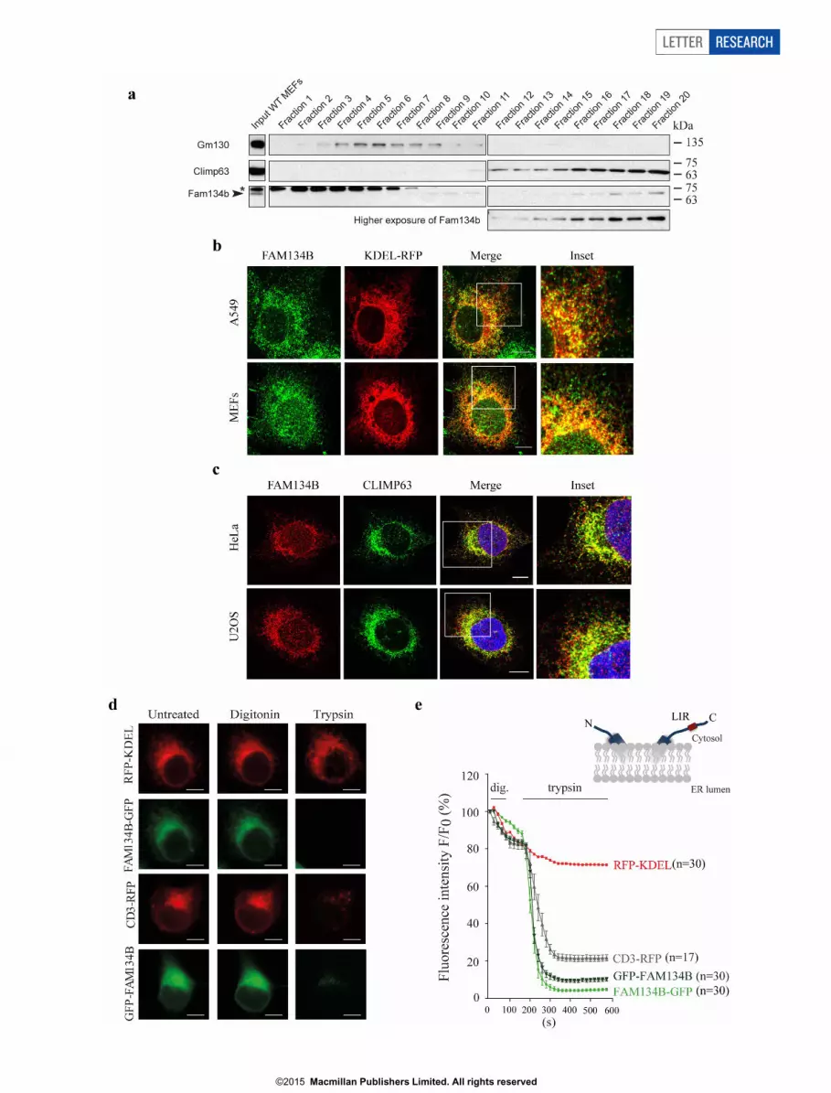
Extended Data Figure 2 | Subcellular localization and topology ofendogenous FAM134B. a, The post-nuclear fraction of wild-type MEF lysatewas loaded on a linear Iodixanol gradient (0–20%). Fractions (top to bottom)were subjected to western blot and analysed with antibodies directed againstendogenous proteins as indicated. Images are from two different gels, while theexposure time is the same. Asterik indicates non-specific band. b, A549 cellsand MEFs were transfected with a KDEL–RFP expression plasmid for 24 h.After fixation, endogenous FAM134B was detected. FAM134B co-localizeswith KDEL–RFP. Representatives of five images are shown. Scale bar, 10 mm.c, HeLa and U2OS cells were fixed and stained for endogenous FAM134Band CLIMP-63. FAM134B co-localizes with CLIMP-63. Representatives offive images are shown. Scale bar, 10 mm. d, FAM134B topology analysis.COS-7 cells transiently overexpressing C-terminal tagged FAM134B(FAM134B–eGFP) or N-terminal tagged FAM134B (eGFP–FAM134B) were
subjected to fluorescence protein protection (FPP) assay. COS-7 cellstransfected with plasmids encoding the luminal ER peptide RFP–KDEL andC-terminal RFP-tagged CD3 (CD3–RFP) served as controls. RFP–KDEL,which localized to the ER lumen, shows fluorescence protein protection upontrypsin administration following digitonin treatment. By contrast, accordingto the known topology of CD3–RFP the RFP-tag faces the cytosol and as aconsequence trypsin treatment abolishes protein fluorescence. Scale bar,10 mm. e, C-terminal RFP-tagged CD3 (CD3–RFP) served as control (RFP-tagfaces the cytosol). A N-terminal tagged FAM134B variant was also subjectedto the FPP assay. A strong decrease in fluorescence was observed for CD3–RFP, FAM134B–eGFP and eGFP–FAM134B after sequenced digitonin andtrypsin treatment, but not for RFP–KDEL (n 5 number of cells, error barsindicate s.e.m.).
RESEARCH LETTER
G2015 Macmillan Publishers Limited. All rights reserved

LETTER RESEARCH
G2015 Macmillan Publishers Limited. All rights reserved

Extended Data Figure 3 | FAM134B is a membrane-shaping protein thatremodels lipid bilayers. a, Liposome co-floatation assays. Proteins weredetected using anti-GST antibodies in immunoblots of sucrose gradientfractions 1 (top) to 6 (bottom). GST–FAM134B, but not GST (Ctrl), floatedwith liposomes in fraction 2. Disease-related truncating mutations S309X andQ145X only partially floated with the liposome fraction. b, Representativetransmission electron microscopy (TEM) images of freeze-fracturedincubations of liposomes with recombinant GST-fusion proteins (GST–FAM134B; GST–FAM134B(Q145X); Ctrl, GST). Scale bar, 200 nm.
c, Distribution of liposome diameters observed by TEM of freeze-fracturedliposome incubations. Incubation with FAM134B and S309X leads to apronounced increase in the relative numbers of smaller structures in compari-son to control (GST) and Q145X. Inset, box plots of data presented in c; y axis islogarithmic. n.s., not significant; ***P , 0.001 one-way ANOVA, error barsindicate s.e.m. (n 5 2,682 for Ctrl, n 5 2,683 for FAM134B, n 5 1,685 forQ145X, n 5 1,612 for S309X, n 5 number of liposomes). Boxes contain50% of the values; minimal, maximal, and median values are marked byvertical lines.
RESEARCH LETTER
G2015 Macmillan Publishers Limited. All rights reserved

LETTER RESEARCH
G2015 Macmillan Publishers Limited. All rights reserved

Extended Data Figure 4 | Fam134b determines ER degradation throughautophagy. a, U2OS cells transiently co-transfected with 0.25 mg of GFP–SEC61 plasmid expressing full length or mutLIR FAM134B–HA at theindicated quantities (mg). Cell lysates were immunoblotted with antibodiesagainst HA, GFP and vinculin. Black arrow-head indicates protein-degradationproducts. b, Wild-type (WT) and Atg5-knockout (KO) MEFs transientlyexpressing mCherry–eGFP–FAM134B were fixed and processed for immuno-fluorescence analysis. Cells with mCherry-positive and simultaneously GFP-negative punctae were counted in three independent experiments (biologicalreplicates). Representative of 50 images is shown. Scale bar, 10 mm. c, d,Atg5-knockout and control MEFs were co-transfected with plasmidsexpressing full-length LIR (c) or mutLIR (d) FAM134B–HA and KDEL–RFP
for 24 h, fixed and processed for immunofluorescence using antibodies againstthe HA tag and Lc3b. Overexpression of FAM134B–HA in wild-type but notin Atg5-knockout MEFs leads to the formation of punctae positive for bothKDEL–RFP and Lc3b. Representatives of five images are shown. Scale bar,10 mm. e, Quantification of cells displaying KDEL–RFP- and Lc3b-positivepunctae shown in c and d. At least 50 cells per experiment from threeindependent experiments (biological replicates) were quantified (error barsindicate s.d.). f, Fam134b is stabilized in starved Atg5-knockout MEFs. Wild-type and Atg5-knockout MEFs were starved in EBSS for the indicated periodsof time. Cell lysates were processed by SDS–PAGE and western blot usingantibodies against Fam134b and vinculin.
RESEARCH LETTER
G2015 Macmillan Publishers Limited. All rights reserved

Extended Data Figure 5 | FAM134B knockdown causes ER expansions.a, Ultrastructural analysis of FAM134B-depleted cells. Constructs expressingcontrol shRNA or a-FAM134B shRNA were lentivirally delivered into U2OScells. Cells were chemically fixed and embedded with Epon resin. Longitu-dinal and transversal sections and a part of the nuclear membrane is shown.FAM134B-depleted cells display ER expansions, particularly in the cellperiphery. The membrane of the nucleus of FAM134B-knockdown cells alsoundergoes expansion. M, mitochondria; N, nucleus; PM, plasma membrane.
Scale bars, 2 mm (left images) and 500 nm (middle and right images) (n 5 150cells). b, c, ER sheets are expanded in FAM134B-deficient cells. Constructsexpressing control shRNA ora-FAM134B shRNA were lentivirally delivered inU2OS cells. After selection, cells were fixed, stained for CLIMP-63 andRTN4A and RTN4B and analysed by fluorescence microscopy. Representativesof five images are shown. Scale bar, 10 mm. d, CLIMP-63 and TRAPa levelsin autophagy-deficient and FAM134B-depleted cells.
LETTER RESEARCH
G2015 Macmillan Publishers Limited. All rights reserved

RESEARCH LETTER
G2015 Macmillan Publishers Limited. All rights reserved

Extended Data Figure 6 | FAM134B is not required for bulk autophagy andaggrephagy. a, Fam134b1/1 and Fam134b–/– MEFs were starved in EBSS for 2h, fixed and stained for Lc3b (using two different anti-Lc3b antibodies toenhance the signal). Representatives of five images are shown. Scale bar, 10 mm.b, Lc3b-positive punctae in 50 cells per experiment from three independentexperiments were counted. *P , 0.0001, one-way ANOVA. c, Long-livingproteins degradation assay. For autophagy induction, cells were starved withEBSS. Protein degradation was assessed in three independent experiments intriplicate. Error bars represent s.e.m. of three independent counts (threetechnical replicates, n 5 20 cells per each replicate, P value is calculated usingt-test, *P , 0.05). d, Transmission electron microscopy of Fam134b1/1 andFam134b–/– cells. Autophagosomal/degradative compartments (that is,autophagosomes, autolysosomes and lysosomes) were counted. Quantificationwas performed by counting 20 cells in three different grids (three biologicalreplicates). Error bars represent s.d., P value is calculated using t-test. e, Lc3blipidation and p62 degradation was analysed in Fam134b1/1 and Fam134b–/–
MEFs treated with 200 nM bafilomycin A1 for the indicated time. f, g, Forautophagy induction, cells were starved with EBSS (f) or treated with thechemical Ku-0063794 (10 mM) (g) for the indicated periods of time.
Bafilomycin A1 was added 1 h before the beginning of the treatment. h, Controland FAM134B-knockdown (shRNA-mediated) U2OS cells were starved inEBSS for 2 h, fixed and stained for LC3B (using two different anti-LC3Bantibodies to enhance the signal). Representatives of five images are shown.Scale bar, 10 mm. i, LC3B-positive punctae in 150 cells per experiment fromthree independent experiments were counted. *P , 0.0001, Mann–WhitneyU-test. j, LC3B lipidation and p62 degradation were analysed in control andFAM134B shRNA cells treated with 200 nM bafilomycin A1 for the indicatedtime. k, l, For autophagy induction, cells were starved with EBSS (k) or treatedwith the chemical Ku-0063794 (10 mM) (l) for the indicated periods of time.Bafilomycin A1 was added 1 h before the beginning of the treatment.m, Fam134b1/1 and Fam134b–/– MEFs were either treated with 5 mg ml21
puromycin for 2 h or treated and subsequently washed and incubated inpuromycin-free medium for 3 h, fixed and stained for ubiquitin (Ub) and p62.Representatives of five images are shown. Scale bar, 10 mm. n, The number ofcells with Ub/p62 punctae before and after puromycin release per 100 cellsper experiment was determined from three independent experiments(biological replicates). Error bars indicate s.d.
LETTER RESEARCH
G2015 Macmillan Publishers Limited. All rights reserved

Extended Data Figure 7 | FAM134B deficiency sensitizes cells to apoptosis.a, b, Fam134b1/1 and Fam134b–/– MEFs were treated with 25 mM CCCP(carbonyl cyanide 3-chlorophenylhydrazone) (a) or starved in EBSS(b) for thetime indicated. Cell lysates were processed by SDS–PAGE and western blotusing antibodies against vinculin, Parp and Fam134b. c, Fam134b1/1 andFam134b–/– MEFs were treated with 2 mg ml21 tunicamycin (Tm) or 1 mMthapsigargin (Tg) for 12 h or left untreated. Cell lysates were processed by SDS–PAGE and western blot using antibodies against vinculin and Parp. d, FACSanalysis for annexin V and propidium iodide (PI) in Fam134b1/1 andFam134b–/– MEFs. Quantifications of annexin V/propidium iodide-positivecells after the indicated experimental settings (EBSS starvation for 8 h,treatment with 1 mM thapsigargin, 2 mg ml21 tunicamycin, 25 mM CCCP for12 h and 200 nM staurosporin (STS) for 6 h). Data are shown as mean 6 s.d. oftwo independent biological replicates; for each biological replicate two
experiments were performed and for each experiment 10,000 cells wereanalysed. **P , 0.01, one-way ANOVA. e, A549 cells stably expressing controland anti-FAM134B no. 1 and no. 2 shRNAs were either starved in EBSS ortreated with 10 mM Ku-0063794, 200 ng ml21 bafilomycin A1 (BafA), 1 mMthapsigargin or 2 mg ml21 tunicamycin for 12 h or left untreated. Cell lysateswere processed by SDS–PAGE and western blot using antibodies againstvinculin and PARP. f, g, FAM134B knockdown induces apoptosis involving themitochondrial pathway. A549 cells stably expressing control and anti-FAM134B no. 2 shRNAs were starved in EBSS for the time indicated. Celllysates were processed by SDS–PAGE and western blot using antibodies againstvinculin, PARP and caspase 8 (f) or caspase 9 (g). a–c, e–g, Filled arrowheadsindicate processed PARP and caspase 8/9; empty arrowheads indicateunprocessed PARP and caspase 8/9.
RESEARCH LETTER
G2015 Macmillan Publishers Limited. All rights reserved

LETTER RESEARCH
G2015 Macmillan Publishers Limited. All rights reserved

Extended Data Figure 8 | Fam134b deficiency does not affect spinal cordmotor neurons. a, Western blot analysis of mouse embryonic tissue lysateswith a Fam134b antibody. b, Transmission electron microscopy analysis ofFam134b1/1 and Fam134b–/– MEFs. Cells lacking Fam134b display anexpanded ER. M, mitochondria. Scale bar, 1 mm; n 5 150 cells. c, Compoundmuscle action potential (CMAP). CMAP latencies were recorded from tailnerves of wild-type (1/1) and knockout (2/2) mice at the ages indicated. Nosignificant differences were observed between Fam134b1/1 and Fam134b2/2
mice. One-way ANOVA, error bars indicate s.e.m.; n 5 6 for 1/1 and 2/2 at6 months; n 5 10 for 1/1 and n 5 13 for 2/2 at 12 months. n.s., notsignificant. d, Motor neurons in Nissl-stained thoracic spinal cord sections (10-
mm thick) of Fam134b1/1 and Fam134b2/2 mice. Scale bar, 20 mm.Representatives of seven images per genotype are shown. e, Quantification ofmotor neuron cell bodies in the ventral horn. Motor neuron number wasunchanged in Fam134b1/1 and Fam134b2/2 mice at an age of 12 and 20months (n 5 7 for 1/1 and n 5 7 for 2/2). n.s., not significant, Student’st-test, error bars indicate s.e.m. f, Normal ultrastructure of motor neurons inspinal cord sections of 12-month-old mice. Arrows in the middle and rightpanels highlight that there were no observed alterations in ER and Golgiarchitecture, respectively. Scale bar, 1 mm. Representatives of three images pergenotype are shown.
RESEARCH LETTER
G2015 Macmillan Publishers Limited. All rights reserved

Extended Data Figure 9 | Ultrastructural analysis of dorsal root ganglianeurons in Fam134b2/2 mice. a–e, Ex vivo analysis of dorsal root ganglia(DRG) neurons. DRG neurons from 3-month-old Fam134b1/1 andFam134b2/2 littermates were cultured for 2 days before transmission electronmicroscopy using the flat embedding approach. a, b, The ultrastructuralarchitecture of the peripheral ER as found in the DRG neuron cell body aboveor below the nucleus (a) and ER (b) adjacent to the nucleus of DRG neurons isshown. Scale bars: a, 500 nm; b, 200 nm. c, Representative examples ofGolgi compartments. Scale bar, 200 nm. (n 5 25 cells for a–c). d, e, Lateral
cross-sections of the axons recognizable by their heavy myelination around theplasma membrane and their emptiness in organelles. Panel d shows a bundleof axons whereas panel e presents a single axon. e, lower panels, longitudinalcross-sections of neurites, which are typically packed with microtubules andER. ER appears as thin black stripes in these images. ax, axon; ER, endoplasmicreticulum; G, Golgi; M, mitochondrium; mt, microtubule; my, myelin; N,nucleus, LD, lipid droplet. Scale bars: d, 1 mm; e, upper panels, 1 mm; lowerpanels, 500 nm. Representatives of 25 images per genotype are shown.
LETTER RESEARCH
G2015 Macmillan Publishers Limited. All rights reserved

Extended Data Figure 10 | Model of FAM134B function. To drive ER-phagy, FAM134B clusters at the edges of cisternal ER. Local enrichment ofFAM134B LIR leads to the recruitment of autophagosomal membranes and
subsequent budding of ER-derived vesicles. Mature autophagosomes fuse withlysosomes leading to the degradation of enclosed ER fragments.
RESEARCH LETTER
G2015 Macmillan Publishers Limited. All rights reserved