reduced activation to implicit affect induction in euthymic bipolar patients: an fmri study
TRANSCRIPT
Journal of Affective Disorders 97 (2007) 109–122www.elsevier.com/locate/jad
Research report
Reduced activation to implicit affect induction ineuthymic bipolar patients: An fMRI study
Gin S. Malhi a,b,c,g,⁎, Jim Lagopoulos a,b,c,g, Adrian M. Owen d, Belinda Ivanovski a,c,Ron Shnier b,e, Perminder Sachdev a,f
a School of Psychiatry, University of New South Wales, Sydney, Australiab Neuroscience Research Group, Mayne Clinical Research Imaging Centre, Prince of Wales Medical Research Institute, Sydney, Australia
c Mood Disorders Unit, Black Dog Institute, Sydney, Australiad Medical Research Council Cognition and Brain Sciences Unit, Cambridge, UK
e Diagnostic Imaging, Mayne Group, Melbourne, Australiaf Neuropsychiatric Institute, Prince of Wales Hospital, Sydney, Australia
g NISAD, Sydney, Australia
Received 6 December 2005; received in revised form 31 May 2006; accepted 2 June 2006Available online 11 July 2006
Abstract
Objective: To examine whether euthymic bipolar patients engage similar or contrasting brain regions as healthy subjects whenresponding to implicit affect induction.Methods: The study examined 10 euthymic patients with bipolar I disorder, and 10 age- and gender-matched healthy subjects usingevent-related functional magnetic resonance imaging (fMRI) while subjects engaged in a modified word-based memory taskdesigned to implicitly evoke negative, positive or no affective change. The activation paradigm involved nominating whether atarget word was contained within a previously presented word list using specified response keys.Results: The fMRI task produced significantly greater activation in healthy subjects as compared to patients in response to bothnegative and positive affect in the anterior and posterior cingulate, medial prefrontal cortex, middle frontal and rightparahippocampal gyri. Only negative affect produced significantly greater activation in the postcentral gyrus, inferior parietallobule, thalamus and putamen and only positive affect achieved the same in the precentral, superior temporal and lingual gyri,precuneus, cuneus, caudate, pons, midbrain and cerebellum. There were no brain regions in which responses were greater inpatients as compared to healthy subjects. There were no statistically significant differences between the groups with respect tospeed or accuracy.Conclusions: Diminished prefrontal, cingulate, limbic and subcortical neural activity in euthymic bipolar patients as compared tohealthy subjects is suggestive of emotional compromise that is independent of cognitive and executive functioning. This finding isof clinical importance and has implications both for the diagnosis and treatment of bipolar disorder. Future studies should aim toreplicate these findings and examine the development of bipolar disorder, investigating in particular the effects of medication.© 2006 Elsevier B.V. All rights reserved.
Keywords: fMRI; Affect; Bipolar disorder; Euthymia; Mood induction
⁎ Corresponding author. Mayne Clinical Research Imaging Centre, Prince of Wales Medical Research Institute, Barker Street, Randwick NSW2052, Australia. Tel.: +61 2 9382 3719; fax: +61 2 9399 1005.
E-mail address: [email protected] (G.S. Malhi).
0165-0327/$ - see front matter © 2006 Elsevier B.V. All rights reserved.doi:10.1016/j.jad.2006.06.005

110 G.S. Malhi et al. / Journal of Affective Disorders 97 (2007) 109–122
1. Introduction
In that sweet mood when pleasant thoughts bringsad thoughts to the mind
William Wordsworth (1770–1850)
The vicissitudes of mood, ranging from despair todelight, are common experiences that can be promptedby circumstance or chance, and as so eloquentlydescribed by Wordsworth can comprise varied andsometimes seemingly contrasting elements. However,even opposing emotions are sometimes miscible, as inbipolar disorder where patients have coterminoussymptoms of dysphoria and hypomania described as‘mixed states’ (Akiskal et al., 2005; Berk et al., 2005).Clinically, such admixtures are difficult to detect, oftenonly manifesting as a subtle functional compromise.Indeed it has become increasingly apparent that‘euthymia’ does not equate to recovery and that akinto its bedfellows, hypomania and depression, it too isassociated with neurocognitive deficits (Ferrier et al.,1999; Olley et al., 2005). Recent studies have identifiedexecutive impairments and memory dysfunction (Malhiet al., in press; Malhi et al., 2004a; Martinez-Aran et al.,2004a,b) that perhaps underpin patient reports of‘diminished emotional reactivity’ and ‘an inability tonegotiate real-world problems’. Functional neuroima-ging studies in bipolar disorder have attempted tolocalise these deficits and have identified a number ofkey brain regions (Adler et al., 2004; Blumberg et al.,2003a; Malhi et al., 2004b,c; Monks et al., 2004).
1.1. Functional neuroimaging studies in bipolar disorder
Early research yielded somewhat disparate findings(see Ketter et al., 2001; Malhi et al., 2004d; Phillips et al.,2003) however, the results from more recent studies havebeenmore consistent (Strakowski et al., 2005). A series ofstudies (Blumberg et al., 2003a,b) that partitioned trait andstate effects by examining bipolar patients in all threephases of the disorder using the colour-naming Strooptask found activations in the prefrontal and dorsal anteriorcingulate cortices across all groups. However, incomparison to controls, patients had blunted activationin a rostral region of the left ventral prefrontal cortex thatwas spatially distinct and independent of mood state, afinding that has been corroborated by a recent functionalmagnetic resonance imaging (fMRI) study that alsoemployed an emotional Stroop paradigm (Malhi et al.,2005). Furthermore, in comparison to euthymic patients,the ventral prefrontal cortex increase in signal was blunted
in patients with elevated mood on the right side butexaggerated in depressed patients on the left side. Theauthors thus opined that bipolar disorder may beassociated with a trait abnormality in the left ventralprefrontal cortex and that additional lateralized abnor-malities may relate to the valence of the mood episode.Extrapolating to a younger age group the same researchersalso identified increased activation in the left thalamusand putamen of bipolar adolescents as compared tohealthy subjects suggesting once again a subcorticaldysfunction in bipolar disorder (Blumberg et al., 2003b).Interestingly, the lack of prefrontal cortical dysfunction inthis age group prompted speculation that this may be adevelopmental abnormality that stems from subcorticaldysfunction involving disruption of inhibitory regulationin the basal ganglia.
State-related differences in emotional processing havealso been reported in two recent studies (Malhi et al.,2004b,c) that adopted an fMRI picture-caption cognition-based mood induction paradigm previously validated inhealthy subjects (Teasdale et al., 1999) as well as patientswith major depression (Kumari et al., 2003). Examiningpatients with bipolar depression and hypomania a patternof subcortical activation emerged that prompted thesuggestion that prefrontal cortex engagement is impairedin patients when unwell resulting in recruitment ofadditional subcortical brain regions.
1.2. Aim of the study
In this study we employed fMRI to contrast the brainregions engaged by euthymic bipolar patients and healthysubjects when responding to implicit affect induction. Weexamined euthymic patients to avoid a mood-stateconfound and employed an implicit design to ensure taskengagement.
On the basis of extant literature and our previousstudies we hypothesised that patients would havediminished activation as compared to healthy subjects,specifically in prefrontal and cingulate cortices and, thatsubcortical structures would be less responsive.
2. Methods
2.1. Subjects
Ten right-handed female patients with bipolar Idisorder were recruited from our Sydney BipolarDisorder Clinic at the Prince of Wales Hospital. A re-search psychiatrist made the diagnoses using theStructured Clinical Interview for DSM-IV (SCID-P)(First et al., 1995) supplemented by case note review. At
111G.S. Malhi et al. / Journal of Affective Disorders 97 (2007) 109–122
the time of participation, all patients had been inremission (no significant symptoms or alteration inmedication status) for a period of at least three months(verified by self-report, clinical assessment and case notereview). Subjects were excluded if they had a history ofsubstance abuse, neurological disease or closed headinjury, an additional Axis-I or any Axis-II psychiatricdiagnosis or a medical disorder currently necessitatingtreatment.
Patients were aged 19–54 years (mean±SD 32.4±10.8 years) and gainfully employed. Symptoms wereassessed using the 17-item Hamilton Depression RatingScale (Hamilton, 1960), the Young Mania Rating Scale(Young et al., 1978), the Montgomery–Asberg Depres-sion Rating Scale (Montgomery and Asberg, 1979), theGlobal Assessment of Functioning scale (AmericanPsychiatric Association, 1994) and the CORE measurefor psychomotor disturbance (Parker and Hadzi-Pavlo-vic, 1996). Patients were defined as euthymic if theyscored 6 or less on the 17-item Hamilton DepressionRating Scale and 6 or less on the Young Mania RatingScale. Patients also completed the following question-naires: the Beck Depression Inventory (Beck et al.,1961) and the Spielberger State-Trait Anxiety Inventory(Spielberger, 1983).
Themean duration of illness from diagnosis was 8.8±5.8 years, and the mean number of previous depressiveepisodes was 4.9±4.1 and manic episodes 3.2±2.6.None of the patients met DSM-IV criteria for rapidcycling. At the time of fMRI scanning, seven patientswere on mood-stabilising psychotropic medications andthree were on no medication. Five patients were taking
Table 1Demographic data and clinical assessment scores for patients and healthy co
Healthy subjects
Mean (SD)
Age (years) 31.7 (11.9)Years educated 15.9 (2.0)IQ 111.4 (5.2)BDI 4.8 (0.8)Ham-D 3.1 (1.0)MADRS 3.2 (0.9)STAI-I 29.4 (4.4)STAI-II 30.8 (4.0)COREa 0.0 (0.0)YMRS 0.8 (0.9)GAF a 91.5 (4.1)a Patients and healthy subjects only differed significantly on CORE and G
Abbreviations: HAM-D (17-item Hamilton Depression Rating Scale); YMDepression Rating Scale); GAF (Global Assessment of Functioning scale) CInventory); STAI (Spielberger State-Trait Anxiety Inventory).
lithium (mean daily dose 1340±230.2 mg) with a meanplasma level of 0.76±0.09 mmol/L. One of thesepatients and one other were each taking lamotrigine100 mg daily with the final patient on mood-stabilisingmedication taking carbamazepine alone (700 mg daily,plasma level of 40.0 μg/mL). Two patients had receivedelectroconvulsive therapy (more than 12 months prior)and seven patients had at least one first-degree relativewith an affective disorder.
Patients were compared with ten female volunteersmatched for age (20–54 years; mean 31.7±11.9 years),handedness (all right-handed), level of education andpremorbid IQ (see Table 1). Comparison subjects werescreened for a history of neurological or psychiatricdisorder (with SCID-NP version) or a family history ofthe same. They also underwent the same clinicalassessments and self-report questionnaires as patientsimmediately prior to scanning.
2.2. fMRI task
To ensure that subjects satisfactorily engaged theimplicit affect-inducing stimuli, the task was con-structed to necessitate a response based on the materialpresented. Patients were not alerted to the affectivecomponents of the task.
2.2.1. Visual word stimuliTwo hundred and forty words were extracted from the
Lang Affective Norms for English Words (ANEW)database (Bradley and Lang, 1999) such that with respectto affect a quarter (sixty) were unambiguously negative
mparison subjects
Euthymic bipolar patients
Mean (SD)
32.4 (10.8)16.1 (2.9)110.2 (5.1)7.6 (1.0)4.2 (1.0)3.6 (0.8)29.6 (5.1)31.5 (4.7)0.4 (0.5)0.9 (0.8)86.5 (8.2)
AF scores with t-test statistic p-values of 0.00 and 0.05 respectively.RS (Young Mania Rating Scale); MADRS (Montgomery–AsbergORE (measure for psychomotor disturbance); BDI (Beck Depression

112 G.S. Malhi et al. / Journal of Affective Disorders 97 (2007) 109–122
and another quarter were positive. The remaining wordswere neutral in terms of their affect and all three groups ofwords were matched with respect to arousal (means andstandard deviations for positive, negative and neutralwords respectively — 5.6 (3.0); 5.4 (2.7); 5.1 (2.0).Negative, positive and neutral lists of 3, 5 or 7 words wereconstructed and this produced 12 positive and 12 negativeword lists with four of each length (3, 5 and 7), and 24matching neutral word lists. Half of the 3, 5 and 7 wordlists were then matched to a target word such that 50% ofthe responses would be affirmative (that the word iscontained within the list). Target words and lists werematched with respect to affect and lists were balanced fororder of presentation and target word position.
2.2.2. Task designA delayed-response working memory paradigm (see
Fig. 1) based on the Sternberg memory task (Sternberg,1969) was constructed using emotionally valent wordscontained within word lists that had been generated asdescribed above. Subjects were instructed to memorisevertical lists of 3, 5 or 7 words presented for 8 s afterwhich the screen was blank (except for a fixation cross-hair) for 4 s. A target word then appeared and waspresented for 2 s during which time the subject had todecide whether or not the target word was part of thepreviously presented word list and respond by pressingspecified reaction time buttons using index (for Yes) andmiddle fingers (for No) of both hands. The target was thenerased and a 1 second delay preceded the appearance ofthe next word list. Prior to scanning, subjects wereinstructed and the task was demonstrated using a practiceset of neutral word stimuli. After scanning, all subjectsrated the affect of a subset of the word stimuli.
Fig. 1. Negative versus neutral: Group analysis activation foci and signal temneutral group analysis images are radiologically orientated with the top imagand depicted in yellow/red denote activations that were greater in healthy subLN — lentiform nuclei, Thal — thalamus, MedFG — medial frontal gyrus,
An event-related task was designed with 15 sbetween successive word-list stimuli and a total runtime of 720 s. Overall, 48 unique computer generatedword lists (16×3 words, 16×5 words and 16×7 words)were presented in an order counterbalanced for valence,using a 50 point Arial font and back-projected in blackink onto a frosted white screen using an LCD videoprojector. Subjects viewed the screen through a mirrorfixed to the head coil and words subtended a visualangle of approximately 8°–15° horizontally dependingon their length. Mean reaction time as well as individualreaction times was acquired using a fibre optic device(response window 50–2000 ms) specifically designedfor use in the MR scanner. Subjects were debriefedfollowing completion of the experiments with respect tothe mood induction effects of the imaging.
2.3. Statistical analyses of affective word-list ratingsand behavioural data
Independent and one-sample t-test comparisons ofmean valence ratings of positive, neutral and negativewords for bipolar patients, healthy controls and anormative reference group were conducted. As thevariance of the normative reference group data could notbe calculated ANOVA analysis utilising valence as awithin-subjects variable could not be conducted. In orderto identify between group differences in performance,reaction time data were analysed using a 2 way ANOVA.
2.3.1. Functional imagingImaging was performed using a 1.5 T Philips Intera
MRI scanner. Sixteen axial slices (7 mm thickness, nogap) parallel to the anterior and posterior commissure
poral display of the thalamic BOLD response. Note: Negative versuse taken at z=14 and the bottom image taken at z=49. Regions labelledjects than in euthymic bipolar patients. [MFG— middle frontal gyrus,PCG — posterior cingulate gyrus.]

Table 2Ratings of positive and negative words
Valenceof words
ANEW Healthy subjects Euthymic bipolar patients
Mean (SD) Mean (SD) Mean (SD)
Positive 8.14 (2.51) 8.01 (0.15) 7.89 (0.15)Negative 2.46 (2.12) 2.31 (0.17) 2.10 (0.21)a
Neutral 5.20 (1.99) 5.12 (0.16) 5.01 (0.17)a Patients rated negative words significantly more negatively.
113G.S. Malhi et al. / Journal of Affective Disorders 97 (2007) 109–122
covering the whole brain were imaged with a temporalresolution of 3 s using a T2 weighted gradient echo EPIsequence (TE=45 ms; TR=3000 ms; matrix=64×64;flip angle=90°). The field of view was 400 mm and theeffective in-plane functional spatial resolution was3.59 mm. For each functional run a total of 240 wholebrain scans were collected. As an aid to localisation of theactivated voxels T1-weighted high-resolution whole brainimages (with the following parameters: TR=28,TE=5 ms, matrix 256×256, FOV 300 mm, acquired
Table 3Tailarach and Tournoux coordinates of regions in patients and healthy subjectto negative versus neutral affect
Brain region Brodmann'sarea
Healthy subjects
Talairach coordinate
x y
Superior frontal gyrus 9/10Right 14 58
16 42Left −12 48
Inferior frontal gyrus 45/47RightLeft −34 22
−38 24−44 20
Posterior cingulate, left 30/31 −8 −54Superior temporal gyrus 38
Right 40 1040 6
Left −42 10−48 −2−44 6
Middle temporal gyrus 21Left −42 −46
−38 −62
Insula, left −40 −22Medial globus pallidus, right 12 2Caudate (head), left −8 4Precuneus, right 7 14 −40Lingual gyrus, right 18Declive (cerebellum), rightCerebellar tonsil, right
a Based on peak z value.
resolution=1.17×1.17×1.0 mm) were acquired. Addi-tionally, high-resolution anatomical images in the plane ofthe functional slices were also acquired to facilitate the co-registration of the functional volumes to a Talairach(Talairach and Tournoux, 1988) calibrated whole brainimage. All subjects had their heads firmly immobilized inthe head coil using forehead straps and foam inserts.
2.4. Statistical analysis of fMRI data
All functional data was pre-processed using SPM99.Images were interrogated and carefully scrutinized formovement and susceptibility artefacts. Datawas correctedfor movement using least square minimisation and wassmoothed using an 8×8×10 (FWHM)Gaussian kernel tocompensate for residual spatial variability and to reducethe effect of variation of MR signal between runs.
Two separate statistical analyses were performed onindividual and group data with respect to affect inductionby negative and positive words.
s showing significant within-group differences in activation in response
Bipolar patients
sa Talairach coordinatesa
z z score x y z z score
20 5.3234 5.4434 5.13
50 26 4 4.60−6 5.48 −38 22 −4 4.55
4 4.24 −46 30 4 4.2112 5.7220 6.03 −6 −60 14 6.78
−22 5.71 52 4 −8 4.06−12 4.63−22 5.05−6 5.24−12 5.15
6 4.21 −54 −4 −20 5.1920 4.56 −56 −14 −4 4.55
−54 0 −10 4.08−4 4.82−4 6.53
4 4.1244 5.91
26 −76 −10 5.0930 −78 −18 4.9222 −60 −38 4.05
114 G.S. Malhi et al. / Journal of Affective Disorders 97 (2007) 109–122
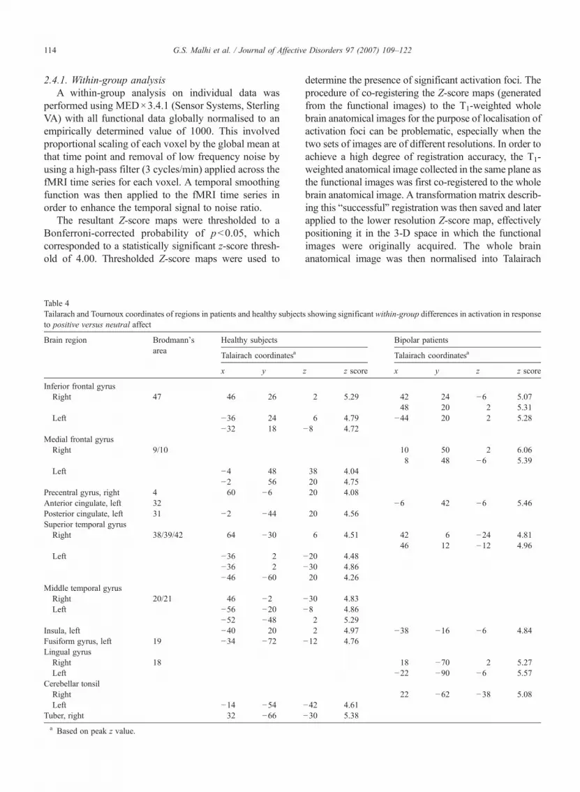
2.4.1. Within-group analysisA within-group analysis on individual data was
performed using MED×3.4.1 (Sensor Systems, SterlingVA) with all functional data globally normalised to anempirically determined value of 1000. This involvedproportional scaling of each voxel by the global mean atthat time point and removal of low frequency noise byusing a high-pass filter (3 cycles/min) applied across thefMRI time series for each voxel. A temporal smoothingfunction was then applied to the fMRI time series inorder to enhance the temporal signal to noise ratio.
The resultant Z-score maps were thresholded to aBonferroni-corrected probability of p<0.05, whichcorresponded to a statistically significant z-score thresh-old of 4.00. Thresholded Z-score maps were used to
Table 4Tailarach and Tournoux coordinates of regions in patients and healthy subjectto positive versus neutral affect
Brain region Brodmann'sarea
Healthy subjects
Talairach coordinatesa
x y
Inferior frontal gyrusRight 47 46 26
Left −36 24−32 18
Medial frontal gyrusRight 9/10
Left −4 48−2 56
Precentral gyrus, right 4 60 −6Anterior cingulate, left 32Posterior cingulate, left 31 −2 −44Superior temporal gyrusRight 38/39/42 64 −30
Left −36 2−36 2−46 −60
Middle temporal gyrusRight 20/21 46 −2Left −56 −20
−52 −48Insula, left −40 20Fusiform gyrus, left 19 −34 −72Lingual gyrusRight 18Left
Cerebellar tonsilRightLeft −14 −54
Tuber, right 32 −66a Based on peak z value.
determine the presence of significant activation foci. Theprocedure of co-registering the Z-score maps (generatedfrom the functional images) to the T1-weighted wholebrain anatomical images for the purpose of localisation ofactivation foci can be problematic, especially when thetwo sets of images are of different resolutions. In order toachieve a high degree of registration accuracy, the T1-weighted anatomical image collected in the same plane asthe functional images was first co-registered to the wholebrain anatomical image. A transformation matrix describ-ing this “successful” registration was then saved and laterapplied to the lower resolution Z-score map, effectivelypositioning it in the 3-D space in which the functionalimages were originally acquired. The whole brainanatomical image was then normalised into Talairach
s showing significant within-group differences in activation in response
Bipolar patients
Talairach coordinatesa
z z score x y z z score
2 5.29 42 24 −6 5.0748 20 2 5.31
6 4.79 −44 20 2 5.28−8 4.72
10 50 2 6.068 48 −6 5.39
38 4.0420 4.7520 4.08
−6 42 −6 5.4620 4.56
6 4.51 42 6 −24 4.8146 12 −12 4.96
−20 4.48−30 4.8620 4.26
−30 4.83−8 4.86
2 5.292 4.97 −38 −16 −6 4.84
−12 4.76
18 −70 2 5.27−22 −90 −6 5.57
22 −62 −38 5.08−42 4.61−30 5.38

115G.S. Malhi et al. / Journal of Affective Disorders 97 (2007) 109–122
space using a piecewise-linear scaling method, and theresultant transformation matrix was saved. The latter wasthen applied to the transformed Z-score map and theresultant Z-score image was co-registered to the Talairachnormalised whole brain anatomical image using AIR(Woods et al., 1993).
2.4.2. Between-group analysisIndividual pre-processed data that had been globally
normalised and linearly detrended was analysed using theGeneral Linear Model (GLM) and the theory of Gaussianrandom fields as implemented in the BrainVoyagersoftware package (Brain Innovations, Netherlands). Abetween-group analysis was performed using a random-effects model to provide an estimate of the error variancefor each condition of interest across subjects, rather thanacross scans thus providing a stronger generalisation tothe population (Holmes and Friston, 1998).
Affective word-related differences between the con-trols and patients for Positive and Negative affectivewords were compared. Individual contrast imagesreflecting activations arising from Positive and Negativevalenced words were created separately and contrastanalysis of the predictors was used to find regions inwhich average activity was higher during the positive ornegative word phases between groups.
Table 5Coordinates of brain regions showing between-group differences for negativ
Brain region Brodmann'sarea
T
x
Middle frontal gyrusRight 9/10Left −
Medial frontal gyrusRight 6Left −
Postcentral gyrusRight 3
Anterior cingulate gyrusRight 32Left −
Posterior cingulate gyrus, right 31Inferior parietal lobule
Left 40 −−
Parahippocampal gyrus, right 30Thalamus
Right
Putamen, left −
All activations favoured healthy subjects.
3. Results
3.1. Affective word-list ratings
There were no statistically significant differencesin mean rating scores for positive words betweenthe patient, healthy control and normative referencegroups. Similarly, comparisons of mean rating scoresof neutral words failed to demonstrate significant dif-ferences between the three groups. However, whencompared to the control group (t8=2.52, p<0.05) andthe normative reference group (t9=−4.99. p<0.01),bipolar patients rated negative words significantlymore negatively. Mean valence rating scores for thenegative words did not differ significantly between thehealthy control and normative reference groups (seeTable 2).
3.2. Behavioural results
The performance of healthy subjects and patients waspredictably better on the 3 word lists (96% and 93%accuracy, respectively) than on the more difficult 7 wordlists (84% and 78%, respectively) however, there was nosignificant group difference in response accuracy[ANOVA: F(2,56)=4.23, p=.11]. Patients had slower
e affect
alairach coordinates
y z p-value
28 28 36 4.08×10⁻¹¹
31 44 12 3.5×10⁻¹³
18 6 49 4.82×10⁻¹⁸
10 5 49 4.76×10⁻¹⁴
34 −18 36 9.63×10⁻¹³
20 −24 49 1.79×10⁻¹¹
3 21 36 8.31×10⁻¹¹
12 18 36 2.83×10⁻¹³
17 −22 36 1.26×10⁻¹²
43 −33 33 2.48×10⁻¹¹
43 −30 36 1.53×10⁻¹¹
17 −37 7 2.1×10⁻¹¹
18 −15 4 4.1×10⁻¹²
21 −15 7 1.68×10⁻¹⁰
16 −36 14 9.5×10⁻¹⁰
21 −23 14 3.75×10⁻¹¹
20 7 15 1.9×10⁻¹¹

Table 6Coordinates of brain regions showing between-group differences forpositive affect
Brain region Brodmann'sarea
Talairach coordinates
x y z p-value
Superior frontal gyrusRight 10 11 43 19 3.3×10⁻⁴
Left −18 56 16 2.1×10⁻⁵
−19 59 27 2.3×10⁻⁴
Middle frontal gyrusRight 9/10 27 47 16 5.0×10⁻⁸
27 54 13 3.0×10⁻⁶
Left −31 53 4 8.0×10⁻⁵
Precentral gyrusRight 4 35 −16 61 1.7×10⁻⁴
48 −11 35 1.3×10⁻⁵
Left −46 −11 35 3.5×10⁻⁴
Anterior cingulate gyrus,right
32 6 34 16 2.3×10⁻⁴
Posterior cingulate gyrus,left
31 −7 −6 43 3.6×10⁻⁴
Superior temporal gyrus,left
22 −61 −29 4 1.6×10⁻⁵
Parahippocampal gyrus,right
30 17 −54 −6 3.8×10⁻⁵
Lingual gyrus, right 18 16 −84 −6 2.6×10⁻⁵
Precuneus and cuneusRight 7/18 12 −73 16 4.9×10⁻⁴
Left −13 −70 35 8.8×10⁻⁵
CaudateRight Body 11 11 16 3.3×10⁻⁴
Left Head −13 17 −1 3.4×10⁻⁴
Pons, right 13 −26 −19 4.9×10⁻⁴
Midbrain, right 5 −39 −11 5.7×10⁻⁴
Cerebellum, right 13 −33 −11 7.6×10⁻⁴
All activations favoured healthy subjects.
116 G.S. Malhi et al. / Journal of Affective Disorders 97 (2007) 109–122
reaction times but the difference was not statisticallysignificant [F(2,56)=3.19, p= .079].
3.3. fMRI results
The results of within-group and between-groupanalyses of the responses to affect induction are presentedin Tables 3–6 and described below.
3.3.1. Within-group analyses
3.3.1.1. Negative versus neutral words (Table 3).Healthy subjects showed significant activation in thesuperior and inferior frontal and superior and middletemporal gyri bilaterally. They also showed left-sidedactivation in the posterior cingulate, insula and head ofcaudate and right-sided activation in the medial globuspallidus and precuneus.
In common with healthy subjects, euthymic bipolarpatients also showed bilateral activation in the inferiorfrontal and superior and middle temporal gyri and hadleft-sided activation in the posterior cingulate. Addi-tionally, patients had right-sided activation in the lingualgyrus and cerebellum but lacked any significant lenti-form nuclei activation.
3.3.1.2. Positive versus neutral words (Table 4).Healthy subjects again showed significant activation inthe inferior frontal and superior and middle temporalgyri bilaterally. They also showed only left-sided acti-vation in the medial frontal and fusiform gyri, posteriorcingulate, insula and cerebellum. Purely right-sidedactivation occurred only in the precentral gyrus.
In contrast patients showed left-sided activation onlyin the anterior cingulate and insula. They also showedbilateral activation in the inferior frontal and lingual gyriand right-sided activation in the cerebellum, medialfrontal and superior temporal gyri.
3.3.1.3. Summary. In response to negative affect bothhealthy subjects and patients showed activations in theinferior frontal, superior and middle temporal andposterior cingulate gyri. Only healthy subjects showedresponses in the medial prefrontal cortex, insula,precuneus and subcortical nuclei and only patientsshowed responses in the lingual gyrus and cerebellum.
In response to positive affect both healthy subjectsand patients showed activations in the insula,cerebellum, and superior temporal and inferior andmedial frontal gyri. Only healthy subjects showedresponses in the precentral, middle temporal, poste-rior cingulate and fusiform gyri and only patients
showed responses in the anterior cingulate and lin-gual gyri.
Of note, inferior frontal and superior temporalactivation occurred in both groups in response to bothnegative and positive affect and both anterior andposterior cingulate responses were all left-sided.
3.3.2. Between-group analysesAll activations in response to both negative and
positive words were greater in healthy subjects ascompared to patients.
3.3.2.1. Negative words (Table 5 and Fig. 1).Activations in response to negative words showedsignificant bilateral differences between patients andhealthy subjects in the postcentral, middle and medialfrontal gyri; right-sided differences in the posteriorcingulate, parahippocampal gyrus and thalamus and

Fig. 2. Positive versus neutral: Group analysis activation foci and signal temporal display of the caudate BOLD response.Note: Positive versus neutral groupanalysis images are radiologically orientatedwith the top image taken at z=14 and the bottom image taken at z=49. Regions labelled and depicted in yellow/red denote activations that were greater in healthy subjects than in euthymic bipolar patients. [MFG — middle frontal gyrus, ACG — anterior cingulategyrus, Caud — caudate, PreC— precuneus, Cn — cuneus, LG— lingual gyrus.]
117G.S. Malhi et al. / Journal of Affective Disorders 97 (2007) 109–122
left-sided differences in the anterior cingulate, inferiorparietal lobule and putamen.
3.3.2.2. Positive words (Table 6 and Fig. 2).Activations in response to positive words showedsignificant bilateral differences between patients andhealthy subjects in the precentral, superior and middlefrontal gyri; right-sided differences in the anteriorcingulate, parahippocampal and lingual gyri, cuneus,body of caudate, pons, midbrain and cerebellum and left-sided differences in the posterior cingulate and superiortemporal gyri, precuneus and head of caudate (Fig. 2).
3.3.2.3. Summary. Significantly greater activationoccurred in healthy subjects as compared to patients inresponse to both negative and positive affect in theanterior and posterior cingulate, medial prefrontalcortex, middle frontal and right parahippocampal gyri.Only negative affect produced significantly greateractivation in the postcentral gyrus, inferior parietallobule, thalamus and putamen and only positive affectachieved the same in the precentral, superior temporaland lingual gyri, precuneus, cuneus, caudate, pons,midbrain and cerebellum. There were no brain regionsin which responses were greater in patients as comparedto healthy subjects.
4. Discussion
The principal finding in this study is that euthymicbipolar patients are less responsive than healthy sub-jects with respect to both negative and positive affectand that the pattern of differences in activation bet-ween the two groups is dependent on affective valence.
However, prior to a detailed discussion of the findingsit is important to acknowledge a number of putativelimitations.
4.1. Limitations
The most important potential confound is that ofmedication, with seven patients taking mood stabilisersat the time of scanning — the most common one beinglithium. Lithium can alter vascular smooth musclecontractility and in the brain this may affect neurovas-cular coupling upon which the BOLD response ispredicated (Dehpour et al., 1995). However, this is morelikely to produce global effects than the specific regionalchanges, although it is important to note that globalblood flow changes can affect local BOLD responsesand the coupling between local neuronal activity andhemodynamics. As only three patients were unmedicat-ed, a sub-analysis of the effect of medication was notpossible.
It is likely that the relatively small sample size alsodiminished the power of the study with respect toidentifying between-group differences in behaviouralresponses such as in reaction times. Performance andBOLD responses can also be affected by anxiety withinthe scanner (Lagopoulos et al., 2005) and it is possiblethat during the experiment patients were more or lessanxious than controls.
Another limitation concerns the use of implicit affectinduction and a specific memory task so as to ensuremeasurable engagement within the scanner. Effortfulgeneration of affect as opposed to passive viewing mayinvolve different brain regions and permit compensatoryrecruitment of additional resources that complicates

118 G.S. Malhi et al. / Journal of Affective Disorders 97 (2007) 109–122
interpretation. However, such confounds may beassociated with the introduction of a cognitive task perse (Phan et al., 2002), and although in this study, thebehavioural responses suggest that patients performedas well as healthy subjects, it is possible that some of thedifferences in activation reflect cognitive variance asopposed to changes in affect.
Finally, wider application of our findings is some-what limited as patients were confined to those suitablefor scanning. Therefore extrapolating to patients withrapid cycling or bipolar II may not be appropriate,especially as co-morbid illnesses such as substancemisuse and anxiety disorders were excluded, and onlyfemales were scanned.
4.2. Responses to both positive and negative words
Differences in activation in the medial prefrontalcortex, anterior and posterior cingulate and parahippo-campal gyrus were present irrespective of whether theaffect induced was negative or positive, suggesting thatthese regions are involved in the processing of emotionalstimuli regardless of valence.
Reduced activity in the medial prefrontal cortex hasbeen reported in euthymic and depressed bipolarpatients, and remitted and depressed unipolar patients(Kruger et al., 2003; Liotti et al., 2002). In healthysubjects, medial prefrontal cortex activation has beenassociated with the appraisal of emotions (; Teasdaleet al., 1999) and with self-referential evaluation (Craiket al., 1999; Kelley et al., 2002; Kircher et al., 2000,2002). The allocation of additional resources to therecovery of emotional information (Schacter et al., 1996)or the need for heightened monitoring and evaluation ofretrieved information within an emotional contextinvolves prefrontal cortex activity (Fletcher and Henson,2001; Henson et al., 1999a,b; Shallice et al., 1994).Therefore, reduced activity in euthymic bipolar patientscan be interpreted as an inability to recruit additionalresources or engage post-retrieval processing because ofprefrontal dysfunction. This is in keeping with findingsfrom recent studies that have detected state and trait-related prefrontal abnormalities (Blumberg et al., 2003a,b; Malhi et al., 2004b,c). However, within-groupanalyses show that affective words as compared toneutral words produced inferior prefrontal cortexactivation in both patients and healthy subjects indicat-ing that language-mediated processing of words withemotional salience (Beauregard et al., 1997; Elliott et al.,2000;Maddock et al., 2001; Strange et al., 2000), and theevaluation of emotional meaning remain intact (Mad-dock et al., 2003). Therefore the groups may differ
simply because of a negativity bias in patients that iseither inherent or stems from sub-syndromal symptom-atology (Cacioppo and Gardner, 1999).
Emotional meaning and motivational information arealso thought to be subservedby the affective division of theanterior cingulate cortex (ACC) (Devinsky et al., 1995),which is partitioned functionally into rostral–ventralaffective (BA24/32 and BA 25) and dorsal cognitive(BA24/32) components (Bush et al., 2002). The ACC isinvolved in attention regulation during cognitive(Devinsky et al., 1995) and emotional processing (Bushet al., 2000; Whalen et al., 1998) and has close links withthe medial prefrontal cortex (BA9/10) that computes andmaintains online information necessary for the choice of anappropriate response, whilst the ACC facilitates imple-mentation of the selected action (Paus, 2001). Previousstudies have shown ventral ACC activation in response toemotionally salient words (Beauregard et al., 1997; Elliottet al., 2000; Tabert et al., 2001) however, in the presentstudy patients were unable to activate the cognitivedivision (BA32) in response to negative words and theaffective division (BA24) in response to positive words tothe same extent as healthy subjects. This suggests thatpatients were unable to process the emotional valence ofpositive words — ultimately a function of the affectivedivision of the ACC, and that the processing of negativewords within its cognitive division was limited (Bushet al., 1998; Drevets and Raichle, 1998). One possibleexplanation is that patients, seemingly ‘recovered’, remainattuned to negative cues such that at a ‘limbic level’ theyrelatemore to negative than positive affect but are impairedwith respect to associated cognitive elaboration. However,the ACC has a variety of sophisticated functions includingfor instance, error detection and conflict monitoring, andso interpretation of our findings is guarded.
Like the ACC, the posterior cingulate cortex (PCC)has a number of putative roles. Its caudal region isrobustly activated during the evaluation of emotionalwords (Maddock et al., 2003) in particular thoseassociated with threat (Maddock, 1999; Maddock andBuonocore, 1997), and is also implicated in themodulation of memory by emotionally arousing stimuli(Andreasen et al., 1995; Grasby et al., 1993). However, anumber of studies of episodic retrieval of emotionallyneutral information have also reported posterior cingu-late activation, suggesting that activity may reflect amore general function (Maratos et al., 2001). In thepresent study, both negative and positive affect producedless PCC activation in patients than in healthy subjects.However, within-group analyses showed that the leftposterior cingulate (BA30/31), specifically its caudalregion, is activated in patients in response to negative

119G.S. Malhi et al. / Journal of Affective Disorders 97 (2007) 109–122
affect whereas positive words failed to elicit anysignificant responses suggesting a valence-dependentdifferential in affective processing. One explanation isthat negative words evoke more complex mentalrepresentations than positive words and are cognitivelymore demanding and that this type of cognitiveprocessing that requires theory of mind (TOM), maybe impaired in bipolar patients (Kerr et al., 2003). Thecaudal posterior cingulate cortex is also stronglyinterconnected with the subgenual cingulate (BA 25)(Van Hoesen and Solodkin, 1993), and has beenrepeatedly implicated in the pathophysiology of mooddisorders (Drevets et al., 1997; Mayberg et al., 1999).However, in keeping with the findings of others (Krugeret al., 2003), diminished activity in this region ineuthymic bipolar patients, suggests that this region is ofgreater pertinence to active depressive states (Liotti et al.,2002).
4.3. Responses to either negative or positive words
Negative affect produced uniquely greater activationin healthy subjects as compared to patients in the rightpostcentral gyrus, left inferior parietal lobule, rightthalamus and left putamen.
Changes in basal ganglia activity are commonlyobserved in imaging studies involving the successfulrecognition of negative emotional stimuli (Fossati et al.,2004) andmemory (Cabeza andNyberg, 2000). Thalamicactivation in response to both negative and positive affecthas been noted in healthy subjects and depressed andhypomanic bipolar patients (Malhi et al., 2004b,c). Leftputamen activation in response to negative words (Fossatiet al., 2004) is thought to be associated with theintegration of internal states and the maintenance ofnegative mood (Critchley et al., 2000). In the presentstudy, diminished basal ganglia activation in euthymicbipolar patients in response to negative words suggeststhat subcortical thalamic processing of negative affectdoes not occur to the same extent as in healthy subjects.One explanation for this is that subcortical processing ofnegative affect is less critical in patients where a negativebias facilitates the generation of a negative emotionalstate, and in keeping with this left putamen activation wasgreater in healthy subjects.
Positive words produced greater activation in healthysubjects in the precentral gyrus, caudate, superiortemporal and lingual gyrus, the cuneus and precuneus,pons, midbrain and cerebellum. Anterior left temporalcortex activation has been shown to occur whenprocessing pictures with positive emotional contentand when processing emotional words (Crosson et al.,
1999; Phan et al., 2002). Specifically, left temporalactivation has been implicated in monitoring theemotional connotation of words (Crosson et al., 2002)and in keeping with this, findings in the present studysuggest that in comparison to healthy subjects, patientshad less left superior temporal activation when proces-sing words with positive affect.
Caudate activity modulates a neural system loopconnecting it to the thalamus and anterior cingulate(Alexander et al., 1990) such that decreased caudatemetabolism has been linked to bipolar depression (Baxteret al., 1989), and increased blood flow that diminishesupon the withdrawal of lithium, has been associated withmania (Goodwin et al., 1997). Greater left-sided activityin the head of the caudate has been reported in bipolarpatients when hypomanic as compared to when euthymic(Blumberg et al., 2000) however, diminished activity ineuthymic patients as compared to healthy subjectssuggests that even when euthymic, bipolar patients areunable to engage the caudate to the same extent as inhealth. One reason for this may be that basal gangliaactivation reflects dopaminergic innervation and theinvolvement of striatal structures in reward, motivationand happiness (Damasio et al., 2000; Davidson and Irwin,1999; George et al., 1996; Rauch et al., 1999; Redouteet al., 2000) and that these processes are less accessible topatients. However, like the thalamus, the caudate relaysmany circuits and fMRI activation may simply reflectmotor function. Alternatively, emotion and motor func-tionmay be coupled whereby the basal ganglia coordinateactions in response to emotions and produce responsepreparedness with respect to approach orwithdrawal froma stimulus, depending on whether it elicits a positive ornegative affect (Panksepp, 1998; Sprengelmeyer et al.,1998). Similarly, responses in the pons, midbrain andcerebellum (Konarski et al., 2005) may reflect motoractivity and emotion-related processing (Fletcher andHenson, 2001; Schmahmann and Sherman, 1998).
4.4. Conclusion
The key finding in the present study is that ofdiminished neural activity in euthymic bipolar patientsas compared to matched healthy subjects. Loss ofprefrontal control in bipolar depressed patients perhapsproduces increased subcortical thalamic and ventralstriatal metabolism that results in limbic disinhibitionand the manifestation of affective symptoms irrespectiveof affect, with only subcortical responses partitioningpositive and negative emotional processing. Therefore,despite equivalent executive functioning and cognitiveappraisal of emotions, euthymic bipolar patients appear

120 G.S. Malhi et al. / Journal of Affective Disorders 97 (2007) 109–122
to engage emotional circuitry less robustly than dohealthy subjects.
Acknowledgements
Australian Rotary Health Research Fund and theNHMRC Program Grant (222708) for financial supportand NISAD for infrastructure support.
References
Adler, C.M., Holland, S.K., Schmithorst, V., Tuchfarber, M.J.,Strakowski, S.M., 2004. Changes in neuronal activation in patientswith bipolar disorder during performance of a working memorytask. Bipolar Disorders 6, 540–549.
Akiskal, H., Benazzi, F., Perugi, G., Rihmer, Z., 2005. Agitated‘unipolar’ depression re-conceptualized as a depressive mixedstate: implications for the antidepressant-suicide controversy.Journal of Affective Disorders 85, 245–258.
Alexander, G.E., Crutcher, M.D., Delong, M.R., 1990. Basal ganglia–thalamocortical circuits: parallel substrates for motor, oculomotor,“prefrontal” and “limbic” functions. Progress in Brain Research 85,119–146.
American Psychiatric Association, 1994. Diagnostic and StatisticalManual of Mental Disorders, Fourth edition. Washington, DC.
Andreasen, N.C., O'leary, D.S., Cizadlo, T., Arndt, S., Rezai, K.,Watkins, G.L., Ponto, L.L.B., Hichwa, R.D., 1995. Rememberingthe past: two facets of episodic memory explored with positronemission tomography. American Journal of Psychiatry 152,1576–1585.
Baxter, L.R.J., Schwartz, J.M., Phelps,M.E.,Mazziotta, J.C., Guze, B.H.,Selin, C.E., Gerner, R.H., Sumida, R.M., 1989. Reduction ofprefrontal cortex glucose metabolism common to three types ofdepression. Archives of General Psychiatry 46, 243–250.
Beauregard, M., Chertkow, H., Bub, D., Murtha, S., Dixon, R., Evans,A., 1997. The neural substrate for concrete, abstract and emotionalword lexica: a positron emission tomography study. Journal ofCognitive Neuroscience 9, 441–461.
Beck, A.T., Ward, C.H., Mendelson, M., Mock, J.E., Erbaugh, J.K.,1961. An inventory for measuring depression. Archives of GeneralPsychiatry 4, 53–63.
Berk, M., Dodd, S., Malhi, G., 2005. ‘Bipolar missed states’: thediagnosis and clinical salience of bipolar mixed states. Australianand New Zealand Journal of Psychiatry 39, 215–221.
Blumberg, H.P., Stern, E., Martinez, D., Ricketts, S., De Asis, J.,White, T., Epstein, J., Mcbride, P., Eidelberg, D., Kocsis, J.H.,Silbersweig, D.A., 2000. Increased anterior cingulate andcaudate activity in bipolar mania. Biological Psychiatry 48,1045–1052.
Blumberg, H.P., Leung, H.C., Skudlarski, P., Lacadie, C.M.,Fredericks, C.A., Harris, B.C., Charney, D.S., Gore, J.C., Krystal,J.H., Peterson, B.S., 2003a. A functional magnetic resonanceimaging study of bipolar disorder: state- and trait-relateddysfunction in ventral prefrontal cortices. Archives of GeneralPsychiatry 60, 601–609.
Blumberg, H.P., Martin, A., Kaufman, J., Leung, H.C., Skudlarski, P.,Lacadie, C., Fulbright, R.K., Gore, J.C., Charney, D.S., Krystal, J.H.,Peterson, B.S., 2003b. Frontostriatal abnormalities in adolescentswith bipolar disorder: preliminary observations from functionalMRI.American Journal of Psychiatry 160, 1345–1347.
Bradley, M.M., Lang, P.J., 1999. Affective norms for English words(ANEW), Gainesville, FL, The NIMH Center for the Study ofEmotion and Attention, University of Florida.
Bush, G., Whalen, P.J., Rosen, B.R., Jenike, M.A., Mcinerney, S.C.,Rauch, S.L., 1998. The counting Stroop: an interference taskspecialized for functional neuroimaging-validation study withfunctional MRI. Human Brain Mapping 6, 270–282.
Bush, G., Luu, C., Posner, M.I., 2000. Cognitive and emotionalinfluences in anterior cingulate cortex. Trends in Cognitive Science4, 215–222.
Bush, G., Vogt, B.A., Holmes, J., Dale, A.M., Greve, D., Jenike, M.A.,Rosen, B.R., 2002. Dorsal and anterior cingulate cortex: a role inreward-based decision making. Proceedings of the NationalAcademy of Sciences of the United States of America 99, 523–528.
Cabeza, R., Nyberg, L., 2000. Imaging cognition II: an empiricalreview of 275 PET and fMRI studies. Journal of CognitiveNeuroscience 12, 1–47.
Cacioppo, J.T., Gardner, W.L., 1999. Emotions. Annual Review ofPsychology, vol. 50. Annual Reviews, US, pp. 191–214.
Craik, F.I.M., Moroz, T.M., Moscovitch, M., Stuss, D.T., Winokur, G.,Tulving, E., Kapur, S., 1999. In search of the self: a positronemission tomography investigation. Psychological Science 10,26–34.
Critchley, H., Daly, E., Phillips, M., Brammer, M., Bullmore, E.,Williams, S., 2000. Explicit and implicit neural mechanisms forprocessing of social information from facial expressions. Afunctional magnetic resonance imaging study. Human BrainMapp 9, 93–105.
Crosson, B., Radonovich, K., Sadek, J.R., Gokcay, D., Bauer, R.M.,Fischler, I.S., Cato, M.A., Maron, L., Auerbach, E.J., Browd, S.R.,Briggs, R.W., 1999. Left-hemisphere processing of emotionalconnotation during word generation. Neuroreport 10, 2449–2455.
Crosson, B., Cato, M.A., Sadek, J.R., Gokcay, D., Bauer, R.M.,Fischler, I.S., Maron, L., Gopinath, K., Auerbach, E.J., Browd,S.R., Briggs, R.W., 2002. Semantic monitoring of words withemotional connotation during fMRI: contribution of anterior leftfrontal cortex. Journal of the International NeuropsychologicalSociety 8, 607–622.
Damasio, A.R.,Grabowski, T.J., Bechara, A.,Damasio, H., Ponto, L.L.B.,Parvizi, J., Hichwa, R.D., 2000. Subcortical and cortical brain activityduring the feeling of self-generated emotions. Nature and Neurosci-ence 3, 1049–1056.
Davidson, R.J., Irwin, W., 1999. The functional neuroanatomy ofemotion and affective style. Trends in Cognitive Science 3, 11–12.
Dehpour, A.R., Ghafourifar, P., Samenian, J., Sadeghipour, H.R., Sadr, S.S., 1995. The effect of lithium on endothelial-dependent relaxation inrat isolated aorta. General Pharmacology 26, 1003–1007.
Devinsky, O., Morrell, M.J., Vogt, B.A., 1995. Contributions ofanterior cingulate cortex to behaviour. Brain 118, 279–306.
Drevets, W.C., Raichle, M.E, 1998. Reciprocal suppression of regionalcerebral blood flow during emotional versus higher cognitiveprocesses: implications for interactions between emotion andcognition. Cognition and Emotion 12, 353–385.
Drevets, W.C., Price, J.L., Simpson, J.R.L., Todd, R.D., Reich, T.,Vannier, M., Raichle, M.E., 1997. Subgenual prefrontal cortexabnormalities in mood disorders. Nature 386, 824–827.
Elliott, R., Rubinsztein, J.S., Sahakian, B.J., Dolan, R.J., 2000.Selective attention to emotional stimuli in a verbal go/no-go task:an fMRI study. Neuroreport 11, 1739–1744.
Ferrier, I.N., Stanton, B.R., Kelly, T.P., Scott, J., 1999. Neuropsycho-logical function in euthymic patients with bipolar disorder. BritishJournal of Psychiatry 175, 246–251.

121G.S. Malhi et al. / Journal of Affective Disorders 97 (2007) 109–122
First, M.B., Spitzer, R.L., Gibbon M., Williams, J.B.W., 1995.Structured Clinical Interview for DSM-IVAxis I Disorders, PatientEdition (SCID-P), Version 2, New York, Biometrics Research.
Fletcher, P.C., Henson, R.N., 2001. Frontal lobes and human memory:insights from functional neuroimaging. Brain 124, 849–881.
Fossati, P., Harvey, P.-O., Bastard, G.L., Ergis, A.-M., Jouvent, R.,Allilaire, J.-F., 2004. Verbal memory performance of patients witha first depressive episode and patients with unipolar and bipolarrecurrent depression. Journal of Psychiatric Research 38, 137–144.
George, M.S., Ketter, T.A., Parekh, P.I., Herscovitch, P., Post, R.M.,1996. Gender differences in regional cerebral blood flow duringtransient self-induced sadness or happiness. Biological Psychiatry40, 859–871.
Goodwin, G.M., Cavanagh, J.T., Glabus, M.F., Kehoe, R.F., O'carroll,R.E., Ebmeier, K.P., 1997. Uptake of 99 mTc-exametazine shownby single photon emission computed tomography before and afterlithium withdrawal in bipolar patients: associations with mania.British Journal of Psychiatry 170, 426–430.
Grasby, P.M., Frith, C.D., Friston, K.J., Bench, C., Frackowiak, R.S.J.,Dolan, R.J., 1993. Functional mapping of brain areas implicated inauditory–verbal memory function. Brain 116, 1–20.
Hamilton, M., 1960. A rating scale for depression. Journal ofNeurology, Neurosurgery and Psychiatry 23.
Henson, R.N., Shallice, T., Dolan, R.J., 1999a. Right prefrontal cortexand episodic memory retrieval: a functional MRI test of themonitoring hypothesis. Brain 122, 1367–1381.
Henson, R.N.A., Rugg, M.D., Shallice, T., Josephs, O., Dolan, R.J.,1999b. Recollection and familiarity in recognition memory: anevent-related functional magnetic resonance imaging study.Journal of Neuroscience 19, 3962–3972.
Holmes, A., Friston, K., 1998. Generalisability, random effects andpopulation inference. Neuroimage 7, S754.
Kelley, W.M., Macrae, C.N., Wyland, C.L., Caglar, S., Inati, S.,Heatherthon, T.F., 2002. Finding the self ? An event-related fMRIstudy. Journal of Cognitive Neuroscience 14, 785–794.
Kerr, N., Dunbar, R.I.M., Bentall, R.P., 2003. Theory of mind deficitsin bipolar affective disorder. Journal of Affective Disorders 73,253–259.
Ketter, T.A., Kimbrell, T.A., George, M.S., Dunn, R.T., Speer, A.M.,Benson, B.E., Willis, M.W., Danielson, A., Frye, M.A., Herscov-itch, P., Post, R.M., 2001. Effects of mood and subtype on cerebralglucose metabolism in treatment-resistant bipolar disorder. Bio-logical Psychiatry 49, 97–109.
Kircher, T.T.J., Senior, C., Phillips, M.L., Benson, P.J., Bullmore, E.T.,Brammer, M., Simmons, A., Williams, S.C.R., Bartels, M., David,A.S., 2000. Towards a functional neuroanatomy of self-processing:effects of faces and words. Cognitive Brain Research 10, 133–144.
Kircher, T.T.J., Brammer, M., Bullmore, E., Simmons, A., Bartels, M.,David, A.S., 2002. The neural correlates of intentional andincidental self processing. Neuropsychologia 40, 683–692.
Konarski, J.Z., Mcintyre, R.S., Grupp, L.A., Kennedy, S.H., 2005. Isthe cerebellum relevant in the circuitry of neuropsychiatricdisorders? Review of Psychiatry and Neuroscience 30, 178–186.
Kruger, S., Seminowicz, D., Goldapple, K., Kennedy, S., Mayberg, H.,2003. State and trait influences on mood regulation in bipolardisorder: blood flow differences with an acute mood challenge.Biological Psychiatry 54, 1274–1283.
Kumari, V., Mitterschiffthaler, M.T., Teasdale, J.D., Malhi, G.S.,Brown, R.G., Giampietro, V., Brammer, M.J., Poon, L., Simmons,A., Williams, S.C.R., Checkley, S.A., Sharma, T., 2003. Neuralabnormalities during cognitive generation of affect in treatment-resistant depression. Biological Psychiatry 54, 777–791.
Lagopoulos, J., Malhi, G., Shnier, R., 2005. A fibre-optic system formeasuring skin conductance in the MRI scanner. Brain BehaviourResearch Methods 37, 657–664.
Lane, R.D., Reiman, E.M., Ahern, G.L., Schwartz, G.E., Davidson, R.J.,1997. Neuroanatomical correlates of happiness, sadness, and disgust.American Journal of Psychiatry 154, 926–933.
Liotti, M.M., Mayberg, H.S., Mcginnis, S.M., Brannan, S.L.,Jerabek, P.P., 2002. Unmasking disease-specific cerebral bloodflow abnormalities: mood challenge in patients with remittedunipolar depression. American Journal of Psychiatry 159,1830–1840.
Maddock, R.J., 1999. The retrosplenial cortex and emotion: newinsights from functional neuroimaging of the human brain. Trendsin Neuroscience 22, 310–316.
Maddock, R.J., Buonocore, M.H., 1997. Activation of left posteriorcingulate gyrus by the auditory presentation of threat-related words:an fMRI study. Psychiatry Research: Neuroimaging 75, 1–14.
Maddock, R.J., Garrett, A.S., Buonocore, M.H., 2001. Rememberingfamiliar people: the posterior cingulate cortex and autobiograph-ical memory retrieval. Neuroscience 104, 667–676.
Maddock, R.J., Garrett, A.S., Buonocore, M.H., 2003. Posteriorcingulate cortex activation by emotional words: fMRI evidencefrom a valence detection task. Human Brain Mapping 18, 30–41.
Malhi, G.S., Ivanovski, B., Szekeres, V., Olley, A., 2004a. Bipolardisorder: it's all in your mind? The neuropsychological profile of abiological disorder. Canadian Journal of Psychiatry — RevueCanadienne de Psychiatrie 49, 813–819.
Malhi, G.S., Lagopoulos, J., Sachdev, P., Mitchell, P.B., Ivanovski, B.,Parker, G.B., 2004b. Cognitive generation of affect in hypomania:an fMRI study. Bipolar Disorders 6, 271–285.
Malhi, G.S., Lagopoulos, J., Ward, P.B., Kumari, V., Mitchell, P.B.,Parker, G.B., Ivanovski, B., Sachdev, P., 2004c. Cognitivegeneration of affect in bipolar depression: an fMRI study.European Journal of Neuroscience 19, 741–754.
Malhi, G.S., Lagopoulos, J., Owen, A.M., Yatham, L.N., 2004d.Bipolaroids: functional imaging in bipolar disorder. BipolarDisorders 110 (Suppl 422), 46–54.
Malhi, G.S., Lagopoulos, J., Sachdev, P.S., Ivanovski, B., Shnier, R.,2005. An emotional Stroop functional MRI study of euthymicbipolar disorder. Bipolar Disorders 7 (Suppl 5), 58–69.
Malhi, G.S., Ivanovski, B., Hadzi-Pavlovic, D., Mitchell, P.B., Vieta,E., Sachdev, P., in press. Neuropsychological deficits andfunctional impairment in bipolar depression, hypomania andeuthymia. Bipolar Disorders 8.
Maratos, E.J., Dolan, R.J., Morris, J.S., Henson, R.N.A., Rugg, M.D.,2001. Neural activity associated with episodic memory foremotional context. Neuropsychologia 39, 910–920.
Martinez-Aran, A., Vieta, E., Colom, F., Torrent, C., Sanchez-Moreno,J., Reinares, M., Benabarre, A., Goikolea, J.M., Brugue, E.,Daban, C., Salamero, M., 2004a. Cognitive impairment ineuthymic bipolar patients: implications for clinical and functionaloutcome. Bipolar Disorders 6, 224–232.
Martinez-Aran, A., Vieta, E., Reinares, M., Colom, F., Torrent, C.,Sanchez-Moreno, J., Benabarre, A., Goikolea, J.M., Comes, M.,Salamero, M., 2004b. Cognitive function across manic orhypomanic, depressed, and euthymic states in bipolar disorder.American Journal of Psychiatry 161, 262–270.
Mayberg, H.S., Liotti, M., Brannan, S.K.,Mcginnis, S., Mahurin, R.K.,Jerabek, P.A., Silva, J.A., Tekell, J.L., Martin, C.C., Lancaster, J.L.,Fox, P.T., 1999. Reciprocal limbic–cortical function and negativemood: converging PET findings in depression and normal sadness.American Journal of Psychiatry 156, 675–682.

122 G.S. Malhi et al. / Journal of Affective Disorders 97 (2007) 109–122
Monks, P.J., Thompson, J.M., Bullmore, E.T., Suckling, J., Brammer,M.J., Williams, S.C., Simmons, A., Giles, N., Lloyd, A.J.,Harrison, C.L., Seal, M., Murray, R.M., et al., 2004. A functionalMRI study of working memory task in euthymic bipolar disorder:evidence for task-specific dysfunction. Bipolar Disorders 6,550–564.
Montgomery, P., Asberg, B., 1979. A new depression scale designed tobe sensitive to change. British Journal of Psychiatry 134, 382–389.
Olley, A., Malhi, G.S., Mitchell, P.B., Batchelor, J., Lagopoulos, J.,Austin, M.-P.V., 2005. When ‘euthymia’ is just not good enough:the neuropsychology of bipolar disorder. Journal of Nervous andMental Disease 193, 323–330.
Panksepp, J., 1998. Affective Neuroscience: the Foundations ofHuman and Animal Emotions. Oxford University Press, London.
Parker, G., Hadzi-Pavlovic, D., 1996. Development and structure ofthe CORE System. In: Parker, G., Hadzi-Pavlovic, D. (Eds.),Melancholia: a Disorder of Movement and Mood. CambridgeUniversity Press, NY.
Paus, T., 2001. Primate anterior cingulate cortex: where motor control,drive and cognition interface. Neuroscience 2, 417–424.
Phan, K.L., Wager, T., Taylor, S.F., Liberzon, I., 2002. Functionalneuroanatomy of emotion: a meta-analysis of emotion activationstudies in PET and fMRI. Neuroimage 16, 331–348.
Phillips, M.L., Drevets, W.C., Rauch, S.L., Lane, R., 2003.Neurobiology of emotion perception II: implications for majorpsychiatric disorders. Biological Psychiatry 54, 515–528.
Rauch, S.C., Shin, L.M., Dougherty, D.D., Alpert, N.M., Orr, S.P.,Lasko, M., Macklin, M.L., Fischman, A.J., Pitman, R.K., 1999.Neural activation during sexual and competitive arousal in healthymen. Psychiatry Research: Neuroimaging 91, 1–10.
Redoute, J., Stoleru, S., Gregoire, M.-C., Costes, N., Cinotti, L.,Lavenne, F., Le Bars, D., Forest, M.G., Pujol, J.-F., 2000. Brainprocessing of visual sexual stimuli in human males. Human BrainMapping 11, 162–177.
Schacter, D.L., Alpert, N.M., Savage, C.R., Rauch, S.L., Albert, M.S.,1996. Conscious recollection and the human hippocampalformation: evidence from positron emission tomography. Proceed-ings of the National Academy of Sciences of the United States ofAmerica 93, 321–325.
Schmahmann, J.D., Sherman, J.C., 1998. The cerebellar cognitiveaffective syndrome. Brain 121, 561–579.
Shallice, T., Fletcher, P., Frith, C.D., Grasby, P., Frackowiak, R.S.J.,Dolan, R.J., 1994. Brain regions associated with acquisition andretrieval of verbal episodic memory. Nature 368, 633–635.
Spielberger, C.D., 1983. Manual for the State-Trait Anxiety Inventory(STAI). Consulting Psychologists Press, Palo Alto.
Sprengelmeyer, R., Rausch, M., Eysel, U.T., Przuntek, H., 1998.Neural structures associated with recognition of facial expressionsof basic emotions. Proceedings of the Royal Society of London—Series B: Biological Sciences 265, 1927–1931.
Sternberg, S., 1969. Memory-scanning: mental processes revealed byreaction-time experiments. American Scientist 57, 421–457.
Strakowski, S.M., Delbello, M., Adler, C.M., 2005. The functionalneuroanatomy of bipolar disorder: a review of neuroimagingfindings. Molecular Psychiatry 10, 105–116.
Strange, B.A., Henson, R.N.A., Friston, K.J., Dolan, R.J., 2000. Brainmechanisms for detecting perceptual, semantic and emotionaldeviance. Neuroimage 12, 425–433.
Tabert, M.H., Borod, J.C., Tang, C.Y., Lange, G., Wei, T.C., Johnson,R., Nusbaum, A.O., Buchsbaum, M.S., 2001. Differentialamygdala activation during emotional decision and recognitionmemory tasks using unpleasant words: an fMRI study. Neurop-sychologia 39, 556–573.
Talairach, J., Tournoux, P., 1988. Co-planar Stereotaxic Atlas of theHuman Brain: Three-dimensional Proportional System. ThiemeMedical, New York.
Teasdale, J.D., Howard, R.J., Cox, S.G., Ha, Y., Brammer, M.J.,Williams, S.C.R., Checkley, S.A., 1999. Functional MRI study ofthe cognitive generation of affect. American Journal of Psychiatry156, 209–215.
Van Hoesen, G.W., Solodkin, A., 1993. Some modular features oftemporal cortex in humans as revealed by pathological changes inAlzheimer's disease. Cerebral Cortex 3, 465–475.
Whalen, P.J., Bush, G., Mcnally, R.J., Wilhelm, S., Mcinerney, S.C.,Jenike, M.A., Rauch, S.L., 1998. The emotional counting Stroopparadigm: a functional magnetic resonance imaging probe of theanterior cingulate affective division. Biological Psychiatry 44,1219–1228.
Woods, R.P., Mazziotta, J.C., Cherry, S.R., 1993. Automated imageregistration. Annals of Nuclear Medicine 7, S70–S71.
Young, R.C., Biggs, J.T., Ziegler, V.E., Meyer, D.A., 1978. A ratingscale for mania: reliability, validity and sensitivity. British Journalof Psychiatry 133, 429–435.