re-emergence of crimean-congo hemorrhagic fever virus in central africa
TRANSCRIPT
Re-Emergence of Crimean-Congo Hemorrhagic FeverVirus in Central AfricaGilda Grard1*, Jan Felix Drexler2, Joseph Fair3, Jean-Jacques Muyembe4, Nathan D. Wolfe3,5, Christian
Drosten2, Eric M. Leroy1,6
1 Centre International de Recherches Medicales de Franceville (CIRMF), Franceville, Gabon, 2 Institute of Virology, University of Bonn Medical Centre, Bonn, Germany,
3 Global Viral Forecasting, San Francisco, California, United States of America, 4 Institut National de Recherche Biomedicale, Kinshasa, Democratic Republic of the Congo,
5 Department of Epidemiology, School of Public Health, University of California Los Angeles, Los Angeles, California, United States of America, 6 Institut de Recherche
pour le Developpement (IRD), UMR 224 (MIVEGEC), IRD/CNRS/UM1, Montpellier, France
Abstract
Background: Crimean-Congo hemorrhagic fever (CCHF) is a severe tick-borne disease well recognized through Europe andAsia where diagnostic tests and medical surveillance are available. However, it is largely neglected in Africa, especially in thetropical rainforest of Central Africa where only sporadic human cases have been reported and date back to more than 30years. We describe here an isolated human case that occurred in the Democratic Republic of the Congo in 2008 andperformed phylogenetic analysis to investigate whether it resulted from a regional re-emergence or from the introductionof a novel virus in the area.
Methods and Findings: Near complete segment S and partial segment M sequences were characterized. Bayesianphylogenetic analysis and datation were performed to investigate the relationship between this new strain and viral strainsfrom Africa, Europe and Asia. The new strain is phylogenetically close to the previously described regional genotype (II) thatappears to be specific to Central Africa. Phylogenetic discrepancy between segment S and M suggested genetic exchangeamong local sublineages, which was dated back to 130–590 years before present.
Conclusions: The phylogenetic analyses presented here suggest ongoing CCHF virus circulation in Central Africa for a longtime despite the absence of reported human cases. Many infections have most probably been overlooked, due to theweakness of healthcare structures and the absence of available diagnostic procedure. However, despite the lack of accurateecological data, the sporadic reporting of human cases could also be partly associated with a specific sylvatic cycle inCentral Africa where deforestation may raise the risks of re-emergence. For these reasons, together with the high risk ofnosocomial transmission, public health authorities’ attention should be drawn to this etiological agent.
Citation: Grard G, Drexler JF, Fair J, Muyembe J-J, Wolfe ND, et al. (2011) Re-Emergence of Crimean-Congo Hemorrhagic Fever Virus in Central Africa. PLoS NeglTrop Dis 5(10): e1350. doi:10.1371/journal.pntd.0001350
Editor: A. Desiree LaBeaud, Children’s Hospital Oakland Research Institute, United States of America
Received April 11, 2011; Accepted August 26, 2011; Published October 11, 2011
Copyright: � 2011 Grard et al. This is an open-access article distributed under the terms of the Creative Commons Attribution License, which permitsunrestricted use, distribution, and reproduction in any medium, provided the original author and source are credited.
Funding: CIRMF is supported by the Government of Gabon, Total-Fina-Elf Gabon, and Ministere des Affaires Etrangeres et Europeennes de la France. NDW issupported by awards from the National Institutes of Health Director’s Pioneer Award (Grant DP1-OD000370). Global Viral Forecasting is supported by Google.org,the Skoll Foundation, the Henry M. Jackson Foundation for the Advancement of Military Medicine, the US Armed Forces Health Surveillance Center Division ofGEIS Operations, and the United States Agency for International Development (USAID) Emerging Pandemic Threats Program, PREDICT project, under the terms ofCooperative Agreement Number GHN-A-OO-09-00010-00. The funders had no role in study design, data collection and analysis, decision to publish, orpreparation of the manuscript.
Competing Interests: The authors have declared that no competing interests exist.
* E-mail: [email protected]
Introduction
Crimean-Congo hemorrhagic fever virus (CCHFV, family
Bunyaviridae, genus Nairovirus) is a tick-borne virus. It causes severe
illness throughout Africa, Asia, Southeast Europe and the Middle
East, with case fatality rates ranging from 3% to 30%. Its
worldwide distribution closely matches that of its main arthropod
vector, ixodid ticks belonging to the genus Hyalomma. Human
infection occurs through tick bites, contact with infected livestock,
or nosocomial transmission. The CCHFV negative-stranded RNA
genome is divided into a small (S), medium (M) and large (L)
segment.
Previous phylogenetic analysis of the S segment clustered strains
into 6 to 7 distinct phylogeographic groups: West Africa in group
I, Central Africa (Uganda and Democratic Republic of Congo
(DRC)) in group II, South Africa and West Africa in group III,
Middle East and Asia (that may be split into 2 distinct groups Asia
1 and Asia 2 [4]) in group IV, Europe and Turkey in group V,
and finally Greece in group VI [1–5]. However, some of these
phylogenetic lineages include strains separated by large spatial
distances (such as South Africa and West Africa) suggesting viral
migration, most likely via migratory birds transporting infected
ticks, or secondary introductions following importation of com-
mercial livestock. Comparative phylogenetic analysis revealed,
with a few exceptions, parallel clustering of the S and L segments,
while M segment reassortment seems more frequent [1,4–6].
During the last 60 years, CCHFV outbreaks have been
described in Asia, the Middle East and the Balkans, where the
virus has become endemic and caused several thousand human
cases. During the last decade, CCHFV has caused human disease
www.plosntds.org 1 October 2011 | Volume 5 | Issue 10 | e1350
in previously unaffected countries (Turkey 2002, Iran 2003, Greece
2008, Georgia 2009) and has re-emerged in countries located
southwest of the Russian Federation after an absence of nearly 30
years [7]. By contrast, fewer than 100 cases have been recorded in
Africa [8], most of them in South Africa [9,10]. In East and West
Africa, enzootic CCHFV circulation has been shown by serological
surveys of cattle and virus isolation from ticks since the 1970s
[11,12] but until the outbreaks in Mauritania in 2004 [13] and
Sudan in 2008 [14], only sporadic human cases had been reported.
In Central African Republic (CAR), limited serological evidences of
CCHFV circulation in Zebu cattle has been provided [15] and
three viral strains were isolated from ticks between 1973 and 1976,
one of which lead to accidental infection of a laboratory worker
[11]. Subsequent isolations from ticks were performed in the
80’s [16] but no human case was reported. Despite the early
identification of human CCHFV infection in DRC (Kisangani,
1956), CCHFV occurrence in Central Africa has not been much
described and only sporadic human cases have been reported. One
month after having isolated the first CCHFV strain (strain Congo
3011) in newborn mice, Dr. Courtois became infected and this was
the last notified case in DRC, from which the strain Congo3010
was isolated [17,18]. The virus was next identified in Uganda
between 1958 and 1978. Fifteen CCHFV strains were isolated from
febrile patients, of which nearly half were laboratory workers
having handled infectious samples [11,17,18]. From the geographic
information associated with the other patients, it can be inferred
that CCHFV was present both in the Entebbe area and in the Arua
district (previous West Nile district) located 350 km North, near the
border of Sudan. Three CCHFV strains were also isolated from
ticks and an early serological survey suggested cattle infection [11].
No other epidemiological or ecological information is available on
CCHFV in Central Africa or its borders, and no further cases have
been recorded.
In 2008, CIRMF (Centre International de Recherches Medi-
cales de Franceville, Gabon) identified CCHFV in a serum sample
received for etiological diagnosis of a case of hemorrhagic fever in
DRC. This is the only identification of CCHV in DRC for more
than 50 years. To determine whether it was due to introduction of
a novel virus or to re-emergence of a local genotype, we
determined the phylogenetic relationships between this virus
(hereafter referred to as Beruwe-2008) and previously described
isolates. Phylogenetic analysis showed that the Beruwe-2008 strain
belonged to the genotype previously identified in this area and thus
suggested that it had re-emerged. Local CCHFV persistence may
have been supported by a sylvatic natural cycle specific to Central
Africa, indicating that countries subject to major deforestation
may note an increasing number of human infections.
Methods
Ethics statementLaboratory investigations were performed subsequently to the
WHO request for surveillance and early alert of hemorrhagic fever
outbreak in Central Africa. Because of the emergency settings
associated with the suspicion of such acute illnesses, no ethics
committee approval or written consent was deemed necessary.
The blood sample was taken by national healthcare workers of the
Lubutu hospital where the patient came for medical care. He was
informed that his blood sample will be further used for diagnostic
investigation and gave his verbal consent. The patient described
here is anonymous. The blood sample was next sent to the Institut
National de Recherche Biomedicale (Kinshasa, DRC) and then to
CIRMF upon WHO authorities. The study was approved by the
scientific committee of CIRMF.
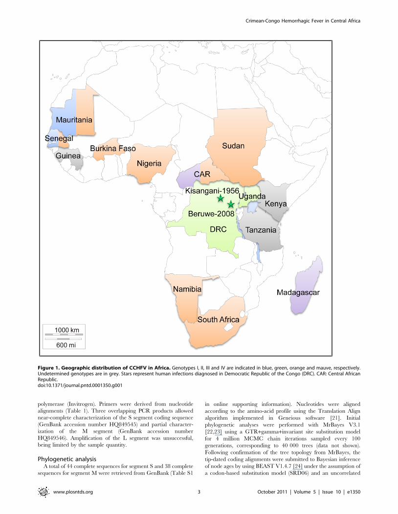
Case descriptionThe patient was a 26-year-old man living in Beruwe (Nord Kivu
province) in DRC, 325 km from Kisangani (Figure 1). He became
ill in the mining area where he worked. He complained of fever
and headache on day 1 and developed moderate bloody diarrhea
on day 2. Epistaxis, oral bleeding and hematuria occurred on day
3. He treated himself with ibuprofen and paracetamol during the
first three days. On day 4 after onset he additionally took quinine
and finally presented with severe asthenia and persistent bleeding
to Lubutu hospital, where the serum sample was taken. At this
stage the patient was subicteric, with bleeding at the venipuncture
site, but had only low-grade fever (37.6uC). He declared no contact
with wild animals during the previous three weeks but he had slept
in the forest. No further information on his outcome was available.
Molecular diagnosisThe patient’s serum was manipulated in biosafety level 4 (BSL-
4) conditions. The serum was first tested for Ebola and Marburg
viruses. As results were negative, investigations were next
performed for CCHFV. RNA was extracted with the QIAamp
viral RNA mini kit (Qiagen, Courtaboeuf, France) according to
manufacturer’s instructions. Reverse transcription (RT) and real-
time PCR amplification were performed with the High Capacity
cDNA RT kit and Taqman universal PCR master mix (Applied
Biosystems - Life Technologies Corporation, Carlsbad, Califor-
nia), and previously reported primers and probes [19]. Conven-
tional one-step RT-PCR was performed with CCHFV primers as
previously reported [20] and with SuperScript III one-step RT-
PCR system and Platinum Taq DNA polymerase (Invitrogen -Life
Technologies Corporation, Carlsbad, California). This yielded a
536-nucleotide fragment in the S segment, sequencing of which
confirmed CCHFV identification.
Genetic characterizationAs viral isolation on Vero cells was unsuccessful, viral RNA was
extracted from the patient’s serum as described above, and was
used for RT-PCR amplification with Platinum Taq DNA
Author Summary
Crimean-Congo hemorrhagic fever virus (CCHFV) is trans-mitted to humans through tick-bite or contact withinfected blood or tissues from livestock, the mainvertebrate hosts in a peri-domestic natural cycle. Withnumerous outbreaks, a high case fatality rate (3%–30%)and a high risk for nosocomial transmission, CCHFVbecame a public health concern in Europe and Asia.However virus surveillance in Africa is difficult due to thelimited sanitary facilities. Especially, CCHFV occurrence inCentral Africa is very poorly described and seems highly incontrast with the temperate to dry environments to whichthe virus is usually associated with. We described a singlehuman infection that occurred in Democratic Republicof the Congo after nearly 50 years of absence. Thephylogenetic analysis suggests that CCHFV enzooticcirculation in the area is still ongoing despite the absenceof notification, and thus reinforces the need for themedical workers and authorities to be aware of theoutbreak risk. The source of infection seemed associatedwith a forest environment while no link with the usualagro-pastoral risk factors could be identified. Moreaccurate ecological data about CCHFV enzootic cycle arerequired to assess the risk of emergence in developingcountries subjected to deforestation.
Crimean-Congo Hemorrhagic Fever in Central Africa
www.plosntds.org 2 October 2011 | Volume 5 | Issue 10 | e1350
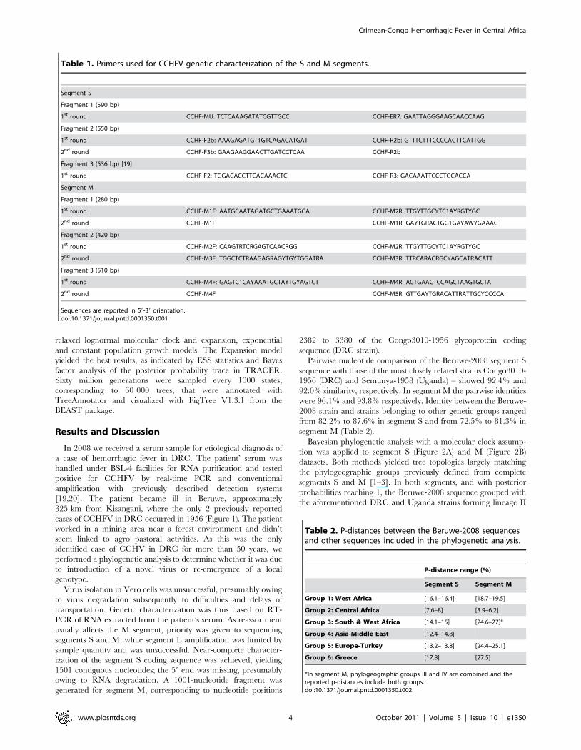
polymerase (Invitrogen). Primers were derived from nucleotide
alignments (Table 1). Three overlapping PCR products allowed
near-complete characterization of the S segment coding sequence
(GenBank accession number HQ849545) and partial character-
ization of the M segment (GenBank accession number
HQ849546). Amplification of the L segment was unsuccessful,
being limited by the sample quantity.
Phylogenetic analysisA total of 44 complete sequences for segment S and 38 complete
sequences for segment M were retrieved from GenBank (Table S1
in online supporting information). Nucleotides were aligned
according to the amino-acid profile using the Translation Align
algorithm implemented in Geneious software [21]. Initial
phylogenetic analyses were performed with MrBayes V3.1
[22,23] using a GTR+gamma+invariant site substitution model
for 4 million MCMC chain iterations sampled every 100
generations, corresponding to 40 000 trees (data not shown).
Following confirmation of the tree topology from MrBayes, the
tip-dated coding alignments were submitted to Bayesian inference
of node ages by using BEAST V1.4.7 [24] under the assumption of
a codon-based substitution model (SRD06) and an uncorrelated
Figure 1. Geographic distribution of CCHFV in Africa. Genotypes I, II, III and IV are indicated in blue, green, orange and mauve, respectively.Undetermined genotypes are in grey. Stars represent human infections diagnosed in Democratic Republic of the Congo (DRC). CAR: Central AfricanRepublic.doi:10.1371/journal.pntd.0001350.g001
Crimean-Congo Hemorrhagic Fever in Central Africa
www.plosntds.org 3 October 2011 | Volume 5 | Issue 10 | e1350
relaxed lognormal molecular clock and expansion, exponential
and constant population growth models. The Expansion model
yielded the best results, as indicated by ESS statistics and Bayes
factor analysis of the posterior probability trace in TRACER.
Sixty million generations were sampled every 1000 states,
corresponding to 60 000 trees, that were annotated with
TreeAnnotator and visualized with FigTree V1.3.1 from the
BEAST package.
Results and Discussion
In 2008 we received a serum sample for etiological diagnosis of
a case of hemorrhagic fever in DRC. The patient’ serum was
handled under BSL-4 facilities for RNA purification and tested
positive for CCHFV by real-time PCR and conventional
amplification with previously described detection systems
[19,20]. The patient became ill in Beruwe, approximately
325 km from Kisangani, where the only 2 previously reported
cases of CCHFV in DRC occurred in 1956 (Figure 1). The patient
worked in a mining area near a forest environment and didn’t
seem linked to agro pastoral activities. As this was the only
identified case of CCHV in DRC for more than 50 years, we
performed a phylogenetic analysis to determine whether it was due
to introduction of a novel virus or re-emergence of a local
genotype.
Virus isolation in Vero cells was unsuccessful, presumably owing
to virus degradation subsequently to difficulties and delays of
transportation. Genetic characterization was thus based on RT-
PCR of RNA extracted from the patient’s serum. As reassortment
usually affects the M segment, priority was given to sequencing
segments S and M, while segment L amplification was limited by
sample quantity and was unsuccessful. Near-complete character-
ization of the segment S coding sequence was achieved, yielding
1501 contiguous nucleotides; the 59 end was missing, presumably
owing to RNA degradation. A 1001-nucleotide fragment was
generated for segment M, corresponding to nucleotide positions
2382 to 3380 of the Congo3010-1956 glycoprotein coding
sequence (DRC strain).
Pairwise nucleotide comparison of the Beruwe-2008 segment S
sequence with those of the most closely related strains Congo3010-
1956 (DRC) and Semunya-1958 (Uganda) – showed 92.4% and
92.0% similarity, respectively. In segment M the pairwise identities
were 96.1% and 93.8% respectively. Identity between the Beruwe-
2008 strain and strains belonging to other genetic groups ranged
from 82.2% to 87.6% in segment S and from 72.5% to 81.3% in
segment M (Table 2).
Bayesian phylogenetic analysis with a molecular clock assump-
tion was applied to segment S (Figure 2A) and M (Figure 2B)
datasets. Both methods yielded tree topologies largely matching
the phylogeographic groups previously defined from complete
segments S and M [1–3]. In both segments, and with posterior
probabilities reaching 1, the Beruwe-2008 sequence grouped with
the aforementioned DRC and Uganda strains forming lineage II
Table 1. Primers used for CCHFV genetic characterization of the S and M segments.
Segment S
Fragment 1 (590 bp)
1st round CCHF-MU: TCTCAAAGATATCGTTGCC CCHF-ER7: GAATTAGGGAAGCAACCAAG
Fragment 2 (550 bp)
1st round CCHF-F2b: AAAGAGATGTTGTCAGACATGAT CCHF-R2b: GTTTCTTTCCCCACTTCATTGG
2nd round CCHF-F3b: GAAGAAGGAACTTGATCCTCAA CCHF-R2b
Fragment 3 (536 bp) [19]
1st round CCHF-F2: TGGACACCTTCACAAACTC CCHF-R3: GACAAATTCCCTGCACCA
Segment M
Fragment 1 (280 bp)
1st round CCHF-M1F: AATGCAATAGATGCTGAAATGCA CCHF-M2R: TTGYTTGCYTC1AYRGTYGC
2nd round CCHF-M1F CCHF-M1R: GAYTGRACTGG1GAYAWYGAAAC
Fragment 2 (420 bp)
1st round CCHF-M2F: CAAGTRTCRGAGTCAACRGG CCHF-M2R: TTGYTTGCYTC1AYRGTYGC
2nd round CCHF-M3F: TGGCTCTRAAGAGRAGYTGYTGGATRA CCHF-M3R: TTRCARACRGCYAGCATRACATT
Fragment 3 (510 bp)
1st round CCHF-M4F: GAGTC1CAYAAATGCTAYTGYAGTCT CCHF-M4R: ACTGAACTCCAGCTAAGTGCTA
2nd round CCHF-M4F CCHF-M5R: GTTGAYTGRACATTRATTGCYCCCCA
Sequences are reported in 59-39 orientation.doi:10.1371/journal.pntd.0001350.t001
Table 2. P-distances between the Beruwe-2008 sequencesand other sequences included in the phylogenetic analysis.
P-distance range (%)
Segment S Segment M
Group 1: West Africa [16.1–16.4] [18.7–19.5]
Group 2: Central Africa [7.6–8] [3.9–6.2]
Group 3: South & West Africa [14.1–15] [24.6–27]*
Group 4: Asia-Middle East [12.4–14.8]
Group 5: Europe-Turkey [13.2–13.8] [24.4–25.1]
Group 6: Greece [17.8] [27.5]
*In segment M, phylogeographic groups III and IV are combined and thereported p-distances include both groups.doi:10.1371/journal.pntd.0001350.t002
Crimean-Congo Hemorrhagic Fever in Central Africa
www.plosntds.org 4 October 2011 | Volume 5 | Issue 10 | e1350
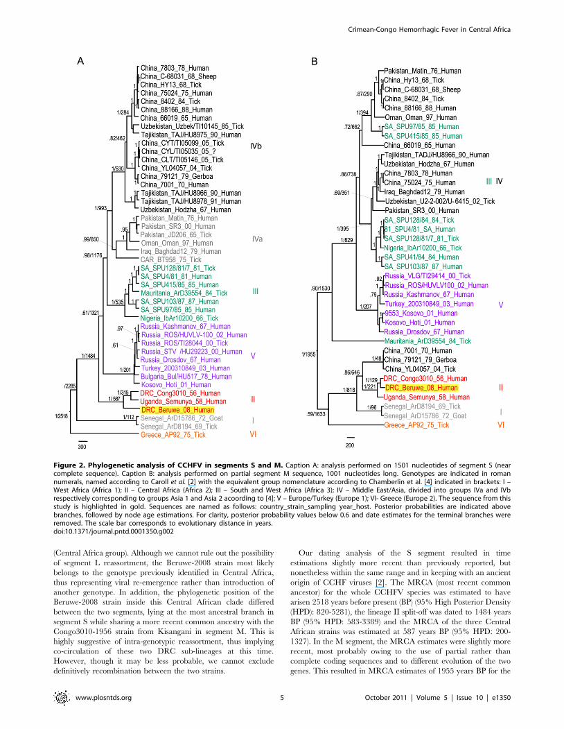
(Central Africa group). Although we cannot rule out the possibility
of segment L reassortment, the Beruwe-2008 strain most likely
belongs to the genotype previously identified in Central Africa,
thus representing viral re-emergence rather than introduction of
another genotype. In addition, the phylogenetic position of the
Beruwe-2008 strain inside this Central African clade differed
between the two segments, lying at the most ancestral branch in
segment S while sharing a more recent common ancestry with the
Congo3010-1956 strain from Kisangani in segment M. This is
highly suggestive of intra-genotypic reassortment, thus implying
co-circulation of these two DRC sub-lineages at this time.
However, though it may be less probable, we cannot exclude
definitively recombination between the two strains.
Our dating analysis of the S segment resulted in time
estimations slightly more recent than previously reported, but
nonetheless within the same range and in keeping with an ancient
origin of CCHF viruses [2]. The MRCA (most recent common
ancestor) for the whole CCHFV species was estimated to have
arisen 2518 years before present (BP) (95% High Posterior Density
(HPD): 820-5281), the lineage II split-off was dated to 1484 years
BP (95% HPD: 583-3389) and the MRCA of the three Central
African strains was estimated at 587 years BP (95% HPD: 200-
1327). In the M segment, the MRCA estimates were slightly more
recent, most probably owing to the use of partial rather than
complete coding sequences and to different evolution of the two
genes. This resulted in MRCA estimates of 1955 years BP for the
Figure 2. Phylogenetic analysis of CCHFV in segments S and M. Caption A: analysis performed on 1501 nucleotides of segment S (nearcomplete sequence). Caption B: analysis performed on partial segment M sequence, 1001 nucleotides long. Genotypes are indicated in romannumerals, named according to Caroll et al. [2] with the equivalent group nomenclature according to Chamberlin et al. [4] indicated in brackets: I –West Africa (Africa 1); II – Central Africa (Africa 2); III – South and West Africa (Africa 3); IV – Middle East/Asia, divided into groups IVa and IVbrespectively corresponding to groups Asia 1 and Asia 2 acoording to [4]; V – Europe/Turkey (Europe 1); VI- Greece (Europe 2). The sequence from thisstudy is highlighted in gold. Sequences are named as follows: country_strain_sampling year_host. Posterior probabilities are indicated abovebranches, followed by node age estimations. For clarity, posterior probability values below 0.6 and date estimates for the terminal branches wereremoved. The scale bar corresponds to evolutionary distance in years.doi:10.1371/journal.pntd.0001350.g002
Crimean-Congo Hemorrhagic Fever in Central Africa
www.plosntds.org 5 October 2011 | Volume 5 | Issue 10 | e1350
whole species (95%HPD: 886-3844), 221 years BP for the three
Central African strains (95%HPD: 114-407) and 129 years BP
(95% HPD: 75-228) for the two DRC strains. The genotype II
split-off was estimated to have occurred 646 years BP, but the
differences in the tree topologies prevented a true node age
comparison with segment S.
CCHV genotype II has been identified only in DRC and
Uganda, while different CCHV lineages have been identified in
neighboring countries to the north. Multiple genotypes have been
identified in CAR, belonging to groups IV and III [2,20], the latter
also being encountered in Sudan [14]. By contrast no other
genotype has been identified in Central Africa, for which reports
on CCHFV are scarce and date back to 30 years. Hence, the data
currently available suggest that genotype II is specific to central
Africa. In DRC, CCHV has been reported only once, 50 years
ago, but our data strongly suggest that the same genotype is still
actively circulating.
Of note, the MRCA estimates presented here are in agreement
with ancient divergence of this lineage (around 1000 years ago),
but whether or not this split-off was linked to virus adaptation to
Central Africa cannot be assessed. However, as the MRCA of the
three strains was dated back to 683 to 243 years BP (Figure 2A and
B respectively), one might reasonably assume that the association
of genotype II with this area goes back to this time period and thus
did not result from very recent introduction. In addition, the co-
circulation of different sub-lineages supports the possibility that
ongoing CCHFV circulation occurred in the same area for some
time. However, as the reassortment event would have taken place
approximately 120 years BP, there is no evidence that CCHFV
has been permanently circulating inside the Beruwe microhabitat,
and we cannot exclude the possibility that this virus was very
recently (re)introduced.
In addition to the CCHFV genotypic specificity for Central
Africa, its occurrence in the tropical rainforest contrasts strongly
with the ecological characteristics of other areas in which CCHFV
has been isolated [11]. Indeed, the enzootic distribution of
CCHFV mostly coincides with temperate to dry or semi-dry
climates in the forests, steppes and savannahs of Eurasia and West,
East and South Africa. In these environments, domestic animals
and their associated ticks are major agents of rural enzootic cycles
affecting nearby human populations [11,25]. Despite the lack of
accurate ecological data, the occurrence of CCHFV in Central
Africa and its apparent genotypic specificity may suggest a
distinctive sylvatic natural cycle in the deep tropical forest
characterized by high rainfall, specific wildlife species, and a low
density of domestic animals. Interestingly, co-speciation or long-
term association with specific tick species has been previously
suggested to explain the geographical distribution of CCHV
genetic variants in Russia and Central Asia [26]. Such a sylvatic
cycle, involving specific vectors and hosts with few contacts with
human populations, could partly explain the lack of outbreaks and
the sporadic nature of recorded human cases. In addition, as
CCHFV is known to have been present in Central Africa for
decades, and as human populations often live in isolated villages,
many human infections may have been overlooked. However
increasing invasion and destruction of rainforest habitats may lead
to a higher risk of human CCHFV cases in future.
Hence, despite 30 years without a single reported case, the data
presented here suggest that CCHFV continues to circulate in
Central Africa. More information on the epidemiology and the
natural cycle of CCHFV in this ecosystem is required to assess its
potential for emergence, notably in Gabon and Republic of the
Congo. However health authorities and medical staff should be
aware of the possibility of viral (re)emergence and of the high risk
of nosocomial transmission.
Supporting Information
Table S1 GenBank accession numbers for the sequences used in
this study. Countries, strains, date of sampling and hosts are
reported along with the associated GenBank accession numbers
for segment S and segment M.
(DOC)
Acknowledgments
The authors thank the members of the Health Ministry of Democratic
Republic of the Congo and the medical experts of the World Health
Organization.
Author Contributions
Conceived and designed the experiments: GG EML. Performed the
experiments: GG. Analyzed the data: GG JFD CD EML. Contributed
reagents/materials/analysis tools: J-JM JFD CD. Wrote the paper: GG
EML JFD CD JF NDW.
References
1. Deyde VM, Khristova ML, Rollin PE, Ksiazek TG, Nichol ST (2006) Crimean-
Congo hemorrhagic fever virus genomics and global diversity. J Virol 80:
8834–8842.
2. Carroll SA, Bird BH, Rollin PE, Nichol ST (2010) Ancient common ancestry of
Crimean-Congo hemorrhagic fever virus. Mol Phylogenet Evol 55: 1103–1110.
3. Hewson R, Chamberlain J, Mioulet V, Lloyd G, Jamil B, et al. (2004) Crimean-
Congo haemorrhagic fever virus: sequence analysis of the small RNA segments
from a collection of viruses world wide. Virus Res 102: 185–189.
4. Chamberlain J, Cook N, Lloyd G, Mioulet V, Tolley H, et al. (2005) Co-
evolutionary patterns of variation in small and large RNA segments of Crimean-
Congo hemorrhagic fever virus. J Gen Virol 86: 3337–3341.
5. Anagnostou V, Papa A (2009) Evolution of Crimean-Congo Hemorrhagic Fever
virus. Infect Genet Evol 9: 948–954.
6. Hewson R, Gmyl A, Gmyl L, Smirnova SE, Karganova G, et al. (2004)
Evidence of segment reassortment in Crimean-Congo haemorrhagic fever virus.
J Gen Virol 85: 3059–3070.
7. Maltezou HC, Andonova L, Andraghetti R, Bouloy M, Ergonul O, et al.
Crimean-Congo hemorrhagic fever in Europe: current situation calls for
preparedness. Euro Surveill 15: 9504.
8. Ergonul O (2006) Crimean-Congo haemorrhagic fever. Lancet Infect Dis 6:
203–214.
9. Swanepoel R, Struthers JK, Shepherd AJ, McGillivray GM, Nel MJ, et al.
(1983) Crimean-congo hemorrhagic fever in South Africa. Am J Trop Med Hyg
32: 1407–1415.
10. Shepherd AJ, Swanepoel R, Shepherd SP, Leman PA, Blackburn NK, et al.
(1985) A nosocomial outbreak of Crimean-Congo haemorrhagic fever at
Tygerberg Hospital. Part V. Virological and serological observations. S Afr
Med J 68: 733–736.
11. Hoogstraal H (1979) The epidemiology of tick-borne Crimean-Congo
hemorrhagic fever in Asia, Europe, and Africa. J Med Entomol 15: 307–417.
12. CRORA viral database. Centre collaborateur OMS de Reference et de
Recherche sur les Arbovirus, Institut Pasteur de Dakar. Available: http://www.
pasteur.fr/recherche/banques/CRORA/virus/v0401010.htm. Accessed 2011
Apr 7.
13. Nabeth P, Cheikh DO, Lo B, Faye O, Vall IO, et al. (2004) Crimean-Congo
hemorrhagic fever, Mauritania. Emerg Infect Dis 10: 2143–2149.
14. Aradaib IE, Erickson BR, Mustafa ME, Khristova ML, Saeed NS, et al.
Nosocomial outbreak of Crimean-Congo hemorrhagic fever, Sudan. Emerg
Infect Dis 16: 837–839.
15. Guilherme JM, Gonella-Legall C, Legall F, Nakoume E, Vincent J (1996)
Seroprevalence of five arboviruses in Zebu cattle in the Central African
Republic. Trans R Soc Trop Med Hyg 90: 31–33.
16. Degallier N, Cornet JP, Saluzzo JF, Germain M, Herve JP, et al. (1985) [Ecology
of tick-borne arboviruses in the Central African Republic]. Bull Soc Pathol Exot
Filiales 78: 296–310.
17. Simpson DI, Knight EM, Courtois G, Williams MC, Weinbren MP, et al. (1967)
Congo virus: a hitherto undescribed virus occurring in Africa. I. Human
isolations–clinical notes. East Afr Med J 44: 86–92.
Crimean-Congo Hemorrhagic Fever in Central Africa
www.plosntds.org 6 October 2011 | Volume 5 | Issue 10 | e1350
18. Woodall JP, Williams MC, Simpson DI (1967) Congo virus: a hitherto
undescribed virus occurring in Africa. II. Identification studies. East Afr Med J44: 93–98.
19. Wolfel R, Paweska JT, Petersen N, Grobbelaar AA, Leman PA, et al. (2007)
Virus detection and monitoring of viral load in Crimean-Congo hemorrhagicfever virus patients. Emerg Infect Dis 13: 1097–1100.
20. Rodriguez LL, Maupin GO, Ksiazek TG, Rollin PE, Khan AS, et al. (1997)Molecular investigation of a multisource outbreak of Crimean-Congo hemor-
rhagic fever in the United Arab Emirates. Am J Trop Med Hyg 57: 512–518.
21. Drummond AJ, Ashton B, Buxton S, Cheung M, Cooper A, et al. (2010)Geneious v5.1. Available: http://www.geneious.com.
22. Gelman A, Rubin DB (1996) Markov chain Monte Carlo methods in
biostatistics. Stat Methods Med Res 5: 339–355.23. Ronquist F, Huelsenbeck JP (2003) MrBayes 3: Bayesian phylogenetic inference
under mixed models. Bioinformatics 19: 1572–1574.
24. Drummond AJ, Rambaut A (2007) BEAST: Bayesian evolutionary analysis bysampling trees. BMC Evol Biol 7: 214.
25. Whitehouse CA (2004) Crimean-Congo hemorrhagic fever. Antiviral Res 64:145–160.
26. Yashina L, Petrova I, Seregin S, Vyshemirskii O, Lvov D, et al. (2003) Genetic
variability of Crimean-Congo haemorrhagic fever virus in Russia and CentralAsia. J Gen Virol 84: 1199–1206.
Crimean-Congo Hemorrhagic Fever in Central Africa
www.plosntds.org 7 October 2011 | Volume 5 | Issue 10 | e1350