rarγ is essential for retinoic acid induced chromatin remodeling and transcriptional activation in...
TRANSCRIPT

Journ
alof
Cell
Scie
nce
RARc is essential for retinoic acid induced chromatinremodeling and transcriptional activation in embryonicstem cells
Vasundhra Kashyap1,4,*, Kristian B. Laursen1,*, Fabienne Brenet2, Agnes J. Viale3, Joseph M. Scandura2 andLorraine J. Gudas1,2,4,`
1Department of Pharmacology, Weill Medical College of Cornell University, 1300 York Avenue, New York, NY 10065, USA2Department of Medicine, Weill Medical College of Cornell University, 1300 York Avenue, New York, NY 10065, USA3Genomics Core Laboratory, Memorial Sloan-Kettering Cancer Center, 1275 York Avenue, New York, NY 10021, USA4Weill Graduate School of Biomedical Sciences, 1300 York Avenue, New York, NY 10065, USA
*These authors contributed equally to this work`Author for correspondence ([email protected])
Accepted 3 December 2012Journal of Cell Science 126, 999–1008� 2013. Published by The Company of Biologists Ltddoi: 10.1242/jcs.119701
SummaryWe have utilized retinoic acid receptor c (gamma) knockout (RARc2/2) embryonic stem (ES) cells as a model system to analyze RARcmediated transcriptional regulation of stem cell differentiation. Most of the transcripts regulated by all-trans retinoic acid (RA) in ES cellsare dependent upon functional RARc signaling. Notably, many of these RA–RARc target genes are implicated in retinoid uptake andmetabolism. For instance, Lrat (lecithin:retinol acyltransferase), Stra6 (stimulated by retinoic acid 6), Crabp2 (cellular retinoic acid binding
protein 2), and Cyp26a1 (cytochrome p450 26a1) transcripts are induced in wild type (WT), but not in RARc2/2 cells. Transcripts for thetranscription factors Pbx1 (pre-B cell leukemia homeobox-1), Wt1 (Wilm’s tumor gene-1), and Meis1 (myeloid ecotropic viral integrationsite-1) increase upon RA treatment of WT, but not RARc2/2 cells. In contrast, Stra8, Dleu7, Leftb, Pitx2, and Cdx1 mRNAs are induced by
RA even in the absence of RARc. Mapping of the epigenetic signature of Meis1 revealed that RA induces a rapid increase in the H3K9/K14ac epigenetic mark at the proximal promoter and at two sites downstream of the transcription start site in WT, but not in RARc2/2 cells.Thus, RA-associated increases in H3K9/K14ac epigenetic marks require RARc and are associated with increased Meis1 transcript levels,
whereas H3K4me3 is present at the Meis1 proximal promoter even in the absence of RARc. In contrast, at the Lrat proximal promoterprimarily the H3K4me3 mark, and not the H3K9/K14ac mark, increases in response to RA, independently of the presence of RARc. Ourdata show major epigenetic changes associated with addition of the RARc agonist RA in ES cells.
Key words: Meis1, Differentiation, Transcription, Nuclear receptor, Retinoic acid receptor, Epigenetics
IntroductionRetinoic acid receptors (RARs) belong to the family of nuclear
receptors that regulates transcription. There are three RAR
isotypes (RARa, RARb, RARc) that heterodimerize with RXRs
(RXRa, RXRb, RXRc) and bind the cis-acting retinoic acid
response elements (RAREs) to execute the biological functions of
RA during embryonic development and postnatally (Clagett-
Dame and Knutson, 2011; Means and Gudas, 1995; Samarut and
Rochette-Egly, 2012). While single RAR mutant mice are viable
and show relatively mild phenotypes, compound mutants of
RARs display an array of congenital abnormalities and die
shortly after birth (Mark et al., 2009).
RARc null mice exhibit growth deficiency (Lohnes et al.,
1993). Recently, RARc was shown to be highly expressed in the
growth plate, and ablation of RARc is associated with reduced
chondrocyte proliferation and decreased expression and
deposition of proteoglycans (Williams et al., 2009). These
findings provide some mechanistic understanding of the growth
retardation phenotype observed in the RARc null mice. RARcregulates hindbrain and axial patterning, and its loss results in
several malformations of the axial skeleton, including
anteriorization of the cervical and thoracic vertebrae (Lohnes
et al., 1993; Wendling et al., 2001). RARc is required for the
formation of normal alveoli and alveoli elastic fibers in the lung
(McGowan et al., 2000). Genetic ablation of RARc results in
male sterility and is associated with squamous metaplasia of
seminal vesicles and the prostate glands and keratinization of
glandular epithelia (Lohnes et al., 1993).
Retinoids also regulate hematopoietic development, which is
dependent on distinct functions mediated by RARa and RARc(Purton, 2007). While RARa induces granulocytic differentiation,
RARc plays a critical role in maintaining the balance between the
self-renewal state of HSCs and their differentiation (Purton, 2007;
Purton et al., 2006). Many of the molecular targets and pathways
downstream of RARc that mediate its effects on hematopoiesis
remain to be determined.
RARc mediates the anti-proliferative and apoptotic effects of
retinoids in certain tissues and cancer cells, such as melanoma
and neuroblastoma cells (Meister et al., 1998; Spanjaard et al.,
1997). RARc is the principal receptor that functions in RA
mediated growth arrest in keratinocytes (Goyette et al., 2000). In
a model of epidermal tumorigenesis, ablation of RARc enhanced
Research Article 999
Journ
alof
Cell
Scie
nce
the tumor incidence of Ras transformed keratinocytes and was
associated with resistance to retinoid mediated growth arrest and
apoptosis (Chen et al., 2004).
Studies conducted in our laboratory have shown that the lack
of both alleles of RARc in F9 teratocarcinoma stem cells is
associated both with impaired differentiation and greatly reduced
expression of genes involved in cell differentiation, such as
Hoxa1, laminin B1, and collagen IV (a1) (Boylan et al., 1993;
Boylan et al., 1995). A microarray analysis of F9 RARc null
teratocarcinoma stem cells revealed novel RARc regulated genes,
reinforcing its important role in retinoid signaling and
differentiation (Su and Gudas, 2008a; Su and Gudas, 2008b).
We have now utilized murine RARc knockout (RARc2/2)
embryonic stem (ES) cells as a model system to study RARcmediated transcriptional regulation in development and cell
differentiation. ES cells are derived from the inner cell mass of
blastocysts and have the unique ability to self-renew under
defined conditions (Smith, 2001). ES cells have a stable genome
and possess the capacity to differentiate into the three germ
layers, thus making them an excellent cell culture system to study
RA mediated differentiation in vitro (Gudas and Wagner, 2011;
Soprano et al., 2007). We previously reported that RARc null ES
cells do not differentiate into parietal endoderm, an epithelial cell
type, in response to RA (Kashyap et al., 2011).
To delineate further the functions of RARc in ES cells, we
performed microarray analysis of wild type (WT) and RARc2/2
ES cells and identified differentially regulated genes. We
characterized the transcriptional and epigenetic regulation of the
RARc target gene Meis1 using chromatin immunoprecipitation
(ChIP) and ChIP-chip technologies. Furthermore, functional
depletion of RARc in WT ES cells, combined with restored
RARc expression in RARc2/2 cells, confirmed the requirement
for RARc in the RA induced transcription of Meis1.
ResultsIdentification of differentially expressed genes in WT and
RARc knockout ES cells by microarray analysis
To identify RARc regulated genes in ES cells, we performed
microarray analyses of ES RARc knockout (RARc2/2)
compared to wild type (WT) ES cells under different culture
conditions. We cultured the WT and RARc2/2 cells with vehicle
control or 1 mM RA for 8 or 24 h. The 8 h and 24 h time points
allowed us to examine the kinetics of RA induced transcript
changes. The 8 h time point also provides the advantage of
capturing early transcript changes that are less likely to be
secondary changes related to cell differentiation.
A total of 152 transcripts were differentially regulated by
threefold or more in untreated WT versus RARc2/2 ES cells
(Fig. 1). Of these 152 differentially expressed transcripts, 80
transcripts showed reduced levels (Fig. 1; supplementary
material Table S2A), and 78 transcripts showed elevated levels
in the RARc2/2 cells compared to the WT cells in vehicle treated
(untreated) conditions (Fig. 1; supplementary material Table
S2B). These data suggest a role for RARc in regulating gene
expression in the absence of the ligand, as we demonstrated in
teratocarcinoma stem cells for RARa (Laursen et al., 2012).
Furthermore, a total of 56 and 72 transcripts were differentially
regulated by threefold or more in WT versus RARc2/2 ES cells
upon 8 and 24 h of RA treatment, respectively (Fig. 1;
supplementary material Table S3A,B, Table S4A,B).
Transcripts are differentially regulated by RA in WT and
RARc knockout cells
To identify the genes that were regulated by RA treatment, we
analyzed the fold changes in WT and RARc2/2 ES cells
independently upon 8 and 24 h of RA treatment in comparison to
the untreated controls for each cell line. A total of 29 and 91
transcripts were increased 2.5-fold in WT ES cells upon 8 and
24 h of RA treatment, respectively, compared to untreated WT
(Fig. 1A; supplementary material Tables S5, S6). The 2.5-fold
cut-off was selected based on advice from the Weill Cornell
Bioinformatics Core staff. Transcript levels of many known RA
target genes increased in WT ES cells in response to RA
treatment, such as genes of the Hoxa and Hoxb clusters and
Cdx1, RARb2, and Gata6 (Fig. 1A,D; supplementary material
Table S5, Table S6A). Transcript levels of 40 genes decreased
upon 24 h of RA treatment in WT cells (Fig. 1C; supplementary
material Table S6B, Table S7B). In accord with previous studies,
transcript levels of Zfp42 (Rex1), a stem cell marker, were
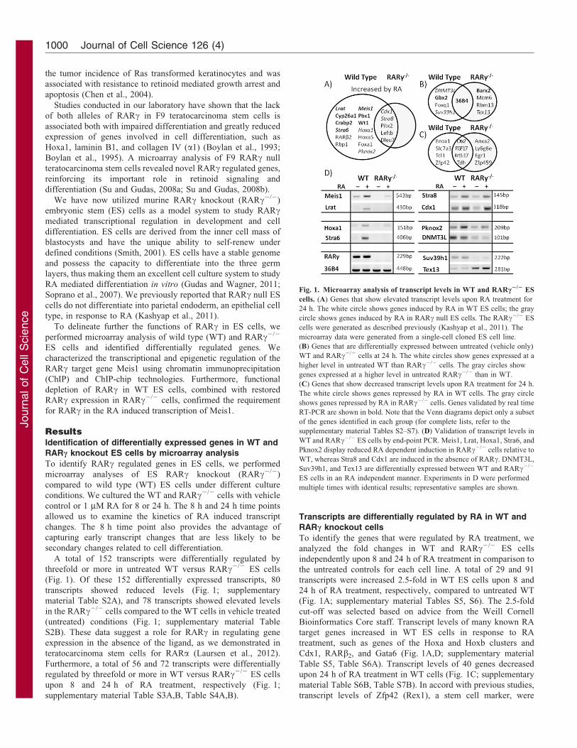
Fig. 1. Microarray analysis of transcript levels in WT and RARc2/2 ES
cells. (A) Genes that show elevated transcript levels upon RA treatment for
24 h. The white circle shows genes induced by RA in WT ES cells; the gray
circle shows genes induced by RA in RARc null ES cells. The RARc2/2 ES
cells were generated as described previously (Kashyap et al., 2011). The
microarray data were generated from a single-cell cloned ES cell line.
(B) Genes that are differentially expressed between untreated (vehicle only)
WT and RARc2/2 cells at 24 h. The white circles show genes expressed at a
higher level in untreated WT than RARc2/2 cells. The gray circles show
genes expressed at a higher level in untreated RARc2/2 than in WT.
(C) Genes that show decreased transcript levels upon RA treatment for 24 h.
The white circle shows genes repressed by RA in WT cells. The gray circle
shows genes repressed by RA in RARc2/2 cells. Genes validated by real time
RT-PCR are shown in bold. Note that the Venn diagrams depict only a subset
of the genes identified in each group (for complete lists, refer to the
supplementary material Tables S2–S7). (D) Validation of transcript levels in
WT and RARc2/2 ES cells by end-point PCR. Meis1, Lrat, Hoxa1, Stra6, and
Pknox2 display reduced RA dependent induction in RARc2/2 cells relative to
WT, whereas Stra8 and Cdx1 are induced in the absence of RARc. DNMT3L,
Suv39h1, and Tex13 are differentially expressed between WT and RARc2/2
ES cells in an RA independent manner. Experiments in D were performed
multiple times with identical results; representative samples are shown.
Journal of Cell Science 126 (4)1000
Journ
alof
Cell
Scie
nce
reduced by 2.7-fold upon 24 h of RA treatment of WT cells
(Fig. 1C; supplementary material Table S6B). Only a few genes(Otx2, FGF17, Krt1-17, and Tdh) showed reduced expressionlevels in both WT and RARc2/2 cells treated with RA for 24 h
(Fig. 1C; supplementary material Table S6B, Table S7B).
In contrast, only 23 transcripts were increased by 2.5-fold ormore in RARc2/2 ES cells upon treatment with RA for 24 hcompared to untreated RARc2/2 (supplementary material Table
S7A). The 8 h RA treatment did not induce significant (.2.5-fold) changes in the levels of any transcripts in the RARc2/2 EScells compared to untreated RARc2/2 ES cells. A large number
of transcripts that were differentially regulated by 2.5-fold ormore in WT cells upon 24 h of RA treatment did not showstatistically significant fold changes of 2.5-fold or more in theRARc2/2 ES cells (Fig. 1A; supplementary material Table S4A).
Thus, RARc is implicated in regulating this group of genes andour results also suggest that other RARs, i.e. RARa and RARb,incompletely compensate for the loss of RARc. However, Stra8,
Dleu7, Leftb, Pitx2, and Cdx1 were induced by RA by more than3.8-fold even in the absence of RARc.
Gene ontology revealed that the vast majority of genes whichexhibit reduced expression in RARc2/2 cells are homeobox
genes involved in morphogenesis, axis formation, and tissuepatterning (Meis1, Pknox2, Pbx1, Foxq1, Gbx2, and Hox genes).RARc plays a key role in axis specification by RA (Bayha et al.,
2009). The induction of Hoxa1, Hoxa2, Hoxb1, and Hoxb2,which are involved in rhombomere/hindbrain formation (Gavalaset al., 2003), is lost in RARc2/2 ES cells. Reduced levels of
Anxa5, Anxa2, F2r, F2rl1, and Gap43 suggest impaired woundhealing in RARc2/2 mice. Also, the transcript levels of severalP450 cytochromes (Cyp1b1, Cyp26a1, and Cyp7b1) are reduced
in RARc2/2 cells, pointing to abnormal metabolism. Finally,reduced expression of Col4a1, Col4a2, Ccnd2, Lama1, Pik3r1,PDGFRa, and Zyxin in RARc2/2 versus WT suggests that focaladhesion may be impaired in RARc2/2 ES cells, which could
lead to increased cellular mobility and/or invasiveness.
RARc regulates RA mediated changes in the transcriptlevels of genes involved in retinoid metabolism, includingStra6, Cyp26a1, Lrat and Crabp2, in ES cells
Because we previously observed alterations in the expression ofseveral genes involved in retinol metabolism during ES
differentiation (Langton and Gudas, 2008), we assessed themRNA levels of several genes that function in the retinoidmetabolism pathway by real time RT-PCR to determine if they
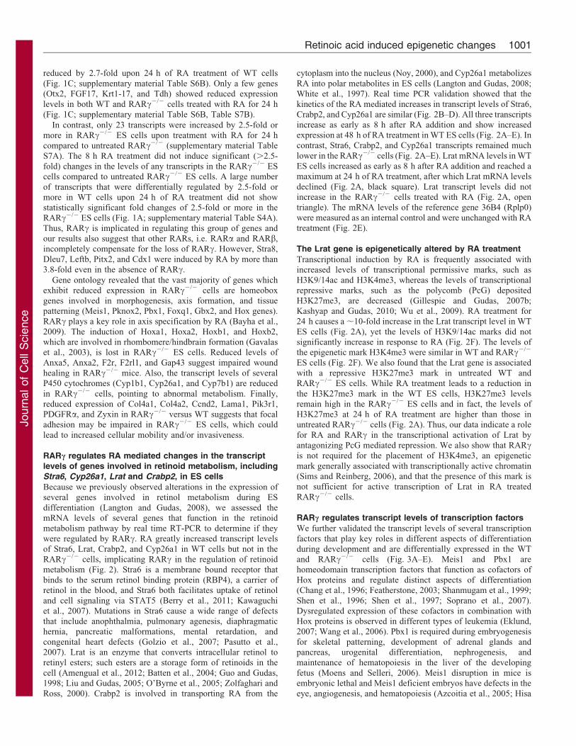
were regulated by RARc. RA greatly increased transcript levelsof Stra6, Lrat, Crabp2, and Cyp26a1 in WT cells but not in theRARc2/2 cells, implicating RARc in the regulation of retinoidmetabolism (Fig. 2). Stra6 is a membrane bound receptor that
binds to the serum retinol binding protein (RBP4), a carrier ofretinol in the blood, and Stra6 both facilitates uptake of retinoland cell signaling via STAT5 (Berry et al., 2011; Kawaguchi
et al., 2007). Mutations in Stra6 cause a wide range of defectsthat include anophthalmia, pulmonary agenesis, diaphragmatichernia, pancreatic malformations, mental retardation, and
congenital heart defects (Golzio et al., 2007; Pasutto et al.,2007). Lrat is an enzyme that converts intracellular retinol toretinyl esters; such esters are a storage form of retinoids in the
cell (Amengual et al., 2012; Batten et al., 2004; Guo and Gudas,1998; Liu and Gudas, 2005; O’Byrne et al., 2005; Zolfaghari andRoss, 2000). Crabp2 is involved in transporting RA from the
cytoplasm into the nucleus (Noy, 2000), and Cyp26a1 metabolizes
RA into polar metabolites in ES cells (Langton and Gudas, 2008;White et al., 1997). Real time PCR validation showed that thekinetics of the RA mediated increases in transcript levels of Stra6,
Crabp2, and Cyp26a1 are similar (Fig. 2B–D). All three transcriptsincrease as early as 8 h after RA addition and show increasedexpression at 48 h of RA treatment in WT ES cells (Fig. 2A–E). Incontrast, Stra6, Crabp2, and Cyp26a1 transcripts remained much
lower in the RARc2/2 cells (Fig. 2A–E). Lrat mRNA levels in WTES cells increased as early as 8 h after RA addition and reached amaximum at 24 h of RA treatment, after which Lrat mRNA levels
declined (Fig. 2A, black square). Lrat transcript levels did notincrease in the RARc2/2 cells treated with RA (Fig. 2A, opentriangle). The mRNA levels of the reference gene 36B4 (Rplp0)
were measured as an internal control and were unchanged with RAtreatment (Fig. 2E).
The Lrat gene is epigenetically altered by RA treatment
Transcriptional induction by RA is frequently associated withincreased levels of transcriptional permissive marks, such asH3K9/14ac and H3K4me3, whereas the levels of transcriptionalrepressive marks, such as the polycomb (PcG) deposited
H3K27me3, are decreased (Gillespie and Gudas, 2007b;Kashyap and Gudas, 2010; Wu et al., 2009). RA treatment for24 h causes a ,10-fold increase in the Lrat transcript level in WT
ES cells (Fig. 2A), yet the levels of H3K9/14ac marks did notsignificantly increase in response to RA (Fig. 2F). The levels ofthe epigenetic mark H3K4me3 were similar in WT and RARc2/2
ES cells (Fig. 2F). We also found that the Lrat gene is associatedwith a repressive H3K27me3 mark in untreated WT andRARc2/2 ES cells. While RA treatment leads to a reduction in
the H3K27me3 mark in the WT ES cells, H3K27me3 levelsremain high in the RARc2/2 ES cells and in fact, the levels ofH3K27me3 at 24 h of RA treatment are higher than those inuntreated RARc2/2 cells (Fig. 2A). Thus, our data indicate a role
for RA and RARc in the transcriptional activation of Lrat byantagonizing PcG mediated repression. We also show that RARcis not required for the placement of H3K4me3, an epigenetic
mark generally associated with transcriptionally active chromatin(Sims and Reinberg, 2006), and that the presence of this mark isnot sufficient for active transcription of Lrat in RA treated
RARc2/2 cells.
RARc regulates transcript levels of transcription factors
We further validated the transcript levels of several transcription
factors that play key roles in different aspects of differentiationduring development and are differentially expressed in the WTand RARc2/2 cells (Fig. 3A–E). Meis1 and Pbx1 arehomeodomain transcription factors that function as cofactors of
Hox proteins and regulate distinct aspects of differentiation(Chang et al., 1996; Featherstone, 2003; Shanmugam et al., 1999;Shen et al., 1996; Shen et al., 1997; Soprano et al., 2007).
Dysregulated expression of these cofactors in combination withHox proteins is observed in different types of leukemia (Eklund,2007; Wang et al., 2006). Pbx1 is required during embryogenesis
for skeletal patterning, development of adrenal glands andpancreas, urogenital differentiation, nephrogenesis, andmaintenance of hematopoiesis in the liver of the developing
fetus (Moens and Selleri, 2006). Meis1 disruption in mice isembryonic lethal and Meis1 deficient embryos have defects in theeye, angiogenesis, and hematopoiesis (Azcoitia et al., 2005; Hisa
Retinoic acid induced epigenetic changes 1001
Journ
alof
Cell
Scie
nce
et al., 2004). Expression of Meis1 is elevated in acute myeloid
leukemias, and Meis1 has oncogenic potential in leukemias that
harbor fusion proteins with the translocation of MLL (mixed
lineage leukemia) family members (Kawagoe et al., 1999; Wong
et al., 2007). Pbx1 and Meis1 mRNA levels increase upon 24 h of
RA treatment in WT cells, and transcript levels continue to
increase up to 48 h of RA treatment (Fig. 3A,B). In contrast, RA
did not increase Pbx1 and Meis1 transcript levels in the
RARc2/2 cells (Fig. 3A,B).
Wt1 (Wilm’s tumor gene) encodes a transcription factor that
has an essential role in the normal development of the
urogenital system, and is mutated in a subset of patients with
Wilm’s tumors, a type of kidney tumor (Kreidberg et al., 1993;
Pelletier et al., 1991a; Pelletier et al., 1991b). RA increased
Wt1 mRNA levels at 48 h in WT cells, but no RA dependent
increase in the Wt1 mRNA levels occurred in the RARc2/2
cells (Fig. 3C).
Gbx2 (gastrulation brain homeobox 2) is required for normal
development of mid/hindbrain region and morphogenesis of the
inner ear (Lin et al., 2005; Wassarman et al., 1997). In WT cells
RA treatment caused less than a twofold increase in the Gbx2
mRNA levels. Gbx2 mRNA levels were consistently lower in
RARc2/2 cells compared to WT cells (Fig. 3D).
Barx1 is a homeodomain transcription factor that is expressed
in the stomach mesenchyme and molar dental cells of
mesenchymal origin (Makarenkova and Meech, 2012). Barx1
mRNA levels were 70-fold lower in untreated WT cells than in
the untreated RARc2/2 cells (Fig. 3E). At 48 h of RA treatment
Barx1 mRNA levels decreased by ,25% in the RARc2/2 cells,
whereas Barx1 transcript levels did not change in WT cells
(Fig. 3E). Thus, RA mediates changes in the transcript levels of
multiple genes involved in differentiation and development that
require RARc. Why these specific genes are highly regulated by
RA in differentiating WT ES cells is not yet clear.
RA induces RARc dependent epigenomic re-organization
of the Meis1 gene in WT cells
Since aberrant regulation of Meis1 is implicated in leukemia
(Kawagoe et al., 1999; Wong et al., 2007) and we are interested
in differentiation therapy for cancer, we next examined the
dynamics of the RA induced increase in Meis1 transcript levels in
further detail. We hypothesized that RA signaling would induce
epigenetic changes at the Meis1 gene and that loss of RARcwould prevent such chromatin changes at this gene. In WT cells
RA induced a rapid increase in H3K9/14ac levels at the proximal
promoter and at specific regions (DS1 and DS2) located
Fig. 2. RA regulated induction of retinoid metabolism genes. (A–E) Transcript levels of Lrat, Stra6, Cyp26a1, Crabp2, and 36B4 (a control) in WT and
RARc2/2 ES cells. Experiments were performed three times using independent RNA samples (& WT; g RARc2/2); the error bars represent the standard error of
the mean (s.e.m.). (F) ChIP-chip heatmap of the Lrat genomic region showing H3K9/14ac, H3K27me3, and H3K4me3 histone marks in WT and RARc2/2 cells
upon increasing times of exposure to RA (0, 1, 8, and 24 h, black triangle). The colors represents log2-transformed ChIP enrichment in ChIP-chip data sets
(replicate means). Columns show genomic loci and rows show IP condition. Statistical significance: *P,0.05, **P,0.01, ***P,0.005. The color scale of the
log2 enrichment is indicated below the ChIP-chip data panel. The Lrat genomic location (blue tones) is indicated schematically at the bottom.
Journal of Cell Science 126 (4)1002

Journ
alof
Cell
Scie
nce
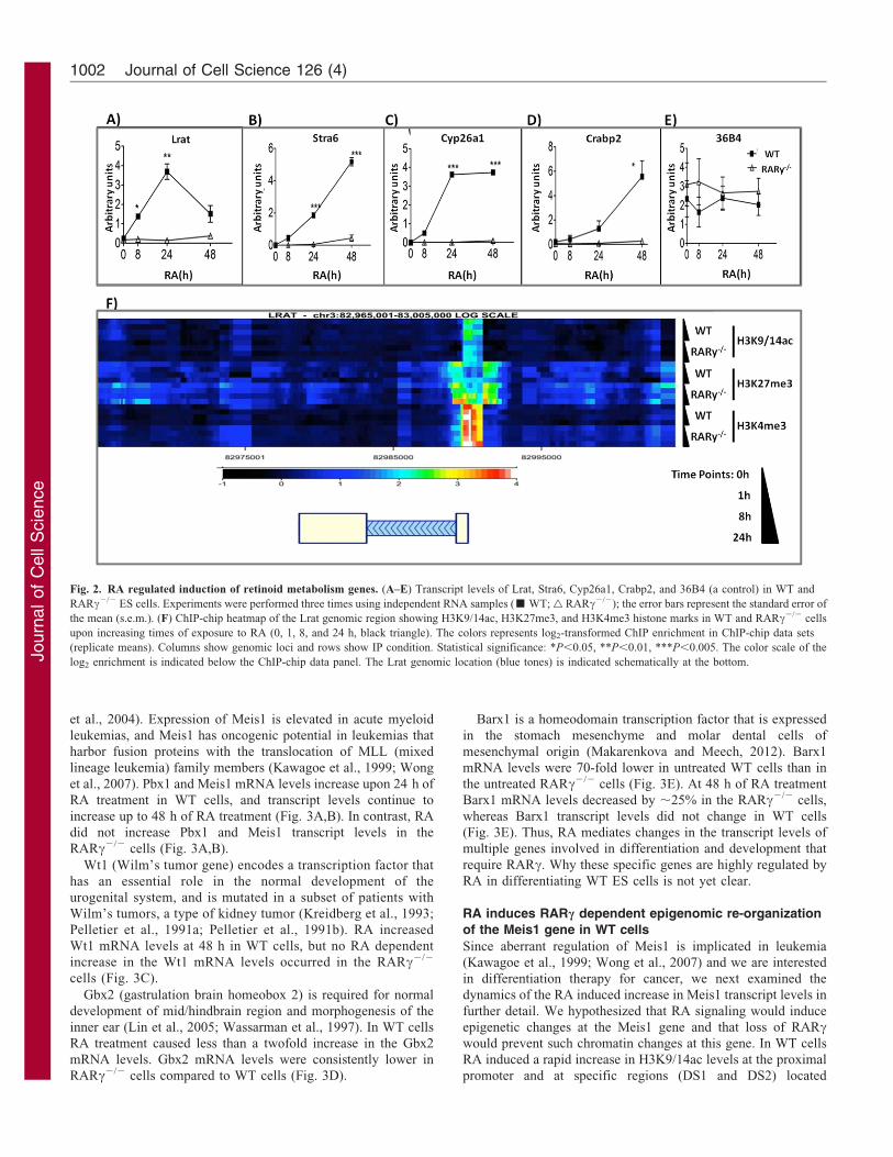
downstream of the transcription start site (TSS) in the Meis1 gene
(Fig. 3F, arrows; Fig. 4A). H3K9/14ac epigenetic marks are
generally associated with transcriptionally active genes
(Jenuwein and Allis, 2001). In contrast, the H3K9/14ac levels
at the promoter and downstream intragenic regions of the Meis1
gene were much lower in the RARc cells compared to the WT
cells (Fig. 3F; Fig. 4A), correlating with Meis1 transcript levels
in WT versus RARc2/2 cells (Fig. 3A). The H3K4me3 mark is
elevated in a region surrounding the promoter of the Meis1 gene
in both untreated and RA treated WT and RARc2/2 cells
(Fig. 3F; Fig. 4C).
Meis1 resides in a bivalent chromatin domain, e.g. it is
associated with both with the repressive H3K27me3 mark
deposited by PcG proteins (Boyer et al., 2006) and with the
permissive H3K4me3 mark described above. By 24 h of RA
treatment there is a reduction in the H3K27me3 mark (Fig. 3F),
and in the levels of Suz12 protein (Fig. 4B) at the promoter and
in the gene body of Meis1 in WT cells. However, in RARc2/2
cells both the H3K27me3 mark and Suz12 levels remain high at
24 h of RA treatment (Fig. 3F; Fig. 4B). Thus, RA signaling via
RARc antagonizes the PcG mediated repression of the Meis1
gene, and the lack of RARc prevents the removal of the
H3K27me3 mark from the Meis1 gene (Fig. 3F).
Taken together, these data suggest that at the Meis1 gene RARc (or
its downstream targets) is required for RA induced changes in the
epigenetic configuration comprising the H3K9/14ac marks. However,
RARc is not needed for the deposition of the H3K4me3 mark at the
Meis1 promoter and the H3K4me3 mark does not correlate with
transcription of Meis1 in RA treated RARc2/2 cells (Fig. 3F).
The Meis1 promoter proximal region appears devoid of
functional retinoic acid response elements (RAREs)
The effects of RA are mediated through RAREs, and consequently
the epigenomic structure exhibits dramatic changes in response to
RA, generating hot-spots in the ChIP-chip maps. We evaluated the
Meis1 proximal promoter region (defined as TSS 2/+ 40 kb), and
identified three sites exhibiting dramatic epigenetic changes in
response to RA (Fig. 3F, arrows). Given the critical role of RARcin the regulation of the Meis1 gene, we further characterized the
levels of retinoic acid receptor c (RARc) and retinoid receptor a(RXRa) at these three sites (Fig. 5A,B). RARc levels were slightly
elevated in WT versus RARc2/2 cells at all evaluated regions, but
showed no specific enrichment at any one particular region. The
levels of RXRa at all of these regions were similar to those of IgG
(Fig. 5A,B versus Fig. 5F). We detected binding of RXRa at the
Cyp26a1 promoter, our positive control (supplementary material
Fig. S1), consistent with our previous studies indicating that
Cyp26a1 is a direct RA target gene (Kashyap et al., 2011).
Consequently, the Meis1 proximal promoter region appears to be
devoid of functional RAREs.
Fig. 3. RA regulated induction of transcription factors. (A–E) Transcript levels in WT and RARc2/2 ES cells of Meis1, Pbx1, Wt1, Gbx2, and Barx1. Experiments were
performed three times using independent RNA samples (filled squares, WT; open triangles, RARc2/2); error bars indicate s.e.m. (F) ChIP-chip heat-map of the Meis1 proximal
promoter region showing H3K9/14ac, H3K27me3, and H3K4me3 histone marks in WT and RARc2/2 cells upon increasing times of exposure to RA (0, 1, 8, and 24 h, black
triangle). The colors represents log2-transformed ChIP enrichment in ChIP-chip data sets (replicate means). DS1 and DS2 indicate downstream sites 1 and 2; PP indicates the
proximal promoter region. Columns show genomic loci and rows show IP condition. Statistical significance: P,0.05, **P,0.01, ***P,0.005. The color scale of the log2
enrichment is indicated below the ChIP-chip data panel. The Meis1 exon (broad) and intron (narrow) locations (blue tones) are indicated schematically at the bottom.
Retinoic acid induced epigenetic changes 1003
Journ
alof
Cell
Scie
nce
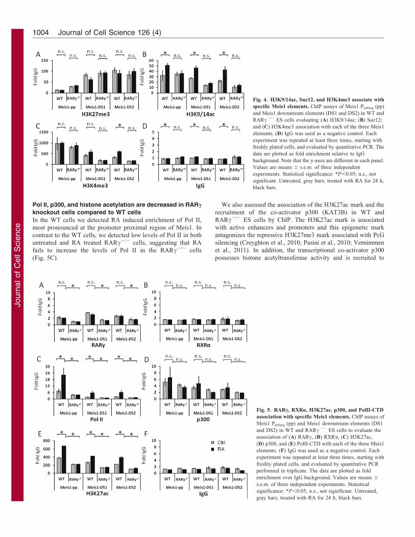
Pol II, p300, and histone acetylation are decreased in RARc
knockout cells compared to WT cells
In the WT cells we detected RA induced enrichment of Pol II,
most pronounced at the promoter proximal region of Meis1. In
contrast to the WT cells, we detected low levels of Pol II in both
untreated and RA treated RARc2/2 cells, suggesting that RA
fails to increase the levels of Pol II in the RARc2/2 cells
(Fig. 5C).
We also assessed the association of the H3K27ac mark and the
recruitment of the co-activator p300 (KAT3B) in WT and
RARc2/2 ES cells by ChIP. The H3K27ac mark is associated
with active enhancers and promoters and this epigenetic mark
antagonizes the repressive H3K27me3 mark associated with PcG
silencing (Creyghton et al., 2010; Pasini et al., 2010; Vernimmen
et al., 2011). In addition, the transcriptional co-activator p300
possesses histone acetyltransferase activity and is recruited to
Fig. 4. H3K9/14ac, Suz12, and H3K4me3 associate with
specific Meis1 elements. ChIP assays of Meis1 Prefseq (pp)
and Meis1 downstream elements (DS1 and DS2) in WT and
RARc2/2 ES cells evaluating (A) H3K9/14ac; (B) Suz12;
and (C) H3K4me3 association with each of the three Meis1
elements. (D) IgG was used as a negative control. Each
experiment was repeated at least three times, starting with
freshly plated cells, and evaluated by quantitative PCR. The
data are plotted as fold enrichment relative to IgG
background. Note that the y-axes are different in each panel.
Values are means 6 s.e.m. of three independent
experiments. Statistical significance: *P,0.05; n.s., not
significant. Untreated, gray bars; treated with RA for 24 h,
black bars.
Fig. 5. RARc, RXRa, H3K27ac, p300, and PolII-CTD
association with specific Meis1 elements. ChIP assays of
Meis1 Prefseq (pp) and Meis1 downstream elements (DS1
and DS2) in WT and RARc2/2 ES cells to evaluate the
association of (A) RARc, (B) RXRa, (C) H3K27ac,
(D) p300, and (E) PolII-CTD with each of the three Meis1
elements. (F) IgG was used as a negative control. Each
experiment was repeated at least three times, starting with
freshly plated cells, and evaluated by quantitative PCR
performed in triplicate. The data are plotted as fold
enrichment over IgG background. Values are means 6
s.e.m. of three independent experiments. Statistical
significance: *P,0.05; n.s., not significant. Untreated,
gray bars; treated with RA for 24 h, black bars.
Journal of Cell Science 126 (4)1004
Journ
alof
Cell
Scie
nce
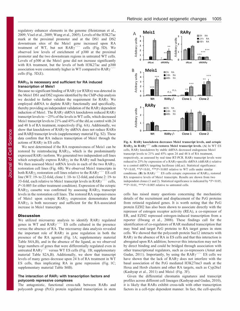
regulatory enhancer elements in the genome (Heintzman et al.,2009; Visel et al., 2009; Wang et al., 2005). Levels of the H3K27acmark at the proximal promoter and at the DS1 and DS2downstream sites of the Meis1 gene increased upon RA
treatment of WT, but not RARc2/2 cells (Fig. 5D). Weobserved low levels of enrichment of p300 at the proximalpromoter and the two downstream regions in untreated WT cells.
Levels of p300 at the Meis1 gene did not increase significantlywith RA treatment, but the levels of both H3K27ac and p300association were consistently higher in WT compared to RARc2/2
cells (Fig. 5D,E).
RARc2 is necessary and sufficient for RA inducedtranscription of Meis1Because no significant binding of RARc (or RXRa) was detected in
the Meis1 DS1 and DS2 regions identified by the ChIP-chip analysiswe decided to further validate the requirement for RARc. Weemployed shRNA to deplete RARc functionally and specifically,
thereby providing an independent validation of the RARc dependentinduction of Meis1. The RARc shRNA knockdown reduced RARctranscript levels to ,25% of the levels in WT cells, which decreased
Meis1 transcript levels to 21% and 45% of the shLuc control with 24and 48 h of RA treatment, respectively (Fig. 6A). Additionally, weshow that knockdown of RARc by shRNA does not reduce RARaand RARb transcript levels (supplementary material Fig. S2). Thesedata confirm that RA induces transcription of Meis1 through theactions of RARc in ES cells.
We next determined if the RA responsiveness of Meis1 can berestored by reintroducing RARc2, which is the predominantlyexpressed RARc isoform. We generated two independent cell lines
which ectopically express RARc2 in the RARc null background.We then assessed Meis1 mRNA levels in each of the two RARc2
restoration cell lines (Fig. 6B). We observed Meis1 transcripts in
both RARc2 restoration cell lines relative to the RARc2/2 ES cellline (WT: 19- to 22-fold, clone 1: 10- to 12-fold, and clone 2: 19- to33-fold, each relative to Meis1 transcript levels in RARc2/2 cells,
P,0.005 for either treatment condition). Expression of the ectopicRARc2 cassette was confirmed by assessing RARc2 transcriptlevels in the restoration cell lines. The restored RA responsivenessof Meis1 upon ectopic RARc2 expression demonstrates that
RARc2 is both necessary and sufficient for the RA-associatedincrease in Meis1 transcripts.
DiscussionWe utilized microarray analysis to identify RARc regulatedgenes in WT and RARc2/2 ES cells cultured in the presenceversus the absence of RA. The microarray data analysis revealed
the important role of RARc in gene regulation in both thepresence of the RA agonist (Fig. 1A; supplementary materialTable S4A,B), and in the absence of the ligand, as we observedlarge numbers of genes that were differentially regulated even in
untreated RARc2/2 versus WT ES cells (Fig. 1B; supplementarymaterial Table S2A,B). Additionally, we show that transcriptlevels of many genes decrease upon 24 h of RA treatment in WT
ES cells, thus implicating RA in gene repression (Fig. 1C;supplementary material Table S6B).
The interaction of RARc with transcription factors andpolycomb repressive complexes
The antagonistic, functional cross-talk between RARs andpolycomb group (PcG) protein regulated transcription in stem
cells has raised many questions concerning the mechanistic
details of the recruitment and displacement of the PcG proteins
from retinoid regulated genes. It is worth noting that the PcGprotein EZH2 has also been shown to associate directly with the
repressor of estrogen receptor activity (REA), a co-repressor of
ER, and EZH2 repressed estrogen-induced transcription from areporter (Hwang et al., 2008). These findings call for the
identification of co-regulators of RAR mediated transcription that
may bind and target PcG proteins to RA target genes in stemcells. We showed that the polycomb protein Suz12 interacts with
RARc in the absence of RA in ES cells and that this interaction isabrogated upon RA addition; however this interaction may not be
by direct binding and could be bridged through association with
other transcriptional regulators, such as co-repressors (Amat andGudas, 2011). Importantly, by using the RARc2/2 ES cells we
have shown that the lack of RARc does not interfere with the
basal association of the PcG mediated H3K27me3 mark at theHoxa and Hoxb clusters and other RA targets, such as Cyp26a1
(Kashyap et al., 2011) and Meis1 (Fig. 3F).
Given the differential chromatin signatures and transcript
profiles across different cell lineages (Kashyap and Gudas, 2010),it is likely that RARs exhibit cross-talk with other transcription
factors in a cell-type dependent manner. In fact, the cell-specific
Fig. 6. RARc knockdown decreases Meis1 transcript levels, and ectopic
RARc2 in RARc2/2 cells restores Meis1 transcript levels. (A) In WT ES
cells, RARc knockdown by stable shRNA decreased endogenous Meis1
transcript levels to 21% and 45% upon 24 and 48 h of RA treatment,
respectively, as assessed by real time RT-PCR. RARc transcript levels were
reduced to 25% by expression of a RARc-specific shRNA (shRARc) relative
to a control shRNA targeting luciferase (shLuc). Statistical significance:
*P,0.05, **P,0.01, ***P,0.005 relative to WT cells under similar
conditions. (B) In RARc2/2 ES cells ectopic expression of RARc2 restored
RA responsive levels of Meis1 transcripts. Results are shown from two
independent clones (1 and 2). Statistical significance is indicated by *P,0.05,
**P,0.01, ***P,0.005 relative to untreated cells.
Retinoic acid induced epigenetic changes 1005
Journ
alof
Cell
Scie
nce
functions of RARs may be executed in conjunction with
transcription factors that play key roles in the biological
functions of their respective lineages. The interplay of RARs
with other transcription factors, such as Foxa1 (Hua et al., 2009),
calls for interrogation and identification of additional
transcription factors that may regulate the functions of RARs in
ES cells. Some of the transcription factors that we have identified
as being transcriptionally activated by RARc (Figs 1, 3) in ES
cells in response to RA are candidates for playing such roles.
RARc is required for RA-associated epigenetic changes at
the Lrat gene
Our data show that RA increases Lrat transcript levels in WT, but
not in RARc2/2 cells (Fig. 2A). We show that RARc is required
for the removal of the H3K27me3 mark from the Lrat gene, and
that failure to deplete the repressive H3K27me3 mark
specifically at the Lrat proximal promoter region is associated
with the lack of transcriptional activation by RA in the RARc2/2
cells (Fig. 2F). RARc is not required for the placement of the
H3K4me3 mark and the presence of this mark is not sufficient for
Lrat transcriptional activation in RARc2/2 cells (Fig. 2F). We
did not detect a DR2 or DR5 RARE within 2 kb 59 or 39 of the
Lrat start site of transcription, suggesting that Lrat may possess
an RARE at some distance from the coding region or that Lrat is
a secondary RAR target gene. The absence of H3K9/14ac marks
in the Lrat proximal promoter region suggests that RA induction
of Lrat is regulated mainly by dissociation of the H3K27me3
repressive mark, a feature which is observed in WT, but not in
RARc2/2 cells (Fig. 2F).
Meis1 transcriptional activation by RA involves loss of
PcG mediated repression and RARc mediated epigenetic
activation
RA signaling in WT ES cells leads to increased association of the
transcriptional activation marks H3K9/14ac at the Meis1 gene,
concomitant with increased levels of Pol II (Figs 3–5). These RA
dependent epigenetic changes are attenuated or absent in the
RARc2/2 cells, in accord with the significantly lower Meis1
transcript levels in RA treated RARc2/2 cells (Figs 3–5).
Importantly, we did not find any correlation between placement
of the H3K4me3 mark and transcriptional activation of Meis1 by
RA. Like the H3K27me3 mark, the H3K4me3 mark is recruited
independently of RARc, thus generating a bivalent domain
(Fig. 3F). In this environment in WT cells the activation of
RARc by RA induces local depletion of the H3K27me3 mark,
thus shifting the balance between repressive and permissive
H3K4me3 histone marks. In addition, the RA induced
recruitment of co-activators favors histone acetylation, further
potentiating transcriptional induction. We confirmed the
requirement for RARc in the induction of Meis1 through
shRARc depletion of RARc (Fig. 6A), but we did not detect
binding of RARc or RXRa in the DS1 and DS2 regions of the
Meis1 proximal promoter region (Fig. 3F; Fig. 5). This indicates
that Meis1 may be an indirect, secondary target of RARc in ES
cells. Alternatively, RAR/RXR binding could occur at an
enhancer region distant (+40 kb) from the Meis1 proximal
promoter region. The presence of a conserved Pbx1 binding site
in the Meis1 proximal promoter region (Magnani et al., 2011)
suggests that RARc may induce Meis1 through or possibly in
cooperation with Pbx1.
Conclusions
Our analysis shows that many genes exhibit reduced expressionupon RA treatment of WT ES cells; in fact, while 91 genes
showed upregulation by RA, 40 genes, including Otx2 andZfp42, exhibited downregulation by RA at 24 h. This points tonon-consensus RA signaling in addition to ligand-inducedtranscription. Several genes, including DNMT3L, Suv39h1, and
Tex13, were differentially expressed between WT and RARc2/2
ES cells independent of RA treatment. Consequently, RARc mayhave ligand-independent functions similar to those recently
reported for RARa (Laursen et al., 2012). Our research dataalso lead to novel conclusions about epigenetic modifications ofRA-responsive genes in ES cells. First, RARc is not required for
placement of the H3K4me3 epigenetic mark in ES cells and thepresence of this mark is not sufficient for transcriptionalactivation of the RA responsive genes Lrat and Meis1.Additionally, the lack of RARc increases the association of the
H3K27me3 mark with the proximal promoter of Meis1, but notwith the downstream elements DS1 and DS2. In conclusion, thesedata provide new insights into the types of RA induced epigenetic
changes in embryonic stem cells.
Materials and MethodsDerivation and culture of the ES cell lines
The cell lines were derived and cultured as described previously (Kashyap et al.,2011). All-trans retinoic acid (RA; Cat. no. 2625, Sigma Chemical Co., MO). RA(1 mM) was added to the cells 24 h after cell-plating and ethanol (0.1%) was usedas a vehicle control.
Generation of RARc knockdown cell lines
Generation of viral particles and transduction of ES cells was previously described(Benoit et al., 2009). In brief, knockdown vectors pLKO shRARc (hair-pin sequence59-CCCAGAGGAAGCCTCTATTTA-39) or pLKO shLuc (control), together withpackaging vectors pCMVD8.9 and pVSV-G (Cat. no. 631530, Clontech, CA), weretransfected into HEK293T cells using Lipofectamine 2000 (Cat. no. 11668,Invitrogen, CA). After overnight recovery the medium was replaced with freshmedium and the cells were allowed to produce virus for an additional 48 h before thesupernatant was harvested, filtered through 0.45 mm filters, and supplemented withpolybrene. WT ES cells were transduced with viral supernatant in a 1:1 ratio with 26growth medium. About 16 h later, the medium was replaced with mediumsupplemented with puromycin (Cat. no. P7255, Sigma Chemical Co., MO) at a finalconcentration of 0.5 mg/ml for 10 days of propagation in the selection medium. Afterthis, puromycin was not included in the medium.
Generation of stable clones
The pRosa26-SV40 mRARc2 expression vector was stably introduced intoRARc2/2 ES cells. In brief, the pRosa26 expression vector, which contains aHygromycin expression cassette was transfected into RARc2/2 ES cells usingLTX Plus reagent (Cat. no. 15338, Invitrogen, CA) according to manufacturer’sinstructions. Selection of stable clones was performed using hygromycin (Cat. no.10687, Invitrogen, CA) at a final concentration of 100 mg/ml for 10 days. Colonieswere picked and screened by PCR using the mRARcE7(+)/mRARcE8(2) primerpair. Successful gDNA purification was evident by a 334 bp PCR product, whereasintegration of the transgene was evident by an additional PCR product of 241 bp.Transgene expression in positive clones was verified by the presence of a 162 bpPCR product using the rb-globin59C/rb-globin39B primer pair, which spans the b-globin intron of the pRosa26-SV40 vector. In addition, the generation of RARcprotein was validated by western blotting (data not shown).
RNA isolation and reverse transcription
Total RNA was extracted using Trizol reagent (Cat. no. 15596, Invitrogen, CA).The RNA was quantitated by optical density at 260 nm. The RNA (1 mg) wasreverse transcribed to cDNA using the Quanta reverse transcription mix (Cat. no.95048, Quanta Biosciences, MD). The cDNA obtained was diluted tenfold and2 ml of diluted cDNA was utilized for quantitative PCR reactions.
Real time PCR and primers
Real time PCR was carried out in a total volume of 20 ml using the Sybr Green mix(Cat. no. 84091, Quanta Biosciences, MD) according to Kashyap et al. (Kashyapet al., 2011). The primers were designed using the UCSC genome browser (http://genome.ucsc.edu/cgi-bin/hgPc) and all real-time PCR primers were designed
Journal of Cell Science 126 (4)1006
Journ
alof
Cell
Scie
nce
around the introns. The primer sequences can be found in supplementary materialTable S1.
Chromatin immunoprecipitation
Cells were treated with RA for 24 h, cross-linked (1% formaldehyde, 10 min),quenched (200 mM glycine, 5 min), washed with ice cold phosphate-bufferedsaline (PBS), and harvested by scraping. ChIP was performed according toGillespie and Gudas (Gillespie and Gudas, 2007a; Gillespie and Gudas, 2007b). Atleast three biological replicate ChIP experiments were performed.
Antibodies and chemicals
Anti-H3K27ac (07-360), anti-H3K4me3 (07-473) and anti-H3K9/14ac (06–599)antibodies were purchased from Millipore (Billerica, MA). Anti-RXRa (D-20, sc-553), anti-p300 (N-15, sc-584), and anti-IgG (sc-2030) antibodies were purchasedfrom Santa Cruz Biotechnology (Santa Cruz, CA). Anti-phospho-Ser-5 carboxyl-terminal domain (CTD) of RNA polymerase II (pCTDser5) was purchased fromCovance Research Products (Richmond, CA). Anti-RARc (ab12012) and Anti-H3K27me3 (ab6002) were purchased from Abcam Inc. (Cambridge, MA).
Microarray expression profiling and analysis
Cells were treated with RA for various times (0, 8 and 24 h) prior to harvesting.Total RNA was isolated using Trizol. RNA quality was assessed using the RNA6000 NanoAssay and a Bioanalyzer 2100 (Agilent). Samples with a 28S/18Sribosomal peak ratio of 1.8–2.0 were considered suitable for labeling. 200 ng oftotal RNA from each sample was labeled using the Illumina Total Prep RNAAmplification kit (Ambion), according to the manufacturer’s instructions. Labeledand fragmented cRNAs (3 mg) were then hybridized to the mouse-ref8 array(Illumina), which incorporates 22,000 transcripts of known mouse genes. The rawdata obtained from the Illumina microarray platform were imported intoGenespring 11 (Agilent) and were normalized using the quantile normalizationprocedure. Following the normalization, the data were filtered for expressionvalues. The data across replicates were averaged and subsequently, the list of genesthat showed statistically significant fold changes (P,0.05) was obtained. Thegeneration of the ChIP-chip data has been previously described (Kashyap et al.,2011). We obtained bioinformatics advice on data analyses from Dr PialiMukherjee at the Epigenomics Core at Weill Cornell Medical College. Geneexpression profiles were deposited at GEO with the accession code GSE43221(http://www.ncbi.nlm.nih.gov/geo/query/acc.cgi?acc5GSE43221).
AcknowledgementsWe thank members of the Gudas laboratory for helpful discussionsabout this research. We thank Dr Pierre Chambon for the RARcknockout mice. We thank Tamara Weissman for editorial assistancein the preparation of this manuscript.
Author contributionsV.K., K.B.L., F.B., A.J.V., and J.M.S. performed experiments. V.K.,K.B.L., J.M.S., and L.J.G. wrote the manuscript. V.K., K.B.L.,A.J.V., J.M.S., and L.J.G. analyzed and interpreted the data.
FundingThis research was supported by National Institutes of Health [grantnumber NIH R01 CA043796 to L.J.G.]; and Weill Cornell funds [toL.J.G.]. Deposited in PMC for release after 12 months.
Supplementary material available online at
http://jcs.biologists.org/lookup/suppl/doi:10.1242/jcs.119701/-/DC1
ReferencesAmat, R. and Gudas, L. J. (2011). RARc is required for correct deposition and removal
of Suz12 and H2A.Z in embryonic stem cells. J. Cell. Physiol. 226, 293-298.
Amengual, J., Golczak, M., Palczewski, K. and von Lintig, J. (2012). Lecithin:retinolacyltransferase is critical for cellular uptake of vitamin A from serum retinol-bindingprotein. J. Biol. Chem. 287, 24216-24227.
Azcoitia, V., Aracil, M., Martınez-A, C. and Torres, M. (2005). The homeodomainprotein Meis1 is essential for definitive hematopoiesis and vascular patterning in themouse embryo. Dev. Biol. 280, 307-320.
Batten, M. L., Imanishi, Y., Maeda, T., Tu, D. C., Moise, A. R., Bronson, D., Possin,D., Van Gelder, R. N., Baehr, W. and Palczewski, K. (2004). Lecithin-retinolacyltransferase is essential for accumulation of all-trans-retinyl esters in the eye andin the liver. J. Biol. Chem. 279, 10422-10432.
Bayha, E., Jørgensen, M. C., Serup, P. and Grapin-Botton, A. (2009). Retinoic acidsignaling organizes endodermal organ specification along the entire antero-posterioraxis. PLoS ONE 4, e5845.
Benoit, Y. D., Lussier, C., Ducharme, P. A., Sivret, S., Schnapp, L. M., Basora, N.
and Beaulieu, J. F. (2009). Integrin alpha8beta1 regulates adhesion, migration andproliferation of human intestinal crypt cells via a predominant RhoA/ROCK-dependent mechanism. Biol. Cell 101, 695-708.
Berry, D. C., Jin, H., Majumdar, A. and Noy, N. (2011). Signaling by vitamin A andretinol-binding protein regulates gene expression to inhibit insulin responses. Proc.
Natl. Acad. Sci. USA 108, 4340-4345.
Boyer, L. A., Plath, K., Zeitlinger, J., Brambrink, T., Medeiros, L. A., Lee, T. I.,Levine, S. S., Wernig, M., Tajonar, A., Ray, M. K. et al. (2006). Polycombcomplexes repress developmental regulators in murine embryonic stem cells. Nature
441, 349-353.
Boylan, J. F., Lohnes, D., Taneja, R., Chambon, P. and Gudas, L. J. (1993). Loss ofretinoic acid receptor gamma function in F9 cells by gene disruption results inaberrant Hoxa-1 expression and differentiation upon retinoic acid treatment. Proc.
Natl. Acad. Sci. USA 90, 9601-9605.
Boylan, J. F., Lufkin, T., Achkar, C. C., Taneja, R., Chambon, P. and Gudas, L. J.
(1995). Targeted disruption of retinoic acid receptor alpha (RAR alpha) and RARgamma results in receptor-specific alterations in retinoic acid-mediated differentiationand retinoic acid metabolism. Mol. Cell. Biol. 15, 843-851.
Chang, C. P., Brocchieri, L., Shen, W. F., Largman, C. and Cleary, M. L. (1996).Pbx modulation of Hox homeodomain amino-terminal arms establishes differentDNA-binding specificities across the Hox locus. Mol. Cell. Biol. 16, 1734-1745.
Chen, C. F., Goyette, P. and Lohnes, D. (2004). RARgamma acts as a tumorsuppressor in mouse keratinocytes. Oncogene 23, 5350-5359.
Clagett-Dame, M. and Knutson, D. (2011). Vitamin A in reproduction anddevelopment. Nutrients 3, 385-428.
Creyghton, M. P., Cheng, A. W., Welstead, G. G., Kooistra, T., Carey, B. W.,Steine, E. J., Hanna, J., Lodato, M. A., Frampton, G. M., Sharp, P. A. et al.
(2010). Histone H3K27ac separates active from poised enhancers and predictsdevelopmental state. Proc. Natl. Acad. Sci. USA 107, 21931-21936.
Eklund, E. A. (2007). The role of HOX genes in malignant myeloid disease. Curr. Opin.
Hematol. 14, 85-89.
Featherstone, M. (2003). HOX proteins and their co-factors in transcriptional regulation.In Advances in Developmental Biology and Biochemistry, Vol. 13, pp. 1-42.
Gavalas, A., Ruhrberg, C., Livet, J., Henderson, C. E. and Krumlauf, R. (2003).Neuronal defects in the hindbrain of Hoxa1, Hoxb1 and Hoxb2 mutants reflectregulatory interactions among these Hox genes. Development 130, 5663-5679.
Gillespie, R. F. and Gudas, L. J. (2007a). Retinoic acid receptor isotype specificity inF9 teratocarcinoma stem cells results from the differential recruitment of coregulatorsto retinoic response elements. J. Biol. Chem. 282, 33421-33434.
Gillespie, R. F. and Gudas, L. J. (2007b). Retinoid regulated association oftranscriptional co-regulators and the polycomb group protein SUZ12 with theretinoic acid response elements of Hoxa1, RARbeta(2), and Cyp26A1 in F9embryonal carcinoma cells. J. Mol. Biol. 372, 298-316.
Golzio, C., Martinovic-Bouriel, J., Thomas, S., Mougou-Zrelli, S., Grattagliano-
Bessieres, B., Bonniere, M., Delahaye, S., Munnich, A., Encha-Razavi, F.,Lyonnet, S. et al. (2007). Matthew-Wood syndrome is caused by truncatingmutations in the retinol-binding protein receptor gene STRA6. Am. J. Hum. Genet. 80,1179-1187.
Goyette, P., Feng Chen, C., Wang, W., Seguin, F. and Lohnes, D. (2000).Characterization of retinoic acid receptor-deficient keratinocytes. J. Biol. Chem. 275,16497-16505.
Gudas, L. J. and Wagner, J. A. (2011). Retinoids regulate stem cell differentiation.J. Cell. Physiol. 226, 322-330.
Guo, X. and Gudas, L. J. (1998). Metabolism of all-trans-retinol in normal human cellstrains and squamous cell carcinoma (SCC) lines from the oral cavity and skin:reduced esterification of retinol in SCC lines. Cancer Res. 58, 166-176.
Heintzman, N. D., Hon, G. C., Hawkins, R. D., Kheradpour, P., Stark, A., Harp,L. F., Ye, Z., Lee, L. K., Stuart, R. K., Ching, C. W. et al. (2009). Histonemodifications at human enhancers reflect global cell-type-specific gene expression.Nature 459, 108-112.
Hisa, T., Spence, S. E., Rachel, R. A., Fujita, M., Nakamura, T., Ward, J. M.,Devor-Henneman, D. E., Saiki, Y., Kutsuna, H., Tessarollo, L. et al. (2004).Hematopoietic, angiogenic and eye defects in Meis1 mutant animals. EMBO J. 23,450-459.
Hua, S., Kittler, R. and White, K. P. (2009). Genomic antagonism between retinoicacid and estrogen signaling in breast cancer. Cell 137, 1259-1271.
Hwang, C., Giri, V. N., Wilkinson, J. C., Wright, C. W., Wilkinson, A. S., Cooney,
K. A. and Duckett, C. S. (2008). EZH2 regulates the transcription of estrogen-responsive genes through association with REA, an estrogen receptor corepressor.Breast Cancer Res. Treat. 107, 235-242.
Jenuwein, T. and Allis, C. D. (2001). Translating the histone code. Science 293, 1074-1080.
Kashyap, V. and Gudas, L. J. (2010). Epigenetic regulatory mechanisms distinguishretinoic acid-mediated transcriptional responses in stem cells and fibroblasts. J. Biol.
Chem. 285, 14534-14548.
Kashyap, V., Gudas, L. J., Brenet, F., Funk, P., Viale, A. and Scandura, J. M.(2011). Epigenomic reorganization of the clustered Hox genes in embryonic stemcells induced by retinoic acid. J. Biol. Chem. 286, 3250-3260.
Kawagoe, H., Humphries, R. K., Blair, A., Sutherland, H. J. and Hogge, D. E.
(1999). Expression of HOX genes, HOX cofactors, and MLL in phenotypically andfunctionally defined subpopulations of leukemic and normal human hematopoieticcells. Leukemia 13, 687-698.
Retinoic acid induced epigenetic changes 1007

Journ
alof
Cell
Scie
nce
Kawaguchi, R., Yu, J., Honda, J., Hu, J., Whitelegge, J., Ping, P., Wiita, P., Bok, D.
and Sun, H. (2007). A membrane receptor for retinol binding protein mediates
cellular uptake of vitamin A. Science 315, 820-825.
Kreidberg, J. A., Sariola, H., Loring, J. M., Maeda, M., Pelletier, J., Housman, D.
and Jaenisch, R. (1993). WT-1 is required for early kidney development. Cell 74,
679-691.
Langton, S. and Gudas, L. J. (2008). CYP26A1 knockout embryonic stem cells exhibit
reduced differentiation and growth arrest in response to retinoic acid. Dev. Biol. 315,
331-354.
Laursen, K. B., Wong, P. M. and Gudas, L. J. (2012). Epigenetic regulation by RARamaintains ligand-independent transcriptional activity. Nucleic Acids Res. 40, 102-115.
Lin, Z., Cantos, R., Patente, M. and Wu, D. K. (2005). Gbx2 is required for the
morphogenesis of the mouse inner ear: a downstream candidate of hindbrain
signaling. Development 132, 2309-2318.
Liu, L. and Gudas, L. J. (2005). Disruption of the lecithin:retinol acyltransferase gene
makes mice more susceptible to vitamin A deficiency. J. Biol. Chem. 280, 40226-
40234.
Lohnes, D., Kastner, P., Dierich, A., Mark, M., LeMeur, M. and Chambon, P.
(1993). Function of retinoic acid receptor gamma in the mouse. Cell 73, 643-658.
Magnani, L., Ballantyne, E. B., Zhang, X. and Lupien, M. (2011). PBX1 genomic
pioneer function drives ERa signaling underlying progression in breast cancer. PLoS
Genet. 7, e1002368.
Makarenkova, H. P. and Meech, R. (2012). Barx homeobox family in muscle
development and regeneration. Int. Rev. Cell Mol. Biol. 297, 117-173.
Mark, M., Ghyselinck, N. B. and Chambon, P. (2009). Function of retinoic acid
receptors during embryonic development. Nucl. Recept. Signal. 7, e002.
McGowan, S., Jackson, S. K., Jenkins-Moore, M., Dai, H. H., Chambon, P. and
Snyder, J. M. (2000). Mice bearing deletions of retinoic acid receptors demonstrate
reduced lung elastin and alveolar numbers. Am. J. Respir. Cell Mol. Biol. 23, 162-167.
Means, A. L. and Gudas, L. J. (1995). The roles of retinoids in vertebrate development.
Annu. Rev. Biochem. 64, 201-233.
Meister, B., Fink, F. M., Hittmair, A., Marth, C. and Widschwendter, M. (1998).
Antiproliferative activity and apoptosis induced by retinoic acid receptor-gamma
selectively binding retinoids in neuroblastoma. Anticancer Res. 18, 1777-1786.
Moens, C. B. and Selleri, L. (2006). Hox cofactors in vertebrate development. Dev.
Biol. 291, 193-206.
Noy, N. (2000). Retinoid-binding proteins: mediators of retinoid action. Biochem. J. 348,
481-495.
O’Byrne, S. M., Wongsiriroj, N., Libien, J., Vogel, S., Goldberg, I. J., Baehr, W.,
Palczewski, K. and Blaner, W. S. (2005). Retinoid absorption and storage is
impaired in mice lacking lecithin:retinol acyltransferase (LRAT). J. Biol. Chem. 280,
35647-35657.
Pasini, D., Malatesta, M., Jung, H. R., Walfridsson, J., Willer, A., Olsson, L., Skotte,
J., Wutz, A., Porse, B., Jensen, O. N. et al. (2010). Characterization of an
antagonistic switch between histone H3 lysine 27 methylation and acetylation in the
transcriptional regulation of Polycomb group target genes. Nucleic Acids Res. 38,
4958-4969.
Pasutto, F., Sticht, H., Hammersen, G., Gillessen-Kaesbach, G., Fitzpatrick, D. R.,
Nurnberg, G., Brasch, F., Schirmer-Zimmermann, H., Tolmie, J. L., Chitayat, D.
et al. (2007). Mutations in STRA6 cause a broad spectrum of malformations
including anophthalmia, congenital heart defects, diaphragmatic hernia, alveolar
capillary dysplasia, lung hypoplasia, and mental retardation. Am. J. Hum. Genet. 80,
550-560.
Pelletier, J., Bruening, W., Li, F. P., Haber, D. A., Glaser, T. and Housman, D. E.
(1991a). WT1 mutations contribute to abnormal genital system development and
hereditary Wilms’ tumour. Nature 353, 431-434.
Pelletier, J., Schalling, M., Buckler, A. J., Rogers, A., Haber, D. A. and Housman,
D. (1991b). Expression of the Wilms’ tumor gene WT1 in the murine urogenital
system. Genes Dev. 5, 1345-1356.
Purton, L. E. (2007). Roles of retinoids and retinoic acid receptors in the regulation of
hematopoietic stem cell self-renewal and differentiation. PPAR Res. 2007, 87934.
Purton, L. E., Dworkin, S., Olsen, G. H., Walkley, C. R., Fabb, S. A., Collins, S. J.
and Chambon, P. (2006). RARgamma is critical for maintaining a balance between
hematopoietic stem cell self-renewal and differentiation. J. Exp. Med. 203, 1283-
1293.
Samarut, E. and Rochette-Egly, C. (2012). Nuclear retinoic acid receptors: conductorsof the retinoic acid symphony during development. Mol. Cell. Endocrinol. 348, 348-360.
Shanmugam, K., Green, N. C., Rambaldi, I., Saragovi, H. U. and Featherstone,
M. S. (1999). PBX and MEIS as non-DNA-binding partners in trimeric complexeswith HOX proteins. Mol. Cell. Biol. 19, 7577-7588.
Shen, W. F., Chang, C. P., Rozenfeld, S., Sauvageau, G., Humphries, R. K., Lu, M.,
Lawrence, H. J., Cleary, M. L. and Largman, C. (1996). Hox homeodomainproteins exhibit selective complex stabilities with Pbx and DNA. Nucleic Acids Res.
24, 898-906.Shen, W. F., Montgomery, J. C., Rozenfeld, S., Moskow, J. J., Lawrence, H. J.,
Buchberg, A. M. and Largman, C. (1997). AbdB-like Hox proteins stabilize DNAbinding by the Meis1 homeodomain proteins. Mol. Cell. Biol. 17, 6448-6458.
Sims, R. J., 3rd and Reinberg, D. (2006). Histone H3 Lys 4 methylation: caught in abind? Genes Dev. 20, 2779-2786.
Smith, A. G. (2001). Embryo-derived stem cells: of mice and men. Annu. Rev. Cell Dev.
Biol. 17, 435-462.Soprano, D. R., Teets, B. W. and Soprano, K. J. (2007). Role of retinoic acid in the
differentiation of embryonal carcinoma and embryonic stem cells. Vitam. Horm. 75,69-95.
Spanjaard, R. A., Ikeda, M., Lee, P. J., Charpentier, B., Chin, W. W. and Eberlein,
T. J. (1997). Specific activation of retinoic acid receptors (RARs) and retinoid Xreceptors reveals a unique role for RARgamma in induction of differentiation andapoptosis of S91 melanoma cells. J. Biol. Chem. 272, 18990-18999.
Su, D. and Gudas, L. J. (2008a). Gene expression profiling elucidates a specific role forRARgamma in the retinoic acid-induced differentiation of F9 teratocarcinoma stemcells. Biochem. Pharmacol. 75, 1129-1160.
Su, D. and Gudas, L. J. (2008b). Retinoic acid receptor gamma activates receptortyrosine kinase Tie1 gene transcription through transcription factor GATA4 in F9stem cells. Exp. Hematol. 36, 624-641.
Vernimmen, D., Lynch, M. D., De Gobbi, M., Garrick, D., Sharpe, J. A., Sloane-
Stanley, J. A., Smith, A. J. and Higgs, D. R. (2011). Polycomb eviction as a newdistant enhancer function. Genes Dev. 25, 1583-1588.
Visel, A., Blow, M. J., Li, Z., Zhang, T., Akiyama, J. A., Holt, A., Plajzer-Frick, I.,
Shoukry, M., Wright, C., Chen, F. et al. (2009). ChIP-seq accurately predicts tissue-specific activity of enhancers. Nature 457, 854-858.
Wang, Q., Carroll, J. S. and Brown, M. (2005). Spatial and temporal recruitment ofandrogen receptor and its coactivators involves chromosomal looping and polymerasetracking. Mol. Cell 19, 631-642.
Wang, G. G., Pasillas, M. P. and Kamps, M. P. (2006). Persistent transactivation bymeis1 replaces hox function in myeloid leukemogenesis models: evidence for co-occupancy of meis1-pbx and hox-pbx complexes on promoters of leukemia-associated genes. Mol. Cell. Biol. 26, 3902-3916.
Wassarman, K. M., Lewandoski, M., Campbell, K., Joyner, A. L., Rubenstein, J. L.,Martinez, S. and Martin, G. R. (1997). Specification of the anterior hindbrain andestablishment of a normal mid/hindbrain organizer is dependent on Gbx2 genefunction. Development 124, 2923-2934.
Wendling, O., Ghyselinck, N. B., Chambon, P. and Mark, M. (2001). Roles ofretinoic acid receptors in early embryonic morphogenesis and hindbrain patterning.Development 128, 2031-2038.
White, J. A., Beckett-Jones, B., Guo, Y. D., Dilworth, F. J., Bonasoro, J., Jones, G.
and Petkovich, M. (1997). cDNA cloning of human retinoic acid-metabolizingenzyme (hP450RAI) identifies a novel family of cytochromes P450. J. Biol. Chem.
272, 18538-18541.Williams, J. A., Kondo, N., Okabe, T., Takeshita, N., Pilchak, D. M., Koyama, E.,
Ochiai, T., Jensen, D., Chu, M. L., Kane, M. A. et al. (2009). Retinoic acidreceptors are required for skeletal growth, matrix homeostasis and growth platefunction in postnatal mouse. Dev. Biol. 328, 315-327.
Wong, P., Iwasaki, M., Somervaille, T. C., So, C. W. and Cleary, M. L. (2007).Meis1 is an essential and rate-limiting regulator of MLL leukemia stem cell potential.Genes Dev. 21, 2762-2774.
Wu, M., Zhang, Y., Wu, N. H. and Shen, Y. F. (2009). Histone marks and chromatinremodelers on the regulation of neurogenin1 gene in RA induced neuronaldifferentiation of P19 cells. J. Cell. Biochem. 107, 264-271.
Zolfaghari, R. and Ross, A. C. (2000). Lecithin:retinol acyltransferase from mouse andrat liver. CDNA cloning and liver-specific regulation by dietary vitamin a and retinoicacid. J. Lipid Res. 41, 2024-2034.
Journal of Cell Science 126 (4)1008