raidd aggregation facilitates apoptotic death of pc12 cells and sympathetic neurons
TRANSCRIPT
RAIDD aggregation facilitates apoptotic death of PC12cells and sympathetic neurons
O Jabado1,4, Q Wang1,4, HJ Rideout1, M Yeasmin1, KX Guo1,K Vekrellis2, S Papantonis1, JM Angelastro3, CM Troy1,3,L Stefanis*,1,2,3
1 Department of Neurology, Columbia University, USA;2 Laboratory of Neurobiology, Institute of Biomedical Research of the Academy
of Athens, Athens, Greece;3 Department of Pathology, Columbia University, USA4 Both these authors have contributed equally to this work* Corresponding author: L Stefanis, Laboratory of Neurobiology, Institute of
Biomedical Research of the Academy of Athens, Soranou Efesiou 4, Papagou11527, Athens, Greece. Tel: þ 30-210-6597214; Fax: þ 30-210-6597545;E-mail: [email protected]
Received 15.4.03; revised 24.10.03; accepted 24.11.03; published online 06.2.04Edited by Dr P Nicotera
AbstractIn human cell lines, the caspase 2 adaptor RAIDD interactsselectively with caspase 2 through its caspase recruitmentdomain (CARD) and leads to caspase 2-dependent death.Whether RAIDD induces such effects in neuronal cells isunknown. We have previously shown that caspase 2 isessential for apoptosis of trophic factor-deprived PC12 cellsand rat sympathetic neurons. We report here that rat RAIDD,cloned from PC12 cells, interacts with rat caspase 2 CARD.RAIDD overexpression induced caspase 2 CARD- andcaspase 9-dependent apoptosis of PC12 cells and sympa-thetic neurons. Apoptosis correlated with the formation ofdiscrete perinuclear aggregates. Both death and aggregatesrequired the expression of full-length RAIDD. Such aggre-gates may enable more effective activation of caspase 2through close proximity. Following trophic deprivation,RAIDD overexpression increased death and aggregateformation. Therefore, RAIDD aggregation is important for itsdeath-promoting effects and may play a role in trophic factorwithdrawal-induced neuronal apoptosis.Cell Death and Differentiation (2004) 11, 618–630.doi:10.1038/sj.cdd.4401397Published online 6 February 2004
Keywords: caspase; neuronal death; caspase recruitment
domain; PC12 cells; sympathetic neurons; aggregate
Abbreviations: AA, amino acid; Ab, antibody; CARD, caspase
recruitment domain; DD, death domain; DED, death effector
domain; MTOC, microtubule organizing center; NMR, nuclear
magnetic resonance; ORF, open-reading frame; WT, wild type
Introduction
Neuronal apoptotic cell death may play a role in a number ofneurological, in particular neurodegenerative, diseases.1,2
Caspases are cysteine-aspartate proteases that act asparticipants and executioners of apoptotic cell death path-ways. In all, 13 members of this family of proteases have beenidentified to date.1,3 All caspases are synthesized as inactiveprecursors, and generally need to be processed at particularaspartate residues in order to assume an active enzymaticconformation as dimers. Various means of classification ofcaspases into different subcategories have been proposed.Caspases that possess an approximately 18 kDa N-terminalregion, which does not form part of the active enzymaticdimer, are classified as ‘prodomain-containing’ caspases. Anumber of these prodomains have been found to interactthrough their caspase recruitment domains (CARDs) withadaptor molecules, and thus lead to autoactivation of therespective caspases through a close proximity type ofinteraction. It is thought that these caspases act, for the mostpart, as ‘initiators’ of cell death pathways, through subsequentprocessing and activation of downstream ‘effector’ caspases,such as caspase 3, or through processing and activation ofmolecules that lead to the engagement of mitochondrialfactors in the apoptotic cascade.2,3 Among prodomain-containing caspases, however, caspase 2 has been some-what of an enigma. In various settings it has been found to beprocessed by caspase 3, situating it at a downstream point inthe apoptotic pathway.4–7 On the other hand, caspase 2interacts through its prodomain with the adaptor moleculesRAIDD/CRADD and ARC,8–11 is processed early in certainapoptotic pathways12 and upon overexpression activates thecaspase 9/3 cascade.13,14 More recent studies have sug-gested that caspase 2 acts upstream of cytochrome crelease.14–17 The particular position of caspase 2 withinapoptotic cascades is therefore unclear, and may differdepending on the apoptotic stimulus and cell type.
We and others have previously shown that caspase 2 isessential for trophic factor deprivation-induced death ofPC12 cells and sympathetic neurons.18–21 Caspase 2processing occurs at the same time point as caspase 3-likeactivation in serum-deprived PC12 cells, and completeinhibition of caspase 3-like activity does not preventcaspase 2 processing.20,22 This suggests that, in this model,caspase 2 processing occurs in a manner independentof caspase 3. Therefore, other mechanisms for caspase 2processing and activation need to be invoked in this apoptoticpathway.
In this paper, we have focused on the caspase 2 adaptorRAIDD/CRADD, which has been shown to interact selectivelywith caspase 2 and to induce apoptosis upon overexpressionin non-neuronal cells.8,9 We have cloned rat RAIDD fromPC12 cells and have examined the effects of overexpressionof various forms of this molecule in PC12 cells and insympathetic neurons. Our results show for the first time thatRAIDD-induced apoptotic death of neuronal cells correlateswith the formation of cytoplasmic perinuclear aggregates.These aggregates may facilitate the activation of caspase 2through close proximity.
Cell Death and Differentiation (2004) 11, 618–630& 2004 Nature Publishing Group All rights reserved 1350-9047/04 $25.00
www.nature.com/cdd
Results
Cloning of rat RAIDD from PC12 cells
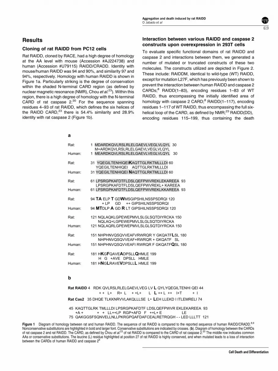
Rat RAIDD, cloned by RACE, had a high degree of homologyat the AA level with mouse (Accession #AJ224738) andhuman (Accession #U79115) RAIDD/CRADD. Identity withmouse/human RAIDD was 94 and 90%, and similarity 97 and94%, respectively. Homology with human RAIDD is shown inFigure 1a. Particularly striking is the degree of conservationwithin the shaded N-terminal CARD region (as defined bynuclear magnetic resonance (NMR), Chou et al.23). Within thisregion, there is a high degree of homology with the N-terminalCARD of rat caspase 2.20 For the sequence spanningresidues 4–93 of rat RAIDD, which defines the six helices ofthe RAIDD CARD,23 there is 54.4% similarity and 28.9%identity with rat caspase 2 (Figure 1b).
Interaction between various RAIDD and caspase 2constructs upon overexpression in 293T cells
To evaluate specific functional domains of rat RAIDD and
caspase 2 and interactions between them, we generated a
number of mutated or truncated constructs of these two
molecules. The constructs utilized are depicted in Figure 2.
These include: RAIDDM, identical to wild-type (WT) RAIDD,
except for mutation L27F, which has previously been shown to
prevent the interaction between human RAIDD and caspase 2
CARDs;8 RAIDD(1–83), encoding residues 1–83 of WT
RAIDD, thus encompassing the initially identified area of
homology with caspase 2 CARD;8 RAIDD(1–117), encoding
residues 1–117 of WT RAIDD, thus encompassing the full six-
helical loop of the CARD, as defined by NMR;23 RAIDD(DD),
encoding residues 115–199, thus containing the death
a
Rat: 1 MDARDKQVLRSLRLELGAEVLVEGLVLQYL 30
M+ARDKQVLRSLRLELGAEVLVEGLVLQYL
Human: 1 MEARDKQVLRSLRLELGAEVLVEGLVLQYL 30
Rat: 31 YQEGILTENHIQEIKAQTTGLRKTMLLLDI 60
YQEGILTENHIQEI AQTTGLRKTMLLLDI
Human: 31 YQEGILTENHIQEI NAQTTGLRKTMLLLDI 60
Rat: 61 LPSRGPKAFDTFLDSLQEFPWVREKLEKAREEA 93
LPSRGPKAFDTFLDSLQEFPWVREKL+ KAREEA
Human: 61 LPSRGPKAFDTFLDSLQEFPWVREKLKKAREEA 93
Rat: 94 TA ELP T GDWMSGIPSHILNSSPSDRQI 120
+ LP GD ++ GIPSHILNSSPSDRQI
Human: 94 MTDLP A GD R LT GIPSHILNSSPSDRQI 120
Rat: 121 NQLAQKLGPEWEPMVLSLGLSQTDIYRCKA 150
NQLAQ+LGPEWEPMVLSLGLSQTDIYRCKA
Human: 121 NQLAQRLGPEWEPMVLSLGLSQTDIYRCKA 150
Rat: 151 NHPHNVQSQVVEAFVRWRQR Y GKQATFLSL 180
NHPHNVQSQVVEAF+RWRQR + GKQATF SL
Human: 151 NHPHNVQSQVVEAFI RWRQR F GKQATFQSL 180
Rat: 181 HKGFQAVEADPSLLQHMLE 199
H G +AVE DPSLL HMLE
Human: 181 HNGLRAVEVDPSLLL HMLE 199
b
Rat RAIDD 4 RDK QVLRSLRLELGAEVLVEG LV L QYLYQEGILTENHI QEI 44
+ + L+ R+ L + +L+ L L ++ L ++ I+T + I
Rat Cas2 35 DHQE TLKKNRVVLAKQLLLSE L- LEH LLEKD I ITLEMRELI 74
45 KAQTTGLRK TMLLLDI LPSRGPKAFDTF LDSLQEFPWVR EKLEKAREEA 93
+A + + + LL++LP RGP+AFD F ++L+ E LE
75 QAKGGSFSQNVELLNLLPKRGPQAFDAFCEALRETRQGH - - LED LLLTT 121
Figure 1 Diagram of homology between rat and human RAIDD. The sequence of rat RAIDD is compared to the reported sequence of human RAIDD/CRADD.8,9
Nonconservative substitutions are highlighted in bold and larger font. Conservative substitutions are indicated by crosses. (b). Diagram of homology between the CARDsof rat caspase 2 and rat RAIDD. The CARD, as defined by Chou et al.23 of rat RAIDD is compared to the CARD of rat caspase 2.20 The middle row indicates commonAAs or conservative substitutions. The leucine (L) residue highlighted at position 27 of rat RAIDD is highly conserved, and when mutated leads to a loss of interactionbetween the CARDs of human RAIDD and caspase 28
Aggregation and death induced by rat RAIDDO Jabado et al
619
Cell Death and Differentiation

domain (DD);8,9 C2(1–135), encoding residues 1–135 of ratcaspase 2, thus encompassing the CARD, but withoutcontaining the full prodomain; C2(1–135M), identical toC2(1–135), except for mutation L57F, which corresponds toresidue L27 of RAIDD (see Figure 1b), and has previouslybeen shown to prevent the interaction between human RAIDDand caspase 2 CARDs.8 All these constructs were Myctagged, except for RAIDD and RAIDDM, which were eitheruntagged or Flag tagged.
We first evaluated the interaction between RAIDD andC2(1–135) or C2(1–135M) by cotransfecting them in 293Tcells, which have the advantage of higher transfectionefficiency. Flag-tagged RAIDD co-immunoprecipitated Myc-tagged C2(1–135), but not C2(1–135 M) (Figure a), consistentwith the results of Duan and Dixit.8 We then evaluatedwhether RAIDDM interacted with C2(1–135). Flag-taggedRAIDDM also co-immunoprecipitated Myc-tagged C2(1–135)(Figure 3b), in apparent contradiction to the results of Duanand Dixit.8 This suggests that the full-length RAIDDM, asopposed to the CARD-containing-only RAIDDM that was usedby Duan and Dixit,8 may assume a different conformation,which allows it to interact with the caspase 2 CARD even in thepresence of the L27F mutation. We also performed immuno-precipitation with the Flag antibody (Ab) after the over-expression of Flag-tagged caspase 9 DN and RAIDD. Nospecific labeling was seen when the blots were probed forRAIDD (data not shown), indicating that, as reportedpreviously,8 there is no physical association between RAIDDand caspase 9.
We conclude that previously observed interactions betweenthe CARDs of human caspase 2 and RAIDD also occur withincells upon overexpression of the rat homologues. However,the specificity of such interactions may be altered in thecontext of the full-length RAIDD molecule.
RAIDD-induced perinuclear aggregate formation
We then analyzed the immunostaining pattern of over-expressed full-length RAIDD in PC12 cells and sympatheticneurons (Figure 4a and b). Cells overexpressing RAIDD wereidentified by a commercial polyclonal Ab raised against anepitope spanning amino acids (AA) 112–126 of human RAIDD(StressGen). When the pCMS-EGFP vector was used foroverexpression, RAIDD immunostaining of PC12 cells andsympathetic neurons was intense only in cells that were alsoEGFP positive (for example, see Figure 4b). The sameRAIDD Ab recognized the expected 23 kDa band uponoverexpression of rat RAIDD in PC12 cells (Figure 4c). Inconjunction, these data indicate that this Ab recognizes
Figure 2 Rat RAIDD and caspase 2 constructs utilized. The gray areas denoteCARDs. The black area in RAIDD denotes the DD. L27 in RAIDD and L57 incaspase 2 are critical residues for CARD interaction.8 C319 represents thecatalytic cysteine on caspase 2. The asterisk on the Flag tag in the RAIDD andRAIDDM constructs indicates that constructs were generated both with andwithout the Flag tag
Figure 3 Interactions between rat RAIDD and caspase 2 constructs uponoverexpression in 293T cells. (a) 293T cells were cotransfected with Flag-taggedRAIDD (FRAIDD) and Myc-tagged C2(1–135) or C2(1–135 M) in equimolarratios. After 24 h, the cells were lysed and immunoprecipitation was performedwith Flag Ab or GAPDH Ab, as an irrelevant (Irr) control (1 mg/ml). Followingrinses, eluted material from original 500mg protein was run on 12% SDS-PAGEgels and immunoblotted with RAIDD (top) or Myc (bottom panel) Abs. Lanes 1, 2and 5 represent lysates (10 mg) of 293T cells, nontransfected 1 or transfected.2,5
Arrowheads denote immunoreactive bands for RAIDD in the upper and Myc inthe bottom panel. Note that RAIDD is immunoprecipitated in both cases (toppanel), whereas only C2(1–135), but not C2(1–135 M), co-immunoprecipitates(bottom panel, compare lane 7 with lane 4). (b) 293T cells were cotransfectedwith Flag-tagged RAIDD or RAIDDM and Myc-tagged C2(1–135) in equimolarratios. Immunoprecipitations were performed as above with Flag or GAPDH Absand Western immunoblots were performed with RAIDD (top) or Myc Abs (bottompanel). The first, second and fifth lanes again denote cell lysates. In both cases,Myc-tagged C2(1–135) co-immunoprecipitates RAIDD (lanes 4 and 7). Eventhough the transfection efficiency is higher for RAIDD, the ratio ofimmunoprecipitated C2(1–135) to RAIDD is equal between the IPs
Aggregation and death induced by rat RAIDDO Jabado et al
620
Cell Death and Differentiation
specifically and reliably overexpressed rat RAIDD. Substan-tially lower levels of endogenous RAIDD were seen byimmunostaining of nontransfected PC12 cells or sympatheticneurons and by Western immunoblot of lysates of untrans-fected PC12 cells (data not shown).
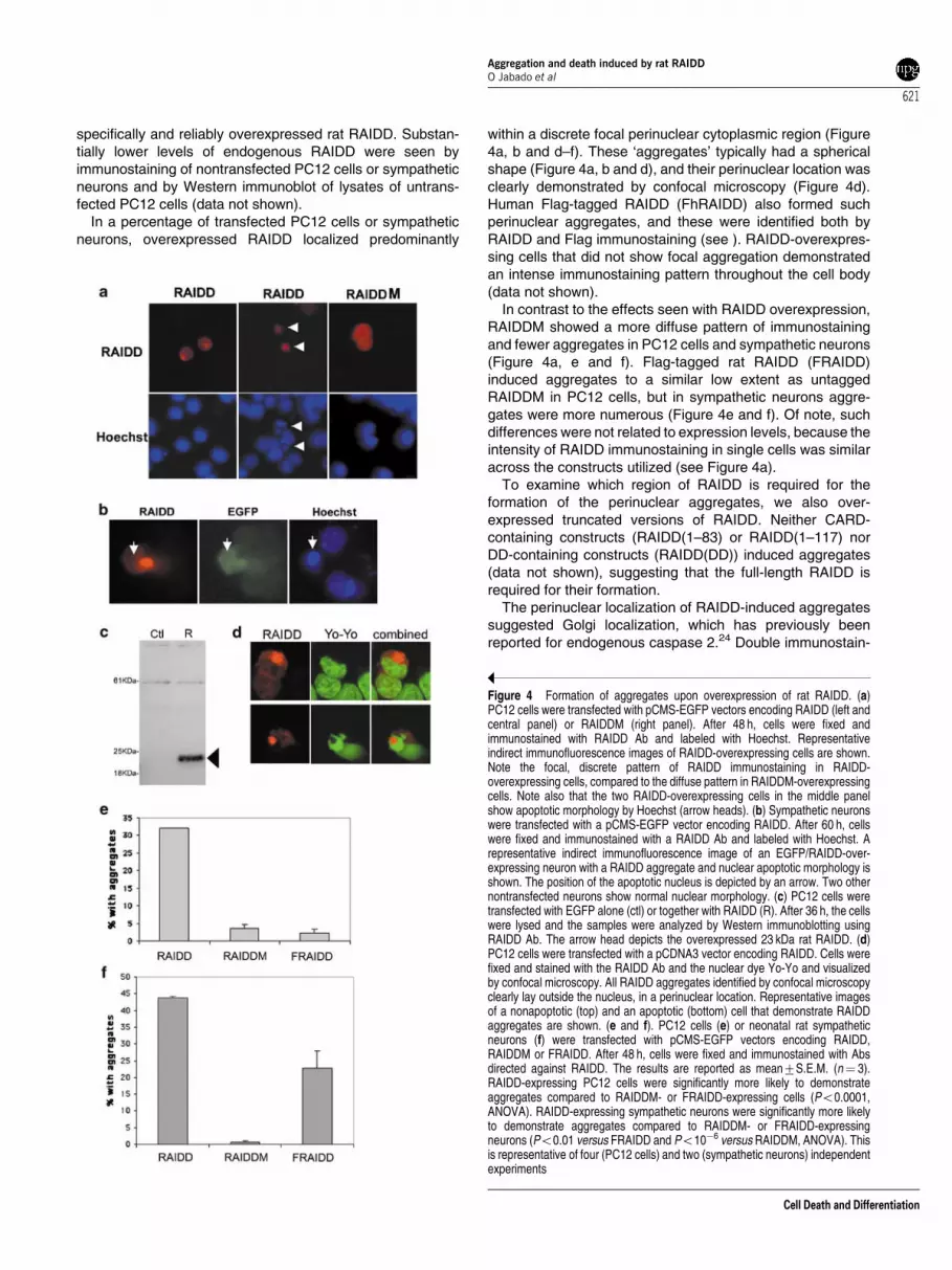
In a percentage of transfected PC12 cells or sympatheticneurons, overexpressed RAIDD localized predominantly
within a discrete focal perinuclear cytoplasmic region (Figure4a, b and d–f). These ‘aggregates’ typically had a sphericalshape (Figure 4a, b and d), and their perinuclear location wasclearly demonstrated by confocal microscopy (Figure 4d).Human Flag-tagged RAIDD (FhRAIDD) also formed suchperinuclear aggregates, and these were identified both byRAIDD and Flag immunostaining (see ). RAIDD-overexpres-sing cells that did not show focal aggregation demonstratedan intense immunostaining pattern throughout the cell body(data not shown).
In contrast to the effects seen with RAIDD overexpression,RAIDDM showed a more diffuse pattern of immunostainingand fewer aggregates in PC12 cells and sympathetic neurons(Figure 4a, e and f). Flag-tagged rat RAIDD (FRAIDD)induced aggregates to a similar low extent as untaggedRAIDDM in PC12 cells, but in sympathetic neurons aggre-gates were more numerous (Figure 4e and f). Of note, suchdifferences were not related to expression levels, because theintensity of RAIDD immunostaining in single cells was similaracross the constructs utilized (see Figure 4a).
To examine which region of RAIDD is required for theformation of the perinuclear aggregates, we also over-expressed truncated versions of RAIDD. Neither CARD-containing constructs (RAIDD(1–83) or RAIDD(1–117) norDD-containing constructs (RAIDD(DD)) induced aggregates(data not shown), suggesting that the full-length RAIDD isrequired for their formation.
The perinuclear localization of RAIDD-induced aggregatessuggested Golgi localization, which has previously beenreported for endogenous caspase 2.24 Double immunostain-
Figure 4 Formation of aggregates upon overexpression of rat RAIDD. (a)PC12 cells were transfected with pCMS-EGFP vectors encoding RAIDD (left andcentral panel) or RAIDDM (right panel). After 48 h, cells were fixed andimmunostained with RAIDD Ab and labeled with Hoechst. Representativeindirect immunofluorescence images of RAIDD-overexpressing cells are shown.Note the focal, discrete pattern of RAIDD immunostaining in RAIDD-overexpressing cells, compared to the diffuse pattern in RAIDDM-overexpressingcells. Note also that the two RAIDD-overexpressing cells in the middle panelshow apoptotic morphology by Hoechst (arrow heads). (b) Sympathetic neuronswere transfected with a pCMS-EGFP vector encoding RAIDD. After 60 h, cellswere fixed and immunostained with a RAIDD Ab and labeled with Hoechst. Arepresentative indirect immunofluorescence image of an EGFP/RAIDD-over-expressing neuron with a RAIDD aggregate and nuclear apoptotic morphology isshown. The position of the apoptotic nucleus is depicted by an arrow. Two othernontransfected neurons show normal nuclear morphology. (c) PC12 cells weretransfected with EGFP alone (ctl) or together with RAIDD (R). After 36 h, the cellswere lysed and the samples were analyzed by Western immunoblotting usingRAIDD Ab. The arrow head depicts the overexpressed 23 kDa rat RAIDD. (d)PC12 cells were transfected with a pCDNA3 vector encoding RAIDD. Cells werefixed and stained with the RAIDD Ab and the nuclear dye Yo-Yo and visualizedby confocal microscopy. All RAIDD aggregates identified by confocal microscopyclearly lay outside the nucleus, in a perinuclear location. Representative imagesof a nonapoptotic (top) and an apoptotic (bottom) cell that demonstrate RAIDDaggregates are shown. (e and f). PC12 cells (e) or neonatal rat sympatheticneurons (f) were transfected with pCMS-EGFP vectors encoding RAIDD,RAIDDM or FRAIDD. After 48 h, cells were fixed and immunostained with Absdirected against RAIDD. The results are reported as mean7S.E.M. (n¼ 3).RAIDD-expressing PC12 cells were significantly more likely to demonstrateaggregates compared to RAIDDM- or FRAIDD-expressing cells (Po0.0001,ANOVA). RAIDD-expressing sympathetic neurons were significantly more likelyto demonstrate aggregates compared to RAIDDM- or FRAIDD-expressingneurons (Po0.01 versus FRAIDD and Po10�6 versus RAIDDM, ANOVA). Thisis representative of four (PC12 cells) and two (sympathetic neurons) independentexperiments
Aggregation and death induced by rat RAIDDO Jabado et al
621
Cell Death and Differentiation

ing with a Golgi marker, GM130, however, and visualizationby confocal microscopy revealed close apposition, but notcolocalization (Figure 5a). The perinuclear localization alsoraised the possibility of an ‘aggresome’, an inclusion formed inthe microtubule organizing center (MTOC) of cells uponoverexpression of misfolded proteins, such as CFTR.25
However, using confocal microscopy, we found little coloca-lization between the MTOC marker g-tubulin and RAIDD-induced aggregates (Figure 5a). Furthermore, there was noincrease in the number of cells with RAIDD-induced aggre-gates upon treatment with the proteasomal inhibitor PSI, orinhibition upon treatment with the microtubule destabilizernocodazole (Figure 5b), as has been reported for aggre-somes.25
We conclude that the full-length rat RAIDD aggregates inperinuclear regions of PC12 cells and sympathetic neurons.The formation of these aggregates is disrupted by the L27Fmutation, and, less so, and depending on the cellular context,by the presence of a Flag tag.
Insolubility of RAIDD aggregates
To further investigate the nature of the RAIDD-inducedaggregates, we transfected PC12 cells with rat RAIDD orRAIDDM, and 48 h later exposed the cultures to a brief periodof detergent extraction, as we have described previously.26
This procedure extracts the soluble components of thecytoplasm, and only relatively insoluble or filamentouscytoplasmic material remains. Following this procedure, thecells were fixed and analyzed for RAIDD immunostaining.Both the low-level endogenous RAIDD immunostaining andthe diffuse immunostaining of overexpressed RAIDD wereabolished. In contrast, the discrete, focal, perinuclear im-munostaining pattern of overexpressed WT RAIDD was stillapparent (Figure 6a). This indicates that RAIDD within theseaggregates has a conformation that renders it resistant todetergent extraction.
To analyze further at the biochemical level this form ofoverexpressed RAIDD, we turned to SH-SY5Y neuroblasto-ma cells, because the higher transfection efficiency in theseneuronal cultures enables more detailed biochemical analy-sis. We first verified that overexpression of the various formsof RAIDD in these cells reproduced the formation ofaggregates, as seen in PC12 cells (data not shown). We thenperformed Western immunoblots of lysates of these cells thathad been transfected with two constructs that induced asubstantial number of aggregates (rat RAIDD, human Flag-tagged RAIDD (FhRAIDD)) or two that did so to a much lesserextent (Flag-tagged rat RAIDDM (FRAIDDM) and Flag-tagged rat RAIDD (FRAIDD)). We separated the lysates indetergent soluble and insoluble, as described previously,26 soas to focus our attention to the pool of RAIDD that is relativelyinsoluble. Although the levels of the various forms of RAIDD inthe detergent-soluble fraction were similar (Figure 6b), therewas a marked increase in detergent-insoluble RAIDD in thecells expressing rat RAIDD or hFRAIDD (Figure 6c). Further-more, an increase in higher molecular weight forms of RAIDD,including forms that did not leave the stacking gel, was seenalmost exclusively in these lysates (Figure 6c).
In conjunction, these results are consistent with the ideathat detergent-insoluble RAIDD accumulates in these aggre-gates.
Apoptosis induced by the overexpression ofRAIDD in PC12 cells and sympathetic neurons
We then examined whether RAIDD overexpression in PC12cells induces apoptotic death, as previously reported for itshuman homologue in other cell types.8,9,14 Overexpression ofWT rat RAIDD induced apoptotic death, albeit variable fromexperiment to experiment and of lower magnitude comparedto previous studies8,9 (Figure 7a and b). The reason for thisvariability is unclear, but may relate to the baseline death in
Figure 5 RAIDD overexpression-induced aggregates/oligomers do not localizeto the Golgi and are not aggresomes. (a). PC12 cells were transfected with apCMS-EGFP (top panel) or a PCDNA3 vector (bottom panel) encoding RAIDD.After 36 h, cells were fixed and immunostained with RAIDD Ab (middle panel)and a monoclonal GM130 Ab to label the Golgi apparatus (top left panel) or amonoclonal g-tubulin Ab to label the MTOC (bottom left pannel, Johnston et al.25).Secondary antibodies used were Cy3 anti-rabbit and Cy5 anti-mouse (top), orCy2 anti-rabbit and Cy3 anti-mouse (bottom). Cells were then visualized under aconfocal microscope. Representative images are shown, with combined imageson the right. Note the lack of colocalization between RAIDD aggregates andGM130 or g-tubulin. The arrow depicts the MTOC and the arrow head a RAIDDaggregate. (b) PC12 cells were transfected with a pCMS-EGFP vector encodingRAIDD. After 24 h, the cell culture medium was replaced with complete mediumwithout additives (nontreated), or containing 5 mM PSI or 10 mg/ml nocodazole.After 7 h, the cells were fixed and immunostained for RAIDD and labeled withHoechst. The percentage of cells bearing aggregates was assessed in eachcondition by indirect immunofluorescence
Aggregation and death induced by rat RAIDDO Jabado et al
622
Cell Death and Differentiation

the cultures (compare apoptosis in EGFP-expressing cells inFigure 7a with that in Figure 7b). Overexpression of Flag-tagged human RAIDD (FhRAIDD),27 kindly provided to us byDrs. Kumar and Shearwin-Whyatt (Adelaide, Australia),induced a similar magnitude of apoptosis in PC12 cells (datanot shown). Interestingly, overexpression of RAIDDM or Flag-tagged rat RAIDD (FRAIDD) induced less apoptotic death(Figure 7a and b). We had similar findings in sympatheticneurons, although in this case FRAIDD induced only slightlylower extent of death compared to RAIDD (Figure 7c).Consistent with the need for an association with the caspase2 CARD for death induction, coexpression of the CARD-containing C2(1–135), which presumably acted as a dominantnegative by preventing the interaction of overexpressedRAIDD with endogenous caspase 2, inhibited RAIDD-induced
death (Figure 7d). Another caspase 2 DN construct, contain-ing a mutation in the catalytic site, also prevented RAIDD-induced apoptosis (data not shown). This death could also beinhibited by the general caspase inhibitor BAF (Figure 7b) orby the coexpression of caspase 9 DN (C9DN, Figure 7d).Expression of truncated forms of rat RAIDD (RAIDD1-83),RAIDD(1–117). RAIDD(DD)) failed to induce apoptosis inPC12 cells or sympathetic neurons (data not shown).
In view of the protective effect of C9DN on RAIDD-induceddeath, we wished to examine further whether elements of the‘intrinsic’ apoptotic pathway were activated following RAIDDoverexpression. We first examined whether there wasevidence of cytochrome c release, a crucial element in such‘intrinsic’ apoptotic pathways. We found that some PC12 cellsthat overexpressed RAIDD showed the loss of mitochondrialcytochrome c staining, similar to what has been observed inother models of neuronal apoptosis (e.g. 7). In contrast, cellsoverexpressing EGFP did not show such loss (Figure 8a). Inaddition, some PC12 cells overexpressing FhRAIDD, but notthose overexpressing EGFP, showed activation of caspase 3,determined by immunostaining with an Ab that specificallyrecognizes activated caspase 3 (Figure 8b). Such results arein agreement with the results of Guo et al.,14 who showed theactivation of elements of the ‘intrinsic’ apoptotic pathwayfollowing human RAIDD overexpression in non-neuronalcells.
We conclude that the full-length rat RAIDD overexpressioninduces apoptotic death of PC12 cells and sympatheticneurons. An association with the caspase 2 CARD isnecessary to induce death, because apoptosis is inhibitedby the coexpression of a DN form of caspase 2 containing onlythe CARD. RAIDD-induced death is also dependent on theactivation of the caspase 9/3 pathway.
Relationship between RAIDD aggregation and celldeath
Given the induction of RAIDD aggregation and cell death uponrat RAIDD or FhRAIDD overexpression, and the relative lackof either of these phenomena upon RAIDDM or FRAIDDoverexpression, we hypothesized that RAIDD aggregationmay be contributing to death. Consistent with this idea, mostcells that showed morphological or biochemical evidence ofactivation of apoptotic pathways expressed RAIDD in anaggregated conformation (see Figures 4 and 8). To look at thiscorrelation in a more quantitative manner, we counted thepercentage of PC12 cells overexpressing nonaggregated oraggregated RAIDD that were apoptotic at baseline, and thenfollowing serum deprivation. We found that in control culturesPC12 cells expressing RAIDD in an aggregated conformationwere 3–4 times more likely to be apoptotic compared to thosethat showed diffuse RAIDD immunostaining. When cultureswere serum deprived, there was further marked potentiationof death in the aggregate-bearing cells (Figure 9a). Overall,there was a 50% potentiation of serum deprivation-induceddeath in RAIDD- compared to EGFP-expressing cells (17.7%serum deprivation-induced apoptosis in EGFP-expressingcells versus 26.4% in RAIDD-expressing cells) (Figure 9a).Interestingly, the percentage of RAIDD-expressing cells
Figure 6 RAIDD-induced aggregates are relatively insoluble. (a) PC12 cellswere transfected with RAIDD, and 2 days later exposed to a detergent buffer for10 min, as described.26,33 The cells were then fixed and immunostained forRAIDD and labeled with Yo-Yo. A representative confocal image is shown,demonstrating the preservation of RAIDD-induced aggregates, whereas thediffuse RAIDD immunostaining is lost. (b and c). SH-SY5Y neuroblastoma cellswere transfected with EGFP (ctl), RAIDD (R), human Flag-tagged RAIDD(FhRAIDD, FhR), Flag-tagged RAIDDM (FRM) or Flag-tagged RAIDD (FR). After2 days, cells were lysed and separated in detergent-soluble (b) and detergent-insoluble components (c). These were run on SDS/PAGE gels andimmunoblotted with anti-RAIDD. Blots were then stripped and immunoblottedwith anti-b-actin. In (b), similar amounts of detergent-soluble RAIDD wereexpressed with the different constructs. Detergent-insoluble RAIDD, however,was substantially higher in cells expressing RAIDD or FhRAIDD (c). The arrowdepicts the end of the stacking gel. The arrow head depicts bands at the top ofthe stacking gel in RAIDD- and FhRAIDD-expressing cell lysates. The asteriskdepicts what is likely a nonspecific band at 60 kDa
Aggregation and death induced by rat RAIDDO Jabado et al
623
Cell Death and Differentiation

bearing aggregates increased about 50% following serumdeprivation (Figure 9a).
To examine whether the formation of aggregates was a lateevent in RAIDD-induced death, which was rather an effect ofthe cellular shrinkage and other alterations that occur inapoptosis, or whether it represented an earlier event with apotential causal role in apoptosis, we performed similarexperiments in the presence of the caspase inhibitor BAF.We reasoned that if BAF prevented the formation ofaggregates, these would be a late event in apoptosis,whereas, if it did not, this would imply that they occurredearly in RAIDD-induced apoptosis. We found that RAIDDoverexpression in the presence of BAF led to an increase inthe percentage of cells bearing aggregates, compared tothose cultures where RAIDD overexpression was performedin the absence of additives (Figure 9b). There was a furthermarked increase of cells bearing aggregates following serumdeprivation in the presence of BAF. Again, as in Figure 9a,there was a substantial increase of aggregation with serumdeprivation alone compared to the control state (Figure 9b).The increase of aggregate formation in BAF-treated culturesis likely due to the fact that cells accumulating aggregates,
which would normally undergo subsequent apoptosis, do notdo so in the presence of caspase inhibition, and thusaccumulate in the cultures.
These results support the idea that aggregation of RAIDDis, at least in part, responsible for its death-promoting effectsin PC12 cells and sympathetic neurons. They further raise thepossibility that some form of RAIDD aggregation may occurphysiologically with trophic deprivation, because, followingthis stimulus, such aggregates increased substantially.
Discussion
In the current paper, we have examined the role of thecaspase 2 interactor RAIDD in mediating PC12 cell andsympathetic neuron apoptosis. As reported in previousstudies with human RAIDD,8,9 the rat homologue inducesapoptosis in these cell culture systems. In agreement with amore recent study,14 overexpression of RAIDD appears toactivate the mitochondrial pathway of apoptosis, as evidencedby cytochrome c release, caspase 3 activation and inhibitionof death by DN caspase 9 or a general pharmacological
Figure 7 RAIDD overexpression induces apoptosis that is dependent on interaction with the caspase 2 CARD and on caspase 9. (a and b). PC12 cells weretransfected with pCMS-EGFP vectors encoding no additional gene (EGFP), RAIDD, RAIDDM or FRAIDD. After 2 days, the cells were fixed and immunostained with Absdirected against RAIDD and labeled with Hoechst. The percentage of EGFP- or RAIDD-positive cells showing nuclear apoptotic morphology was assessed. (a) and (b)are representative experiments, and demonstrate the variability in RAIDD-induced apoptosis. Results are reported as mean7S.E.M. (n¼ 3). In (a) and (b), RAIDDinduced significantly more apoptotic death compared to EGFP, RAIDDM or FRAIDD (Po0.001, ANOVA). In (b), BAF (50 mM, Enzyme Systems Products), applied atthe time of replenishing the medium after transfection, significantly protected against RAIDD-induced death (Po0.0005, ANOVA). (c) Neonatal rat sympathetic neuronswere cultured for 4 days and then were transfected with pCMS-EGFP vectors encoding no additional gene, RAIDD, RAIDDM or FRAIDD. After 2 days, the cells werefixed and immunostained with Abs directed against RAIDD and labeled with Hoechst. Results are reported as mean7S.E.M. (n¼ 4). RAIDD induced significantly moredeath compared to EGFP or RAIDDM (Po0.05, ANOVA). There was no significant difference between the death induced by FRAIDD and that induced by the otherconstructs. This is representative of two independent experiments. (d) PC12 cells were transfected with a pCMS-EGFP vector encoding EGFP alone or cotransfected, ina 1 : 3 ratio, with a PCDNA3 vector encoding RAIDD and pCMS-EGFP vectors encoding Myc-tagged PAI2MT, Myc-tagged C2(1–135) or Flag-tagged caspase 9 DN(C9DN). After 48 h, the cells were fixed and immunostained for Myc (RAIDDþ PAI2MT and RAIDDþC2(1–135)) or Flag (RAIDDþC9DN) and labeled with Hoechst.The percentage of EGFP (for EGFP alone), Myc (for RAIDDþ PAI2MT and RAIDDþC2(1–135)) and Flag (for RAIDDþC9DN)-positive cells that showed features ofnuclear apoptosis was assessed. Results are reported as mean7S.E.M. (n¼ 3). There was significantly more apoptosis in cells expressing PAI2MT compared to allother conditions (Po0.05, ANOVA)
Aggregation and death induced by rat RAIDDO Jabado et al
624
Cell Death and Differentiation

caspase inhibitor. In addition, C2(1–135), a truncated form ofcaspase 2, containing the RAIDD-binding CARD domain,blocked RAIDD-induced apoptotic death. It should be notedthat this DN approach is very specific, because the CARD ofcaspase 2 that comprises this construct has no appreciablehomology with other caspases. Therefore, C2(1–135) inhibitsonly signaling related to caspase 2, and not other caspases,and prevents the association between overexpressed RAIDDand endogenous caspase 2. As RAIDD interacts directly withcaspase 2, but not caspase 9, we propose a model in which
following RAIDD overexpression there is initial RAIDD/caspase 2 interaction, which can be blocked by thecoexpression of C2(1–135). Subsequently, there is theactivation of the mitochondrial pathway of apoptosis (seeschematic diagram of apoptotic pathway induced by RAIDDoverexpression in Figure 10). Such a sequence of events hasbeen recently shown with caspase 2 overexpression.13,14 Weshould note that, based on our studies, we cannot exclude thepossibility that, in addition to apoptosis, other forms of deathoccur with RAIDD overexpression.
Figure 8 RAIDD overexpression induces loss of mitochondrial cytochrome cand caspase 3 activation. (a) A pCMS-EGFP vector encoding EGFP alone or aPCDNA3 vector encoding RAIDD were transfected into PC12 cells. After 48 h,the cells were fixed and immunostained for cytochrome c alone (top panel) or forRAIDD and cytochrome c (bottom panel) and counterstained with Hoechst. A celloverexpressing RAIDD in an aggregated conformation (left panel, depicted by anarrow head) that has lost cytochrome c staining (middle panel) and showsnuclear apoptosis (right panel), and another cell with diffuse RAIDDimmunostaining without the loss of cytochrome c staining or apoptoticmorphology (arrow) are shown in the bottom panel. As a control, a representativeimage of an EGFP-overexpressing cell with maintained cytochrome c staining(arrow head) is shown in the top panel. (b) A pCMS-EGFP vector encodingEGFP alone or a pCNX2 vector encoding Flag-tagged human RAIDD (FhRAIDD)were transfected into PC12 cells. After 48 h, the cells were fixed, immunostainedfor Flag and the active form of caspase 3 and labeled with Hoechst. Tworepresentative indirect immunofluorescence images of RAIDD-overexpressingcells with positive activated caspase 3 are shown in the middle and lower panel.The cell indicated by the arrow head in the middle panel is nonapoptotic, whereasthe one in the lower one is apoptotic. Note the focal perinuclear staining pattern ofFlag. A representative image of an EGFP-overexpressing cell (arrow head)showing the absence of activated caspase 3 is shown in the top panel
Figure 9 RAIDD-induced aggregate formation facilitates serum deprivation-induced PC12 cell apoptosis. (a) PC12 cells were transfected with a pCMS-EGFP vector encoding EGFP alone or, in addition, RAIDD. After 24 h, the cellswere either deprived of serum (S.D.) or maintained in serum (ctl). After 24 h, thecells were fixed and immunostained with RAIDD Ab and labeled with Hoechst.More than 300 EGFP- or RAIDD-overexpressing cells were counted in twoindividual wells for each condition. In the case of RAIDD-expressing cells, thesewere analyzed, in addition to apoptosis, for the presence or absence ofaggregates (agg). The results are reported as mean7S.E.M. (n¼ 2). RAIDD-expressing cells with aggregates were significantly more likely to be apoptoticcompared to those without aggregates, both at baseline and following serumdeprivation (Po0.0005, ANOVA). Similar results were achieved in threeindependent experiments. (b) PC12 cells were transfected with a pCMS-EGFPvector encoding RAIDD. After 24 h, the cells were either deprived of serum (S.D.)or maintained in serum (ctl) in the presence or absence of BAF. After 24 h, thecells were fixed and immunostained with RAIDD Ab and labeled with Hoechst.More than 150 RAIDD-overexpressing cells were counted in two individual wellsfor each condition. The results are reported as mean 7S.E.M. (n¼ 2). Thepresence of BAF led to a significant increase in the percentage of cells bearingaggregates both in the serum-containing (Po0.05, Student’s t-test) and serum-deprived state (Po0.005, Student’s t-test). Serum deprivation alone led to asignificant increase in aggregate formation compared to the ctl state (Po0.05,Student’s t-test). Similar results were achieved in two independent experiments
Aggregation and death induced by rat RAIDDO Jabado et al
625
Cell Death and Differentiation

How could overexpressed RAIDD interact with endogenouscaspase 2 in order to begin to activate this pathway in PC12cells and sympathetic neurons? An unexpected clue hascome from our observation that even though two forms of ratRAIDD (RAIDD and RAIDDM) interact to the same extent withthe caspase 2 CARD when they are overexpressed in 293Tcells (Figure 3b), they have significant differences in theirability to induce death in PC12 cells or sympathetic neurons.The extent of death correlates with the formation of RAIDDaggregates, which are much more frequent with RAIDD, asopposed to RAIDDM, overexpression. Similarly, even thoughboth Flag-tagged human RAIDD (FhRAIDD) and Flag-taggedrat RAIDD (FRAIDD) both may interact with the caspase 2CARD (27, Figure 3a), only the human form, which formsaggregates in PC12 cells, induces substantial cell death. Thedifference between these two C-terminal Flag-tagged RAIDDconstructs may be related to the divergence of the rat andhuman isoforms at the C-terminus (see Figure 1a) that,combined with the highly charged nature of the Flag tag, mayalter the tertiary structure of RAIDD and its ability to fold uponitself. Truncated forms of rat RAIDD, which fail to induceaggregates, also fail to induce death in PC12 cells orsympathetic neurons. Therefore, in both PC12 cells andsympathetic neurons, there is a direct correlation between theability of a RAIDD construct to form perinuclear aggregates
and to induce cell death (see Table 1). In addition, aggregate-bearing PC12 cells are much more likely to show nuclear andbiochemical features of apoptosis compared to cells showingdiffuse RAIDD immunostaining or those expressing EGFP,both at baseline and following serum deprivation (see Figures4, 8 and 9). All these results point to a tight link betweenRAIDD aggregation and apoptosis.
The correlation between aggregate formation and apopto-sis could signify that aggregate formation is the consequence,rather than the cause, of the apoptotic process. However, anumber of observations suggest that this is not the case.Many aggregate-bearing cells did not show signs of nuclearapoptosis (see Figure 4a, left panel, Figure 4d, top panel, andFigure 9a; in the latter, in the presence of serum, more than60% of cells with aggregates were nonapoptotic), indicatingthat formation of aggregates is an earlier event. When RAIDDoverexpression was performed in the presence of BAF, therewas a significant increase in the percentage of aggregate-bearing cells, especially in the serum-deprived state, suggest-ing that cells with aggregates accumulate if apoptosis isinhibited. It appears therefore that aggregate formationprecedes apoptosis and, in view of the tight correlationbetween the two phenomena mentioned above, plays acausative role in the death pathway. We hypothesize thatthese aggregates enable the recruitment of endogenouscaspase 2 in an area of close proximity, leading to caspase 2activation, similar to what has previously been proposed forthe activation of caspase 9 following Apaf-1 oligomerization.28
Owing to the potential crucial role of RAIDD aggregateformation in the propagation of apoptosis, we have investi-gated in more detail the nature of these aggregates. Despitetheir perinuclear localization, we have found no evidence thatthey are contained within Golgi, or that they representaggresomes, as defined by Johnston et al.25 The presenceof such aggregates therefore raises a note of caution, andsuggests that not all perinuclear inclusion-like structures thatare formed following overexpression of certain proteins areaggresomes. These structures may be a reflection of theability of RAIDD to oligomerize, or may represent complexesof RAIDD with other proteins. Truncated RAIDD constructs,containing part or the full extent of the CARD or the DD, did notform such aggregates. This suggests that both the CARD andthe DD of rat RAIDD are required for aggregate formation.
Our data contrast with those previously reported byShearwin-Whyatt et al.27 In that study, full-length human
Figure 10 Schematic diagram of RAIDD overexpression-induced death
Table 1 Summary of the effects of the expression of various RAIDD constructs
PC12 cells Sympathetic neurons SH-SY5Y cells
Construct Aggregation Cell death Aggregation Cell death Aggregation
RAIDD ++ + ++ + ++RAIDDM � 7 � � NTFRAIDD � 7 + 7 �FRAIDDM � NT NT NT �RAIDD(1–83) � � � � NTRAIDD(1–117) � � � � NTRAIDD(DD) � � � � NTFhRAIDD ++ + ++ NT ++
++, marked effects; +, moderate; 7, variable effects; –, no effect. NT¼ not tested. Cell death assessments were not performed in SH-SY5Y cells. Note that in everycase there is a correlation between the ability of a construct to induce aggregates and to cause death
Aggregation and death induced by rat RAIDDO Jabado et al
626
Cell Death and Differentiation

RAIDD did not induce aggregates, but rather a diffusedistribution of RAIDD, whereas CARD-containing truncatedRAIDD formed filaments.27 In addition to full-length ratRAIDD, we have used the same human RAIDD construct asin that study (FhRAIDD), and have found that in the neuronalcell types we have studied it induces the same perinuclearaggregates as the rat isoform. It is likely that, at least in part,the differences in the results obtained reflect differences in thecell types studied. Our results, showing that full-length RAIDDhas the ability to form aggregates in neuronal cells, providenovel insights into the way in which RAIDD may participate inneuronal apoptotic pathways.
A number of other CARD- or death effector domain (DED)-containing proteins have been shown to oligomerize and toform filamentous structures in cells upon overexpres-sion.27,29,30 In many cases such structures are associatedwith the induction of cell death.29 The perinuclear location andthe spherical nature of the RAIDD-induced aggregatesobserved in our study differentiate them from other CARD-induced structures, which have a filamentous, thread-likestructure.27,29,30 Our results indicate an association betweenthe perinuclear aggregates and the death induced by RAIDD.Whether such structures are formed during trophic depriva-tion-induced apoptosis is not known, but their possiblephysiological relevance is supported by the findings thatRAIDD overexpression acts synergistically with serum depri-vation to induce death of PC12 cells, and that the aggregatesthemselves increase substantially upon trophic deprivation.This increase is even more marked in the presence of BAF,indicating that it is not a consequence of trophic factordeprivation-induced apoptosis, but rather an earlier event,with a possible causal role in this type of death (see Figures 9aand 9b). The fact that BAF did not prevent aggregateformation in the control or serum-deprived state indicatesthat these aggregates are not dependent on caspaseactivation for their formation. Thus, if they are formedphysiologically during trophic deprivation, this is likely tooccur upstream of caspase activation.
In conclusion, we show here that overexpression of RAIDDin PC12 cells and sympathetic neurons induces cell death thatis dependent on the caspase 2 CARD and activation of thecaspase 9/3 cascade. The formation of RAIDD perinuclearaggregates facilitates apoptosis, likely by bringing caspase 2molecules in close proximity, leading to their activation. Theinduction of such RAIDD overexpression-induced aggregateswith trophic deprivation and the acceleration of trophic factordeprivation-induced death with RAIDD overexpression sug-gest that RAIDD, through the formation of aggregates/oligomers, may participate in this apoptotic pathway.
Materials and Methods
Cloning of rat RAIDD/CRADD
We extracted total RNA from PC12 cells using the Trizol reagent andperformed RT-PCR (Roche) to generate PC12 cDNA. We then used anested PCR approach to generate a fragment of rat RAIDD. For the firstPCR reaction, we used primers RAIDD51 (50-GCAGATTAAC-CAGCTGGC-30) and RAIDD31 (50-ACAGCCCGCAGCCCGTTGTG-30),and for the second one primers RAIDD52 (50-CTGTCT-
CTGGGACTGTCCCA-30) and RAIDD32 (50-GGAAGGTGGCCTGC-TTCCCG-30). These sequences were selected because they showed ahigh degree of homology between the published human (Accession#U79115) and mouse (Accession #AJ224738) sequences. The nestedPCR reaction resulted in a product of about 130 base pairs (bps),corresponding to bps 506–634 of the human RAIDD sequence. The PCRproduct was cloned into a TA cloning vector (Invitrogen) and sequenced.Based on the sequence, two primers (RAIDDR5: 50-AAGGCCAAC-CATCCCCACAA-30, and RAIDDR3: 50-AAAGGCCTCCACCACCTGC-GA-30) were constructed and used for 30 and 50 prime RACE (Clontech)respectively, as per the manufacturer’s protocol. This resulted in theidentification of an upstream initiation ATG site and a downstream stopTGA site matching the ones in human and mouse RAIDD. Based on theidentified sequences, we constructed two primers, RAID55 (50-GACGGA-GAAATGGATGCCAGA-30) and RAIDDF (50-GGTCTGGTCACTCCAG-CATGT-30), spanning the open-reading frame (ORF) of rat RAIDD,and performed PCR with PC12 cDNA as template (941C� 5 min�1 cycle, (941C� 1 minþ 581C� 1 minþ 721C� 1 min)� 35 cycles,721C� 10 min� 1 cycle). The approximately 600 bp PCR product wascloned into TA and sequenced. For expression studies, rat RAIDD wassubcloned into the EcoRI site of pCMS-EGFP (Clontech) or pcDNA3(Invitrogen), and orientation was verified by sequencing.
Other RAIDD constructs
Various RAIDD constructs were generated by PCR, with full-length RAIDDas the template.
Mutant RAIDD (RAIDDM), with a substitution of residue 27 (L27F), wasgenerated by PCR-based site-directed mutagenesis, cloned into TA, andsubcloned into the EcoRI site of pCMS-EGFP.
C-terminal Flag-tagged versions of RAIDD and RAIDDM weregenerated by performing PCR with primers RAIDDFlag5 (50-CGGTCAC-CATGGATGCCAGAGACAAG-30) and RAIDDFlag3 (CGGTGACCT-CATTTGTCATCATCGTCCTTGTAGTCCATCTCCAGCATGTGCTG-GAG-30), cloning into TA, and subcloning into the EcoRI site of pCMS-EGFP.
RAIDD(1–83) (truncated rat RAIDD encompassing residues 1–83) wasgenerated by PCR, using primers RAIDD5myc (50-TGACGCGTGAAATG-GATGCCAGAGAC-30) and RAIDD53 (50-AGTCATCTTACCCAGG-GAAATTC-30), and subcloned into the MluI–XbaI sites of a modifiedpCMS-EGFP vector containing an in-frame Myc sequence upstream of theMluI site. Another amino-terminal RAIDD construct, RAIDD(1–117),encompassing AA 1–117, was similarly constructed using primersRAIDD5myc and RONup3 (50-GCTCTAGATCAGTCTGATGGG-GAGCTGTT-30). RAIDD(DD) (truncated rat RAIDD encompassing theDD (residues 115–199)) was generated by PCR, using primers RAIDDT35(50-TGACGCGTCCATCAGACCGGCAGATTAACC-30) and RAIDDF, andsimilarly subcloned into Myc-containing pCMS-EGFP. Sequencing wasperformed for all PCR-generated products.
A pCXN2 vector encoding carboxy-terminal Flag-tagged human RAIDD(FhRAIDD, 27) was kindly provided to us by Drs. Sharad Kumar andShearwin-Whyatt (Adelaide, Australia).
Caspase constructs
C2(1–135), a N-terminal myc-tagged construct encompassing the CARDof caspase 2 (AA 1–135) was generated by PCR, using rat caspase 2(Stefanis et al.20) as a template, and primers NEDMyc5 (50-TGACGCG-TAGGATGGCGGCGTCGAGCG-30) and NEDT3A (50-TCATGTGCTGGA-TATCAGAGA-30). The product was cloned into TA and subcloned into the
Aggregation and death induced by rat RAIDDO Jabado et al
627
Cell Death and Differentiation
MluI–XbaI sites of Myc-containing pCMS-EGFP. C2(1–135M), identical toC2(1–135), except for an AA substitution at residue 57 (L57F), wasgenerated by PCR-based site-directed mutagenesis using externalprimers NEDMyc5 and NEDT3A and rat caspase 2 as template,and similarly subcloned into Myc-containing pCMS-EGFP. Bothconstructs were subsequently subcloned into the EcoRI–XbaI sites ofpcDNA3.
Flag-tagged Caspase 9 DN (C327A) in pcDNA3 was generated andsubcloned into pCMS-EGFP or pcDNA3 as described previously.31 Incertain experiments we also used a pCMS-EGFP vector encoding Myc-PAI2MT, to control for the effects of overexpression of an additionalprotein apart from EGFP. PAI2MT is a serpin that lacks the catalytic site,and is therefore not expected to have biological activity and, in particular,to affect the apoptotic pathway. In multiple experiments there was nosignificant difference between the death induced in PAI2MT/EGFP-expressing cells compared to those expressing EGFP alone. We obtaineda pEUK vector encoding PAI-2 from Dr. Philip Bird (Victoria, Australia).PAI2MT, with a mutation at the catalytic site (R380A), was generated byPCR-based site-directed mutagenesis and subcloned into the MluI–NotIsites of the Myc-containing pCMS-EGFP vector.
Cell culture
PC12 cells were grown as described previously,32,20,22 at 7.5%. CO2 onrat-tail collagen-coated dishes in RPMI 1640 medium (Life Sciences)containing 1% penicillin/streptomycin (P/S), 5% fetal calf serum (FCS) and10% heat-inactivated horse serum (HS) (complete medium). In certainexperiments PC12 cells were grown on glass coverslips (CarolinaBiological Supplies). Sympathetic neuron cultures were derived fromsympathetic ganglia of 1- to 2-day-old rat pups.18,20 Followingtrypsinization, the ganglia were plated on 24-well collagen-coated dishes,at 0.5–1 ganglia/dish, at 5% CO2 in RPMI 1640 medium containing 1% P/S, 5% HS and 100 ng/ml human recombinant NGF (generously providedby Genentech), or mouse NGF (Upstate Biotechnology). At 1 day afterplating uridine and 5-fluorodeoxyuridine (10 mM each) were added. 293Tcells and SH-SY5Y neuroblastoma cells were grown at 5% CO2 in 100 mmdishes in DMEM (Life Sciences) with 1% P/S and 10% FCS.
Transfection
Transient overexpression of the various genes of interest in PC12 cells orof 293T and SH-SY5Y cells was achieved by Lipofectamine-mediatedtransfection (Lipofectamine 2000, Life Sciences), following the manufac-turer’s recommendations. For sympathetic neuron transfection, 3–5 daysafter plating we combined 3 mg of each DNA in 30ml of Opti-MEM (LifeSciences) with 4.8 ml of Lipofectamine 2000 (Life Sciences) in 150 ml Opti-MEM. After 1–2 h, we combined the DNA-Lipofectamine mixture with900 ml of Opti-MEM with 100 ng/ml NGF, and applied the total volume of1080 ml to six wells in a 24-well dish (180 ml/well). The cultures were left for4–12 h in this transfection medium, and then this was replaced by thestandard RPMI medium.
Trophic deprivation
PC12 cells were mechanically dissociated from 100 mm or 35 mm dishesafter five rinses with serum-free RPMI 1640 medium and washed with thesame medium 3–4 times by centrifugation and resuspension. PC12 cellswere then replated in collagen-coated 24-well or 35 mm dishes.20,22
Immunofluorescence
PC12 cells or sympathetic neurons were fixed in freshly prepared 3.7%formaldehyde for 25 min at 41C, and then incubated with 10% normal goatserum with 0.4% Triton X-100 to block nonspecific binding, followed byincubation with the primary Ab for 1 h at room temperature. Specificantibodies used were: anti-RAIDD (rabbit polyclonal, StressGen AAP-270,1 : 150), anti-Myc (9E10 mouse monoclonal, derived from a mousehybridoma cell line, 1 : 1), anti-Flag (mouse monoclonal M2, Sigma,1 : 400), anti-GM130 (mouse monoclonal, Transduction Laboratories,1 : 100), anti-active caspase 3 (rabbit polyclonal, Cell Signaling, 1 : 50),anti-cytochrome c (mouse monoclonal, Pharmingen, 1 : 500) and anti-g-tubulin (mouse monoclonal, Sigma, 1 : 400). Following incubation withfluorescent secondary antibodies (Cy2, 1 : 100; or Cy3, 1 : 250, JacksonImmunoResearch), cells were rinsed in PBS. In some cases, Hoechst33342 (Sigma, 1 mg/ml) or Yo-Yo (Molecular Probes, 1 : 1500) was addedfor 20 min at RT, followed by rinses in PBS. The cells were then visualizedin an inverted microscope (Leica DM IRB) at � 40. Pictures were taken onslide film using an Olympus SC35 camera, developed, and scanned on aSprint 35 scanner operated by Adobe Photoshop. Cells plated oncoverslips were placed on glass slides and visualized by confocalmicroscopy (Zeiss LSM410), or on an upright fluorescence microscope(Zeiss Axioskop 2 Plus) equipped with a digital Spot camera. Identicalexposure times were used across conditions.
In certain experiments, in order to examine specifically in situ detergent-insoluble components, we incubated the cultures for 10 min in a detergentsolution prior to fixation. This buffer contained 85 mM Pipes, pH 6.94,10 mM EGTA, 1 mM MgCl2, protease inhibitor cocktail (Roche) and 0.1%Triton X-100.26,33 Following incubation in this buffer, cells were rinsed inPBS and fixed and immunostained as above.
Assessment of cell death
Our first approach for assessing survival/death was to use the pCMS-EGFP vector encoding EGFP and the gene of interest under separatepromoters, and count the number of green fluorescent cells over time.However, we discovered that most of the genes of interest were expressedonly in a small to moderate proportion of EGFP-fluorescing cells. We havetherefore used instead staining with the nuclear dye Hoechst incombination with appropriate immunostaining in order to assess apoptoticcell death in cells that express specific proteins. We counted at least 100immunopositive PC12 cells or sympathetic neurons per well, in at leastthree different wells per condition, and evaluated these cells for thepresence of the characteristic features of apoptosis: nuclear shrinkage,condensation and clumping. Results are reported as mean7S.E.M.Statistical comparisons were carried out using one-way ANOVA withNewman–Keuls post hoc comparisons or Student’s t-test.
Preparation of cell lysates for Westernimmunoblotting or immunoprecipitation
For generation of crude soluble cell lysates, PC12 cells were rinsed in coldPBS and then collected in buffer A (25 mM HEPES (pH 7.5), 5 mM EDTA,1 mM EGTA, 5 mM MgCl2, 2 mM DTT, 10 mg/ml each of pepstatin andleupeptin and 1 mM PMSF). The cellular material was left for 20�min onice and then was sonicated on ice. The lysate was centrifuged for 20 min at160 000� g, and the supernatant was used for Western immuno-blotting.20
To separate lysates into detergent-soluble and -insoluble components,cultures were lysed in a low-detergent buffer containing 0.5% Triton X-100,
Aggregation and death induced by rat RAIDDO Jabado et al
628
Cell Death and Differentiation

25 mM HEPES, 5 mM MgCl2, 1 mM EDTA, 1 mM EGTA and proteaseinhibitors (Complete, Roche) on ice for 20 min. Detergent-insolublematerial was pelleted by ultracentrifugation at 100 000� g for 30 min andresuspended in SDS SB containing 5% b-mercaptoethanol and boiled for10 min.
For immunoprecipitation experiments, 293T cells in 100 mm disheswere washed in PBS, and then lysed in RIPA buffer (150 mM NaCl, 1%NP-40, 0.5% deoxycholate, 0.1% SDS, 50 mM Tris, pH 8.0, with proteaseinhibitors (Complete, Roche)).
Protein concentrations were measured using the Bradford assay(BioRad).
Western immunoblotting
Protein (50–100 mg) was separated by SDS-PAGE (12%, unlessspecified) and transferred to nitrocellulose membranes. The membraneswere blocked in 5% nonfat milk for 1 h at room temperature and probedovernight at 41C with antibodies raised against RAIDD (1 : 500) or Myc(1 : 10). Protein bands were visualized with horseradish peroxidase-conjugated secondary antibodies (Pierce) and enhanced chemilumines-cence (Pierce). To control for protein loading, in certain cases themembranes were stripped and reprobed with mouse anti-b-actin (Sigma,1 : 20 000).
Immunoprecipitation
We immunoprecipitated 293T cell lysates (1 mg) with 1mg of anti-Flagmonoclonal Ab or with 1 mg of anti-GAPDH (mouse monoclonal,Chemicon) as a negative control. Following preclearing, we incubatedthe lysates with the antibodies in a rotator for 2 h at 41C. We then added30ml of protein G slurry (Calbiochem) and incubated for an additional 2 h.Pellets were recovered following a quick spin, and washed five times inRIPA buffer. After the last wash, pellets were resuspended in 30–40 ml ofSDS SB without b-mercaptoethanol and antigens were eluted byincubation at 851C for 5 min. Following a quick spin to pellet the beads,the elute was transferred to another tube, and was incubated for 1 h at371C, with the addition of b-mercaptoethanol to denature the proteins.Western immunoblotting was then performed as above.
Acknowledgements
This work was supported by a grant from the March of Dimes Foundation(Grant # FY99-318 to LS). Additional support was provided by a WellcomeBurroughs Career Award in Biomedical Sciences (LS), the Lowenstein,Matheson and Parkinson’s Disease Foundations (LS), the NIH/NINDS(CMT) and the MDA (CMT). We would like to thank Drs. Lloyd Greene,Michael Shelanski, Isabelle Lang-Rollin and David Park for helpfuldiscussions, Drs. Sharad Kumar and Shearwin-Whyatt for the humanRAIDD construct and Dr. Philip Bird for the PAI2 construct.
References
1. Rideout HJ and Stefanis L (2001) Caspase inhibition: a potential therapeuticstrategy in neurological diseases. Histol Histopathol 16: 895–908
2. Troy CM and Salvesen GS (2002) Caspases on the brain. J Neurosci Res 69:145–150
3. Shi Y (2002) Mechanisms of caspase activation and inhibition during apoptosis.Mol Cell 9: 459–470
4. Van de Craen M, Declercq W, Van den brande I, Fiers W and Vandenabeele P(1999) The proteolytic procaspase activation network: an in vitro analysis. CellDeath Differ 6: 1117–1124
5. Slee EA, Harte MT, Kluck RM, Wolf BB, Casiano CA, Newmeyer DD, WangHG, Reed JC, Nicholson DW, Alnemri ES, Green DR and Martin SJ (1999)Ordering the cytochrome c-initiated caspase cascade: hierarchical activation ofcaspases-2, -3, -6, -7, -8, and -10 in a caspase-9-dependent manner. J CellBiol 144: 281–292
6. Li H, Bergeron L, Cryns V, Pasternack MS, Zhu H, Shi L, Greenberg A andYuan J (1997) Activation of caspase-2 in apoptosis. J Biol Chem 272: 21010–21017
7. Keramaris E, Stefanis L, MacLaurin J, Harada N, Takaku K, Ishikawa T, TaketoMM, Robertson GS, Nicholson DW, Slack RS and Park DS (2000) Involvementof caspase 3 in apoptotic death of cortical neurons evoked by DNA damage.Mol Cell Neurosci 15: 368–379
8. Duan H and Dixit VM (1997) RAIDD is a new ‘death’ adaptor molecule. Nature385: 86–89
9. Ahmad M, Srinivasula SM, Wang L, Talanian RV, Litwack G, Fernandes-Alnemri T and Alnemri ES (1997) CRADD, a novel human apoptotic adaptormolecule for caspase-2, and FasL/tumor necrosis factor receptor-interactingprotein RIP. Cancer Res 57: 615–619
10. Koseki T, Inohara N, Chen S and Nunez G (1999) ARC, an inhibitor ofapoptosis expressed in skeletal muscle and heart that interacts selectively withcaspases. Proc Natl Acad Sci USA 95: 5156–5160
11. Dowds TA and Sabban EL (2001) Endogenous and exogenous ARC in serumwithdrawal mediated PC12 cell apoptosis: a new pro-apoptotic role for ARC.Cell Death Differ 8: 640–648
12. Harvey NL, Butt AJ and Kumar S (1997) Functional activation of Nedd2/ICH-1(caspase-2) is an early process in apoptosis. J Biol Chem 272:13134–13139
13. Paroni G, Henderson C, Schneider C and Brancolini C (2001) Caspase-2-induced apoptosis is dependent on caspase-9, but its processing during UV- ortumor necrosis factor-dependent cell death requires caspase-3. J Biol Chem276: 21907–21915
14. Guo Y, Srinivasula SM, Druilhe A, Fernandes-Alnemri T and Alnemri ES (2002)Caspase-2 induces apoptosis by releasing proapoptotic proteins frommitochondria. J Biol Chem 277: 13430–13437
15. Paroni G, Henderson C, Schneider C and Brancolini C (2002) Caspase-2 cantrigger cytochrome C release and apoptosis from the nucleus. J Biol Chem 277:15147–15161
16. Lassus P, Opitz-Araya X and Lazebnik Y (2002) Requirement for caspase-2 instress-induced apoptosis before mitochondrial permeabilization. Science 297:1352–1354
17. Robertson JD, Enoksson M, Suomela M, Zhivotovsky B and Orrenius S (2002)Caspase-2 acts upstream of mitochondria to promote cytochrome crelease during etoposide-induced apoptosis. J Biol Chem 277:29803–29809
18. Troy CM, Stefanis L, Greene LA and Shelanski ML (1997) Nedd2 is required forapoptosis after trophic factor withdrawal, but not superoxide dismutase (SOD1)down-regulation, in sympathetic neurons and PC12 cells. J Neurosci 17: 1911–1918
19. Troy CM, Rabacchi SA, Hohl JB, Angelastro JM, Greene LA and Shelanski ML(2001) Death in the balance: alternative participation of the caspase-2 and -9pathways in neuronal death induced by nerve growth factor deprivation.J Neurosci 21: 5007–5016
20. Stefanis L, Troy CM, Qi H, Shelanski ML and Greene LA (1998) Caspase-2(Nedd-2) processing and death of trophic factor-deprived PC12 cells andsympathetic neurons occur independently of caspase-3 (CPP32)-like activity.J Neurosci 18: 9204–9215
21. Haviv R, Lindenboim L, Yuan J and Stein R (1998) Need for caspase-2in apoptosis of growth-factor-deprived PC12 cells. J Neurosci Res 52:491–497
22. Stefanis L, Park DS, Yan CYI, Farinelli SE, Troy CM, Shelanski ML andGreene LA (1996) Induction of CPP32-like activity in PC12 cells by withdrawalof trophic support: dissociation from apoptosis. J. Biol. Chem. 271: 30663–30671
23. Chou JJ, Matsuo H, Duan H and Wagner G (1998) Solution structure of theRAIDD CARD and model for CARD/CARD interaction in caspase-2 andcaspase-9 recruitment. Cell 94: 171–180
Aggregation and death induced by rat RAIDDO Jabado et al
629
Cell Death and Differentiation
24. Mancini M, Machamer CE, Roy S, Nicholson DW, Thornberry NA,Casciola-Rosen LA and Rosen A (2000) Caspase-2 is localized at theGolgi complex and cleaves golgin-160 during apoptosis. J Cell Biol 149:603–612
25. Johnston JA, Ward CL and Kopito RR (1998) Aggresomes: a cellular responseto misfolded proteins. J Cell Biol 143: 1883–1898
26. Rideout HJ and Stefanis L (2002) Proteasomal inhibition-induced inclusionformation and death in cortical neurons require transcription and ubiquitination.Mol Cell Neurosci 21: 223–238
27. Shearwin-Whyatt LM, Harvey NL and Kumar S (2000) Subcellular localizationand CARD-dependent oligomerization of the death adaptor RAIDD. Cell DeathDiffer 7: 155–165
28. Srinivasula SM, Ahmad M, Fernandes-Alnemri T and Alnemri ES (1998)Autoactivation of procaspase-9 by Apaf-1-mediated oligomerization. Mol Cell 1:949–957
29. Siegel RM, Martin DA, Zheng L, Ng SY, Bertin J, Cohen J and Lenardo MJ(1998) Death-effector filaments: novel cytoplasmic structures that recruitcaspases and trigger apoptosis. J Cell Biol 141: 1243–1253
30. Guiet C and Vito P (2000) Caspase recruitment domain (CARD)-dependentcytoplasmic filaments mediate bcl10-induced NF-kB activation. J Cell Biol 148:1131–1139
31. Angelastro JM, Moon NY, Liu DX, Yang AS, Greene LA and Franke TF (2001)Characterization of a novel isoform of caspase-9 that inhibits apoptosis. J BiolChem 276: 12190–12200
32. Greene LA and Tischler AS (1976) Establishment of a noradrenergic clonal lineof rat adrenal pheochromocytoma cells which respond to nerve growth factor.Proc Natl Acad Sci USA 73: 2424–2428
33. Cappalletti G, Maggioni MG and Maci R (1999) Influence of MPP+ on the stateof tubulin polymerisation in NGF-differentiated PC12 cells. J Neurosci Res 56:28–35
Aggregation and death induced by rat RAIDDO Jabado et al
630
Cell Death and Differentiation