rage recycles at the plasma membrane in s100b secretory vesicles and promotes schwann cells...
TRANSCRIPT

ORIGINAL ARTICLE 60J o u r n a l o fJ o u r n a l o f
CellularPhysiologyCellularPhysiology
RAGE Recycles at the PlasmaMembrane in S100B SecretoryVesicles and Promotes SchwannCells Morphological Changes
LORENA PERRONE,1,2* GIANFRANCO PELUSO,3 AND MARIAROSA AB MELONE41Department of Neurology, University of Michigan, Ann Arbor, Michigan2Department of Anatomy and Cell Biology, Wayne State University, Detroit, Michigan3Institute of Protein Biochemistry, IBP-CNR, Naples, Italy4Department of Neurological Sciences, Second University of Naples, Naples, Italy
RAGE is a multiligand receptor of the immunoglobulin superfamily involved in regeneration of injured peripheral nerve and cell motility.RAGE is implicated in the development of various chronic diseases, such as neurodegenerative disorders, inflammatory responses, anddiabetic complications. The correlation between RAGE endocytic trafficking and RAGE function is still uninvestigated. S100B is one of theligands of RAGE. The molecular mechanisms responsible of S100B translocation in exocytic vesicles are still poorly investigated. In thepresent study we elucidate the role of RAGE endocytic trafficking in promoting S100B secretion in Schwann cells. Here we show thatRAGE-induced secretion of S100B requires phosphorylated caveolin1-dependent endocytosis of RAGE. Endocytosis of RAGE inresponse to ligand binding promotes the fusion of endosomes with S100B-positive secretory vesicles. Src promotes the fusion ofendosomes with S100B-secretory vesicles. Inhibition of src induces RAGE degradation. RAGE-mediated src activation induces cav1phosphorylation and relocalization in the perinuclear compartment. RAGE signaling and recycling are required for S100-induced Schwanncells morphological changes and are inhibited by high-glucose, suggesting a possible link between diabetes and peripheral nerve injury.Indeed, high glucose inhibits RAGE-mediated src activation. Src inhibition blocks RAGE recycling, S100B secretion, and morphologicalchanges. In summary, we identified a novel pathway of vesicular trafficking required for the amplification of RAGE signaling andcytoskeleton dynamics that is potentially involved in the regeneration of injured peripheral nerve.J. Cell. Physiol. 217: 60–71, 2008. � 2008 Wiley-Liss, Inc.
This article includes Supplementary Material available from theauthors upon request or via the Internet at http://www.interscience.wiley.com/jpages/0021-9541/suppmat.
Abbreviations: AGE, advanced glycation endproducts; RAGE,receptor of advanced glycation endproducts; HMGB1, highmobilitygroup B1; IP, immunoprecipitate; PBS, phosphate buffered saline;TX-100, Triton X-100; PFA, parafolmaldeide; BSA, bovine serumalbumin; dyn2, dynamin 2; PP1, pyrazolo pyrimidine-type inhibitor1; PBS-CM, PBS solution containing 1.8 mMCa2þ and 0.5 mMMg2þ;rt, room temperature; HG, high glucose; LG, low glucose; EGF,endothelial growth factor.
Contract grant sponsor: NIH5P60, National Institute of Diabetes &Digestive & Kidney Diseases.Contract grant number: N. DK20572.
*Correspondence to: Lorena Perrone, Department of Anatomyand Cell Biology, Wayne State University, 540 E. Canfield, 8336Scott Hall, Detroit, MI. E-mail: [email protected]
Received 10 December 2007; Accepted 14 March 2008
DOI: 10.1002/jcp.21474
RAGE is a multiligand receptor of the immunoglobulinsuperfamily of cell surface molecules. RAGE ligands includeAGEs, S100/calgranulins, HMGB1, amyloid-b peptides, and thefamily of b-sheets fibrils (Bierhaus et al., 2005). Amplification ofRAGE-mediated signaling is implicated in the development ofsuch chronic diseases as neurodegenerative disorders,inflammatory responses, and diabetic complications (Bierhauset al., 2005; Vincent et al., 2007). RAGE also exerts physiologicalfunctions and its activity is required during regeneration ofinjured peripheral nerves (Rong et al., 2004a,b). Schwann cells(SC) are involved in the repair of nerve fibers (Dobrowsky et al.,2005) and in diabetic neuropathy (Kalichman et al., 1998).However, thus far, no study has addressed RAGE-mediatedfunction in SC.
Although several studies have analyzed RAGE–mediatedsignaling (Bierhaus et al., 2005), the link between RAGEactivation and RAGE endocytic trafficking has been poorlyinvestigated. There is just one study showing that interaction ofthe amyloid-b peptide (Ab) with RAGE in vascular endothelialcells leads to transport of Ab across the blood-brain barrierthereby inducing the expression of pro-inflammatory cytokines(Deane et al., 2003).
The members of the S100 protein family lack the classicalsignal sequence for secretion. Thus, different S100 proteins usedistinct translocation pathways. Translocation of S100B invesicles plays an important role in the assembly of signalingcomplexes that activate specific signaling pathways (Daveyet al., 2001). Interestingly, S100B relocates from cytosol tovesicles and induces cell motility (Mbele et al., 2002). However,the molecular mechanisms responsible for S100B translocationin vesicles have yet to be completely elucidated.
� 2 0 0 8 W I L E Y - L I S S , I N C .
Recent studies have examined the role of RAGE in cellmigration (Reddy et al., 2006; Riuzzi et al., 2006; Chavakis et al.,2007; Dumitriu et al., 2007; Orlova et al., 2007; Yang et al.,2007) and in the dynamics of actin cytoskeleton (Dumitriu et al.,2007). Both cell migration and changes in cell morphologyrequire actin polymerization together with focal adhesion

R A G E R E C Y C L I N G I N D U C E S S 1 0 0 B S E C R E T I O N 61
assembly and disassembly (Kruchten and McNiven, 2006).Dynamins exert a key function in modulating the dynamics ofadhesion during migration and morphological changes, anddynamin is implicated in both clathrin-coated pits-mediated andcaveolae-mediated endocytosis (Kruchten and McNiven,2006). Phosphorylation and dephosphorylation of focaladhesion kinase (Fak) is important for the turnover of focaladhesion and subsequent reorganization of the actincytoskeleton necessary for cellular movement (Kruchten andMcNiven, 2006). By interacting with Fak, dyn2 is necessary forfocal adhesion turnover (Kruchten and McNiven, 2006).Cytoskeletal and morphological changes are important for SCdifferentiation, which leads to myelinization of nervous fibers(Berti et al., 2006).
Given the relevance of endocytic trafficking in thereceptor-mediated response, the role of RAGE and SC in therepair of injured nerves and in the development of neuropathy,and the lack of studies in these areas, we examined theendocytic trafficking of RAGE in SC. We demonstrate thatendocytosis of RAGE in response to ligand binding promotesthe fusion of endosomes with S100B-positive secretoryvesicles. Fusion of endosomes with S100B-secretory vesicles isregulated by src. RAGE activation leads also to translocation ofphosphorylated cav1 in the perinuclear compartment, where itco-localizes with dyn. We also show that RAGE triggeringinduces Fak activation and SC morphological changes.Interestingly, RAGE-induced src activation and SCmorphological changes are inhibited by high glucose (HG)concentrations, thereby suggesting a molecular mechanismwhereby the repair of injured nerves is delayed in diabetes.
Materials and MethodsReagents
Cell culture reagents were purchased from GIBCO BRL(Invitrogen Corporation, Carlsbad, CA). NHS-LC-biotin,NHS-SS-biotin, Neutravidin beads, and HRP-linked streptavidinwere from Pierce Chemical Co. (Rockford, IL). Biotin-conjugatedanti rabbit IgG, methyl-b-cyclodestrin, glutathione, and all otherreagents, unless otherwise stated, were purchased fromSigma–Aldrich (St. Louis, MI). Anti-RAGE-blocking antibody, whichblocks receptor-ligand interaction, was from RD System(Minneapolis, MN, catalog number: AF1179). The src-specificinhibitor pyrazolo pyrimidine-type inhibitor 1 (PP1) (Schindleret al., 1999) was from Biomol (Plymouth Meeting, PA). S100isolated from bovine brain, was obtained from Calbiochem (EMDBiosciences, San Diego, CA, catalog number: 559284).
Biotinylation assay and biotin internalization assay
Primary SC were isolated from sciatic nerves of 3-day-old HarlanSprague–Dawley rats and cultured as described previously (Mikolet al., 2002). The advantage of these cells is that they can be frozenand re-cultured without losing their phenotype. Indeed, these cellsdo not develop replicative senescence during culturing (Mathonet al., 2001). Two different preparations were used for theexperiments described. Various aliquots from two differentpreparations were de-frozen and cultured until passage 5.Twenty-four hours before stimulationwith S100 (20mgml�1), cellswere serum-starved in Schwann cell-defined medium (SCDM)(50% DMEM, 50% Ham’s F12, 10 mgml�1 transferrin, 10 mMputrescine, 20 nM progesterone, 30 nM sodium selenite). Toinvestigate the half-life of surface RAGE, cells were biotinylatedwith NHS-LC biotin and processed as described previously(Zurzolo et al., 1993; Perrone et al., 2005). After biotinylation, cellswere stimulated at 378C with bovine brain-derived S100, awell-defined activator of RAGE (Hofmann et al., 1999; Vincentet al., 2007). Cells were lysed in RIPA buffer. Biotinylated antigenswere pull-down with streptavidin-agarose beads. After boiling the
JOURNAL OF CELLULAR PHYSIOLOGY
beads in Laemmli buffer, supernatants were analyzed bySDS–PAGE and fluorography with preflashed films. To control thetotal amount of RAGE, 1/10 of the supernatant was analyzed byWestern blotting. Densitometry was carried out within the linearrange of the films.
For endocytosis assay, cells were biotinylated using cleavablebiotin: sulphosuccinimidyl-6-(biotinamide) hexanoate(NHS-SS-biotin) as described elsewhere (Perrone et al., 2005).Samples were incubated at 378C for different times in the presenceof bovine brain S100, whereas control filters were kept at 48C.After incubation, cells were lysed in RIPA buffer (10 mM Tris/ClpH 8, 1% TX-100, 0.1% DOC, 0.1% SDS, 140 mMNaCl, and 1 mMPMSF). Biotinylated antigens were pulled-down withstreptavidin-agarose beads. After the beadswere boiled in Laemmlibuffer, supernatants were analyzed by SDS–PAGE andfluorography with preflashed films. To control the total amount ofRAGE, 1/10 of the supernatant was analyzed by Western blotting.Densitometry was carried out within the linear range of the films.
Antibody-mediated internalization assay: receptorclustering-mediated internalization
Antibody-mediated internalization is a technique that has beenextensively applied to investigate the endocytic fate of variousreceptors and membrane-associated proteins (Tiruppathi et al.,1997; Piazza et al., 2005; Millan et al., 2006; Tampellini et al., 2007).
Schwann cells were growth on glass coverslips coated with1 mgml�1 poly-L-lysine and starved in SCDM for 24 h. Rabbitanti-RAGE antibody (1mgml�1) (H-300, Santa Cruz Biotechnology,Inc., Santa Cruz, CA) was added to the SC for 30 min at 48Cin incubation medium (DMEM, 10 mM Hepes pH 7.5, 0.2% BSA).The specificity of this antibody has been previously verified and ithas been employed to detect RAGE subcellular localization invesicles by co-localization experiments and immunofluorescenceanalysis (Hermani et al., 2006). For internalization studies, cellswere fixed either immediately after incubation at 48C (0 min,indicating surface labeled RAGE) or following incubation at 378Cfor various times to induce the internalization of RAGE-antibodycomplex (Tampellini et al., 2007). Indeed, the incubation of the cellsat 48C completely block the endocytic machinery and RAGEligands are not internalized (Schmidt et al., 1993). Briefly, cells werewashed twice with ice cold PBS containing 1.8 mMCa2þ and 0.5 mMMg2þ (PBS/CM) to eliminate the unbound antibody, incubated at378C in SCDM for different times, then washed twice with ice coldPBS/CM and fixed for 20 min with 4% paraformaldehyde (PFA)in PBS. To verify that in the absence of incubation at 378C theantibody labels only surface RAGE, while it is internalized togetherwith RAGE following incubation at 378C, we performed a confocalZ stack analysis (Tampellini et al., 2007). Control cells (0 min) andcells incubated for 5 min at 378C were permeabilized (see below)and the localization of the RAGE-antibody complex andphosphorylated cav1 were investigated by immunofluorescenceand confocal analysis performing Z-stack sections from the bottomof the cells (adhesion of the cells to the cover slips) to the top of thecells. Optical sections were acquired at 0.7 mm thickness(supplemental Figs. S1 and S2). For confocal analysis, cells weretreated for 5 min with PBS/CM containing 0.15% TX-100, blockedwith PBS/CM, 0.05% TX-100 and 1% bovine albumin for 30 min.Cells were incubated at room temperature for 1 h withbiotin-conjugated anti rabbit IgG (1:400) (Sigma–Aldrich, catalognumber: B7389) and the appropriate antibody for the doublestaining: mouse anti-phospho-cav-1 (1:25) (Tyr14 BD; BDTransduction, BD Biosciences, catalog number 611338), mouseanti-Rab11 (1:200) (BD Biosciences. Clone 47, catalog number610656), mouse anti-S100 (1:100) (Chemicon, Millipore, Bilerica,MA, catalog number MAB079-1). Fluorescein isothiocyanate(FITC)-conjugated streptavidin and Texas-Red-conjugatedanti-mouse IgG were used for staining (Molecular Probes,Invitrogen, Carlsbad, CA).

62 P E R R O N E E T A L .
Lysosomes were detected by incubating the cells withLysoTracker Red (1:10,000) (Molecular Probes, Invitrogen, catalognumber L-7528) 30 min before antibody-mediated RAGEinternalization. Specimens were mounted in ProLong goldmounting medium containing DAPI (Invitrogen).
Analysis of immunofluorescence was performed with anOlympus FluoView 500 laser scanning confocal microscope.Samples were visualized with an Olympus IX-71 invertedmicroscope using a 60� oil-immersion lens, magnified two timeswith FluoView version 5.0 software and scanned sequentially tomaximize signal separation. DAPI, AlexaFluor 488 and AlexaFluor594 fluorescence were excited with a 405 nm blue laser diode,488 nm argon blue and 543 nm helium neon laser, respectively.Emissions were separated with 430–460 nm, 505–525 nm, and610þ nm barrier filters.
Immunofluorescence analysis after stimulation withbovine brain S100
To obtain synchronous stimulation of surface RAGE, cells werestimulatedwith bovine brain S100 at 48C for 30min. Unbound S100were washed with ice-cold PBS/CM, and cells were incubated at378C for different times. Cells were fixed in 4% PFA in PBS/CM for20 min. They were then washed twice with PBS/CM and oncewithNH4Cl 5mM in PBS/CM to quench the excess PFA.Cells werepermeabilized 5minwith TX-100 0.15% in PBS/CMand blocked for30 min in PBS/CM containing 0.05% TX-100 and 1% BSA. Cellswere incubated for 1 h at room temperature with the primaryantibody: rabbit anti-dynamin (H-300) (1:100) (SantaCruzBiotechnology, Inc., catalog number sc-11362); mouse anti-phospho cav1 (1:25) (Tyr14 BD Transduction, BD Biosciences,catalog number 611338). They were then washed three times for10 min with PBS/CM and incubated for 1 h at room temperaturewith the fluorochrome-labeled secondary antibody (MolecularProbes, Invitrogen). The morphology of SC was examined underthe same conditions. Immunofluorescence was analyzed with anOlympus FluoView 500 laser scanning confocal microscope asdescribed above.
Treatment with high glucose
To investigate the effect of high glucose on SC morphology, cellswere growth in 50 mM glucose for 24 h, serum-starved in 50 mMglucose for an additional 24 h before stimulation.
Immunoprecipitation and immunoblot analysis
Cells were washed twice with ice-cold PBS/CM and lysed asdescribed elsewhere (Jori et al., 2003). Samples wereimmunoprecipitated with the following antibodies: rabbitanti-cav-1 1:100 (N-20, Santa Cruz Biotechnology, Inc., catalognumber sc-894), rabbit polyclonal anti-RAGE 1:100 (H-300, SantaCruz Biotechnology, Inc., catalog number sc-5563), mouseanti-c-src 1:100 (Upstate/Millipore, catalog number 05-184).
Western blot analysis was carried out with the followingantibodies: mouse anti-phospho-cav-1 1:500 (Tyr14, BDTransduction, catalog number 611338), rabbit anti-cav-1 1:1,000(BD Transduction, catalog number 610406), mouse anti-RAGE1:500 (RD Systems, catalog number MAB1179), mouse anti-S100B1:1,000 (Chemicon, catalog number MAB079-1), HRP-conjugatedstreptavidin 1:5,000 (Pierce Chemical Co., catalog number N200).Rabbit anti-phospho Fak (Santa Cruz Biotechnology, Inc., catalognumber sc-16563-R), anti-Fak (BD Transduction, catalog number610088). Rabbit anti phospho-src (1:1,000) (Calbiochem, EMDBiosciences, catalog number 569373). Mouse anti-c-src 1:1,000(Santa Cruz Biotechnology, Inc., catalog number sc-5266).
To inhibit RAGE-S100 interaction, cells were incubated with ablocking antibody against rat RAGE (25 mg/ml) (RD SystemMinneapolis, MN, catalog number: AF1179) for 30min at 48C.Cellswere then treated with bovine-brain S100 20 mg/ml at 378C.Anti-RAGE blocking antibodies from RD System were previously
JOURNAL OF CELLULAR PHYSIOLOGY
used to inhibit the interaction of RAGE with the ligand (Hermaniet al., 2006; Sakaguchi et al., 2008). The blocking antibody is notsuitable for immunofluorescence analysis as indicated by themanufacturer instruction and a previous report (Hermani et al.,2006).
ResultsRAGE is internalized and recycles at the plasmamembrane after stimulation with S100
To correlate the endocytic trafficking of RAGE with itsphysiological function in SC, we analyzed the endocytictrafficking of endogenous RAGE in primary cultures of SC. TodeterminewhetherRAGE is degradedor recycles to the plasmamembrane after S100 stimulation,we used an endocytosis assaybased on the use of a cleavable biotin analog (Perrone et al.,2005). After biotinylation, SC were treated with bovine brainS100 (20mg/ml), awell-definedRAGE activator (Hofmann et al.,1999; Vincent et al., 2007), for 0, 5, 20, and 60 min in duplicate.The sample treated with glutathione shows whether thereceptor is endocytosed because internalized receptor is notaccessible to glutathione. The sample not treated withglutathione represents the total amount of biotinylatedreceptor during stimulation. After stimulation with S100 for5 and 20 min, biotinylated RAGE was in an endocyticcompartment (Fig. 1A, lanes 3–6). After 60 min of stimulation,biotinylated RAGE was again available for glutathione cleavage,whereas the total amount of biotinylated RAGEwas reduced byonly 20% (Fig. 1A, lanes 7, 8). Thus, about 80%of RAGE recyclesat the plasma membrane after 60 min of S100 stimulation.Biotinylation does not affect RAGE subcellular localization orsignaling either under steady-state conditions or uponstimulation with S100 (data not shown).
We next analyzed the half-life of RAGE with and withoutS100 stimulation. Schwann cells were treated withcycloheximide (CHX, 50 mg/ml) and incubated with andwithout S100. In the absence of S100, RAGE decreased after 3 hof incubation with CHX and was greatly reduced after 16 h(Fig. 1B, lanes 5 and 6). On the contrary, RAGEwas unvaried upto 16 h of incubation with CHX in the presence of S100(Fig. 1B, lanes 1–4).
We monitored the endocytic trafficking of RAGE usingantibody ligation-induced internalization (Tiruppathi et al.,1997; Piazza et al., 2005; Millan et al., 2006; Tampellini et al.,2007). The anti-RAGE antibody used in this assay induces boththe internalization (Fig. 1C) and signaling of RAGE (Fig. 2A, lane4). Cells were incubated 30 min at 48C with anti-RAGEantibody, washed and incubated at 378C for 5 min. InternalizedRAGE was targeted to a perinuclear compartment andco-localized with Rab11 after 5 min of incubation at 378C(Fig. 1C). After stimulation with S100 both internalized RAGEand Rab11 were localized in the perinuclear compartment,whereas in the absence of S100, RAGEwas distributed over thewhole cell surface (Fig. 1C). Internalized RAGE did notco-localize with lysosomes (data not shown). These datademonstrate that RAGE is targeted to the recycling pathwayafter interaction with the ligand.
Src-mediated phosphorylation of cav-1 is required forRAGE recycling at the plasma membrane
Phosphorylated cav-1 is involved in endocytic trafficking(Tiruppathi et al., 1997; del Pozo et al., 2005). Since RAGEactivation results in src-mediated phosphorylation of cav-1 inendothelial smooth muscle cells (Reddy et al., 2006), we soughtto examinewhether stimulation of RAGE induces src-mediatedcav1 phosphorylation in SC. Src was activated after 5 minof treatment with bovine brain S100 (Fig. 2A, lane 2). Srcactivation was RAGE-dependent. Indeed, a RAGE-blockingantibody strongly reduced S100-induced src activation

Fig. 1. RAGE is targeted to the recycling pathway upon stimulation. A: SCwere surface biotinylated withNHS-SS-biotin and then incubated at37-C with bovine brain S100 (20 mgml�1). Biotinylated and total RAGE were detected byWestern blotting. Quantification of threeindependent experiments is shown at the bottom of the figure. B: SC were treated with cycloheximide (15 mg/ml) and S100 (20 mgml�1) asindicated.RAGEwasdetectedbyWesternblotting.Quantificationofthreeindependentexperimentsisshownatthebottomofthefigure.C:RAGEinternalization (green) was induced by antibody-mediated internalization. The localization of internalized RAGE (green) and Rab11 (red) wasevaluated by immunofluorescence and confocal analysis. The scale bar indicates 10mm. These experiments are representative of at least threeindependent experiments. [Color figure can be viewed in the online issue, which is available at www.interscience.wiley.com.]
R A G E R E C Y C L I N G I N D U C E S S 1 0 0 B S E C R E T I O N 63
(Fig. 2A, lane 3). On the contrary, unspecific IgG did not inhibitS100-mediated src phosphorylation (Fig. 2A, lane 5). RAGEtriggering by an antibody against the extracellular domain ofRAGE also induced src phosphorylation, which confirms thatsrc-induction is RAGE-dependent (Fig. 2A, lane 4).
We next investigated whether RAGE activation inducescav-1 phosphorylation. Cav-1 phosphorylation increasedfourfold after 5 min of stimulation with bovine brain S100(Fig. 2B, compare lanes 1 and 2). Cav1 phosphorylation wassrc-dependent. Indeed, the src-specific inhibitor PP1 (Khanet al., 2006) totally abolished S100-mediated cav1phosphorylation (Fig. 2B, lane 3). The antibody against theextracellular domain of RAGE also increased cav1phosphorylation sixfold thereby demonstrating that RAGEtriggering induces cav1 phosphorylation (Fig. 2B, lane 4). Toassess whether phosphorylated cav1 is involved in RAGE
JOURNAL OF CELLULAR PHYSIOLOGY
endocytic trafficking, we investigated the localization ofphosphorylated cav1 and RAGE by antibody ligation-inducedinternalization. RAGEdid not co-localizewith phospho-cav-1 inthe absence of activation at 378C (Fig. 2C),whereas internalizedRAGE co-localized with phospho-cav-1 in a perinuclearcompartment after 5 and 20min of incubation at 378C (Fig. 2C).Phosphorylated cav1 was relocated in the perinuclearcompartment after RAGE activation, whereas it was localizedexclusively at the plasma membrane in the absence of RAGEtriggering (Fig. 2C). RAGE and phosphorylated cav1co-localized in a perinuclear region also after stimulation withbovine brain S100 (data not shown). Confocal Z stack imagesdemonstrate that endocytosis of surface RAGE does not occurat 48C. Indeed, the uptake of anti-RAGE antibody is blockedwhen cells are fixed at 0 min. Following permeabilization of thecells, the staining of RAGE-antibody complex is evident only in

ig. 2. Phosphorylated cav-1 targets RAGE to the recycling pathways. A: SCwere stimulatedwith S100 (20mgml�1) for the indicated timewithndwithout an anti-RAGE blocking antibody (BLAb), anti-RAGE activating antibody, or unspecific IgG. Protein extracts were immunoprecipitatedithamouseanti-srcantibody.PhosphorylatedsrcandsrcweredetectedbyWesternblotting.Quantificationofthreeindependentexperimentsishown at the bottom of the figure. B: SC were stimulated as in (A). Protein extracts were immunoprecipitated with an anti-cav1 antibody.hosphorylatedcav1andcav1weredetectedbyWesternblotting.Quantificationof three independentexperiments is shownatthebottomof thegure. C: Internalization of RAGE (green) was induced by antibody-mediated internalization and incubation at 37-C for the indicated times.ternalized RAGE (green) and phosphorylated cav-1 (red) were detected by immunofluorescence and confocal microscopy. The scale bardicates 10mm.BlAbU anti-Rageblockingantibody. [Colorfigure canbeviewed in theonline issue,which is available atwww.interscience.wiley.
64 P E R R O N E E T A L .
FawsPfiInin
com.]JOURNAL OF CELLULAR PHYSIOLOGY

R A G E R E C Y C L I N G I N D U C E S S 1 0 0 B S E C R E T I O N 65
sections corresponding to the cell surface and does notco-localize with phosphorylated cav1 in any of the Z-scansections (supplemental Fig. S1). Incubation at 378C for 5 minleads to relocation of RAGE-antibody complex to theintracellular compartment, as demonstrated by confocal Z scanimages following permeabilization of the cells. There is noevident staining of RAGE-antibody complex at the plasmamembrane (top sections), while it is localized in an intracellularcompartment where it co-localizes with phosphorylated cav-1(supplemental Fig. S2). In the absence of cell permeabilization,the staining of RAGE-antibody complex is evident at 0 min,while it is strongly reduced following 5 min of incubation at378C (data not shown). Since after incubation at 378C thestaining of RAGE-antibody complex is evident only followingpermeabilization of the cells, we further demonstrate that theuptake of anti-RAGE antibody is blocked at 48Cand that RAGE-antibody complex is internalized following incubation at 378C.
We next investigated whether phosphorylation of cav-1plays a functional role in RAGE endocytic trafficking. To thisaim, cells were pre-treated with 5 mM PP1 for 45 min to inhibitsrc-mediated cav-1 phosphorylation (Khan et al., 2006). Thisexperiment confirmed that RAGE recycles at the plasmamembrane after 60 min of stimulation with bovine brain S100(Fig. 3A, lanes 1–4). Biotinylated RAGE was greatly reduced inthe presence of PP1 and glutathione cleavage (about 85%compared with control time 0 min without glutathionecleavage). It was also greatly reduced in the presence of PP1 andin the absence of glutathione cleavage (about 80% comparedwith control time 0 min), suggesting that src-inhibition inducesRAGE degradation after S100 stimulation. We usedbiotinylation-based degradation assays (Jullien et al., 2002;Tanowitz and Von Zastrow, 2002; Chen et al., 2005; Tampelliniet al., 2007) to determine whether inhibition of src activationchanges the trafficking fate of RAGE. Cells were biotinylatedand stimulated with bovine brain S100 at 378C for differenttimes in the presence and absence of PP1. Without PP1,biotinylatedRAGEwas reduced (by about 30%) after 3 hof S100stimulation. On the contrary, in the presence of PP1,biotinylated RAGE was reduced by about 72% after 1 h of S100stimulation (Fig. 3B). Using antibody-cross linking-mediatedinternalization, we found that RAGE is targeted to lysosomesafter 30min of S100 stimulation in the presence of PP1 (Fig. 3C).
We then examined the localization of internalized RAGE andRab11 after 5 min of antibody-cross linking-mediatedinternalization both with and without PP1-induced srcinhibition. We observed a reduction of RAGE-Rab11co-localization in the presence of PP1 compared to the controlstimulated in the absence of PP1 (Fig. 3D).
Accordingly, the endocytosis assay showed that about20% of biotinylated RAGE recycles at the plasma membrane(Fig. 3A). These data demonstrate that inhibition of src changesthe trafficking fate of internalized RAGE. Indeed, in thepresence of PP1, about 80% of RAGE was targeted fordegradation, whereas only 20% was still targeted to therecycling pathway.
RAGE-induced secretion of S100B requires src activation
Although RAGE induces S100B relocation in vesicles inendothelial cells (Hsieh et al., 2004), the trafficking pathwayresponsible for S100B translocation in vesicles is unknown.Since SC express endogenous S100B, we investigated whetherRAGE triggering affects S100B exocytic trafficking using theantibody-mediated internalization assay. As shown inFigure 4A,RAGEand S100Bdid not co-localize in the absence ofinduction of endocytosis by incubation of the cells at 378C (time0-min in Fig. 4A). Internalized RAGE co-localized with S100B invesicle-like structures in the perinuclear region upon 5 minof internalization at 378C. RAGE and S100B still co-localized
JOURNAL OF CELLULAR PHYSIOLOGY
after 20 and 60 min of internalization and were distributedthroughout the cytoplasm. The relocalization of S100B invesicles was mediated by RAGE triggering. Indeed, in cellstreated with an unspecific IgG and incubated at 378C for 5 min,no vesicles were S100B-positive, whereas S100B appeared inthe cytoplasm.
PP1 inhibited RAGE co-localization with endogenousS100B (Fig. 4B). S100B and internalized RAGEwere localized ina perinuclear compartment after 60 min of RAGE triggering inthe presence of PP1; however, the co-localization was very lowand S100B and RAGE staining was weak (Fig. 4B). S100Brelocation in vesicles is Ca2þ-dependent in astrocytoma cells(Mbele et al., 2002). RAGE triggering in the presence of 10 mMEGTA inhibited S100B co-localization with RAGE (data notshown), which is in agreement with a previous finding (Daveyet al., 2001).
We also investigated the RAGE-S100B association inco-immunoprecipitation experiments using an anti-RAGEantibody. Stimulationwith exogenous bovine brain S100 greatlyincreased the amount of S100B in the immunocomplex (aboutsevenfold induction compared with unstimulated cells)(Fig. 4C, lane 2). Although we do not know whether RAGEco-immunoprecipitates with endogenous or extra-cellularS100B or both, this observation could suggest that RAGE doesnot dissociate from extracellular S100. In agreement, RAGE isresponsible for the transcytosis of the b-amyloid peptide fromthe apical to the basolateral surface in endothelial cells, furtherdemonstrating that RAGE does not dissociate from the ligandduring its endocytic trafficking (Mackic et al., 1998).
Astrocytes secrete S100B consequent to metabolic stress(Gerlach et al., 2006). Since SC express endogenous S100B, wesought to determine whether RAGE activation inducessecretion of endogenous S100B. To this aim, we stimulatedRAGE using antibody-mediated internalization to identifyendogenous S100B in the media, and usedWestern blotting toevaluate S100B secretion after tri-fluoro acetic acid (TCA)precipitation of the culture medium (Pachydaki et al., 2006). Toquantify the cell number stimulated in the different conditions,we measured the amount of actin by Western blot(Fig. 4D, bottom). S100B was not detectable in the media in theabsence of stimulation or after 20 min of RAGE triggering,whereas it was secreted after 60 and 120 min of RAGEtriggering (Fig. 4D, lanes 1–4). S100B secretion was src-dependent andwas completely abolished in the presence of PP1(Fig. 4D, lane 5). Treatment of SCwith PP1 in the presence of anunrelated antibody instead of anti-RAGE antibody did notinduce S100B secretion (Fig. 4D, lane 6), which confirms thatRAGE triggering is required for S100B secretion.
High glucose inhibits RAGE-induced src activation
RAGE plays a key role in the development of diabetes (Bierhauset al., 2005). However, the effect of a HG concentration onRAGE function in SC is unknown. In an attempt to shed light onthis issue, we first analyzed the effect of HGonRAGE-mediatedsrc activation after S100 stimulation. In culture medium with aphysiological glucose concentration (5.5 mM¼ LG), src wasactivated in response to S100 stimulation, whereas 50 mMglucose (HG) inhibited RAGE-mediated src activation to thesame extent as PP1 (Fig. 5A lanes 1–4, and 6). High glucose didnot affect src phosphorylation in the absence of S100stimulation (Fig. 5A, lane 5), neither did it affect cell viability(data not shown). We next investigated Fak phosphorylationafter RAGE triggering. Fak activation was delayed despite srcphosphorylation; it occurred after 10 min of RAGE triggeringand persisted up to 15 min of stimulation (Fig. 5B, lanes 1–4).Inhibition of src activation with PP1 completely abolished Fakphosphorylation (Fig. 5B, lane 5). High glucose also inhibitedRAGE-mediated Fak activation (Fig. 5B, lane 6). It did not affect

Fig. 3. srcactivation isnecessary forRAGErecyclingat theplasmamembrane.A:SCwere incubatedwith src-specific inhibitorPP1.Control andPP1-treatedcellswere surfacebiotinylatedwithNHS-SS-biotinand incubatedat37-CwithS100 (20mgml�1) for the indicated times.BiotinylatedRAGEwasdetectedbyWesternblottingafterpull-downwithavidinbeads (top).TotalRAGEwasanalyzedbyWesternblotting.Quantificationofthree independent experiments is shown at the right of the figure. B: Cells were surface biotinylated with NHS-LC-biotin and incubated at 37-CwithS100 (20mgml�1) for the indicated times.BiotinylatedRAGEwasdetectedbyWesternblottingafterpull-downwithavidinbeads (top).TotalRAGEwasevaluatedbyWesternblotting.Quantificationof three independentexperiments is shownattherightof thefigure.C: InternalizationofRAGE (green) was induced by antibody-mediated internalization. Cells were incubated at 37-C for 30 min with PP1. Control cells were fixedimmediatelyafterantibody-mediatedRAGEinternalization.LysosomesweredetectedbyincubatingthecellswithLysotraker(red).Thescalebarindicates10mm.D: InternalizationofRAGEwascarriedoutas in(C)bothwithandwithoutPP1.The localizationof internalizedRAGE(green)andRab11 (red) was evaluated with immunofluorescence and confocal microscopy. These experiments are representative of at least threeindependent experiments. [Color figure can be viewed in the online issue, which is available at www.interscience.wiley.com.]
JOURNAL OF CELLULAR PHYSIOLOGY

Fig. 4. InternalizedRAGEco-localizeswithendogenousS100B.A,B: InternalizationofRAGEwas inducedbyantibody-mediated internalization.The localizationof internalizedRAGE(green)andS100B(red)wasevaluatedwith immunofluorescenceandconfocalmicroscopy.Ascontrol, cellswere treated with unspecific IgG instead of with anti-RAGE antibody and incubated at 37-C for 5 min (bottom). B: RAGE internalization wascarried out as in (A). Cells were treated with PP1. The localization of internalized RAGE (green) and S100B (red) was evaluated withimmunofluorescenceandconfocalmicroscopy.The scalebar indicates10mm.C:SCwere stimulatedwithS100 for20min.Proteinextractswereimmunoprecipitated using an anti-RAGE antibody. S100B and RAGE were detected byWestern blotting. Quantification of three independentexperiments is shown at the bottom of the figure. D: Internalization of RAGE was induced by antibody-mediated internalization both in thepresence and absence of PP1. As control, cells were incubated with unspecific IgG instead of anti-RAGE antibody and incubated at 37-C in thepresence of PP1. Cells were incubated with fresh medium after antibody-mediated RAGE internalization. The medium was collected at theindicated time andwas precipitated usingTCA. The presence of S100Bwas evaluatedwithWestern blotting. The amount of actinwas evaluatedwith Western blotting as a control of the number of cells stimulated. These experiments are representative of at least three independentexperiments. [Color figure can be viewed in the online issue, which is available at www.interscience.wiley.com.]
R A G E R E C Y C L I N G I N D U C E S S 1 0 0 B S E C R E T I O N 67
Fak phosphorylation in the absence of S100 stimulation(Fig. 5B, lane 7). This is in line with the finding that HG inhibitsFak activation in rat peritoneal mesothelial cells (Tamuraet al., 2003).
JOURNAL OF CELLULAR PHYSIOLOGY
Phosphatidylinositol 3-kinase (PI3K) activation induces Fakphosphorylation in SC (Cheng et al., 2000). We thereforeinvestigated whether the PI3 kinase pathway is involved inRAGE-mediated Fak phosphorylation. The PI3K inhibitor
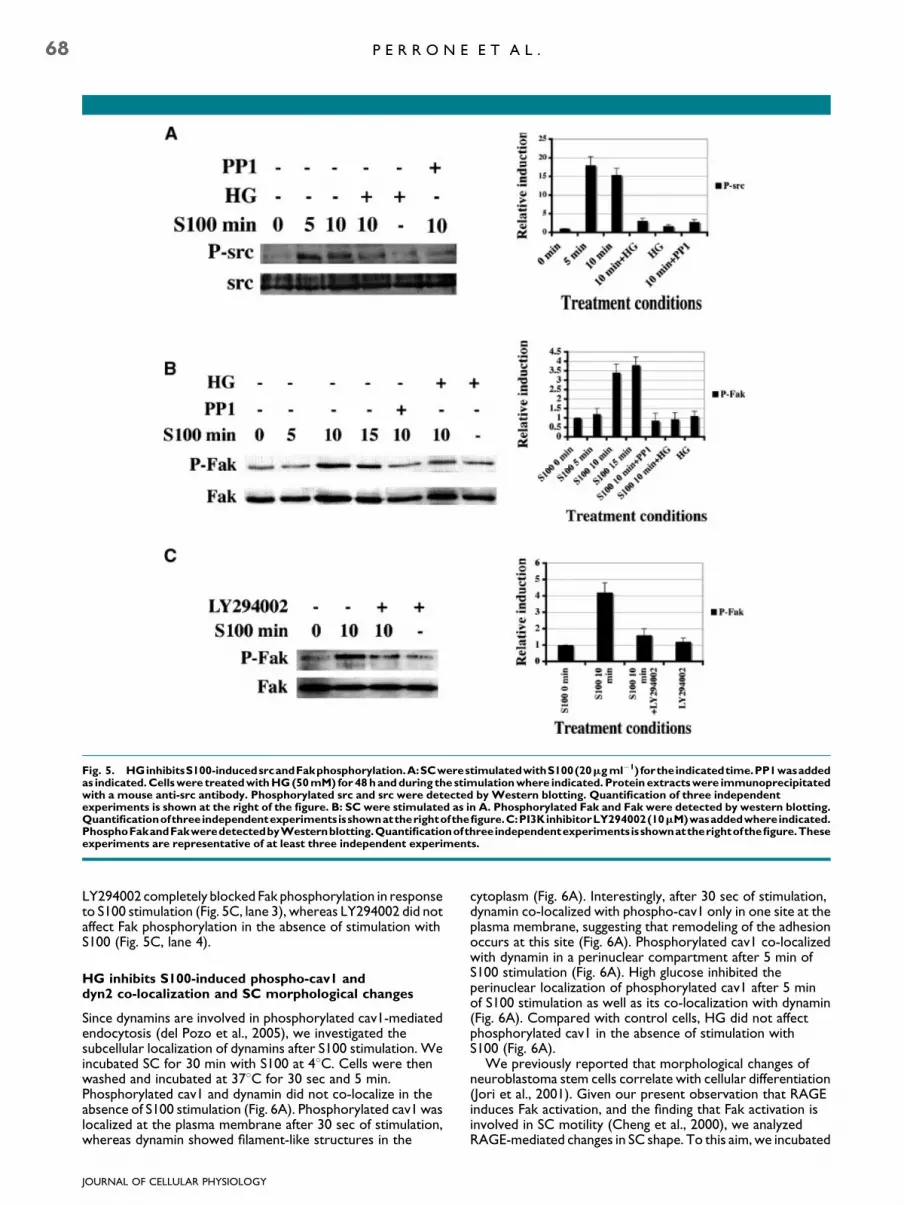
Fig. 5. HGinhibitsS100-inducedsrcandFakphosphorylation.A:SCwerestimulatedwithS100(20mgml�1) fortheindicatedtime.PP1wasaddedas indicated.Cellswere treatedwithHG(50mM) for48handduring the stimulationwhere indicated. Protein extractswere immunoprecipitatedwith a mouse anti-src antibody. Phosphorylated src and src were detected byWestern blotting. Quantification of three independentexperiments is shown at the right of the figure. B: SC were stimulated as in A. Phosphorylated Fak and Fak were detected by western blotting.Quantificationofthreeindependentexperimentsisshownattherightofthefigure.C:PI3KinhibitorLY294002(10mM)wasaddedwhereindicated.PhosphoFakandFakweredetectedbyWesternblotting.Quantificationofthreeindependentexperimentsisshownattherightofthefigure.Theseexperiments are representative of at least three independent experiments.
68 P E R R O N E E T A L .
LY294002 completely blocked Fak phosphorylation in responseto S100 stimulation (Fig. 5C, lane 3), whereas LY294002 did notaffect Fak phosphorylation in the absence of stimulation withS100 (Fig. 5C, lane 4).
HG inhibits S100-induced phospho-cav1 anddyn2 co-localization and SC morphological changes
Since dynamins are involved in phosphorylated cav1-mediatedendocytosis (del Pozo et al., 2005), we investigated thesubcellular localization of dynamins after S100 stimulation. Weincubated SC for 30 min with S100 at 48C. Cells were thenwashed and incubated at 378C for 30 sec and 5 min.Phosphorylated cav1 and dynamin did not co-localize in theabsence of S100 stimulation (Fig. 6A). Phosphorylated cav1 waslocalized at the plasma membrane after 30 sec of stimulation,whereas dynamin showed filament-like structures in the
JOURNAL OF CELLULAR PHYSIOLOGY
cytoplasm (Fig. 6A). Interestingly, after 30 sec of stimulation,dynamin co-localized with phospho-cav1 only in one site at theplasma membrane, suggesting that remodeling of the adhesionoccurs at this site (Fig. 6A). Phosphorylated cav1 co-localizedwith dynamin in a perinuclear compartment after 5 min ofS100 stimulation (Fig. 6A). High glucose inhibited theperinuclear localization of phosphorylated cav1 after 5 minof S100 stimulation as well as its co-localization with dynamin(Fig. 6A). Compared with control cells, HG did not affectphosphorylated cav1 in the absence of stimulation withS100 (Fig. 6A).
We previously reported that morphological changes ofneuroblastoma stem cells correlate with cellular differentiation(Jori et al., 2001). Given our present observation that RAGEinduces Fak activation, and the finding that Fak activation isinvolved in SC motility (Cheng et al., 2000), we analyzedRAGE-mediated changes in SC shape. To this aim, we incubated

Fig. 6. HG inhibits S100-induced SC morphological changes. A: SCwere stimulatedwith S100 (20mgml�1) at 4-C for 30min, washed andincubated at 37-C for the indicated time. HG (50 mM) was added asindicated. As a control, SC were incubated with HG in the absence ofS100 (bottom). The intracellular localization of phosphorylated cav1(red) and dyn2 (green) was analyzed by indirect immunofluorescenceand confocal microscopy. B: SC were stimulated as in (A). Changesin cell shape were analyzed by phase contrast microscopy. Theseexperiments are representative of at least three independentexperiments. LGU low glucose; HGUhigh glucose. [Color figure canbe viewed in the online issue, which is available at www.interscience.wiley.com.]
R A G E R E C Y C L I N G I N D U C E S S 1 0 0 B S E C R E T I O N 69
the cells for 30 min with bovine brain S100 at 48C, and afterremoving unbound S100 by washing, we incubated the cells for30 min at 378C. Cells were fixed in 4% PFA and we examinedtheir morphology by phase-contrast microscopy. Schwann cellsacquired their characteristic bipolar shape after stimulationwith S100, whereas they had a flattened fibroblast-like shape inthe absence of S100 stimulation (Fig. 6B, top). High glucosepartially inhibited the bipolar shape induced by S100. Indeed, inthe presence of S100 and a HG concentration, SC started toelongate and to acquire the bipolar shape, although theyretained a flat fibroblast-like morphology (Fig. 6B, bottomright). Without S100 stimulation and under HG conditions, SCassumed a fibroblast-like shape (Fig. 6B, bottom left).
JOURNAL OF CELLULAR PHYSIOLOGY
Discussion
A previous study suggests that SC-mediated S100B secretion isrequired for peripheral nerve regeneration. Indeed, antibodiesagainst S100B block peripheral nerve regeneration in vivo(Rong et al., 2004a). However, SC-mediated S100B secretion isstill poorly investigated. SC are known to secrete a 12-kDaprotein that induces neurite outgrowth in PC12 cells (Bamptonand Taylor, 2005). In this report we demonstrate for the firsttime that SC secrete S100B. Also glioblastoma cells (Daveyet al., 2001) and astrocytes (Gerlach et al., 2006) secrete S100B.However, the molecular mechanisms governing S100Bsecretion are still poorly understood. Several lines of evidencepreviously suggested that RAGE might be involved in therelocation in vesicles of various S100 family proteins. Indeed,stimulation of prostate cancer cells with the RAGE ligandsS100A8/9 induced the co-location of RAGE with S100A8/9 invesicles (Hermani et al., 2006). In endothelial cells, translocationof endogenous S100 proteins in vesicles is RAGE-dependent(Hsieh et al., 2004). The addition of exogenous S100A13induced RAGE co-localization with endogenous S100A13, andpre-treatment with soluble RAGE inhibited S100A13relocation in vesicles, which suggested that RAGE is requiredfor S100A13 internalization and relocation of endogenousS100A13 in vesicles (Hsieh et al., 2004). However, neitherRAGE internalization nor S100A13-RAGE co-localization wasdemonstrated. We provide the first demonstration that RAGEtriggering induces RAGE internalization and the fusion ofRAGE-containing endocytic vesicles with S100B-positivesecretory vesicles (see model shown in Fig. 7A). Relocation of aspecific S100 protein in vesicles was found to occur only whenRAGEwas stimulated with the same extracellular S100 protein,whereas it was inhibited by stimulation with a different memberof S100 family (Hsieh et al., 2004). We show that triggering ofRAGE with an anti-RAGE antibody induces S100B relocation inRAGE-positive endocytic vesicles. It is conceivable thatdifferent S100 proteins interact with specific proteins therebyactivating specific trafficking pathways and inhibiting thetranslocation in vesicles of anothermember of the S100 proteinfamily. Anti-RAGE antibody does not induce this effect. Herewe demonstrate that anti-RAGE antibody induces RAGEinternalization and signaling, as well as co-localization ofinternalized RAGE with endogenous S100B, which results insecretion of S100B. We also show that RAGE recyclingcorrelates with S100B secretion. Indeed, as shown inFigure 7A, src orchestrates a hitherto unknown vesicularpathway that leads to RAGE recycling at the plasmamembrane,fusion with S100B-containing vesicles, and S100B secretion.Inhibition of src activation leads to RAGE degradation andinhibits RAGE-S100B co-localization in vesicles as well as S100Bsecretion. In agreement with ourmodel, it has been shown thatRAGE triggers the internalization and the transcytosis of theAmyloid-b 1–40 peptide from the apical to the basolateralmembrane in endothelial cells (Mackic et al., 1998). Indeed,Mackic et al. (1998) demonstrate that the degradation of theamyloid peptide is very low in endothelial cells, furthersupporting that internalized RAGE is not targeted tolysosomes. Eventual differences in the quantification of b-amyloid transcytosis in endothelial cells and RAGE recycling inSC can be due to the different methods used to investigateRAGE trafficking and/or the different cell type analyzed. In ourexperiments SC are not polarized, so we cannot evaluatewhether RAGE is targeted to a transcytotic pathway. However,in agreement with our data in correlation with the Mackic et al.(1998) report, it has been demonstrated that src, cav-1 and dynplay a role in caveolae-mediated transendothelial transport(Shajahan et al., 2004a,b). Furthermore, RAGE co-fractionateswith caveolae-rich membrane in endothelial cells (Lisanti et al.,1994) and we found that RAGE is lipid rafts associated and

Fig. 7. Model for the autocrine stimulation of SC induced by RAGEtriggering. A: Activation of RAGE induces src-mediatedphosphorylation of cav-1 that targets RAGE to the recycling pathwayin Rab11-positive vesicles. Internalized RAGE is targeted to vesiclescontaining endogenousS100B,which is secreted in response toRAGEactivation. B: RAGE triggering induces src-mediated cav1phosphorylation and re-location in the perinuclear compartmenttogether with dyn2. Src orchestrates RAGE recycling, S100Bsecretion and morphological changes.
70 P E R R O N E E T A L .
interact with cav-1 in SC (data not shown). Interestingly, theendocytic pathway here described for RAGE has beenpreviously demonstrated for the gp60 receptor. Indeed,interaction of gp60 with either albumin or a gp60-specificantibody leads to src and cav-1 phosphorylation and inducesgp60 internalization and targeting to the transcytotic pathway inendothelial cells (Tiruppathi et al., 1997). Further supportingour data compared with the Mackic et al. (1998) study, thefunction of Rab11 in transcytosis has also been demonstrated(Ducharme et al., 2007). Here we show that src-mediatedphosphorylation of cav-1 is necessary for RAGE recycling atplasma membrane, while inhibition of src activation leads toRAGE targeting to lysosomes. The function of src-mediatedphosphorylation of cav-1 in inhibiting receptor degradation andpromoting a perinuclear targeting has been previouslydescribed for the EGF receptor (EGFR) (Khan et al., 2006).Indeed, it is well described that EGFR can follow two alternatepathways: be targeted to lysosomes for degradation or betransported to a perinuclear compartment via caveolae (Khanet al., 2006). Thus, the previous literature strongly support ourobservations summarized in Figure 7A. However, here wedescribe for the first time the role of cav-1 phosphorylation inthe endocytic trafficking of RAGE and its function in promotingS100B secretion.
JOURNAL OF CELLULAR PHYSIOLOGY
Here we show that RAGE triggering induces plasmamembrane rearrangements. In agreement, S100B-mediatedRAGE activation induces cell motility (Reddy et al., 2006).Receptor recycling and redistribution of membrane proteinsplay a role in cell motility (Jones et al., 2006). Indeed, wedemonstrate that RAGE recycles through he Rab11 pathway.RAGE triggering induces phosphorylated cav1 translocation tothe perinuclear compartment and co-localizationwith dynamin.Our data are in line with the finding that the dyn2 and Rab11pathways contribute to cell migration (Jones et al., 2006).
Changes of cell shape require a coordinate disassembly andassembly of focal adhesions (Kruchten and McNiven, 2006).Interestingly, increment of cav1 phosphorylation inhibits Fakand causes disassembly of the actin cytoskeleton in(myo)fibroblasts (Swaney et al., 2006). In agreement, wedemonstrate that phosphorylation of cav-1 is required forRAGE recycling, suggesting that RAGE-mediated src activationand subsequent cav-1 phosphorylation are responsible forS100-mediated SC morphological changes. Indeed, inhibitionof src activation by high glucose treatment abolishesS100-mediated morphological changes of SC.
Our study shows that changes in SC shape correlate withS100B relocation in vesicles and S100B secretion. In agreementwith this finding, relocation of S100B in vesicle-like structurescorrelates with membrane rearrangements in astrocytomacells (Mbele et al., 2002). As shown in Figure 7B, we hypothesizea model whereby RAGE first induces an increase of cav1phosphorylation thereby leading to disassembly of the focaladhesions. Next, perinuclear translocation of phosphorylatedcav1 leads to rearrangement of focal adhesions. Indeed,endocytic trafficking of RAGE induces rearrangements of theplasma membrane. In the presence of S100, SC acquire thecharacteristic bipolar shape (Caddick et al., 2006), and show adifferentiated-like phenotypewith a reduction of the cytoplasm(Previtali et al., 2003). Changes in cell shape and cytoskeletonrearrangements are required to acquire the differentiate SCphenotype necessary to induce myelination (Previtali et al.,2003). SC myelinating phenotype is characterized by theexpression of fibronectin (Akassoglou et al., 2002). We foundthat S100-mediated RAGE activation induces the mRNAexpression of fibronectin (data not shown). Schwann cells,RAGE and S100 are required for peripheral nerve regeneration(Rong et al., 2004a,b; Dobrowsky et al., 2005). In agreementwith our data, src is activated in SC after rat peripheral nerveinjury (Zhao et al., 2003). We suggest that RAGE plays a keyfunction in SC modifications that are important duringregeneration of injured nerves.
We also demonstrate that HG inhibits RAGE-mediated srcphosphorylation. Moreover, RAGE-mediated changes in SCshape do not occur under HG conditions. Given theinvolvement of src in cell motility (Cutrupi et al., 2000), thisfinding supports the concept that RAGE-mediated srcactivation plays a role in changes in SC shape. We demonstratethat inhibition of src activation induces RAGE degradation andblocks S100B secretion. In line with this observation, HGinhibits S100B secretion in astrocytes (Nardin et al., 2007).Further support for the physiological relevance of our datacomes from the finding that regeneration of the injured sciaticnerve is delayed or fails in diabetes (Zochodne et al., 2007).Moreover, HG induces abnormal SC proliferation leading to anot differentiated phenotype that is characteristic of diabeticneuropathy (Almhanna et al., 2002). In agreement with theseobservations, we found that HG inhibits RAGE-inducedfibronectin expression (data not shown).
In summary, we have identified a new vesicular pathway ofRAGE recycling and S100B secretion, thereby providing insightsinto the role of RAGE in acquiring a pro-myelinating phenotype,while high glucose abolishes RAGE mediated effects on SC byinhibiting src activation.

R A G E R E C Y C L I N G I N D U C E S S 1 0 0 B S E C R E T I O N 71
Acknowledgments
This work utilized the Morphology and Image Analysis Core ofthe Michigan Diabetes Research and Training Center funded byNIH5P60 DK20572 from the National Institute of Diabetes &Digestive &KidneyDiseases.We thankC. Backus for assistancewith SC. We also thank ME Bianchi, L Wrabetz, and L Franchifor the helpful discussion, and Jean Ann Gilder for text editing.LP would like to dedicate this work to the heart and soul ofSilvana Geremia.
Literature Cited
Akassoglou K, Yu WM, Akpinar P, Strickland S. 2002. Fibrin inhibits peripheral nerveremyelination by regulating Schwann cell differentiation. Neuron 33:861–875.
Almhanna K, Wilkins PL, Bavis JR, Harwalkar S, Berti-Mattera LN. 2002. Hyperglycemiatriggers abnormal signaling and proliferative responses in Schwann cells. Neurochem Res27:1341–1347.
Bampton ET, Taylor JS. 2005. Effects of Schwann cell secreted factors on PC12 cellneuritogenesis and survival. J Neurobiol 63:29–48.
Berti C, Nodari A, Wrabetz L, Feltri ML. 2006. Role of integrins in peripheral nerves andhereditary neuropathies. Neuromolecular Med 8:191–204.
BierhausA,Humpert PM,MorcosM,WendtT,ChavakisT, ArnoldB, SternDM,NawrothPP.2005. Understanding RAGE, the receptor for advanced glycation end products. J Mol Med83:876–886.
Caddick J, Kingham PJ, Gardiner NJ,Wiberg M, Terenghi G. 2006. Phenotypic and functionalcharacteristics of mesenchymal stem cells differentiated along a Schwann cell lineage. Glia54:840–849.
Chavakis E, Hain A, Vinci M, Carmona G, Bianchi ME, Vajkoczy P, Zeiher AM, Chavakis T,Dimmeler S. 2007. High-mobility group box 1 activates integrin-dependent homing ofendothelial progenitor cells. Circ Res 100:204–212.
Chen ZY, Ieraci A, Tanowitz M, Lee FS. 2005. A novel endocytic recycling signal distinguishesbiological responses of Trk neurotrophin receptors. Mol Biol Cell 16:5761–5772.
Cheng HL, Steinway ML, Russell JW, Feldman EL. 2000. GTPases and phosphatidylinositol3-kinase are critical for insulin-like growth factor-I-mediated Schwann cell motility. J BiolChem 275:27197–27204.
Cutrupi S, Baldanzi G, Gramaglia D, Maffe A, Schaap D, Giraudo E, van Blitterswijk W,Bussolino F, Comoglio PM, Graziani A. 2000. Src-mediated activation of alpha-diacylglycerol kinase is required for hepatocyte growth factor-induced cell motility.EMBO J 19:4614–4622.
Davey GE, Murmann P, Heizmann CW. 2001. Intracellular Ca2þ and Zn2þ levels regulatethe alternative cell density-dependent secretion of S100B in human glioblastoma cells. J BiolChem 276:30819–30826.
Deane R,DuYan S, SubmamaryanRK, LaRue B, Jovanovic S, Hogg E,WelchD,Manness L, LinC, Yu J, Zhu H, Ghiso J, Frangione B, Stern A, Schmidt AM, Armstrong D, Arnold B,Liliensiek B, Nawroth P, Hofman F, Kindy M, Stern D, Zlokovic B. 2003. RAGE mediatesamyloid-beta peptide transport across the blood-brain barrier and accumulation in brain.Nat Med 9:907–913.
del Pozo MA, Balasubramanian N, Alderson NB, Kiosses WB, Grande-Garcia A, AndersonRG, Schwartz MA. 2005. Phospho-caveolin-1 mediates integrin-regulated membranedomain internalization. Nat Cell Biol 7:901–908.
Dobrowsky RT, Rouen S, Yu C. 2005. Altered neurotrophism in diabetic neuropathy:Spelunking the caves of peripheral nerve. J Pharmacol Exp Ther 313:485–491.
Ducharme NA, Williams JA, Oztan A, Apodaca G, Lapierre LA, Goldenring JR. 2007.Rab11-FIP2 regulates differentiable steps in transcytosis. Am J Physiol Cell Physiol293:C1059–C1072.
Dumitriu IE, Bianchi ME, Bacci M, Manfredi AA, Rovere-Querini P. 2007. The secretion ofHMGB1 is required for the migration of maturing dendritic cells. J Leukoc Biol 81:84–91.
Gerlach R, DemelG, Konig HG,Gross U, Prehn JH, Raabe A, Seifert V, Kogel D. 2006. Activesecretion of S100B from astrocytes during metabolic stress. Neuroscience141:1697–1701.
Hermani A, De Servi B, Medunjanin S, Tessier PA, Mayer D. 2006. S100A8 and S100A9activate MAP kinase and NF-kappaB signaling pathways and trigger translocation of RAGEin human prostate cancer cells. Exp Cell Res 312:184–197.
Hofmann MA, Drury S, Fu C, Qu W, Taguchi A, Lu Y, Avila C, Kambham N, Bierhaus A,Nawroth P, Neurath MF, Slattery T, Beach D, McClary J, Nagashima M, Morser J, Stern D,Schmidt AM. 1999. RAGE mediates a novel proinflammatory axis: A central cell surfacereceptor for S100/calgranulin polypeptides. Cell 97:889–901.
Hsieh HL, Schafer BW, Weigle B, Heizmann CW. 2004. S100 protein translocation inresponse to extracellular S100 is mediated by receptor for advanced glycationendproducts in human endothelial cells. Biochem Biophys Res Commun 316:949–959.
Jones MC, Caswell PT, Norman JC. 2006. Endocytic recycling pathways: Emerging regulatorsof cell migration. Curr Opin Cell Biol 18:549–557.
Jori FP, Galderisi U, Piegari E, Peluso G, Cipollaro M, Cascino A, Giordano A, Melone MA.2001. RB2/p130 ectopic gene expression in neuroblastoma stem cells: Evidence of cell-faterestriction and induction of differentiation. Biochem J 360:569–577.
Jori FP, Galderisi U, Piegari E, Cipollaro M, Cascino A, Peluso G, Cotrufo R, Giordano A,Melone MA. 2003. EGF-responsive rat neural stem cells: Molecular follow-up of neuronand astrocyte differentiation in vitro. J Cell Physiol 195:220–233.
Jullien J, Guili V, Reichardt LF, Rudkin BB. 2002. Molecular kinetics of nerve growth factorreceptor trafficking and activation. J Biol Chem 277:38700–38708.
Kalichman MW, Powell HC, Mizisin AP. 1998. Reactive, degenerative, and proliferativeSchwann cell responses in experimental galactose and human diabetic neuropathy. ActaNeuropathol (Berl) 95:47–56.
Khan EM, Heidinger JM, Levy M, Lisanti MP, Ravid T, Goldkorn T. 2006. Epidermal growthfactor receptor exposed to oxidative stress undergoes Src- and caveolin-1-dependentperinuclear trafficking. J Biol Chem 281:14486–14493.
Kruchten AE, McNiven MA. 2006. Dynamin as a mover and pincher during cell migration andinvasion. J Cell Sci 119:1683–1690.
JOURNAL OF CELLULAR PHYSIOLOGY
Lisanti MP, Scherer PE, Vidugiriene J, Tang Z, Hermanowski-Vosatka A, Tu YH, Cook RF,Sargiacomo M. 1994. Characterization of caveolin-rich membrane domains isolatedfrom an endothelial-rich source: Implications for human disease. J Biol Chem126:111–126.
Mackic JB, Stins M, McComb JG, Calero M, Ghiso J, Kim KS, Yan SD, Stern D, Schmidt AM,Frangione B, Zlokovic BV. 1998. Human blood-brain barrier receptors for Alzheimer’samyloid-beta1-40. Asymmetrical binding, endocytosis, and transcytosis at the apical side ofbrain microvascular endothelial cell monolayer. J Clin Invest 102:734–743.
Mathon NF, Malcolm DS, Harrisingh MC, Cheng L, Lloyd AC. 2001. Lack of replicativesenescence in normal rodent glia. Science 291:872–875.
MbeleGO,Deloulme JC, Gentil BJ, DelphinC, FerroM,Garin J, TakahashiM, Baudier J. 2002.The zinc- and calcium-binding S100B interacts and co-localizes with IQGAP1 duringdynamic rearrangement of cell membranes. J Biol Chem 277:49998–50007.
Mikol DD, Scherer SS, Duckett SJ, Hong HL, Feldman EL. 2002. Schwann cell caveolin-1expression increases during myelination and decreases after axotomy. Glia38:191–199.
Millan J, Hewlett L, Glyn M, Toomre D, Clark P, Ridley AJ. 2006. Lymphocyte transcellularmigration occurs through recruitment of endothelial ICAM-1 to caveola- and F-actin-richdomains. Nat Cell Biol 8:113–123.
Nardin P, Tramontina F, Leite MC, Tramontina AC, Quincozes-Santos A, de Almeida LM,Battastini AM, Gottfried C, Goncalves CA. 2007. S100B content and secretion decrease inastrocytes cultured in high-glucose medium. Neurochem Int 50:774–782.
Orlova VV, Choi EY, Xie C, Chavakis E, Bierhaus A, Ihanus E, Ballantyne CM, Gahmberg CG,Bianchi ME, Nawroth PP, Chavakis T. 2007. A novel pathway of HMGB1-mediatedinflammatory cell recruitment that requires Mac-1-integrin. EMBO J 26:1129–1139.
Pachydaki SI, Tari SR, Lee SE, Ma W, Tseng JJ, Sosunov AA, Cataldergirmen G, Scarmeas N,Caspersen C, Chang S, Schiff WM, Schmidt AM, Barile GR. 2006. Upregulation of RAGEand its ligands in proliferative retinal disease. EXp Eye Res 82:807–815.
Perrone L, Paladino S, Mazzone M, Nitsch L, Gulisano M, Zurzolo C. 2005. Functionalinteraction between p75NTR and TrkA: The endocytic trafficking of p75NTR is driven byTrkA and regulates TrkA-mediated signalling. Biochem J 385:233–241.
Piazza T, Cha E, Bongarzone I, Canevari S, Bolognesi A, Polito L, Bargellesi A, Sassi F, Ferrini S,Fabbi M. 2005. Internalization and recycling of ALCAM/CD166 detected by a fully humansingle-chain recombinant antibody. J Cell Sci 118:1515–1525.
Previtali SC, Nodari A, Taveggia C, Pardini C, Dina G, Villa A, Wrabetz L, Quattrini A, FeltriML. 2003. Expression of laminin receptors in schwann cell differentiation: Evidence fordistinct roles. J Neurosci 23:5520–5530.
ReddyMA, Li S, Sahar S, KimYS,XuZG, Lanting L,NatarajanR. 2006. Key role of Src kinase inS100B-induced activation of the receptor for advanced glycation end products in vascularsmooth muscle cells. J Biol Chem 281:13685–13693.
Riuzzi F, Sorci G, Donato R. 2006. The amphoterin (HMGB1)/receptor for advancedglycation end products (RAGE) pair modulates myoblast proliferation, apoptosis,adhesiveness, migration, and invasiveness. Functional inactivation of RAGE in L6myoblastsresults in tumor formation in vivo. J Biol Chem 281:8242–8253.
Rong LL, Trojaborg W, QuW, Kostov K, Yan SD, Gooch C, Szabolcs M, Hays AP, SchmidtAM. 2004a. Antagonism of RAGE suppresses peripheral nerve regeneration. FASEB J18:1812–1817.
Rong LL, Yan SF, Wendt T, Hans D, Pachydaki S, Bucciarelli LG, Adebayo A, Qu W, Lu Y,Kostov K, Lalla E, Yan SD, Gooch C, Szabolcs M, Trojaborg W, Hays AP, Schmidt AM.2004b. RAGE modulates peripheral nerve regeneration via recruitment of bothinflammatory and axonal outgrowth pathways. FASEB J 18:1818–1825.
Sakaguchi M, Sonegawa H, Murata H, Kitazoe M, Futami J, Kataoka K, Yamada H, Huh NH.2008. S100A11, a dual mediator for growth regulation of human keratinocytes. Mol BiolCell 19:78–85.
Schindler T, Sicheri F, Pico A, Gazit A, Levitzki A, Kuriyan J. 1999. Crystal structure ofHck in complex with a Src family-selective tyrosine kinase inhibitor. Mol Cell 3:639–648.
Schmidt AM, Yan SD, Brett J, Mora R, Nowygrod R, Stern D. 1993. Regulation of humanmononuclear phagocyte migration by cell surface-binding proteins for advanced glycationend products. J Clin Invest 91:2155–2168.
Shajahan AN, Timblin BK, Sandoval R, Tiruppathi C, Malik AB, Minshall RD. 2004a. Role ofSrc-induced dynamin-2 phosphorylation in caveolae-mediated endocytosis in endothelialcells. J Biol Chem 279:20392–20400.
Shajahan AN, Tiruppathi C, Smrcka AV, Malik AB, Minshall RD. 2004b. Gbetagammaactivation of Src induces caveolae-mediated endocytosis in endothelial cells. J Biol Chem279:48055–48062.
Swaney JS, Patel HH, Yokoyama U, Head BP, Roth DM, Insel PA. 2006. Focal adhesions in(myo)fibroblasts scaffold adenylyl cyclase with phosphorylated caveolin. J Biol Chem281:17173–17179.
Tampellini D, Magrane J, Takahashi RH, Li F, Lin MT, Almeida CG, Gouras GK. 2007.Internalized antibodies to the Abeta domain of APP reduce neuronal Abeta and protectagainst synaptic alterations. J Biol Chem 282:18895–18906.
Tamura M, Osajima A, Nakayamada S, Anai H, Kabashima N, Kanegae K, Ota T, Tanaka Y,Nakashima Y. 2003. High glucose levels inhibit focal adhesion kinase-mediated woundhealing of rat peritoneal mesothelial cells. Kidney Int 63:722–731.
Tanowitz M, Von Zastrow M. 2002. Ubiquitination-independent trafficking of G protein-coupled receptors to lysosomes. J Biol Chem 277:50219–50222.
Tiruppathi C, Song W, Bergenfeldt M, Sass P, Malik AB. 1997. Gp60 activation mediatesalbumin transcytosis in endothelial cells by tyrosine kinase-dependent pathway. J BiolChem 272:25968–25975.
Vincent AM, Perrone L, Sullivan KA, Backus C, Sastry AM, Lastoskie C, Feldman EL. 2007.Receptor for advanced glycation end products activation injures primary sensory neuronsvia oxidative stress. Endocrinology 148:548–558.
YangD,ChenQ, YangH,TraceyKJ, BustinM,Oppenheim JJ. 2007.Highmobility groupbox-1protein induces themigration and activation of human dendritic cells and acts as an alarmin.J Leukoc Biol 81:59–66.
Zhao YL, Takagawa K, Oya T, Yang HF, Gao Z, Kawaguchi M, Ishii Y, Sasaoka T, Owada K,Furuta I, Sasahara M. 2003. Active Src expression is induced after rat peripheral nerveinjury. Glia 42:184–193.
Zochodne DW, Guo GF, Magnowski B, Bangash M. 2007. Regenerative failure of diabeticnerves bridging transection injuries. Diabetes Metab Res Rev 23:490–496.
Zurzolo C, Lisanti MP, Caras IW, Nitsch L, Rodriguez-Boulan E. 1993.Glycosylphosphatidylinositol-anchored proteins are preferentially targeted to thebasolateral surface in Fischer rat thyroid epithelial cells. J Cell Biol 121:1031–1039.